structure of the skin epidermis stratum basale stratum lucidum stratum corneum dermis ...
TRANSCRIPT

Chapter 5:The Integumentary System
Gabriela AguiarFernando Graziano
Kevin Gonzales.
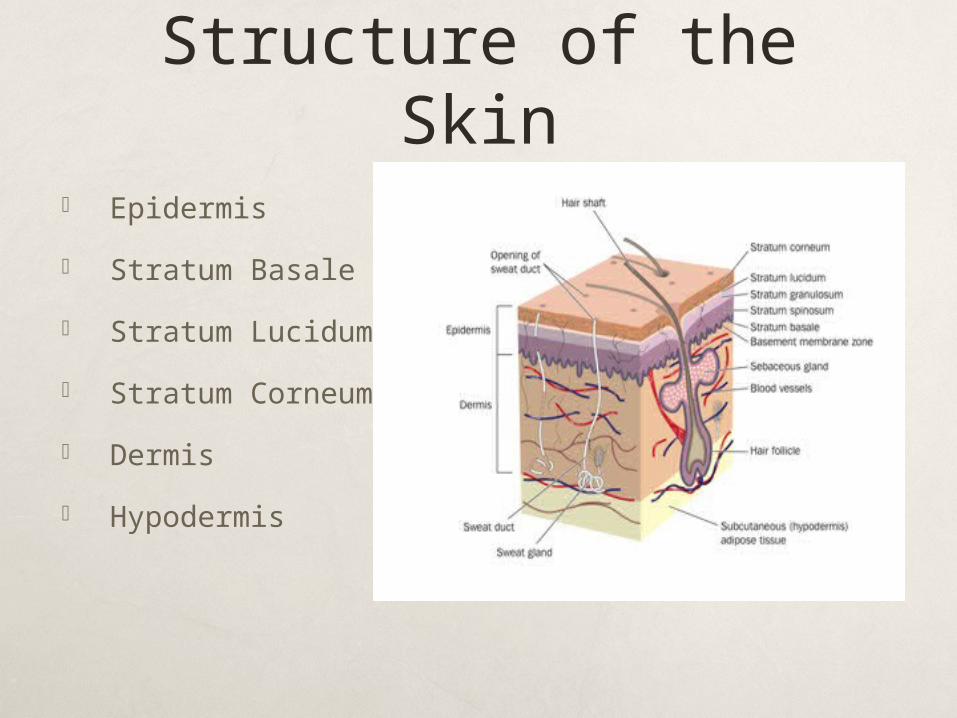
Structure of the Skin Epidermis
Stratum Basale
Stratum Lucidum
Stratum Corneum
Dermis
Hypodermis

Epidermis The epidermis on the outside. This is made from
layers of cells with a basal layer, which is always forming new cells through cell division. The new cells gradually move towards the surface, which takes 1-2 months. As they move up they gradually die, become flattened and develop keratin and the outermost layer of flat dead cells is being continually worn away by friction. The keratin and oil from the sebaceous glands help to make the skin waterproof.
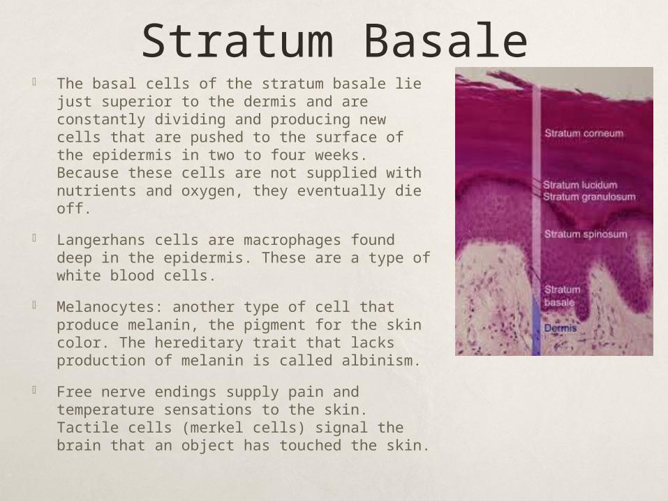
Stratum Basale The basal cells of the stratum basale lie just
superior to the dermis and are constantly dividing and producing new cells that are pushed to the surface of the epidermis in two to four weeks. Because these cells are not supplied with nutrients and oxygen, they eventually die off.
Langerhans cells are macrophages found deep in the epidermis. These are a type of white blood cells.
Melanocytes: another type of cell that produce melanin, the pigment for the skin color. The hereditary trait that lacks production of melanin is called albinism.
Free nerve endings supply pain and temperature sensations to the skin. Tactile cells (merkel cells) signal the brain that an object has touched the skin.
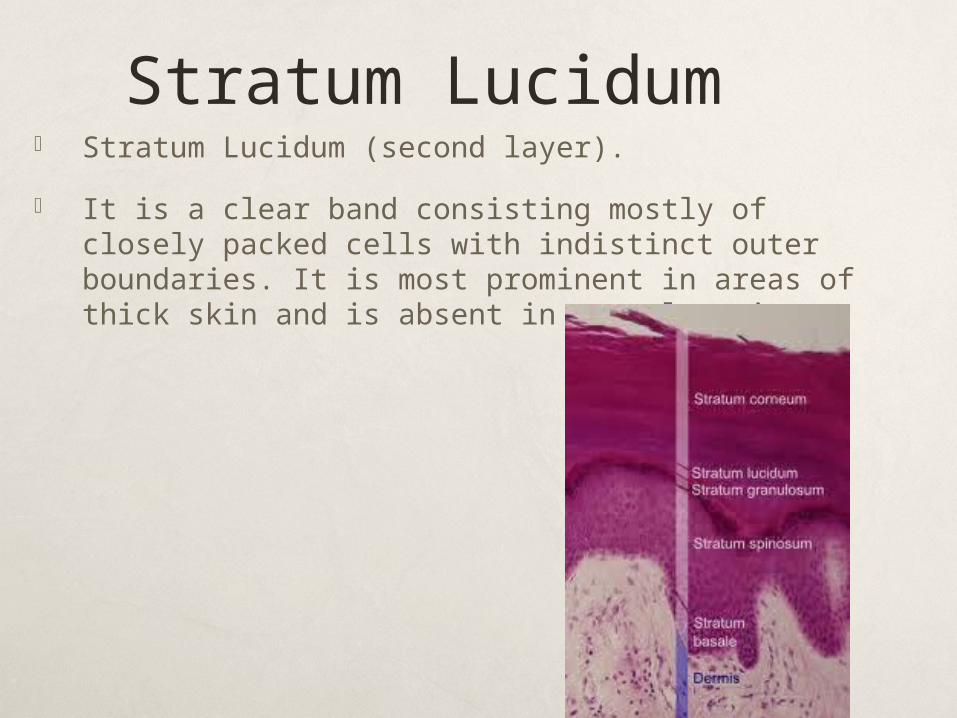
Stratum Lucidum Stratum Lucidum (second layer).
It is a clear band consisting mostly of closely packed cells with indistinct outer boundaries. It is most prominent in areas of thick skin and is absent in some locations.
Stratum Corneum Stratum Corneum (outermost layer). It consists of
varying layers of flat, closely packed, dead cells that are constantly being lost as a result of abrasion -- for example, by friction with clothing. The lost cells are constantly being replaced by cells from deeper layers of the epidermis. Cellular production of fibrous, waterproof protein called Keratin.
Dermis The dermis is the inner layer. Composed of dense
irregular connective tissue. The following tissues and structures can all be found in the dermis: Connective tissue – packs and binds the other structures in
the skin. Elastic fibers – make the skin resilient. Capillaries – tiny blood vessels. Muscle fibers – to move the position of the hairs. Sensory cells – to sense touch, pressure, heat, cold and pain. Nerve fibers – to activate muscles and glands and relay
messages from the sensory cells to the brain. Pigment cells which produce Melanin – a very dark pigment. Sweat glands which open onto the surface as pores Hair follicles – pits in the epidermis in which hairs grow.
Sebaceous glands – produce oil to keep hair follicle free from dust and bacteria, and to help to waterproof the skin.
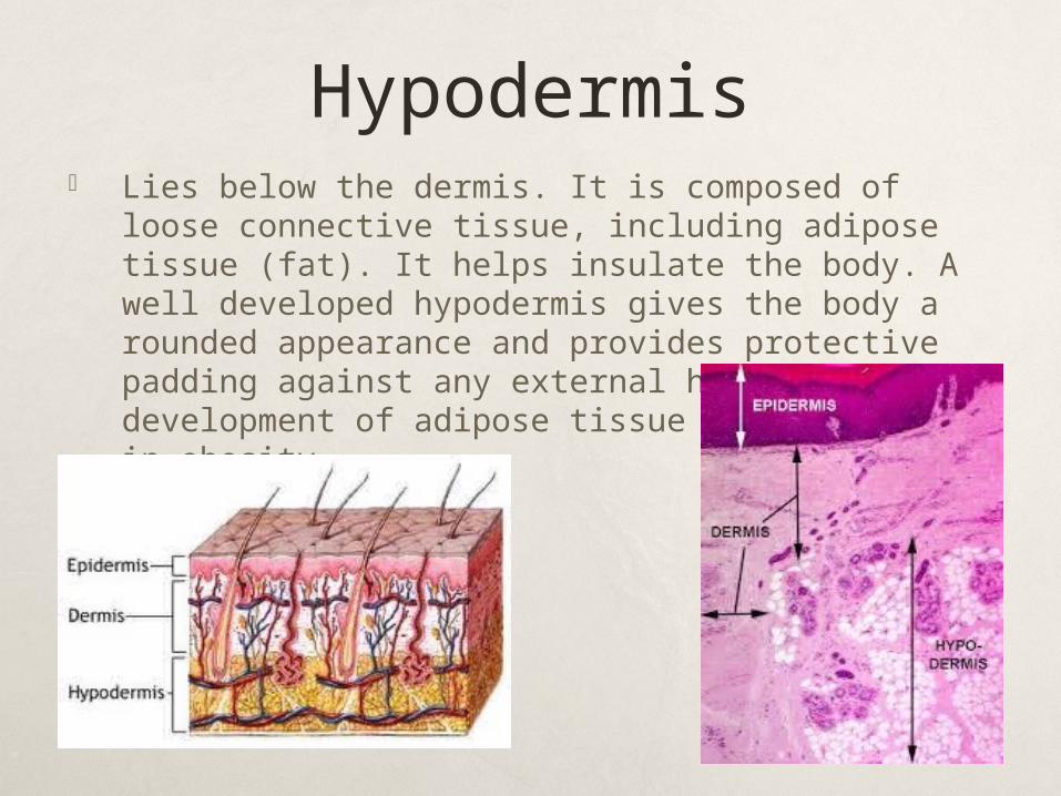
Hypodermis Lies below the dermis. It is composed of loose
connective tissue, including adipose tissue (fat). It helps insulate the body. A well developed hypodermis gives the body a rounded appearance and provides protective padding against any external harm. Excessive development of adipose tissue layers results in obesity.
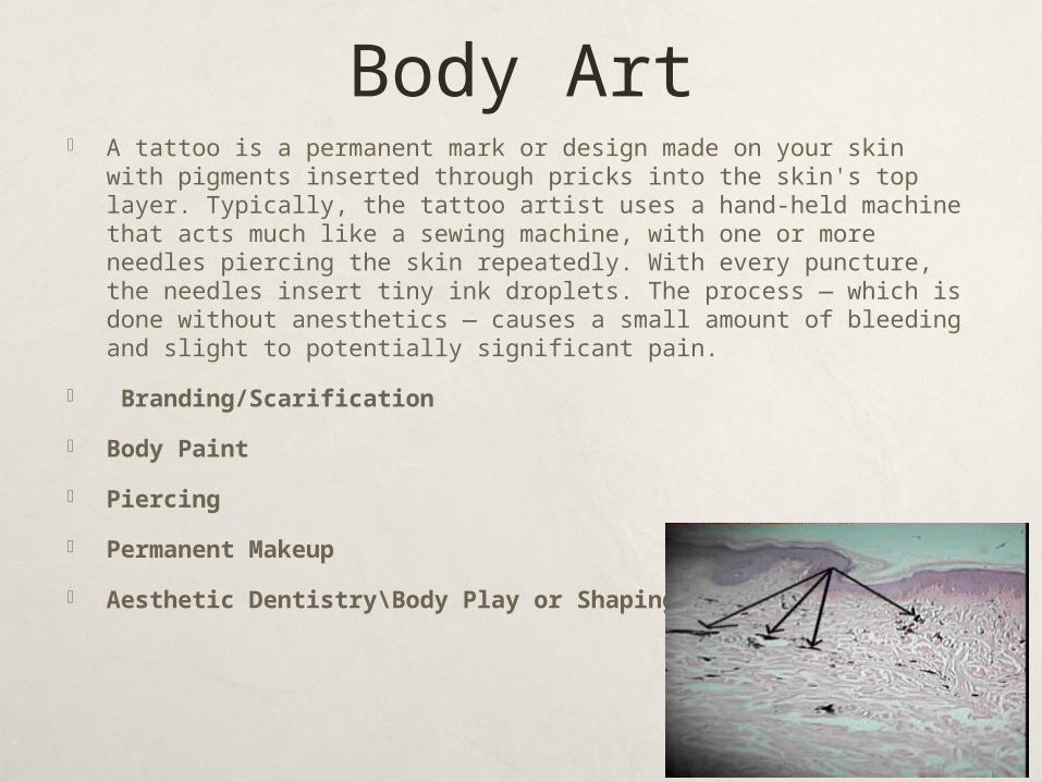
Body Art A tattoo is a permanent mark or design made on your skin with
pigments inserted through pricks into the skin's top layer. Typically, the tattoo artist uses a hand-held machine that acts much like a sewing machine, with one or more needles piercing the skin repeatedly. With every puncture, the needles insert tiny ink droplets. The process — which is done without anesthetics — causes a small amount of bleeding and slight to potentially significant pain.
Branding/Scarification
Body Paint
Piercing
Permanent Makeup
Aesthetic Dentistry\Body Play or Shaping
Accessory Structure of the Skin
Hair and Nails
Glands ( sweat glands, mammary glands)
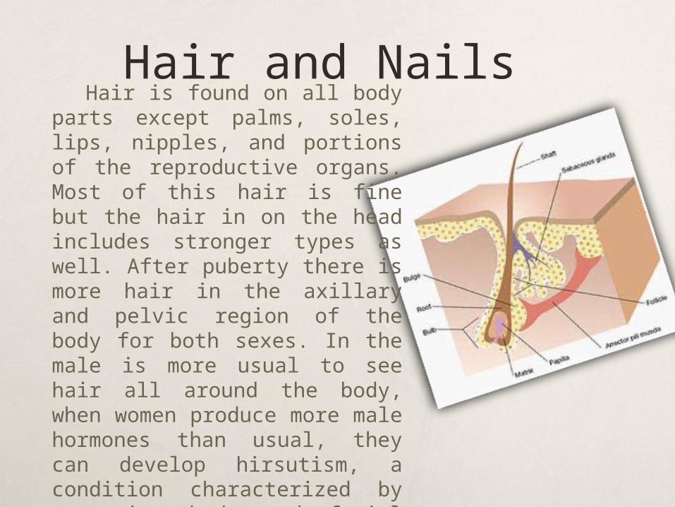
Hair and NailsHair is found on all body
parts except palms, soles, lips, nipples, and portions of the reproductive organs. Most of this hair is fine but the hair in on the head includes stronger types as well. After puberty there is more hair in the axillary and pelvic region of the body for both sexes. In the male is more usual to see hair all around the body, when women produce more male hormones than usual, they can develop hirsutism, a condition characterized by excessive body and facial hair. Hair projects from complex structures called hair follicles.
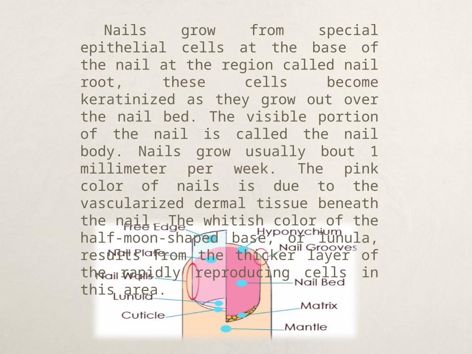
Nails grow from special epithelial cells at the base of the nail at the region called nail root, these cells become keratinized as they grow out over the nail bed. The visible portion of the nail is called the nail body. Nails grow usually bout 1 millimeter per week. The pink color of nails is due to the vascularized dermal tissue beneath the nail. The whitish color of the half-moon-shaped base, or lunula, results from the thicker layer of the rapidly reproducing cells in this area.
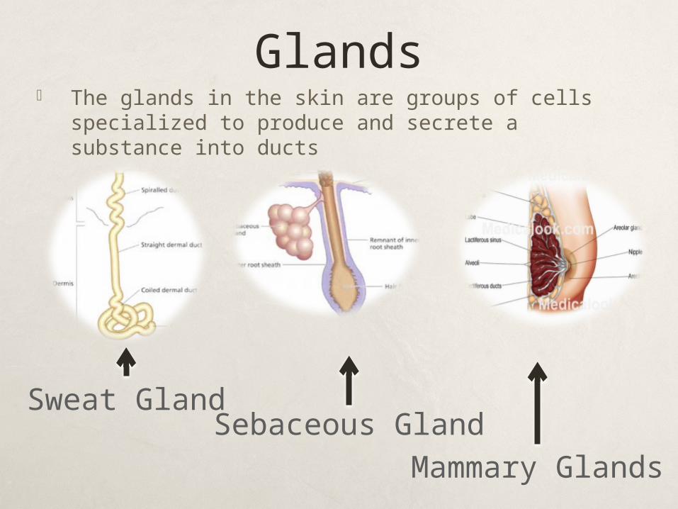
Glands The glands in the skin are groups of cells specialized
to produce and secrete a substance into ducts
Sweat GlandSebaceous Gland
Mammary Glands
Sweat Glands
Sweat gland are present in all regions of the skin, there can be as many as 90 glands per square centimeter on the leg, 400 glands per square centimeter on the palm and soles and even a greater number on the fingertips. A sweat gland is tubular, the tubule is coiled, particularly at its origin within the dermis. These glands become active when the person is under stress.
Sebaceous Glands Most sebaceous gland are associated with a hair
follicle. These glands secrete an oily substance called sebum that flows into the follicle and then onto the skin surface, this secretion lubricates the hair and the skin, and helps waterproof them. Sebum also helps to kill bacteria on the skin.
Acne vulgaris, is the most common form of acne, is an inflammation of the sebaceous glands that most often occur during adolescence. Hormonal changes during puberty cause the sebaceous gland to become more active during that period.
Mammary Glands
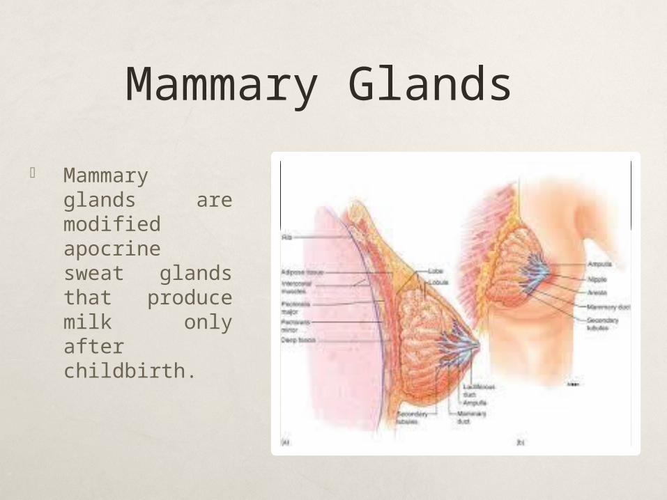
Mammary glands are modified apocrine sweat glands that produce milk only after childbirth.
Disorders of the Skin Athlete’s foot
Impetigo
Eczema
Dandruff
Urticaria
Skin Cancer ( melanomas)
Wound healing
Burns
Disorders that are more annoying than life threating.
Athletes foot: is a fungal infection that usually involves the skin of the toes and soles.
Impetigo: is highly contagious occurring often in young children. Caused by bacterial infection that results in pustules that crust over.
Psoriasis: chronic condition, possibly hereditary, in which the skin develops pink or reddish patches covered with scaled due to overactive cell division.
Eczema: an inflammation of the skin, is caused by sensitivity to various chemicals, heat or dryness.
Continue… Eczema: an inflammation of the skin, is caused by
sensitivity to various chemicals, heat or dryness
Dandruff: skin disorder not caused by dry scalp, as thought, but by accelerated rate of keratinization in certain areas of the scalp, producing flaking and itching.
Urticaria (hives): allergic reaction characterized by the appearance of reddish, elevated patches and often itching.
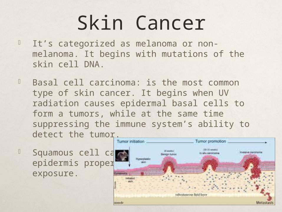
Skin Cancer It’s categorized as melanoma or non-melanoma. It
begins with mutations of the skin cell DNA.
Basal cell carcinoma: is the most common type of skin cancer. It begins when UV radiation causes epidermal basal cells to form a tumors, while at the same time suppressing the immune system’s ability to detect the tumor.
Squamous cell carcinoma: begins in the epidermis proper. Triggered by excessive UV exposure.
… Melanoma: more likely to be malignant starts in the
melanocytes and has appearance of an unusual mole. It is dark, circular and confined. It may hurt, itch or feel numb.
Kaposi’s sarcoma: is a form of skin cancer most likely seen in patients with AIDS. It appears as red, blue or black spots on the skin. This responds to treatment to drugs called “ AIDS cocktails”
Moles & Warts: are usually not cancerous. Moles are due to an overgrowth of the melanocytes and warts are due to a viral infection.
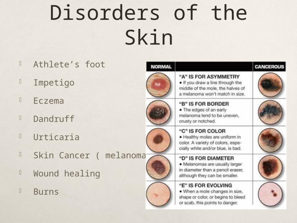
Sings of melanoma(ABCDE) rule
Asymmetrical – irregular shape
Borders- no borders and have notches or indentation in them.
Color- uneven, several colors.
Diameter- greater than 6mm.
Elevation- is elevated above surface of the skin.
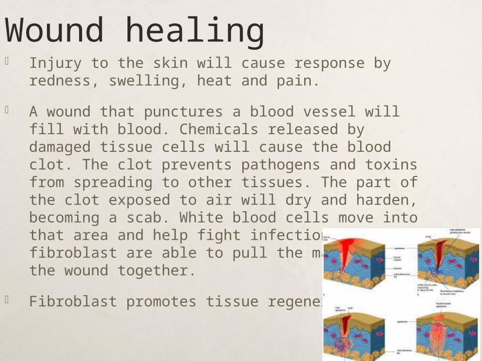
Wound healing Injury to the skin will cause response by redness,
swelling, heat and pain.
A wound that punctures a blood vessel will fill with blood. Chemicals released by damaged tissue cells will cause the blood clot. The clot prevents pathogens and toxins from spreading to other tissues. The part of the clot exposed to air will dry and harden, becoming a scab. White blood cells move into that area and help fight infection and fibroblast are able to pull the margins of the wound together.
Fibroblast promotes tissue regeneration.
Burns It’s caused by heat, radioactive, chemicals or
electrical agents.
Two factors affect burn severity: the depth of the burn and the extent of the burned area.
Major concerns with burns are: lost of fluids, heat loss and bacterial infection.
As soon as damaged skin is removed, skin grafting begins. Auto-grafting is when skin is taken from other parts of the body to replace damaged skin. Heterografting, is when the affected person receives skin from another person.
Degrees Burns 1st degree: only epidermis is affected. Redness and pain,
but no swelling or blisters. ( moderate sunburn)
2nd degree: extends through the entire epidermis and part of the dermis. Redness, pain and blisters. Scarring is common.
3rd degree: destroy entire thickness of the skin. Surface is leathery, brown, black, red, white or tan. No pain because the pain receptors have been destroyed as have blood vessels, sweat glands, sebaceous glands and hair follicles.
4th degree: involves tissue down to the bone. Chances of surviving this degree of burn are minimal unless it affected a small part of the body.
Effects of AgingAs aging occurs, the epidermis maintains its
thickness, but the rate of cell mitosis decreases. The dermis becomes thinner, the dermal papillae flatten, and the epidermis is held less tightly to the dermis so that the skin is looser. Adipose tissue in the hypodermis of the face and hands also decreases, which means that older people are most likely to feel cold.
As a person ages, the number of melanocytes decreases. This causes the hair to turn gray and the skin to become paler. In contrast, some of the remaining pigment cells are larger, and pigmented blotches appear on the skin.
Homeostasis Functions of the skin
Hyperthermia
Hypothermia
Functions of the skin Skin has a protective function- the melanocytes
protects us from UV radiation and skins outer dead cells prevent bacterial invasion. Oily secretions are acidic and retards the growth of the bacteria. The cells in the epidermis alerts immune system of their presence.
Skin helps regulate water loss- skin is waterproof, thereby preventing water loss and water entering when skin is immersed.
Skin assists the function of the urinary system- sweat glands secrete water through sensible and insensible perspiration. This contains small amounts of salt, ammonia, urea, and other wastes.
Skin produces vitamin D- skin function is useful to digestive and skeletal system. When skin is exposed to UV light, it produces vitamin D. this is converted to a hormone called calcitroil in which helps regulate calcium and phosphorus in the body.
Skin gathers sensory information- the sensory receptors in the dermis and epidermis specialized for touch, pressure, pain, hot and cold, are associated with the nervous system. Receptors also account for the communication in between people.
Skin helps regulate body temperature- when the temperature is too hot, blood vessels will dilate, more blood is brought to the surface for cooling. When the temperature is cold, blood vessels constrict so less blood is brought to the skin. When the temperature is extremely below normal, muscles will start to shrivel and if it’s for a long period of time, frostbite will occur.
Hyperthermia & Hypothermia
Hypothermia: body temperature below normal. Uncontrolled shivering, incoherent speech, lack of coordination, pulse rate slows, hallucinations occur, breathing becomes shallow. This is a 50% mortality rate.
Hyperthermia: body temperature above normal.
Heat exhaustion: blood pressure may be low and salts may be lost due to profuse sweating.
Heat stroke: elevated body temperature up to 110 F with no sweating.
Fever: is an immune system response by a bacterial infection.

Video! http://www.youtube.com/watch?v=MeTaBniB0ok