the host defense of drosophila melanogaster · drosophila antimicrobial peptides. name, number of...
TRANSCRIPT

ANRV306-IY25-24 ARI 11 February 2007 13:14
The Host Defense ofDrosophila melanogasterBruno Lemaitre1 and Jules Hoffmann2
1Centre de Genetique Moleculaire, CNRS, 91198 Gif-sur-Yvette, France;email: [email protected] de Biologie Moleculaire et Cellulaire, UPR 9022 du CNRS, 67084Strasbourg Cedex, France; email: [email protected]
Annu. Rev. Immunol. 2007. 25:697–743
First published online as a Review in Advance onJanuary 2, 2007
The Annual Review of Immunology is online atimmunol.annualreviews.org
This article’s doi:10.1146/annurev.immunol.25.022106.141615
Copyright c© 2007 by Annual Reviews.All rights reserved
0732-0582/07/0423-0697$20.00
Key Words
insect immunity, Toll, Imd, recognition, pathogens
AbstractTo combat infection, the fruit fly Drosophila melanogaster relies onmultiple innate defense reactions, many of which are shared withhigher organisms. These reactions include the use of physical bar-riers together with local and systemic immune responses. First, ep-ithelia, such as those beneath the cuticle, in the alimentary tract, andin tracheae, act both as a physical barrier and local defense againstpathogens by producing antimicrobial peptides and reactive oxygenspecies. Second, specialized hemocytes participate in phagocytosisand encapsulation of foreign intruders in the hemolymph. Finally,the fat body, a functional equivalent of the mammalian liver, pro-duces humoral response molecules including antimicrobial peptides.Here we review our current knowledge of the molecular mecha-nisms underlying Drosophila defense reactions together with strate-gies evolved by pathogens to evade them.
697
Ann
u. R
ev. I
mm
unol
. 200
7.25
:697
-743
. Dow
nloa
ded
from
arj
ourn
als.
annu
alre
view
s.or
gby
Sw
iss
Aca
dem
ic L
ibra
ry C
onso
rtia
on
01/2
2/09
. For
per
sona
l use
onl
y.
ANRV306-IY25-24 ARI 11 February 2007 13:14
AMP: antimicrobialpeptide
RNAi: RNAinterference
INTRODUCTION
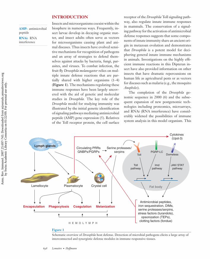
Insects and microorganisms coexist within thebiosphere in numerous ways. Frequently, in-sect larvae develop in decaying organic mat-ter, and insect adults often serve as vectorsfor microorganisms causing plant and ani-mal diseases. Thus insects have evolved sensi-tive mechanisms for recognition of pathogensand an array of strategies to defend them-selves against attacks by bacteria, fungi, par-asites, and viruses. To combat infection, thefruit fly Drosophila melanogaster relies on mul-tiple innate defense reactions that are par-tially shared with higher organisms (1–4)(Figure 1). The mechanisms regulating theseimmune responses have been largely uncov-ered with the aid of genetic and molecularstudies in Drosophila. The key role of theDrosophila model for studying immunity wasillustrated by the initial genetic identificationof signaling pathways mediating antimicrobialpeptide (AMP) gene expression (5). Relativesof the Toll receptor protein, the cell surface
Lymph glands
Lamellocyte Plasmatocyte Crystal cell
Encapsulation Phagocytosis Coagulation Melanization
Toll
pathway
Imd
pathway
JAK/ STAT
pathway
DomelessPGRP-LC
Toll
Antimicrobial peptides,
iron sequestration, DIMs,
serine proteases/serpins,
stress factors (turandots),
opsonization (TEPs),
clotting factors (fondue)
Serine proteases/
serpins
Cytokines
(Upd-3)
Circulating PRRs
GNBPs/PGRPs
Fat body
H E M O L Y M P H
Figure 1Schematic overview of Drosophila host defense. Detection of microbial pathogens elicits a large array ofinterconnected and synergistic defense modules in immune-responsive tissues.
receptor of the Drosophila Toll signaling path-way, also regulate innate immune responsesin mammals. The conservation of a signal-ing pathway for the activation of antimicrobialdefense responses suggests that some compo-nents of innate immunity share an ancient ori-gin in metazoan evolution and demonstratesthat Drosophila is a potent model for deci-phering general innate immune mechanismsin animals. Investigations on the highly effi-cient immune reactions in this Dipteran in-sect have also provided information on otherinsects that have dramatic repercussions onhuman life as agricultural pests or as vectorsfor diseases such as malaria (e.g., the mosquitoAnopheles).
The completion of the Drosophila ge-nomic sequence in 2000 (6) and the subse-quent expansion of new postgenomic tech-nologies including proteomics, microarrays,and RNAi (RNA interference) have consid-erably widened the possibilities of immunesystem analysis in this model organism. This
698 Lemaitre · Hoffmann
Ann
u. R
ev. I
mm
unol
. 200
7.25
:697
-743
. Dow
nloa
ded
from
arj
ourn
als.
annu
alre
view
s.or
gby
Sw
iss
Aca
dem
ic L
ibra
ry C
onso
rtia
on
01/2
2/09
. For
per
sona
l use
onl
y.

ANRV306-IY25-24 ARI 11 February 2007 13:14
article presents an overview of our currentknowledge of the Drosophila immune responsein the context of two fundamental ques-tions: (a) What are the molecular mechanismsunderlying the defense reactions? (b) Howdoes each of these mechanistic modules con-tribute to defense during an infection, andwhat strategies have been developed by thepathogens to evade them?
REPERTOIRE OF DROSOPHILADEFENSE MECHANISMS
A hallmark of the Drosophila host defense, andthat of most other holometabolous insects,is the challenge-induced synthesis and secre-tion of potent AMPs that accumulate in thehemolymph where they oppose invading mi-croorganisms. Although synthesis of AMPsis probably common to all metazoans, secre-tion of these molecules into the hemolymphis not a general phenomenon. We refer it toas the “systemic immune response,” which isby far the best analyzed among Drosophila im-mune reactions, and analyze it from the syn-thesis of immune effectors to recognition ofinfection. Epithelial immunity, i.e., the fightagainst invading microorganisms at the levelof the barrier epithelia, is now understoodto significantly contribute to the protectionof Drosophila. This response is analyzed nextboth in terms of AMP and reactive oxygenspecies (ROS) production. A subsequent sec-tion deals with the cellular response by thehemocytes, especially their role in phagocy-tosis and encapsulation of parasites. The finalsection is devoted to two reactions, coagula-tion and melanization, which are activated im-mediately upon injury.
The Systemic Immune Response
Injection of bacteria into the body cavity in-duces the appearance of antimicrobial activityin the hemolymph of Drosophila. This activitypersists for several days and can confer protec-tion against a second challenge by pathogenicbacteria (7). This reaction mainly consists of
ROS: reactiveoxygen species
AMP production by the fat body, which isa major immune-responsive tissue that origi-nates from the mesoderm during embryoge-nesis and acquires its immune competence atthe onset of the first larval stage. Due to itslarge size and its location inside the open cir-culatory system of the insect body cavity, thefat body represents a powerful organ for thesynthesis and secretion of peptides into thehemolymph, where they readily reach theireffective concentrations.
Immune effectors. Among the variousmolecules produced by the fat body in re-sponse to infection, AMPs are the best char-acterized. Some 20 immune-inducible AMPs,which can be grouped into seven classes,have been identified (Figure 2). They aresmall (<10 kDa), with the exception of the25 kDa Attacin, and cationic and exhibita broad range of activities against bacte-ria and/or fungi (8). Diptericin, Drosocin,and Attacin are very effective against Gram-negative bacteria (9–11). Defensin is activeagainst Gram-positive germs (12), whereasDrosomycin and Metchnikowin are antifun-gal agents (13, 14). Cecropin A1 acts againstboth bacteria and some fungi (15, 16). De-fensins and Cecropins have been reportedfrom many insects, whereas Drosomycin andMetchnikowin have so far been identifiedonly in Drosophilidae (8). The insect AMPsare membrane-active, and their precise modeof action at the membrane level is still un-der investigation. Some AMPs are very sta-ble owing to the presence of intramolecu-lar disulfide bridges and are still detected inthe hemolymph several weeks after challenge(17). Experiments with transgenic flies over-expressing a single AMP provided support fora critical role of AMPs in resistance to infec-tion in Drosophila (18). However, the particu-lar contributions of each of these AMPs havenot been tested, as loss-of-function mutantsfor AMP genes are not available to date.
Recent large-scale analyses, at the tran-scriptome and proteome levels, have re-vealed that in addition to that of AMPs, the
www.annualreviews.org • Host Defense of Drosophila melanogaster 699
Ann
u. R
ev. I
mm
unol
. 200
7.25
:697
-743
. Dow
nloa
ded
from
arj
ourn
als.
annu
alre
view
s.or
gby
Sw
iss
Aca
dem
ic L
ibra
ry C
onso
rtia
on
01/2
2/09
. For
per
sona
l use
onl
y.

ANRV306-IY25-24 ARI 11 February 2007 13:14
Figure 2Drosophila antimicrobial peptides. Name, number of genes in the genome, antimicrobial activities,estimated concentration in the hemolymph after bacterial injection, and 3-D structure (8) (nd, notdetermined).
DIM: Drosophilaimmune molecule
production of many peptides and proteins isupregulated after septic injury (19–25).1 Someof these are involved in the regulation of the
1A list of all Drosophila immune genes can be foundon the web at http://www.cgm.cnrs-gif.fr/immunity/enindex.html.
systemic immune response itself (e.g., signal-ing components). Another group of proteins(opsonins, components of the melanizationor clotting system) participates in distinct de-fense mechanisms, while an additional groupincludes putative immune effectors. Amongthese are 17 members of the DIM family
700 Lemaitre · Hoffmann
Ann
u. R
ev. I
mm
unol
. 200
7.25
:697
-743
. Dow
nloa
ded
from
arj
ourn
als.
annu
alre
view
s.or
gby
Sw
iss
Aca
dem
ic L
ibra
ry C
onso
rtia
on
01/2
2/09
. For
per
sona
l use
onl
y.

ANRV306-IY25-24 ARI 11 February 2007 13:14
(Drosophila immune molecule) and 8 Turandotproteins, which are small peptides of unknownfunctions secreted by the fat body (17, 25–27). One catalase gene, two transferrin genes,and one iron transporter gene are also in-duced following septic injury, pointing to arole of ROS and iron sequestration in the con-trol of microbial development (19, 28). Ironis essential for most invading microorganismsduring the course of an infection, and bothanimals and plants have elaborated immunestrategies that limit iron availability to themicroorganisms.
Collectively, the systemic antimicrobial re-sponse represents a dramatic change in geneexpression that not only results in the pro-duction of antimicrobial molecules in thehemolymph, but also participates in other im-mune mechanisms. Major challenges are tounderstand the relative contribution of theseimmune effectors to the total host defense, totest their specificity against certain pathogenclasses, and to determine potential synergiesbetween them.
Regulation. The massive expression of novelpeptides/polypeptides that occurs followinginfection is primarily regulated at the tran-scriptional level. The cloning in the early1990s of the genes in D. melanogaster thatencode AMPs followed by promoter map-ping experiments with the Cecropin A1 andDiptericin genes revealed the presence ofDNA motifs required for immune inducibil-ity. These include a combination of NF-κBbinding sites, GATA binding sites recognizedby the transcription factor Serpent, and a lesswell-defined motif called R1 (29–33). Promi-nent among these motifs are the κB responseelements, which confer immune inducibility(34–36). Three NF-κB/Rel-like proteins areencoded in the Drosophila genome. Dorsal andDif, encoded by two clustered genes, containan N-terminal Rel DNA binding domain anda C-terminal transactivator domain, whereasRelish is similar to mammalian p105, con-sisting of an N-terminal Rel domain and aC-terminal inhibitory ankyrin repeat domain
(37–39a). Gel shift assays have shown that thethree proteins (Relish, Dorsal, and Dif ) bindto κB sites and can transactivate some of theAMP genes in cell culture (37, 38, 40, 41). Fur-thermore, genetic studies have demonstratedthe key roles of these transactivators in theregulation of AMP genes via two distinct sig-naling pathways, referred to as Toll and Imdpathways.
The Toll pathway. The Toll pathway is anevolutionarily conserved signaling cascadethat plays a key role in the establishmentof the dorso-ventral axis of the Drosophilaembryo, as well as in several other devel-opmental processes (42). Canonic compo-nents of this pathway are the extracellu-lar cytokine Spatzle (which shares structuralsimilarities with the nerve growth factor,NGF), the transmembrane receptor Toll, theTube and MyD88 adaptors, the Pelle kinase,Cactus (the Drosophila homolog of IκB), andthe Dorsal and Dif transactivators (42, 43)(Figure 3). Deletion of any of these compo-nents (except for Cactus and Dorsal) causesa similar immune-deficient phenotype char-acterized by the lack of expression of severalimmune genes, including the antifungal pep-tide Drosomycin gene, and a marked suscep-tibility to fungal and Gram-positive bacterialinfection (44, 44a). Dif and Dorsal seem toplay redundant roles in the control of Dro-somycin at the larval stage, whereas Dif is suf-ficient to mediate Toll activation in adults(45–47). Subtle roles for Dorsal have beenproposed at the adult stage (48). Many compo-nents of the Toll pathway are themselves up-regulated in a Toll-dependent manner uponinfection (44, 49). The induction of the in-hibitor molecule, Cactus, establishes a nega-tive feedback on this cascade (50). Unlike ver-tebrate Toll-like receptors (TLRs), Toll doesnot function as a pattern recognition recep-tor but is activated by the cytokine Spatzle(44, 51, 52). The Drosophila genome encodeseight other Toll proteins, none clearly impli-cated in fly immunity (53, 54). The DrosophilaToll pathway shares significant similarities
www.annualreviews.org • Host Defense of Drosophila melanogaster 701
Ann
u. R
ev. I
mm
unol
. 200
7.25
:697
-743
. Dow
nloa
ded
from
arj
ourn
als.
annu
alre
view
s.or
gby
Sw
iss
Aca
dem
ic L
ibra
ry C
onso
rtia
on
01/2
2/09
. For
per
sona
l use
onl
y.

ANRV306-IY25-24 ARI 11 February 2007 13:14
Time (h)
Exp
ressio
n
Time (h)
Exp
ressio
n
6 12 24 48 3 6 12 24
Injury
Gram-positive
bacterial infection
(M. luteus)
Gram-negative
bacterial infection
(E. coli )
Diptericin (Imd read-out)Drosomycin (Toll read-out)
Fungi
Yeast
Gram-negative
bacteria
Gram-positive
bacteria
Polymeric
peptidoglycan
Monomeric
peptidoglycan
Necrotic
Persephone
GNBP3
GNBP1
PGRP-SA
PGRP-SD
Lysine-type
peptidoglycan
DAP-type
peptidoglycan
PGRP-LE
PGRP-LCaPGRP-LCx
HEMOLYMPHSpätzle
Pro-Spätzle
Toll
PGRP-LCx
PGRP-LCx
FAT BODY CELL
dMyD88
TubePelle
DIFDorsal
Cactus
dFADD
Dredd
Imd
dIAP2
dTAB2
dTAK1
IKKγ
IKKβ
RelishJNK
pathway
Drosomycin and
other antimicrobials
Diptericin and
other antimicrobials
DD
TIR
BIRRING
Rel
DEDDED
DDDD DDDD
DEDDEDDED
TIR
DD DD
DD DD
ANK
ANK
Rel
DED
SPE
NUCLEUS
702 Lemaitre · Hoffmann
Ann
u. R
ev. I
mm
unol
. 200
7.25
:697
-743
. Dow
nloa
ded
from
arj
ourn
als.
annu
alre
view
s.or
gby
Sw
iss
Aca
dem
ic L
ibra
ry C
onso
rtia
on
01/2
2/09
. For
per
sona
l use
onl
y.

ANRV306-IY25-24 ARI 11 February 2007 13:14
with the signaling cascade activated down-stream of Interleukin-1 and the TLRs, point-ing to a common ancestry of these immunemechanisms.
The Imd pathway. This pathway was initiallydefined by the identification of a mutationnamed immune deficiency (imd ) that impairedthe expression of several antibacterial pep-tide genes but only marginally affected Dro-somycin induction (55–57). imd flies succumbto Gram-negative bacteria but are more re-sistant to fungi and Gram-positive bacteriathan are Toll mutant flies. imd encodes adeath domain–containing protein similar tothat of Receptor Interacting Protein (RIP) ofthe tumor necrosis factor receptor (TNF-R)pathway, and its overexpression triggers thetranscription/induction of antibacterial pep-
TLR: Toll-likereceptor
TNF-R: tumornecrosis factorreceptor
tide genes in the absence of an infection(58). Genetic screens and reverse genetic ap-proaches have led to the identification ofeight additional canonical components of theImd pathway: the PGRP-LC receptor (59–61); the Mitogen-Activated Protein 3 kinase(MAP3K) TAK1 (62, 63); TAB2 (64–66);DIAP2, a member of Inhibitor of Apop-tosis proteins (64, 65, 67); IKKβ/ird5 andIKKγ/Kenny, which form a Drosophila equiva-lent of the mammalian IKK signalosome (68–70); the dFADD adaptor (71, 72); the Dreddcaspase (73); and the transcription factorRelish (74). Mutations affecting these factorsgenerate an immune deficiency similar to thatof imd. Noninfected imd-deficient flies areperfectly viable, and to date no developmen-tal role has been attributed to this pathway.The Imd pathway shares some similarities
←−−−−−−−−−−−−−−−−−−−−−−−−−−−−−−−−−−−−−−−−−−−−−−−−−−−−−−−−−−−−−−−−−−−−−Figure 3Model of Toll and Imd pathway activation. (Top) Antimicrobial peptide genes are regulated by a balancebetween two signaling pathways: the Toll pathway that is largely activated by fungi and Gram-positivebacteria, and the Imd pathway that is mainly activated by Gram-negative bacteria. According to the κBsites present in their promoters, antimicrobial peptide genes are more sensitive to either the Toll cascade(e.g., Drosomycin) or the Imd cascade (e.g., Diptericin) or are coregulated. Toll pathway: The Toll receptoris activated upon binding with a cleaved form of Spatzle that is processed by proteolytic cascadesactivated by secreted recognition molecules (PGRP-SA, PGRP-SD, GNBP1, GNBP3). Three distinctrecognition modules that involve the sensing of Gram-positive bacteria (PGRP-SA, PGRP-SD,GNBP1), Glucan (GNBP3), and entomopathogenic fungi (via the direct cleavage of an SP) converge tothe activation of SPE that cleaves Spatzle. Mature Spatzle binds as a dimer to Toll, thereby inducing itsdimerization at the plasma membrane (51, 52). This causes the recruitment of three intracellular Deathdomain–containing proteins, MyD88, Tube, and Pelle, the latter also being a kinase (43). By amechanism still uncharacterized, Cactus is phosphorylated, then degraded by the proteasome (50). As aconsequence, the Rel transcription factors Dif and Dorsal are released and move from the cytoplasm tothe nucleus (37, 142). Among the less well-understood aspects of this cascade are the respective roles ofthe multiple Spatzle isoforms, the mechanisms that link Pelle activation to Cactus degradation, and thecomplex regulation of Cactus isoforms. Imd pathway: Upon direct binding with bacterial elicitors(monomeric or polymeric DAP-type PGN), PGRP-LC recruits the adaptor Imd (127, 272). Imd theninteracts with dFADD, which itself binds the apical caspase Dredd (273). This caspase has been proposedto associate with Relish, which it might cleave directly once Relish is phosphorylated (274, 275). AfterRelish cleavage, the Rel domain translocates to the nucleus, whereas the inhibitory domain remainsstable in the cytoplasm. Relish is phosphorylated by the IKK signaling complex (68), which is itselfthought to be activated by TAK1 and its adaptor TAB2 in an Imd- and possibly dFADD-dependentmanner. The Ring domain protein DIAP2 may activate dTAK1. At present the mechanisms that link Imdto the IKK complex and the precise role of TAK1 and DIAP2 are not known (see text). TIR (Toll-IL1receptor domain), DD (death domain), DED (death-effector domain), ANK (ANKyrin repeats), Rel (Relhomology domain), RING (RING finger domain), BIR (Baculovirus IAP repeat), SPE (Spatzleprocessing enzyme). (Bottom) Differential expression of Drosomycin (Toll target) and Diptericin (Imdtarget) genes in response to injection of E. coli (Gram-negative bacteria) or M. luteus (Gram-positivebacteria) (83). Diptericin shows an acute phase profile, whereas Drosomycin exhibits a late and sustainedexpression pattern.
www.annualreviews.org • Host Defense of Drosophila melanogaster 703
Ann
u. R
ev. I
mm
unol
. 200
7.25
:697
-743
. Dow
nloa
ded
from
arj
ourn
als.
annu
alre
view
s.or
gby
Sw
iss
Aca
dem
ic L
ibra
ry C
onso
rtia
on
01/2
2/09
. For
per
sona
l use
onl
y.

ANRV306-IY25-24 ARI 11 February 2007 13:14
MAPK:mitogen-activatedprotein kinase
with the vertebrate TNF-R pathway. Severalproteins were recently identified that affectthe Imd pathway (64, 75–80a). The molecularorganization of the Imd pathway is, however,as yet poorly defined owing to difficulties inperforming epistatic analyses and to the ab-sence of data on the subcellular localizationof most of its components (Figure 3).
Interactions between Toll and Imd pathways.
To date, the Toll and Imd pathways are the solereported intracellular cascades of Drosophilathat are activated by microbial ligands. Mi-croarray studies using imd /Toll double mutantflies have shown that these cascades regulatealmost 80% of the genes induced upon sep-tic injury, reflecting their important contribu-tion in survival from infection (49). An openquestion is whether the Toll and Imd pathwayscan synergize to increase the levels of induc-tion of some of the immune-response genes,possibly via the formation of Dif/Relish het-erodimers. Bioinformatic analysis has recentlyidentified κB binding sites specific for eitherToll or Imd pathway activation (81). Althoughsome immune-induced genes are clearly de-pendent on one pathway only (Diptericin forImd, DIM1 for Toll), others can apparentlybe induced by both cascades. Most notably,Drosomycin, which is a convenient read-outfor Toll pathway activation, receives a modestinput from the Imd pathway in the systemicresponse and is solely activated by the Imdpathway in the local response (73, 82). Thesusceptibility of Toll and imd mutants to differ-ent classes of microorganisms is explained bythe differential activation of these two path-ways by distinct microorganisms as well as bythe existence of specific immune target genes.Another important aspect of the antimicrobialresponse is the temporal activation of thesetwo pathways. Genes regulated by Imd gen-erally show an acute phase profile, whereasToll target genes exhibit a late and sustainedexpression pattern (21, 47, 83) (Figure 3).
Finally, a number of developmental andphysiological factors affect Toll and Imdsignaling by influencing the maturation of
the fat body. This is reflected by the im-mune inducibility of Diptericin in larvae,which increases with age and is dependenton ecdysone, the key molting hormone of in-sects (84). All mutations affecting ecdysonemetabolism can thus indirectly affect the im-mune response. This effect complicates im-mune studies and may explain a number ofconflicting reports (85).
The JAK/STAT pathway. The JAK/STATpathway, originally identified through its rolein embryonic segmentation, has three maincellular components: the receptor Domeless,the Janus Kinase ( JAK) Hopscotch, and theSTAT transcription factor (86). The first ev-idence for an involvement of the JAK/STATpathway in insect immunity came from stud-ies performed in the mosquito Anopheles in-dicating that after an immune challenge theSTAT protein accumulates in the nucleus(87). Subsequent gene expression profile stud-ies identified a subset of Drosophila immune-responsive genes that are regulated by theJAK/STAT pathway, namely the genes encod-ing the complement-like protein Tep2 and theTurandot stress genes (21, 88, 89). Turandot areDrosophila-specific genes of unknown func-tion that are induced by various stress con-ditions, especially by septic injury (26, 27).The transcriptional regulation of these genesis complex, with additional inputs from boththe Imd and MAPK (mitogen-activated pro-tein kinase) pathways (90).
JAK/STAT-deficient flies are as resistant tobacterial and fungal infections as are wild-typeflies, and they express a normal AMP profile.They are, however, sensitive to infectionwith the Drosophila C virus (91). It has beenproposed that the JAK/STAT pathway couldrespond to tissue damage and that ahemocyte-released cytokine, Unpaired-3(Upd-3), activates this pathway by bindingto the fat body Domeless receptor duringinfection (89). The precise role of thispathway and its overall contribution to thehost defense remain to be established.
704 Lemaitre · Hoffmann
Ann
u. R
ev. I
mm
unol
. 200
7.25
:697
-743
. Dow
nloa
ded
from
arj
ourn
als.
annu
alre
view
s.or
gby
Sw
iss
Aca
dem
ic L
ibra
ry C
onso
rtia
on
01/2
2/09
. For
per
sona
l use
onl
y.

ANRV306-IY25-24 ARI 11 February 2007 13:14
Other pathways. The JNK pathway reg-ulates many developmental processes inDrosophila and is required for proper woundhealing of the epidermis (92, 93). Microarrayanalysis of S2 cells, a hemocyte-derived cellline, has shown that TAK1 activates the JNKpathway in response to bacteria via the JNKkinase basket (21). These data are consistentwith a model where Imd signaling bifurcatesdownstream of TAK1, activating both JNKand IKK signaling (Figure 3). Some negativefeedbacks between the Imd-Relish and Imd-JNK branches have been reported (94, 95). InS2 cells, JNK-dependent immune genes en-code many proteins involved in cytoskeletonremodeling, in keeping with a role in hemo-cyte activation (21). A role for the JNK path-way in AMP gene expression by the fat bodyhas been proposed (96, 97).
The p38 stress pathway has been im-plicated in the immune response of plants,Caenorhabditis elegans, and mammals (98, 99).The Drosophila genome encodes two ho-mologs of p38, and cell culture experimentsidentified several MAPKs acting upstream(100, 101). Although there is no in vivo evi-dence for a role of this pathway in the antimi-crobial response, its implication in immunefunctions remains possible. Eiger/Wengenare Drosophila proteins with similarities tomammalian TNF and TNF-R, respectively(102, 103). Flies carrying a mutation in thesegenes are fully viable and do not display ob-vious immune defects. Overexpression of theeiger and wengen genes, however, leads toapoptosis via activation of the JNK pathway.The physiological function of these proteinshas not yet been determined.
Microbial recognition. Injection of Gram-positive or Gram-negative bacteria and fungiinduces different patterns of AMP expres-sion, indicating that microbial recognitionmechanisms can differentiate between variousclasses of invaders (83). This property favorsadapted effector responses via the selective ac-tivation of the Toll and Imd pathways. Mi-crobial detection is a process that ultimately
PRR: patternrecognition receptor
PGRP:peptidoglycanrecognition proteins
GNBP:Gram-negativebinding protein
proPO:prophenoloxidase
PGN:peptidoglycan
DAP:meso-diaminopimelicacid
LPS:lipopolysaccharide
requires direct contact between a host pro-tein called pattern recognition receptor (PRR)and a microbial molecule. Genetic studieshave shown that microbial recognition act-ing upstream of the Toll and Imd pathways isachieved through peptidoglycan recognitionproteins (PGRPs) and Gram-negative bind-ing proteins (GNBPs), two families of PRRsinitially identified in larger insects for theircapacity to bind microbial ligands and to ac-tivate the proPO (prophenoloxidase) cascade(104–106).
Bacterial recognition by PGRPs. InDrosophila, recognition of bacteria is achievedthrough the sensing of specific forms ofpeptidoglycan (PGN) by PGRPs. PGN isan essential glucopeptidic polymer restrictedto the cell wall of both Gram-negativeand Gram-positive bacteria (Figure 4a). Itconsists of long glycan chains of alternatingN-acetylglucosamine and N-acetylmuramicacid residues that are cross-linked to eachother by short peptide bridges (107). PGN isa highly complex and fast-evolving moleculewith marked differences from one bacteriumto another. PGN from Gram-negativebacteria differs from most Gram-positivePGN by the replacement of lysine withmeso-diaminopimelic acid (DAP) at thethird position in the peptide chain. Thereis, however, a subclass of Gram-positivebacteria including Bacillus species, whichproduces DAP-type PGN. Another majordifference between Gram-negative andGram-positive PGN is its localization withinthe cell wall. Gram-negative PGN consists ofa single layer and is hidden in the periplasmicspace underneath the outer membrane andlipopolysaccharide (LPS) layer, whereas PGNfrom Gram-positive bacteria is multilayeredand exposed at the bacterial surface.
The Imd pathway is activated by DAP-type PGN, whereas the Toll pathway is acti-vated by Lys-type PGN, as demonstrated bystudies using highly purified bacterial com-pounds (108). In contrast to vertebrates, LPSendotoxin, the major component of the
www.annualreviews.org • Host Defense of Drosophila melanogaster 705
Ann
u. R
ev. I
mm
unol
. 200
7.25
:697
-743
. Dow
nloa
ded
from
arj
ourn
als.
annu
alre
view
s.or
gby
Sw
iss
Aca
dem
ic L
ibra
ry C
onso
rtia
on
01/2
2/09
. For
per
sona
l use
onl
y.

ANRV306-IY25-24 ARI 11 February 2007 13:14
Gram-negative cell envelope, has no ef-fect on Toll and Imd pathway activ-ity, and previous positive results were ex-plained by the presence of PGN contami-nants in commercial LPS preparations (108,109).
Further studies have demonstrated thatboth polymeric and monomeric DAP-typePGN can activate the Imd pathway. A spe-cific monomer, GlcNAc-MurNAc(anhydro)-L-Ala-γ-D-Glu-meso-DAP-D-Ala monomer,also known as tracheal cytotoxin (TCT), was
706 Lemaitre · Hoffmann
Ann
u. R
ev. I
mm
unol
. 200
7.25
:697
-743
. Dow
nloa
ded
from
arj
ourn
als.
annu
alre
view
s.or
gby
Sw
iss
Aca
dem
ic L
ibra
ry C
onso
rtia
on
01/2
2/09
. For
per
sona
l use
onl
y.

ANRV306-IY25-24 ARI 11 February 2007 13:14
identified as the minimal PGN motif capa-ble of efficient induction of the Imd path-way (109, 110). TCT provides an ideal “signa-ture” of Gram-negative bacteria because thismuropeptide is located at the end of the PGNstrand and is released from PGN during cellgrowth and division (Figure 4a). In contrast,the minimum structure needed to activate theToll pathway is a muropeptide dimer of lysine-type PGN (111).
PGRPs are highly conserved from insectsto mammals and share a 160 amino acid do-main (the PGRP domain) with similarities tobacteriophage T7 lysozyme, a zinc-dependentN-acetylmuramoyl-l-alanine amidase (112–114) (Figure 4b). Sequence analysis of the13 Drosophila PGRPs points to the existenceof two subgroups with either recognition orenzymatic properties. Members of the firstgroup (PGRP-SA, SD, LA, LC, LD, LE,and LF) lack zinc-binding residues requiredfor amidase activity but still retain the abil-ity to bind and recognize PGN and functionas PRRs. In contrast, proteins from the othersubgroup of PGRPs, referred to as catalyticPGRPs, have demonstrated (PGRP-SC1, LB,SB1) or predicted (SC2, SB2) zinc-dependentamidase activity that removes peptides fromthe glycan chains, thereby reducing or elim-inating the biological activity of PGN (115–116a). Some of these PGRPs modulate theimmune response by scavenging PGN (115–117). The tertiary structures of four PGRPshave now been solved (118–123). A promi-
nent feature is the presence of an extendedsurface groove in the PGRP domain, whichincludes a zinc-finger cage in the catalyticPGRP-LB. The structures of PGRP-LE andPGRP-LCa/x in complex with TCT show aninteraction between the peptide stem of PGNand the PGRP groove (Figure 4c). In contrastto other PGRPs, the PGRP domain of PGRP-LCa does not possess a typical PGN-dockinggroove, in agreement with its role as a core-ceptor sensing monomeric DAP-type PGN(121).
Recognition of Gram-negative bacteria by
PGRP-LE/LC. PGRP-LC-deficient fliesdisplay a strongly reduced expression ofantibacterial peptide genes in response toGram-negative bacteria. This effect is similarto, albeit weaker than, that observed for imd-deficient mutants. Epistatic and phenotypicanalyses indicated that PGRP-LC is themajor receptor of the Imd pathway (59). Al-ternative splicing of PGRP-LC can producethree proteins (LCa, LCx, LCy) that sharethe same intracellular (signaling) domain buthave distinct extracellular (sensing) domains(109, 112). Studies in cell culture using RNAispecific for each isoform have shown thatPGRP-LCx is required for recognition ofpolymeric PGN, whereas both PGRP-LCaand PGRP-LCx are mandatory for detectionof monomeric PGN (109). The current beliefis that signaling is achieved by association ofat least two PGRP-LC molecules in close
←−−−−−−−−−−−−−−−−−−−−−−−−−−−−−−−−−−−−−−−−−−−−−−−−−−−−−−−−−−−−−−−−−−−−−Figure 4Recognition upstream of the Toll and Imd pathways. (a) Structure of E. coli PGN. PGN is a complexheteropolymer consisting of long glycan chains of alternating N-acetylglucosamine (GlcNAc) andN-acetylmuramic acid (MurNAc) residues, connected by short peptide bridges (107). All the terminalMurNAc residues ending the E. coli PGN glycan chains have a unique internal 1,6-anhydro bond. TheTCT monomer (indicated in blue) occurs naturally in Gram-negative bacteria at the extremity of allglycan strands and is consequently present in approximately 5% of the GlcNAc-MurNAc units.Lysosymes catalyze the cleavage of the β-1,4-glycosidic bond between the MurNAc and GlcNAcresidues (orange arrow) while amidase PGRPs remove the peptidic bridge from the sugar backbone (bluearrow). (b) Schematic structure of some PGRPs and GNBPs. SP, signal peptide; TM, transmembranedomain; PGRP, PGRP domain. GNPBs have an N-terminal domain that has been shown to bindβ(1,3)-glucan and a C-terminal domain with homology to glucanase. (c) Three-dimensional structure ofPGRP-LE with TCT (120). A prominent feature is the presence of an extended surface groove in thePGRP domain that interacts with the peptide stem of TCT (in color).
www.annualreviews.org • Host Defense of Drosophila melanogaster 707
Ann
u. R
ev. I
mm
unol
. 200
7.25
:697
-743
. Dow
nloa
ded
from
arj
ourn
als.
annu
alre
view
s.or
gby
Sw
iss
Aca
dem
ic L
ibra
ry C
onso
rtia
on
01/2
2/09
. For
per
sona
l use
onl
y.
ANRV306-IY25-24 ARI 11 February 2007 13:14
proximity through binding of polymericPGN. Such an interaction cannot occurwith monomeric PGN, and in this casePGRP-LCa is expected to act as an adaptor(121, 124). This model is supported by thecrystallization of TCT in complex with bothPGRP-LCa and LCx (122).
PGRP-LE encodes a PGRP with affin-ity to DAP-type PGN and is expressed bothextra- and intracellularly (125, 126). A frag-ment of PGRP-LE corresponding to thePGRP domain alone functions extracellularlyto enhance PGRP-LC-mediated PGN recog-nition on the cell surface, a role evocative ofthat of mammalian CD14 in binding of LPS toTLR4 (127). A full-length form of PGRP-LEis also present in the cytoplasm and acts as anintracellular receptor for monomeric PGN,thus bypassing the requirement for PGRP-LC (127). Monomeric PGN probably gainsaccess to the cytoplasm by virtue of its smallsize. Thus, both PGRP-LC and PGRP-LEcould participate in the sensing of Gram-negative bacteria upstream of the Imd path-way (Figure 3).
Downregulation of the Imd pathway by
amidase PGRPs. Injection of Gram-negativebacteria induces a transient expression of an-tibacterial peptide genes (Figure 3), suggest-ing the existence of a mechanism to down-regulate the immune response. Recent studiesin Drosophila revealed a key role for amidasePGRPs in controlling the level of Imd path-way activity by degrading PGN (116, 117).PGRP-LB and PGRP-SC are efficient ami-dases that remove peptides from the glycanchains and thereby convert Gram-negativePGN into nonimmunostimulatory fragments.PGRP-LB is active only on DAP-type PGN,whereas PGRP-SC digests both DAP- andLys-type PGN (115, 116). RNAi extinctionof PGRP-SC1/2 or PGRP-LB leads to higherDiptericin expression after systemic infectioncompared to that of wild-type flies. PGRP-LBencodes a secreted protein and is regulated bythe Imd pathway. The presence of PGRP-LBin the hemolymph establishes a negative feed-
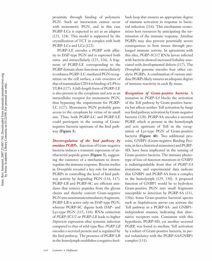
back loop that ensures an appropriate degreeof immune activation in response to bacte-rial infection (116). This mechanism econo-mizes host resources by anticipating the ter-mination of the immune response. AmidasePGRPs may also prevent potentially severeconsequences to host tissues through pro-longed immune activity. In agreement withthis idea, PGRP-SC1/2 RNAi larvae infectedwith bacteria showed increased lethality asso-ciated with developmental defects (117). TheDrosophila genome encodes four other cat-alytic PGRPs. A combination of various ami-dase PGRPs likely ensures an adequate degreeof immune reactivity in each fly tissue.
Recognition of Gram-positive bacteria. Amutation in PGRP-SA blocks the activationof the Toll pathway by Gram-positive bacte-ria but affects neither Toll activation by funginor Imd pathway activation by Gram-negativebacteria (128). PGRP-SA encodes a secretedPGRP, which is present in the hemolymphand acts upstream of Toll in the recog-nition of Lys-type PGN of Gram-positivebacteria (Figure 4b). Two additional pro-teins, GNBP1 (Gram-negative Binding Pro-tein, in fact a historical misnomer) and PGRP-SD, have been implicated in the sensing ofGram-positive bacteria. The immune pheno-type of loss-of-function mutations in GNBP1is indistinguishable from that of PGRP-SAmutations, and experimental data indicatethat GNBP1 and PGRP-SA form a complexin the hemolymph (129, 130). A proposedfunction of GNBP1 would be to hydrolyzeGram-positive PGN into small fragmentssusceptible to detection by PGRP-SA (111,130a). Some Gram-positive bacterial speciessuch as Staphylococcus aureus can activate theToll pathway in a PGRP-SA- and GNBP1-independent manner, indicating that alter-native receptors exist. Consistent with thishypothesis, PGRP-SD, yet another secretedPGRP, was found to mediate Toll activationby a subset of Gram-positive bacteria, in par-tial redundancy with the PGRP-SA/GNBP1complex (131).
708 Lemaitre · Hoffmann
Ann
u. R
ev. I
mm
unol
. 200
7.25
:697
-743
. Dow
nloa
ded
from
arj
ourn
als.
annu
alre
view
s.or
gby
Sw
iss
Aca
dem
ic L
ibra
ry C
onso
rtia
on
01/2
2/09
. For
per
sona
l use
onl
y.
ANRV306-IY25-24 ARI 11 February 2007 13:14
Fungal recognition. Recent studies point toa role of GNBP proteins in the detectionof fungal infection (81). Members of thisfamily display a significant overall homologyto bacterial glucanases (131a) (Figure 4b).They contain an N-terminal domain thatbinds to β(1,3)-glucan and a C-terminal do-main that is homologous to the catalytic do-main of β-glucanase. However, the absenceof conserved key residues in the catalytic sitesuggests that this domain is not functional.The Drosophila genome contains three con-sensus members of the GNBP family andthree related members containing only anN-terminal domain similar to that of GNBP3.The GNBP3 protein sequence is highly sim-ilar to that of lepidopteran β Glucan Recog-nition Proteins known to bind fungal β(1,3)-glucans (131b). A role of GNBP3 in fungaldetection is supported by the observation thatGNBP3 mutants are sensitive to fungal in-fection and fail to activate the Toll path-way (131c). According to this observation,GNBP3 is a circulating PRR acting upstreamof the Toll pathway in fungal detection.
Activation of Toll by serine proteases. TheToll receptor is activated upon bindingof a cleaved form of Spatzle (51, 52).The immune-induced cleavage of Spatzle isthought to be triggered by secreted PRRsthat activate a proteolytic cascade, a processconceptually similar to vertebrate blood co-agulation or complement activation cascades(132). These proteolytic cascades have a func-tional core consisting of several serine pro-teases (SPs) that undergo zymogen activation,followed by cleavage of a substrate by the ter-minal downstream protease. SP cascades al-low the shaping and amplification of extra-cellular signals and are involved in complexregulatory mechanisms, in which inappropri-ate activation is prevented by the action ofSP inhibitors (serpins) (133). The Drosophilagenome encodes more than 200 SPs and ser-ine protease homologs (SPHs), most of whichhave as yet uncharacterized functions (134).SPHs represent one quarter of all Drosophila
SP: serine protease
SPH: serineprotease homolog
SP-related proteins and are thought to pos-sess regulatory functions (135, 136). ManySPs that function in insect immunity containa Clip-domain N-terminal to the catalytic do-main (137). This domain is only found in in-sect SPs and is believed to play a regulatoryrole in the sequential activation of SPs.
The activation of Spatzle during the im-mune response is achieved by a set of SPsdistinct from those mediating Toll activationduring embryonic development (44). Recentstudies demonstrate the existence of a com-mon core of SPs and SPHs acting upstreamof Spatzle in the immune response that inte-grate signals sent by various secreted recogni-tion molecules via more specialized SPs (138).SPE, an immune-regulated Clip-domain SP,has been identified as the Spatzle processingenzyme (138, 139). SPE presents 89% aminoacid identity with Easter, the enzyme that pro-cesses Spatzle in the embryo. Toll activationby entomopathogenic fungi is independent ofPGRP-SA, but is mediated by an extracellu-lar cascade involving the Clip-SP Persephone(140) and the inhibitory serpin Necrotic (141).Overexpression of persephone or loss of func-tion of necrotic is sufficient to trigger Spatzle-dependent activation of Toll in the absence ofan immune challenge.
Recognition: concluding remarks. The pastfew years have seen the identification of theDrosophila PRRs regulating the Toll and Imdpathways and of the microbial ligands thatthey recognize. From an evolutionary point ofview, it is interesting to note that insects useGNBPs and PGRPs as PRRs because theseare proteins derived from enzymes known todegrade microbial cell wall products. Thus,the roles of these molecules may have evolvedfrom microbicidal to recognition.
The organization of the Toll and the Imdpathways differs, in that the former is acti-vated by secreted PRRs whereas the latteris activated through membrane-bound PRRs.This difference of architecture could reflectthe mode of recognition: (a) direct for theToll pathway for which PRRs can bind to
www.annualreviews.org • Host Defense of Drosophila melanogaster 709
Ann
u. R
ev. I
mm
unol
. 200
7.25
:697
-743
. Dow
nloa
ded
from
arj
ourn
als.
annu
alre
view
s.or
gby
Sw
iss
Aca
dem
ic L
ibra
ry C
onso
rtia
on
01/2
2/09
. For
per
sona
l use
onl
y.

ANRV306-IY25-24 ARI 11 February 2007 13:14
microbial ligands exposed at the surface ofGram-positive bacteria and fungi, or (b) indi-rect for the Imd pathway, which is activated byPGN released from the periplasm of Gram-negative bacteria.
Finally, it cannot be formally excludedthat these pathways are activated through therecognition of host molecules (e.g., moleculesreleased at the wound site). The existenceof endogenous ligands is also supported bythe observation that larvae with melanotic tu-mors display a significant AMP gene expres-sion level in the fat body in the absence of aninfection (142–144). One report suggests thatendogenous DNA can activate the Imd path-way based on the observation that a fly line de-ficient in DNase II exhibits a higher Diptericinexpression level in the absence of an infectioncompared to that of wild-type flies (145).
Epithelial Immunity in Drosophila
Because the barrier epithelia are in constantcontact with large numbers of microorgan-isms, these surfaces must be armed with effi-cient systems for microbial recognition andcontrol. This is especially true for insectssuch as Drosophila, which live on decayingmatter and feed on fermenting medium. InDrosophila, both the gut and trachea, two mainroutes of infection, are lined with a chitinousmatrix. Furthermore, the gut lumen is an en-vironment hostile to microbial colonizationdue to its physical and physiological proper-ties and the secretion of lysozymes (146, 147).In addition, local production of AMPs andROS provides two complementary inducibledefense mechanisms in the gut (Figure 5).
Local AMP expression. Expression ofAMPs has occasionally been reported in ep-ithelia of other insect species. In the silkwormlarva Bombyx mori, Cecropin transcription isinduced in the epithelial cells underlying thecuticle, when it is lightly abraded in the pres-ence of live bacteria (148). In Drosophila, theuse of GFP reporter transgenes has revealedthat AMP genes are expressed in several
surface epithelia that are in contact withthe external environment (82, 149). Theseinclude the epidermis, the reproductive sys-tem, the respiratory tract, and the digestivetract. This AMP synthesis is referred toas the local immune response as opposed tothe systemic response. One can distinguishbetween constitutive and inducible AMPexpression in epithelia. In the first case,the AMP gene is expressed constitutivelyin a defined tissue, and its transcription isnot upregulated during microbial infection.This is the case for Drosomycin in salivaryglands and in the female spermatheca, andfor Cecropin in the male ejaculatory duct (82).This constitutive expression is not regulatedby NF-κB pathways but by various tissue-specific transcription factors such as thehomeobox-containing protein Caudal (150,151). In virgin females, mating stimulates theexpression of some AMP genes (152).
The second form is the inducible lo-cal AMP gene expression. This response istriggered upon natural infection by Gram-negative bacteria and is mediated by the Imdpathway (82, 153). For example, Drosomycinand Diptericin are induced in both trachea andgut via the Imd pathway in response to lo-cal infection by bacteria such as Erwinia caro-tovora (154). Like the systemic response, thelocal immune response is mediated throughthe recognition of Gram-negative PGN byPGRP-LC (116). It remains to be clarifiedwhy commensal bacteria or ingested PGNdoes not generate a state of permanent im-mune activation in tissues such as the gut.A central role in bacterial tolerance of thegut has been attributed to amidase PGRPs,as they scavenge PGN released by commen-sals (116, 117) (Figure 5). Because Gram-negative PGN is hidden in the periplasmicspace underneath the outer LPS membraneand bacteria residing in the gut have a lowdivision rate, commensals may well releaseonly low amounts of PGN that can readilybe hydrolyzed by amidase PGRPs. This im-plies the important concept of a threshold re-sponse for local immune activation in order to
710 Lemaitre · Hoffmann
Ann
u. R
ev. I
mm
unol
. 200
7.25
:697
-743
. Dow
nloa
ded
from
arj
ourn
als.
annu
alre
view
s.or
gby
Sw
iss
Aca
dem
ic L
ibra
ry C
onso
rtia
on
01/2
2/09
. For
per
sona
l use
onl
y.

ANRV306-IY25-24 ARI 11 February 2007 13:14
ROS2O-
dDuox
PGRP-LC
AMP genes
PGRP-LB
Imdpathway
Amidaseactivity
Antimicrobialpeptides
AprA
H O2 2
HClOCl-
H O2
ROS-sensitivemicrobes
ROS-resistantmicrobes
PGRPLB
PMPM
ROS production AMP production
IRC
Figure 5Gut immune response. Local production of ROS (left) and AMPs (right) provides two inducible defensemechanisms in the gut. ROS are produced by the Duox protein and are detoxified by the IRC catalase(156–158). Enzymatic assays demonstrated that the PHD domain of Drosophila DUOX can transformH2O2 into the highly microbicidal HOCl. AMPs (e.g., Diptericin or Attacin) are produced by epithelialcells under the control of the Imd pathway upon recognition of PGN released by Gram-negative bacteria(116, 117). Amidase PGRPs degrade PGN in nonimmunostimulatory fragments, thereby reducing thegut immune reactivity. Bacteria such as Pseudomonas entomophila secrete an abundant protease (AprA) thatdegrades AMPs (238). PM, peritrophic matrix.
differentiate between commensal microor-ganisms and invading pathogens.
To date, no implication of the Toll path-way in the local immune response has beendemonstrated, and there is no evidence thatAMPs are induced in epithelia in response toLys-type Gram-positive bacteria or fungi.
ROS production. In mammals an immedi-ate epithelial response to pathogen assault isthe generation of ROS. In Drosophila, naturalinfections with bacteria also induce rapid ROSsynthesis in the gut, and the dynamic cycle ofROS generation and elimination appears tobe vital.
The Duox proteins form a conserved fam-ily of molecules that contain, in addition to the
NADPH domain, an N-terminal extracellularperoxidase domain (PHD) that can produceROS in a regulated manner (155). Inactivationof the Duox gene by RNAi blocks ROS pro-duction in the gut of infected flies, suggestingthat this enzyme is the sole source of epithelialROS (156). Duox RNAi flies rapidly succumbto oral infection by the Gram-negative bacte-ria E. carotovora, and this lethality is associatedwith an inability to control bacterial growth.Excessive ROS production, which is delete-rious to the host, is prevented in Drosophilaby immune responsive catalase (IRC). Silenc-ing of IRC by RNAi results in higher ROSproduction and fly lethality, indicating thatIRC provides an antioxidant defense systemin Drosophila (157). The IRC and Duox phe-notypes demonstrate that a fine redox balance
www.annualreviews.org • Host Defense of Drosophila melanogaster 711
Ann
u. R
ev. I
mm
unol
. 200
7.25
:697
-743
. Dow
nloa
ded
from
arj
ourn
als.
annu
alre
view
s.or
gby
Sw
iss
Aca
dem
ic L
ibra
ry C
onso
rtia
on
01/2
2/09
. For
per
sona
l use
onl
y.

ANRV306-IY25-24 ARI 11 February 2007 13:14
Figure 6Drosophila hematopoiesis and hemocyte functions. The lymph gland contains a large number ofhemocyte progenitors that can differentiate into three hemocyte types with distinct functions (indicatedon the right). Many factors that regulate key steps of hematopoietic lineage commitment have beenidentified: serpent (GATA factor), U-shaped (Friend-of-GATA), Lozenge (Runx1 homolog), Glial CellsMissing 1 and 2, PVR (homologous to RTK receptors), and Collier (EBF ortholog) (164, 165, 193).Given their similarities to mammalian hematopoietic factors, these studies point to a conservation ofmolecular bases for blood-lineage determination in mammalian and Drosophila hematopoiesis. TheJAK-STAT, Ras/Raf/MAPK, and Toll signaling pathways have also been implicated in hemocytedifferentiation and proliferation, but their exact function is not yet known (276, 277).
is critical for control of microorganisms in thegut lumen. This ROS-dependent gut immu-nity is not affected by the Imd pathway andprovides an additional barrier against ingestedmicroorganisms (158) (Figure 5).
Cellular Response
The body cavity of Drosophila, like that ofall arthropods, is filled with a circulatinghemolymph that contains both free-floatingand sessile blood cells (hemocytes). Drosophilalarvae contain several thousand hemocytes,which can be divided into the following threecell types on the basis of their structuraland functional features: plasmatocytes, crys-tal cells, and lamellocytes (Figure 6) (159,
160). Plasmatocytes represent 90%–95% ofall mature larval hemocytes and function inthe phagocytic removal of dead cells and mi-crobial pathogens. Lamellocytes are relativelylarge, flat, and adherent cells that primar-ily function in encapsulation and neutraliza-tion of objects too large to be phagocytosed.Lamellocytes are not found in embryos andadults, and they are rarely observed in healthylarvae, although large numbers of these cellscan be induced to differentiate from hemocyteprecursors upon infection of larvae with par-asitoid wasp eggs. Crystal cells constitute 5%of the larval hemocytes and are nonphagocyticcells involved in the melanization process.Mature crystal cells express proPOs, whichare oxidoreductases related to hemocyanins
712 Lemaitre · Hoffmann
Ann
u. R
ev. I
mm
unol
. 200
7.25
:697
-743
. Dow
nloa
ded
from
arj
ourn
als.
annu
alre
view
s.or
gby
Sw
iss
Aca
dem
ic L
ibra
ry C
onso
rtia
on
01/2
2/09
. For
per
sona
l use
onl
y.

ANRV306-IY25-24 ARI 11 February 2007 13:14
and which mediate melanization. They arefragile, readily disrupt, and release their con-tents into the hemolymph upon activation.They thus function as storage cells for thelarge amounts of proPO present in their cy-toplasm in crystallized form (161).
Hematopoiesis. In Drosophila, mature plas-matocytes arise from two spatially and tem-porally distinct phases of hematopoietic de-velopment, one early in the embryonic headmesoderm and another during larval develop-ment in a specialized organ called the lymphgland (162–166). The embryonic phase ofhematopoiesis gives rise to the mature cir-culating hemocytes of larval stages whereaslymph gland plasmatocytes, under normal,nonimmune conditions, do not enter circu-lation until metamorphosis. At the onset ofmetamorphosis, the lymph gland releases alarge number of actively phagocytosing plas-matocytes, called pupal macrophages, whichplay a critical role in tissue remodeling as theyphagocytose doomed larval cells. Once in cir-culation, these lymph gland–derived hemo-cytes, along with a subset of hemocytes de-rived from embryonic cells, persist into theadult stage. Many factors that regulate hemo-cyte differentiation and development havebeen investigated both at embryonic and lar-val stages (164, 165) (Figure 6). Recent stud-ies have revealed that the primary lymphgland lobes are compartmentalized into threedistinct zones: a posterior signaling centerthat acts as an organizer, a medullary zonecontaining hemocyte precursors, and a cor-tical zone containing differentiating hemo-cytes (Figure 6) (167, 168). The posteriorsecondary lobes serve as a reservoir for pro-hemocytes in a semiquiescent phase, but havea less well-defined organization than the pri-mary lobes. Hemocyte proliferation is ob-served mostly in the cortical zone of the lymphgland and is barely detected among circulat-ing cells. Both proliferation and differentia-tion can be modulated by developmental andimmune stimuli. In the adults, lymph glandsare absent and no hemocyte proliferation is
dsRNA:double-strandedRNA
observed, leaving a uniform population of sev-eral thousand mature plasmatocytes.
Blood cell immune functions. Phagocyto-sis and encapsulation are two major mecha-nisms of the cellular response.
Phagocytosis. In Drosophila, plasmatocytesare responsible for the disposal of both mi-croorganisms and apoptotic cells. They caninternalize a large variety of particles suchas bacteria, yeast, Sephadex beads, double-stranded RNA (dsRNA), or ink particleswithin minutes. The first step of phagocytosisis the attachment of the phagocyte to the tar-geted particle, followed by cytoskeleton mod-ification, internalization, and destruction ofthe engulfed target within phagosomes.
To date, phagocytosis has been shown toinvolve several types of receptor proteins.These include members of the scavenger re-ceptor family (dSR-CI), the EGF-domainprotein Eater, and the IgSF-domain proteinDscam (169–172). Proteins related to CD36appear to favor phagocytosis, and two reportshave proposed that PGRP family memberscould also play a role in this process (61,173, 174). The immune role of phagocyto-sis receptors is particularly well documentedin the case of Eater, which is expressed exclu-sively on plasmatocytes (and prohemocytes)where it binds to and helps to internalize abroad range of bacteria (171). Importantly,Eater-deficient flies show a severe reductionof phagocytosis of Gram-negative and Gram-positive bacteria. An LPS recognition pro-tein (LRP) with six EGF repeats was recentlyidentified in the beetle Holotrichia diomphalia(175). LRP is a protein that is secreted into thehemolymph and aggregates Gram-negativebacteria through its association with LPS.This points to the role of EGF-like repeat-containing proteins to phagocytose and ag-gregate bacteria in insects.
The existence of entry receptors specificfor certain pathogens is supported by theobservation that knock-down of pestes, amember of the CD36 family of scavenger
www.annualreviews.org • Host Defense of Drosophila melanogaster 713
Ann
u. R
ev. I
mm
unol
. 200
7.25
:697
-743
. Dow
nloa
ded
from
arj
ourn
als.
annu
alre
view
s.or
gby
Sw
iss
Aca
dem
ic L
ibra
ry C
onso
rtia
on
01/2
2/09
. For
per
sona
l use
onl
y.

ANRV306-IY25-24 ARI 11 February 2007 13:14
TEP:thioester-containingprotein
receptors, blocks entry of M. fortuitum and L.monocytogenes in S2 cells, whereas it does notaffect uptake of E. coli or S. aureus (176).
Another receptor that binds microor-ganisms and participates in phagocytosis isDscam. It encodes a member of the Ig su-perfamily with essential function in neuroninterconnection (172). The Dscam gene com-prises a cluster of variable exons flanked byconstant exons, which can theoretically gen-erate by alternative splicing as many as 19,000isoforms. Secreted isoforms of Dscam weredetected in the hemolymph, and hemocyte-specific Dscam silencing reduces the phago-cytic uptake of bacteria. The molecular diver-sity of Dscam transcripts is highly conservedacross major insect orders, pointing to a con-served role of this gene.
Systematic RNAi screens in S2 cells identi-fied many Drosophila genes required for the in-ternalization of various microorganisms (61,176–179). As expected, a large number ofthese genes affect actin cytoskeleton organi-zation (cdc42, Arp2/3complex, actin cappingproteins, cofilin) and vesicle trafficking as wellas other essential cell functions. Among therare in vivo studies, a genetic analysis hasclearly demonstrated a role for the WASp ho-molog D-Scar in phagocytosis, while Chick-adee, the Drosophila homolog of the G-actinsequestering protein Profilin, negatively reg-ulates this process (180). The observation thatphagocytosis was only reduced by 40% in D-Scar flies underlines the complexity of thephagocytosis process, and the possibility thatmultiple internalization pathways coexist inDrosophila hemocytes.
In mammals, the engulfed target is de-stroyed within phagosomes by lysosomalenzymes, ROS, and nitric oxide as well asantimicrobial factors such as defensins. Themechanisms that kill the microorganisms in-side the vacuoles have been poorly inves-tigated in Drosophila. DNase II enzymesare highly conserved proteins that are re-quired for the degradation of DNA withinphagolysosomes. Flies depleted in DNase IIshow an increased susceptibility to infection
with both Gram-positive and Gram-negativebacteria, although this phenotype could notbe definitely linked to a phagocytosis defect(181).
Opsonization. The Drosophila genome har-bors six genes coding for proteins struc-turally related to the complement alpha2-macroglobulin family (88). Five of these genescontain a canonical thioester motif and arereferred to as thioester-containing proteins(TEP1 to TEP5); the sixth member, TEP6or Mcr (Macroglobulin-complement related),lacks the cystein residue that forms the char-acteristic thioester of TEPs. The TEP fam-ily members possess a signal peptide indi-cating that they are secreted, and three ofthem, TEP1, TEP2, and TEP4, are upreg-ulated upon infection (88). It has been pro-posed that TEPs function during the immuneresponse as opsonins to promote phagocy-tosis and/or protease inhibitors. An opsoninfunction for a TEP member has been docu-mented in Anopheles gambiae where TEP1 isalso involved in parasite killing of Plasmodium(182). Recently, the contribution of DrosophilaTEPs to phagocytosis has been investigated inS2 cells using RNAi (178). TEP2 and TEP3are required for efficient phagocytosis of theGram-negative bacteria E. coli and the Gram-positive bacteria Staphylococcus aureus, respec-tively, and Mcr for successful binding to thesurface of C. albicans and for efficient inter-nalization of this fungus. Other opsonizationfactors could be secreted isoforms of Dscamor so far uncharacterized proteins related tothe LRP of H. diomphalia.
Encapsulation. Encapsulation is a dramaticdefense reaction against invading parasitesthat is mediated by lamellocytes in Drosophilalarvae. The encapsulation reaction has es-sentially been analyzed using wasps that laytheir eggs into the hemocoel of larvae, butit can also be induced by the injection of anoil droplet (Figure 6, and see Figure 8b,c)(165, 183). The wasp egg is detected by plas-matocytes, which exert a permanent immune
714 Lemaitre · Hoffmann
Ann
u. R
ev. I
mm
unol
. 200
7.25
:697
-743
. Dow
nloa
ded
from
arj
ourn
als.
annu
alre
view
s.or
gby
Sw
iss
Aca
dem
ic L
ibra
ry C
onso
rtia
on
01/2
2/09
. For
per
sona
l use
onl
y.
ANRV306-IY25-24 ARI 11 February 2007 13:14
surveillance in circulation (184). They attachto the egg chorion and induce within a fewhours, through unknown signaling molecules,a strong cellular reaction in the lymph gland,with an increase in proliferation and the mas-sive differentiation of lamellocytes from pro-hemocytes of the medullary zone and sec-ondary lobes (168). Lamellocytes are releasedfrom the lymph gland and then form a multi-layered capsule around the invader, a processthat is ultimately accompanied by blackeningof the capsule due to melanization. Withinthe capsule, the parasite is eventually killed,possibly by the local production of cytotoxicproducts such as ROS and intermediates ofthe melanization cascade (185), but the ex-act cause of death is not known. The molecu-lar mechanisms underlying the whole processof encapsulation are virtually unknown. Useof thermosensitive alleles of the myospheroidgene, which encodes an integrin subunit, re-duced the efficiency of capsule formationwithout affecting lamellocyte differentiation.A role for integrins in this process is not un-expected as lamellocytes aggregate and sticktogether through septate junctions in orderto build a capsule (186). This is further sup-ported by studies in Lepidoptera (187). Twomembers of the Rho GTPase family, Rac1and Rac2, which regulate many aspects of cy-toskeleton remodeling, have also been shownto participate in this process. In Rac2 mu-tants, plasmatocytes adhere to the parasitoidegg but fail to spread, and septate junctionsdo not assemble, possibly due to defective lo-calization of the Protein 4.1 homolog Coracle(188). Rac1 and the Jun Kinase Basket regu-late the formation of actin- and focal adhesionkinase (FAK)-rich placodes in hemocytes andare both required for the proper encapsula-tion of parasitoid wasp eggs (189). Hemeseis a transmembrane glycophorin-like proteinwith an expression restricted to the cell sur-face of hemocytes and to the hematopoieticorgans (190). Depletion of Hemese by RNAihas no obvious effect under normal condi-tions, but the cellular response to parasiticwasps is much enhanced, suggesting a mod-
ulatory role in the activation or recruitmentof hemocytes. Microarray analysis identifiedmany genes whose expression is upregulatedafter wasp infection. These genes are promis-ing candidates for the analysis of the encapsu-lation process (191).
Encapsulation is a fascinating immune re-action that requires communication betweendistant organs and involves different hemo-cyte lineages. The mechanism by which thewasp eggs are recognized by the Drosophilaimmune system is not known. Since para-sitoids and Drosophila are phylogenetically re-lated, wasp eggs may not be easily detectedthrough PRRs in contrast to fungi or bacte-ria. Early experiments showed that Drosophilalarvae encapsulate transplanted tissues fromthe same species when they are mechanicallydamaged. Tissue fragments with intact base-ment membrane remain free in circulation(192). This suggests that the destruction of thebasement membrane is sufficient to induce anencapsulation reaction. Hemocytes could rec-ognize intruders due to the absence of a factorfound on their own basement membrane. Afurther question is the nature of the signalingmolecules that trigger lamellocyte differenti-ation within the lymph gland. An attractivehypothesis holds that upon sensing the waspegg, plasmatocytes send a cytokine to the pos-terior signaling center in the lymph gland toinduce lamellocyte specification in neighbor-ing cells (193).
Melanotic pseudotumors. The melanoticpseudotumor—or melanotic tumor—pheno-type is characterized by the presence of blackbodies either free-floating within the bodycavity, or attached to internal organs (194,194a). These noninvasive pseudotumors arerare in wild-type flies but can be frequentin some genetic backgrounds. Melanoticcapsules share many features with capsulesthat form around a parasite, as they containlayers of melanized lamellocytes. Watson hasproposed a distinction between two typesof melanotic tumor mutants (195). Class 1includes mutants in which melanotic tumors
www.annualreviews.org • Host Defense of Drosophila melanogaster 715
Ann
u. R
ev. I
mm
unol
. 200
7.25
:697
-743
. Dow
nloa
ded
from
arj
ourn
als.
annu
alre
view
s.or
gby
Sw
iss
Aca
dem
ic L
ibra
ry C
onso
rtia
on
01/2
2/09
. For
per
sona
l use
onl
y.

ANRV306-IY25-24 ARI 11 February 2007 13:14
result from an “autoimmune response” or theresponse of an apparently normal immunesystem to an abnormal target tissue. It wasproposed that disruption of the basementmembrane that lines tissues could lead tomelanotic tumors by inducing hemocyteadhesion and capsule formation (192). Class 2mutants display overactivation of hemocytesresulting in the formation of capsules. Manymutations known to activate the fly immunesystem (such as Toll and JAK gain-of-functionmutations) belong to this class (142, 196).
Other hemocyte immune functions. Like thefat body, hemocytes store various moleculesthat can be released upon infection. Plasma-tocytes express immune molecules such as theblood clotting factor Hemolectin or the Tollligand Spatzle, whereas crystal cells containthe enzymes required for the melanizationcascade (161, 186, 197). Circulating plasma-tocytes express many components of the ex-tracellular matrix (Collagen IV, Peroxidasin,etc.) and may contribute to the formation ofbasal membranes (198).
Septic injury also triggers the expression ofantibacterial peptide genes via the Imd path-way in a subset of circulating plasmatocytes(199). Their contribution to hemolymph an-timicrobial activity is probably minimal. BothS2 and mbn-2 cell lines that derive fromhemocytes express AMP genes in the presenceof Gram-negative PGN and Drosomycin in re-sponse to Spatzle (43, 110, 200). In these cells,the Imd pathway also activates the JNK path-way that may participate in hemocyte acti-vation through cytoskeleton remodeling (21,96).
Hemocytes are believed to play sig-naling functions between distant immune-responsive tissues, in particular via the pro-duction of cytokines (e.g., Upd-3) (89).
COAGULATION ANDMELANIZATION
Physical breakage of the arthropod cuticle im-mediately induces hemolymph clotting and
melanization. Although these reactions arewell characterized in other arthropods (201,202), progress in our understanding of thesereactions in Drosophila has been made onlyrecently.
Coagulation
Clotting is critical in limiting hemolymph lossand initiating wound healing in insects as invertebrates. It is also an important immunedefense, quickly forming a secondary bar-rier to infection, immobilizing bacteria andthereby promoting their killing. In Drosophilalarvae, a clot composed of fibers trappinghemocytes is rapidly generated at the site ofinjury (Figure 7a,c). This reaction is inde-pendent of melanization because it still oc-curs in proPO-deficient mutants (see below)(203). It is, however, assumed that cross-linking enzymes, including proPO itself andtransglutaminase, may be involved in hard-ening of clots (204, 205). Subsequent stepsin wound closure include melanization andepithelial movements (Figure 7c) (92, 93).One plasmatocyte-specific gene, hemolectin,has been demonstrated to be required for ef-ficient clot formation in Drosophila (197, 203).Hemolectin is a large protein with severaldomains that are also present in other clot-ting factors (Figure 7b). It is a major com-ponent of the fibers (Figure 7b). A pull-down assay and proteomic studies have iden-tified additional proteins present in the clot(203, 204). One of these, Fondue, is an abun-dant hemolymph protein regulated by theToll pathway, which exhibits multiple repeatblocks. Depletion of fondue by RNAi in-duced clotting defects (144). Fondue is not in-volved in the formation of primary clot fibers,but rather in the subsequent cross-linkingof these fibers. Wounding of fondue-RNAior hemolectin larvae or flies does not lead toincreased mortality compared to challengedcontrols. Thus it appears that impairment ofclotting in vivo leads to more subtle pheno-types, such as the formation of larger scabs(144, 203).
716 Lemaitre · Hoffmann
Ann
u. R
ev. I
mm
unol
. 200
7.25
:697
-743
. Dow
nloa
ded
from
arj
ourn
als.
annu
alre
view
s.or
gby
Sw
iss
Aca
dem
ic L
ibra
ry C
onso
rtia
on
01/2
2/09
. For
per
sona
l use
onl
y.

ANRV306-IY25-24 ARI 11 February 2007 13:14
Figure 7Clotting reaction in Drosophila. (a) A Drosophila clot with fibers and incorporated plasmatocytes.(Courtesy of Ulrich Theopold, Stockholm University.) (b) Structure of Hemolectin. Hemolectin is alarge protein with domains that are also present in other clotting factors (197). Hemolectin is a majorcomponent of Drosophila clotting fibers. This figure was done using the SMART bioinformatic tool(EMBL, Heidelberg) for identifications and positions of domains. (c) A model for clot formation at aninjury site. Upon injury, plasmatocytes immediately release Hemolectin and other proteins that form clotfibers. Cross-linkage of these fibers occurs with the help of proteins such as Fondue, transglutaminase,and proPO, the latter being released by crystal cells.
Melanization
An immediate immune response in Drosophilais the melanization reaction observed at thesite of cuticular injury or on the surface ofparasites invading the hemocoel (Figure 6).This blackening reaction results from the denovo synthesis and deposition of melanin. Itis generally assumed that melanization playsan important role in arthropod defense re-actions such as wound healing, encapsula-tion, sequestration of microorganisms, andthe production of toxic intermediates thatare speculated to kill invading microorgan-isms (206–208). Melanization requires the ac-tivation of proPO, an enzyme that catalyzes
PO: phenoloxidase
PPAE:prophenoloxidaseactivating enzyme
the oxidation of mono- and diphenols toorthoquinones, which polymerize nonenzy-matically to melanin. Enzymatically inactiveproPO is cleaved into active phenoloxidase(PO) by an SP known as prophenoloxidase ac-tivating enzyme (PPAE). PPAE also exists asan inactive zymogen that is itself stimulatedthrough a stepwise process involving otherSPs. Studies with other insect species indicatethat the melanization cascade is triggered byinjury or by recognition through PRRs of mi-crobial ligands, such as PGN, β(1,3)-glucan,and LPS (209–212). Consequently, the proPOcascade is an efficient nonself-recognition sys-tem in invertebrates.
www.annualreviews.org • Host Defense of Drosophila melanogaster 717
Ann
u. R
ev. I
mm
unol
. 200
7.25
:697
-743
. Dow
nloa
ded
from
arj
ourn
als.
annu
alre
view
s.or
gby
Sw
iss
Aca
dem
ic L
ibra
ry C
onso
rtia
on
01/2
2/09
. For
per
sona
l use
onl
y.

ANRV306-IY25-24 ARI 11 February 2007 13:14
The Drosophila genome encodes three pro-POs: DoxA1, DoxA3, and CG8193. DoxA1and CG8193 are expressed in crystal cellswhile DoxA3 is exclusively expressed in lamel-locytes and as a consequence may participatein melanization during encapsulation (186).Melanization at injury sites in larvae is me-diated exclusively by crystal cells and is im-paired in three classical hemocyte mutants:domino, which lacks hemocytes (213); Blackcells, which has aberrant crystal cells; andlozenge, which lacks crystal cells (161). Noneof the classical immune pathways (Toll, Imd,JAK-STAT) is involved in the rapid release ofPO by crystal cells, but the rupture of crystalcells and subsequent melanization are blockedwhen the function of the RhoA GTPase is al-tered (81). The source of PO in adults thatare devoid of crystal cells is not known. Re-cently, two Clip domain-containing SPs, MP1and the crystal cell-specific MP2/sp7/PAE,and one serpin, Serpin27A, have been im-plicated in the melanization cascade (143,214–217). Inactivation of MP1 and MP2 re-duces the level of PO activity after immunechallenge, while excessive melanization is ob-served in Serpin27A-deficient mutants. Therole of Serpin27A is to restrict melanizationto the site of injury and prevent systemicmelanization. These analyses also suggest thatMP1 is involved in the defense against bothbacteria and fungi, whereas MP2 is more spe-cific for the antifungal response (217). Exceptfor these reports, our information on the orga-nization of proteolytic cascades that regulateproPO activation is still scarce, and a possiblelink with PRRs remains to be established.
DROSOPHILA IMMUNITY ANDPATHOGEN EVASIONSTRATEGIES
Immune defense mechanisms are selected fortheir capacity to confer resistance to microor-ganisms and parasites encountered in the wild.Their organization somehow reflects the evo-lution of interactions between the host andits pathogens. It is therefore important to un-
derstand how an immune system works in re-sponse to its pathogens and the diseases theyinflict on the host. A major difficulty for a de-tailed understanding of the fly immune sys-tem is our ignorance of the impact of vari-ous pathogens on Drosophila populations. Thissituation is complicated by the fact that D.melanogaster belongs to a fruit fly species thatrecently spread across the world and is oftenassociated with human habitats. This broadecological niche does not help to narrow downthe list of natural Drosophila pathogens andtheir effects. However, a number of Drosophilapathogens have been identified in laboratoriesor in the wild (218), some of which are cur-rently used to analyze the immune responseunder more physiological conditions.
In the second section of this review, wefirst describe various infection models usedto study Drosophila immunity and then discussthe evasion strategies developed by pathogensto escape the immune response, thereby re-vealing the complexity of host pathogen in-teractions in an insect model.
Immunity to Bacteria
Immunity to septic injury. Our knowledgeof the Drosophila immune response is essen-tially based on the analysis of host reactionsfollowing injection of nonpathogenic bacte-ria (e.g., E. coli or Micrococcus luteus) either bypricking the body wall with a sharp needlecoated with bacteria or by the microinjectionof a precise dose of microbes directly into thebody cavity of the insect. As discussed above,directly introducing a microorganism into thehemocoel triggers a rapid response that allowsmonitoring of the multiple facets of the in-teractions between a microorganism and thesystemic immune defense (219). The expres-sion of AMP genes is usually proportional tothe number of bacteria injected and reachesa plateau or even decreases when flies are in-jected with massive doses of bacteria (108).There is no equivalent of an overactive in-flammatory response in adult Drosophila com-parable to the septic shock in mammals, and
718 Lemaitre · Hoffmann
Ann
u. R
ev. I
mm
unol
. 200
7.25
:697
-743
. Dow
nloa
ded
from
arj
ourn
als.
annu
alre
view
s.or
gby
Sw
iss
Aca
dem
ic L
ibra
ry C
onso
rtia
on
01/2
2/09
. For
per
sona
l use
onl
y.

ANRV306-IY25-24 ARI 11 February 2007 13:14
flies are resistant to injection of large amountsof bacteria such as E. coli.
Mutations affecting the Imd or the Tollpathways cause the most severe immune de-ficiency to bacterial challenge, and deathis always associated with excessive bacterialgrowth (44). Death of the host usually oc-curs when numbers of bacteria increase be-yond 106 per fly. Inhibition of phagocytosisthrough injection of Sephadex beads that sat-urate the plasmatocytes has no effect in wild-type adults but increases the susceptibility ofimd mutant flies to E. coli (220). Similarly, aneffect of melanization against bacteria can bedetected when a mutation affecting this pro-cess is combined with Toll or Imd pathwaymutations (55, 217). Survival experiments inlarvae carrying combinations of mutations af-fecting hemocytes (domino), the antibacterialresponse (imd), and melanization (Black cells)after an immune challenge with E. coli estab-lish the relevance of all three immune reac-tions in larvae—AMP production, phagocyto-sis, and melanization—and point to a synergybetween them to fight infection (213).
Two fast killers: Pseudomonas aeruginosaand Serratia marcescens. Unlike the situa-tion described above, injection of a small num-ber of cells of Pseudomonas aeruginosa or Ser-ratia marcescens into the hemolymph is fatalto wild-type flies (7, 221–223). Both bacte-ria are opportunistic and versatile pathogensthat have developed the ability to adapt toa large number of environmental conditions.The reasons why these two bacterial speciesare so pathogenic to flies are not clearly estab-lished. Following injection, P. aeruginosa in-vades and degrades fly tissues and uses themas a nutrient source (224). A prior injectionwith nonpathogenic variants partially protectsagainst a subsequent infection by P. aeruginosa,and this effect is Toll- and Imd-dependent.Mutants deficient for Toll and Imd pathwaysare more susceptible to P. aeruginosa than arewild-type flies (224, 225). This indicates thatP. aeruginosa is somewhat sensitive to the flyhumoral response, but can ultimately over-
come it. A microarray analysis indicated thatAMP gene expression is delayed when fliesare challenged with a pathogenic strain of P.aeruginosa as compared to nonpathogenic bac-teria (225). Altogether these data point to theexistence of a complex set of strategies usedby P. aeruginosa to suppress and escape the im-mune response.
Many P. aeruginosa mutants with decreasedpathogenicity have been identified, but noneof these mutations leads to a complete lossof virulence (219, 221, 226). Toxins such asphenazine and pyoverdine, as well as injectionof virulence factors into host cells throughthe Type III secretion system, participate invirulence. A recent study indicates that P.aeruginosa suppresses its own phagocytosis byDrosophila hemocytes through the injection,via the Type III apparatus, of a Rho-GAPGTPase toxin that impairs hemocyte function(227, 228).
Two slow killers: Mycobacterium mar-inum and Salmonella typhimurium. My-cobacterium marinum, a pathogenic bacteriumclosely related to M. tuberculosis, causes atuberculosis-like disease in fish and frogs. Itis lethal to flies within 8 days following injec-tion of a low dose (229). M. marinum initiallyproliferates inside plasmatocytes and spreadslater in the course of infection to the whole or-ganism. Intracellular M. marinum blocks vac-uolar acidification, suggesting that it success-fully subverts fly plasmatocytes as it does withvertebrate macrophages. Consistent with anintracellular localization of this bacterium, in-jection of M. marinum does not trigger AMPgene expression and as a consequence imd andToll mutant flies do not exhibit higher sus-ceptibility than wild-type flies. Flies infectedwith M. marinum progressively lose energyreserves, and this wasting-like process partic-ipates in the pathogenesis (229a).
Similarly, injection of Salmonella ty-phimurium causes death within 7–9 days.Bacterial growth in the hemolymph is limitedby the Imd pathway (230). Like M. marinum,
www.annualreviews.org • Host Defense of Drosophila melanogaster 719
Ann
u. R
ev. I
mm
unol
. 200
7.25
:697
-743
. Dow
nloa
ded
from
arj
ourn
als.
annu
alre
view
s.or
gby
Sw
iss
Aca
dem
ic L
ibra
ry C
onso
rtia
on
01/2
2/09
. For
per
sona
l use
onl
y.

ANRV306-IY25-24 ARI 11 February 2007 13:14
Ecc15: Erwiniacarotovora 15
this bacterium survives in hemocytes and im-pairs their function, leading to slow death.
Although M. marinum and S. typhimuriumare not true Drosophila pathogens, these stud-ies suggest that one mechanism used by bac-teria to resist the systemic immune responseis to reside inside hemocytes and to alter theirphagocytic function, similar to intracellularpathogens in vertebrates.
Drosophila as a model host to identify bacte-
rial virulence factors. The good correlationbetween virulence factors of P. aeruginosa re-quired to infect Drosophila and those requiredto infect vertebrates, as well as the observationthat many human pathogenic bacteria have abroad host range including invertebrates, haslaunched the use of D. melanogaster and C. ele-gans as alternative model hosts (219, 231, 232).Indeed, several studies suggest that Drosophilacan be used to screen for virulence factorsand to analyze the complex interactions be-tween pathogenic bacteria and the innate hostdefense (219, 221–230, 233–234). Recently,high-throughput RNAi screens in S2 cellshave been developed to systematically screenfor host components required for bacterial in-ternalization and spreading. Such large-scalescreens have been applied to L. monocytogenes,Legionella pneumophila, and Mycobacteriumfortuitum (176–179, 233, 234).
Even though the requirement for some vir-ulence factors depends on a specific host, in-vertebrate models may now provide a pow-erful tool to reveal new aspects of bacterialpathogenesis.
Immunity after oral bacterial infection.Injuries are probably not a frequent source ofentry of pathogens in Drosophila, unlike oralinfection upon ingestion. One limitation ofapproaches using injection is that they bypassthe initial steps of naturally occurring infec-tions including bacterial colonization, persis-tence, and the host local immune response. Inaddition, the microorganisms generally usedare not true pathogens of Drosophila. To over-come this limitation, the use of natural oral
infections with infectious bacteria has beendeveloped.
Flora and bacterial pathogens of
Drosophila. Drosophila flies live on fer-menting medium and as a consequence theirgut flora will reflect bacteria living in theirfood (218). It is not clear whether a stable gutflora exists in this insect. The study of fliesraised axenically (deprived of their normalflora) points to a contribution of this florato the physiology of these insects (218). Todate, the influence of this natural gut flora onthe Drosophila immune system has not beeninvestigated.
Fruit flies have been involved in the trans-mission of many phytopathogenic bacteriaand probably participate in their dissemina-tion in the environment (218). Recently, afew bacterial strains were found to be able totrigger an immune response in flies or to bepathogenic after oral infection. These includeS. marcescens Db11, Erwinia carotovora 15(Ecc15), and Pseudomonas entomophila. Thesebacteria can establish a persistent interactionwith their host.
S. marcescens Db11. S. marcescens Db11 wasisolated from moribund flies in the labora-tory (222). Oral infection by S. marcescenskills flies within 4 to 8 days. This bacteriumpersists in the gut where it triggers localAMP gene expression and crosses the in-testinal barrier to reach the hemocoel with-out, however, eliciting a systemic immuneresponse (81). S. marcescens may not be de-tected by the fly immune system as a result ofa low release of PGN into the hemolymph.Two host defense mechanisms contribute tohost survival against S. marcescens: (a) localAMP expression in the gut and (b) phagocy-tosis of S. marcescens in the hemolymph (81,171).
Erwinia carotovora 15 (Ecc15). A Gram-negative bacterial strain, Ecc15, was initiallyidentified for its capacity to persist in the
720 Lemaitre · Hoffmann
Ann
u. R
ev. I
mm
unol
. 200
7.25
:697
-743
. Dow
nloa
ded
from
arj
ourn
als.
annu
alre
view
s.or
gby
Sw
iss
Aca
dem
ic L
ibra
ry C
onso
rtia
on
01/2
2/09
. For
per
sona
l use
onl
y.
ANRV306-IY25-24 ARI 11 February 2007 13:14
Drosophila gut and to trigger both local andsystemic immune responses following oral in-fection (82, 154). Both responses are mediatedby the Imd pathway. E. carotovora are phy-topathogenic bacteria that cause soft rot infruit and use insects as vectors. Ecc15 persistsin the gut of larvae while not killing them.This effect is mediated by a single gene, Er-winia virulence factor (Evf ) (235). Evf plays adirect role in gut persistence as its transfer intoother Gram-negative bacteria enables themto persist in the Drosophila gut and to elicita systemic immune response (236). Evf en-codes a novel protein with no homology inthe databases. The exact mechanism by whichEvf promotes bacterial persistence in the gutis not known. That Evf is found only in a sub-set of E. carotovora strains that have infectiousproperties toward Drosophila suggests that itis an example of a gene that promotes sur-vival and dissemination of bacteria in theirenvironment.
P. entomophila. P. entomophila is a novelPseudomonas species collected from a fly inGuadeloupe (237). In contrast to Ecc15, P. en-tomophila is highly pathogenic for Drosophila aswell as for other insect species by destroyingthe gut. In addition, this bacterium induces afood-uptake blockage in Drosophila larvae. Al-though P. entomophila triggers both local andsystemic activation of the Imd pathway, onlythe gut antimicrobial response contributes tohost survival after oral infection (238). Onemechanism used by P. entomophila to coun-teract the gut immune response is the secre-tion of an abundant protease, AprA, that de-grades AMPs produced by gut epithelia andthereby promotes bacterial persistence (238).S. marcescens also expresses a protease (222).Taken together, these results suggest that pro-teases may represent a common strategy usedby pathogens to circumvent the local antimi-crobial host defense of insects (Figure 5).Many pseudomonads and other bacteria ex-press proteases similar to AprA but are notable to infect Drosophila by oral ingestion.This indicates that P. entomophila virulence is
multifactorial, AprA being one virulence fac-tor among others. The complete sequencingof the P. entomophila genome has revealed theexistence of large sets of genes encoding pu-tative virulence factors such as proteases, li-pases, toxins, and proteins involved in alginatesynthesis (239).
Gut infection and fat body immune re-
sponse. Both Ecc15 and P. entomophila cantrigger a strong systemic immune responsein Drosophila after oral infection, pointing tothe existence of a signaling mechanism fromthe gut to the fat body. This immune re-sponse correlates with the capacity of thesebacterial species to persist and multiply in-side the gut and does not appear to rely onphysical crossing of the gut wall (236, 237).It has been proposed that this systemic im-mune response is mediated by the transloca-tion of small PGN fragments from the gutlumen to the hemolymph (116). This view issupported by the observation that ingestion ofmonomeric PGN can stimulate a strong sys-temic immune response in PGRP-LB RNAiflies that have reduced amidase activity andare unable to degrade PGN to its nonim-munogenic form. Transfer of PGN wouldprovide an indirect mechanism for recogni-tion of Gram-negative bacteria that may ex-plain the existence of different PGRP-LC iso-forms devoted to the detection of monomericPGN. Alternatively, one study points to a keyrole of nitric oxide as a signaling molecule, inthat ingestion of nitric oxide is sufficient totrigger an immune response in the absence ofan infection (240, 241).
Finally, a role for hemocytes in the sig-naling between gut infection and fat bodyhas been proposed based on the observationthat domino larvae lacking hemocytes failedto activate a systemic immune response af-ter Ecc15 infection (154). Cells that expresshemocyte markers are embedded in the firstloop of the midgut (B. Lemaitre, unpublisheddata). These Drosophila hemocytes might actsimilarly to vertebrate M-cells that have animmune function in the vertebrate digestive
www.annualreviews.org • Host Defense of Drosophila melanogaster 721
Ann
u. R
ev. I
mm
unol
. 200
7.25
:697
-743
. Dow
nloa
ded
from
arj
ourn
als.
annu
alre
view
s.or
gby
Sw
iss
Aca
dem
ic L
ibra
ry C
onso
rtia
on
01/2
2/09
. For
per
sona
l use
onl
y.

ANRV306-IY25-24 ARI 11 February 2007 13:14
tract by sampling the contents of the gutlumen.
Natural bacterial infection: concluding re-
marks. The use of naturally infectious bacte-ria has revealed the important contribution ofthe local immune defense (81, 158, 238). Fromthis point of view, note that local AMP synthe-sis in epithelia is a conserved defense mech-anism within the animal kingdom, whereassystemic AMP production by the fat body isprobably a more recent adaptation becauseit appears to be specific to holometabolousinsects. Natural infection studies also revealcommon strategies used by bacteria to subvertthe insect immune system such as the pro-duction of proteases. Why a given bacterialspecies can trigger an immune response uponingestion is not yet understood. This could bea simple consequence of bacterial persistencein a compartment of the gut (leading to a localincrease of a bacterial elicitor) or could reflectspecific properties of the bacteria (release ofPGN). Furthermore, P. entomophila infectiontriggers a systemic immune response that hasno overt function against bacteria remainingin the gut lumen.
Bacterial endosymbionts: Wolbachia. Incontrast to other insect groups, heritable sym-bionts are uncommon in Drosophila species,possibly reflecting a robust innate immune re-sponse that eliminates many bacteria (218).The most frequent endosymbionts are Wol-bachia, which are intracellular bacteria presentin 30% of all laboratory stocks as well as inpopulations in the wild. Wolbachia parasites,which can only be transmitted vertically froma female host to her offspring, have evolvedstrategies for maximizing their transmission,such as changing the sex of male fly embryos.They colonize many different tissues but havea specific tropism to reproductive tissues toensure their transmission. The recent discov-ery of PGRP-LE as an intracellular sensor ofGram-negative bacteria opens the possibili-ties of an immune defense to intracellular bac-teria such as Wolbachia (127).
Immunity to Fungi
Extensive ecological studies have been per-formed on the association between flies andyeasts. In the wild, flies also play a role in thedissemination of phytopathogenic fungi. Todate, the fly immune response has been ana-lyzed in detail in response to two fungi: Beau-veria bassiana and Candida albicans.
B. bassiana. B. bassiana is an ento-mopathogenic fungus that infects manyinsect species by penetrating their cuticle.Conidia of B. bassiana germinate on the hostcuticle and differentiate to form an appresso-rium. An infecting hypha penetrates throughthe host cuticle and eventually reaches thehemocoel. Studies on Metarhizium anisopliae,a fungus related to B. bassiana, have shownthat entry into the host involves enzymaticdegradation mediated by chitinases, lipases,and proteases such as PR1 (242). Naturalinfection of Drosophila by B. bassiana (initiatedby applying fungal spores on the cuticle) leadsto the expression of the antifungal peptidegenes Drosomycin and Metchnikowin and otherToll-dependent immune genes through aselective activation of the Toll pathway (19,83). Toll-deficient mutants show increasedsusceptibility to B. bassiana, which cannot berescued by ectopic expression of Drosomycin(18). Thus, the mechanisms mediating Tollresistance against B. bassiana have yet tobe established. Finally, melanization is alsobelieved to play an important role in theinsect host defense mechanism against fungi(214, 217).
Activation of the Toll pathway by fungi ismediated by GNBP3 via the sensing of β(1,3)-glucan. Curiously, even GNBP3 mutant fliesupregulate their Toll pathway when they be-come infected with B. bassiana (131c). In thiscondition, the Toll pathway is thought to betriggered by a fungal protease PR1, which canlead to cleavage of the host SP Persephone.In a second step, Persephone activates the SPcascade acting upstream of Toll. Thus, fliesmay sense infection by B. bassiana through
722 Lemaitre · Hoffmann
Ann
u. R
ev. I
mm
unol
. 200
7.25
:697
-743
. Dow
nloa
ded
from
arj
ourn
als.
annu
alre
view
s.or
gby
Sw
iss
Aca
dem
ic L
ibra
ry C
onso
rtia
on
01/2
2/09
. For
per
sona
l use
onl
y.

ANRV306-IY25-24 ARI 11 February 2007 13:14
the production of a fungal virulence factor re-quired for entry.
C. albicans. C. albicans is an opportunistichuman pathogen. It is a dimorphic fungus,and it has been suggested that the yeast-to-hyphae transition plays a pivotal role in itsvirulence. Injection of C. albicans into wild-type flies induces moderate killing, but Toll-deficient mutants are highly sensitive to thisfungus (243). C. albicans proliferates exten-sively as pseudohyphae, in a manner similarto that which occurs in a mammalian hostmodel. Toll pathway activation by C. albicansrequires GNBP3 (131c). In addition, C. al-bicans is rapidly phagocytosed by hemocytes,and experiments in S2 cells have shown thatthis process is enhanced by Mcr (178).
Immunity to Parasitoid Wasps
Parasitoids are insects, mostly wasps(Hymenoptera), that parasitize invertebrates,mainly insects (244). In contrast to para-sites, they ultimately kill their hosts by theravenous feeding of the preimaginal stagesof the parasitoids. Depending on the species,females lay one or more eggs into their host.Upon completion of their development,adult parasitoids emerge, mate, and dispersein the environment in search of hosts tocomplete their life cycle (Figure 8a). Ofthe approximately 50 known species ofDrosophila parasitoids, those belonging to theLeptopilina, Asobara, and Ganapsis genera havebeen studied most intensively. Environmentalstudies indicate that there is a high degreeof parasitization of Drosophila populations bywasps (244). More recently, immunologicalaspects of Drosophila-parasitoid relationshipshave been analyzed. In Drosophila, cellular en-capsulation is the most important componentof the immune response against parasitoids(Figure 8b,c). D. melanogaster and Drosophilayakuba resistance to infestations of Leptopilinaboulardi are both under the genetic control ofone major gene (245, 245a). For successfulparasitism, parasitoids must circumvent the
host immunity using passive and/or activemechanisms.
Immune local evasion. Immune localevasion is achieved when the parasitoid eggdevelops in a host site inaccessible to hemo-cytes or is embedded into host tissues. Theegg can also use a type of molecular disguisethat prevents hosts from recognizing it asnonself or can be covered by factors causinga local inactivation of the host defense. Anexample of local evasion is observed in Aso-bara tabida, a braconid wasp that parasitizesDrosophila larvae in Europe and North Amer-ica (246, 247). Parasitization by A. tabidaprovokes a transient activation of the hostimmune system. However, eggs of A. tabidapossess a sticky chorion permitting theirattachment to host tissues. Attachment of thechorion leads to the complete embeddingof the parasitoid egg within the host tissues,preventing hemocytes from agglutinatingat the egg surface. Thus A. tabida eggs areinitially recognized by the host, but thereseems to be a race between the immune re-action and the parasitoid evasion mechanismto avoid encapsulation (248). The molecularbasis of this evasion mechanism is notknown.
Active immune suppression. The activesuppression of the immune response gener-ally involves substances that are introducedinto the host by the female wasp at the timeof oviposition. These mechanisms involve fe-male products such as ovarian protein ven-oms, virus-like particles, or polydnaviruses.One of the best examples of active immunesuppression is observed in the interaction be-tween D. melanogaster and the parasitic waspsL. boulardi or L. heterotoma. Most strains ofthese wasp species are able to bypass the im-mune response of D. melanogaster. They sup-press the fly encapsulation response notablyby changing lamellocyte morphology froma discoidal to a bipolar shape, which pre-sumably diminishes their ability to adhereand to form capsules (Figure 8d ) (249–251).
www.annualreviews.org • Host Defense of Drosophila melanogaster 723
Ann
u. R
ev. I
mm
unol
. 200
7.25
:697
-743
. Dow
nloa
ded
from
arj
ourn
als.
annu
alre
view
s.or
gby
Sw
iss
Aca
dem
ic L
ibra
ry C
onso
rtia
on
01/2
2/09
. For
per
sona
l use
onl
y.

ANRV306-IY25-24 ARI 11 February 2007 13:14
Figure 8Drosophila infection by parasitoid wasp. (a) The parasitic wasp Leptopilina boulardi infects Drosophila secondinstar larvae (L2) and deposits eggs within the body cavity. (b, c) Encapsulation of the egg is the majorhost defense against parasitoid infection. The black capsules are visible through the larval cuticle (b). P,plasmatocyte. (d ) L. boulardi can suppress the Drosophila encapsulation response by altering lamellocyte(L) morphology from a discoidal to a bipolar shape. L, normal lamellocyte; mod. L, bipolar-shapedlamellocyte. Courtesy of Aurore Dubuffet (a–c) and Corinne Labrosse (d ), University of Tours, France.
Virus-like particles (VLPs) produced in thefemale long gland and the reservoir are in-jected together with the eggs and have beenassociated with immune suppression. P4, oneof the proteins present in the long glands, wasrecently isolated from L. boulardi and demon-strated to be a Rho-GAP domain-containing
protein (252, 253). Rho-GAP proteins are act-ing as inhibitors of Rho GTPases that areinvolved in the actin cytoskeleton reorgani-zation. The observations that (a) the abilityto detect the P4 protein in female parasitoidscorrelates with their virulence and (b) its injec-tion in the host alters lamellocyte morphology
724 Lemaitre · Hoffmann
Ann
u. R
ev. I
mm
unol
. 200
7.25
:697
-743
. Dow
nloa
ded
from
arj
ourn
als.
annu
alre
view
s.or
gby
Sw
iss
Aca
dem
ic L
ibra
ry C
onso
rtia
on
01/2
2/09
. For
per
sona
l use
onl
y.

ANRV306-IY25-24 ARI 11 February 2007 13:14
establish this protein as a main immune-suppressive factor of L. boulardi. Similarly,a 40-kDa protein with immune-suppressiveproperties was identified at the surface ofVLPs in L. heterotoma (254, 255).
Another strategy of immune suppression isobserved with Asobara citri, a parasitoid devoidof VLPs. A. citri appears to suppress the hostimmune system by directly affecting thehematopoietic organ and, consequently, thenumber of circulating hemocytes (256). In-deed, the size of the anterior lymph glands isstrongly reduced in larvae of D. melanogasterparasitized by A. citri. The molecular mecha-nism underlying lymph gland necrosis causedby A. citri is not known.
ANTIVIRAL IMMUNITY
Viruses are important natural pathogens ofD. melanogaster: 40% of all flies are infectedwith horizontally transmitted viruses, and ver-tical transmission of viruses is also commonin this species (218, 257). More than 25 dis-tinct Drosophila viruses have been identified,all RNA viruses (218, 257a). Although thereis an extensive literature on the association be-tween viruses and Drosophila, only the immuneresponse to viruses, especially to the Sigmaand Drosophila C virus (DCV), has been in-vestigated. Drosophila host reactions to virusesare clearly distinct from those involved in thedefense against microbes or parasites. To date,RNAi is the sole immune effector mecha-nism directed against viruses identified in thisinsect.
Sigma Virus
The Sigma virus, a rhabdovirus relatedto vesicular stomatitis virus of horses, iswidespread in natural populations of D.melanogaster and is normally transmitted ver-tically from fly to fly via the egg. Viruses can,however, also be artificially transmitted by in-jection of tissue extracts derived from an in-fected fly. Flies carrying the Sigma virus ap-pear normal, but die after benign exposure
DCV: Drosophila Cvirus
to carbon dioxide. Natural populations of D.melanogaster are often polymorphic for non-permissive alleles of genes that are known col-lectively as ref (refractory) and that preventvirus replication in cells. Ref(2)P, the best-characterized among the five ref genes iden-tified, encodes a PEST domain protein thatis highly variable in sequence between al-leles of natural populations (258, 259). A nullmutation in ref(2)P is permissive to the Sigmavirus, but causes male sterility, indicating thatthis gene has an endogenous function in flies.The molecular mechanism by which the re-strictive allele of ref(2)P blocks Sigma repli-cation is not known, although direct inter-action has been suggested between ref(2)Pprotein encoded by restrictive alleles and theviral RNA polymerase (260). Because theSigma virus can mutate to overcome the flyresistance to infection resulting from a re-fractory phenotype, there is probably an armsrace between the viral genome and the hostgenome. Collectively, these studies indicatethat a high polymorphism of ref genes playsan important role at the level of populationsto limit Sigma infections.
Drosophila C Virus (DCV)
DCV is a nonenveloped single-stranded RNAvirus similar to vertebrate picornaviruses.Flies isolated in different parts of the worlddisplay varying susceptibility to this virus.It is horizontally transmitted through con-tact or ingestion, but to date only injectionshave been used to ensure reproducible in-fection and lethality under laboratory con-ditions. Genome screening with S2 cellshas identified a large number of Drosophilagenes required for virus entry and repli-cation, notably genes encoding componentsof the clathrin-mediated endocytic pathway(261–262a).
Proteomic and microarray analyses wereperformed on DCV-injected flies to analyzethe global immune response to DCV. Onemolecule, pherokine-2, was found to be in-duced at the posttranscriptional level in the
www.annualreviews.org • Host Defense of Drosophila melanogaster 725
Ann
u. R
ev. I
mm
unol
. 200
7.25
:697
-743
. Dow
nloa
ded
from
arj
ourn
als.
annu
alre
view
s.or
gby
Sw
iss
Aca
dem
ic L
ibra
ry C
onso
rtia
on
01/2
2/09
. For
per
sona
l use
onl
y.

ANRV306-IY25-24 ARI 11 February 2007 13:14
hemolymph of DCV-infected flies (263). Thehigh expression of pherokine-2 during meta-morphosis pointed to a possible role of thisgene in tissue remodeling. DCV infectiontriggers a transcriptional response in flies thatsignificantly differs from that triggered bybacteria or fungi (91). Some of the DCV-specific responsive genes are regulated by theJAK-STAT pathway, a cascade that plays animportant role in the antiviral response inmammals. One of the DCV-responsive genes,vir1, is induced 24 h after viral infection inthe ventral epidermis, but not in the fat body,in a JAK-STAT-dependent manner. A rolefor the JAK-STAT pathway in antiviral de-fense is also supported by the observation thatflies deficient in the JAK kinase Hopscotchexhibit increased susceptibility to DCV andcontain a higher viral load. These data sug-gest that flies produce antiviral molecules in aJAK-STAT-dependent manner, although, todate, none of the identified DCV-regulatedgenes has been shown to manifest direct an-tiviral activity. DCV infection does not in-duce expression of the JAK-STAT-dependentstress-induced gene Turandots, at least in thefirst 3 days of infection before tissue dam-age becomes apparent. Some componentsof the Toll pathway (Toll and Dif, but notPelle and Tube) have also been implicatedin the resistance against Drosophila X virus(264).
These studies support the existence of anantiviral signaling response in Drosophila andraise many new questions (265). For one, themechanisms by which Drosophila recognizesviral infection have not yet been worked out.In particular, it will be important to determinewhether the transcriptional response to viralinfection is triggered by intracellular patternrecognition receptors and/or can depend onsignals from virus-induced tissue damage. Asecond point of importance will be the iden-tification of antiviral molecules in Drosophila.Finally, it seems appropriate to study the im-mune response after natural viral infection be-cause the route of entry may influence the hostresponse: Only few genes were induced in mi-
croarray studies after oral ingestion of DCV(266), in contrast to the robust response ob-served after DCV injection (91).
RNAi as an Antiviral ImmuneResponse Against RNA Viruses
RNAi provides an antiviral defense in plantsand animals that is able to seek out and destroyviral RNA. RNAi is initiated by the recogni-tion of dsRNA through Dicer 2, which cleavesnewly synthesized viral dsRNA, generatingsiRNAs (small interfering RNA) that “guide”the RISC complex to specifically recognizeand degrade viral RNA. Recent reports haveunderlined the importance of this mechanismin the Drosophila antiviral response. Mutationsin argonaute-2 or Dicer 2, which affect theRNAi pathway, increased the susceptibility offlies to a large number of RNA viruses includ-ing DCV, Flock House virus (FHV), Sinbis,and Drosophila X virus (267–269a). Successfulinfection and killing of Drosophila by the in-sect nodavirus or FHV are strictly dependenton the viral protein B2, a potent inhibitor ofdsRNA processing by Dicer. A significant roleof RNAi in the Drosophila antiviral defenseis further supported by the observation thatthree genes involved in the RNAi responseagainst viruses, but not their housekeepingmiRNA paralogs, are in the top 3% of fast-evolving genes in the D. melanogaster genome,which is often a signature of a host-pathogenarms race (270).
Although still preliminary, these studiessuggest the existence of two types of responsesto virus infection: degradation of viral RNAby RNAi and cytokine activation through theJAK-STAT pathway of a number of genes,which may counter viral infection.
PERSPECTIVES
In recent years considerable progress hasbeen made in our understanding of the im-mune response of Drosophila, now one of thebest-characterized host defense systems ofmetazoans. The success of this model largely
726 Lemaitre · Hoffmann
Ann
u. R
ev. I
mm
unol
. 200
7.25
:697
-743
. Dow
nloa
ded
from
arj
ourn
als.
annu
alre
view
s.or
gby
Sw
iss
Aca
dem
ic L
ibra
ry C
onso
rtia
on
01/2
2/09
. For
per
sona
l use
onl
y.
ANRV306-IY25-24 ARI 11 February 2007 13:14
relies on the possibility of applying geneticand genomic approaches to this organism.This situation will probably prevail in thefuture because Drosophila genetics remainamong the most powerful and cost-effectiveapproaches when compared to other models.Indeed, injection of dsRNA, which openedthe route to functional studies in other insects,has its limitations. In particular, the injectionprocedure can perturb the response underinvestigation, and this may be critical whenanalyzing subtle aspects of the immune sys-tem. Ultimately, classical genetics, includingthe generation of animals carrying multiplemutations in defined genetic backgrounds, arefeasible in Drosophila, but not in other insects.Such methodology thus remains mandatoryto the generation and validation of soliddata.
An attractive feature of the fly immune re-sponse is the existence of multiple defense“modules” that can be deployed in a coor-dinated response against distinct pathogens.The prominent immune reactions, such asAMP production, iron sequestration, phago-cytosis, and ROS production, appear to beshared with vertebrates, whereas others, suchas melanization and encapsulation by lamel-locytes, are restricted to invertebrates. An-alyzing the precise molecular mechanismsunderlying each of these reactions remainsan essential task for our full understanding ofthe fly immune response. In parallel, investi-gations concentrating on the global immuneresponse to one specific pathogen are comple-mentary since they provide a more integratedview of the immune system demonstratinghow these defense “modules” interact. Thehope is that these combined approaches willreveal more coherence among the many as-pects of fly defense mechanisms, which stillappear dissociated. In the future, addressingthe interactions between immunity and otherphysiological functions (e.g., metabolism, oradaptation to environmental stress) will as-sist in discerning the relationships of theimmune system with other aspects of flylife.
The remarkable similarities betweenDrosophila host defense and essential facetsof vertebrate innate immunity clearly pointto a common ancestry in evolution. Thisis particularly striking with regard to theintracellular signaling pathways activatedupon infections. A precise understanding ofthe evolution of innate immune responses inmetazoans will obviously require the study ofthese defense reactions in other invertebratespecies and should allow discriminationbetween recent acquisitions and conservedmechanisms. From this point of view, the re-cent genomic sequencing of several Drosophilaspecies as well as that of other insects hasalso launched Drosophila as a model forstudying microevolution between closelyrelated species. Several interesting studies,not discussed in this review, are investigatingthe genetic basis of immune competencevariations among Drosophila populationsand the diversity of immune genes amonginsect species. Major differences can beobserved between closely related species.To illustrate this, some Drosophila species,such as D. pseudoobscura, lack the capacity toencapsulate parasitoids due to the absenceof lamellocytes (183). It has been proposedthat this natural case of constitutive immunedeficiency could represent an unexpected andrisky strategy: The absence of a particularimmune “module” may be beneficial as iteconomizes valuable resources allocated tothis mechanism. Thus, studies comparingimmune systems of related species or evenpopulations of D. melanogaster may revealdifferences in host defense strategies. Thiscomparison will probably underline theimportance of ecology in shaping an im-mune system and should lead to a betterunderstanding of adaptation. As Jiggins &Kim point out (271), there is no reportedspecialist fungal or bacterial pathogen forD. melanogaster. This may be related to itsecology as Drosophila have short life spans andlive on ephemeral food patches. It is possiblethat most D. melanogaster pathogens aregeneralists infecting many species, which in
www.annualreviews.org • Host Defense of Drosophila melanogaster 727
Ann
u. R
ev. I
mm
unol
. 200
7.25
:697
-743
. Dow
nloa
ded
from
arj
ourn
als.
annu
alre
view
s.or
gby
Sw
iss
Aca
dem
ic L
ibra
ry C
onso
rtia
on
01/2
2/09
. For
per
sona
l use
onl
y.

ANRV306-IY25-24 ARI 11 February 2007 13:14
turn restricts the opportunity for coevolution.This mode of life has strong implicationsfor determining the nature of the immune
system. Future studies may well revealinteresting links between Drosophila immunedefense and the ecology of this insect.
ACKNOWLEDGMENTS
The authors apologize for their inability to quote many additional contributions due to spacelimitations. We thank Charles Hetru for graphics conception; Christoph Scherfer, MarieMeister, Anna Zaidman, and Marylene Poirie for critical reading; and Ulrich Theopold,Corinne Labrosse, and Aurore Dubuffet for providing images.
LITERATURE CITED
1. Tzou P, De Gregorio E, Lemaitre B. 2002. How Drosophila combats microbial infection:a model to study innate immunity and host-pathogen interactions. Curr. Opin. Microbiol.5:102–10
2. Hultmark D. 2003. Drosophila immunity: paths and patterns. Curr. Opin. Immunol. 15:12–19
3. Hoffmann JA. 2003. The immune response of Drosophila. Nature 426:33–384. Brennan CA, Anderson KV. 2004. Drosophila: the genetics of innate immune recognition
and response. Annu. Rev. Immunol. 22:457–835. Lemaitre B. 2004. The road to Toll. Nat. Rev. Immunol. 4:521–276. Rubin GM, Yandell MD, Wortman JR, Gabor Miklos GL, Nelson CR, et al. 2000.
Comparative genomics of the eukaryotes. Science 287:2204–157. Boman HG, Nilsson I, Rasmuson B. 1972. Inducible antibacterial defense system in
Drosophila. Nature 237:232–358. Imler JL, Bulet P. 2005. Antimicrobial peptides in Drosophila: structures, activities and
gene regulation. Chem. Immunol. Allergy 86:1–219. Wicker C, Reichhart JM, Hoffmann D, Hultmark D, Samakovlis C, Hoffmann JA.
1990. Insect immunity: characterization of a Drosophila cDNA encoding a novel memberof the diptericin family of immune peptides. J. Biol. Chem. 265:22493–98
10. Asling B, Dushay MS, Hultmark D. 1995. Identification of early genes in the Drosophilaimmune response by PCR-based differential display: the Attacin A gene and the evolu-tion of attacin-like proteins. Insect. Biochem. Mol. Biol. 25:511–18
11. Bulet P, Dimarcq JL, Hetru C, Lagueux M, Charlet M, et al. 1993. A novel inducibleantibacterial peptide of Drosophila carries an O-glycosylated substitution. J. Biol. Chem.268:14893–97
12. Dimarcq JL, Hoffmann D, Meister M, Bulet P, Lanot R, et al. 1994. Characterizationand transcriptional profiles of a Drosophila gene encoding an insect defensin. A study ininsect immunity. Eur. J. Biochem. 221:201–9
13. Fehlbaum P, Bulet P, Michaut L, Lagueux M, Broeckaert W, et al. 1994. Insect immu-nity: Septic injury of Drosophila induces the synthesis of a potent antifungal peptide withsequence homology to plant antifungal peptides. J. Biol. Chem. 269:33159–63
14. Levashina E, Ohresser S, Bulet P, Reichhart J, Hetru C, Hoffmann J. 1995. Metch-nikowin, a novel immune-inducible proline-rich peptide from Drosophila with antibac-terial and antifungal properties. Eur. J. Biochem. 233:694–700
15. Kylsten P, Samakovlis C, Hultmark D. 1990. The cecropin locus in Drosophila: a compactgene cluster involved in the response to infection. EMBO J. 9:217–24
728 Lemaitre · Hoffmann
Ann
u. R
ev. I
mm
unol
. 200
7.25
:697
-743
. Dow
nloa
ded
from
arj
ourn
als.
annu
alre
view
s.or
gby
Sw
iss
Aca
dem
ic L
ibra
ry C
onso
rtia
on
01/2
2/09
. For
per
sona
l use
onl
y.

ANRV306-IY25-24 ARI 11 February 2007 13:14
16. Ekengren S, Hultmark D. 1999. Drosophila cecropin as an antifungal agent. Insect.Biochem. Mol. Biol. 29:965–72
17. Uttenweiler-Joseph S, Moniatte M, Lagueux M, Van Dorsselaer A, Hoffmann JA,Bulet P. 1998. Differential display of peptides induced during the immune response ofDrosophila: a matrix-assisted laser desorption ionization time-of-flight mass spectrometrystudy. Proc. Natl. Acad. Sci. USA 95:11342–47
18. Tzou P, Reichhart JM, Lemaitre B. 2002. Constitutive expression of a single antimicro-bial peptide can restore wild-type resistance to infection in immuno-deficient Drosophilamutants. Proc. Natl. Acad. Sci. USA 99:2152–57
19. De Gregorio E, Spellman PT, Rubin GM, Lemaitre B. 2001. Genome-wide analysis ofthe Drosophila immune response by using oligonucleotide microarrays. Proc. Natl. Acad.Sci. USA 98:12590–95
20. Irving P, Troxler L, Heuer TS, Belvin M, Kopczynski C, et al. 2001. A genome-wideanalysis of immune responses in Drosophila. Proc. Natl. Acad. Sci. USA 98:15119–24
21. Boutros M, Agaisse H, Perrimon N. 2002. Sequential activation of signaling pathwaysduring innate immune responses in Drosophila. Dev. Cell 3:711–22
22. Vierstraete E, Cerstiaens A, Baggerman G, Van den Bergh G, De Loof A, SchoofsL. 2003. Proteomics in Drosophila melanogaster: first 2D database of larval hemolymphproteins. Biochem. Biophys. Res. Commun. 304:831–38
23. Vierstraete E, Verleyen P, Baggerman G, D’Hertog W, Van den Bergh G, et al. 2004. Aproteomic approach for the analysis of instantly released wound and immune proteinsin Drosophila melanogaster hemolymph. Proc. Natl. Acad. Sci. USA 101:470–75
24. Levy F, Bulet P, Ehret-Sabatier L. 2004. Proteomic analysis of the systemic immuneresponse of Drosophila. Mol. Cell. Proteomics 3:156–66
25. Levy F, Rabel D, Charlet M, Bulet P, Hoffmann JA, Ehret-Sabatier L. 2004. Peptidomicand proteomic analyses of the systemic immune response of Drosophila. Biochimie 86:607–16
26. Ekengren S, Tryselius Y, Dushay MS, Liu G, Steiner H, Hultmark D. 2001. A humoralstress response in Drosophila. Curr. Biol. 11:714–18
27. Ekengren S, Hultmark D. 2001. A family of Turandot-related genes in the humoralstress response of Drosophila. Biochem. Biophys. Res. Commun. 284:998–1003
28. Yoshiga T, Georgieva T, Dunkov BC, Harizanova N, Ralchev K, Law JH. 1999.Drosophila melanogaster transferrin. Cloning, deduced protein sequence, expression dur-ing the life cycle, gene localization and up-regulation on bacterial infection. Eur. J.Biochem. 260:414–20
29. Kadalayil L, Petersen UM, Engstrom Y. 1997. Adjacent GATA and κB-like motifs reg-ulate the expression of a Drosophila immune gene. Nucleic Acids Res. 25:1233–39
30. Petersen UM, Kadalayil L, Rehorn KP, Hoshizaki DK, Reuter R, Engstrom Y. 1999.Serpent regulates Drosophila immunity genes in the larval fat body through an essentialGATA motif. EMBO J. 18:4013–22
31. Tingvall TO, Roos E, Engstrom Y. 2001. The GATA factor Serpent is required for theonset of the humoral immune response in Drosophila embryos. Proc. Natl. Acad. Sci. USA98:3884–88
32. Uvell H, Engstrom Y. 2003. Functional characterization of a novel promoter elementrequired for an innate immune response in Drosophila. Mol. Cell. Biol. 23:8272–81
33. Senger K, Armstrong GW, Rowell WJ, Kwan JM, Markstein M, Levine M. 2004.Immunity regulatory DNAs share common organizational features in Drosophila. Mol.Cell 13:19–32
www.annualreviews.org • Host Defense of Drosophila melanogaster 729
Ann
u. R
ev. I
mm
unol
. 200
7.25
:697
-743
. Dow
nloa
ded
from
arj
ourn
als.
annu
alre
view
s.or
gby
Sw
iss
Aca
dem
ic L
ibra
ry C
onso
rtia
on
01/2
2/09
. For
per
sona
l use
onl
y.

ANRV306-IY25-24 ARI 11 February 2007 13:14
34. Kappler C, Meister M, Lagueux M, Gateff E, Hoffmann JA, Reichhart JM. 1993. In-sect immunity. Two 17bp repeats nesting a κB-related sequence confer inducibility tothe diptericin gene and bind a polypeptide in bacteria-challenged Drosophila. EMBO J.12:1561–68
35. Engstrom Y, Kadalayil L, Sun SC, Samakovlis C, Hultmark D, Faye I. 1993. κB-likemotifs regulate the induction of immune genes in Drosophila. J. Mol. Biol. 232:327–33
36. Meister M, Braun A, Kappler C, Reichhart JM, Hoffmann JA. 1994. Insect immunity.A transgenic analysis in Drosophila defines several functional domains in the diptericinpromoter. EMBO J. 13:5958–66
37. Ip YT, Reach M, Engstrom Y, Kadalayil L, Cai H, et al. 1993. Dif, a dorsal-related genethat mediates an immune response in Drosophila. Cell 75:753–63
38. Reichhart JM, Georgel P, Meister M, Lemaitre B, Kappler C, Hoffmann JA. 1993.Expression and nuclear translocation of the rel/NF-kB-related morphogen dorsal duringthe immune response of Drosophila. C.R. Acad. Sci. III 316:1218–24
39. Dushay MS, Asling B, Hultmark D. 1996. Origins of immunity: Relish, a compound Rel-like gene in the antibacterial defense of Drosophila. Proc. Natl. Acad. Sci. USA 93:10343–47
39a. Steward R. 1987. Dorsal, an embryonic polarity gene in Drosophila, is homologous tothe vertebrate proto-oncogene c-rel. Science 238:692–94
40. Petersen UM, Bjorklund G, Ip YT, Engstrom Y. 1995. The dorsal related immunityfactor, Dif, is a sequence specific trans-activator of Drosophila cecropin gene expression.EMBO J. 14:3146–58
41. Gross I, Georgel P, Kappler C, Reichhart JM, Hoffmann JA. 1996. Drosophila immunity:a comparative analysis of the Rel proteins dorsal and Dif in the induction of the genesencoding diptericin and cecropin. Nucleic Acids Res. 24:1238–45
42. Belvin MP, Anderson KV. 1996. A conserved signaling pathway: the Drosophila Toll-dorsal pathway. Annu. Rev. Cell Dev. Biol. 12:393–416
43. Tauszig-Delamasure S, Bilak H, Capovilla M, Hoffmann JA, Imler JL. 2002. DrosophilaMyD88 is required for the response to fungal and Gram-positive bacterial infections.Nat. Immunol. 3:91–97
44. Lemaitre B, Nicolas E, Michaut L, Reichhart JM, Hoffmann JA. 1996. The dorsoventralregulatory gene cassette Spatzle/Toll/cactus controls the potent antifungal response inDrosophila adults. Cell 86:973–83
44a. Rutschmann S, Kilinc A, Ferrandon D. 2002. Cutting edge: The Toll pathway is requiredfor resistance to gram-positive bacterial infections in Drosophila. J. Immunol. 168:1542–46
45. Meng X, Khanuja BS, Ip YT. 1999. Toll receptor-mediated Drosophila immune responserequires Dif, an NF-κB factor. Genes Dev. 13:792–97
46. Manfruelli P, Reichhart JM, Steward R, Hoffmann JA, Lemaitre B. 1999. A mosaicanalysis in Drosophila fat body cells of the control of antimicrobial peptide genes by theRel proteins Dorsal and DIF. EMBO J. 18:3380–91
47. Rutschmann S, Jung AC, Hetru C, Reichhart JM, Hoffmann JA, Ferrandon D. 2000.The Rel protein DIF mediates the antifungal but not the antibacterial host defense inDrosophila. Immunity 12:569–80
48. Bettencourt R, Asha H, Dearolf C, Ip YT. 2004. Hemolymph-dependent and-independent responses in Drosophila immune tissue. J. Cell. Biochem. 92:849–63
49. De Gregorio E, Spellman PT, Tzou P, Rubin GM, Lemaitre B. 2002. The Toll andImd pathways are the major regulators of the immune response in Drosophila. EMBO J.21:2568–79
730 Lemaitre · Hoffmann
Ann
u. R
ev. I
mm
unol
. 200
7.25
:697
-743
. Dow
nloa
ded
from
arj
ourn
als.
annu
alre
view
s.or
gby
Sw
iss
Aca
dem
ic L
ibra
ry C
onso
rtia
on
01/2
2/09
. For
per
sona
l use
onl
y.

ANRV306-IY25-24 ARI 11 February 2007 13:14
50. Nicolas E, Reichhart JM, Hoffmann JA, Lemaitre B. 1998. In vivo regulation of the IκBhomologue cactus during the immune response of Drosophila. J. Biol. Chem. 273:10463–69
51. Weber AN, Tauszig-Delamasure S, Hoffmann JA, Lelievre E, Gascan H, et al. 2003.Binding of the Drosophila cytokine Spatzle to Toll is direct and establishes signaling. Nat.Immunol. 4:794–800
52. Hu X, Yagi Y, Tanji T, Zhou S, Ip YT. 2004. Multimerization and interaction of Tolland Spatzle in Drosophila. Proc. Natl. Acad. Sci. USA 101:9369–74
53. Tauszig S, Jouanguy E, Hoffmann JA, Imler JL. 2000. Toll-related receptors and thecontrol of antimicrobial peptide expression in Drosophila. Proc. Natl. Acad. Sci. USA97:10520–25
54. Ooi JY, Yagi Y, Hu X, Ip YT. 2002. The Drosophila Toll-9 activates a constitutive an-timicrobial defense. EMBO Rep. 3:82–87
55. Lemaitre B, Kromer-Metzger E, Michaut L, Nicolas E, Meister M, et al. 1995. A re-cessive mutation, immune deficiency (imd), defines two distinct control pathways in theDrosophila host defense. Proc. Natl. Acad. Sci. USA 92:9365–469
56. Corbo JC, Levine M. 1996. Characterization of an immunodeficiency mutant inDrosophila. Mech. Dev. 55:211–20
57. Levashina E, Ohresser S, Lemaitre B, Imler J. 1998. Two distinct pathways can controlexpression of the Drosophila antimicrobial peptide metchnikowin. J. Mol. Biol. 278:515–27
58. Georgel P, Naitza S, Kappler C, Ferrandon D, Zachary D, et al. 2001. Drosophila immunedeficiency (IMD) is a death domain protein that activates antibacterial defense and canpromote apoptosis. Dev. Cell 1:503–14
59. Gottar M, Gobert V, Michel T, Belvin M, Duyk G, et al. 2002. The Drosophila immuneresponse against Gram-negative bacteria is mediated by a peptidoglycan recognitionprotein. Nature 416:640–44
60. Choe KM, Werner T, Stoven S, Hultmark D, Anderson KV. 2002. Requirement for apeptidoglycan recognition protein (PGRP) in Relish activation and antibacterial immuneresponses in Drosophila. Science 296:359–62
61. Ramet M, Manfruelli P, Pearson A, Mathey-Prevot B, Ezekowitz RA. 2002. Functionalgenomic analysis of phagocytosis and identification of a Drosophila receptor for E. coli.Nature 416:644–48
62. Vidal S, Khush RS, Leulier F, Tzou P, Nakamura M, Lemaitre B. 2001. Mutations inthe Drosophila dTAK1 gene reveal a conserved function for MAPKKKs in the controlof rel/NF-κB-dependent innate immune responses. Genes Dev. 15:1900–12
63. Silverman N, Zhou R, Erlich RL, Hunter M, Bernstein E, et al. 2003. Immune activationof NF-κB and JNK requires Drosophila TAK1. J. Biol. Chem. 278:48928–34
64. Gesellchen V, Kuttenkeuler D, Steckel M, Pelte N, Boutros M. 2005. An RNA interfer-ence screen identifies Inhibitor of Apoptosis Protein 2 as a regulator of innate immunesignaling in Drosophila. EMBO Rep. 6:979–84
65. Kleino A, Valanne S, Ulvila J, Kallio J, Myllymaki H, et al. 2005. Inhibitor of apoptosis2 and TAK1-binding protein are components of the Drosophila Imd pathway. EMBO J.24:3423–34
66. Zhuang ZH, Sun L, Kong L, Hu JH, Yu MC, et al. 2006. Drosophila TAB2 is requiredfor the immune activation of JNK and NF-κB. Cell. Signal. 18:964–70
67. Leulier F, Lhocine N, Lemaitre B, Meier P. 2006. The Drosophila IAP DIAP2 functionsin innate immunity and is essential to resist gram-negative bacterial infection. Mol. Cell.Biol. 26:7821–31
www.annualreviews.org • Host Defense of Drosophila melanogaster 731
Ann
u. R
ev. I
mm
unol
. 200
7.25
:697
-743
. Dow
nloa
ded
from
arj
ourn
als.
annu
alre
view
s.or
gby
Sw
iss
Aca
dem
ic L
ibra
ry C
onso
rtia
on
01/2
2/09
. For
per
sona
l use
onl
y.

ANRV306-IY25-24 ARI 11 February 2007 13:14
68. Silverman N, Zhou R, Stoven S, Pandey N, Hultmark D, Maniatis T. 2000. A DrosophilaIκB kinase complex required for Relish cleavage and antibacterial immunity. Genes Dev.14:2461–71
69. Rutschmann S, Jung AC, Zhou R, Silverman N, Hoffmann JA, Ferrandon D. 2000.Role of Drosophila IKKγ in a Toll-independent antibacterial immune response. NatureImmunol. 1:342–47
70. Lu Y, Wu LP, Anderson KV. 2001. The antibacterial arm of the Drosophila innateimmune response requires an IκB kinase. Genes Dev. 15:104–10
71. Leulier F, Vidal S, Saigo K, Ueda R, Lemaitre B. 2002. Inducible expression of double-stranded RNA reveals a role for dFADD in the regulation of the antibacterial responsein Drosophila adults. Curr. Biol. 12:996–1000
72. Naitza S, Rosse C, Kappler C, Georgel P, Belvin M, et al. 2002. The Drosophila immunedefense against gram-negative infection requires the death protein dFADD. Immunity17:575–81
73. Leulier F, Rodriguez A, Khush RS, Abrams JM, Lemaitre B. 2000. The Drosophila caspaseDredd is required to resist gram-negative bacterial infection. EMBO Rep. 1:353–58
74. Hedengren M, Asling B, Dushay MS, Ando I, Ekengren S, et al. 1999. Relish, a centralfactor in the control of humoral but not cellular immunity in Drosophila. Mol. Cell 4:827–37
75. Khush RS, Cornwell WD, Uram JN, Lemaitre B. 2002. A ubiquitin-proteasome pathwayrepresses the Drosophila immune deficiency signaling cascade. Curr. Biol. 12:1728–37
76. Cha GH, Cho KS, Lee JH, Kim M, Kim E, et al. 2003. Discrete functions of TRAF1and TRAF2 in Drosophila melanogaster mediated by c-Jun N-terminal kinase and NF-κB-dependent signaling pathways. Mol. Cell. Biol. 23:7982–91
77. Foley E, O’Farrell PH. 2004. Functional dissection of an innate immune response by agenome-wide RNAi screen. PLoS Biol. 2:e203
78. Zhou R, Silverman N, Hong M, Liao DS, Chung Y, et al. 2005. The role of ubiquiti-nation in Drosophila innate immunity. J. Biol. Chem. 280:34048–55
79. Yagi Y, Ip YT. 2005. Helicase89B is a Mot1p/BTAF1 homologue that mediates anantimicrobial response in Drosophila. EMBO Rep. 6:1088–94
80. Tsuda M, Langmann C, Harden N, Aigaki T. 2005. The RING-finger scaffold proteinplenty of SH3s targets TAK1 to control immunity signaling in Drosophila. EMBO Rep.6:1082–87
80a. Kim M, Lee JH, Lee SY, Kim E, Chung J. 2006. Caspar, a suppressor of antibacterialimmunity in Drosophila. Proc. Natl. Acad. Sci. USA 103:16358–63
81. Bangham J, Jiggins F, Lemaitre B. 2006. Insect immunity: the postgenomic era. Immu-nity 25:1–5
82. Tzou P, Ohresser S, Ferrandon D, Capovilla M, Reichhart JM, et al. 2000. Tissue-specific inducible expression of antimicrobial peptide genes in Drosophila surface epithe-lia. Immunity 13:737–48
83. Lemaitre B, Reichhart J, Hoffmann J. 1997. Drosophila host defense: differential induc-tion of antimicrobial peptide genes after infection by various classes of microorganisms.Proc. Natl. Acad. Sci. USA 94:14614–19
84. Meister M, Richards G. 1996. Ecdysone and insect immunity: the maturation of theinducibility of the diptericin gene in Drosophila larvae. Insect. Biochem. Mol. Biol. 26:155–60
85. Ligoxygakis P, Bulet P, Reichhart JM. 2002. Critical evaluation of the role of the Toll-like receptor 18-Wheeler in the host defense of Drosophila. EMBO Rep. 3:666–73
732 Lemaitre · Hoffmann
Ann
u. R
ev. I
mm
unol
. 200
7.25
:697
-743
. Dow
nloa
ded
from
arj
ourn
als.
annu
alre
view
s.or
gby
Sw
iss
Aca
dem
ic L
ibra
ry C
onso
rtia
on
01/2
2/09
. For
per
sona
l use
onl
y.

ANRV306-IY25-24 ARI 11 February 2007 13:14
86. Agaisse H, Perrimon N. 2004. The roles of JAK/STAT signaling in Drosophila immuneresponses. Immunol. Rev. 198:72–82
87. Barillas-Mury C, Han YS, Seeley D, Kafatos FC. 1999. Anopheles gambiae Ag-STAT, anew insect member of the STAT family, is activated in response to bacterial infection.EMBO J. 18:959–67
88. Lagueux M, Perrodou E, Levashina EA, Capovilla M, Hoffmann JA. 2000. Constitutiveexpression of a complement-like protein in Toll and JAK gain-of-function mutants ofDrosophila. Proc. Natl. Acad. Sci. USA 97:11427–32
89. Agaisse H, Petersen UM, Boutros M, Mathey-Prevot B, Perrimon N. 2003. Signalingrole of hemocytes in Drosophila JAK/STAT-dependent response to septic injury. Dev.Cell 5:441–50
90. Brun S, Vidal S, Spellman P, Takahashi K, Tricoire H, Lemaitre B. 2006. The MAPKKKMekk1 regulates the expression of Turandot stress genes in response to septic injury inDrosophila. Genes Cells 11:397–407
91. Dostert C, Jouanguy E, Irving P, Troxler L, Galiana-Arnoux D, et al. 2005. The Jak-STAT signaling pathway is required but not sufficient for the antiviral response ofDrosophila. Nat. Immunol. 6:946–53
92. Ramet M, Lanot R, Zachary D, Manfruelli P. 2001. JNK signaling pathway is requiredfor efficient wound healing in Drosophila. Dev. Biol. 241:145–56
93. Galko MJ, Krasnow MA. 2004. Cellular and genetic analysis of wound healing inDrosophila larvae. PLoS Biol. 2:e239
94. Park JM, Brady H, Ruocco MG, Sun H, Williams D, et al. 2004. Targeting of TAK1 bythe NF-κB protein Relish regulates the JNK-mediated immune response in Drosophila.Genes Dev. 18:584–94
95. Kim T, Yoon J, Cho H, Lee WB, Kim J, et al. 2005. Downregulation of lipopolysaccha-ride response in Drosophila by negative crosstalk between the AP1 and NF-κB signalingmodules. Nat. Immunol. 6:211–18
96. Kallio J, Leinonen A, Ulvila J, Valanne S, Ezekowitz RA, Ramet M. 2005. Functionalanalysis of immune response genes in Drosophila identifies JNK pathway as a regulatorof antimicrobial peptide gene expression in S2 cells. Microbes Infect. 7:811–19
97. Delaney JR, Stoven S, Uvell H, Anderson KV, Engstrom Y, Mlodzik M. 2006. Coopera-tive control of Drosophila immune responses by the JNK and NF-κB signaling pathways.EMBO J. 25:3068–77
98. Asai T, Tena G, Plotnikova J, Willmann M, Chiu W, et al. 2002. MAP kinase signalingcascade in Arabidopsis innate immunity. Nature 415:977–83
99. Kim DH, Feinbaum R, Alloing G, Emerson FE, Garsin DA, et al. 2002. A conservedp38 MAP kinase pathway in Caenorhabditis elegans innate immunity. Science 297:977–83
100. Craig CR, Fink JL, Yagi Y, Ip YT, Cagan RL. 2004. A Drosophila p38 orthologue isrequired for environmental stress responses. EMBO Rep. 5:1058–63
101. Zhuang ZH, Zhou Y, Yu MC, Silverman N, Ge BX. 2005. Regulation of Drosophila p38activation by specific MAP2 kinase and MAP3 kinase in response to different stimuli.Cell. Signal. 18:441–48
102. Igaki T, Kanda H, Yamamoto-Goto Y, Kanuka H, Kuranaga E, et al. 2002. Eiger, a TNFsuperfamily ligand that triggers the Drosophila JNK pathway. EMBO J. 21:3009–18
103. Moreno E, Yan M, Basler K. 2002. Evolution of TNF signaling mechanisms: JNK-dependent apoptosis triggered by Eiger, the Drosophila homolog of the TNF superfamily.Curr. Biol. 12:1263–68
104. Yoshida H, Kinoshita K, Ashida M. 1996. Purification of a peptidoglycan recognitionprotein from hemolymph of the silkworm, Bombyx mori. J. Biol. Chem. 271:13854–60
www.annualreviews.org • Host Defense of Drosophila melanogaster 733
Ann
u. R
ev. I
mm
unol
. 200
7.25
:697
-743
. Dow
nloa
ded
from
arj
ourn
als.
annu
alre
view
s.or
gby
Sw
iss
Aca
dem
ic L
ibra
ry C
onso
rtia
on
01/2
2/09
. For
per
sona
l use
onl
y.
ANRV306-IY25-24 ARI 11 February 2007 13:14
105. Lee WJ, Lee JD, Kravchenko VV, Ulevitch RJ, Brey PT. 1996. Purification and molec-ular cloning of an inducible gram-negative bacteria-binding protein from the silkworm,Bombyx mori. Proc. Natl. Acad. Sci. USA 93:7888–93
106. Kang D, Liu G, Lundstrom A, Gelius E, Steiner H. 1998. A peptidoglycan recognitionprotein in innate immunity conserved from insects to humans. Proc. Natl. Acad. Sci. USA95:10078–82
107. Mengin-Lecreulx D, Lemaitre B. 2005. Structure and metabolism of peptidoglycan andmolecular requirements allowing its detection by the Drosophila innate immune system.J. Endotoxin Res. 11:105–11
108. Leulier F, Parquet C, Pili-Floury S, Ryu JH, Caroff M, et al. 2003. The Drosophila im-mune system detects bacteria through specific peptidoglycan recognition. Nat. Immunol.4:478–84
109. Kaneko T, Goldman WE, Mellroth P, Steiner H, Fukase K, et al. 2004. Monomeric andpolymeric gram-negative peptidoglycan but not purified LPS stimulate the DrosophilaIMD pathway. Immunity 20:637–49
110. Stenbak CR, Ryu JH, Leulier F, Pili-Floury S, Parquet C, et al. 2004. Peptidoglycanmolecular requirements allowing detection by the Drosophila immune deficiency path-way. J. Immunol. 173:7339–48
111. Filipe SR, Tomasz A, Ligoxygakis P. 2005. Requirements of peptidoglycan structurethat allow detection by the Drosophila Toll pathway. EMBO Rep. 6:327–33
112. Werner T, Liu G, Kang D, Ekengren S, Steiner H, Hultmark D. 2000. A family ofpeptidoglycan recognition proteins in the fruit fly Drosophila melanogaster. Proc. Natl.Acad. Sci. USA 97:13772–77
113. Steiner H. 2004. Peptidoglycan recognition proteins: on and off switches for innateimmunity. Immunol. Rev. 198:83–96
114. Royet J, Reichhart JM, Hoffmann JA. 2005. Sensing and signaling during infection inDrosophila. Curr. Opin. Immunol. 17:11–17
115. Mellroth P, Karlsson J, Steiner H. 2003. A scavenger function for a Drosophila peptido-glycan recognition protein. J. Biol. Chem. 278:7059–64
116. Zaidman-Remy A, Herve M, Poidevin M, Pili-Floury S, Kim MS, et al. 2006. TheDrosophila amidase PGRP-LB modulates the immune response to bacterial infection.Immunity 24:463–73
116a. Mellroth P, Steiner H. 2006. PGRP-SB1: an N-acetylmuramoyl l-alanine amidase withantibacterial activity. Biochem. Biophys. Res. Commun. 350:994–99
117. Bischoff V, Vignal C, Duvic B, Boneca IG, Hoffmann JA, Royet J. 2006. Downregulationof the Drosophila immune response by peptidoglycan-recognition proteins SC1 and SC2.PLoS Pathog. 2:e14
118. Kim MS, Byun M, Oh BH. 2003. Crystal structure of peptidoglycan recognition proteinLB from Drosophila melanogaster. Nat. Immunol. 4:787–93
119. Chang CI, Pili-Floury S, Herve M, Parquet C, Chelliah Y, et al. 2004. A Drosophilapattern recognition receptor contains a peptidoglycan docking groove and unusual L,D-carboxypeptidase activity. PLoS Biol. 2:e277
120. Lim JH, Kim MS, Kim HE, Yano T, Oshima Y, et al. 2006. Structural basis for preferen-tial recognition of diaminopimelic acid-type peptidoglycan by a subset of peptidoglycanrecognition proteins. J. Biol. Chem. 281:8286–95
121. Chang CI, Ihara K, Chelliah Y, Mengin-Lecreulx D, Wakatsuki S, Deisenhofer J.2005. Structure of the ectodomain of Drosophila peptidoglycan-recognition protein LCasuggests a molecular mechanism for pattern recognition. Proc. Natl. Acad. Sci. USA102:10279–84
734 Lemaitre · Hoffmann
Ann
u. R
ev. I
mm
unol
. 200
7.25
:697
-743
. Dow
nloa
ded
from
arj
ourn
als.
annu
alre
view
s.or
gby
Sw
iss
Aca
dem
ic L
ibra
ry C
onso
rtia
on
01/2
2/09
. For
per
sona
l use
onl
y.

ANRV306-IY25-24 ARI 11 February 2007 13:14
122. Chang CI, Chelliah Y, Borek D, Mengin-Lecreulx D, Deisenhofer J. 2006. Structure oftracheal cytotoxin in complex with a heterodimeric pattern-recognition receptor. Science311:1761–64
123. Reiser JB, Teyton L, Wilson IA. 2004. Crystal structure of the Drosophila peptidoglycanrecognition protein (PGRP)-SA at 1.56 A resolution. J. Mol. Biol. 340:909–17
124. Mellroth P, Karlsson J, Hakansson J, Schultz N, Goldman WE, Steiner H. 2005.Ligand-induced dimerization of Drosophila peptidoglycan recognition proteins in vitro.Proc. Natl. Acad. Sci. USA 102:6455–60
125. Takehana A, Katsuyama T, Yano T, Oshima Y, Takada H, et al. 2002. Overexpres-sion of a pattern-recognition receptor, peptidoglycan-recognition protein-LE, acti-vates imd/relish-mediated antibacterial defense and the prophenoloxidase cascade inDrosophila larvae. Proc. Natl. Acad. Sci. USA 99:13705–10
126. Takehana A, Yano T, Mita S, Kotani A, Oshima Y, Kurata S. 2004. Peptidoglycan recog-nition protein (PGRP)-LE and PGRP-LC act synergistically in Drosophila immunity.EMBO J. 23:4690–700
127. Kaneko T, Yano T, Aggarwal K, Lim JH, Ueda K, et al. 2006. PGRP-LC and PGRP-LEhave essential yet distinct functions in the Drosophila immune response to monomericDAP-type peptidoglycan. Nat. Immunol. 7:715–23
128. Michel T, Reichhart JM, Hoffmann JA, Royet J. 2001. Drosophila Toll is activated byGram-positive bacteria through a circulating peptidoglycan recognition protein. Nature414:756–59
129. Gobert V, Gottar M, Matskevich AA, Rutschmann S, Royet J, et al. 2003. Dual activationof the Drosophila Toll pathway by two pattern recognition receptors. Science 302:2126–30
130. Pili-Floury S, Leulier F, Takahashi K, Saigo K, Samain E, et al. 2004. In vivo RNAianalysis reveals an unexpected role for GNBP1 in the defense against Gram-positivebacterial infection in Drosophila adults. J. Biol. Chem. 279:12848–53
130a. Wang L, Weber AN, Atilano ML, Filipe SR, Gay NJ, Ligoxygakis P. 2006. Sensing ofGram-positive bacteria in Drosophila: GNBP1 is needed to process and present pepti-doglycan to PGRP-SA. EMBO J. 25:5005–14
131. Bischoff V, Vignal C, Boneca IG, Michel T, Hoffmann JA, Royet J. 2004. Function ofthe Drosophila pattern-recognition receptor PGRP-SD in the detection of Gram-positivebacteria. Nat. Immunol. 5:1175–80
131a. Kim YS, Ryu JH, Han SJ, Choi KH, Nam KB, et al. 2000. Gram-negative bacteria-binding protein, a pattern recognition receptor for lipopolysaccharide and β-1,3-glucanthat mediates the signaling for the induction of innate immune genes in Drosophilamelanogaster cells. J. Biol. Chem. 275:32721–72
131b. Ochiai M, Ashida M. 1988. Purification of a β-1,3-glucan recognition protein in theprophenoloxidase activating system from hemolymph of the silkworm, Bombyx mori. J.Biol. Chem. 263:12056–62
131c. Gottar M, Gobert V, Matskevich A, Reichhart JM, Wang C, et al. 2006. Dual detectionof fungal infections in Drosophila by recognition of glucans and sensing of virulencefactors. Cell 127:1425–37
132. Krem MM, Cera ED. 2002. Evolution of enzyme cascades from embryonic developmentto blood coagulation. Trends Biochem. Sci. 27:67–74
133. Reichhart JM. 2005. Tip of another iceberg: Drosophila serpins. Trends Cell Biol. 15:659–65
134. Ross J, Jiang H, Kanost MR, Wang Y. 2003. Serine proteases and their homologs inthe Drosophila melanogaster genome: an initial analysis of sequence conservation andphylogenetic relationships. Gene 304:117–31
www.annualreviews.org • Host Defense of Drosophila melanogaster 735
Ann
u. R
ev. I
mm
unol
. 200
7.25
:697
-743
. Dow
nloa
ded
from
arj
ourn
als.
annu
alre
view
s.or
gby
Sw
iss
Aca
dem
ic L
ibra
ry C
onso
rtia
on
01/2
2/09
. For
per
sona
l use
onl
y.

ANRV306-IY25-24 ARI 11 February 2007 13:14
135. Lee KY, Zhang R, Kim MS, Park JW, Park HY, et al. 2002. A zymogen form ofmasquerade-like serine proteinase homologue is cleaved during prophenoloxidase acti-vation by Ca2+ in coleopteran and Tenebrio molitor larvae. Eur. J. Biochem. 269:4375–83
136. Yu XQ, Jiang H, Wang Y, Kanost MR. 2003. Nonproteolytic serine proteinase homologsare involved in prophenoloxidase activation in the tobacco hornworm, Manduca sexta.Insect. Biochem. Mol. Biol. 33:197–208
137. Piao S, Song YL, Kim JH, Park SY, Park JW, et al. 2005. Crystal structure of a clip-domain serine protease and functional roles of the clip domains. EMBO J. 24:4404–14
138. Kambris Z, Brun S, Jang IH, Nam HJ, Romeo Y, et al. 2006. Drosophila immunity: Alarge-scale in vivo RNAi screen identifies five serine proteases required for Toll activa-tion. Curr. Biol. 16:808–13
139. Jang IH, Chosa N, Kim SH, Nam HJ, Lemaitre B, et al. 2006. A Spatzle-processingenzyme required for Toll signaling activation in Drosophila innate immunity. Dev. Cell10:45–55
140. Ligoxygakis P, Pelte N, Hoffmann JA, Reichhart JM. 2002. Activation of DrosophilaToll during fungal infection by a blood serine protease. Science 297:114–16
141. Levashina EA, Langley E, Green C, Gubb D, Ashburner M, et al. 1999. Constitu-tive activation of Toll-mediated antifungal defense in serpin-deficient Drosophila. Science285:1917–19
142. Lemaitre B, Meister M, Govind S, Georgel P, Steward R, et al. 1995. Functional analysisand regulation of nuclear import of dorsal during the immune response in Drosophila.EMBO J. 14:536–45
143. Ligoxygakis P, Pelte N, Ji C, Leclerc V, Duvic B, et al. 2002. A serpin mutant links Tollactivation to melanization in the host defense of Drosophila. EMBO J. 21:6330–37
144. Scherfer C, Qazi MR, Takahashi K, Ueda R, Dushay MS, et al. 2006. The Toll immune-regulated Drosophila protein Fondue is involved in hemolymph clotting and pupariumformation. Dev. Biol. 295:156–63
145. Mukae N, Yokoyama H, Yokokura T, Sakoyama Y, Nagata S. 2002. Activation of theinnate immunity in Drosophila by endogenous chromosomal DNA that escaped apoptoticdegradation. Genes Dev. 16:2662–71
146. Daffre S, Kylsten P, Samakovlis C, Hultmark D. 1994. The lysozyme locus in Drosophilamelanogaster: an expanded gene family adapted for expression in the digestive tract. Mol.Gen. Genet. 242:152–62
147. Hultmark D. 1996. Insect lysozymes. EXS 75:87–102148. Brey PT, Lee WJ, Yamakawa M, Koizumi Y, Perrot S, et al. 1993. Role of the integument
in insect immunity: epicuticular abrasion and induction of cecropin synthesis in cuticularepithelial cells. Proc. Natl. Acad. Sci. USA 90:6275–79
149. Ferrandon D, Jung AC, Criqui M, Lemaitre B, Uttenweiler-Joseph S, et al. 1998. Adrosomycin-GFP reporter transgene reveals a local immune response in Drosophila thatis not dependent on the Toll pathway. EMBO J. 17:1217–27
150. Ryu JH, Nam KB, Oh CT, Nam HJ, Kim SH, et al. 2004. The homeobox gene Cau-dal regulates constitutive local expression of antimicrobial peptide genes in Drosophilaepithelia. Mol. Cell. Biol. 24:172–85
151. Han SH, Ryu JH, Oh CT, Nam KB, Nam HJ, et al. 2004. The moleskin gene productis essential for Caudal-mediated constitutive antifungal Drosomycin gene expression inDrosophila epithelia. Insect. Mol. Biol. 13:323–27
152. Peng J, Zipperlen P, Kubli E. 2005. Drosophila sex peptide stimulates female innateimmune system after mating via the Toll and Imd pathways. Curr. Biol. 15:1690–94
736 Lemaitre · Hoffmann
Ann
u. R
ev. I
mm
unol
. 200
7.25
:697
-743
. Dow
nloa
ded
from
arj
ourn
als.
annu
alre
view
s.or
gby
Sw
iss
Aca
dem
ic L
ibra
ry C
onso
rtia
on
01/2
2/09
. For
per
sona
l use
onl
y.

ANRV306-IY25-24 ARI 11 February 2007 13:14
153. Onfelt Tingvall T, Roos E, Engstrom Y. 2001. The imd gene is required for local Cecropinexpression in Drosophila barrier epithelia. EMBO Rep. 2:239–43
154. Basset A, Khush RS, Braun A, Gardan L, Boccard F, et al. 2000. The phytopathogenicbacteria, Erwinia carotovora, infects Drosophila and activates an immune response. Proc.Natl. Acad. Sci. USA 97:3376–81
155. Ritsick DR, Edens WA, McCoy JW, Lambeth JD. 2004. The use of model systems tostudy biological functions of Nox/Duox enzymes. Biochem. Soc. Symp. 2004:85–96
156. Ha EM, Oh CT, Bae YS, Lee WJ. 2005. A direct role for dual oxidase in Drosophila gutimmunity. Science 310:847–50
157. Ha EM, Oh CT, Ryu JH, Bae YS, Kang SW, et al. 2005. An antioxidant system requiredfor host protection against gut infection in Drosophila. Dev. Cell 8:125–32
158. Ryu JH, Ha EM, Oh CT, Seol JH, Brey PT, et al. 2006. An essential complementaryrole of NF-κB pathway to microbicidal oxidants in Drosophila gut immunity. EMBO J.25:3693–701
159. Rizki TM. 1978. The circulatory system and associated cells and tissues. In The Genet-ics and Biology of Drosophila, ed. M Ashburner, TRF Wright, pp. 397–452. New York:Academic
160. Lanot R, Zachary D, Holder F, Meister M. 2000. Post-embryonic hematopoiesis inDrosophila. Dev. Biol. 230:243–57
161. Rizki T, Rizki R, Grell E. 1980. A mutant affecting the crystal cells in Drosophilamelanogaster. Roux’s Arch. Dev. Biol. 188:91–99
162. Tepass U, Fessler LI, Aziz A, Hartenstein V. 1994. Embryonic origin of hemocytes andtheir relationship to cell death in Drosophila. Development 120:1829–37
163. Holz A, Bossinger B, Strasser T, Janning W, Klapper R. 2003. The two origins ofhemocytes in Drosophila. Development 130:4955–62
164. Evans CJ, Hartenstein V, Banerjee U. 2003. Thicker than blood: conserved mechanismsin Drosophila and vertebrate hematopoiesis. Dev. Cell 5:673–90
165. Meister M. 2004. Blood cells of Drosophila: cell lineages and role in host defense. Curr.Opin. Immunol. 16:10–15
166. Shresta R, Gateff E. 1982. Ultrastructure and cytochemistry of the cell-types in thetumorous hematopoietic organs and the hemolymph of the mutant lethal (1) malignblood neoplasm (l(1)mbn) of Drosophila melanogaster. Dev. Growth Differ. 24:83–98
167. Lebestky T, Jung SH, Banerjee U. 2003. A Serrate-expressing signaling center controlsDrosophila hematopoiesis. Genes Dev. 17:348–53
168. Jung SH, Evans CJ, Uemura C, Banerjee U. 2005. The Drosophila lymph gland as adevelopmental model of hematopoiesis. Development 132:2521–33
169. Ramet M, Pearson A, Manfruelli P, Li X, Koziel H, et al. 2001. Drosophila scavengerreceptor CI is a pattern recognition receptor for bacteria. Immunity 15:1027–38
170. Pearson A, Lux A, Krieger M. 1995. Expression cloning of dSR-CI, a class Cmacrophage-specific scavenger receptor from Drosophila melanogaster. Proc. Natl. Acad.Sci. USA 92:4056–60
171. Kocks C, Cho JH, Nehme N, Ulvila J, Pearson AM, et al. 2005. Eater, a transmembraneprotein mediating phagocytosis of bacterial pathogens in Drosophila. Cell 123:335–46
172. Watson FL, Puttmann-Holgado R, Thomas F, Lamar DL, Hughes M, et al. 2005.Extensive diversity of Ig-superfamily proteins in the immune system of insects. Science309:1874–78
173. Franc N, Dimarcq J, Lagueux M, Hoffmann J, Ezekowitz R. 1996. Croquemort, anovel Drosophila hemocyte/macrophage receptor that recognizes apoptotic cells. Immu-nity 4:431–43
www.annualreviews.org • Host Defense of Drosophila melanogaster 737
Ann
u. R
ev. I
mm
unol
. 200
7.25
:697
-743
. Dow
nloa
ded
from
arj
ourn
als.
annu
alre
view
s.or
gby
Sw
iss
Aca
dem
ic L
ibra
ry C
onso
rtia
on
01/2
2/09
. For
per
sona
l use
onl
y.

ANRV306-IY25-24 ARI 11 February 2007 13:14
174. Garver LS, Wu J, Wu LP. 2006. The peptidoglycan recognition protein PGRP-SC1a isessential for Toll signaling and phagocytosis of Staphylococcus aureus in Drosophila. Proc.Natl. Acad. Sci. USA 103:660–65
175. Ju JS, Cho MH, Brade L, Kim JH, Park JW, et al. 2006. A novel 40-kDa proteincontaining six repeats of an epidermal growth factor-like domain functions as a patternrecognition protein for lipopolysaccharide. J. Immunol. 177:1838–45
176. Philips JA, Rubin EJ, Perrimon N. 2005. Drosophila RNAi screen reveals CD36 familymember required for mycobacterial infection. Science 309:1251–53
177. Agaisse H, Burrack LS, Philips JA, Rubin EJ, Perrimon N, Higgins DE. 2005. Genome-wide RNAi screen for host factors required for intracellular bacterial infection. Science309:1248–51
178. Stroschein-Stevenson SL, Foley E, O’Farrell PH, Johnson AD. 2006. Identification ofDrosophila gene products required for phagocytosis of Candida albicans. PLoS Biol. 4:e4
179. Benghezal M, Fauvarque MO, Tournebize R, Froquet R, Marchetti A, et al. 2006. Spe-cific host genes required for the killing of Klebsiella bacteria by phagocytes. Cell. Microbiol.8:139–48
180. Pearson AM, Baksa K, Ramet M, Protas M, McKee M, et al. 2003. Identification ofcytoskeletal regulatory proteins required for efficient phagocytosis in Drosophila. MicrobesInfect. 5:815–24
181. Seong CS, Varela-Ramirez A, Aguilera RJ. 2006. DNase II deficiency impairs innateimmune function in Drosophila. Cell. Immunol. 240:5–13
182. Blandin S, Shiao SH, Moita LF, Janse CJ, Waters AP, et al. 2004. Complement-likeprotein TEP1 is a determinant of vectorial capacity in the malaria vector Anophelesgambiae. Cell 116:661–70
183. Eslin P, Doury G. 2006. The fly Drosophila subobscura: a natural case of innate immunitydeficiency. Dev. Comp. Immunol. 30:977–83
184. Russo J, Dupas S, Frey F, Carton Y, Brehelin M. 1996. Insect immunity: early eventsin the encapsulation process of parasitoid (Leptopilina boulardi) eggs in resistant andsusceptible strains of Drosophila. Parasitology 112:135–42
185. Nappi AJ, Vass E, Frey F, Carton Y. 1995. Superoxide anion generation in Drosophiladuring melanotic encapsulation of parasites. Eur. J. Cell Biol. 68:450–56
186. Irving P, Ubeda JM, Doucet D, Troxler L, Lagueux M, et al. 2005. New insights intoDrosophila larval haemocyte functions through genome-wide analysis. Cell. Microbiol.7:335–50
187. Lavine MD, Strand MR. 2003. Haemocytes from Pseudoplusia includens express multipleα and β integrin subunits. Insect. Mol. Biol. 12:441–52
188. Williams MJ, Ando I, Hultmark D. 2005. Drosophila melanogaster Rac2 is necessary fora proper cellular immune response. Genes Cells 10:813–23
189. Williams MJ, Wiklund ML, Wikman S, Hultmark D. 2006. Rac1 signaling in theDrosophila larval cellular immune response. J. Cell Sci. 119:2015–24
190. Kurucz E, Zettervall C, Sinka R, Vilmos P, Pivarcsi A, et al. 2003. Hemese, a hemocyte-specific transmembrane protein, affects the cellular immune response in Drosophila. Proc.Natl. Acad. Sci. USA 100:2622–27
191. Wertheim B, Kraaijeveld AR, Schuster E, Blanc E, Hopkins M, et al. 2005. Genome-wide gene expression in response to parasitoid attack in Drosophila. Genome Biol. 6:R94
192. Rizki RM, Rizki TM. 1980. Hemocyte responses to implanted tissues in Drosophilamelanogaster larvae. Roux Arch. Dev. Biol. 189:207–13
193. Crozatier M, Ubeda JM, Vincent A, Meister M. 2004. Cellular immune response toparasitization in Drosophila requires the EBF orthologue collier. PLoS Biol. 2:e196
738 Lemaitre · Hoffmann
Ann
u. R
ev. I
mm
unol
. 200
7.25
:697
-743
. Dow
nloa
ded
from
arj
ourn
als.
annu
alre
view
s.or
gby
Sw
iss
Aca
dem
ic L
ibra
ry C
onso
rtia
on
01/2
2/09
. For
per
sona
l use
onl
y.

ANRV306-IY25-24 ARI 11 February 2007 13:14
194. Sparrow JC. 1978. Melanotic “tumours.” In The Genetics and Biology of Drosophila, ed. MAshburner, TRF Wright, 2B:277–313. London: Academic
194a. Minakhina S, Steward R. 2006. Melanotic mutants in Drosophila: pathways and pheno-types. Genetics 174:253–63
195. Watson KL, Johnson TK, Denell RE. 1991. Lethal(1) aberrant immune response mutationsleading to melanotic tumor formation in Drosophila melanogaster. Dev. Genet. 12:173–87
196. Hanratty WP, Dearolf CR. 1993. The Drosophila Tumorous-lethal hematopoietic onco-gene is a dominant mutation in the hopscotch locus. Mol. Gen. Genet. 238:33–37
197. Goto A, Kadowaki T, Kitagawa Y. 2003. Drosophila hemolectin gene is expressed in em-bryonic and larval hemocytes and its knock down causes bleeding defects. Dev. Biol.264:582–91
198. Fessler LI, Nelson RE, Fessler JH. 1994. Drosophila extracellular matrix. Methods Enzy-mol. 245:271–94
199. Reichhart JM, Meister M, Dimarcq JL, Zachary D, Hoffmann D, et al. 1992. Insectimmunity: developmental and inducible activity of the Drosophila diptericin promoter.EMBO J. 11:1469–77
200. Samakovlis C, Asling B, Boman HG, Gateff E, Hultmark D. 1992. In vitro induction ofcecropin genes: an immune response in a Drosophila blood cell line. Biochem. Biophys. Res.Commun. 188:1169–75
201. Muta T, Iwanaga S. 1996. The role of hemolymph coagulation in innate immunity.Curr. Opin. Immunol. 8:41–47
202. Soderhall K, Cerenius L, Johansson MW. 1996. The prophenoloxidase activating sys-tem in invertebrates. In New Directions in Invertebrate Immunology, ed. K Soderhall, SIwanaga, GR Vasta, pp. 229–53. Fair Haven, NJ: SOS Publ.
203. Scherfer C, Karlsson C, Loseva O, Bidla G, Goto A, et al. 2004. Isolation and charac-terization of hemolymph clotting factors in Drosophila melanogaster by a pullout method.Curr. Biol. 14:625–29
204. Karlsson C, Korayem AM, Scherfer C, Loseva O, Dushay MS, Theopold U. 2004.Proteomic analysis of the Drosophila larval hemolymph clot. J. Biol. Chem. 279:52033–41
205. Bidla G, Lindgren M, Theopold U, Dushay MS. 2005. Hemolymph coagulation andphenoloxidase in Drosophila larvae. Dev. Comp. Immunol. 29:669–79
206. Nappi AJ, Vass E. 1993. Melanogenesis and the generation of cytotoxic molecules duringinsect cellular immune reactions. Pigment Cell Res. 6:117–26
207. Ashida M. 1990. The prophenoloxidase cascade in insect immunity. Res. Immunol.141:908–10
208. Soderhall K, Cerenius L. 1998. Role of prophenoloxidase-activating system in inverte-brate immunity. Curr. Opin. Immunol. 10:23–28
209. Ochiai M, Ashida M. 1999. A pattern recognition protein for peptidoglycan. Cloningthe cDNA and the gene of the silkworm, Bombyx mori. J. Biol. Chem. 274:11854–58
210. Ochiai M, Ashida M. 2000. A pattern-recognition protein for β-1,3-glucan. The bindingdomain and the cDNA cloning of β-1,3-glucan recognition protein from the silkworm,Bombyx mori. J. Biol. Chem. 275:4995–5002
211. Ma C, Kanost MR. 2000. A β1,3-glucan recognition protein from an insect, Manducasexta, agglutinates microorganisms and activates the phenoloxidase cascade. J. Biol. Chem.275:7505–14
212. Lee MH, Osaki T, Lee JY, Baek MJ, Zhang R, et al. 2003. Peptidoglycan recognitionproteins involved in 1,3-β-D-glucan-dependent prophenoloxidase activation system ofinsect. J. Biol. Chem. 279:3218–27
www.annualreviews.org • Host Defense of Drosophila melanogaster 739
Ann
u. R
ev. I
mm
unol
. 200
7.25
:697
-743
. Dow
nloa
ded
from
arj
ourn
als.
annu
alre
view
s.or
gby
Sw
iss
Aca
dem
ic L
ibra
ry C
onso
rtia
on
01/2
2/09
. For
per
sona
l use
onl
y.

ANRV306-IY25-24 ARI 11 February 2007 13:14
213. Braun A, Hoffmann JA, Meister M. 1998. Analysis of the Drosophila host defense in dominomutant larvae, which are devoid of hemocytes. Proc. Natl. Acad. Sci. USA 95:14337–42
214. De Gregorio E, Han SJ, Lee WJ, Baek MJ, Osaki T, et al. 2002. An immune-responsiveSerpin regulates the melanization cascade in Drosophila. Dev. Cell 3:581–92
215. Castillejo-Lopez C, Hacker U. 2005. The serine protease Sp7 is expressed in blood cellsand regulates the melanization reaction in Drosophila. Biochem. Biophys. Res. Commun.338:1075–82
216. Leclerc V, Pelte N, El Chamy L, Martinelli C, Ligoxygakis P, et al. 2005. Prophenolox-idase activation is not required for survival to microbial infections in Drosophila. EMBORep. 7:231–35
217. Tang H, Kambris Z, Lemaitre B, Hashimoto C. 2006. Two proteases defining a melaniza-tion cascade in the immune system of Drosophila. J. Biol. Chem. 281:28097–104
218. Ashburner M, Golic KG, Hawley RS. 2005. Parasites, pests, and diseases. In Drosophila,A Laboratory Handbook, ed. M Ashburner, pp. 1285–333. New York: Cold Spring HarborLab. Press. 2nd ed.
219. Vodovar N, Acosta C, Lemaitre B, Boccard F. 2004. Drosophila: a polyvalent model todecipher host-pathogen interactions. Trends Microbiol. 12:235–42
220. Elrod-Erickson M, Mishra S, Schneider D. 2000. Interactions between the cellular andhumoral immune responses in Drosophila. Curr. Biol. 10:781–84
221. D’Argenio DA, Gallagher LA, Berg CA, Manoil C. 2001. Drosophila as a model host forPseudomonas aeruginosa infection. J. Bacteriol. 183:1466–71
222. Flyg C, Kenne K, Boman HG. 1980. Insect pathogenic properties of Serratia marcescens:phage-resistant mutants with a decreased resistance to Cecropia immunity and a de-creased virulence to Drosophila. J. Gen. Microbiol. 120:173–81
223. Kurz CL, Chauvet S, Andres E, Aurouze M, Vallet I, et al. 2003. Virulence factors ofthe human opportunistic pathogen Serratia marcescens identified by in vivo screening.EMBO J. 22:1451–60
224. Lau GW, Goumnerov BC, Walendziewicz CL, Hewitson J, Xiao W, et al. 2003. TheDrosophila melanogaster Toll pathway participates in resistance to infection by the gram-negative human pathogen Pseudomonas aeruginosa. Infect. Immun. 71:4059–66
225. Apidianakis Y, Mindrinos MN, Xiao W, Lau GW, Baldini RL, et al. 2005. Profilingearly infection responses: Pseudomonas aeruginosa eludes host defenses by suppressingantimicrobial peptide gene expression. Proc. Natl. Acad. Sci. USA 102:2573–78
226. Chugani SA, Whiteley M, Lee KM, D’Argenio D, Manoil C, Greenberg EP. 2001.QscR, a modulator of quorum-sensing signal synthesis and virulence in Pseudomonasaeruginosa. Proc. Natl. Acad. Sci. USA 98:2752–57
227. Fauvarque MO, Bergeret E, Chabert J, Dacheux D, Satre M, Attree I. 2002. Role andactivation of type III secretion system genes in Pseudomonas aeruginosa–induced Drosophilakilling. Microb. Pathog. 32:287–95
228. Avet-Rochex A, Bergeret E, Attree I, Meister M, Fauvarque MO. 2005. Suppression ofDrosophila cellular immunity by directed expression of the ExoS toxin GAP domain ofPseudomonas aeruginosa. Cell. Microbiol. 7:799–810
229. Dionne MS, Ghori N, Schneider DS. 2003. Drosophila melanogaster is a geneticallytractable model host for Mycobacterium marinum. Infect. Immun. 71:3540–50
229a. Dionne MS, Pham LN, Shirasu-Hiza M, Schneider DS. 2006. Akt and FOXO dysreg-ulation contribute to infection-induced wasting in Drosophila. Curr. Biol. 16:1977–85
230. Brandt SM, Dionne MS, Khush RS, Pham LN, Vigdal TJ, Schneider DS. 2004. Secretedbacterial effectors and host-produced Eiger/TNF drive death in a Salmonella-infectedfruit fly. PLoS Biol. 2:e418
740 Lemaitre · Hoffmann
Ann
u. R
ev. I
mm
unol
. 200
7.25
:697
-743
. Dow
nloa
ded
from
arj
ourn
als.
annu
alre
view
s.or
gby
Sw
iss
Aca
dem
ic L
ibra
ry C
onso
rtia
on
01/2
2/09
. For
per
sona
l use
onl
y.

ANRV306-IY25-24 ARI 11 February 2007 13:14
231. Rahme LG, Ausubel FM, Cao H, Drenkard E, Goumnerov BC, et al. 2000. Plants andanimals share functionally common bacterial virulence factors. Proc. Natl. Acad. Sci. USA97:8815–21
232. Ewbank JJ. 2002. Tackling both sides of the host-pathogen equation with Caenorhabditiselegans. Microbes Infect. 4:247–56
233. Cheng LW, Portnoy DA. 2003. Drosophila S2 cells: an alternative infection model forListeria monocytogenes. Cell. Microbiol. 5:875–85
234. Elwell C, Engel JN. 2005. Drosophila melanogaster S2 cells: a model system to studyChlamydia interaction with host cells. Cell. Microbiol. 7:725–39
235. Basset A, Tzou P, Lemaitre B, Boccard F. 2003. A single gene that promotes interactionsof a phytopathogenic bacterium with its insect vector, Drosophila melanogaster. EMBORep. 4:205–9
236. Muniz CA, Jaillard D, Lemaitre B, Boccard F. 2007. Erwinia carotovora Evf antagonizesthe elimination of bacteria in the gut of Drosophila larvae. Cell. Microbiol. 9:106–19
237. Vodovar N, Vinals M, Liehl P, Basset A, Degrouard J, et al. 2005. Drosophila host defenseafter oral infection by an entomopathogenic Pseudomonas species. Proc. Natl. Acad. Sci.USA 102:11414–19
238. Liehl P, Blight M, Vodovar N, Boccard F, Lemaitre B. 2006. Prevalence of local immuneresponse against oral infection in a Drosophila/Pseudomonas infection model. PLoS Pathog.2:e56
239. Vodovar N, Vallenet D, Cruveiller S, Rouy Z, Barbe V, et al. 2006. Complete genome se-quence of the entomopathogenic and metabolically versatile soil bacterium Pseudomonasentomophila. Nat. Biotechnol. 24:673–79
240. Nappi AJ, Vass E, Frey F, Carton Y. 2000. Nitric oxide involvement in Drosophila im-munity. Nitric Oxide 4:423–30
241. Foley E, O’Farrell PH. 2003. Nitric oxide contributes to induction of innate immuneresponses to gram-negative bacteria in Drosophila. Genes Dev. 17:115–25
242. Clarkson JM, Charnley AK. 1996. New insights into the mechanisms of fungal patho-genesis in insects. Trends Microbiol. 4:197–203
243. Alarco AM, Marcil A, Chen J, Suter B, Thomas D, Whiteway M. 2004. Immune-deficient Drosophila melanogaster: a model for the innate immune response to humanfungal pathogens. J. Immunol. 172:5622–28
244. Carton Y, Bouletreau M, van Alphen JJM, van Lenteren JC. 1986. The Drosophila par-asitic wasps. In The Genetics and Biology of Drosophila, ed. M Ashburner, HL Carson, JNThompson, pp. 347–94. New York: Academic
245. Hita M, Espagne E, Lemeunier F, Pascual L, Carton Y, et al. 2006. Mapping candidategenes for Drosophila melanogaster resistance to the parasitoid wasp Leptopilina boulardi.Genet. Res. 88:81–91
245a. Dubuffet A, Dupas S, Frey F, Drezen JM, Poirie M, Carton Y. 2007. Genetic interactionsbetween the parasitoid wasp Leptopilina boulardi and its Drosophila hosts. Heredity. 98:21–27
246. Kraaijeveld AR, van Alphen JJM. 1994. Geographical variation in resistance of theparasitoids Asobara tabida against encapsulation by Drosophila melanogaster larvae: themechanisms explored. Physiol. Entomol. 19:9–14
247. Prevost G, Eslin P, Doury G, Moreau SJ, Guillot S. 2005. Asobara, braconid parasitoidsof Drosophila larvae: unusual strategies to avoid encapsulation without VLPs. J. Insect.Physiol. 51:171–79
www.annualreviews.org • Host Defense of Drosophila melanogaster 741
Ann
u. R
ev. I
mm
unol
. 200
7.25
:697
-743
. Dow
nloa
ded
from
arj
ourn
als.
annu
alre
view
s.or
gby
Sw
iss
Aca
dem
ic L
ibra
ry C
onso
rtia
on
01/2
2/09
. For
per
sona
l use
onl
y.

ANRV306-IY25-24 ARI 11 February 2007 13:14
248. Eslin P, Prevost G. 2000. Racing against host’s immunity defenses: a likely strategy forpassive evasion of encapsulation in Asobara tabida parasitoids. J. Insect. Physiol. 46:1161–67
249. Rizki RM, Rizki TM. 1984. Selective destruction of a host blood cell type by a parasitoidwasp. Proc. Natl. Acad. Sci. USA 81:6154–58
250. Rizki RM, Rizki TM. 1990. Parasitoid virus-like particles destroy Drosophila cellularimmunity. Proc. Natl. Acad. Sci. USA 87:8388–92
251. Rizki TM, Rizki RM, Carton Y. 1990. Leptopilina heterotoma and L. boulardi: strategiesto avoid cellular defense responses of Drosophila melanogaster. Exp. Parasitol. 70:466–75
252. Labrosse C, Eslin P, Doury G, Drezen JM, Poirie M. 2005. Haemocyte changes in D.melanogaster in response to long gland components of the parasitoid wasp Leptopilinaboulardi: a Rho-GAP protein as an important factor. J. Insect. Physiol. 51:161–70
253. Labrosse C, Stasiak K, Lesobre J, Grangeia A, Huguet E, et al. 2005. A RhoGAP proteinas a main immune suppressive factor in the Leptopilina boulardi (Hymenoptera, Figitidae)–Drosophila melanogaster interaction. Insect. Biochem. Mol. Biol. 35:93–103
254. Chiu H, Morales J, Govind S. 2006. Identification and immuno-electron microscopylocalization of p40, a protein component of immunosuppressive virus-like particles fromLeptopilina heterotoma, a virulent parasitoid wasp of Drosophila. J. Gen. Virol. 87:461–70
255. Morales J, Chiu H, Oo T, Plaza R, Hoskins S, Govind S. 2005. Biogenesis, structure, andimmune-suppressive effects of virus-like particles of a Drosophila parasitoid, Leptopilinavictoriae. J. Insect. Physiol. 51:181–95
256. Moreau SJ, Eslin P, Giordanengo P, Doury G. 2003. Comparative study of the strategiesevolved by two parasitoids of the genus Asobara to avoid the immune response of thehost, Drosophila melanogaster. Dev. Comp. Immunol. 27:273–82
257. Brun G, Plus N. 1980. The viruses of Drosophila. In The Genetics and Biology of Drosophila,ed. M Ashburner, TRF Wright, pp. 625–702. New York: Academic
257a. Habayeb MS, Ekengren SK, Hultmark D. 2006. Nora virus, a persistent virus inDrosophila, defines a new picorna-like virus family. J. Gen. Virol. 87:3045–51
258. Contamine D, Petitjean AM, Ashburner M. 1989. Genetic resistance to viral infection:the molecular cloning of a Drosophila gene that restricts infection by the rhabdovirussigma. Genetics 123:525–33
259. Wayne ML, Contamine D, Kreitman M. 1996. Molecular population genetics of ref(2)P,a locus which confers viral resistance in Drosophila. Mol. Biol. Evol. 13:191–99
260. Wyers F, Petitjean AM, Dru P, Gay P, Contamine D. 1995. Localization of domainswithin the Drosophila Ref(2)P protein involved in the intracellular control of sigma rhab-dovirus multiplication. J. Virol. 69:4463–70
261. Cherry S, Perrimon N. 2004. Entry is a rate-limiting step for viral infection in a Drosophilamelanogaster model of pathogenesis. Nat. Immunol. 5:81–87
262. Cherry S, Doukas T, Armknecht S, Whelan S, Wang H, et al. 2005. Genome-wide RNAiscreen reveals a specific sensitivity of IRES-containing RNA viruses to host translationinhibition. Genes Dev. 19:445–52
262a. Cherry S, Kunte A, Wang H, Coyne C, Rawson RB, Perrimon N. 2006. COPI activitycoupled with fatty acid biosynthesis is required for viral replication. PLoS Pathog. 2:e102
263. Sabatier L, Jouanguy E, Dostert C, Zachary D, Dimarcq JL, et al. 2003. Pherokine-2and -3. Eur. J. Biochem. 270:3398–407
264. Zambon RA, Nandakumar M, Vakharia VN, Wu LP. 2005. The Toll pathway is impor-tant for an antiviral response in Drosophila. Proc. Natl. Acad. Sci. USA 102:7257–62
265. Cherry S, Silverman N. 2006. Host-pathogen interactions in Drosophila: new tricks froman old friend. Nat. Immunol. 7:911–17
742 Lemaitre · Hoffmann
Ann
u. R
ev. I
mm
unol
. 200
7.25
:697
-743
. Dow
nloa
ded
from
arj
ourn
als.
annu
alre
view
s.or
gby
Sw
iss
Aca
dem
ic L
ibra
ry C
onso
rtia
on
01/2
2/09
. For
per
sona
l use
onl
y.

ANRV306-IY25-24 ARI 11 February 2007 13:14
266. Roxstrom-Lindquist K, Terenius O, Faye I. 2004. Parasite-specific immune response inadult Drosophila melanogaster: a genomic study. EMBO Rep. 5:207–12
267. Wang XH, Aliyari R, Li WX, Li HW, Kim K, et al. 2006. RNA interference directsinnate immunity against viruses in adult Drosophila. Science 312:452–54
268. Galiana-Arnoux D, Dostert C, Schneemann A, Hoffmann JA, Imler JL. 2006. Essentialfunction in vivo for Dicer-2 in host defense against RNA viruses in Drosophila. Nat.Immunol. 7:590–97
269. Zambon RA, Vakharia VN, Wu LP. 2006. RNAi is an antiviral immune response againsta dsRNA virus in Drosophila melanogaster. Cell. Microbiol. 8:880–89
269a. van Rij RP, Saleh MC, Berry B, Foo C, Houk A, et al. 2006. The RNA silencing en-donuclease Argonaute 2 mediates specific antiviral immunity in Drosophila melanogaster.Genes Dev. 20:2985–95
270. Obbard DJ, Jiggins FM, Halligan DL, Little TJ. 2006. Natural selection drives ex-tremely rapid evolution in antiviral RNAi genes. Curr. Biol. 16:580–85
271. Jiggins FM, Kim KW. 2005. The evolution of antifungal peptides in Drosophila. Genetics171:1847–59
272. Choe KM, Lee H, Anderson KV. 2005. Drosophila peptidoglycan recognition proteinLC (PGRP-LC) acts as a signal-transducing innate immune receptor. Proc. Natl. Acad.Sci. USA 102:1122–26
273. Hu S, Yang X. 2000. dFADD, a novel death domain–containing adapter protein for theDrosophila caspase DREDD. J. Biol. Chem. 275:30761–64
274. Stoven S, Ando I, Kadalayil L, Engstrom Y, Hultmark D. 2000. Activation of theDrosophila NF-κB factor Relish by rapid endoproteolytic cleavage. EMBO Rep. 1:347–52
275. Stoven S, Silverman N, Junell A, Hedengren-Olcott M, Erturk D, et al. 2003. Caspase-mediated processing of the Drosophila NF-κB factor Relish. Proc. Natl. Acad. Sci. USA100:5991–96
276. Sorrentino RP, Melk JP, Govind S. 2004. Genetic analysis of contributions of dorsalgroup and JAK-Stat92E pathway genes to larval hemocyte concentration and the eggencapsulation response in Drosophila. Genetics 166:1343–56
277. Huang L, Ohsako S, Tanda S. 2005. The lesswright mutation activates Rel-relatedproteins, leading to overproduction of larval hemocytes in Drosophila melanogaster. Dev.Biol. 280:407–20
www.annualreviews.org • Host Defense of Drosophila melanogaster 743
Ann
u. R
ev. I
mm
unol
. 200
7.25
:697
-743
. Dow
nloa
ded
from
arj
ourn
als.
annu
alre
view
s.or
gby
Sw
iss
Aca
dem
ic L
ibra
ry C
onso
rtia
on
01/2
2/09
. For
per
sona
l use
onl
y.
AR306-FM ARI 13 February 2007 11:22
Annual Review ofImmunology
Volume 25, 2007Contents
FrontispiecePeter C. Doherty � � � � � � � � � � � � � � � � � � � � � � � � � � � � � � � � � � � � � � � � � � � � � � � � � � � � � � � � � � � � � � � � � � � � � � � � � � � � � x
Challenged by Complexity: My Twentieth Century in ImmunologyPeter C. Doherty � � � � � � � � � � � � � � � � � � � � � � � � � � � � � � � � � � � � � � � � � � � � � � � � � � � � � � � � � � � � � � � � � � � � � � � � � � � � �1
The Impact of Glycosylation on the Biological Function and Structureof Human ImmunoglobulinsJames N. Arnold, Mark R. Wormald, Robert B. Sim, Pauline M. Rudd,
and Raymond A. Dwek � � � � � � � � � � � � � � � � � � � � � � � � � � � � � � � � � � � � � � � � � � � � � � � � � � � � � � � � � � � � � � � � � � 21
The Multiple Roles of Osteoclasts in Host Defense: Bone Remodelingand Hematopoietic Stem Cell MobilizationOrit Kollet, Ayelet Dar, and Tsvee Lapidot � � � � � � � � � � � � � � � � � � � � � � � � � � � � � � � � � � � � � � � � � � � � � � � 51
Flying Under the Radar: The Immunobiology of Hepatitis CLynn B. Dustin and Charles M. Rice � � � � � � � � � � � � � � � � � � � � � � � � � � � � � � � � � � � � � � � � � � � � � � � � � � � � � 71
Resolution Phase of Inflammation: Novel EndogenousAnti-Inflammatory and Proresolving Lipid Mediators and PathwaysCharles N. Serhan � � � � � � � � � � � � � � � � � � � � � � � � � � � � � � � � � � � � � � � � � � � � � � � � � � � � � � � � � � � � � � � � � � � � � � � �101
Immunobiology of Allogeneic Hematopoietic Stem CellTransplantationLisbeth A. Welniak, Bruce R. Blazar, and William J. Murphy � � � � � � � � � � � � � � � � � � � � � � �139
Effector and Memory CTL DifferentiationMatthew A. Williams and Michael J. Bevan � � � � � � � � � � � � � � � � � � � � � � � � � � � � � � � � � � � � � � � � � � �171
TSLP: An Epithelial Cell Cytokine that Regulates T CellDifferentiation by Conditioning Dendritic Cell MaturationYong-Jun Liu, Vasilli Soumelis, Norihiko Watanabe, Tomoki Ito,
Yui-Hsi Wang, Rene de Waal Malefyt, Miyuki Omori, Baohua Zhou,and Steven F. Ziegler � � � � � � � � � � � � � � � � � � � � � � � � � � � � � � � � � � � � � � � � � � � � � � � � � � � � � � � � � � � � � � � � � � �193
Discovery and Biology of IL-23 and IL-27: Related but FunctionallyDistinct Regulators of InflammationRobert A. Kastelein, Christopher A. Hunter, and Daniel J. Cua � � � � � � � � � � � � � � � � � � � � � �221
v
Ann
u. R
ev. I
mm
unol
. 200
7.25
:697
-743
. Dow
nloa
ded
from
arj
ourn
als.
annu
alre
view
s.or
gby
Sw
iss
Aca
dem
ic L
ibra
ry C
onso
rtia
on
01/2
2/09
. For
per
sona
l use
onl
y.

AR306-FM ARI 13 February 2007 11:22
Improving T Cell Therapy for CancerAnn M. Leen, Cliona M. Rooney, and Aaron E. Foster � � � � � � � � � � � � � � � � � � � � � � � � � � � � � � � �243
Immunosuppressive Strategies that are Mediated by Tumor CellsGabriel A. Rabinovich, Dmitry Gabrilovich, and Eduardo M. Sotomayor � � � � � � � � � � � �267
The Biology of NKT CellsAlbert Bendelac, Paul B. Savage, and Luc Teyton � � � � � � � � � � � � � � � � � � � � � � � � � � � � � � � � � � � � � �297
Regulation of Cellular and Humoral Immune Responses by the SLAMand SAP Families of MoleculesCindy S. Ma, Kim E. Nichols, and Stuart G. Tangye � � � � � � � � � � � � � � � � � � � � � � � � � � � � � � � � � �337
Mucosal Dendritic CellsAkiko Iwasaki � � � � � � � � � � � � � � � � � � � � � � � � � � � � � � � � � � � � � � � � � � � � � � � � � � � � � � � � � � � � � � � � � � � � � � � � � � � � �381
Immunologically Active Autoantigens: The Role of Toll-LikeReceptors in the Development of Chronic Inflammatory DiseaseAnn Marshak-Rothstein and Ian R. Rifkin � � � � � � � � � � � � � � � � � � � � � � � � � � � � � � � � � � � � � � � � � � � � �419
The Immunobiology of SARSJun Chen and Kanta Subbarao � � � � � � � � � � � � � � � � � � � � � � � � � � � � � � � � � � � � � � � � � � � � � � � � � � � � � � � � � �443
Nonreceptor Protein-Tyrosine Phosphatases in Immune Cell SignalingLily I. Pao, Karen Badour, Katherine A. Siminovitch, and Benjamin G. Neel � � � � � � �473
Fc Receptor-Like MoleculesRandall S. Davis � � � � � � � � � � � � � � � � � � � � � � � � � � � � � � � � � � � � � � � � � � � � � � � � � � � � � � � � � � � � � � � � � � � � � � � � � �525
The Death Domain Superfamily in Intracellular Signaling of Apoptosisand InflammationHyun Ho Park, Yu-Chih Lo, Su-Chang Lin, Liwei Wang, Jin Kuk Yang,
and Hao Wu � � � � � � � � � � � � � � � � � � � � � � � � � � � � � � � � � � � � � � � � � � � � � � � � � � � � � � � � � � � � � � � � � � � � � � � � � � � �561
Cellular Responses to Viral Infection in Humans: Lessons fromEpstein-Barr VirusAndrew D. Hislop, Graham S. Taylor, Delphine Sauce, and Alan B. Rickinson � � � � � �587
Structural Basis of Integrin Regulation and SignalingBing-Hao Luo, Christopher V. Carman, and Timothy A. Springer � � � � � � � � � � � � � � � � � � �619
Zoned Out: Functional Mapping of Stromal SignalingMicroenvironments in the ThymusHoward T. Petrie and Juan Carlos Zuniga-Pflucker � � � � � � � � � � � � � � � � � � � � � � � � � � � � � � � � � � �649
T Cells as a Self-Referential, Sensory OrganMark M. Davis, Michelle Krogsgaard, Morgan Huse, Johannes Huppa,
Bjoern F. Lillemeier, and Qi-jing Li � � � � � � � � � � � � � � � � � � � � � � � � � � � � � � � � � � � � � � � � � � � � � � � � � � �681
The Host Defense of Drosophila melanogasterBruno Lemaitre and Jules Hoffmann � � � � � � � � � � � � � � � � � � � � � � � � � � � � � � � � � � � � � � � � � � � � � � � � � � �697
vi Contents
Ann
u. R
ev. I
mm
unol
. 200
7.25
:697
-743
. Dow
nloa
ded
from
arj
ourn
als.
annu
alre
view
s.or
gby
Sw
iss
Aca
dem
ic L
ibra
ry C
onso
rtia
on
01/2
2/09
. For
per
sona
l use
onl
y.

AR306-FM ARI 13 February 2007 11:22
Ontogeny of the Hematopoietic SystemAna Cumano and Isabelle Godin � � � � � � � � � � � � � � � � � � � � � � � � � � � � � � � � � � � � � � � � � � � � � � � � � � � � � � � �745
Chemokine:Receptor Structure, Interactions, and AntagonismSamantha J. Allen, Susan E. Crown, and Tracy M. Handel � � � � � � � � � � � � � � � � � � � � � � � � � �787
IL-17 Family Cytokines and the Expanding Diversity of EffectorT Cell LineagesCasey T. Weaver, Robin D. Hatton, Paul R. Mangan, and Laurie E. Harrington � � �821
Indexes
Cumulative Index of Contributing Authors, Volumes 15–25 � � � � � � � � � � � � � � � � � � � � � � � �853
Cumulative Index of Chapter Titles, Volumes 15–25 � � � � � � � � � � � � � � � � � � � � � � � � � � � � � � � � �860
Errata
An online log of corrections to Annual Review of Immunology chapters (if any, 1997 tothe present) may be found at http://immunol.annualreviews.org/errata.shtml
Contents vii
Ann
u. R
ev. I
mm
unol
. 200
7.25
:697
-743
. Dow
nloa
ded
from
arj
ourn
als.
annu
alre
view
s.or
gby
Sw
iss
Aca
dem
ic L
ibra
ry C
onso
rtia
on
01/2
2/09
. For
per
sona
l use
onl
y.