the national antimicrobial resistance monitoring system · pdf filewhole genome sequencing ......
TRANSCRIPT
The National Antimicrobial Resistance Monitoring System
Manual of Laboratory Methods
THIRD EDITION
2016
11/10/2016 2
Table of Contents
Introduction ............................................................................................................................... 3 Equipment and Materials .......................................................................................................... 4 Salmonella & Escherichia coli .................................................................................................. 7 Campylobacter ........................................................................................................................ 11 Enterococcus ........................................................................................................................... 16 Whole Genome Sequencing .................................................................................................... 19 Quality Controls for Antimicrobial Susceptibility Testing ..................................................... 19 Equipment and Method Quality Control................................................................................. 20 Criteria for Retesting of Isolates ............................................................................................. 20 References ............................................................................................................................... 23 Acknowledgments of Contributors ......................................................................................... 25
11/10/2016 3
Introduction The National Antimicrobial Resistance Monitoring System – Enteric Bacteria (NARMS) was established in 1996 as a collaborative effort between the Food and Drug Administration's Center for Veterinary Medicine (FDA-CVM), U.S. Department of Agriculture Food Safety Inspection Service (USDA-FSIS), and the Centers for Disease Control and Prevention (CDC). The NARMS program monitors select enteric bacteria in humans, animals and retail meats for changes in resistance to various antimicrobial drugs of human and veterinary importance.
The primary objectives of NARMS are to:
1. Monitor trends in antimicrobial resistance among enteric bacteria from humans, retail meats, and animals
2. Disseminate timely information on antimicrobial resistance to stakeholders in the U.S. and abroad to promote interventions that reduce resistance among foodborne bacteria
3. Conduct research to better understand the emergence, persistence, and spread of antimicrobial resistance
4. Provide data that assist the FDA in making decisions related to the approval of safe and effective antimicrobial drugs for animals
Additionally, NARMS provides a national source of enteric bacterial isolates that are valuable for research such as diagnostic test development, discovering new genes and molecular mechanisms associated with resistance, studying mobile gene elements, and virulence and colonization studies.
Point of Contact: Sonya Bodeis Jones, M.S. U.S. Food and Drug Administration Center for Veterinary Medicine [email protected]
11/10/2016 4
Equipment and Materials
1. Equipment and Supplies
Aerobic incubator, 36±1°C AIM AutoInoculator (Trek1) Anaerobe jars (Mart Microbiology B.V.) Anoxomat® automatic anaerobic system (Spiral Biotech) ARIS with auto reader (Trek1) Bax® Q7 Instrument Campylobacter MIC panels (CAMPY) and seals (Trek1) Cryovials Densicheck (bioMérieux) Dosing Heads (Trek1) Dry Ice Extended length pipette tips Gram positive MIC panels and seals (CMV#AGPF) (Trek1) Gram negative MIC panels and seals (CMV#AGNF) (Trek1) Light Cycler (Roche) Microaerobic gas mixture (85% nitrogen, 10% carbon dioxide, 5% oxygen) Microaerobic incubators 36+1°C and/or 42°C Mirror viewing device Re-sealable (Ziploc-type) bags Sensititre AutoInoculator (Nephelometer & Dispenser) (Trek1) Sensitouch Manual plate reader with SWIN computer (Trek1) Sterile plate spreaders Thermal Cycler Vitek 2 Compact (V2C) (bioMérieux) Vizion plate reader (Trek1) Vortex mixer
2. Media and Reagents
95% Ethanol Bactidrop ninhydrin (Remel) Bax® System Real-time PCR Assay Kits (Dupont Nutrition and Health) Biochemicals (arginine, arabinose, sucrose, MDGP, motility) Bolton’s Campylobacter Enrichment Broth (Oxoid) Brain Heart Infusion (BHI) agar Brain Heart Infusion broth Brucella broth with 15-20% glycerol Campy-Cefex Agar (Hardy Diagnostics) Campy-CVA Agar (Remel) Cation-Adjusted Mueller-Hinton broth +TES, 5 ml (Trek2)
1 ThermoFisher Scientific/Trek Diagnostics
11/10/2016 5
Cation-Adjusted Mueller-Hinton broth + TES, 11 ml (Trek2) Chromagar ECC selective medium Enterococcosel Agar Eosin Methylene Blue (EMB) Agar Defibrinated sheep blood Demineralized water , 5ml (Trek2) Hydrogen peroxide Indoxyl acetate (Remel) Luria Broth (LB) with 30% glycerol MacConkey Agar (MAC) Mueller-Hinton broth + TES + lysed horse blood, 11mL (Trek2) Nutrient Agar slants (NA) Oxidase identification sticks (Oxoid) Phosphate buffered saline or water Salmonella antisera Sensititre 0.5 McFarland polymer turbidity standard (Trek2) Sodium Hippurate, 96% (Alfa Aesar) Trypticase Soy Agar (TSA) slants Trypticase Soy Agar (TSA) with 5% defibrinated sheep blood (blood agar plate, BAP) or (sheep blood agar, SBA) Trypticase Soy Broth with 15-20% Glycerol Wang’s Campylobacter Freezing Media
2 ThermoFisher Scientific/Trek Diagnostics
11/10/2016 6
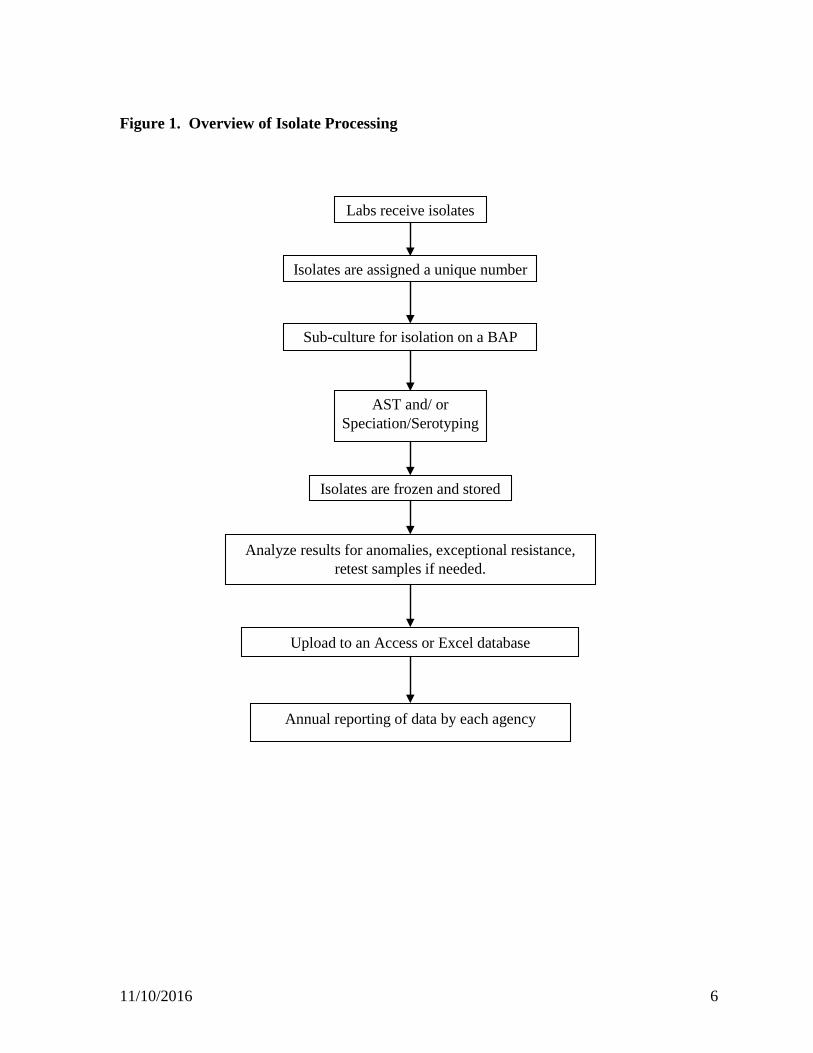
Figure 1. Overview of Isolate Processing
Labs receive isolates
Isolates are assigned a unique number
Sub-culture for isolation on a BAP
Isolates are frozen and stored
AST and/ or Speciation/Serotyping
Analyze results for anomalies, exceptional resistance, retest samples if needed.
Upload to an Access or Excel database
Annual reporting of data by each agency
11/10/2016 7
Salmonella & Escherichia coli A. Receipt of Isolates
Isolates are received from human, retail meats, and food animal sources. They are received either on agar slants or frozen in transport vials. Submission sheets and/or electronic files are included with shipments. Each agency works with Salmonella and Escherichia coli from different sources; therefore, the acquisition of isolates is different. For additional information please consult each agency’s website. CDC - Human Salmonella isolates are received from public health laboratories in all 50 states. (http://www.cdc.gov/narms). In addition, CDC receives Shigella and non-cholerae Vibrio from the state labs and uses the method described below for antimicrobial susceptibility testing of those isolates. FDA - Retail meat Salmonella and E. coli isolates are received frozen by the FDA/CVM/NARMS laboratory from the retail meat FoodNet surveillance sites. Each isolate is assigned a unique CVM/NARMS number prior to analysis. (http://www.fda.gov/AnimalVeterinary/SafetyHealth/AntimicrobialResistance/NationalAntimicrobialResistanceMonitoringSystem/default.htm). FDA (Denver Laboratory) – Salmonella isolates are received from ORA laboratories. Isolates are from Domestic/Import food and feed samples. USDA FSIS – Salmonella isolates from the Pathogen Reduction/Hazard Analysis and Critical Control Point (PR/HACCP) programs were isolated by FSIS Field Service Laboratories following FSIS Microbiology Laboratory Guidebook methods. Salmonella and E. coli isolates from post-mortem animal cecal contents of dairy cattle, beef cattle, swine, broilers and turkeys from federally inspected slaughter and processing plants (Cecal Sampling Program) were isolated by FSIS Eastern Laboratory. All isolates are delivered to the FSIS Microbiology Characterization Branch for analysis.
B. Subculturing for Isolate Purity
Isolates are subcultured for purity on a BAP or MAC and incubated overnight at 36 ±1ºC. If a culture appears contaminated or contains more than one strain type, additional selective media are used or additional isolation steps may be taken. If any isolate appears non-viable upon initial recovery, enrichment media may be used to promote the growth of the target organism. Once a pure culture is obtained, isolates are tested using the Vitek2 Compact (V2C), serology, or other identification methods.

11/10/2016 8
C. Isolate Storage
CDC - For long-term storage of Salmonella and E. coli, CDC prepares a suspension from a BAP in the appropriate medium and aliquots the suspended isolate into a cryovial for freezing. FDA - Since FDA receives and stores all isolates frozen, the isolates are streaked for purity twice, tested and refrozen. All isolates are stored in a -70 to -80°C freezer. USDA FSIS – Salmonella isolates from PR/HACCP and the Cecal Sampling Program are lyophilized and maintained for long-term storage at the FSIS Eastern Laboratory. E. coli isolates from the Cecal Sampling Program are delivered in cryovials containing BHI broth with glycerol to FDA/CVM Office of Research in Laurel, MD for storage and for any further testing.
D. Identification and Speciation of Salmonella and E. coli
CDC - Salmonella are identified at the primary site (state level) using standard biochemical methods, and serotyped according to the Kauffman-White scheme. Mixed or questionable isolates are confirmed by a CDC reference laboratory. FDA - Salmonella speciation is performed using Whole Genome Sequencing. Discrepant serotypes received from the state labs vs. Pulsed-field Gel Electrophoresis (PFGE) are serotyped according to the Kauffman-White scheme. Escherichia coli isolates are confirmed on the V2C. USDA FSIS - Salmonella isolates are confirmed using the V2C and serotyped on site by FSIS using molecular typing methods. Isolates that are not reportable by molecular detection as per FSIS MLG 4.08, Appendix 1.03 are sent to the National Veterinary Service Laboratory (NVSL, Ames, IA) for serotyping. Escherichia coli from the Cecal Sampling Program are confirmed with real-time PCR targeting the gadE gene.
E. Antimicrobial Susceptibility Testing of Salmonella and E. coli
1. Gently invert a Sensititre 0.5 McFarland Polymer Turbidity Standard several times and place it in the nephelometer.
2. Press CALIBRATE on the keypad. This action will standardize the nephelometer to the center green LED light on the auto-inoculator.
3. Prepare a gram negative Sensititre™ panel (CMV#AGNF) for each isolate according to the manufacturer instructions. Ensure that the desiccant inside the package is orange. If the desiccant is green, discard the panel. Label each panel with appropriate identifying information. Place the panel in the auto-inoculator or AIM.
11/10/2016 9
Steps 4 through 11 should be completed within 15 minutes.
4. Prepare the inoculum by selecting a few colonies from the BAP and suspend in a pre-labeled tube of 5 ml demineralized water. Vortex well.
5. Adjust to a 0.5 McFarland equivalent by using the Sensititre nephelometer.
6. Transfer 10 µl of the suspension (20µl for non-cholerae Vibrio; CDC only) to a sterile 11 ml tube of Sensititre Cation- Adjusted Mueller-Hinton Broth with TES. This transfer is performed with a sterile, extra long pipette tip. Vortex well.
7. Aseptically replace the MHB tube cap with a disposable dosing head.
8. Open the dosing head clamp, invert the inoculated MHB tube and insert it in the clamp.
9. In response to the auto-inoculator display “Enter Pattern #”, press “001”, and then ENTER to dispense 50 µl into each well of the gram negative MIC panel. For the AIM, choose the plate configuration for 50 µl.
10. Press enter again to start dispensing.
11. Once the MIC panel is inoculated, remove the panel from the auto-inoculator and cover it with the provided clear plastic adhesive seal. Ensure that all wells are covered.
12. Repeat steps 4 through 11 (excluding step 9) for the rest of the test isolates and controls. Note: The testing panel should be inoculated and placed in the incubator within 15 minutes of preparing the cell suspension.
13. At least weekly, perform colony counts from the panel’s positive control well (see Colony Counts, p.20).
14. Incubate panels in the ARIS with the auto-reader or an aerobic incubator (stack no more than 4 high) at 35 ±1ºC for 18 hours.
15. Panels should be read by the ARIS auto reader or visually using the Sensitouch or Vizion at 18 hours. ARIS procedures are provided in the ARIS manual from Thermo Fisher Scientific/Trek Diagnostics.
16. In a timely manner, review the minimum inhibitory concentration (MIC) results for abnormalities as outlined in the section Criteria for Repeat Testing. For antimicrobials where no MIC is detected by the ARIS, the plates should be visually inspected. MIC’s for the folate pathway inhibitors should be confirmed manually using a mirror viewing device.
F. Interpretive Criteria
For categorizing susceptibility, breakpoints were adopted from the most recent version of the CLSI document M100 or based on NARMS data when needed (e.g., streptomycin). Refer to the NARMS Integrated Report Data Tables for a reference table showing Salmonella and E. coli breakpoints.
11/10/2016 10
G. Quality Control (QC) for testing Salmonella and E. coli
1. Antimicrobial susceptibility testing a. Control strains: E. coli ATCC 25922, Enterococcus faecalis ATCC 29212,
Staphylococcus aureus ATCC 29213, and Pseudomonas aeruginosa ATCC 27853.
b. Frequency: The QC strains are tested either daily or weekly and whenever a new lot of sterile water, MHB, or panels are used.
c. Control strain storage: Maintain stock cultures at -20ºC or below for prolong storage. Store subcultured isolates at 2 to 8ºC as appropriate for the organism type. The control strain is plated from the freezer onto a BAP at least monthly and then is subcultured once prior to testing.
d. Media: For each new lot number of panel, uninoculated MHB is dispensed into the panel and the panel is then incubated for a sterility test.
e. Criteria for repeat testing: If more than 1 out of 20 or 3 out of 30 MICs for each antimicrobial agent/QC organism combination is outside of acceptable MIC limits, it is repeated for 5 consecutive test runs. CLSI guidelines (M07-A10) are followed in the event of further QC failures.
2. Quality Assurance
a. Each agency has the option to participate in the WHO External Quality Assurance System (EQAS) annually to test samples for identification, antimicrobial susceptibility and serotyping. The results are recorded and posted on the WHO EQAS website.
b. Inter-Agency:AST and identification methods are exchanged bi-annually among agencies for comparability of results.
3. All final data are audited internally within each individual agency to ensure the accurate reporting of results.

11/10/2016 11
Campylobacter A. Receipt of Isolates
Isolates are received from human, retail meats, and food animal sources. They may be received on agar slants, frozen in transport vials or by primary isolation from chicken carcass rinses and turkey sponges collected by the Food Safety and Inspection Service (FSIS). Submission sheets and/or electronic files are included with the isolate shipments. Each agency works with Campylobacter from different sources; therefore, the acquisition of isolates is different for each group. A brief summary is given below. A more detailed description can be found at each agency’s website. CDC - Human Campylobacter isolates are received from ten state health departments that participate in the Foodborne Diseases Active Surveillance Network (FoodNet). Each isolate is assigned a unique CDC/NARMS number prior to analysis. (http://www.cdc.gov/narms) FDA - Retail meat Campylobacter isolates are received frozen by the FDA/CVM/NARMS laboratory from the retail meat FoodNet surveillance sites. Each isolate is assigned a unique CVM/NARMS number prior to analysis. (http://www.fda.gov/AnimalVeterinary/SafetyHealth/AntimicrobialResistance/NationalAntimicrobialResistanceMonitoringSystem/default.htm) USDA FSIS - Campylobacter isolates from the Pathogen Reduction/Hazard Analysis and Critical Control Point (PR/HACCP) programs were isolated by FSIS Field Service Laboratories following FSIS Microbiology Laboratory Guidebook methods. Campylobacter isolates from post-mortem animal cecal contents of dairy cattle, beef cattle, swine, broilers and turkeys from federally inspected slaughter and processing plants (Cecal Sampling Program) were isolated by FSIS Eastern Laboratory. All isolates are delivered to the FSIS Microbiology Characterization Branch for analysis.
B. Subculturing for Isolate Purity
1. Sub-culture isolate onto a BAP and streak for isolation. Incubate plate under microaerophilic conditions (85% N2, 10% CO2, 5% O2) for 24 hours at 42°C. Alternatively, plates can be incubated at 36+1°C for 36-48 hours.
2. If there is typical Campylobacter growth, select a single, well-isolated colony and streak for isolation on another BAP and incubate as above.
3. If there is sufficient growth of a pure culture, the isolate is processed for identification, susceptibility testing, and freezing. Additional plates may be inoculated if more growth is needed.
4. For contaminated or mixed cultures, sub-culture on Campy-Cefex agar or a selective Campy agar until a pure isolate is obtained.
11/10/2016 12
5. Cultures remain viable up to one week on plates stored at 4°C in a microaerophilic atmosphere.
C. Recovery of Isolates Showing No Growth
1. Prepare Bolton broth with 5% lysed horse blood (no selective supplement) and aseptically dispense 2 ml into sterile 15 ml disposable centrifuge tubes (or dispense 20-25 ml into 50 ml tubes).
2. For frozen stock, thaw the entire culture vial and add it to the prepared tube of broth. For plated stock, use a sterile cotton swab to remove all growth from a plate and suspend it in broth.
3. With the screw cap loose, incubate the inoculated broth tube at 36+1°C in a microaerophilic environment for 18-24 hours.
4. After incubation, use a swab (or dispense 75 µl) to transfer the broth to a BAP. Use a loop to streak the remainder of the plate for isolation. Incubate the plate as above.
5. Re-incubate the Bolton broth at 42°C for an additional 1-2 days and streak again onto a BAP in case the initial plate fails to produce growth.
6. If one of the plates above shows typical Campylobacter colonies, proceed with bacterial subculturing as previously described.
7. If there is no growth or the growth is obviously atypical, record as a non-viable sample and, if possible, request the FoodNet site to re-submit the original isolate.
D. Isolate Storage
For long term storage of a Campylobacter isolates, use a fresh (< 48 h/ 36+1°C or < 24 h/42°C) culture on a BAP to prepare a heavy homogeneous suspension in a cryoprotective medium and store at -80°C. Two isolate storage options are recommended:
1. Prepare cell suspension in defibrinated sheep blood. Flash-freeze tubes in an
ethanol/dry ice bath before transferring to -80°C.
2. Prepare cell suspension in Brucella broth with 15-20% glycerol or Wang’s storage medium (Brucella broth, 10% sheep blood, 15% glycerol). Transition tubes at 4°C for approximately 30 min before transferring to -80°C.
E. Identification and Speciation of Campylobacter
Currently, each agency uses different methods for final isolate identification and speciation. To ensure comparability, each laboratory participates in several quality assurance programs (see Quality Assurance section below). A brief description of each agency’s protocol is as follows.
11/10/2016 13
CDC After isolates are confirmed as Campylobacter by a genus specific PCR, they are screened for jejuni/coli speciation using PCR (Linton, et al. 1997; Pruckler, J. et al. 2006). Isolates that are positive by a genus specific PCR and jejuni/coli negative are screened using a series of biochemical tests. Tests include hippurate hydrolysis, catalase and indoxyl acetate. The results of the biochemical tests determine which species-specific PCR will be used to identify the isolate.
FDA Campylobacter speciation is performed using Whole Genome Sequencing (WGS). Discrepant speciation results received from the state labs vs. WGS are further tested by PCR using the following method: 1. Isolates are identified by PCR using the HIP and CC primers (Linton, et al. 1997;
Zhao, et al. 2001). DNA templates are prepared using a heated cell lysate.
2. If speciation is not determined, PCR is repeated using HIP and CC primers with a new DNA prep.
3. If PCR fails to provide a definitive species, the assay is repeated using additional primers (Burnett, et al. 2002; Gonzales, et al. 1997; Wang, et al. 2002).
USDA FSIS Campylobacter isolates are identified at the genus level following FSIS MLG 41.04 methods for PR/HACCP samples or methodology for cecal samples. Campylobacter speciation for all isolates is performed using the BAX® System Q7 instrument and real-time Campylobacter identification kits (DuPont Nutrition and Health) which detect C. coli, C. lari, and C. jejuni.
F. Antimicrobial Susceptibility Testing of Campylobacter
1. Set up a rack with one 5 ml Mueller-Hinton broth tube and one 11 ml Mueller-Hinton broth containing lysed horse blood for each isolate to be tested.
2. Prepare Sensititre™ CAMPY panels by removing the foil wrapper and labeling with the isolate number. Make sure the desiccant inside the wrapper is orange. Do not use the panel if desiccant is green.
3. Calibrate nephelometer using the 0.5 McFarland turbidity standard. When using the Sensititre™ nephelometer, calibration is complete when the center green nephelometer LED is lit.
Steps 4 through 10 should be completed within 30 minutes.
4. Use a sterile swab to pick several colonies from a BAP incubated for 48 h/36+1°C or 24 h/42°C. Suspend colonies into 5 ml Mueller-Hinton broth, vortex, and adjust to a 0.5 McFarland standard.
5. Transfer 100 µl of the 0.5 McFarland Mueller-Hinton suspension above (see Colony Counts, p.20) into 11 ml Mueller-Hinton with lysed horse blood using an extra long tip, then vortex.
11/10/2016 14
6. Open the dosing head clamp, invert the inoculated Mueller-Hinton broth with lysed horse blood and set inside the clamp.
7. Transfer 100 µl to each well of the panel by using the auto-inoculator (Program #011-ENTER). For the AIM, choose plate configuration for 100 µl.
8. Press ENTER again to start dispensing.
9. Remove the panel once the dispensing has completed and cover the panel with the provided perforated seal making sure a perforated hole is situated above each well.
10. Incubate panels in a microaerophilic atmosphere (85% N2, 10% CO2, 5% O2) at 36+1°C for 48 h or at 42°C for 24 h. Do not stack panels more than four high.
11. Perform colony counts from the panel’s positive control well on at least a weekly basis as described above.
12. After incubation, read the panels by visual inspection with ample light preferably using a magnifying plate reader. Growth appears as a deposit of cells at the bottom of a well or as turbid growth. The minimum inhibitory concentration (MIC) is recorded as the lowest concentration of antimicrobial that inhibits growth. Occasionally, a precipitate or amorphous “speck” may be seen in a well and should not be considered as bacterial growth when determining the MIC.
13. After results are read, check the QC strain to make sure the QC ranges are within acceptable limits. If they are not, follow corrective procedures recommended by the Clinical and Laboratory Standards Institute (CLSI Document M07-A10).
14. In a timely, manner, review the minimum inhibitory concentration (MIC) results for abnormalities as outlined in the section Criteria for Repeat Testing.
G. Interpretive Criteria
For categorizing susceptibility, breakpoints were adopted by epidemiological cut-off values. Refer to the NARMS Integrated Report Data Tables for a reference table of interpretive criteria used for Campylobacter.
H. Quality Control (QC) for testing Campylobacter
1. Antimicrobial susceptibility testing
a. Control strain: C. jejuni ATCC 33560
b. Frequency: The control strain is included with every batch of testing
c. Control strain storage: The control strain is plated from the freezer onto a BAP at least weekly and then is subcultured at least once prior to testing.
d. Media: For each new lot number of panel, uninoculated MHB with blood is dispensed into the panel and the panel is then incubated for a sterility test.
11/10/2016 15
2. Quality Assurance
a. Each agency has the option to participate annually in the World Health Organization (WHO) External Quality Assurance System (EQAS) to test samples for identification and antimicrobial susceptibility. The results are recorded and posted on the WHO EQAS website.
b. Each agency has the option to participate in the National Campylobacter and Helicobacter Reference Laboratory Quality Assurance Program for Campylobacter spp. Identification sponsored by the CDC.
c. Inter-Agency:AST and identification methods are exchanged bi-annually among agencies for comparability of results.
3. All final data are audited internally within each individual agency to ensure the accurate reporting of results.
11/10/2016 16
Enterococcus A. Receipt of Isolates
Isolates are received from retail meats, and food animal sources. They are received either frozen in transport vials; or they are recovered by culturing chicken carcass rinse samples collected by FSIS. Submission sheets and/or electronic files are included with shipments. Each agency works with Enterococcus from different sources; therefore, the acquisition of isolates is different for each group. For additional information please consult each agency’s website. CDC- Human Enterococcus isolates are no longer tested. FDA - Retail meat Enterococcus isolates are received frozen by the FDA/CVM/NARMS laboratory from the retail meat FoodNet surveillance sites. Each isolate is assigned a unique CVM/NARMS number prior to analysis. (http://www.fda.gov/AnimalVeterinary/SafetyHealth/AntimicrobialResistance/NationalAntimicrobialResistanceMonitoringSystem/default.htm) USDA FSIS - Enterococcus isolates from post-mortem animal cecal contents of dairy cattle, beef cattle, swine, broilers and turkeys from federally inspected slaughter and processing plants (Cecal Sampling Program) were isolated by FSIS Eastern Laboratory. All isolates are delivered to the FSIS Microbiology Characterization Branch for analysis.
B. Subculturing for Isolate Purity
1. Subculture isolate onto a BAP.
2. Incubate the BAP overnight at 36±1°C for 18-24 hours.
3. Pick a single colony and subculture to 2nd BAP, incubate as above.
If there is no growth on the plate after 24 hours, re-incubate the plate for 48 hours. If there is no growth after 48 hours, go back to the original culture and re-streak for isolation. If there is still no growth, record no growth and ask site to resend. If an isolate displays contamination/mixed culture, a selective media (Enterococcosel Agar) may be used to isolate and purify Enterococcus from the sample.
C. Isolate Storage
FDA - Since FDA receives and stores all isolates frozen, the isolate is streaked for purity twice, tested and refrozen. All isolates are stored in a -70 to -80ºC freezer. USDA FSIS - Isolates are stored on nutrient agar slants until they are tested. After testing, they are stored in Brain Heart Infusion broth with glycerol and are sent to FDA/CVM Office of Research in Laurel, MD for storage and any further testing.
11/10/2016 17
D. Identification and Speciation of Enterococcus FDA - Isolates are identified using the V2C using an isolated colony from the 2nd BAP plate. If the V2C fails to produce a definitive identification, additional testing is done using molecular identification methods. USDA FSIS - All isolates are identified using the V2C.
E. Antimicrobial Susceptibility Testing of Enterococcus
1. Gently invert several times a Sensititre 0.5 McFarland Polymer Turbidity Standard and place it in the Sensititre nephelometer.
2. Press CALIBRATE on the keypad. Calibration is complete when the center green nephelometer LED is lit.
3. Label a gram positive Sensititre™ panel (CMV#AGPF) with the isolate number. Ensure the desiccant is orange. Do not use the panel if the desiccant is green.
Steps 4 through 12 should be completed within 30 minutes.
4. Using a sterile swab, transfer several colonies from a pure culture on an 18-24 hr blood agar plate to a sterile 5 ml tube of demineralized water. Vortex well.
5. Adjust the suspension to a 0.5 McFarland equivalent by using the Sensititre nephelometer.
6. Transfer 10 µl of the suspension to a sterile 11 ml tube of Sensititre Cation-Adjusted Mueller-Hinton Broth with TES. This transfer is performed with a sterile, extra long pipette tip. Vortex well.
7. Aseptically replace the Mueller-Hinton tube cap with a disposable dosing head.
8. Place the panel in the Sensititre autoinoculator.
9. Open the dosing head clamp, invert the inoculated Mueller-Hinton tube and insert it in the clamp.
10. In response to the autoinoculator display “Enter Pattern #”, press “001", and then ENTER to dispense 50µl into each well of a Gram positive MIC panel. For the AIM, choose the plate configuration for 50 µl.
11. Press ENTER again to start dispensing.
12. Remove the panel and seal it with a clear plastic adhesive seal. Ensure that all wells are covered.
13. Colony counts are performed at least weekly (see Colony Counts, p.20).
14. Repeat steps 4-12 for each additional test isolate and QC isolate to be tested.
15. Incubate panels in the ARIS with the auto-reader or an aerobic incubator (stack no more than 4 high) at 36 ±1ºC for 18 hours.
11/10/2016 18
16. Panels should be read by the ARIS auto reader or visually using the Sensitouch at 18 hours. ARIS procedures are provided in the ARIS manual from Thermo Fisher Scientific/Trek Diagnostics.
17. If the ARIS is used, Linezolid and Nitrofurantoin should be read manually at 18 hours and Vancomycin should be read manually at 24 hours.
18. After results are read, check QC strain results to make sure they are within limits. If they are not, follow corrective procedures recommended by CLSI (M07-A10).
19. In a timely manner, review the minimum inhibitory concentration (MIC) results for abnormalities as outlined in the section Criteria for Repeat Testing.
F. Interpretive Criteria
For categorizing susceptibility, breakpoints were adopted from the most recent version of the CLSI document M100. Refer to the NARMS Integrated Report Data Tables for a reference table showing Enterococcus breakpoints.
G. Quality Control (QC) for testing Enterococcus
1. Antimicrobial Susceptibility Testing
a. Control Strains- E. faecalis ATCC 29212, E. faecalis ATCC 51299, E. coli ATCC 25922 and Staphylococcus aureus ATCC 29213.
b. Frequency: The QC strains are tested either daily or weekly and whenever a new lot of sterile water, MHB, or panels are used.
c. Control strain storage- Maintain stock cultures at -80°C for prolonged storage. The control strain is plated from the freezer onto a BAP. A second pass on BAP is completed.
d. Media- For each new lot number of panel, uninoculated MHB is dispensed into the panel and the panel is then incubated for a sterility test.
2. Quality Assurance
Inter-Agency: AST and identification methods are exchanged bi-annually among agencies for comparability of results.
3. All final data are audited internally within each individual agency to ensure the accurate reporting of results.
11/10/2016 19
Whole Genome Sequencing
NARMS Salmonella, Campylobacter, and select E. coli are sequenced as part of routine surveillance on the Illumina MiSeq and HiSeq platforms, with data made publicly accessible in GenBank bioproject PRJNA290865. Resistance genotype information is also being reported on the NARMS website as part of NARMS Now. Salmonella isolates that are analyzed in Denver Laboratory are sequenced on the Illumina MiSeq platform. The data is accessible in GenBank bioproject PRJNA186035.
Quality Controls for Antimicrobial Susceptibility Testing
The overall performance of the test system should be monitored by running QC each day the test is performed or, if satisfactory performance is documented (see CLSI criteria in Document M07-A10, Chapter 4), testing may be done weekly. Colony counts should also be performed weekly to ensure the final inoculum concentration is accurate. Performance is satisfactory if no more than 3 out of 30 consecutive results for a given antimicrobial/QC organism combination are outside of the acceptable limits (CLSI Document M100 and M45 for Campylobacter). If more than 3/30 results are out of control, try to determine the cause of the error and immediately retest the antimicrobial/QC strain combination for a total of 5 consecutive test days.
• If all 5 results are satisfactory, no further action is required.
• If any of the 5 results are unsatisfactory, additional sources of error should be
investigated and the other NARMS laboratories should be contacted. It may be necessary to obtain a new QC strain or test materials, review test procedures, or obtain instrument service. Until the problem is resolved, reporting results and continued testing will depend on a careful assessment of the facts in consultation with the other NARMS laboratories.
In addition to routine QC testing, the CLSI (Document M07-A10, Chapter 4) directs that QC testing should be run on each new batch of reagents and panels and that one panel should be incubated uninoculated to check for sterility. If the QC fails, the lot should be rejected. The frozen stocks of quality control strains used for in vitro testing are obtained directly from the American Type Culture Collection (ATCC). Since repeated passage in vitro can alter strain characteristics, in-house QC strain stocks should be replenished only by purchase of fresh stock from a strain repository such as the ATCC.
11/10/2016 20
Equipment and Method Quality Control
Auto Inoculator or AIM
To ensure the equipment is well maintained, annual preventative maintenance is performed on each piece of equipment by a ThermoFisher Scientific/Trek Diagnostics service technician. In the event problems arise with equipment, service calls are placed to ThermoFisher Scientific/TREK Diagnostics. Loaner equipment is sent to the lab while the equipment is being serviced.
Colony Counts
1. Colony counts are recorded for each test run. At a minimum, colony counts are performed on the first test isolate and at least one QC organism each day antimicrobial susceptibility testing is performed.
2. From the positive control well of the inoculated panel, transfer 10 µl using a pipettor to 10 ml water, broth or appropriate diluent and mix by vortexing.
3. Spread plate 100 µl from the inoculated water, broth or diluent to each of two BAPs. Ensure there is not excessive moisture on the plate surface prior to plating the suspension.
4. Incubate colony count plates with MIC panels at the appropriate temperataure for that organism.
5. Average the number of colonies on the two plates and then multiply by 104 to obtain the total number of CFU/ml.
6. The inoculum should be between 1.0 x10⁵ – 1.0x10⁶ CFU/ml.
Criteria for Retesting of Isolates
Repeat testing of an isolate must be done when one or more of the following conditions occur:
• No growth on panel • Growth in all wells • Multiple skip patterns • Apparent contamination in wells or isolate preparation • Unlikely or discordant susceptibility results (Table 1).
If an isolate is retested, data for all antibiotics should be replaced with the new test results. Categorical changes may require a third test (and may indicate a mixed culture). Uncommon test results (Table 2) may represent emerging resistance phenotypes. Retesting is encouraged.
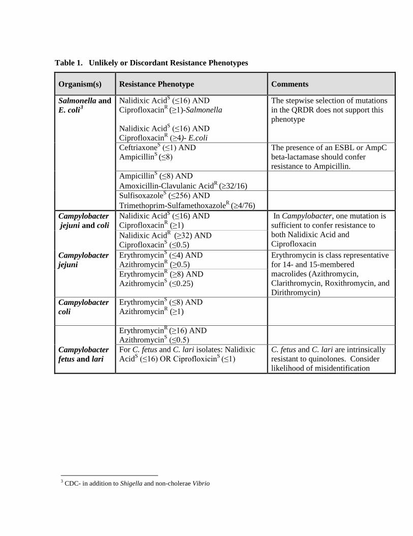
Table 1. Unlikely or Discordant Resistance Phenotypes
Organism(s)
Resistance Phenotype
Comments
Salmonella and E. coli3
Nalidixic AcidS (≤16) AND CiprofloxacinR (≥1)-Salmonella Nalidixic AcidS (≤16) AND CiprofloxacinR (≥4)- E.coli
The stepwise selection of mutations in the QRDR does not support this phenotype
CeftriaxoneS (≤1) AND AmpicillinS (≤8)
The presence of an ESBL or AmpC beta-lactamase should confer resistance to Ampicillin.
AmpicillinS (≤8) AND Amoxicillin-Clavulanic AcidR (≥32/16)
SulfisoxazoleS (≤256) AND Trimethoprim-SulfamethoxazoleR (≥4/76)
Campylobacter Nalidixic AcidS (≤16) AND CiprofloxacinR (≥1) jejuni and coli
In Campylobacter, one mutation is sufficient to confer resistance to both Nalidixic Acid and Ciprofloxacin Erythromycin is class representative for 14- and 15-membered macrolides (Azithromycin, Clarithromycin, Roxithromycin, and Dirithromycin)
Nalidixic AcidR (≥32) AND CiprofloxacinS (≤0.5)
Campylobacter jejuni
ErythromycinS (≤4) AND AzithromycinR (≥0.5) ErythromycinR (≥8) AND AzithromycinS (≤0.25)
Campylobacter coli
ErythromycinS (≤8) AND AzithromycinR (≥1)
ErythromycinR (≥16) AND
AzithromycinS (≤0.5)
Campylobacter fetus and lari
For C. fetus and C. lari isolates: Nalidixic AcidS (≤16) OR CiprofloxicinS (≤1)
C. fetus and C. lari are intrinsically resistant to quinolones. Consider likelihood of misidentification
3 CDC- in addition to Shigella and non-cholerae Vibrio
11/10/2016 22
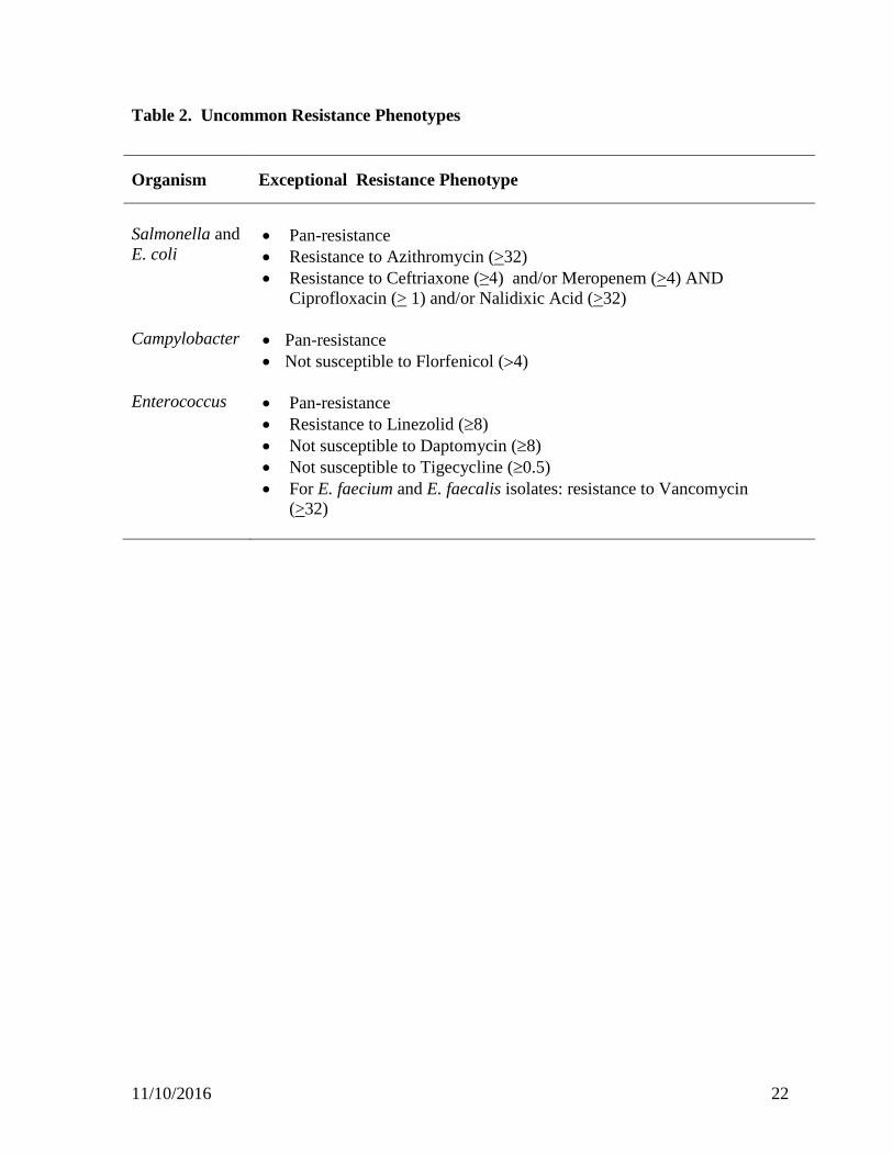
Table 2. Uncommon Resistance Phenotypes
Organism Exceptional Resistance Phenotype
Salmonella and E. coli
• Pan-resistance • Resistance to Azithromycin (>32) • Resistance to Ceftriaxone (≥4) and/or Meropenem (>4) AND
Ciprofloxacin (> 1) and/or Nalidixic Acid (>32)
Campylobacter • Pan-resistance • Not susceptible to Florfenicol (>4)
Enterococcus • Pan-resistance
• Resistance to Linezolid (≥8) • Not susceptible to Daptomycin (≥8) • Not susceptible to Tigecycline (≥0.5) • For E. faecium and E. faecalis isolates: resistance to Vancomycin
(>32)
11/10/2016 23
References
Burnett, et al. 2002. Speciating Campylobacter jejuni and Campylobacter coli isolates from poultry and humans using six PCR-based assays. FEMS Microbiology Letters 216:201-209. CLSI. Methods for Dilution Antimicrobial Susceptibility Tests for Bacteria That Grow Aerobically; Approved Standard-Tenth Edition. CLSI document M07-A10. Wayne, PA: Clinical and Laboratory Standards Institute; 2015. CLSI. Performance Standards for Antimicrobial Disk and Dilution Susceptibility Tests for Bacteria Isolated From Animals; Approved Standard – Fourth Edition. CLSI document VET01-A4 (replaces M31-A3). Wayne, PA: Clinical and Laboratory Standards Institute; 2013. CLSI. Methods for Antimicrobial Dilution and Disk Susceptibility Testing of Infrequently Isolated or Fastidious Bacteria; Approved Guideline. CLSI document M45-A3. Wayne, PA: Clinical and Laboratory Standards Institute; 2015. CLSI. Performance Standards for Antimicrobial Susceptibility Testing. 26th ed. CLSI supplement M100-S26. Wayne, PA: Clinical and Laboratory Standards Institute; 2016. FDA. National Antimicrobial Resistance Monitoring System – Enteric Bacteria (NARMS): 2005 Executive Report. Rockville, MD: U.S. Department of Health and Human Services, Food and Drug Administration, 2009. FDA. NARMS Integrated Report: 2012-2013 http://www.fda.gov/AnimalVeterinary/SafetyHealth/AntimicrobialResistance/NationalAntimicrobialResistanceMonitoringSystem/ucm059103.htm FSIS. Microbiology Laboratory Guidebook (MLG) http://www.fsis.usda.gov/wps/portal/fsis/topics/science/laboratories-and-procedures/guidebooks-and-methods/microbiology-laboratory-guidebook/microbiology-laboratory-guidebook Gonzales, et al. 1997. Specific identification of the enteropathogens Campylobacter jejuni and Campylobacter coli by using a PCR test based on the ceuE gene encoding a putative virulence determinant. JCM 35: 759-763. Jackson, C.R., P.J. Fedorka-Cray, and J.B. Barret. 2004. Use of a genus- and species-specific multiplex PCR for identification of Enteroccoci. J. Clin. Microbiol. 42: 3558-3565. Linton, et al. 1996. Rapid identification by PCR of the genus Campylobacter of five Campylobacter species enteropathogenic for man and animals. Res. Microbiol. 147: 707-718.
11/10/2016 24
Linton, et al. 1997. PCR detection, identification to species level, and fingerprinting of Campylobacter jejuni and Campylobacter coli direct from diarrheic samples. JCM 35:2568-2572. Pruckler, J., et al. 2006. Comparison of four real-time PCR methods for the identification of the genus Campylobacter and speciation of C. jejuni and C. coli. ASM 106th General meeting; PosterC282. Wang, et al. 2002. Colony multiplex PCR assay for identification and differentiation of Campylobacter jejuni, C. coli, C. lari, C. upsaliensis, and C. fetus subsp. fetus. JCM 40:4744-4747. Zhao, et al. 2001. Prevalence of Campylobacter spp., Escherichia coli, and Salmonella serovars in retail chicken, turkey, pork, and beef from the Greater Washington, D.C., area. AEM 67:5431-5436.

11/10/2016 25
Acknowledgments of Contributors CDC Jean Whichard Christy Bennett
Christina Scheel Andre McCullough Amelia Dicknese Davina Campbell
FDA Patrick McDermott
Shaohua Zhao Gregory Tyson Sherry Ayers Stuart Gaines Sonya Bodeis Jones Crystal Rice-Trujillo Shenia Young Gordon Martin Jacqueline Hernandez Sampa Mukherjee Cong Li Jason Abbott Chih-Hao Hsu Mercedes Loftis
FSIS Jovita Haro
Cesar Morales Sutawee (Aor) Thitaram Glenn Tillman Rachel Whitaker
CIPARS Danielle Daignault Andrea Desruisseau