the herpes simplex virus 1 vhs protein enhances translation of viral true late mrnas and virus
TRANSCRIPT

JOURNAL OF VIROLOGY, June 2011, p. 5363–5373 Vol. 85, No. 110022-538X/11/$12.00 doi:10.1128/JVI.00115-11Copyright © 2011, American Society for Microbiology. All Rights Reserved.
The Herpes Simplex Virus 1 vhs Protein Enhances Translation ofViral True Late mRNAs and Virus Production in a Cell
Type-Dependent Manner�†Bianca Dauber,1 Jerry Pelletier,2 and James R. Smiley1*
Li Ka Shing Institute of Virology, Department of Medical Microbiology and Immunology, University of Alberta, Edmonton, Alberta,Canada T6G 2S2,1 and Biochemistry Department and McGill Cancer Center, McGill University, Montreal, Quebec, Canada H3G 1Y62
Received 17 January 2011/Accepted 16 March 2011
The herpes simplex virus 1 (HSV-1) virion host shutoff protein (vhs) degrades viral and cellular mRNAs.Here, we demonstrate for the first time that vhs also boosts translation of viral true late mRNAs in a celltype-dependent manner and that this effect determines the viral growth phenotype in the respective celltype. Our study was prompted by the detection of stress granules, indicators of stalled translationinitiation, in cells infected with vhs mutants but not in wild-type-virus-infected cells. Accumulation of truelate-gene products gC and US11 was strongly reduced in the absence of vhs in HeLa cells and several otherrestrictive cell lines but not in Vero and other permissive cells and was independent of phosphorylationof the � subunit of eukaryotic initiation factor 2 (eIF2�). Polysome analysis showed that gC and US11transcripts were poorly translated in vhs-null-virus-infected HeLa cells, while translation of a cellularmRNA was not affected. Interestingly, hippuristanol, an eIF4A inhibitor, produced a similar phenotype inHeLa cells infected with wild-type HSV-1, while Vero cells were much more resistant to the inhibitor. Theseresults suggest that translation of true late-gene transcripts is particularly sensitive to conditions oflimited access to translation factors and that vhs is able either to prevent the limiting conditions or tofacilitate translation initiation under these conditions. The varied permissivity of cell lines to vhs-nullinfection may stem from differences in the resilience of the translation machinery or the ability to controlthe accumulation of mRNAs.
Herpes simplex virus 1 (HSV-1), a large, enveloped DNAvirus, is a master in subverting host cell functions for its ownbenefit. In particular, HSV-1 employs several mechanisms toblock expression of cellular proteins and facilitate efficientsynthesis of viral proteins (reviewed in references 29 and 55).The viral protein that executes the earliest attack on cellulargene expression is the virion host shutoff (vhs) protein (re-viewed in reference 54).
The vhs protein, encoded by the UL41 gene, is part of thetegument and as such is delivered into the cytoplasm afterfusion of the virion envelope with the host cell membrane (43).It is an endoribonuclease that degrades both cellular and viralmRNA (24, 32). The degradation of cellular mRNA presum-ably improves the access of viral mRNAs to the cellular trans-lation machinery. The vhs-mediated increased turnover of viralmRNAs is thought to sharpen the transition between the suc-cessive expression of viral immediate-early (IE), early (E), andlate (L) genes (32).
vhs is able to degrade all types of RNA in an in vitro system(10, 60, 61, 63); however, in infected cells vhs degrades onlymRNA and spares other RNA species (24, 32). In an effort toexplain this mRNA specificity, vhs has been shown to associatewith the translation initiation complex eIF4F, which binds the
5� cap structure of mRNAs (33). In particular, vhs interactswith the eIF4F component eIF4AII, an ATP-dependent RNAhelicase and the helicase cofactors eIF4H and eIF4B (7, 11).These observations suggest that interactions with eIF4AII,eIF4H, and eIF4B target vhs to actively translating mRNAs.This model is underscored by the finding that knockdown ofeIF4H prevented vhs-mediated mRNA decay in cell culture(47).
vhs-mediated shutoff of host protein synthesis occurs mainlyin the immediate-early and early phases of infection. Later ininfection, the nuclease activity of newly synthesized vhs ap-pears to be dampened by the viral proteins VP16 and VP22(22, 25, 48, 52). This is thought to be necessary to ensuresufficient accumulation of viral late-gene transcripts and effi-cient virus production.
Since vhs destabilizes most mRNAs, it reduces synthesis ofproteins involved in the innate and adaptive immune re-sponses. This vhs-mediated downregulation is most likely thereason for the capacity of vhs to dampen the type I interferon(IFN) system (8, 31, 35), block activation of dendritic cells (45),and reduce production of proinflammatory cytokines andchemokines (58). These factors might account at least in partfor the crucial role of vhs in HSV-1 virulence and pathogenicity(reviewed in reference 54). For example, vhs mutants arestrongly attenuated in the corneas and central nervous systemof mice (26, 34, 36, 56, 57). Accordingly, replication and viru-lence of vhs mutants is enhanced in knockout mice lacking IFNreceptors (8, 26) or STAT1 (34, 36). However, even in IFN-����/� and STAT�/� mice, the vhs mutant is still attenuatedrelative to the wild-type (WT) virus (26, 34, 36), suggesting that
* Corresponding author. Mailing address: Department of MedicalMicrobiology & Immunology, 632 Heritage Medical Research Centre,University of Alberta, Edmonton, Alberta, Canada T6G 2S2. Phone:(780) 492-4070. Fax: (780) 492-9828. E-mail: [email protected].
† Supplemental material for this article may be found at http://jvi.asm.org/.
� Published ahead of print on 23 March 2011.
5363
Dow
nloa
ded
from
http
s://j
ourn
als.
asm
.org
/jour
nal/j
vi o
n 17
Nov
embe
r 20
21 b
y 61
.254
.216
.91.

vhs contributes to efficient replication beyond its role as mod-ulator of the immune response.
In cell culture, the attenuation of vhs-deficient HSV-1 is lesspronounced than in mice. Similar to what was observed in themouse model, the reduced virus yield in mouse embryonicfibroblasts (MEFs) is not completely due to IFN signaling,since virus titers of a vhs mutant in multicycle growth curveanalyses were only partially restored in IFN-���R�/� MEFs(35). Knockout of the double-stranded-RNA (dsRNA)-depen-dent protein kinase R (PKR) protein did not alleviate thisdefect (35). vhs-deficient HSV-1 is also slightly attenuated insingle-step growth curves in Vero cells, which lack IFN-� andIFN-� genes (39, 40, 53), suggesting an IFN-independentgrowth defect. The longstanding hypothesis advanced to ex-plain this inherent growth defect is that the inappropriateoveraccumulation of IE and E mRNAs might impair replica-tion (32), but the exact mechanism by which this impairmentmight be achieved has not been thoroughly examined so far.
A potential clue to resolving this question came from twopublications that point to a novel role for vhs in modulatingtranslation. First, our group showed that vhs modulates trans-lation in a bicistronic reporter gene assay without significantlyaltering the level of the respective mRNA (44). While expres-sion of the 5� cistron was inhibited by vhs, expression of the 3�cistron was dependent on the sequence of the intergenic re-gion. Expression driven by the encephalomyocarditis virus(EMCV) internal ribosome entry site (IRES) was suppressed,whereas a mutant EMCV IRES and the cellular ApaF1, BiP,and DAP5 IRES elements facilitated increased expression inthe presence of vhs. Interestingly, the 5� untranslated region(UTR) of the HSV-1 mRNAs for UL12, thymidine kinase(TK), or glycoprotein C (gC) also mediated vhs-dependentexpression of the 3� cistron, suggesting that vhs may be in-volved in boosting translation of viral mRNAs during infection(44). Second, Esclatine and colleagues reported that vhs-defi-cient HSV-1 (F) induces formation stress granules (SGs) (9),which are considered diagnostic for the accumulation of stalledtranslation initiation intermediates (20, 21). SGs form as aresponse to various forms of stress (reviewed in reference 1).Depending on the type of stress, the serine/threonine kinasesPKR, PKR-like endoplasmic reticulum kinase (PERK), gen-eral control nonrepressed 2 (GCN2), and heme-regulated in-hibitor (HRI) are activated and phosphorylate the translationinitiation factor eIF2� (reviewed in reference 46). This eventstalls the initiation process at the point of loading the initiatortRNAiMet onto the small ribosomal subunit (reviewed in refer-ence 16) and results in accumulation of 48S preinitiation com-plexes containing mRNA, the translation initiation complexeIF4F, poly(A) binding protein (PABP), eIF3, eIF1, eIF1A,and the small ribosomal subunit (19). The preinitiation com-plexes are then assembled into SGs by proteins such as TIA-1and TIAR (20, 21). Notably, SG formation and inhibition oftranslation can also occur independent of eIF2� phosphoryla-tion, for example, by blocking the activity of eIF4A with thenovel compounds hippuristanol and pateamine (2, 3, 28). SinceSGs are dynamic structures, mRNAs shuttle between SGs andpolysomes to resume translation or are targeted to processingbodies for degradation (reviewed in reference 1).
Based on these observations, we hypothesized that inactiva-tion of vhs may impair translation of viral proteins accounting
for the reduced virus yield. To test this hypothesis, we exam-ined the effect of vhs mutations on SG formation, viral repli-cation, and viral gene expression in various cell types. Wereport here that infection with vhs-deficient HSV-1 led to SGformation, reduced virus yield, and decreased accumulation oftrue late-gene products in HeLa cells and several additionalcell lines but not in Vero cells. Furthermore, we determinedthat the reduced late-gene expression was due to reducedtranslational activity on true late-gene transcripts in HeLacells. This block in translation initiation was specific for viraltranscripts, i.e., cellular mRNAs were not affected. Inhibitorstudies implied that the resilience of the translational machin-ery of the respective cell type might be critical for translationof true late-gene transcript in the absence of vhs.
Taken together, these findings suggest that in many celltypes vhs plays an important role in boosting translation oflate-gene products and that the lack of this activity mightaccount for the inherent growth defect of vhs mutants.
MATERIALS AND METHODS
Cells and viruses. HeLa (cervical carcinoma), Vero (African green monkeykidney), U2OS (osteosarcoma), HEp2, and SK-N-SH (neuroblastoma) cells andHFF TEL 12 (telomerase immortalized human foreskin fibroblasts; kind giftfrom W. Bresnahan) were maintained in Dulbecco’s modified Eagle Mediumsupplemented with 10% heat-inactivated fetal bovine serum, 100 U/ml penicillin,and 100 �g/ml streptomycin. Human embryo lung (HEL) fibroblasts were cul-tured as described above except for the addition of 1 mM sodium pyruvate. Thewild-type HSV-1 strains used in this study were KOS and its plaque-purifiedderivative KOS1.1 (17), and the vhs mutants were KOS-derived �Sma, whichcontains a 588-nucleotide deletion in the UL41 gene (40), vhs1, which containsa point mutation in the UL41 gene (39), and GFP vhs�, in which all but the last7 codons of the UL41 open reading frame (ORF) are deleted and replaced bygreen fluorescent protein (GFP), so that GFP is under the control of the vhspromoter (4). Viruses were propagated, and the titer in Vero cells was deter-mined. Virus absorption, infection, and mock infection were carried out at 37°Cat a multiplicity of 10 PFU/cell.
Drug treatments. Phosphonoacetic acid (PAA; Sigma) was used at 300 �g/mlstarting at 1 h postinfection (hpi) to inhibit viral DNA replication. Thapsigargin(Sigma) was used at 1 �M for 1 h prior to fixing to induce endoplasmic reticulumstress. Hippuristanol (a polyoxygenated streroid isolated from the coral Isishippuris [3]) in dimethyl sulfoxide (DMSO) was used at concentrations rangingfrom 20 nM to 4,000 nM starting at 1 hpi to inhibit eIF4A activity. Correspondingamounts of DMSO were used as controls.
Immunofluorescence (IF) microscopy. Cells grown on coverslips were mockinfected or infected with the respective viruses at a multiplicity of infection(MOI) of 10. At the indicated time points, cells were washed with phosphate-buffered saline (PBS), fixed with 3.65% paraformaldehyde for 15 min, perme-abilized with ice-cold methanol for 10 min, washed with PBS, and blocked with5% fetal bovine serum (FBS) or 2.5% human serum. Proteins were stained withprimary antibodies specific for HSV-1 ICP4 (mouse, P1101; Virusys Corpora-tion) and cellular TIA-1 (goat, C-20; Santa Cruz), TIAR (goat, C-18; SantaCruz), PABP (mouse, 10E10; Santa Cruz), G3BP (mouse, 611126; BD Trans-duction Laboratories), and eIF4A1 (rabbit, ab31217; Abcam). Primary antibod-ies were detected by suitable secondary antibodies coupled to Alexa 488, Alexa555, or Alexa 647 (Invitrogen). Images were obtained using an Axiovert 200 Mfluorescence microscope, an ApoTome optical sectioning device (Zeiss), and theAxioVision 4.5 program (Zeiss). All image processing was performed usingPhotoshop CS3 (Adobe).
Western blot analysis. Mock-infected and infected cells were lysed in radio-immunoprecipitation assay (RIPA) buffer (1% Igepal, 150 mM NaCl, 20 mMTris-HCl [pH 7.5], 1 mM EDTA, 0.1% SDS, 0.25% sodium deoxycholate, 1�Roche protease inhibitor, 10 mM Na–�-glycerophosphate, 2 mM Na3VO4, 1 mMNaF). Cleared lysates were analyzed by 7.5% or 10% sodium dodecyl sulfate-polyacrylamide gel electrophoresis (SDS-PAGE), transferred to a nitrocellulosemembrane (Hybond ECL; Amersham), and immunoblotted with primary anti-bodies specific for the HSV-1 proteins gC (mouse, P1104; Virusys Corporation),gB (mouse, P1123; Virusys), ICP4 (mouse, P1101; Virusys Corporation), ICP27(mouse, P1113; Virusys Corporation), ICP34.5 (rabbit; kind gift from Ian Mohr),
5364 DAUBER ET AL. J. VIROL.
Dow
nloa
ded
from
http
s://j
ourn
als.
asm
.org
/jour
nal/j
vi o
n 17
Nov
embe
r 20
21 b
y 61
.254
.216
.91.

US11 (mouse; kind gift from Bernard Roizman), UL47 (rabbit; kind gift fromGillian Elliott), thymidine kinase (TK) (rabbit; kind gift from William C. Sum-mers), and VP16 (mouse, LP1; kind gift from Tony Minson) and the cellularproteins �-actin (mouse, A5441; Sigma), phospho-eIF2� (rabbit, 9721; Cell Sig-naling), and eIF2� (rabbit, 9722; Cell Signaling). Primary antibodies were de-tected by suitable secondary horseradish peroxidase (HRP)-conjugated antibod-ies using an enhanced chemiluminescence protocol (GE Healthcare) (see Fig. 3and 7) or suitable secondary antibodies coupled to Alexa Fluor 680 (Invitrogen)or IRDye800 (Rockland), using the Odyssey infrared imaging system (LI-COR)(see Fig. S2 in the supplemental material).
RNA extraction and Northern blot analysis. Total RNA was isolated fromcells in 60-mm culture dishes by use of TRIzol reagent (Invitrogen) according tothe manufacturer’s instructions. RNA samples (10 �g) were subjected to 1.2%agarose-formaldehyde gel electrophoresis, visualized by SYBR gold staining, andtransferred to a Genescreen membrane (NEN). Hybridizations of gC, US11, andp56 probes, radiolabeled with 32P by random priming, were performed usingExpressHyb (Clontech) according to the user’s manual. The gC and US11 probeswere generated by PCR amplification of viral DNA using the primers 5� CCCAAACCCAAGAACAACAC and 5� TGTTCGTCAGGACCTCCTCT (gC) or5� GGCGACCCAGATGGTTACTT and 5� GCGAGCCGTACGTGGTTC(US11). The p56 probe has previously been described (30). �-Actin transcriptswere detected with a 32P-labeled oligonucleotide, 5� AATGACCAGCGCGGCGATCTCTTCTTCCATT. Hybridization was carried out in modified Westneatsolution (6.6% SDS, 250 mM MOPS [morpholinepropanesulfonic acid] [pH 7.0],5� Denhardt’s solution, 1 mM EDTA) at 53°C. Signals were analyzed with aFujifilm FLA-5100 phosphorimager and Image Gauge version 4.22.
Polysome gradient. HeLa or Vero cells from two 100-mm culture dishes weremock infected or infected with KOS1.1 or �Sma at an MOI of 10. Cells werelysed at 12 hpi in lysis buffer (250 mM sucrose, 25 mM KCl, 5 mM MgCl2, 50 mMTris-HCl [pH 7.4], 0.5% Triton X-100, 100 �g/ml cycloheximide, RNaseOUT).Cleared supernatant was incubated with MgCl2 (final concentration, 10 mM) orEDTA (final concentration, 25 mM) for 10 min on ice. Lysates were layered ontogradients of 10 to 50% sucrose solution in 50 mM Tris-HCl (pH 7.4), 25 mMKCl, 1.5 mM MgCl2, 100 �g/ml cycloheximide, 2 mM dithiothreitol (DTT), andRNaseOUT and centrifuged in an SW40 rotor for 105 min at 38,000 rpm and4°C. Twenty fractions were collected from the top of the gradient, and theirabsorbance at 254 nm was measured. Samples were proteinase K treated, andRNA was phenol-chloroform extracted. Half of each RNA sample was subjectedto Northern blot analysis as described above to determine the distribution ofselected RNA species across the gradient.
RESULTS
Formation of stress granules in response to infection withvhs-deficient HSV-1 is cell type dependent. Esclatine et al.showed that infection of HEp-2 and HFF cells with the �UL41mutant virus but not wild-type HSV-1(F) led to the formationof cytoplasmic structures that stained positive for the SGmarker antigens TIA-1/TIAR (9). We asked whether thesecytoplasmic structures are true SGs and whether SG formationis a universal response to vhs-deficient HSV-1. Therefore, weinfected cells with wild-type and vhs-deficient HSV-1 at anMOI of 10. Indirect immunofluorescence microscopy usingantibodies against TIA-1 showed that �Sma but not KOS1.1infection induced formation of cytoplasmic granules in HeLacells at 16 h postinfection (hpi) (Fig. 1A). As expected, at latetime points postinfection ICP4 localized to viral replicationcompartments and DAPI (4�,6-diamidino-2-phenylindole)staining demonstrated chromatin marginalization. The cyto-plasmic structures observed in �Sma-infected HeLa cells alsostained positive for a second key SG marker, TIAR (see Fig.S1a in the supplemental material). Furthermore, immuno-staining against TIA-1 or TIAR demonstrated that the cyto-plasmic structures were also induced in response to the addi-tional vhs mutants vhs1 and GFPvhs� (Fig. S1B and C) but notin response to parental KOS (Fig. S1D), confirming that theobserved phenotype is due to the loss of vhs function and not
due to other derivative-specific differences. Costaining of�Sma-infected HeLa cells with TIA-1/G3BP, G3BP/eIF4A,and PABP/eIF4A (Fig. 1B) revealed that the cytoplasmicstructures contain G3BP, eIF4A, and PABP and hence arebona fide SGs. We also observed SG formation in �Sma-infected HEp-2, SK-N-SH, HFF, U2OS, and HEL cells (Fig.1C). However, SG formation was less prominent in U2OS andHEL cells than in HeLa, HEp-2, and SK-N-SH cells, and HFFcells showed an intermediate phenotype. Surprisingly, how-ever, we were not able to detect any SGs in �Sma-infectedVero cells (Fig. 1D). Although the TIA-1 signal in KOS1.1-infected Vero cells appeared somewhat speckled, these struc-tures were mainly found in the nucleus and the cytoplasmicTIA signal was mostly dispersed. Therefore, this patternclearly differed from SGs as seen in �Sma-infected HeLa cells.We considered the possibility that Vero cells are inherentlyunable to form SGs. However, endoplasmic reticulum stressinduced by thapsigargin led to strong SG formation (Fig. 1D).This experiment verified that Vero cells are capable of formingSGs and suggests that Vero cells may have either differentmechanisms to deal with the stress induced by vhs-deficientHSV-1 or the level of stress may be below the specific thresh-old to induce SG formation. One distinguishing feature ofVero cells is that they do not express IFN-�/� (6). However,IFN-�/� treatment did not induce SGs in HeLa cells (data notshown), making it unlikely that IFN-�/� signaling is responsi-ble for the difference between HeLa and Vero cells. As HeLaand Vero cells are two of the most widely used cell lines inHSV-1 research, we felt that the striking difference in SGformation in response to vhs-deficient HSV-1 warranted fur-ther investigation.
SGs form late in infection and require viral DNA replica-tion. To assess when SGs form during infection, we infectedHeLa cells with �Sma at an MOI of 10 and stained for TIA-1and the viral protein ICP4 at various time points. SGs formedas early as 8 hpi (Fig. 2A). The fraction of ICP4-positive SG-containing cells increased over time to greater than 50% at 16hpi (Fig. 2A), suggesting that SG formation is triggered duringthe phase of late-gene expression. To further test this hypoth-esis, we treated �Sma-infected HeLa cells with phosphono-acetic acid (PAA), which inhibits viral DNA replication andconsequentially transcription of true late genes. This treatmentefficiently blocked formation of SGs (Fig. 2B), indicating thatone or more events linked to viral DNA replication, such asaccumulation of viral true late-gene transcripts, triggers SGformation.
Attenuated virus production in the absence of vhs is celltype dependent. In order to test whether the difference in SGformation in response to �Sma is reflected in differences inviral replication between HeLa and Vero cells, we performeda single cycle growth curve analysis. HeLa and Vero cells wereinfected with HSV-1 KOS1.1 or �Sma at an MOI of 10, andvirus titers were determined at 1, 6, 12, 18, and 24 hpi. WhileKOS1.1 and �Sma replicated to similar titers in Vero cells,�Sma virus titers were reduced about 50-fold compared to thewild-type virus in HeLa cells (Fig. 3A). In HEp-2, HFF, andSK-N-SH cells, �Sma showed a growth phenotype similar tothat observed in HeLa cells, with a difference in viral titers ofvhs-deficient and wild-type virus between 29- and 45-fold (Fig.
VOL. 85, 2011 vhs-MEDIATED TRANSLATION OF TRUE LATE-GENE PRODUCTS 5365
Dow
nloa
ded
from
http
s://j
ourn
als.
asm
.org
/jour
nal/j
vi o
n 17
Nov
embe
r 20
21 b
y 61
.254
.216
.91.

3B), whereas U2OS and HEL cells showed a phenotype similarto that of Vero cells, supporting �Sma virus replication totiters about 2- or 6-fold lower than those for KOS1.1, respec-tively (Fig. 3C). We are confident that during this single-step
replication cycle, the impact of IFN signaling was negligible, asdemonstrated by the low to nondetectable mRNA levels ofIFN-stimulated gene 56 under the conditions used as describedbelow (see Fig. 5).
FIG. 1. Characterization of stress granule (SG) formation in response to infection with wild-type or vhs-deficient HSV-1. (A) HeLa cells weremock treated or infected with HSV-1 KOS1.1 or �Sma at an MOI of 10. At 16 hours postinfection (hpi), cells were stained for ICP4 and TIA-1.DNA was stained with DAPI. The merged images (upper row) show ICP4 (pink), TIA-1 (green), and nuclear DNA (blue), and the images in thelower row show the TIA-1 signal in white. (B) Costaining of �Sma-infected HeLa cells against several SG marker proteins. HeLa cells wereinfected with �Sma at an MOI of 10. At 16 hpi, cells were stained for TIA-1 (green) and G3BP (red), G3BP (green) and eIF4A (red), or PABP(green) and eIF4A (red). (C) HEp-2, SK-N-SH, HFF, U2OS, and HEL cells were infected with HSV-1 �Sma and stained as described for panelA. (D) Vero cells were mock treated, infected with HSV-1 KOS1.1 or �Sma at an MOI of 10, or treated with 1 �M thapsigargin for 1 h. At 16hours postinfection (hpi), cells were stained as described for panel A.
5366 DAUBER ET AL. J. VIROL.
Dow
nloa
ded
from
http
s://j
ourn
als.
asm
.org
/jour
nal/j
vi o
n 17
Nov
embe
r 20
21 b
y 61
.254
.216
.91.

vhs is necessary for efficient accumulation of true late geneproducts in HeLa and other restrictive cell lines but not inVero and other permissive cells. As vhs-deficient HSV-1 in-duces formation of SGs and formation of SGs is indicative ofstalled translation initiation (reviewed in reference 1), we hy-pothesized that translation of viral transcripts might be re-duced in the absence of vhs. To test this hypothesis, we exam-ined lysates of infected HeLa or Vero cells by Western blotanalysis. Detection of the viral proteins ICP4 (immediate-early), thymidine kinase (TK; early), and ICP34.5 (leaky late)showed that these proteins are expressed equally well in HeLaand Vero cells infected with KOS1.1 or the vhs mutant �Sma,GFPvhs�, or vhs1 at 6 and 12 hpi (Fig. 4A). The amounts ofthe leaky-late gene products VP16 and glycoprotein B (gB)were slightly reduced in the absence of vhs in HeLa cells butnot in Vero cells (Fig. 4A). The true late-gene products gly-coprotein C (gC) and US11, however, were strongly reduced in�Sma-infected HeLa cells compared to the level in KOS1.1-infected HeLa cells, while no difference in gC and US11 pro-tein levels was observed in Vero cells (Fig. 4A). Lower gC andUS11 protein levels in �Sma-infected HeLa cells were not dueto differences in the accumulation of the respective mRNAs, asshown by Northern blot analysis of RNA extracted from HeLaand Vero cells infected with KOS1.1 or �Sma for 6 or 12 h(Fig. 4C). Given that both transcripts accumulate with true latekinetics, this observation furthermore excluded a potential de-fect in DNA replication of the vhs mutant. Reduced expressionwas also observed for another true late-gene product, UL47(see Fig. S2 in the supplemental material). Figure S2 alsoshows that KOS displays the same phenotype as its plaque-purified derivative KOS1.1. Further Western blot analysesdemonstrated that HFF, SK-N-SH, and HEp2 cells show aHeLa-like expression profile, with reduced expression of gCand US11 in the absence of vhs (Fig. 4B), whereas late-geneexpression in U2OS and HEL cells was more similar to that inVero cells, although the level of gC but not US11 protein wasslightly reduced in U2OS cells (Fig. 4B). We also looked atICP27 levels since gC expression strictly depends on ICP27(38, 50). However, ICP27 was present in equal amounts in allsamples (Fig. S2).
eIF2� phosphorylation and p56 induction do not correlatewith reduced late-gene expression. Phosphorylation of thetranslation initiation factor eIF2� is a common cause for re-duced protein expression in virus-infected cells and has beenshown to be responsible for reduced late-gene expression withvhs-deficient HSV-2 (62). However, we found that eIF2� isphosphorylated to similar extents in the presence and absenceof vhs in HeLa cells (Fig. 4A). Consistent with this observation,expression of ICP34.5, a viral cofactor of eIF2� dephosphor-ylation (14, 15), does not depend on the presence of vhs (Fig.4A). It is possible that phosphorylation of eIF2� in �Sma-infected U2OS and HFF cells contributes to the reduced ex-pression of gC (Fig. 4B), despite efficient expression of ICP34.5(data not shown). However, comparison of Western blot re-sults from all cell types tested determined that the level ofeIF2� phosphorylation did not correlate with reduced late-gene expression in HeLa, SK-N-SH, and Hep2 cells. We alsotested a possible involvement of p56 (ISG56, or IFIT1) in thereduced late-gene expression during vhs-null infection. ISG56can be induced directly by virus infection or by IFN-�/� sig-naling and has been shown to interfere with translation bybinding to the translation initiation factor eIF3 (12, 18). RNAfrom cells treated with 1,000 U IFN-�, mock treated, or in-fected with KOS1.1 or �Sma at an MOI of 10 was extracted at6 and 12 hpi and analyzed by Northern blotting with probesspecific for p56 and �-actin (Fig. 5). As expected, �-actinmRNA levels were reduced in the presence of wild-type vhs.Compared to the IFN-� treated cells, only a weak signal forp56 mRNA was detected in �Sma-infected HeLa and Hep2cells, whereas no p56 mRNA was detected in all other celltypes tested. It is not clear whether these low levels of p56affect translation, and we cannot rule out that p56 contributesslightly to the phenotype in HeLa and Hep2 cells. However,similar to eIF2� phosphorylation, p56 induction was not foundto strictly correlate to the reduced late-gene expression phe-notype when results from all cell types were compared.
vhs increases translation of late-gene transcripts in HeLacells. The experimental data gathered so far support the hy-pothesis that late-gene expression is blocked at the level oftranslation. Therefore, we directly assessed the translational
FIG. 2. (A) Dependency of SGs formation on the stage of infection. HeLa cells were infected with HSV-1 �Sma at an MOI of 10, fixed at theindicated time points postinfection, and stained for TIA-1 and ICP4. The percentage of cells containing SGs was calculated by counting ca. 300ICP4-positive cells in several different fields. (B) HeLa cells were infected with HSV-1 �Sma at an MOI of 10 and mock treated or treated with300 �g/ml phosphonoacetic acid (PAA) starting at 1 hpi. At 16 hpi, cells were stained for TIA-1 (green), ICP4 (pink), and DNA (blue).
VOL. 85, 2011 vhs-MEDIATED TRANSLATION OF TRUE LATE-GENE PRODUCTS 5367
Dow
nloa
ded
from
http
s://j
ourn
als.
asm
.org
/jour
nal/j
vi o
n 17
Nov
embe
r 20
21 b
y 61
.254
.216
.91.

activity on gC and US11 mRNAs. We mock treated or infectedHeLa or Vero cells with KOS1.1 or �Sma, prepared lysates at12 hpi, and analyzed polysome profiles by sucrose density cen-trifugation. As a control, we also examined the sedimentationprofiles of lysates that were treated with EDTA to dissociatethe polysomes. RNA was extracted from fractions 1 to 20 (topto bottom) and subjected to agarose gel electrophoresis. Stain-
FIG. 4. Analyses of protein profiles in different cell types infected withwild-type and vhs mutant HSV-1. (A) HeLa or Vero cells were mocktreated or infected with KOS1.1 or the vhs mutant �Sma, GFPvhs�, orvhs1. Lysates were prepared at 6 and 12 hpi and analyzed by immuno-blotting with antibodies specific for gC, US11, phospho-eIF2�, totaleIF2�, �-actin, gB, VP16, ICP34.5, TK, and ICP4. (B) HFF, SK-N-SH,HEp2, HEL, and U2OS cells were mock treated or infected with KOS1.1or �Sma. Lysates were prepared 6 and 12 hpi and analyzed by immuno-blotting with antibodies specific for gC, US11, phospho-eIF2�, eIF2�, andICP4. (C) HeLa or Vero cells were mock treated or infected with KOS1.1or �Sma. RNA was extracted at 6 and 12 hpi and analyzed by Northernblot with probes specific for gC or US11 mRNA.
FIG. 3. Single-cycle growth curve analyses. HeLa and Vero cells(A), HEp2, HFF, and SK-N-SH cells (B), or HEL and U2OS cells(C) were infected with KOS1.1 or �Sma at an MOI of 10. At theindicated time points, cells and supernatants were harvested. Viraltiters were determined by a plaque assay for Vero cells. The graphsrepresent the averages of results from three independent experiments.Error bars indicate the standard deviations.
5368 DAUBER ET AL. J. VIROL.
Dow
nloa
ded
from
http
s://j
ourn
als.
asm
.org
/jour
nal/j
vi o
n 17
Nov
embe
r 20
21 b
y 61
.254
.216
.91.

ing of rRNA with SYBR gold showed that 40S ribosomalsubunits are found in fractions 3 and 4, 60S-80S complexes arefound in fractions 5 to 7, and higher fractions represent poly-somes (see Fig. S3A and S4A in the supplemental material).
The RNA was further analyzed by Northern blotting withprobes specific for gC and US11 mRNA and cellular �-actinmRNA and detected by phosphorimaging (Fig. S3B, C, and Dand S4B, C, and D). The distribution of the respective mRNAsignal over the gradients is graphically represented in Fig. 6 asthe percentage of total mRNA signal present in each fraction.As expected from the Western blot data, viral gC and US11mRNA from both KOS1.1 and �Sma-infected Vero cells sedi-mented in the fractions representing polysomes, indicating ef-ficient translation initiation (Fig. 6D and E). Although thepeak for gC mRNA from �Sma-infected Vero cells was slightlyshifted toward smaller fractions, the majority of the mRNAwas still in the polysome fraction. The gC and US11 mRNAfrom KOS1.1-infected HeLa cells was also mainly found inpolysome fractions, although with more of the mRNA signal atlower fractions than for KOS1.1-infected Vero cells (Fig. 6Aand B). In contrast, gC and US11 mRNA from �Sma-infectedHeLa cells was shifted toward fractions representing mono-somes or preinitiation complexes and overlapped with mRNAsfrom EDTA-dissociated polysomes (Fig. 6A and B). This re-sult indicated that in the absence of vhs in HeLa cells, trans-lation initiation on viral true late transcripts is strongly com-promised, whereas vhs has little impact on translationefficiency in Vero cells.
Reduced translation is specific for viral transcripts. Inter-estingly, vhs is not required for the translation of cellular �-ac-tin mRNA, as the majority of the mRNA was found in poly-some fractions in both mock-treated and �Sma-infected HeLa
FIG. 5. Induction of p56 mRNA. Vero, HeLa, HEp2, SK-N-SH,HFF, U2OS, and HEL cells were mock treated, infected with KOS1.1or �Sma at an MOI of 10, or treated with 1,000 U IFN-� 6 h prior toharvest. RNA was extracted at 6 and 12 hpi and analyzed by Northernblotting with probes specific for p56 and �-actin mRNA.
FIG. 6. Polysome analyses. HeLa cells (A, B, C) or Vero cells (D, E, F) were mock treated or infected with KOS1.1 or �Sma at an MOI of10. Cells were lysed 12 hpi, incubated with or without EDTA, and analyzed by sedimentation through a 10 to 50% sucrose gradient. Samples weretaken from top to bottom, and extracted RNA was analyzed by Northern blotting for the viral mRNAs for gC (A, D) and US11 (B, E) or thecellular �-actin mRNA (C, F). Bands were quantified by a phosphorimager, and the distribution of the mRNAs across the gradient is shown asa percentage of the total signal of the respective mRNA. The graphs represent one out of three comparable experiments.
VOL. 85, 2011 vhs-MEDIATED TRANSLATION OF TRUE LATE-GENE PRODUCTS 5369
Dow
nloa
ded
from
http
s://j
ourn
als.
asm
.org
/jour
nal/j
vi o
n 17
Nov
embe
r 20
21 b
y 61
.254
.216
.91.
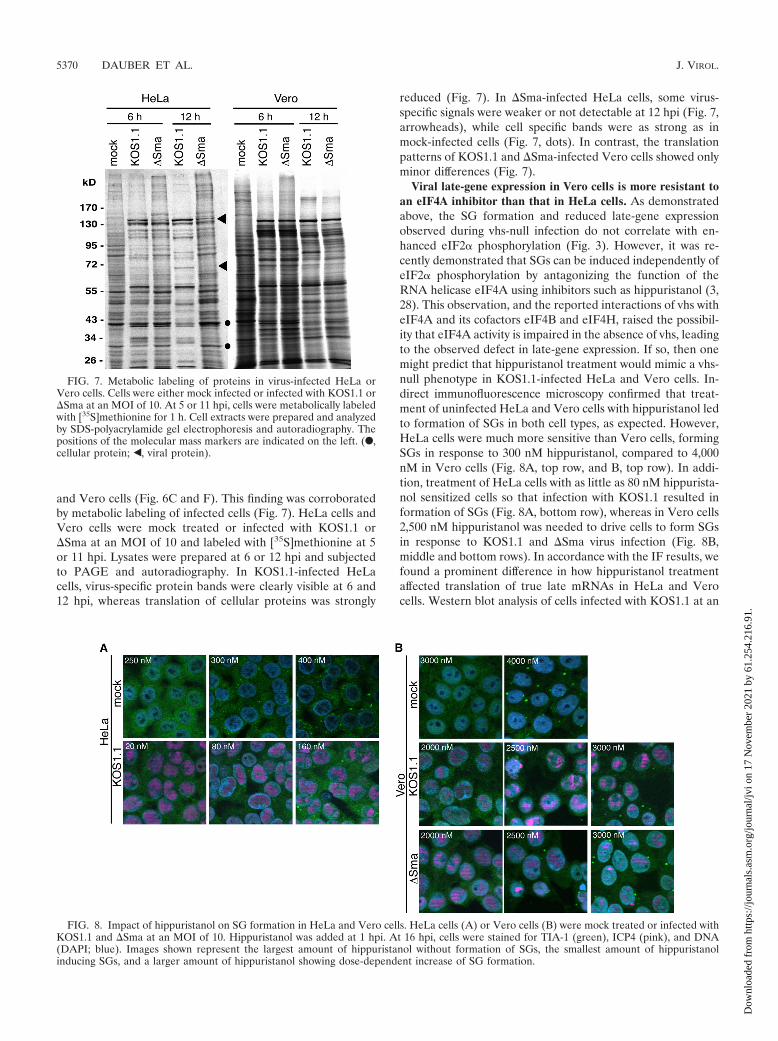
and Vero cells (Fig. 6C and F). This finding was corroboratedby metabolic labeling of infected cells (Fig. 7). HeLa cells andVero cells were mock treated or infected with KOS1.1 or�Sma at an MOI of 10 and labeled with [35S]methionine at 5or 11 hpi. Lysates were prepared at 6 or 12 hpi and subjectedto PAGE and autoradiography. In KOS1.1-infected HeLacells, virus-specific protein bands were clearly visible at 6 and12 hpi, whereas translation of cellular proteins was strongly
reduced (Fig. 7). In �Sma-infected HeLa cells, some virus-specific signals were weaker or not detectable at 12 hpi (Fig. 7,arrowheads), while cell specific bands were as strong as inmock-infected cells (Fig. 7, dots). In contrast, the translationpatterns of KOS1.1 and �Sma-infected Vero cells showed onlyminor differences (Fig. 7).
Viral late-gene expression in Vero cells is more resistant toan eIF4A inhibitor than that in HeLa cells. As demonstratedabove, the SG formation and reduced late-gene expressionobserved during vhs-null infection do not correlate with en-hanced eIF2� phosphorylation (Fig. 3). However, it was re-cently demonstrated that SGs can be induced independently ofeIF2� phosphorylation by antagonizing the function of theRNA helicase eIF4A using inhibitors such as hippuristanol (3,28). This observation, and the reported interactions of vhs witheIF4A and its cofactors eIF4B and eIF4H, raised the possibil-ity that eIF4A activity is impaired in the absence of vhs, leadingto the observed defect in late-gene expression. If so, then onemight predict that hippuristanol treatment would mimic a vhs-null phenotype in KOS1.1-infected HeLa and Vero cells. In-direct immunofluorescence microscopy confirmed that treat-ment of uninfected HeLa and Vero cells with hippuristanol ledto formation of SGs in both cell types, as expected. However,HeLa cells were much more sensitive than Vero cells, formingSGs in response to 300 nM hippuristanol, compared to 4,000nM in Vero cells (Fig. 8A, top row, and B, top row). In addi-tion, treatment of HeLa cells with as little as 80 nM hippurista-nol sensitized cells so that infection with KOS1.1 resulted information of SGs (Fig. 8A, bottom row), whereas in Vero cells2,500 nM hippuristanol was needed to drive cells to form SGsin response to KOS1.1 and �Sma virus infection (Fig. 8B,middle and bottom rows). In accordance with the IF results, wefound a prominent difference in how hippuristanol treatmentaffected translation of true late mRNAs in HeLa and Verocells. Western blot analysis of cells infected with KOS1.1 at an
FIG. 7. Metabolic labeling of proteins in virus-infected HeLa orVero cells. Cells were either mock infected or infected with KOS1.1 or�Sma at an MOI of 10. At 5 or 11 hpi, cells were metabolically labeledwith [35S]methionine for 1 h. Cell extracts were prepared and analyzedby SDS-polyacrylamide gel electrophoresis and autoradiography. Thepositions of the molecular mass markers are indicated on the left. (F,cellular protein; Š, viral protein).
FIG. 8. Impact of hippuristanol on SG formation in HeLa and Vero cells. HeLa cells (A) or Vero cells (B) were mock treated or infected withKOS1.1 and �Sma at an MOI of 10. Hippuristanol was added at 1 hpi. At 16 hpi, cells were stained for TIA-1 (green), ICP4 (pink), and DNA(DAPI; blue). Images shown represent the largest amount of hippuristanol without formation of SGs, the smallest amount of hippuristanolinducing SGs, and a larger amount of hippuristanol showing dose-dependent increase of SG formation.
5370 DAUBER ET AL. J. VIROL.
Dow
nloa
ded
from
http
s://j
ourn
als.
asm
.org
/jour
nal/j
vi o
n 17
Nov
embe
r 20
21 b
y 61
.254
.216
.91.

MOI of 10 for 12 h showed that in HeLa cells as little as 20 nMhippuristanol decreased gC and US11 levels and 80 nM hip-puristanol blocked accumulation of these proteins (Fig. 9A).At these concentrations, hippuristanol had only minor effectson accumulation of ICP34.5 and ICP4 and did not affect actinlevels (Fig. 9A). Northern blot analysis confirmed that hip-puristanol treatment did not affect gC or US11 mRNA levels(Fig. 9C), indicating that the reduced protein expression is dueto the effect of hippuristanol on translation and not on tran-scription or mRNA stability. Furthermore, it has been re-ported that hippuristanol does not induce phosphorylation ofeIF2� (28). Similar to what was observed in the induction ofSGs, far larger amounts of hippuristanol were required in Verocells (1,280 nM) to significantly reduce gC and US11 levelsduring KOS1.1 and �Sma infection (Fig. 9B). These resultssuggest that translation in Vero cells is more resilient duringconditions of reduced availability of translation factors thantranslation in HeLa cells. The apparently selective effect of lowdoses of hippuristanol on viral late- versus IE- and E-gene
expression in HeLa cells further implies either that translationof late-gene transcripts is more sensitive to restrictive condi-tions than translation of IE and E mRNAs or that conditionsbecome more restrictive during the late stages of infection.
DISCUSSION
It is well established that vhs degrades viral and cellularmRNAs during the early stages of infection. However, there ismounting evidence that vhs also plays a role in translation.First, vhs associates with the translation initiation factorseIF4A, eIF4B, and eIF4H (7, 11). Furthermore, we have pre-viously shown that vhs increases translation of the second cis-tron driven by several IRES elements or viral 5� UTRs in abicistronic reporter gene assay (44). Finally, infection withvhs-deficient HSV-1 leads to formation of TIA-1/TIAR-posi-tive cytoplasmic foci, presumably SGs (9), which might indicatestalled translation initiation. In this study, we demonstratedthat vhs boosts translation of viral late-gene products and pro-motes efficient virus production in a cell type-dependent man-ner and that the requirement for vhs might be determined bythe resilience of the translation machinery in the respective celltype. This novel role of vhs during HSV-1 infection is sup-ported by the following findings. (i) HSV-1 with mutated vhs orpartial or complete deletion of the vhs ORF, but not wild-typeviruses, triggered formation of SGs in all cell types testedexcept Vero cells. (ii) The vhs mutant �Sma was attenuated inseveral cell lines, including HeLa cells, but not in Vero, HEL,or U2OS cells. (iii) Expression of true late genes was stronglyreduced in the absence of functional vhs in HeLa and otherrestrictive cells but not in Vero, HEL, or U2OS cells, showinga strong correlation with the results of the growth curve. (iv)Reduced gene expression in �Sma-infected HeLa cells wasdue to decreased translation initiation on the respective viralmRNAs. (v) This effect was specific for late viral transcriptssince translation of cellular mRNAs was not affected in theabsence of vhs. (vi) Treatment of HeLa and Vero cells with theeIF4A inhibitor hippuristanol induced SG formation and re-duced late-gene expression during KOS1.1 infection, mimick-ing the phenotype of a �Sma infection. The effective dose forHeLa cells was far lower than that for Vero cells, indicatingthat the resilience of the translational machinery differs be-tween HeLa and Vero cells. This difference might be the crit-ical factor for translation of true late-gene transcript in theabsence of vhs and might determine the scale of the inherentgrowth defect of vhs-deficient HSV-1.
It is not yet clear whether SG formation directly impedesvirus production or, alternatively, whether the SGs are only areadout of the stalled translation initiation. TIA-1�/� orTIAR�/� MEFs have been shown to support replication ofHSV-1 (strain 129) to higher titers than WT MEFs (27), indi-cating that TIA-1 and TIAR have a negative effect on viralreplication. On the other hand, Wylie and coworkers showedthat knockout of TIA-1 or TIAR in MEFs did not restorereplication of an HSV-2 vhs mutant to wild-type titers (62).However, they did not determine whether the knockouts re-sulted in an increase in virus production relative to that inwild-type MEFs and, if so, whether this increase was differentfor HSV-2 and the vhs mutant. While it will be interesting tostudy the �Sma phenotype in the absence of TIA-1 or TIAR,
FIG. 9. Impact of hippuristanol treatment on expression of viralproteins in HeLa and Vero cells. HeLa cells (A) and Vero cells(B) were mock treated or infected with KOS1.1 or �Sma at an MOI of10. At 1 hpi, cells were treated with hippuristanol as indicated ortreated with DMSO as a control. Lysates were prepared at 12 hpi andanalyzed by immunoblotting with antibodies specific for gC, US11,ICP34.5, ICP4, and �-actin. (C) Hippuristanol treatment does notreduce the level of gC mRNA. HeLa cells were mock treated orinfected with KOS1.1 or �Sma at an MOI of 10. At 1 hpi, cells weretreated with hippuristanol as indicated or treated with DMSO as acontrol. RNA was extracted at 12 hpi and analyzed by Northern blot-ting for the viral gC mRNA or the cellular �-actin mRNA.
VOL. 85, 2011 vhs-MEDIATED TRANSLATION OF TRUE LATE-GENE PRODUCTS 5371
Dow
nloa
ded
from
http
s://j
ourn
als.
asm
.org
/jour
nal/j
vi o
n 17
Nov
embe
r 20
21 b
y 61
.254
.216
.91.

we speculate that SG formation is more likely a readout thana cause of the �Sma phenotype because the reduced gC andUS11 protein levels are observed as early as 6 hpi, whereas SGformation starts between 8 and 10 hpi.
We suggest that the accumulation of high levels of viral latemRNAs following the onset of viral DNA replication over-whelms the capacity of the translational initiation machinery,leading to the observed translational defect in restrictive cells.The observation that hippuristanol generates a similar pheno-type in cells infected with wild-type virus is consistent with thishypothesis and further suggests that eIF4A may be one of thelimiting factors. In this context, it is interesting to note that the5� UTRs of many HSV mRNAs adopt quite stable secondarystructures because of the overall GC-rich nature of the HSVgenome. For example, the 5� UTRs of ICP4, TK, ICP34.5, gC,and US11 mRNAs have predicted values of �125, �31, �103,�63, and �80 kcal/mole, respectively, as estimated by theMfold algorithm. Most 5� UTRs of well-translated cellularmRNAs have little or no secondary structure, with predictedfree energies between 0 and �30 kcal/mole (5), whereas sec-ondary structures with a free energy of �50 kcal/mole or lowerimpose a strong block on ribosome scanning (23, 37). Forhighly structured 5� UTRs, optimal helicase activity is abso-lutely critical for initiation of translation (59). Thus, it seemsquite plausible that eIF4A could become limiting due to themassive accumulation of highly structured viral mRNAs duringthe late stages of infection. As shown in Fig. 8 and 9, Vero cellscan deal with over 10 times more hippuristanol than HeLa cellsbefore the balance is tipped toward SG formation and a blockin translation of viral late-gene transcripts. These observationssuggest that translation in Vero cells is more resistant to re-duced eIF4A availability than translation in HeLa cells. There-fore, vhs would be less critical for late-gene expression underthese circumstances. It is also possible that Vero cells are moreefficient at degrading excess mRNA to avoid overloading of thetranslation machinery.
Interestingly, the translational blockade observed in HeLacells in the absence of vhs appears to selectively impair trans-lation of viral true late mRNAs, sparing other mRNAs that arepresent in the infected cells. Thus, cellular �-actin mRNAremains associated with large polysomes late during vhs-nullinfection (Fig. 6), and many other cellular and viral mRNAsare also translationally active as judged by [35S]methionineincorporation (Fig. 7, 12-hpi lanes). How might this selectiveblockade arise? One possibility is that some structural featureof late mRNAs places them at a disadvantage relative to othermRNAs. Alternatively, perhaps true late transcripts fail to beefficiently translated simply because they accumulate aftertranslation initiation factors have become limiting. Accordingto the latter model, the limiting factors are stably sequesteredby the preexisting cellular and viral mRNAs and are not readilyrecycled in the absence of vhs. Further studies are required todistinguish between these and other possibilities.
There are several possibilities for how vhs boosts translationinitiation on true late mRNAs in restrictive cells. The simplestis that vhs prevents “mRNA overload” during the late stages ofinfection by eliminating host mRNAs and promoting the decayof viral IE and E transcripts. A second (and related) possibilityis that vhs promotes recycling of translation initiation factorsby shortening the half-life of viral mRNAs, allowing transfer of
factors from the viral IE, E, and leaky late mRNAs madebefore DNA replication onto true late mRNAs. A third pos-sibility is that vhs plays a more active role beyond reducingmRNA levels and recycling translation initiation factors.eIF4B and eIF4H are cofactors of eIF4A that increase thehelicase activity, and their requirement correlates with thedegree of secondary structure in the 5� UTRs of mRNAs (41,42, 51). vhs has been shown to interact with eIF4A, eIF4B, andeIF4H (7, 11). Therefore, we speculate that vhs might act as anadaptor to bring helicase and cofactors together and therebyincrease unwinding of the secondary structure. vhs has nodefined RNA-binding capacity, so how would it assemble he-licase and cofactors preferably on true late-gene transcripts?Although we cannot yet answer this question, it is possible thatvhs is targeted to mRNAs as a complex with VP16 and VP22,of which the latter has been shown to bind to mRNAs (49).Furthermore, late in infection the RNase activity of vhs wouldbe dampened in a complex with VP16 and VP22 (13, 22, 25,48, 52).
These three models for how vhs boosts translation of truelate-gene transcripts are not mutually exclusive, and we arecurrently investigating the potential role of eIF4A availability,RNA accumulation, 5� UTR structure, and interaction of vhswith translation initiation factors.
ACKNOWLEDGMENTS
We thank Holly Saffran for technical support.This research was supported by operating grants from the Canadian
Institutes for Health Research to J.R.S. (FRN 37995) and from theCanadian Cancer Society Research Institute to J.P. (20066). B.D. wassupported by a postdoctoral fellowship from Alberta Innovates—Health Solutions, and J.R.S. is a Canada Research Chair in MolecularVirology.
REFERENCES
1. Anderson, P., and N. Kedersha. 2008. Stress granules: the Tao of RNAtriage. Trends Biochem. Sci. 33:141–150.
2. Bordeleau, M. E., et al. 2006. RNA-mediated sequestration of the RNAhelicase eIF4A by Pateamine A inhibits translation initiation. Chem. Biol.13:1287–1295.
3. Bordeleau, M. E., et al. 2006. Functional characterization of IRESes by aninhibitor of the RNA helicase eIF4A. Nat. Chem. Biol. 2:213–220.
4. Corcoran, J. A., W. L. Hsu, and J. R. Smiley. 2006. Herpes simplex virusICP27 is required for virus-induced stabilization of the ARE-containingIEX-1 mRNA encoded by the human IER3 gene. J. Virol. 80:9720–9729.
5. Davuluri, R. V., Y. Suzuki, S. Sugano, and M. Q. Zhang. 2000. CARTclassification of human 5� UTR sequences. Genome Res. 10:1807–1816.
6. Diaz, M. O., et al. 1988. Homozygous deletion of the alpha- and beta1-interferon genes in human leukemia and derived cell lines. Proc. Natl.Acad. Sci. U. S. A. 85:5259–5263.
7. Doepker, R. C., W. L. Hsu, H. A. Saffran, and J. R. Smiley. 2004. Herpessimplex virus virion host shutoff protein is stimulated by translation initiationfactors eIF4B and eIF4H. J. Virol. 78:4684–4699.
8. Duerst, R. J., and L. A. Morrison. 2004. Herpes simplex virus 2 virion hostshutoff protein interferes with type I interferon production and responsive-ness. Virology 322:158–167.
9. Esclatine, A., B. Taddeo, and B. Roizman. 2004. Herpes simplex virus 1induces cytoplasmic accumulation of TIA-1/TIAR and both synthesis andcytoplasmic accumulation of tristetraprolin, two cellular proteins that bindand destabilize AU-rich RNAs. J. Virol. 78:8582–8592.
10. Everly, D. N., Jr., P. Feng, I. S. Mian, and G. S. Read. 2002. mRNAdegradation by the virion host shutoff (Vhs) protein of herpes simplex virus:genetic and biochemical evidence that Vhs is a nuclease. J. Virol. 76:8560–8571.
11. Feng, P., D. N. Everly, Jr., and G. S. Read. 2005. mRNA decay during herpessimplex virus (HSV) infections: protein-protein interactions involving theHSV virion host shutoff protein and translation factors eIF4H and eIF4A.J. Virol. 79:9651–9664.
12. Guo, J., D. J. Hui, W. C. Merrick, and G. C. Sen. 2000. A new pathway oftranslational regulation mediated by eukaryotic initiation factor 3. EMBO J.19:6891–6899.
5372 DAUBER ET AL. J. VIROL.
Dow
nloa
ded
from
http
s://j
ourn
als.
asm
.org
/jour
nal/j
vi o
n 17
Nov
embe
r 20
21 b
y 61
.254
.216
.91.

13. Hafezi, W., E. Bernard, R. Cook, and G. Elliott. 2005. Herpes simplex virustegument protein VP22 contains an internal VP16 interaction domain and aC-terminal domain that are both required for VP22 assembly into the virusparticle. J. Virol. 79:13082–13093.
14. He, B., M. Gross, and B. Roizman. 1998. The gamma134.5 protein of herpessimplex virus 1 has the structural and functional attributes of a proteinphosphatase 1 regulatory subunit and is present in a high molecular weightcomplex with the enzyme in infected cells. J. Biol. Chem. 273:20737–20743.
15. He, B., M. Gross, and B. Roizman. 1997. The gamma(1)34.5 protein ofherpes simplex virus 1 complexes with protein phosphatase 1alpha to de-phosphorylate the alpha subunit of the eukaryotic translation initiation fac-tor 2 and preclude the shutoff of protein synthesis by double-stranded RNA-activated protein kinase. Proc. Natl. Acad. Sci. U. S. A. 94:843–848.
16. Hershey, J. W. 1991. Translational control in mammalian cells. Annu. Rev.Biochem. 60:717–755.
17. Hughes, R. G., Jr., and W. H. Munyon. 1975. Temperature-sensitive mutantsof herpes simplex virus type 1 defective in lysis but not in transformation.J. Virol. 16:275–283.
18. Hui, D. J., C. R. Bhasker, W. C. Merrick, and G. C. Sen. 2003. Viralstress-inducible protein p56 inhibits translation by blocking the interaction ofeIF3 with the ternary complex eIF2.GTP. Met-tRNAi. J. Biol. Chem. 278:39477–39482.
19. Kedersha, N., et al. 2002. Evidence that ternary complex (eIF2-GTP-tRNA(i) (Met))-deficient preinitiation complexes are core constituents ofmammalian stress granules. Mol. Biol. Cell 13:195–210.
20. Kedersha, N., et al. 2000. Dynamic shuttling of TIA-1 accompanies therecruitment of mRNA to mammalian stress granules. J. Cell Biol. 151:1257–1268.
21. Kedersha, N. L., M. Gupta, W. Li, I. Miller, and P. Anderson. 1999. RNA-binding proteins TIA-1 and TIAR link the phosphorylation of eIF-2 alpha tothe assembly of mammalian stress granules. J. Cell Biol. 147:1431–1442.
22. Knez, J., P. T. Bilan, and J. P. Capone. 2003. A single amino acid substitu-tion in herpes simplex virus type 1 VP16 inhibits binding to the virion hostshutoff protein and is incompatible with virus growth. J. Virol. 77:2892–2902.
23. Kozak, M. 1989. Circumstances and mechanisms of inhibition of translationby secondary structure in eucaryotic mRNAs. Mol. Cell. Biol. 9:5134–5142.
24. Kwong, A. D., and N. Frenkel. 1987. Herpes simplex virus-infected cellscontain a function(s) that destabilizes both host and viral mRNAs. Proc.Natl. Acad. Sci. U. S. A. 84:1926–1930.
25. Lam, Q., et al. 1996. Herpes simplex virus VP16 rescues viral mRNA fromdestruction by the virion host shutoff function. EMBO J. 15:2575–2581.
26. Leib, D. A., et al. 1999. Interferons regulate the phenotype of wild-type andmutant herpes simplex viruses in vivo. J. Exp. Med. 189:663–672.
27. Li, W., et al. 2002. Cell proteins TIA-1 and TIAR interact with the 3�stem-loop of the West Nile virus complementary minus-strand RNA andfacilitate virus replication. J. Virol. 76:11989–12000.
28. Mazroui, R., et al. 2006. Inhibition of ribosome recruitment induces stressgranule formation independently of eukaryotic initiation factor 2alpha phos-phorylation. Mol. Biol. Cell 17:4212–4219.
29. Mohr, I. 2006. Phosphorylation and dephosphorylation events that regulateviral mRNA translation. Virus Res. 119:89–99.
30. Mossman, K. L., et al. 2001. Herpes simplex virus triggers and then disarmsa host antiviral response. J. Virol. 75:750–758.
31. Murphy, J. A., R. J. Duerst, T. J. Smith, and L. A. Morrison. 2003. Herpessimplex virus type 2 virion host shutoff protein regulates alpha/beta inter-feron but not adaptive immune responses during primary infection in vivo.J. Virol. 77:9337–9345.
32. Oroskar, A. A., and G. S. Read. 1989. Control of mRNA stability by thevirion host shutoff function of herpes simplex virus. J. Virol. 63:1897–1906.
33. Page, H. G., and G. S. Read. 2010. The virion host shutoff endonuclease(UL41) of herpes simplex virus interacts with the cellular cap-binding com-plex eIF4F. J. Virol. 84:6886–6890.
34. Pasieka, T. J., et al. 2009. Host responses to wild-type and attenuated herpessimplex virus infection in the absence of Stat1. J. Virol. 83:2075–2087.
35. Pasieka, T. J., et al. 2008. Herpes simplex virus virion host shutoff attenuatesestablishment of the antiviral state. J. Virol. 82:5527–5535.
36. Pasieka, T. J., B. Lu, and D. A. Leib. 2008. Enhanced pathogenesis of anattenuated herpes simplex virus for mice lacking Stat1. J. Virol. 82:6052–6055.
37. Pelletier, J., and N. Sonenberg. 1985. Insertion mutagenesis to increasesecondary structure within the 5� noncoding region of a eukaryotic mRNAreduces translational efficiency. Cell 40:515–526.
38. Perkins, K. D., J. Gregonis, S. Borge, and S. A. Rice. 2003. Transactivationof a viral target gene by herpes simplex virus ICP27 is posttranscriptional anddoes not require the endogenous promoter or polyadenylation site. J. Virol.77:9872–9884.
39. Read, G. S., and N. Frenkel. 1983. Herpes simplex virus mutants defective in
the virion-associated shutoff of host polypeptide synthesis and exhibitingabnormal synthesis of alpha (immediate early) viral polypeptides. J. Virol.46:498–512.
40. Read, G. S., B. M. Karr, and K. Knight. 1993. Isolation of a herpes simplexvirus type 1 mutant with a deletion in the virion host shutoff gene andidentification of multiple forms of the vhs (UL41) polypeptide. J. Virol.67:7149–7160.
41. Rogers, G. W., Jr., N. J. Richter, W. F. Lima, and W. C. Merrick. 2001.Modulation of the helicase activity of eIF4A by eIF4B, eIF4H, and eIF4F.J. Biol. Chem. 276:30914–30922.
42. Rogers, G. W., Jr., N. J. Richter, and W. C. Merrick. 1999. Biochemical andkinetic characterization of the RNA helicase activity of eukaryotic initiationfactor 4A. J. Biol. Chem. 274:12236–12244.
43. Roizman, B., D. M. Knipe, and R. J. Whitley. 2007. Herpes simplex viruses,p. 2501–2601. In D. M. Knipe and P. M. Howley (ed.), Fields virology, 5th ed.Lippincott Williams & Wilkins, Philadelphia, PA.
44. Saffran, H. A., G. S. Read, and J. R. Smiley. 2010. Evidence for translationalregulation by the herpes simplex virus virion host shutoff protein. J. Virol.84:6041–6049.
45. Samady, L., et al. 2003. Deletion of the virion host shutoff protein (vhs) fromherpes simplex virus (HSV) relieves the viral block to dendritic cell activa-tion: potential of vhs� HSV vectors for dendritic cell-mediated immunother-apy. J. Virol. 77:3768–3776.
46. Samuel, C. E. 1993. The eIF-2 alpha protein kinases, regulators of transla-tion in eukaryotes from yeasts to humans. J. Biol. Chem. 268:7603–7606.
47. Sarma, N., D. Agarwal, L. A. Shiflett, and G. S. Read. 2008. Small interferingRNAs that deplete the cellular translation factor eIF4H impede mRNAdegradation by the virion host shutoff protein of herpes simplex virus. J. Vi-rol. 82:6600–6609.
48. Sciortino, M. T., et al. 2007. Replication-competent herpes simplex virus 1isolates selected from cells transfected with a bacterial artificial chromosomeDNA lacking only the UL49 gene vary with respect to the defect in the UL41gene encoding host shutoff RNase. J. Virol. 81:10924–10932.
49. Sciortino, M. T., B. Taddeo, A. P. Poon, A. Mastino, and B. Roizman. 2002.Of the three tegument proteins that package mRNA in herpes simplexvirions, one (VP22) transports the mRNA to uninfected cells for expressionprior to viral infection. Proc. Natl. Acad. Sci. U. S. A. 99:8318–8323.
50. Sedlackova, L., et al. 2010. Identification of an ICP27-responsive element inthe coding region of a herpes simplex virus type 1 late gene. J. Virol.84:2707–2718.
51. Shahbazian, D., et al. 2010. Control of cell survival and proliferation bymammalian eukaryotic initiation factor 4B. Mol. Cell. Biol. 30:1478–1485.
52. Smibert, C. A., B. Popova, P. Xiao, J. P. Capone, and J. R. Smiley. 1994.Herpes simplex virus VP16 forms a complex with the virion host shutoffprotein vhs. J. Virol. 68:2339–2346.
53. Smibert, C. A., and J. R. Smiley. 1990. Differential regulation of endogenousand transduced beta-globin genes during infection of erythroid cells with aherpes simplex virus type 1 recombinant. J. Virol. 64:3882–3894.
54. Smiley, J. R. 2004. Herpes simplex virus virion host shutoff protein: immuneevasion mediated by a viral RNase? J. Virol. 78:1063–1068.
55. Smith, R. W., S. V. Graham, and N. K. Gray. 2008. Regulation of translationinitiation by herpesviruses. Biochem. Soc. Trans. 36:701–707.
56. Smith, T. J., L. A. Morrison, and D. A. Leib. 2002. Pathogenesis of herpessimplex virus type 2 virion host shutoff (vhs) mutants. J. Virol. 76:2054–2061.
57. Strelow, L., T. Smith, and D. Leib. 1997. The virion host shutoff function ofherpes simplex virus type 1 plays a role in corneal invasion and functionsindependently of the cell cycle. Virology 231:28–34.
58. Suzutani, T., et al. 2000. The role of the UL41 gene of herpes simplex virustype 1 in evasion of non-specific host defence mechanisms during primaryinfection. J. Gen. Virol. 81:1763–1771.
59. Svitkin, Y. V., et al. 2001. The requirement for eukaryotic initiation factor 4A(elF4A) in translation is in direct proportion to the degree of mRNA 5�secondary structure. RNA 7:382–394.
60. Taddeo, B., and B. Roizman. 2006. The virion host shutoff protein (UL41) ofherpes simplex virus 1 is an endoribonuclease with a substrate specificitysimilar to that of RNase A. J. Virol. 80:9341–9345.
61. Taddeo, B., W. Zhang, and B. Roizman. 2006. The U(L)41 protein of herpessimplex virus 1 degrades RNA by endonucleolytic cleavage in absence ofother cellular or viral proteins. Proc. Natl. Acad. Sci. U. S. A. 103:2827–2832.
62. Wylie, K. M., J. E. Schrimpf, and L. A. Morrison. 2009. Increased eIF2alphaphosphorylation attenuates replication of herpes simplex virus 2 vhs mutantsin mouse embryonic fibroblasts and correlates with reduced accumulation ofthe PKR antagonist ICP34.5. J. Virol. 83:9151–9162.
63. Zelus, B. D., R. S. Stewart, and J. Ross. 1996. The virion host shutoff proteinof herpes simplex virus type 1: messenger ribonucleolytic activity in vitro.J. Virol. 70:2411–2419.
VOL. 85, 2011 vhs-MEDIATED TRANSLATION OF TRUE LATE-GENE PRODUCTS 5373
Dow
nloa
ded
from
http
s://j
ourn
als.
asm
.org
/jour
nal/j
vi o
n 17
Nov
embe
r 20
21 b
y 61
.254
.216
.91.