negative and positive mrna splicing elements act ...jvi.asm.org/content/82/8/3921.full.pdf · over...
TRANSCRIPT
JOURNAL OF VIROLOGY, Apr. 2008, p. 3921–3931 Vol. 82, No. 80022-538X/08/$08.00�0 doi:10.1128/JVI.01558-07Copyright © 2008, American Society for Microbiology. All Rights Reserved.
Negative and Positive mRNA Splicing Elements Act Competitively ToRegulate Human Immunodeficiency Virus Type 1 Vif Gene Expression�
C. M. Exline, Z. Feng, and C. M. Stoltzfus*Department of Microbiology, University of Iowa, Iowa City, Iowa 52242
Received 17 July 2007/Accepted 1 February 2008
Over 40 different human immunodeficiency virus type 1 (HIV-1) mRNAs are produced by alternative splicingof the primary HIV-1 RNA transcripts. In addition, approximately half of the viral RNA remains unspliced andis used as genomic RNA and as mRNA for the Gag and Pol gene products. Regulation of splicing at the HIV-13� splice sites (3�ss) requires suboptimal polypyrimidine tracts, and positive or negative regulation occursthrough the binding of cellular factors to cis-acting splicing regulatory elements. We have previously shownthat splicing at HIV-1 3�ss A1, which produces single-spliced vif mRNA and promotes the inclusion of HIV exon2 into both completely and incompletely spliced viral mRNAs, is increased by optimizing the 5� splice site (5�ss)downstream of exon 2 (5�ss D2). Here we show that the mutations within 5�ss D2 that are predicted to loweror increase the affinity of the 5�ss for U1 snRNP result in reduced or increased Vif expression, respectively.Splicing at 5�ss D2 was not necessary for the effect of 5�ss D2 on Vif expression. In addition, we have found thatmutations of the GGGG motif proximal to the 5�ss D2 increase exon 2 inclusion and Vif expression. Finally,we report the presence of a novel exonic splicing enhancer (ESE) element within the 5�-proximal region of exon2 that facilitates both exon inclusion and Vif expression. This ESE binds specifically to the cellular SR proteinSRp75. Our results suggest that the 5�ss D2, the proximal GGGG silencer, and the ESE act competitively todetermine the level of vif mRNA splicing and Vif expression. We propose that these positive and negativesplicing elements act together to allow the accumulation of vif mRNA and unspliced HIV-1 mRNA, compatiblewith optimal virus replication.
Human immunodeficiency virus type 1 (HIV-1) primaryRNA transcripts are alternatively spliced, to generate over 40different mRNAs of three different size classes, unspliced�9-kb mRNAs, incompletely spliced �4-kb mRNAs, and com-pletely spliced �1.8-kb mRNAs (Fig. 1) (for a review, seereference 28). The unspliced viral RNA is used as genomicRNA and as mRNA for the Gag and Pol gene products (for areview, see reference 7). The incompletely spliced size classincludes mRNAs for Vif, Vpr, single-exon Tat, and Env/Vpu,and the completely spliced size class includes mRNAs for two-exon Tat, Rev, and Nef. Four different 5� splice sites (5�ss) andeight different 3� splice sites (3�ss) are used to produce thedifferent alternatively spliced HIV-1 mRNAs, which arepresent in different amounts in the infected cells (21). Thelocations and sequences of most of these splice sites are highlyconserved in all clades of group M HIV-1 and in HIV-1 strainsin groups N and O. The extent to which these 5�ss and 3�ss areused is dependent on the relative strengths of the splice sitesand on the presence of cis splicing elements within the viralgenome. The cis elements within the HIV-1 genome includeboth exonic splicing silencers (ESS) and intronic splicing si-lencers (ISS) and exonic splicing enhancers (ESE).
We have previously shown that the splicing of tat mRNA,which is present in relatively low abundance, is regulated bytwo different ESS within the first tat coding exon that repressessplicing at 3�ss A3 and in an ESS in the second tat/rev codingexon that represses splicing at 3�ss A7 (4, 11, 26, 27). In addi-
tion, an ISS upstream of 3�ss A7 regulates tat/rev mRNA splic-ing (30). The first tat coding exon and the second tat/rev codingexon also contain ESE that selectively increase splicing at 3�ssA3 and A7, respectively (4, 26, 27, 35). The splicing of vprmRNA, which is also present in low abundance in infectedcells, is regulated by an ESS that represses splicing at 3�ss A2(6, 18). In addition, splicing at 3�ss A2 is facilitated by thepresence of a downstream 5�ss (5�ss D3), a result predicted bythe exon definition hypothesis, which proposes that the defi-nition of exons is an early step in splicing and precedes thedefinition of introns (5, 10, 22). The use of the 5�ss D3 resultsin the inclusion of a small 74-nucleotide (nt) noncoding exon(exon 3) between 3�ss A2 and 5�ss D3 into a fraction of theHIV-1 mRNAs. Mutations that optimize 5�ss D3 result inincreased splicing at 3�ss A2 and increased exon 3 inclusion (6).
vif mRNA is also present in low abundance in HIV-1-in-fected cells. Regulation of vif mRNA splicing may be impor-tant to maintain Vif expression at the levels necessary to in-hibit the accumulation and packaging of APOBEC3G andAPOBEC3F deoxycytidine deaminases (14, 20, 25, 34) withoutinhibiting virus protein processing and virus replication, whichhas been shown to occur at higher Vif levels (2). We havepreviously shown that splicing at 3�ss A1, the splice site used togenerate vif mRNA, is limited by the presence of a suboptimaldownstream 5�ss, since the mutation of 5�ss D2 to a consensus5�ss results in an HIV-1 phenotype with excessive splicing, adramatic increase in the expression of Vif, and the inclusion ofthe 50-nt noncoding exon 2 between 3�ss A1 and 5�ss D2 intoa majority of the spliced viral mRNAs (17). In contrast, in thewild-type infected cells, exon 2 is included into only a fractionof viral mRNAs (21). It has also recently been shown that exon2 contains two potential ESE elements (ESEM1 and ESEM2)
* Corresponding author. Mailing address: Department of Microbi-ology, University of Iowa, Iowa City, IA 52242. Phone: (319) 335-7793.Fax: (319) 335-9006. E-mail: [email protected].
� Published ahead of print on 13 February 2008.
3921
on August 9, 2018 by guest
http://jvi.asm.org/
Dow
nloaded from

with the sequence UGGAAAG; mutations of these two ele-ments resulted in a selective decrease in exon 2 inclusion.Surprisingly, in the context of an infectious provirus, these ESEmutations were reported to not significantly affect the levels ofvif mRNA and Vif expression, suggesting that ESEM1 andESEM2 affect exon 2 definition and exon 2 inclusion but notthe creation of vif mRNA (13).
In this study, we investigated splicing elements affecting bothexon 2 definition and HIV-1 Vif expression by creating muta-tions that strengthen or weaken 5�ss D2, mutations within exon2, and mutations downstream of 5�ss D2. Based on our results,we propose that 5�ss D2, a GGGG splicing silencer proximal to5�ss D2, and an ESE act competitively by interacting with hostfactors to determine the level of vif mRNA splicing and Vifexpression. In addition, we propose that these elements acttogether to allow the accumulation of unspliced mRNA levelscompatible with optimal virus replication.
MATERIALS AND METHODS
Plasmids. The infectious HIV-1 molecular clone pNL4-3 was obtained fromthe NIH AIDS Research and Reference Program (1). pNLD2up has been de-scribed previously (17). The mutant derivatives of pNL4-3 shown in Fig. 2 weregenerated using a QuikChange site-directed mutagenesis kit (Stratagene) asdescribed previously (18). The �-galactosidase expression plasmid pCMV110was obtained from Tom Hope, Northwestern University School of Medicine.
Cell transfection. 293T cells growing in Dulbecco’s modified Eagle’s mediumwith 10% fetal bovine serum were transfected with pNL4-3 or with mutants ofpNL4-3, using calcium phosphate precipitation as previously described (18). Tomonitor transfection efficiencies, cells were cotransfected with a lacZ expressionplasmid, CMV110. The cells and culture medium were harvested at 24 to 48 hposttransfection. Aliquots of the cell extracts were used to determine �-galac-
tosidase-specific enzyme activity (23). The remainder of the cells were disruptedby using Tri-Reagent (Molecular Research Center, Inc.), and total cellular RNAand protein were isolated according to the protocol supplied by the manufac-turer.
Analysis of viral RNA species. Reverse transcription (RT)-PCR was used todetect HIV-1 mRNA species in the �1.8-kb and �4-kb mRNA size classes, andNorthern blotting analysis of the total RNA was performed as previously de-scribed (18). Blots were hybridized to a 32P-labeled probe generated from the422-nt XhoI/BamHI restriction fragment in the 3� untranslated region ofpNL4-3, which is present in all HIV-1 mRNAs.
Quantitative real-time RT-PCR analysis of vif mRNA. cDNA was synthesizedfrom total cellular RNA, using Moloney murine leukemia virus reverse trans-criptase (RT) and random hexamers, as previously described (6). Real-time PCRanalysis to quantify vif and �-actin mRNAs was performed with an AppliedBiosystems model 7300 real-time PCR system using an absolute RNA quantifi-cation method. PCR amplification was followed by detection with SYBR Green.Primers used for specific amplification of vif mRNA were D1-A1 (forward),GGCGACTGGGACAGC, and Vif body (reverse), CACACAATCATCACCTGCC. Primers used for the amplification of �-actin mRNA were �-actin-5�(forward), GCTCCTCCTGAGCGCAAG, and �-actin-3� (reverse), CATCTGCTGGAAGGTGGACA. Ratios of the vif mRNA concentration to the �-actinmRNA concentration were then determined. Data for the pNL4-3 mutants werethen normalized to wild-type values after corrections were made for the relativetransfection efficiencies based on the �-galactosidase assays of transfected cellextracts.
Immunoblotting. Cellular proteins (50 �g) were resolved by sodium dodecylsulfate (SDS)-polyacrylamide gel electrophoresis, transferred to nitrocellulosemembrane via the semidry transfer method, and immunoblotted. �-Tubulin wasdetected with monoclonal antibody (MAb) E7 (Developmental Studies Hybrid-oma Bank, University of Iowa) diluted 1:1,000. Vif was detected using eitherMAb 319 diluted 1:50 or polyclonal Ab 2221 diluted 1:1,000. Both Vif antibodieswere obtained from the NIH AIDS Research and Reference Reagent Program.Blots were developed with an ECLPlus Western blotting detection system (Am-ersham) using peroxidase-conjugated sheep anti-mouse immunoglobulin G(MAb E7 and MAb 319) or donkey anti-rabbit immunoglobulin G (Ab 2221).
FIG. 1. Viral RNA species produced within HIV-1-infected cells. HIV-1 genes are shown relative to the long terminal repeats (LTR). The viralgenomic or �9-kb unspliced mRNA shows the location of 5�ss and 3�ss within the pNL4-3 infectious plasmid. The incompletely and completelyspliced HIV-1 viral mRNAs (�4-kb and 1.8-kb size classes) are shown as black boxes. Spliced mRNAs are denoted by the translated open readingframes and by the exon content. The incompletely spliced mRNAs, denoted by an I, are differentiated from completely spliced mRNAs by theinclusion of the intron between 5�ss D4 and 3�ss A7. Either one or both of the noncoding exons 2 and 3 are included in a fraction of all �4-kband �1.8-kb mRNA species, with the exception of vif mRNA. The locations of the primer pairs used for qRT-PCR analysis of vif mRNA (D1A1and VifBody) and RT-PCR analysis of 1.8-kb mRNA (BSS and SJ4.7A) are indicated.
3922 EXLINE ET AL. J. VIROL.
on August 9, 2018 by guest
http://jvi.asm.org/
Dow
nloaded from

RT assays and virus production. Cell-free supernatants were assayed for RTactivity by [�-32P]dTTP incorporation as previously described (33). Incorpora-tion of radioactivity was quantitated using an Instant Imager (Packard) program.RT data were normalized to that of the wild type after correction was made fortransfection efficiency based on �-galactosidase assays of extracts from trans-fected cells.
In vitro RNA splicing. The pdsx�E and pdsxASLV plasmids were a gift fromM. Caputi, Florida Atlantic University, and have been described previously (29,31). Plasmids for in vitro splicing templates were constructed by inserting an-nealed oligonucleotides into the ClaI site of dsx�E (sequence GGACAGCAGAGATCCAGT for dsxWT and GGACAGCAGTCTAGGTGG for dsxA1M).Plasmids were linearized with HindIII for dsx�E derivatives or with BamHI fordsxASLV and used as templates for the in vitro transcription of 32P-labeled RNAsubstrates, using T7 polymerase. In vitro splicing assays of purified substrateswere carried out essentially as described previously (3).
UV cross-linking and immunoprecipitation. The wild-type NL4-3 sequence(GGA CAG CAG AGA TCC AGT) or the mutant sequence corresponding toESEVifm (GGA CAG CAG AAA GCC) were cloned into the pBluescript SK�plasmid at the KpnI site. In vitro transcription templates (65 nt) were created byPCR amplification of the sequence between the T7 RNA promoter and thesequence downstream of the insertion. 32P-labeled RNA substrates (48 nt) werethen synthesized from the PCR product. For UV cross-linking assays, 1 � 105
cpm of RNA substrates were incubated in the presence of the HeLa nuclearextracts (HNE), at the indicated volumes at 30°C for 30 min and cross-linked tobound protein by UV irradiation for 15 min. Unbound substrate was digestedwith 25 �g RNase A (Fermentas), and the 32P-labeled RNA-protein complexeswere resolved on 10% polyacrylamide gels. For immunoprecipitation reactions,1 � 106 cpm substrate was incubated with 20 �l HNE and cross-linked, andunbound RNA was digested with 100 �g RNase A. After samples were digested,they were incubated at 4°C for 2 h with either 2 �g of pan-SR MAb 16H3(Zymed) or 1 �g of MAb �-SRp75 (Ptglabs). Complexes were then bound toprotein A-conjugated agarose beads, and unbound proteins were removed bywashing the beads four times with 20 mM Tris-HCl–0.5 M NaCl–1% NP-40 andonce with 20 mM Tris-HCl–0.15 M NaCl–1% NP-40. The bound fractions weresolubilized in 5 mM Tris-7 mM SDS-10 mM dithiothreitol-5% glycerol (pH 6.8),heated at 100°C for 3 min, and resolved on a 10% polyacrylamide gel. Theimmunoprecipitation products were then visualized by autoradiography.
RESULTS
Down mutations of 5�ss D2 decrease exon 2 inclusion andlevels of vif mRNA and Vif protein expression. To test the roleof the downstream 5�ss in Vif expression, we created 5�ss D2mutations within the infectious HIV-1 plasmid pNL4-3, and
the mutant plasmid was predicted to bind to U1 snRNP withreduced affinity compared to that of the wild type (Fig. 2). Toprevent splicing at 5�ss D2, we changed the invariant GU toGC (pNLD2GC). We also changed additional bases, as shown inFig. 2, that were predicted to either reduce or increase the affinityof U1 snRNP for 5�ss D2 compared to that of pNLD2GC(pNLD2GCdown and pNLD2GC-G5, respectively). The mutantplasmids were transfected into 293T cells, and cells and superna-tants from the transfected cells were harvested at 24 to 48 hposttransfection. The extent of exon inclusion was determined bypolyacrylamide gel electrophoresis of the RT-PCR products ob-tained from �1.8-kb mRNA (Fig. 3A). As controls for exon 2inclusion, we compared the profiles of the three down mutationsof 5�ss D2 to that of the wild-type pNL4-3 plasmid and to that ofthe previously described pNLD2up mutant plasmid. This mutantplasmid is predicted to bind to U1 snRNA with a significantlyhigher affinity than that of the wild type (Fig. 2). As expected fromour previous results, exon 2 was spliced into most, if not all,pNLD2up �1.8-kb mRNAs, which include nef, rev, and tatmRNA species (17). This can be seen by the absence of RNAspecies 1.5.7, 1.4b, 1.4a, and 1.4.7 and by the prominence of RNAspecies 1.2.5.7, 1.2.4b, 1.2.4a, and 1.2.4.7. Exon 2 was also in-cluded into most of the pNLD2up plasmid �4.0-kb size classmRNAs (data not shown). In contrast, the three GU-to-GC mu-tants did not include exon 2 into the HIV-1 mRNAs. This can beseen in Fig. 3A by the absence of RNA species 1.2.5.7 and 1.2.4.7,indicating that splicing at D2 was prevented by all three of theGU-to-GC mutants. Exon 2 was also not included into mRNAsfrom the �4.0-kb mRNA size class (data not shown).
We next determined the effect of the D2 GC mutations onvif mRNA levels by quantitative real-time RT-PCR (qRT-PCR) assays (Fig. 3B). Each of the GC mutants producedlower levels of vif mRNA than the wild type, with the highestlevels seen with pNLD2GC (�50% of the wild type) and thelowest levels seen with pNLD2GCdown (�10% of the wildtype). The pNLD2GC-G5 mutant demonstrated intermediatelevels (�25% of the wild type). In contrast, pNLD2-up, with an
FIG. 2. Mutations of pNL4-3 used in this study. The sequence of exon 2 is shown in uppercase letters, and the surrounding intron sequencesare shown in lowercase letters. The location of the changes in sequence for each of the mutants is shown. The predicted �G0
37 in kcal/mol for theeight base pairs of U1 snRNA binding to wild-type and mutated 5�ss D2 was calculated by the method described by Serra and Turner (24).
VOL. 82, 2008 REGULATION OF HIV-1 Vif mRNA SPLICING 3923
on August 9, 2018 by guest
http://jvi.asm.org/
Dow
nloaded from
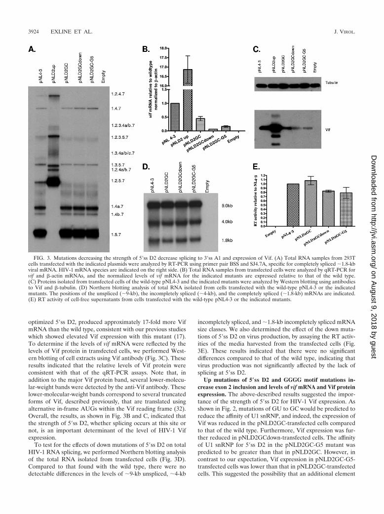
optimized 5�ss D2, produced approximately 17-fold more VifmRNA than the wild type, consistent with our previous studieswhich showed elevated Vif expression with this mutant (17).To determine if the levels of vif mRNA were reflected by thelevels of Vif protein in transfected cells, we performed West-ern blotting of cell extracts using Vif antibody (Fig. 3C). Theseresults indicated that the relative levels of Vif protein wereconsistent with that of the qRT-PCR assays. Note that, inaddition to the major Vif protein band, several lower-molecu-lar-weight bands were detected by the anti-Vif antibody. Theselower-molecular-weight bands correspond to several truncatedforms of Vif, described previously, that are translated usingalternative in-frame AUGs within the Vif reading frame (32).Overall, the results, as shown in Fig. 3B and C, indicated thatthe strength of 5�ss D2, whether splicing occurs at this site ornot, is an important determinant of the level of HIV-1 Vifexpression.
To test for the effects of down mutations of 5�ss D2 on totalHIV-1 RNA splicing, we performed Northern blotting analysisof the total RNA isolated from transfected cells (Fig. 3D).Compared to that found with the wild type, there were nodetectable differences in the levels of �9-kb unspliced, �4-kb
incompletely spliced, and �1.8-kb incompletely spliced mRNAsize classes. We also determined the effect of the down muta-tions of 5�ss D2 on virus production, by assaying the RT activ-ities of the media harvested from the transfected cells (Fig.3E). These results indicated that there were no significantdifferences compared to that of the wild type, indicating thatvirus production was not significantly affected by the lack ofsplicing at 5�ss D2.
Up mutations of 5�ss D2 and GGGG motif mutations in-crease exon 2 inclusion and levels of vif mRNA and Vif proteinexpression. The above-described results suggested the impor-tance of the strength of 5�ss D2 for HIV-1 Vif expression. Asshown in Fig. 2, mutations of GU to GC would be predicted toreduce the affinity of U1 snRNP, and indeed, the expression ofVif was reduced in the pNLD2GC-transfected cells comparedto that of the wild type. Furthermore, Vif expression was fur-ther reduced in pNLD2GCdown-transfected cells. The affinityof U1 snRNP for 5�ss D2 in the pNLD2GC-G5 mutant waspredicted to be greater than that in pNLD2GC. However, incontrast to our expectation, Vif expression in pNLD2GC-G5-transfected cells was lower than that in pNLD2GC-transfectedcells. This suggested the possibility that an additional element
FIG. 3. Mutations decreasing the strength of 5�ss D2 decrease splicing to 3�ss A1 and expression of Vif. (A) Total RNA samples from 293Tcells transfected with the indicated plasmids were analyzed by RT-PCR using primer pair BSS and SJ4.7A, specific for completely spliced �1.8-kbviral mRNA. HIV-1 mRNA species are indicated on the right side. (B) Total RNA samples from transfected cells were analyzed by qRT-PCR forvif and �-actin mRNAs, and the normalized levels of vif mRNA for the indicated mutants are expressed relative to that of the wild type.(C) Proteins isolated from transfected cells of the wild-type pNL4-3 and the indicated mutants were analyzed by Western blotting using antibodiesto Vif and �-tubulin. (D) Northern blotting analysis of total RNA isolated from cells transfected with the wild-type pNL4-3 or the indicatedmutants. The positions of the unspliced (�9-kb), the incompletely spliced (�4-kb), and the completely spliced (�1.8-kb) mRNAs are indicated.(E) RT activity of cell-free supernatants from cells transfected with the wild-type pNL4-3 or the indicated mutants.
3924 EXLINE ET AL. J. VIROL.
on August 9, 2018 by guest
http://jvi.asm.org/
Dow
nloaded from

or elements may regulate the usage of 5�ss D2. Previous studieshave shown that a GGGG motif proximal to the 5�ss of thebrain region-specific C1 cassette exon of the N-methyl-D-aspartic acid (NMDA)-type glutamate receptor NR1 subunit(GRIN1) transcript acts to silence inclusion of the C1 exon (9).Several GGGG motifs in the intron sequences upstream anddownstream of alternative exon 4 of the HLA-DQB1 gene playa role in the differential inclusion of exon 3 into mRNA (15).In the HIV-1 genome, there is a GGGG motif which overlapsthe last base of the U1 snRNP binding site and is just proximalto the 5�ss D2 sequence. In the pNLD2GC-G5 mutant, thenumber of G’s was increased, resulting in a GGGGG se-quence.
We tested for the effects of the potential 5�ss-proximal GGGGsilencer motif on splicing and on Vif expression by creating themutations shown in Fig. 2. As shown in Fig. 4A, mutations withinthe first three bases of the potential silencer motif (pNLG4M,pNLG4968T, pNLG4969T, and pNLG4970T) resulted in the in-creased inclusion of exon 2, as evidenced by increased amounts ofthe RNA species 1.2.5.7, 1.2.4a/b.7, and 1.2.4.7 relative to those ofthe wild-type pNL4-3. These mutants demonstrated significantlyless exon 2 inclusion than pNLD2up, which is predicted to inac-tivate the putative GGGG silencer and increase the affinity of D2for U1 snRNP. On the other hand, a G-to-T mutation of the lastbase of the GGGG motif (pNLG4971T) had little effect on exon2 inclusion. The effects of the GGGG mutations were also seenwith studies of vif mRNA levels (Fig. 4B) and Vif protein expres-sion (Fig. 4C). Mutants with changes within the GGGG motif
that increased exon inclusion exhibited vif mRNA levels two- tofourfold higher than the wild type but considerably lower thanpNLD2up, which in these experiments expressed vif mRNAlevels approximately 15-fold higher than the wild type. ThepNLG4971T mutant, on the other hand, did not demonstrate anelevation of Vif expression relative to that of the wild type. In-terestingly, the pNLD2-G5 mutant, in which the length of theGGGG motif was increased but in which the predicted affinity of5�ss D2 for U1 snRNP was also increased, showed elevated in-clusion of exon 2 (Fig. 4A) and increased Vif expression (Fig. 4Band C).
We next compared the effects of the GGGG mutations andthe up mutations of 5�ss D2 on total HIV-1 splicing by usingNorthern blotting analysis of the total RNA from transfectedcells (Fig. 4D) and analysis of virus production, by RT assays ofcell supernatants (Fig. 4E). We have previously reported thatpNLD2up demonstrates an excessive splicing phenotype char-acterized by greatly reduced levels of unspliced viral RNA andreduced levels of Gag protein (17). In the experiment shown inFig. 4D, we found that in addition to the normal band corre-sponding to the wild-type �4-kb mRNA band, a slower-mi-grating band was detected that corresponded to the size ex-pected for vif mRNA. This upper band was not detectable inthe wild-type RNA because of the normally low abundance ofvif mRNA. The pNLG4M mutant, which inactivates theGGGG motif but does not increase the predicted affinity of D2for U1 snRNP, and the pNLD2-G5 mutant, which is predictedto increase the affinity of D2 for U1 snRNP but not impinge on
FIG. 4. Mutations increasing the strength of 5�ss D2 or in a GGGG motif proximal to 5�ss D2 increase splicing at 3�ss A1 and the expressionof Vif while decreasing virus production. (A) Total RNA samples from 293T cells transfected with the indicated plasmids were analyzed byRT-PCR using primers specific for completely spliced �1.8-kb viral mRNA. HIV-1 mRNA species are indicated on the right side. (B) Total RNAsamples from transfected cells were analyzed by qRT-PCR for vif and �-actin mRNAs, and the normalized levels of vif mRNA for the indicatedmutants are expressed relative to that of the wild type. (C) Proteins isolated from transfected cells of the wild-type pNL4-3 and the indicatedmutants were analyzed by Western blotting using antibodies to Vif and �-tubulin. (D) Northern blotting of total RNA isolated from cellstransfected with the wild-type pNL4-3 and the indicated mutants. The positions of unspliced (�9-kb), incompletely spliced (vif, �4-kb), andcompletely spliced (�1.8-kb) mRNAs are indicated. (E) RT activity of cell-free supernatants from cells transfected with the wild-type pNL4-3 orthe indicated mutants.
VOL. 82, 2008 REGULATION OF HIV-1 Vif mRNA SPLICING 3925
on August 9, 2018 by guest
http://jvi.asm.org/
Dow
nloaded from

the GGGG motif, also demonstrated increased levels of �4-kbvif mRNAs. In addition, these two mutants produced virusparticles at levels between that of the wild type and that of thepNLD2up mutant, whose virus particle production in this ex-periment was approximately eightfold reduced compared tothat of the wild type (Fig. 4E). We concluded from theseresults that the level of splicing at 3�ss A1 is determined byboth the affinity of U1 snRNP for 5�ss D2 and the GGGGsilencer 3� proximal to 5�ss D2. Both exon 2 inclusion and vifmRNA levels are increased by mutations within the 5�ss D2that increase its affinity for U1 snRNP and by mutations of theGGGG silencer motif. Increased splicing at 3�ss A1 is concom-itant with reduced virus production, and the extent of thereduction correlates with the extent to which the viral RNA isspliced.
An ESE binding selectively to SRp75 promotes exon 2 in-clusion and levels of vif mRNA and Vif protein expression. Asdiscussed above, several ESS and ESE elements that promoteor inhibit RNA splicing have previously been identified withinthe HIV-1 genome. Therefore, we tested for the possibility that
exon 2 contained additional cis elements that contributed tothe regulation of splicing at 3�ss A1. Preliminary experimentsperformed in the context of a gag/pol-deleted pNL4-3 plasmidindicated that point mutations and deletions within the 5�-proximal region of exon 2 resulted in a decreased inclusion ofexon 2 into HIV-1 mRNAs (data not shown). Based on thesedata, mutations downstream of 3�ss A1 at nt 4923 and 4925 inpNL4-3 were introduced to create the pNL-ESEVifm mutant.This mutant was transfected into 293T cells, and at 24 to 48 hposttransfection, cells and cell supernatants were harvested.RT-PCR was used to detect viral RNA species in the mutant�1.8-kb HIV-1 mRNA size class, and this result was comparedto that of the wild-type pNL4-3 and pNLD2GC (Fig. 5A). ThemRNA species profile of pNL-ESEVifm was similar to that ofpNLD2GC, indicating an almost complete absence of exon 2inclusion. A decrease in exon 2 inclusion to undetectable levelswas also observed with the �4.0-kb mRNA size class (data notshown). Analysis of total RNA from pNL-ESEVif by qRT-PCR indicated that the vif mRNA was present at a level thatwas approximately 3% of that of the wild type (Fig. 5B). Pro-
FIG. 5. A mutation within exon 2 decreases splicing to 3�ss A1 and expression of Vif. (A) Total RNA samples from 293T cells transfected withthe indicated plasmids were analyzed by RT-PCR using primers specific for completely spliced �1.8-kb mRNA. HIV-1 mRNA species areindicated on the right. (B) Total RNA samples from transfected cells were analyzed by qRT-PCR for vif and �-actin mRNAs, and the normalizedlevels of vif mRNA for the indicated mutants are expressed relative to that of the wild type. (C) Proteins isolated from transfected cells of thewild-type pNL4-3 and the indicated mutants were analyzed by Western blotting using antibodies to Vif and �-tubulin. (D) Northern blotting oftotal RNA isolated from cells transfected with the wild-type pNL4-3 and the indicated mutants. The positions of unspliced (�9-kb), incompletelyspliced (�4-kb), and completely spliced (�1.8-kb) mRNAs are indicated on the right. (E) Reverse transcriptase activity of cell-free supernatantsfrom cells transfected with the wild-type pNL4-3 or the indicated mutants.
3926 EXLINE ET AL. J. VIROL.
on August 9, 2018 by guest
http://jvi.asm.org/
Dow
nloaded from
tein expression, as determined by Western blotting analysis ofcell lysates, detected little or no Vif and was in agreement withthe qRT-PCR data (Fig. 5C).
Analysis of pNL-ESEVifm RNA by Northern blotting indi-cated that the relative amounts of �9-kb, �4-kb, and �1.8-kbsize class mRNAs were similar to those of the wild type (Fig.5D). Furthermore, mutant virus particle production was nearwild-type levels, as determined by RT assays of transfected cellsupernatants (Fig. 5E). Thus, the mutations in pNL-ESEVifm,except for the reduced levels of Vif mRNA and Vif protein, didnot affect virus replication.
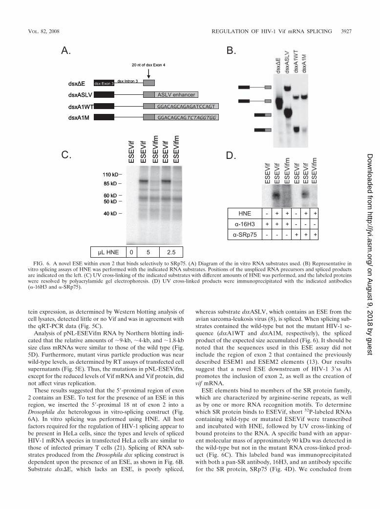
These results suggested that the 5�-proximal region of exon2 contains an ESE. To test for the presence of an ESE in thisregion, we inserted the 5�-proximal 18 nt of exon 2 into aDrosophila dsx heterologous in vitro-splicing construct (Fig.6A). In vitro splicing was performed using HNE. All hostfactors required for the regulation of HIV-1 splicing appear tobe present in HeLa cells, since the types and levels of splicedHIV-1 mRNA species in transfected HeLa cells are similar tothose of infected primary T cells (21). Splicing of RNA sub-strates produced from the Drosophila dsx splicing construct isdependent upon the presence of an ESE, as shown in Fig. 6B.Substrate dsx�E, which lacks an ESE, is poorly spliced,
whereas substrate dsxASLV, which contains an ESE from theavian sarcoma-leukosis virus (8), is spliced. When splicing sub-strates contained the wild-type but not the mutant HIV-1 se-quence (dsxA1WT and dsxA1M, respectively), the splicedproduct of the expected size accumulated (Fig. 6). It should benoted that the sequences used in this ESE assay did notinclude the region of exon 2 that contained the previouslydescribed ESEM1 and ESEM2 elements (13). Our resultssuggest that a novel ESE downstream of HIV-1 3�ss A1promotes the inclusion of exon 2, as well as the creation ofvif mRNA.
ESE elements bind to members of the SR protein family,which are characterized by arginine-serine repeats, as wellas by one or more RNA recognition motifs. To determinewhich SR protein binds to ESEVif, short 32P-labeled RNAscontaining wild-type or mutated ESEVif were transcribedand incubated with HNE, followed by UV cross-linking ofbound proteins to the RNA. A specific band with an appar-ent molecular mass of approximately 90 kDa was detected inthe wild-type but not in the mutant RNA cross-linked prod-uct (Fig. 6C). This labeled band was immunoprecipitatedwith both a pan-SR antibody, 16H3, and an antibody specificfor the SR protein, SRp75 (Fig. 4D). We concluded from
FIG. 6. A novel ESE within exon 2 that binds selectively to SRp75. (A) Diagram of the in vitro RNA substrates used. (B) Representative invitro splicing assays of HNE was performed with the indicated RNA substrates. Positions of the unspliced RNA precursors and spliced productsare indicated on the left. (C) UV cross-linking of the indicated substrates with different amounts of HNE was performed, and the labeled proteinswere resolved by polyacrylamide gel electrophoresis. (D) UV cross-linked products were immunoprecipitated with the indicated antibodies(�-16H3 and �-SRp75).
VOL. 82, 2008 REGULATION OF HIV-1 Vif mRNA SPLICING 3927
on August 9, 2018 by guest
http://jvi.asm.org/
Dow
nloaded from

our results that ESEVif specifically binds to SRp75 and notto other SR proteins.
ESE, GGGG, and 5�ss D2 act competitively to determinesplicing at 3�ss A1 and the level of Vif expression. To deter-mine the relative strengths of the three elements regulatingsplicing at 3�ss A1 and the level of Vif expression (downstreamsuboptimal 5�ss D2, GGGG silencer, and ESEVif), we createddouble mutants in the pNL4-3 backbone, with both the ESEVifmutation (Em) and either the D2up or the G4 mutation(pNLEm2up and pNLEmG4M, respectively). These two dou-ble mutants were transfected into 293T cells, and the cells wereanalyzed for the Vif gene expression and virus production. Todetermine the level of exon 2 inclusion, we used RT-PCR toamplify the �1.8-kb size class mRNAs (Fig. 7A). The results ofthis experiment indicated that the inclusion of exon 2 in thepNLEm2up double mutant was elevated and did not appear tobe significantly different from than that in pNLD2up. On theother hand, the inclusion of exon 2 in the pNLEmG4M doublemutant was reduced compared to that in pNLG4M and moresimilar to that of the wild type. This was indicated by a relativedecrease in mRNA species, which include exon 2 (1.2.5.7,1.2.4a.7, 1.2.4b.7, and 1.2.4.7), and a relative increase inmRNA species, in which exon 2 is skipped (1.5.7, 1.4a.7, 1.4b.7,
and 1.4.7). The pNL-ESEVifm mutant, as shown in Fig. 5A,demonstrated little or no inclusion of exon 2 into viral mRNAs.
We next analyzed Vif expression in cells transfected with thedouble mutants. We determined vif mRNA levels by qRT-PCR(Fig. 7B). This experiment indicated that pNLEm2up pro-duced somewhat less vif mRNA (approximately 12-fold higherthan that of the wild-type) than pNLD2up (19-fold higher thanthat of the wild type). The pNLEmG4M mutant producedwild-type levels of vif mRNA compared to that of the single-GGGG silencer pNLG4M mutant, which, in these experi-ments, produced vif mRNA at a level approximately fourfoldhigher than that of the wild type. The pNL-ESEVifm mutant,as shown in Fig. 5B, produced very little vif mRNA. As shownin Fig. 7C, the relative levels of Vif protein in extracts fromtransfected cells were consistent with the relative levels ofmRNA seen in Fig. 7B.
We next determined the effect of the ESE double mutationson unspliced, incompletely spliced, and completely spliced vi-ral RNA levels, by Northern blotting (Fig. 7D). As describedabove and elsewhere, little unspliced mRNA was present incells transfected with the pNLD2up mutant. In addition, asshown in Fig. 4D, a prominent mRNA band was present whichmigrated slightly slower than the 4.0-kb mRNA band and at
FIG. 7. Negative and positive splicing elements act competitively to affect splicing to 3�ss A1, the expression of Vif, and virus production.(A) Total RNA samples from 293T cells transfected with the indicated plasmids were analyzed by RT-PCR using primers specific for completelyspliced �1.8-kb viral mRNA. HIV-1 mRNA species are indicated on the right side. (B) Total RNA samples from transfected cells were analyzedby qRT-PCR for vif and �-actin mRNAs, and the normalized levels of vif mRNA for the indicated mutants are expressed relative to that of thewild type. (C) Proteins isolated from transfected cells of the wild-type pNL4-3 and the indicated mutants were analyzed by Western blotting usingantibodies to Vif and �-tubulin. (D) Northern blotting of total RNA isolated from cells transfected with the wild-type pNL4-3 and the indicatedmutants. The positions of unspliced (�9-kb), incompletely spliced (vif, �4-kb), and completely spliced (�1.8-kb) mRNAs are indicated. (E) RTactivity of cell-free supernatants from cells transfected with the wild-type pNL4-3 or the indicated mutants.
3928 EXLINE ET AL. J. VIROL.
on August 9, 2018 by guest
http://jvi.asm.org/
Dow
nloaded from

the size expected for single-spliced vif mRNA. Cells trans-fected by the pNLEm2up double mutant also exhibited aprominent vif mRNA band. However, in these cells, there wasa higher level of unspliced mRNA than in pNLD2up-trans-fected cells. In cells transfected with the pNLG4M GGGGsilencer mutant, a distinct vif mRNA band was present, as wellas reduced levels of unspliced mRNA, compared to that ofthe wild type as shown in Fig. 4D. On the other hand, a vifmRNA band was not detected in cells transfected with thepNLEmG4M double mutant. Furthermore, these cells ap-peared to accumulate an increased level of unspliced mRNAcomparable to that of the wild type. In contrast to the datashown in Fig. 4D, the intensity of the 1.8-kb mRNA bands ofall the mutant RNA samples seen in Fig. 7D appeared to bereduced relative to that of the 4.0-kb mRNA bands. This dif-ference was not reproduced in other repetitions of this exper-iment.
Finally, we compared the effect of the ESE double mutantson virus production to that of the single mutants by performingRT assays of supernatants from the transfected cells (Fig. 7E).The pNLEm2up double mutant produced significantly higherlevels of particles than the pNLD2up mutant, consistent withthe higher relative accumulation of 9-kb unspliced RNA in thepNLEm2up-transfected cells than in the pNLD2up-transfectedcells, as shown in Fig. 7D. Similarly, the pNLEmG4M doublemutant produced higher levels of particles than pNLG4M,consistent with the higher relative accumulation of unsplicedmRNA observed for pNLEmG4M-transfected cells than forpNLG4M-transfected cells, as shown in Fig. 7D.
From the results with the ESEVifm/D2up and the ESEVifm/D2G4M double mutants, we concluded that the suboptimal5�ss D2, the GGGG silencer, and ESEVif enhancer act com-petitively to determine the level of splicing at 3�ss A1. In thisway, the levels of exon 2 inclusion and vif mRNA and Vifprotein expression are regulated. In addition, the overall level
of HIV-1 splicing is maintained to allow the accumulation ofunspliced mRNA compatible with optimal virus production.
DISCUSSION
Based on the results of this study and previous results, wepropose the model shown in Fig. 8 for the regulation of splicingat 3�ss A1. To date, three different positively acting exon 2ESEs have been identified. Two 5�-distal ESEs (ESEM1 andESEM2) contain the sequence UGGAAAG (13). In this re-port, we have shown evidence for a third ESE (ESEVif) withinthe 5�-proximal region of exon 2. ESEM1 and ESEM2 havebeen reported to facilitate exon 2 inclusion into mRNAs butnot to affect the expression of vif mRNA (13). In contrast, weshowed in this report that ESEVif promotes the production ofvif mRNA, as well as exon 2 inclusion. We have also detecteda negatively acting GGGG silencer element proximal to 5�ssD2 which represses both exon 2 inclusion and vif mRNA pro-duction. A third splicing element is the suboptimal down-stream 5�ss D2 itself; the affinity of this splice site for U1snRNP determines its influence on splicing at 3�ss A1. We haveshown that the ESEVif, the GGGG silencer element, anddownstream 5�ss act competitively to regulate vif mRNA levels.
In the model shown in Fig. 8, we propose that the regulationof splicing at 3�ss A1 involves the interaction of multiple cel-lular factors binding to the cis splicing elements within the viralgenome. It has been reported that ESEM1 and ESEM2 bind tothe SR protein ASF/SF2 (13). We have shown herein thatESEVif specifically binds to the SR protein SRp75, and thissuggests that this protein is responsible for the activation ofsplicing at 3�ss A1. It has been previously shown by Han et al.(9) that the hnRNP H and F proteins bind to GGGG motifsproximal to 5�ss of the C1 exon of the GRIN1 transcript butthat, in this context, these proteins appear to act as antagonistsof splicing silencing rather than as silencers themselves. This
FIG. 8. Proposed model for vif mRNA splicing regulation. The results presented in this study and in a previous study (13) indicate that splicingat 3�ss A1 and 5�ss D2 is regulated at an early step of splicing by positively acting ESEs that promote splicing (dashed lines) and a negatively actingGGGG silencer (solid line) that inhibits splicing. In addition, the wild-type 5�ss D2 is suboptimal; mutagenesis that lowers the affinity of 5�ss D2for U1 snRNP inhibits splicing at 3�ss A1 (solid line), whereas mutagenesis that increases the affinity of 5�ss D2 for U1 snRNP promotes splicingat 3�ss A1 (dashed line). ESEM1 and ESEM2 have been previously reported to be binding sites for the SR protein SF2/ASF (13). ESEVif bindsselectively to the SRp75 protein. The silencing activity of the GGGG motif is thought to be mediated by a cellular factor or factors that have notyet been identified. We cannot conclude yet whether the major effects of the GGGG motif and ESEVif are on 5�ss recognition, 3�ss recognition,or recognition of both splice sites. The overall outcome of these competing positive and negative factors binding downstream of 3�ss A1 may beto determine the binding affinity of U2AF65/U2AF35 to the polypyrimidine tract (Py)n and 3�ss A1, which is one of the earliest steps in splicing.
VOL. 82, 2008 REGULATION OF HIV-1 Vif mRNA SPLICING 3929
on August 9, 2018 by guest
http://jvi.asm.org/
Dow
nloaded from

result suggested that the 5�ss proximal GGGG motif may me-diate silencing by binding to another cellular protein or pro-teins. It was speculated by Han et al. that hnRNP A1 maymediate silencing by cooperative binding to GGGG motifs andUAGG ESS elements within the C1 exon of the GRIN1 tran-script (9). This does not appear to be the case with the HIV-15�ss proximal GGGG silencer, since there are no UAGG se-quences within HIV-1 exon 2, and to date, we have not iden-tified any other sequences within HIV-1 noncoding exon 2 thatwould allow such cooperative interactions with hnRNP A1.
We showed that G-to-T mutations of the second and third Gresidue of the GGGG motif (G4969T and G4970T) resulted inincreased exon 2 inclusion and a three- to fourfold increase invif mRNA levels relative to that of the wild type. A G-to-Tmutation of the first G residue resulted in a somewhat lowerincrease of vif mRNA level, whereas a G-to-T mutation of thefourth G residue of the GGGG motif had little or no effect onexon 2 inclusion or vif mRNA level. It should be noted thatmutations of the first residue of the GGGG motif are alsopredicted to change the affinity of 5�ss D2 for U1 snRNP.Therefore, the contribution of each of the two elements to thesplicing changes produced by these mutations cannot be dis-cerned. It has previously been shown that several GGGG mo-tifs in the intron sequences upstream and downstream of thealternative exon 4 of the HLA-DQB1 gene play a role inthe differential inclusion of exon 4 into mRNA. Mutations ofthe upstream GGGG motifs result in decreased exon 4 inclu-sion, suggesting that they act as intronic splicing enhancers.Alternatively, these mutations may change the structural con-formation necessary for splicing. In contrast, mutations of thedownstream 5�ss-proximal GGGG motif result in increasedsplicing, suggesting that in this case, the motif acts as a splicingsilencer. It was found in this study that point mutations withinthe second and third G residues had the most effect on splicingenhancement, but similar mutations in the 5�ss-proximalGGGG silencer were not tested (15). In the case of the brainregion-specific C1 cassette exon of the GRIN1 transcript, mu-tation of the central two G residues of the 5�-proximal GGGGmotif was shown to inactivate the silencer activity, but theeffect of mutating only the first or fourth G residues of theGGGG motif was not tested (9).
Our studies indicate that, in general, the levels of vif mRNAin the absence of changes in ESEVif and the GGGG silencervary according to the predicted binding affinity of U1 snRNPfor 5�ss D2. An exception to this general rule is thepNLD2GC-G5 mutant, which would be expected to expresslevels of vif mRNA higher than those of pNLD2GC, based onits predicted U1 snRNP binding. The anomalous behavior ofpNLD2GC-G5 may be explained by the increased length of theGGGG motif, resulting in increased silencer activity relative tothat of U1 snRNP binding. Interestingly, this same G5 mutantin the context of the wild-type GU sequence (pNLD2-G5)demonstrated an elevated splicing phenotype, as expectedfrom the predicted increased affinity of the mutated 5�ss D2 toU1 snRNP. This suggests that in the context of pNLD2-G5, U1snRNP binding is dominant over the extended GGGG motifcreated by the G5 nt change. Further characterization of theGGGG silencer and its hypothetical binding protein(s) is re-quired to fully understand this phenomenon.
It is possible that the 5�ss mutations could affect HIV-1
mRNA stability in addition to their effects on RNA splicing.Indeed, it has been shown previously that in the context of asubgenomic HIV-1 env expression construct containing only5�ss D5 and 3�ss A7, mutations inactivating 5�ss D5 result indestabilization of the unspliced env RNA (12). In the contextof the complete HIV-1 genome, mutations that inactivated anormally cryptic 5�ss within the gag/pol gene resulted in adramatic reduction in the steady-state level of unspliced RNA(16). In our experiments, we have not seen a reduction insteady-state levels of unspliced RNA as a result of inactivating5�ss D2. We have also not detected significant differences be-tween the wild type and the 5�ss D2 mutants in terms of therate of decay of vif mRNA after the addition of the transcrip-tion inhibitor actinomycin D (Z. Feng and C. M. Stoltzfus,unpublished data). Thus, our results suggest that the primaryeffects of the mutations we have studied are on HIV-1 mRNAsplicing and not on RNA stability.
Both the removal of repression of splicing at 3�ss A1 byinactivation of the GGGG silencer element and the increase inthe strength of 5�ss D2 resulted in decreased virus production.This presumably is due to a skewing of the balance of normalHIV-1 splicing, resulting in a relative decrease in the level ofunspliced mRNA and a consequent decrease in the level ofGag and Gag-Pol proteins as we have previously shown for theD2up mutant (17) and the D2A3 mutant, which has a G-to-Achange at position �3 of D2 (19). The negative effects of theGGGG mutations on virus production can be partially or com-pletely abrogated by second-site mutations within ESEVif.This is further evidence that the splicing elements act compet-itively to allow optimal levels of virus replication.
The results presented here and elsewhere have indicatedthat the presence or absence of noncoding exon 2 or 3 withinthe 5� leaders of viral mRNAs does not significantly affect virusproduction or mRNA stability or the expression of viralmRNAs (17, 18). Thus, the inclusion of exon 2 and exon 3 intosome of the HIV-1 mRNAs during virus replication may be aby-product and reflect the necessity of a functional down-stream 5�ss D2 and D3 for the expression of optimum levels ofVif and Vpr, respectively. In the case of Vif, optimal levels ofexpression in infected cells may be necessary both to pre-vent the accumulation and packaging of APOBEC3G andAPOBEC3F deoxycytidine deaminases (14, 20, 25, 34) and toavoid the accumulation of excess Vif, which has been shown toinhibit virus protein processing and virus replication (2).
It is curious that the level of splicing at 3�ss A1 is regulatedby such a complex set of splicing elements, which involve threedifferent ESEs, the GGGG silencer, and a suboptimal down-stream 5�ss. The importance of these three elements is sug-gested by their sequence conservation in all HIV-1 clades.There are several possibilities to explain the evolution of thisseemingly cumbersome system to regulate vif splicing. First,exon 2 and the flanking vif mRNA intron sequences overlapthe integrase protein reading frame. Because of this overlap,the HIV-1 sequence must be compatible with both the require-ments for optimum Vif levels and for integrase function. Thus,the necessity of both functions for efficient virus replicationmay require multiple positive and negative cis splicing ele-ments to achieve the necessary level of splicing to produceoptimal levels of vif mRNA. Second, the presence of multiplesplicing elements may be important to allow regulation of the
3930 EXLINE ET AL. J. VIROL.
on August 9, 2018 by guest
http://jvi.asm.org/
Dow
nloaded from
levels of Vif in different cell types or at different stages of viruslife cycle. This regulation may be affected by the relative con-centrations of the RNA binding proteins that bind to the cog-nate negative and positive RNA splicing elements.
ACKNOWLEDGMENTS
We thank Dibyakanti Mandal for critical evaluation of the manu-script. We acknowledge the NIH AIDS Research and Reference Re-agent Program for supplying HIV-1-related reagents. We thank MaxCaputi, Florida Atlantic University, for the dsx splicing template plas-mids and Tom Hope, Northwestern University School of Medicine, forthe pCMV110 �-galactosidase expression plasmid.
HeLa cells were obtained from the National Cell Culture Center,which is sponsored by the National Center for Research Resources ofthe NIH. Monoclonal antibody E7 was obtained from the Develop-ment Studies Hybridoma Bank, developed under the auspices of theNICHD and maintained by the University of Iowa, Department ofBiological Sciences, Iowa City, IA.
This research was supported by PHS grant AI36073 from the Na-tional Institute of Allergy and Infectious Diseases.
C.M.E. was supported by predoctoral training grant T32AI007533from the National Institute of Allergy and Infectious Diseases.
REFERENCES
1. Adachi, A., H. E. Gendelman, S. Koenig, T. Folks, R. Willey, A. Rabson, andM. A. Martin. 1986. Production of acquired immunodeficiency syndrome-associated retrovirus in human and nonhuman cells transfected with aninfectious molecular clone. J. Virol. 59:284–291.
2. Akari, H., M. Fujita, S. Kao, M. A. Khan, M. Shehu-Xhilaga, A. Adachi, andK. Strebel. 2004. High level expression of human immunodeficiency virustype-1 Vif inhibits virus infectivity by modulating proteolytic processing ofthe Gag precursor at the p2/nucleocapsid processing site. J. Biol. Chem.279:12355–12362.
3. Amendt, B. A., D. Hesslein, L.-J. Chang, and C. M. Stoltzfus. 1994. Presenceof negative and positive cis-acting RNA splicing elements within and flankingthe first tat coding exon of the human immunodeficiency virus type 1. Mol.Cell Biol. 14:3960–3970.
4. Amendt, B. A., Z.-H. Si, and C. M. Stoltzfus. 1995. Presence of exon splicingsilencers within HIV-1 tat exon 2 and tat/rev exon 3: evidence for inhibitionmediated by cellular factors. Mol. Cell. Biol. 15:4606–4615.
5. Berget, S. M. 1995. Exon recognition in vertebrate splicing. J. Biol. Chem.270:2411–2414.
6. Bilodeau, P. S., J. K. Domsic, A. Mayeda, A. R. Krainer, and C. M. Stoltzfus.2001. RNA splicing at human immunodeficiency virus type 1 3� splice site A2is regulated by binding of hnRNP A/B proteins to an exonic splicing silencerelement. J. Virol. 75:8487–8497.
7. Butsch, M., and K. Boris-Lawrie. 2002. Destiny of unspliced retroviral RNA:ribosome and/or virion? J. Virol. 76:3089–3094.
8. Fu, X.-D., R. A. Katz, A. M. Skalka, and T. Maniatis. 1991. The role ofbranchpoint and 3�-exon sequences in the control of balanced splicing ofavian retrovirus RNA. Genes Dev. 5:211–220.
9. Han, K., G. Yeo, P. An, C. B. Burge, and P. J. Grabowski. 2005. A combi-natorial code for splicing silencing: UAGG and GGGG motifs. PLoS Biol.3:e158.
10. Hoffman, B. E., and P. J. Grabowski. 1992. U1 snRNP targets an essentialsplicing factor, U2AF65, to the 3� splice site by a network of interactionsspanning the exon. Genes Dev. 6:2554–2568.
11. Jacquenet, S., A. Mereau, P. S. Bilodeau, L. Damier, C. M. Stoltzfus, and C.Branlant. 2001. A second exon splicing silencer within human immunodefi-ciency virus type 1 tat exon 2 represses splicing of Tat mRNA and bindsprotein hnRNP H. J. Biol. Chem. 276:40464–40475.
12. Kammler, S., C. Leurs, M. Freund, J. Krummheuer, K. Seidel, T. O. Tange,M. K. Lund, J. Kjems, A. Scheid, and H. Schaal. 2001. The sequencecomplementarity between HIV-1 5� splice site SD4 and U1 snRNA deter-mines the steady-state level of an unstable env pre-mRNA. RNA 7:421–434.
13. Kammler, S., M. Otte, I. Hauber, J. Kjems, J. Hauber, and H. Schaal. 2006.The strength of the HIV-1 3� splice sites affects Rev. function. Retrovirology3:89.
14. Kao, S., M. A. Khan, E. Miyagi, R. Plishka, A. Buckler-White, and K.Strebel. 2003. The human immunodeficiency virus type 1 Vif protein reducesintracellular expression and inhibits packaging of APOBEC3G (CEM15), acellular inhibitor of virus infectivity. J. Virol. 77:11398–11407.
15. Kralovicova, J., and I. Vorechovsky. 2006. Position-dependent repressionand promotion of DQB1 intron 3 splicing by GGGG motifs. J. Immunol.176:2381–2388.
16. Lutzelberger, M., L. S. Reinert, A. T. Das, B. Berkhout, and J. Kjems. 2006.A novel splice donor site in the gag-pol gene is required for HIV-1 RNAstability. J. Biol. Chem. 281:18644–18651.
17. Madsen, J. M., and C. M. Stoltzfus. 2006. A suboptimal 5� splice sitedownstream of HIV-1 splice site A1 is required for unspliced viral mRNAaccumulation and efficient virus replication. Retrovirology 3:10.
18. Madsen, J. M., and C. M. Stoltzfus. 2005. An exonic splicing silencer down-stream of 3� splice site A2 is required for efficient human immunodeficiencyvirus type 1 replication. J. Virol. 79:10478–10486.
19. Mandal, D., Z. Feng, and C. M. Stoltzfus. 2008. Gag-processing defect ofhuman immunodeficiency virus type 1 integrase E246 and G247 mutants iscaused by activation of an overlapping 5� splice site. J. Virol. 82:1600–1604.
20. Marin, M., K. M. Rose, S. L. Kozak, and D. Kabat. 2003. HIV-1 Vif proteinbinds the editing enzyme APOBEC3G and induces its degradation. Nat.Med. 9:1398–1403.
21. Purcell, D. F. J., and M. A. Martin. 1993. Alternative splicing of humanimmunodeficiency virus type 1 mRNA modulates viral protein expression,replication and infectivity. J. Virol. 67:6365–6378.
22. Robberson, B. L., G. J. Cote, and S. M. Berget. 1990. Exon definition mayfacilitate splice site selection in RNAs with multiple exons. Mol. Cell. Biol.10:84–94.
23. Sambrook, J., E. F. Fritsch, and T. Maniatis. 1989. Molecular cloning: alaboratory manual, 2nd ed. Cold Spring Harbor Laboratory Press, ColdSpring Harbor, NY.
24. Serra, M., and D. Turner. 1995. Predicting thermodynamic properties ofRNA. Methods Enzymol. 259:242–261.
25. Sheehy, A. M., N. C. Gaddis, and M. H. Malim. 2003. The antiretroviralenzyme APOBEC3G is degraded by the proteasome in response to HIV-1Vif. Nat. Med. 4:1397–1400.
26. Si, Z.-H., D. Rauch, and C. M. Stoltzfus. 1998. The exon splicing silencer inhuman immunodeficiency virus type 1 Tat exon 3 is bipartite and acts earlyin spliceosome assembly. Mol. Cell. Biol. 18:5404–5413.
27. Staffa, A., and A. Cochrane. 1995. Identification of positive and negativesplicing regulatory elements within the terminal tat/rev exon of HIV-1. Mol.Cell. Biol. 15:4597–4605.
28. Stoltzfus, C. M., and J. M. Madsen. 2006. Role of viral splicing elements andcellular RNA binding proteins in regulation of HIV-1 alternative RNAsplicing. Curr. HIV Res. 4:43–55.
29. Tanaka, K., A. Watakabe, and Y. Shimura. 1994. Polypurine sequenceswithin a downstream exon function as a splicing enhancer. Mol. Cell. Biol.14:1347–1354.
30. Tange, T. O., C. K. Damgaard, S. Guth, J. Valcarcel, and J. Kjems. 2001.The hnRNP A1 protein regulates HIV-1 tat splicing via a novel intronsilencer element. EMBO J. 20:5748–5758.
31. Tian, M., and T. Maniatis. 1993. A splicing enhancer complex controlsalternative splicing of doublesex pre-mRNA. Cell 74:105–114.
32. Wang, H., A. Sakurai, B. Khamsri, T. Uchiyama, H. Gu, A. Adachi, and M.Fujita. 2005. Unique characteristics of HIV-1 Vif expression. Microbes In-fect. 7:385–390.
33. Willey, R. L., D. H. Smith, L. A. Lasky, T. S. Theodore, P. L. Earl, B. Moss,D. J. Capon, and M. A. Martin. 1988. In vitro mutagenesis identifies a regionwithin the envelope gene of the human immunodeficiency virus that iscritical for infectivity. J. Virol. 62:139–147.
34. Yu, X., Y. Yu, B. Liu, K. Luo, W. Kong, P. Mao, and X.-F. Yu. 2003. Inductionof APOBEC3G ubiquitation and degradation by an HIV-1 Vif-Cul5-SCFcomplex. Science 302:1056–1060.
35. Zahler, A. M., C. K. Damgaard, J. Kjems, and M. Caputi. 2004. SC35 andheterogeneous nuclear ribonucleoprotein A/B proteins bind to a juxtaposedexonic splicing enhancer/exonic splicing silencer element to regulate HIV-1tat exon 2 splicing. J. Biol. Chem. 279:10077–10084.
VOL. 82, 2008 REGULATION OF HIV-1 Vif mRNA SPLICING 3931
on August 9, 2018 by guest
http://jvi.asm.org/
Dow
nloaded from