differential inhibition of human immunodeficiency virus
TRANSCRIPT

PRECLINICAL STUDIES/DRUG DEVELOPMENT
Differential Inhibition of Human Immunodeficiency VirusType 1 in Peripheral Blood Mononuclear Cells and TZM-bl
Cells by Endotoxin-Mediated Chemokine and GammaInterferon Production
Anthony R. Geonnotti,1 Miroslawa Bilska,1 Xing Yuan,1 Christina Ochsenbauer,2 Tara G. Edmonds,3
John C. Kappes,2,3 Hua-Xin Liao,4 Barton F. Haynes,4 and David C. Montefiori1
Abstract
Bacterial lipopolysaccharide (endotoxin) is a frequent contaminant of biological specimens and is also known tobe a potent inducer of b-chemokines and other soluble factors that inhibit HIV-1 infection in vitro. Thoughlipopolysaccharide (LPS) has been shown to stimulate the production of soluble HIV-1 inhibitors in cultures ofmonocyte-derived macrophages, the ability of LPS to induce similar inhibitors in other cell types is poorlycharacterized. Here we show that LPS exhibits potent anti-HIV activity in phytohemagglutinin-stimulatedperipheral blood mononuclear cells (PBMCs) but has no detectable anti-HIV-1 activity in TZM-bl cells. The anti-HIV-1 activity of LPS in PBMCs was strongly associated with the production of b-chemokines from CD14-positive monocytes. Culture supernatants from LPS-stimulated PBMCs exhibited potent anti-HIV-1 activitywhen added to TZM-bl cells but, in this case, the antiviral activity appeared to be related to IFN-g rather than tob-chemokines. These observations indicate that LPS stimulates PBMCs to produce a complex array of solubleHIV-1 inhibitors, including b-chemokines and IFN-g, that differentially inhibit HIV-1 depending on the targetcell type. The results also highlight the need to use endotoxin-free specimens to avoid artifacts when assessingHIV-1-specific neutralizing antibodies in PBMC-based assays.
Introduction
Neutralizing antibody (Ab) assays for HIV-1 arewidely utilized to study the immune response in in-
fected individuals, to quantify monoclonal Ab performance,to explore viral antigenic diversity, and to evaluate vaccineimmunogens in preclinical and clinical trials. For many years,neutralizing Ab assays quantified a reduction in HIV-1 in-fection in mitogen-stimulated peripheral blood mononuclearcells (PBMCs); however, this system is both resource intensiveand difficult to standardize and validate because of substan-tial genetic and phenotypic variations in uncloned virusstocks and wide donor–donor variability in PBMCs for de-tecting neutralization.1,2 To address these concerns, and tofurther maximize assay performance, substantial resourceshave been invested in the development of a new neutralizing
Ab assay technology that utilizes molecularly cloned, Env-pseudotyped viruses and a genetically engineered cell line(TZM-bl) that contains a stable Tat-inducible luciferase (Luc)reporter gene.3–6 This latter assay has been highly standard-ized, optimized, and validated, and it allows for geneticanalysis and manipulation of clonal viruses to probe neu-tralization determinants in greater detail than before. As aresult, the TZM-bl assay and other similar assay technologies7
have been widely implemented as the primary quantitativetool to assess neutralizing Abs against HIV-1.
Recent studies have shown that several neutralizing Absexhibit much greater potency in the PBMC assay than in theTZM-bl assay.2,8–12 This has raised concern that the TZM-blassay falsely underestimates the neutralizing activity ofsome Abs. Without a clinically established and measure-able humoral immune correlate of HIV-1 protection, these
1Department of Surgery, Duke University Medical Center, Durham, North Carolina.2Department of Medicine, University of Alabama at Birmingham, Birmingham, Alabama.3Department of Pathology, University of Alabama at Birmingham, Birmingham, Alabama.4Department of Medicine, Duke University Medical Center, Durham, North Carolina.
AIDS RESEARCH AND HUMAN RETROVIRUSESVolume 26, Number 3, 2010ª Mary Ann Liebert, Inc.DOI: 10.1089=aid.2009.0186
279

differences create uncertainty as to which assay provides amore meaningful assessment of Ab-mediated HIV-1 neutral-ization.2,5,13
Multiple factors could account for the discrepancy in Abpotency between the PBMCs and TZM-bl assays. Some ofthese factors include differences in CCR5 density,14 CD4:CCR5 ratio,2 Fcg receptor expression,15 effects of cellularproteins incorporated into the viral membrane,16–19 and viralentry mechanisms20 between the two cell types. Additionally,chemokine and cytokine production may play a substantialrole in cases in which samples are contaminated with endo-toxin. Bacterial lipopolysaccharide (LPS), also known as en-dotoxin, is an essential structural component of the outermembrane of gram-negative bacteria that when released canassociate with serum carrier proteins and bind to a CD14=TLR4=MD-2 receptor complex on monocytes and macro-phages.21–25 This binding activates an innate inflammatoryresponse that leads to the production of chemokines, cyto-kines, and other unidentified soluble factors in cultures ofmonocyte-derived macrophages that inhibit R5 and X4 strainsof HIV-1 in vitro.26–29 Macrophage inflammatory protein-1a(MIP-1a), MIP-1b, and regulated on activation, normal T cellexpressed and secreted factor (RANTES) have been identifiedas the primary chemokines responsible for R5 HIV-1 inhibi-tion by LPS.28 These CCR5 ligands have been shown to blockHIV-1 infection by a combination of steric hindrance andCCR5 downregulation.29–32 LPS-induced soluble factors re-sponsible for X4 HIV-1 inhibition remain to be identified andappear to be independent of stromal cell-derived factor-1(SDF-1), interferon (IFN)-a, IFN-b, macrophage-derived che-mokine, leukemia inhibitory factor, and tumor necrosis factor(TNF)-a.29,33
Epithelial HeLa cell lines, such as TZM-bl, express TLR4but lack CD14 and MD-234,35 and thus should be resistant toLPS activation. Here we show that LPS exhibits potent anti-HIV activity in PBMCs but not in TZM-bl cells. Moreover,we show that b-chemokine production from CD14-positivemonocytes accounts for the antiviral effect of LPS in PBMCsand we demonstrate that these b-chemokines have little or noanti-HIV-1 activity in TZM-bl cells. Finally, we provide evi-dence that LPS stimulates PBMCs to produce IFN-g and thatthis cytokine is capable of inhibiting HIV-1 in TZM-bl cells butnot in PBMCs.
Materials and Methods
Cells
TZM-bl ( JC53-BL) cells were obtained from the NIH AIDSResearch and Reference Reagent Program (ARRRP) as con-tributed by John Kappes and Xiaoyun Wu. These cells are agenetically engineered HeLa cell clone that expresses CD4,CXCR4, and CCR5 and contains Tat-responsive reporter genesfor firefly luciferase (Luc) and Escherichia coli b-galactosidaseunder regulatory control of an HIV-1 long terminal repeat.36,37
TZM-bl cells were confirmed to be CD14 negative by flowcytometry (data not shown). 293T=17 cells were obtained fromthe American Type Culture Collection (catalog no. 11268).TZM-bl cells and 293T=17 cells were maintained in Dulbecco’smodified Eagle’s medium (Gibco) containing 10% heat-inactivated fetal bovine serum, 25 mM HEPES, and 50mggentamicin=ml in vented T-75 culture flasks (Corning-Costar).Cultures were incubated at 378C in a humidified 5% CO2–95%
air environment. TZM-bl cell monolayers were split 1:10 atconfluence by treatment with 0.25% trypsin, 1 mM EDTA(Invitrogen). The JC.6, JC.10, and JC.37 engineered HeLa celllines36 were a generous gift of Drs. David Kabat and EmilyPlatt and were maintained in the same manner as TZM-bl cells.
PBMCs were obtained via leukopheresis of healthy donorsand cryopreserved in 1-ml aliquots at a density of 2�107
cells=ml. Before use, aliquots of PBMCs were thawed in a roomtemperature water bath and incubated in growth medium(RPMI 1640, 20% fetal bovine serum) containing 5mg=mlphytohemagglutinin-P (PHA-P), 5% human interleukin-2(IL-2), and 50mg gentamicin=ml for 1 day at 378C in uprightT-75 flasks placed in a humidified 5% CO2=95% air environ-ment. The cells were suspended by vigorous pipetting todislodge adherent cells, washed, and placed in fresh growthmedium containing IL-2 but no PHA-P prior to use. Unlessindicated otherwise, PBMCs were used for neutralizationassays and for virus propagation immediately after this 1-daystimulation with PHA-P.
Viruses
This study utilized replication-competent infectious mo-lecular clones (IMC) of HIV-1 in which a Renilla luciferasereporter gene was inserted between env and nef, without ab-rogating nef expression. Furthermore, viruses were en-gineered such that env genes of choice can be inserted in cis inan isogenic backbone in which all other viral proteins wereexpressed, and in which the reporter gene is geneticallystable38 (T.G. Edmonds et al., unpublished observations);this type of construct is referred to as Env-IMC-LucR re-porter virus. All Env-IMC-LucR viruses included in thisstudy are based on NL4-3 and expressed the respective ecto-domains (i.e., all of gp120 and the ectodomain andmembrane-spanning domain of gp41) of the Env of choice.The cytoplasmic domain of gp41 was derived from NL4-3,thereby avoiding chimerisms in tat, rev, and vpu. These pro-viral plasmids are designated pNL-LucR.T2A-Env.ecto(wherein ‘‘Env’’ is placed by the name of the respective in-serted env gene). Details on the construction and characteris-tics of the viruses will be reported separately39 (T.G. Edmondset al., unpublished observations). The Env-IMC-LucR viruseswere generated by transfection in 293T cells of proviral DNA.Where noted, several experiments utilized Env-IMC-LucRvirus that was propagated by a subsequent passage inPBMCs. All viruses were titrated to achieve an empiricallydetermined tissue culture infectious dose (TCID) that pro-duced optimal relative luminescence units (RLU) in viruscontrol wells in the neutralization assay (>10�backgroundRLU of cell control wells without evidence of virus-inducedcytopathic effects). All viruses except those expressing WEAUenv utilize CCR5 (R5) as coreceptor; WEAU Env utilizes bothCCR5 and CXCR4 (R5=X4). BaL and SF162 Env are consid-ered Tier 1 Envs for possessing a high neutralization-sensitivephenotype when assayed with HIV-1-positive serum sam-ples, whereas WITO, WEAU, CH040, CH058, and CH077 Envare considerably less sensitive to neutralization and are con-sidered Tier 2 Envs.4 The WITO, WEAU, CH040, CH058, andCH077 envs represent the env genes of the transmitted=founder HIV-1 strains from sexual transmission in the re-spective patients40–42 (C. Ochsenbauer et al., unpublishedobservations).
280 GEONNOTTI ET AL.

LPS standards
The primary source of endotoxin used in this study was a‘‘smooth’’ phenotype LPS from E. coli O55:B5 (3.8 EU=ng)(N185, Lonza, Switzerland). This preparation was producedas Control Standard Endotoxin and is referenced against theUSP Reference Standard Endotoxin. Three other smooth LPSpreparations were used for comparison: E. coli O55:B5(3.9 EU=ng) (L6529, Sigma); E. coli 0127:B8 (1.9 EU=ng) (L4516,Sigma); and Salmonella enterica serotype typhimurium(2.2 EU=ng) (L6143, Sigma, St. Louis, MO). An Re mutantrough strain variant of S. enterica serotype typhimurium(1.7 EU=ng) (L9516, Sigma) was also tested to determine ifbacterial colony phenotype (i.e., smooth or rough) had anyeffect on antiviral activity. Prototypical LPS consists of lipid A,an oligosaccharide core component, and a highly variablepolysaccharide O-antigen. Rough strains of LPS have trun-cated, or nonexistent, O-antigen and are present in varyingproportions among different species of gram-negative bacte-ria.41 The smooth vs. rough nomenclature refers to the ap-pearance of the bacterial colonies. Because lipid A is thebioactive moiety, both smooth and rough versions of LPS arestrongly immunogenic; however, there is recent evidence thatrough LPS may potentiate a broader immune response since itrequires only a TLR-4=MD-2 complex and can signal in theabsence of CD14.43,44 All endotoxin preparations were storedin glass vials, used within 4 weeks of reconstitution, andvortexed vigorously for �15 min prior to use. Polymyxin Bsulfate was purchased from EMD Biosciences (San Diego, CA).
Recombinant chemokines and Abs
Human recombinant MIP-1a (CCL3), MIP-1a (LD78b iso-form, CCL3L1), MIP-1b (CCL4), RANTES (CCL5), IFN-g,macrophage-derived chemokine (MDC, CCL22), and affinitypurified Abs to human MIP-1a, MIP-1b, RANTES, and IFN-gwere purchased from R&D Systems (Minneapolis, MN).Quantikine ELISA kits for the detection of human MIP-1a,MIP-1b, RANTES, MDC, IFN-g, and SDF-1 (CXCL12) werealso purchased from R&D Systems. Monoclonal Ab IgG1b12was a gift from Dennis Burton (Scripps Research Institute, LaJolla, CA). Monoclonal Abs 2G12, 2F5, and 4E10 were pur-chased from Polymun Scientific GmbH (Vienna, Austria).HIVIG (human immunodeficiency virus immune globulin) isa purified IgG fraction prepared from pooled plasma ofasymptomatic, HIV-1 Ab-positive donors with CD4þ countsabove 400 cells=ml; this product is available from the NIHAIDS Research and Reference Reagent Program.
Monocyte depletion
Following 1-day PHA stimulation, PBMCs from a singledonor were depleted of monocytes by using a DynabeadsCD14þ depletion kit (Invitrogen, Carlsbad, CA) per themanufacturer’s instructions. Prior to depletion, *17% ofPBMCs were CD14þ; following depletion, <1% CD14þ cellsremained by flow cytometry (data not shown). Cells wereused immediately following depletion.
Endotoxin detection
Endotoxin levels were measured by using a commerciallyavailable limulus amoebocyte lysate (LAL) kinetic chromo-genic assay (Kinetic-QCL, Lonza, Switzerland). In kinetic LAL
assays, endotoxin activates a proenzyme in LAL that cleavesp-nitroaniline (pNA) from a colorless substrate. The amountof endotoxin present determines the rate of pNA release. Theresulting color change was continuously measured photo-metrically at 405 nm and compared to a standard curve ofknown endotoxin amounts.
Neutralization assays
Neutralization in the TZM-bl cell assay was measured as areduction in firefly luciferase (Luc) reporter gene expressionafter a single round of infection with Env-pseudotyped viru-ses as described previously.3,6,45 Briefly, virus was incubatedwith serial threefold dilutions of test sample (eight dilutionstotal) in duplicate in a total volume of 150 ml for 1 h at 378C in96-well flat-bottom culture plates. Freshly trypsinized cells(10,000 cells in 100 ml of growth medium containing 10 mg=mlDEAE dextran) were added to each well. One set of controlwells received cells plus virus (virus control) and another setreceived cells only (background control). After a 48-h incu-bation, 100 ml of cells was transferred to a 96-well black solidplate (Costar) for measurement of luminescence using theBritelite Plus Luminescence Reporter Gene Assay System(PerkinElmer Life Sciences).
Neutralization of HIV-1 in the PBMC assay was measuredas a reduction in LucR reporter gene expression after multiplerounds of virus replication. Virus was incubated with serial 3-fold dilutions of test sample (eight dilutions total) in duplicatein a total volume of 150 ml of IL-2-containing growth mediumfor 1 h at 378C in a 96-well U-bottom culture plate. One-day-old PHA-PBMCs (2�105 cells in 50ml of IL-2-containinggrowth medium) were added to each well. One set of controlwells received cells plus virus (virus control) and another setreceived cells only (background control). After a 4-day incu-bation, 100ml of cells was transferred to a 96-well white solidplates (Costar) for measurements of Renilla luciferase lumi-nescence using the ViviRen Live Cell Substrate (Promega). Forb-chemokine blocking experiments, a final concentration of3750 EU=ml of LPS was added to all wells containing testsamples.
For both assays, the inhibitory concentration (IC50 and IC80)of test samples was defined as the sample concentration atwhich RLUs were reduced by either 50% or 80% compared tovirus control wells after subtraction of background RLUs incell control wells. IC50 was used in the TZM-bl assay becauseit provides optimal sensitivity and precision of measure-ments. IC80 was used as the more reliable measure in thePBMC assay.46
Supernatant transfer experiments
To test the antiviral activity of LPS-conditioned culturesupernatants, PBMCs were incubated overnight in the pres-ence of serial dilutions of LPS in 96-well U-bottom cultureplates. Conditioned culture supernatants (80ml) were trans-ferred to corresponding wells of fresh 96-well flat-bottomculture plates to which virus and TZM-bl cells were added asdescribed above for neutralization assays. RLUs were mea-sured 48 h later.
Endotoxin spike experiments
Monoclonal Abs and HIVIG were spiked with a relativelyhigh concentration of LPS (30,000 EU=ml, 7.89mg) immediately
ENDOTOXIN-MEDIATED INHIBITION OF HIV-1 281
prior to testing for neutralizing activity in TZM-bl cells andPBMCs. Nonspiked samples were assayed in parallel ascontrols.
Results
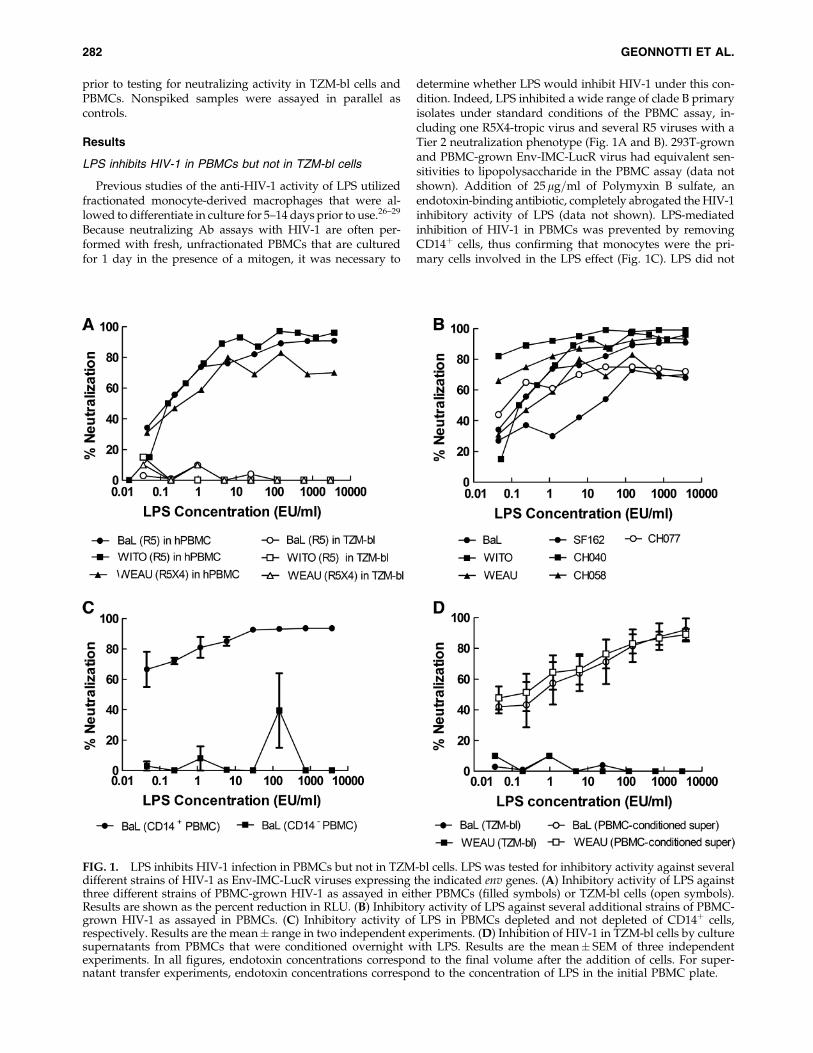
LPS inhibits HIV-1 in PBMCs but not in TZM-bl cells
Previous studies of the anti-HIV-1 activity of LPS utilizedfractionated monocyte-derived macrophages that were al-lowed to differentiate in culture for 5–14 days prior to use.26–29
Because neutralizing Ab assays with HIV-1 are often per-formed with fresh, unfractionated PBMCs that are culturedfor 1 day in the presence of a mitogen, it was necessary to
determine whether LPS would inhibit HIV-1 under this con-dition. Indeed, LPS inhibited a wide range of clade B primaryisolates under standard conditions of the PBMC assay, in-cluding one R5X4-tropic virus and several R5 viruses with aTier 2 neutralization phenotype (Fig. 1A and B). 293T-grownand PBMC-grown Env-IMC-LucR virus had equivalent sen-sitivities to lipopolysaccharide in the PBMC assay (data notshown). Addition of 25mg=ml of Polymyxin B sulfate, anendotoxin-binding antibiotic, completely abrogated the HIV-1inhibitory activity of LPS (data not shown). LPS-mediatedinhibition of HIV-1 in PBMCs was prevented by removingCD14þ cells, thus confirming that monocytes were the pri-mary cells involved in the LPS effect (Fig. 1C). LPS did not
FIG. 1. LPS inhibits HIV-1 infection in PBMCs but not in TZM-bl cells. LPS was tested for inhibitory activity against severaldifferent strains of HIV-1 as Env-IMC-LucR viruses expressing the indicated env genes. (A) Inhibitory activity of LPS againstthree different strains of PBMC-grown HIV-1 as assayed in either PBMCs (filled symbols) or TZM-bl cells (open symbols).Results are shown as the percent reduction in RLU. (B) Inhibitory activity of LPS against several additional strains of PBMC-grown HIV-1 as assayed in PBMCs. (C) Inhibitory activity of LPS in PBMCs depleted and not depleted of CD14þ cells,respectively. Results are the mean� range in two independent experiments. (D) Inhibition of HIV-1 in TZM-bl cells by culturesupernatants from PBMCs that were conditioned overnight with LPS. Results are the mean� SEM of three independentexperiments. In all figures, endotoxin concentrations correspond to the final volume after the addition of cells. For super-natant transfer experiments, endotoxin concentrations correspond to the concentration of LPS in the initial PBMC plate.
282 GEONNOTTI ET AL.
inhibit HIV-1 in the TZM-bl assay (Fig. 1A); however, su-pernatants from PBMCs that were incubated overnight withLPS exhibited potent HIV-1 inhibition activity in the TZM-blassay (Fig. 1D). This latter observation is consistent withprevious reports showing that LPS inhibits HIV-1 by stimu-lating the production of soluble inhibitory factors.28,29
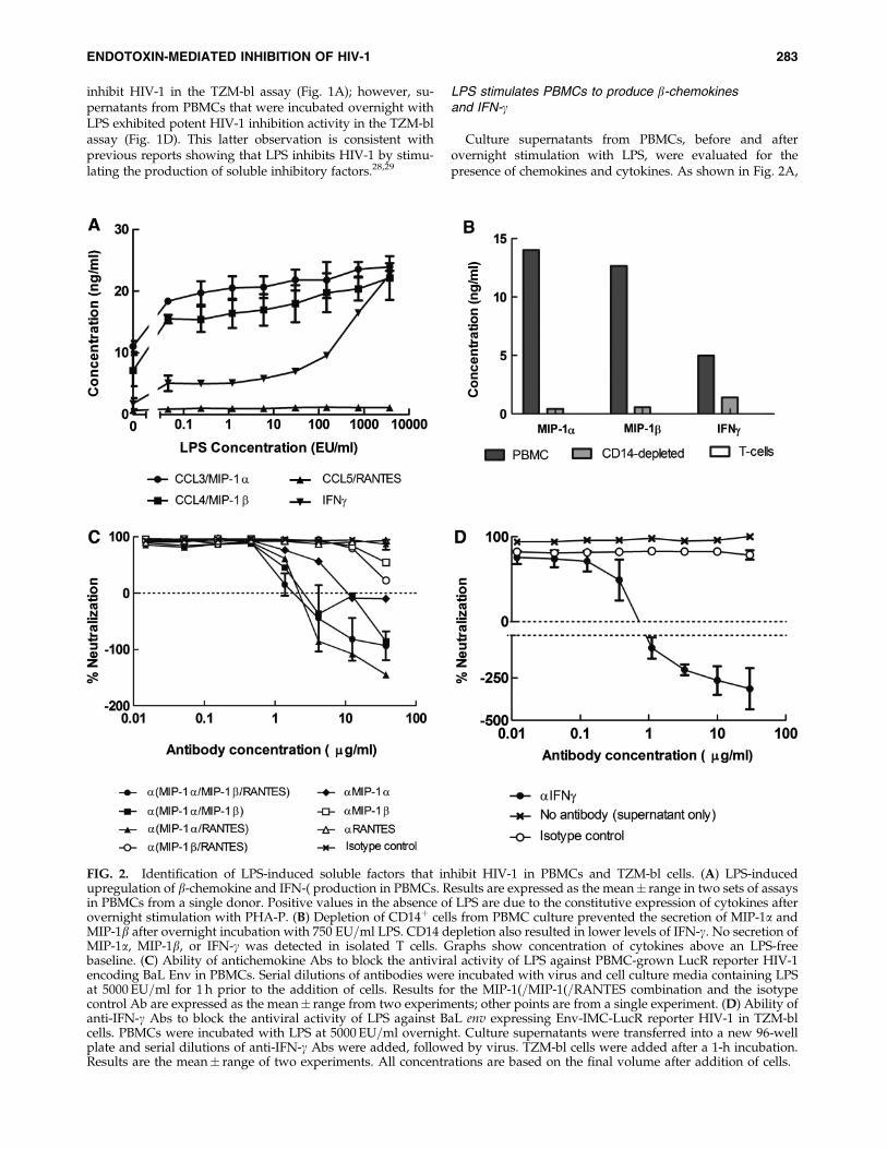
LPS stimulates PBMCs to produce b-chemokinesand IFN-g
Culture supernatants from PBMCs, before and afterovernight stimulation with LPS, were evaluated for thepresence of chemokines and cytokines. As shown in Fig. 2A,
FIG. 2. Identification of LPS-induced soluble factors that inhibit HIV-1 in PBMCs and TZM-bl cells. (A) LPS-inducedupregulation of b-chemokine and IFN-( production in PBMCs. Results are expressed as the mean� range in two sets of assaysin PBMCs from a single donor. Positive values in the absence of LPS are due to the constitutive expression of cytokines afterovernight stimulation with PHA-P. (B) Depletion of CD14þ cells from PBMC culture prevented the secretion of MIP-1a andMIP-1b after overnight incubation with 750 EU=ml LPS. CD14 depletion also resulted in lower levels of IFN-g. No secretion ofMIP-1a, MIP-1b, or IFN-g was detected in isolated T cells. Graphs show concentration of cytokines above an LPS-freebaseline. (C) Ability of antichemokine Abs to block the antiviral activity of LPS against PBMC-grown LucR reporter HIV-1encoding BaL Env in PBMCs. Serial dilutions of antibodies were incubated with virus and cell culture media containing LPSat 5000 EU=ml for 1 h prior to the addition of cells. Results for the MIP-1(=MIP-1(=RANTES combination and the isotypecontrol Ab are expressed as the mean� range from two experiments; other points are from a single experiment. (D) Ability ofanti-IFN-g Abs to block the antiviral activity of LPS against BaL env expressing Env-IMC-LucR reporter HIV-1 in TZM-blcells. PBMCs were incubated with LPS at 5000 EU=ml overnight. Culture supernatants were transferred into a new 96-wellplate and serial dilutions of anti-IFN-g Abs were added, followed by virus. TZM-bl cells were added after a 1-h incubation.Results are the mean� range of two experiments. All concentrations are based on the final volume after addition of cells.
ENDOTOXIN-MEDIATED INHIBITION OF HIV-1 283

LPS stimulated the secretion of MIP-1a, MIP-1b, and IFN-ginto the culture medium. Removal of CD14þ cells fromPBMCs prevented the secretion of b-chemokines and reducedthe amount of IFN-g produced (Fig. 2B). LPS-induced secre-tion of MIP-1a, MIP-1b, or IFN-g was not detected in a cultureof isolated T cells from PBMCs (Fig. 2B). This suggests that aCD14-negative population other than T cells is partly re-sponsible for IFN-g secretion in response to LPS. Althoughsuggested to have a role in X4-tropic HIV-1 inhibition,47–49
MDC expression was not affected by LPS stimulation (datanot shown); SDF-1a was not detected in PBMC supernatants(data not shown).
HIV-1 is differentially inhibited by b-chemokinesand IFN-g in PBMCs and TZM-bl cells
To determine a possible link between the LPS-inducedupregulation of chemokines and cytokines and the anti-HIV-1activity of LPS, we tested whether the chemokines and cyto-kines as pure proteins were inhibitory toward HIV-1 in ourassays. As shown in Table 1, recombinant human MIP-1a,MIP-1a=LD78b, MIP-b, and RANTES were all inhibitory to-ward multiple R5 stains HIV-1 in the PBMC assay. In partic-ular, we confirmed previous reports30,31,50–54 that the LD78bisoform of MIP-1a (CCL3L1) is the most potent inhibitory b-chemokine against HIV-1. This LD78b isoform of MIP-1a islikely the primary chemokine responsible for LPS-mediatedinhibition of R5 viruses in PBMCs, as its IC80 values (Table 1)were well within the levels of MIP-1a detected in PBMC cul-ture (Fig. 2A). RANTES also exhibited a potent inhibitoryeffect against multiple R5 viruses, followed in potency byMIP-1a. MIP-b was the least active chemokine, inhibiting onlytwo of the six R5 viruses tested and requiring relatively highconcentrations for inhibition. None of the b-chemokines in-hibited the reporter virus encoding the R5=X4-tropic WEAUEnv. IFN-g and MDC at concentrations as high as 1250 ng=mlhad no inhibitory effect on HIV-1 infection in PBMCs.
None of the b-chemokines inhibited the reporter virusesencoding the Envs for BaL, WITO, and WEAU or the R5 Env-pseudovirus PVO.4 in the TZM-bl assay at well above phys-iologic concentrations (�1250 ng=ml), with one exception:NL-LucR.T2A-WITO.ecto infection was inhibited by MIP-1aLD78b=CCL3L1 at an IC50 of 845 ng=ml. To understand theapparent resistance of TZM-bl cells to b-chemokine-mediatedviral inhibition, further tests were conducted using three ad-ditional HeLa cell lines that were engineered similarly to theTZM-bl line, but with lower densities of CCR5 on their sur-
face.36 As shown in Fig. 3, sensitivity to b-chemokine-medi-ated viral inhibition was directly related to cellular coreceptordensity, with lower density associated with more potent in-hibition. This outcome strongly suggests that TZM-bl cells areresistant to b-chemokines because of their much higher levelsof CCR5 on the surface. In contrast, IFN-g potently inhibitedBaL(R5) and WEAU (R5X4) in TZM-bl cells, with IC50 valuesof 580 pg=ml and 150 pg=ml, respectively (data not shown).These effective concentrations are well within the IFN-g levelsin LPS-stimulated PBMC culture supernatants (Fig. 2A).
To further confirm a role for b-chemokines and IFN-g in theLPS-mediated anti-HIV activity observed here, additionalexperiments were performed in which we attempted to blockthe antiviral activity of LPS by adding antichemokine and anti-IFN-g Abs. As shown in Fig. 2C, anti-MIP-1a Abs completelyabolished the antiviral activity of LPS in the PBMC assay atantibody concentrations above 10mg=ml. High concentrationsof anti-MIP-1b Ab only partially reversed the antiviral activityof LPS, whereas anti-RANTES Ab had no measurable effect.These results support the notion that MIP-1a was the domi-nant LPS-induced chemokine responsible for HIV-1 inhibitionin the PBMC assay. Nonetheless, our results indicate that MIP-1b and RANTES contribute to this inhibition in so much ascombinations of antichemokine Abs were more effective thanany single antichemokine Ab alone (Fig. 2C). In particular,anti-MIP-1a, when combined with either anti-MIP-1b or anti-RANTES, was more effective than when tested alone. Finally,anti-IFN-g Abs completely abolished the HIV-1 inhibitory ac-tivity of LPS-conditioned PBMC culture supernatants in theTZM-bl assay (Fig. 2D), whereas anti-b-chemokine antibodieshad no effect (data not shown), strongly suggesting that IFN-gis responsible for the anti-HIV-1 activity of LPS-conditionedPBMC supernatant in this assay.
It should be noted that in some cases, the addition of an-tichemokine and anti-IFN-g Abs appeared to enhance virusinfection, resulting in negative inhibition values (i.e., RLU intest wells were higher than those in the virus control wells thatwere used as our measure for zero inhibition). We attributethis effect to constitutive levels of b-chemokine and IFN-gproduction in PHA-P-stimulated PBMCs (Fig. 1A), whichwould partially inhibit infection in the virus control wells butnot in wells containing antichemokine and anti-IFN-g Abs.
Variables that affect the HIV-1-inhibitory activity of LPS
We tested the HIV-1 inhibitory activity of both smooth andrough LPS from three different strains of bacteria (E. coli
Table 1. Inhibition of HIV-1 by b-Chemokines in the PBMC Assay
IC80 (ng=ml) in PBMCsa
Recombinant protein Bal R5 SF162 R5 WITO R5 CH040 R5 CH058 R5 CH077 R5 WEAU R5X4
MIP-1a 400 >1250 120 126 >1250 >1250 >1250MIP-1a=LD78b 13 43 10 7 18 109 >1250MIP-1b >1250 >1250 1160 615 >1250 >1250 >1250RANTES 66 171 36 37 75 >1250 >1250IFN-g >5000 ND ND ND ND ND >5000MDC >1250 ND >1250 ND ND ND >1250
aChemokines and IFN-g were assayed in PBMCs against PBMC-grown NL-LucR.T2A-Env.ecto reporter HIV-1 expressing the indicatedenvs. Values are the concentration at which RLU were reduced 80% compared to virus control wells after subtraction of background RLU incell control wells. ND, inhibition experiments were not done with these viruses.
284 GEONNOTTI ET AL.

O55:B5, E. coli 0127:B8, and S. enterica) by using PBMCs fromdifferent donors. There was little difference in antiviral ac-tivity among the four smooth LPS strains; however, largedifferences were seen between smooth and rough LPS strainsand in the sensitivity of donor PBMCs to LPS-mediated in-hibition of HIV-1 (Fig. 4). The activity of LPS differed in eachdonor PBMCs. The tested smooth strains of LPS produced50% inhibition of Bal env expressing LucR reporter HIV-1 inonly 50–65% of randomly selected donor PBMCs, with large
variations in IC50 values. Eighty percent inhibition, withsimilar IC80 variation, was achieved only in 25–30% of testedPBMCs. In contrast, the rough LPS strain inhibitedHIV-1> 80% in all (100%) tested donor PBMCs (Fig. 4), al-though with substantial variation among IC80 values. Thesefindings suggest that there are genetic differences in donorPBMCs that affect the anti-HIV-1 activity of LPS in vitro.Additionally, they support the current hypothesis thatrough LPS, through its ability to mediate CD14-independent
FIG. 3. The potency of the recombinant human b-chemokine CCL3L1 against HIV-1 NL-LucR.T2A-BaL.ecto was evaluatedin four different HeLa cell lines with previously established levels of CCR5: JC.10 cells (2.0�103 molecules CCR5=cell),JC.37 cells (1.5�104 molecules CCR5=cell), JC.10 cells (2.7�104 molecules CCR5=cell), and TZM-bl=JC.53 cells (1.3�105 mol-ecules CCR5=cell).36 Viral inhibition was measured by a reduction in Renilla luciferase expression after 48 h incubation.
FIG. 4. PBMC donors have differential sensitivity to LPS-mediated inhibition of HIV-1. Bars show the percentage of testedPBMC donors in which LPS inhibited HIV-1 NL-LucR.T2A-BaL.ecto�50% (white bars) or�80% (gray bars). The 80% bars aresuperimposed on the 50% bars, as any PBMCs with 80% inhibition also showed 50% inhibition. Values inset within verticalbars represent median IC50 or IC80 values with ranges in parentheses to show variation; all values are in EU=ml. Twenty-onedifferent PBMCs were tested for O55:B5b, O127:B8, and S. enterica; 10 different PBMCs were tested for O55:B5a and S. enterica(R). O55:B5a and O55:B5b are LPS preparations manufactured by Lonza and Sigma, respectively.
ENDOTOXIN-MEDIATED INHIBITION OF HIV-1 285

signaling, may potentiate immune responses in a broaderrange of cell types.43,44 However, these demonstrated differ-ences in PBMC response to LPS cannot be solely attributed tovariations in CD14 expression or function, as a range of LPS-mediated inhibitory responses was seen among PBMC donorseven with the rough strain of LPS (Fig. 4).
Because of variation between laboratories in the length oftime that PBMCs are stimulated with PHA-P prior to use inneutralization assays, we examined how differences in thelength of time of stimulation affects LPS-mediated HIV-1 in-hibition. PBMCs that had been stimulated for 4 days showed asubstantial decrease in sensitivity to LPS inhibition (Fig. 5A).Also, PBMC infectivity was markedly decreased by longerstimulation times: luminescence values from virus controlwells 4 days after addition of the same amount of HIV-1NL-LucR.T2A-BaL.ecto were *75,000 with 1-day PHA-stimulated PBMCs and only *2000 with 4-day stimulatedPBMCs. This phenomenon was repeatable with differentdonor PBMCs. Longer stimulation times also reduced theamount of MIP-1a and MIP-1b produced by the PBMCs inresponse to LPS by approximately 3- and 6-fold, respectively.Interestingly, 4-day stimulated PBMCs did not produce anyINF-g in response to LPS (Fig. 5B).
Endotoxin contamination increases monoclonalAb potency in the PBMC assay
We tested the effect of endotoxin contamination on severalhuman monoclonal antibodies spanning a diverse range ofepitope specificities: IgG1b12 recognizes a complex epitopeoverlapping the CD4-binding domain,55–57 2G12 binds amannose cluster on the outer domain of gp120 involvingmultiple N-linked glycans,58–60 and the epitopes for 2F561–63
and 4E1064,65 are adjacent to each other in the membrane-proximal ectodomain of gp41. The effect of endotoxin on anIgG fraction of pooled sera from HIV-1 Ab-positive donors
(HIVIG) was also tested. All reagents were spiked with a large(30,000 EU=ml) amount of endotoxin and evaluated for anti-viral activity in both the TZM-bl and PBMC assay. LPS con-tamination increased the potency of the samples by*100-foldin the PBMC assay but had no measureable effect in the TZM-bl assay (Fig. 6).
Discussion
Our results confirm and extend previous reports26–29,66
that LPS has potent and broad inhibitory activity againstboth R5 and R5X4 strains of HIV-1. Previous studies exam-ined the anti-HIV-1 activity of LPS by using cultures offractionated monocyte-derived macrophages for LPS stimu-lation. Here we show that LPS inhibits HIV-1 by stimulatingthe production of chemokines in the monocyte population ofcryopreserved PBMCs that were thawed and used after 1day in culture in the presence of PHA-P and IL-2. We alsodemonstrate that LPS has no direct anti-HIV-1 activity inTZM-bl cells, probably because these cells lack both CD14and MD-2.34,35 The differential effect of LPS in the two celltypes could cause discrepancies in the measured neutraliza-tion potency of endotoxin-contaminated reagents. It is notknown to what extent, if any, endotoxin contaminationcontributed to recent reports of Abs possessing greaterneutralization potency in PBMCs compared to TZM-blcells.2,8–12 Indeed, several other factors might contribute to anapparent diminished sensitivity of the TZM-bl assay for de-tecting neutralizing Abs.2,14,15 At a minimum, any reagent(mAb, Ig fraction, human=animal sera, or antiviral com-pound) that demonstrates, or has demonstrated, potentneutralization only in a PBMC or macrophage-based assayshould be tested for endotoxin contamination to rule outpossible artifactual results. Additionally, it seems prudent toensure that in the future, only endotoxin-free reagents areused in the PBMC assay.
FIG. 5. Prolonged stimulation of PBMCs with PHA-P diminishes the antiviral effect of LPS against HIV-1 NL-LucR.T2A-BaL.ecto (A) and the amount of cytokines released (B). PBMCs from two different donors were stimulated with PHA-P eitherovernight (filled symbols) or for 4 days (open symbols) prior to use in a neutralization assay with LPS. Data represent meanvalues from two independent experiments.
286 GEONNOTTI ET AL.

Our results indicate that b-chemokines MIP-1a and, to alesser extent, MIP-1b and RANTES are the major effectors ofLPS-mediated inhibition of HIV-1 in PBMCs. We also con-firmed prior reports that the LD78b (CCL3L1) isoform of MIP-1a is the most potent anti-HIV-1 chemokine,30,31,51–54 and wesuccessfully demonstrated that blocking b-chemokines abro-gates LPS-mediated HIV-1 inhibition. HIV-1 inhibition by b-chemokines is thought to occur through a combination ofCCR5 steric hindrance, downregulation, and=or dimerizationmechanisms.31 HIV-1 and b-chemokines have been shownto compete for CCR5 binding; the antifusion activity of b-chemokines decreases with increased CCR5 expression.67
Because TZM-bl cells express *100�more CCR5 on theirsurface than PBMCs,2 high CCR5 density could explain thelack of HIV-1 inhibitory activity of b-chemokines in the TZM-bl assay. This hypothesis was tested and confirmed usingadditional engineered HeLa cell lines that were developedalong with the parental TZM-bl cell line, JC.53, but that ex-press lower CCR5 densities on their surface.36 EngineeredHeLa cells with lower CCR5 densities were more sensitive tob-chemokine-mediated viral inhibition, and receptor densitywas directly related to the potency of b-chemokine-mediatedviral inhibition. (Fig. 3)
Interestingly, the anti-HIV-1 activity of LPS-conditionedPBMC culture supernatants in the TZM-bl assay appeared tobe mediated by IFN-g. IFN-g has been shown to inhibit HIV-1replication in cultured macrophages27,68 and in other celllines.27 However, the apparent inhibitory effect of IFN-g onHIV-1 may be due to its actions on TZM-bl cells. IFN-g hasalso been shown to retard cellular growth, inhibit DNA syn-thesis, and be moderately toxic to HeLa cell culture at con-centrations >5 ng=ml) 69,70; the combination of these effectscould potentially interfere with the TZM-bl assay and result ina reduction of RLU. Some toxicity and growth inhibitionwere seen at higher tested concentrations of IFN-g; however,HIV-1 inhibition was detected at concentrations permissiveto monolayer formation. The wide variability of endotoxin-mediated inhibition of HIV-1 across PBMC donors is not
unexpected as LPS is known to cause disparate cytokine re-sponses across individuals.71 The reasons behind this varia-tion are likely multifactorial and may include knowndifferences in gene copy number of CCL3L1,50 the most po-tent anti-HIV b-chemokine, as well as possible desensitizingmutations in the gene encoding TLR472 and=or the down-stream signaling protein IRAK-4.73
The soluble factor responsible for LPS-mediated inhibitionof the R5X4-tropic virus expressing WEAU Env in the PBMCassay remains unknown. WEAU Env was completely resis-tant to inhibition by b-chemokines and IFN-g. Although SDF-1a is a natural ligand for CXCR4 and blocks X4 HIV-1 virusentry,75,76 it is produced in stromal cells rather than PBMCs.29
Several studies have reported that antibodies to MDC=CCL22can block soluble factor-mediated inhibition of X4 viruses47–49;however, we, and others,29,76 did not detect any inhibition ofR5 or R5=X4 HIV-1 with MDC, nor did we detect an increasein MDC production after LPS stimulation of PBMCs (data notshown). The type I interferons, IFN-a and IFN-b, have beenshown to inhibit X4 viruses in PBMCs,77,78 but well abovephysiologic concentrations.29 Additionally, we did not detectan increase in IFN-a production following LPS stimulation ofPBMCs. LPS has been shown to downregulate the surfaceexpression of CD4, CCR5, and CXCR4 on monocyte-derivedmacrophages.29 Recent reports have suggested that CXCR4binds LPS and has a role as a complementary LPS receptor tothe CD14=TLR4=MD-2 main sensing complex.79,80 As such,LPS may competitively inhibit X4-tropic HIV-1 binding toCXCR4. A combination of b-chemokines, receptor down-regulation, and competitive inhibition by LPS might explaindual-tropic viral inhibition by endotoxin.
Detection of endotoxin is relatively straightforward by anyof several commercially available LAL assay kits. Samplescontaining serum or high protein content should be checkedfor assay inhibition per the manufacturer’s instructions, asserum=protein can inhibit LPS detection and cause false-negative readings. Care should be taken in the manufactureand=or purification of any reagent used in neutralization
FIG. 6. High concentrations of LPS have no effect on the potency of neutralizing Abs when assayed in TZM-bl cells.Neutralizing monoclonal Abs (b12, 2G12, 2F5, 4E10) and a neutralizing polyclonal antiserum (HIVIG) were spiked with30,000 EU=ml of E. coli O55:B5 LPS prior to assay. Neutralizing activity of the spiked samples and corresponding nonspikessamples was assayed against PBMC-grown NL-LucR.T2A-BaL.ecto in PBMCs (A) and in TZM-bl cells (B). Gray bars, LPSabsent; black bars, LPS present. For comparison, the median IC80 of E. coli O55:B5 LPS in identical donor PBMCs was 1.56EU=ml.
ENDOTOXIN-MEDIATED INHIBITION OF HIV-1 287
assays as downstream endotoxin removal is very difficult insmall sample volumes. Bacterial LPSs strongly associate withproteins in solution and are stable at a wide range of tem-perature and pH. An excellent review of endotoxin removalmethods is available.81 However, because both PBMC re-sponse to endotoxin and HIV-1 sensitivity to b-chemokinesvary, there is no generic ‘‘safe’’ threshold of endotoxin. In ourexperience, LPS concentrations of �0.1 EU=ml (*29 pg=ml)are permissible with negligible effects on HIV-1 infectionin vitro. Removal of CD14þ cells from PBMC culture is also aneffective means of abrogating LPS-mediated HIV-1 inhibitionin cases in which use of contaminated reagents is unavoid-able; however, note that CD14� PBMCs have been shown tohave reduced Ab-dependent cell-mediated virus inhibition(ADCVI) activity.82
Finally, we note that our recommended safe level of endo-toxin (*29 pg=ml) for avoiding artifacts in HIV-1 neutraliza-tion assays compares well with recent measurements of thephysiologic LPS concentration in plasma from normal, HIV-1-negative individuals.83–85 However, recent studies have re-vealed that HIV-1-infected persons have increased levels ofplasma LPS due to microbial translocation of bacteria throughthe gut mucosa.83–86 Based on our results, these increasedplasma LPS levels may affect measurements of the neutralizingability of HIV-1-positive serum and plasma samples in somePBMC-based assays by inducing antiviral chemokine release.Additionally, our results document a robust immunologicalcascade in primary cells after exposure to low concentrations ofLPS and thereby add support to the hypothesis that LPS in-troduced via microbial translocation in vivo may contribute toimmune activation in HIV-1-infected individuals.82–89 How-ever, although LPS has profound inhibitory effects on HIV-1in vitro, the effect, if any, of LPS-induced immunostim-ulation and subsequent antiviral chemokine release on viralreplication, diversity, and=or pathogenesis in vivo is yet undeter-mined. Finally, it is interesting to speculate as to whether varia-tions in the sensitivity of donor PBMCs to in vitro LPS stimulationwould translate to in vivo variations in the immunostimulatoryresponse to HIV-1-induced microbial translocation.
In summary, endotoxin contamination of serological sam-ples mediates a release of antiviral chemokines in susceptiblePBMCs and can cause false-positive results in neutralizingantibody assays done in PBMCs. Although this phenomenonis variable depending on donor PBMCs and LPS phenotype, itcan occur at very low concentrations of endotoxin. Therefore,to ensure the correct assessment of tested samples all reagentsused in the PBMC assay should be endotoxin free. This rec-ommendation should also be followed when assessing HIV-1neutralization in any cell type known to secrete b-chemokinesin response to LPS stimulation, such as macrophages. HIV-1neutralization assays in TZM-bl cells were unaffected by en-dotoxin contamination; the effect of endotoxin on otherpseudovirus-based assay technologies utilizing engineeredcell targets was not evaluated. Our study reinforces the needfor continued standardization and validation of a variety ofassay technologies to test the HIV-1 neutralizing ability ofserologic reagents.
Acknowledgments
We gratefully acknowledge support from the HIVVaccine Clinical Trials Network (HVTN) (NIH AI46705), the
Center for HIV=AIDS Vaccine Immunology (CHAVI) (NIHAI067854), the Bill and Melinda Gates Foundation Colla-boration for Vaccine Discovery (Grant 38619), and the Pre-clinical Branch, Divisions of AIDS, U.S. National Institutes ofHealth (AI30034). We appreciate the advice on experimentaldesign and data analysis provided by the Duke Center forAIDS Research (CFAR) Flow Cytometry Core, an NIH-funded program (P30 AI 64518). We also thank Drs. Carl Al-ving and Gabriel Perez for helpful insights, Dr. Thomas Dennyfor providing PBMCs, and Drs. David Kabat and Emily Plattfor providing the additional engineered HeLa cell lines.
Author Disclosure Statement
No competing financial interests exist.
References
1. Montefiori DC, Morris L, Ferrari G, and Mascola JR:Neutralizing and other antiviral antibodies in HIV-1 in-fection and vaccination. Curr Opin HIV AIDS 2007;2:169–176.
2. Polonis VR, Brown BK, Borges AR, et al.: Recent advances inthe characterization of HIV-1 neutralization assays forstandardized evaluation of the antibody response to infec-tion and vaccination. Virology 2008;375:315–320.
3. Li M, Gao F, Mascola JR, et al.: Human immunodeficiencyvirus type 1 env clones from acute and early subtype B in-fections for standardized assessments of vaccine-elicitedneutralizing antibodies. J Virol 2005;79:10108–10125.
4. Mascola JR, D’Souza P, Gilbert P, et al.: Recommendationsfor the design and use of standard virus panels to assessneutralizing antibody responses elicited by candidatehuman immunodeficiency virus type 1 vaccines. J Virol2005;79:10103–10107.
5. Montefiori D, Sattentau Q, Flores J, Esparza J, and Mascola J:Antibody-based HIV-1 vaccines: Recent developments andfuture directions. PLoS Med 2007;4:e348.
6. Montefiori DC: Measuring HIV neutralization in a luciferasereporter gene assay. Methods Mol Biol 2009;485:395–405.
7. Richman DD, Wrin T, Little SJ, and Petropoulos CJ: Rapidevolution of the neutralizing antibody response to HIV type1 infection. Proc Natl Acad Sci USA 2003;100:4144–4149.
8. Binley JM, Wrin T, Korber B, et al.: Comprehensive cross-clade neutralization analysis of a panel of anti-human im-munodeficiency virus type 1 monoclonal antibodies. J Virol2004;78:13232–13252.
9. Brown BK, Karasavvas N, Beck Z, et al.: Monoclonal anti-bodies to phosphatidylinositol phosphate neutralize humanimmunodeficiency virus type 1: Role of phosphate-bindingsubsites. J Virol 2007;81:2087–2091.
10. Choudhry V, Zhang M-Y, Sidorov IA, et al.: Cross-reactiveHIV-1 neutralizing monoclonal antibodies selected byscreening of an immune human phage library against anenvelope glycoprotein (gp140) isolated from a patient (R2)with broadly HIV-1 neutralizing antibodies. Virology2007;363:79–90.
11. Zhang M-Y, Vu BK, Choudhary A, et al.: Cross-reactivehuman immunodeficiency virus type 1-neutralizing humanmonoclonal antibody that recognizes a novel conformationalepitope on gp41 and lacks reactivity against self-antigens.J Virol 2008;82:6869–6879.
12. Mann AM, Rusert P, Berlinger L, Kuster H, Gunthard HF, andTrkola A: HIV sensitivity to neutralization is determined
288 GEONNOTTI ET AL.

by target and virus producer cell properties. AIDS 2009;23:1659–1667.
13. Fenyo EM, Heath A, Dispinseri S, et al.: International net-work for comparison of HIV neutralization assays: TheNeutNet report. PLoS ONE 2009;4:e4505.
14. Choudhry V, Zhang M-Y, Harris I, et al.: Increased efficacyof HIV-1 neutralization by antibodies at low CCR5 surfaceconcentration. Biochem Biophys Res Commun 2006;348:1107–1115.
15. Perez LG, Costa MR, Todd CA, Haynes BF, and MontefioriDC: Utilization of IgG Fc receptors by human immunode-ficiency virus type 1: A specific role for antibodies againstthe membrane proximal external region of gp41. J Virol2009;83:7397–7410.
16. Beausejour Y and Tremblay MJ: Susceptibility of HIV type 1to the fusion inhibitor T-20 is reduced on insertion of hostintercellular adhesion molecule 1 in the virus membrane.J Infect Dis 2004;190:894–902.
17. Losier M, Fortin JF, Cantin R, Bergeron MG, and TremblayMJ: Virion-bound ICAM-1 and activated LFA-1: A combi-nation of factors conferring resistance to neutralization bysera from human immunodeficiency virus type 1-infectedindividuals independently of the disease status and phase.Clin Immunol 2003;108:111–118.
18. Rizzuto CD and Sodroski JG: Contribution of virion ICAM-1to human immunodeficiency virus infectivity and sensitivityto neutralization. J Virol 1997;71:4847–4851.
19. Louder MK, Sambor A, Chertova E, et al.: HIV-1 envelopepseudotyped viral vectors and infectious molecular clonesexpressing the same envelope glycoprotein have a similarneutralization phenotype, but culture in peripheral bloodmononuclear cells is associated with decreased neutraliza-tion sensitivity. Virology 2005;339:226–238.
20. Miyauchi K, Kim Y, Latinovic O, Morozov V, and MelikyanGB: HIV enters cells via endocytosis and dynamin-dependentfusion with endosomes. Cell 2009;137:433–444.
21. Hoshino K, Takeuchi O, Kawai T, et al.: Cutting edge: Toll-like receptor 4 (TLR4)-deficient mice are hyporesponsive tolipopolysaccharide: Evidence for TLR4 as the Lps geneproduct. J Immunol 1999;162:3749–3752.
22. Nagai Y, Akashi S, Nagafuku M, et al.: Essential role ofMD-2 in LPS responsiveness and TLR4 distribution. NatImmunol 2002;3:667–672.
23. Poltorak A, He X, Smirnova I, et al.: Defective LPS signalingin C3H=HeJ and C57BL=10ScCr mice: Mutations in Tlr4gene. Science 1998;282:2085–2088.
24. Wright SD: CD14 and innate recognition of bacteria. J Im-munol 1995;155:6–8.
25. Wright SD, Ramos RA, Tobias PS, Ulevitch RJ, and MathisonJC: CD14, a receptor for complexes of lipopolysaccharide(LPS) and LPS binding protein. Science 1990;249:1431–1433.
26. Bernstein MS, Tong-Starksen SE, and Locksley RM: Activa-tion of human monocyte-derived macrophages with lipo-polysaccharide decreases human immunodeficiency virusreplication in vitro at the level of gene expression. J ClinInvest 1991;88:540–545.
27. Kornbluth RS, Oh PS, Munis JR, Cleveland PH, and RichmanDD: Interferons and bacterial lipopolysaccharide protectmacrophages from productive infection by human immu-nodeficiency virus in vitro. J Exp Med 1989;169:1137–1151.
28. Verani A, Scarlatti G, Comar M, et al.: C-C chemokines re-leased by lipopolysaccharide (LPS)-stimulated human mac-rophages suppress HIV-1 infection in both macrophages andT cells. J Exp Med 1997;185:805–816.
29. Verani A, Sironi F, Siccardi AG, Lusso P, and Vercelli D:Inhibition of CXCR4-tropic HIV-1 infection by lipopolysac-charide: Evidence of different mechanisms in macrophagesand T lymphocytes. J Immunol 2002;168:6388–6395.
30. Aquaro S, Menten P, Struyf S, et al.: The LD78b isoform ofMIP-1a is the most potent CC-chemokine in inhibiting CCR5-dependent human immunodeficiency virus type 1 replicationin human macrophages. J Virol 2001;75:4402–4406.
31. Menten P, Wuyts A, and Van Damme J: Macrophage in-flammatory protein-1. Cytokine Growth Factor Rev 2002;13:455–481.
32. Worgall S, Connor R, Kaner RJ, et al.: Expression and use ofhuman immunodeficiency virus type 1 coreceptors byhuman alveolar macrophages. J Virol 1999;73:5865–5874.
33. Mikulak J, Gianolini M, Versmisse P, Pancino G, Lusso P,and Verani A: Biological and physical characterization of theX4 HIV-1 suppressive factor secreted by LPS-stimulatedhuman macrophages. Virology 2009;390:37–44.
34. Thibault S, Tardif MR, Barat C, and Tremblay MJ: TLR2signaling renders quiescent naive and memory CD4þ T cellsmore susceptible to productive infection with X4 and R5HIV-Type 1. J Immunol 2007;179:4357–4366.
35. Wyllie DH, Kiss-Toth E, Visintin A, et al.: Evidence for anaccessory protein function for toll-like receptor 1 in anti-bacterial responses. J Immunol 2000;165:7125–7132.
36. Platt EJ, Wehrly K, Kuhmann SE, Chesebro B, and Kabat D:Effects of CCR5 and CD4 cell surface concentrations on in-fections by macrophage tropic isolates of human immuno-deficiency virus type 1. J Virol 1998;72:2855–2864.
37. Wei X, Decker JM, Liu H, et al.: Emergence of resistant hu-man immunodeficiency virus type 1 in patients receivingfusion inhibitor (T-20) monotherapy. Antimicrob AgentsChemother 2002;46:1896–1905.
38. Edmonds T, Ding D, Conway J, et al.: Rapid and quantitativeanalysis of HIV-1 neutralization in primary cells via Renillaluciferase-expressing infectious molecular clones. Presentedat the Conference on Retroviruses and Opportunistic Infec-tions, 2009, Montreal, Quebec, Canada.
39. Ochesenbauer C and Kappes JC: New virologic reagents forneutralizing antibody assays. Curr Opin HIV AIDS 2009;4:418–425.
40. Keele BF, Giorgi EE, Salazar-Gonzalez JF, et al.: Identificationand characterization of transmitted and early founder virusenvelopes in primary HIV-1 infection. Proc Natl Acad SciUSA 2008;105:7552–7557.
41. Salazar-Gonzalez JF, Salazar MG, Keele BF, et al.: Geneticidentity, biological phenotype, and evolutionary pathwaysof transmitted=founder viruses in acute and early HIV-1infection. J Exp Med 2009;206:1273–1289.
42. Ochsenbauer-Jambor C, Ding H, Keele BF, et al.: Generationand biological characterization of infectious molecularclones derived from clade B HIV-1 transmitted=founderviruses. Presented at the Conference on Retroviruses andOpportunistic Infections, 2009, Montreal, Quebec, Canada.
43. Godowski PJ: A smooth operator for LPS responses. NatImmunol 2005;6:544–546.
44. Jiang Z, Georgel P, Du X, et al.: CD14 is required forMyD88-independent LPS signaling. Nat Immunol 2005;6:565–570.
45. Montefiori DC: Evaluating neutralizing antibodies againstHIV, SIV, and SHIV in luciferase reporter gene assays, In:Current Protocols in Immunology (Coligan JE, Kruisbeek AM,Shevach EM, Strober W, Coico R, Eds.). John Wiley & Sons,2004, New York, pp. 12.11.1–12.11.15.
ENDOTOXIN-MEDIATED INHIBITION OF HIV-1 289

46. Bures R, Gaitan A, Zhu T, et al.: Immunization withrecombinant canarypox vectors expressing membrane-anchored glycoprotein 120 followed by glycoprotein 160boosting fails to generate antibodies that neutralize R5 pri-mary isolates of human immunodeficiency virus type 1.AIDS Res Hum Retroviruses 2000;16:2019–2035.
47. Abdelwahab SF, Cocchi F, Bagley KC, et al.: HIV-1-suppressive factors are secreted by CD4þ T cells duringprimary immune responses. Proc Natl Acad Sci USA2003;100:15006–15010.
48. Cocchi F, DeVico AL, Garzino-Demo A, Arya SK, Gallo RC,and Lusso P: Identification of RANTES, MIP-1a, and MIP-1bas the major HIV-suppressive factors produced by CD8þ Tcells. Science 1995;270:1811–1815.
49. Pal R, Garzino-Demo A, Markham PD, et al.: Inhibition ofHIV-1 infection by the b-chemokine MDC. Science 1997;278:695–698.
50. Urban TJ, Weintrob AC, Fellay J, et al.: CCL3L1 andHIV=AIDS susceptibility. Nat Med 2009;15:1110–1112.
51. Menten P, Struyf S, Schutyser E, et al.: The LD78beta isoformof MIP-1alpha is the most potent CCR5 agonist and HIV-1-inhibiting chemokine. J Clin Invest 1999;104:R1–5.
52. Nakao M, Nomiyama H, and Shimada K: Structures ofhuman genes coding for cytokine LD78 and their expression.Mol Cell Biol 1990;10:3646–3658.
53. Nibbs RJ, Yang J, Landau NR, Mao JH, and Graham GJ:LD78b, a non-allelic variant of human MIP-1alpha(LD78alpha), has enhanced receptor interactions and potentHIV suppressive activity. J Biol Chem 1999;274:17478–17483.
54. Struyf S, Menten P, Lenaerts JP, et al.: Diverging bindingcapacities of natural LD78b isoforms of macrophage in-flammatory protein-1a to the CC chemokine receptors 1, 3and 5 affect their anti-HIV-1 activity and chemotactic po-tencies for neutrophils and eosinophils. Eur J Immunol2001;31:2170–2178.
55. Burton DR, Pyati J, Koduri R, et al.: Efficient neutralization ofprimary isolates of HIV-1 by a recombinant human mono-clonal antibody. Science 1994;266:1024–1027.
56. Mo H, Stamatatos L, Ip JE, et al.: Human immunodeficiencyvirus type 1 mutants that escape neutralization by humanmonoclonal antibody IgG1b12. J Virol 1997;71:6869–6874.
57. Pantophlet R, Ollmann Saphire E, Poignard P, Parren PW,Wilson IA, and Burton DR: Fine mapping of the interactionof neutralizing and nonneutralizing monoclonal antibodieswith the CD4 binding site of human immunodeficiencyvirus type 1 gp120. J Virol 2003;77:642–658.
58. Calarese DA, Scanlan CN, Zwick MB, et al.: Antibody do-main exchange is an immunological solution to carbohy-drate cluster recognition. Science 2003;300:2065–2071.
59. Sanders RW, Venturi M, Schiffner L, et al.: The mannose-dependent epitope for neutralizing antibody 2G12 onhuman immunodeficiency virus type 1 glycoprotein gp120.J Virol 2002;76:7293–7305.
60. Scanlan CN, Pantophlet R, Wormald MR, et al.: The broadlyneutralizing anti-human immunodeficiency virus type 1 anti-body 2G12 recognizes a cluster of alpha1–2 mannose residueson the outer face of gp120. J Virol 2002;76:7306–7321.
61. Barbato G, Bianchi E, Ingallinella P, et al.: Structural analysisof the epitope of the anti-HIV antibody 2F5 sheds light intoits mechanism of neutralization and HIV fusion. J Mol Biol2003;330:1101–1115.
62. Muster T, Steindl F, Purtscher M, et al.: A conserved neu-tralizing epitope on gp41 of human immunodeficiency virustype 1. J Virol 1993;67:6642–6647.
63. Purtscher M, Trkola A, Gruber G, et al.: A broadly neutral-izing human monoclonal antibody against gp41 of humanimmunodeficiency virus type 1. AIDS Res Hum Retroviruses1994;10:1651–1658.
64. Stiegler G, Kunert R, Purtscher M, et al.: A potent cross-cladeneutralizing human monoclonal antibody against a novelepitope on gp41 of human immunodeficiency virus type 1.AIDS Res Hum Retroviruses 2001;17:1757–1765.
65. Zwick MB, Labrijn AF, Wang M, et al.: Broadly neutralizingantibodies targeted to the membrane-proximal external re-gion of human immunodeficiency virus type 1 glycoproteingp41. J Virol 2001;75:10892–10905.
66. Schmidtmayerova H, Sherry B, and Bukrinsky M: Chemo-kines and HIV replication. Nature 1996;382:767.
67. Dragic T, Litwin V, Allaway GP, et al.: HIV-1 entry intoCD4þ cells is mediated by the chemokine receptor CC-CKR-5. Nature 1996;381:667–673.
68. Moriuchi H, Moriuchi M, Combadiere C, Murphy PM, andFauci AS: CD8þ T-cell-derived soluble factor(s), but notbeta-chemokines RANTES, MIP-1 alpha, and MIP-1 beta,suppress HIV-1 replication in monocyte=macrophages. ProcNatl Acad Sci USA 1996;93:15341–15345.
69. Morrison RP: Differential sensitivities of Chlamydia tracho-matis strains to inhibitory effects of gamma interferon. InfectImmun 2000;68:6038–6040.
70. Um S-J, Kim E-J, Hwang E-S, Kim S-J, Namkoong S-E, andPark J-S: Antiproliferative effects of retinoic acid=interferonin cervical carcinoma cell lines: Cooperative growth sup-pression of IRF-1 and p53. Int J Cancer 2000;85:416–423.
71. Miller SI, Ernst RK, and Bader MW: LPS, TLR4 and infec-tious disease diversity. Nat Rev Microbiol 2005;3:36–46.
72. Arbour NC, Lorenz E, Schutte BC, et al.: TLR4 mutations areassociated with endotoxin hyporesponsiveness in humans.Nat Genet 2000;25:187–191.
73. Picard C, Puel A, Bonnet M, et al.: Pyogenic bacterial infec-tions in humans with IRAK-4 deficiency. Science 2003;299:2076–2079.
74. Bleul CC, Farzan M, Choe H, et al.: The lymphocyte che-moattractant SDF-1 is a ligand for LESTR=fusin and blocksHIV-1 entry. Nature 1996;382:829–833.
75. Oberlin E, Amara A, Bachelerie F, et al.: The CXC chemokineSDF-1 is the ligand for LESTR=fusin and prevents infectionby T-cell-line-adapted HIV-1. Nature 1996;382:833–835.
76. Lee B, Rucker J, Doms RW, et al.: b-Chemokine MDC andHIV-1 infection. Science 1998;281:487a.
77. Ho DD, Hartshorn KL, Rota TR, et al.: Recombinant humaninterferon alpha-A suppresses HTLV-III replication in vitro.Lancet 1985;1:602–604.
78. Yamamoto JK, Barre-Sinoussi F, Bolton V, Pedersen NC, andGardner MB: Human alpha- and beta-interferon but notgamma- suppress the in vitro replication of LAV, HTLV-III,and ARV-2. J Interferon Res 1986;6:143–152.
79. Triantafilou K, Triantafilou M, and Dedrick RL: A CD14-independent LPS receptor cluster. Nat Immunol 2001;2:338–345.
80. Triantafilou M, Lepper PM, Briault CD, et al.: Chemokinereceptor 4 (CXCR4) is part of the lipopolysaccharide "sen-sing apparatus." Eur J Immunol 2008;38:192–203.
81. Magalhaes PO, Lopes AM, Mazzola PG, Rangel-Yagui C,Penna TC, and Pessoa A, Jr: Methods of endotoxin removalfrom biological preparations: A review. J Pharm Sci 2007;10:388–404.
82. Forthal DN, Landucci G, Cole KS, Marthas M, Becerra JC,and Van Rompay K: Rhesus macaque polyclonal and
290 GEONNOTTI ET AL.

monoclonal antibodies inhibit simian immunodeficiencyvirus in the presence of human or autologous Rhesus ef-fector cells. J Virol 2006;80:9217–9225.
83. Ancuta P, Kamat A, Kunstman KJ, et al.: Microbial translo-cation is associated with increased monocyte activation anddementia in AIDS patients. PLoS ONE 2008;3:e2516.
84. Brenchley JM, Price DA, Schacker TW, et al.: Microbialtranslocation is a cause of systemic immune activation inchronic HIV infection. Nat Med 2006;12:1365–1371.
85. Lester RT, Yao X-D, Ball TB, et al.: HIV-1 RNA dysregulatesthe natural TLR response to subclinical endotoxemia inKenyan female sex-workers. PLoS ONE 2009;4:e5644.
86. Jiang W, Lederman MM, Hunt P, et al.: Plasma levels ofbacterial DNA correlate with immune activation andthe magnitude of immune restoration in persons withantiretroviral-treated HIV infection. J Infect Dis 2009;199:1177–1185.
87. Mandl JN, Barry AP, Vanderford TH, et al.: Divergent TLR7and TLR9 signaling and type I interferon production dis-tinguish pathogenic and nonpathogenic AIDS virus infec-tions. Nat Med 2008;14:1077–1087.
88. Pandrea I, Gaufin T, Brenchley JM, et al.: Cutting edge: Ex-perimentally induced immune activation in natural hosts ofsimian immunodeficiency virus induces significant increasesin viral replication and CD4þ T cell depletion. J Immunol2008;181:6687–6691.
89. Marchetti G, Bellistri GM, Borghi E, et al.: Microbialtranslocation is associated with sustained failure inCD4þ T-cell reconstitution in HIV-infected patients on long-term highly active antiretroviral therapy. AIDS 2008;22:2035–2038.
Address correspondence to:David Montefiori
Department of SurgeryLaboratory for AIDS Vaccine Research and Development
P.O. Box 2926Duke University Medical CenterDurham, North Carolina 27710
E-mail: [email protected]
ENDOTOXIN-MEDIATED INHIBITION OF HIV-1 291
