the electrophysiologic effects of lowand highspecific antibody (dsa) or one of a group of test...
TRANSCRIPT

The Electrophysiologic Effects of Low and High
Digoxin Concentrations on Isolated Mammalian Cardiac
Tissue: Reversal by Digoxin-Specific Antibody
WILLIAM J. MANDEL, J. THOMASBIGGER, JR., and VINCENT P. BUTLER, JR.From the Departments of Pharmacology and Medicine, College of Physiciansand Surgeons, Columbia University, New York 10032
A B S T R A C T The effects of digoxin on electrophysio-logic properties were evaluated in isolated perfusedcardiac tissue. In canine Purkinje fiber (PF)-ventricu-lar muscle (VM) preparations, control measurements,using microelectrode technique, were made of: restingpotential (RP), action potential (AP) amplitude, rateof rise, overshoot, duration (APD), membrane re-sponsiveness, conduction velocity (CV), and refractoryperiod. The preparation was then exposed to 1 X 10- Mdigoxin and repeat measurements were carried out every15 min. At slow (30/min) rates of stimulation APDinitially prolonged then markedly shortened. With morerapid stimulation (75 and 120/min) no initial APDpro-longation was observed. When stimulated at 75/min, RPand AP rate of rise, amplitude, and CV remained nearcontrol values for 60-75 min then rapidly decreased un-til electrical inexcitability (110±15 min). At that timefibers were perfused with serum containing digoxin-specific antibody (DSA) or one of a group of test solu-tions. In the preparations exposed to DSA, membranecharacteristics improved by 15 min, and by 60 min ap-proximated control values. No beneficial effect was seenwith the various test solutions. DSA also reversed di-goxin-induced enhanced phase 4 depolarization in PF.
Effective (ERP) and Functional (FRP) refractoryperiods of rabbit atrioventricular (AV) node prepara-tions were measured in the control state. The tissue was
This work was completed during Dr. Mandel's tenureas the Martha Lyon Slater Fellow of the New York HeartAssociation. Dr. Bigger is Senior Investigator of the NewYork Heart Association. Dr. Butler is recipient of a Re-search Career Development Award (HE 11315) from theU. S. Public Health Service.
Presented in part at the Spring Meeting of the AmericanSociety for Pharmacology and Experimental Therapeutics,Atlantic City, N. J., 1970 and at the VIth World Congressof Cardiology, London, England, Sept. 1970.
Received for publication 30 August 1971 and in revisedform 9 December 1971.
then exposed to 1 X 10' M digoxin and refractory pe-riod measurements repeated. At a time when AV con-duction prolonged by 20%, associated with marked pro-longation of ERP and FRP, DSA or various test solu-tions were perfused. The prolongation in ERP, FRP,and AV conduction time rapidly returned to normal onlyin the DSAperfused tissue. It is concluded that DSAhasthe ability to reverse pronounced toxic electrophysio-logical effects of digoxin in in vitro cardiac tissue.
INTRODUCTION
Since Withering's discovery of the benefits of Foxglove(1), the digitalis glycosides, as a group, have beenamong the most useful pharmacologic agents availableto the physician. The effects of several digitalis prepara-tions on the electrophysiologic properties of the intactanimal have been thoroughly investigated (2-8). How-ever, many variables occur in this setting, such aschanges in potassium concentration, pH, or autonomicnervous system activity. Studying isolated cardiac tissue,using the microelectrode technique develop by Ling andGerard (9), controls these variables so that one maymore clearly delineate the electrophysiologic propertiesof digitalis. Although studies to determine the electro-physiologic properties of digoxin in isolated tissue hadnot to date been carried out, studies with strophanthinhave shown that membrane resistance of cardiacPurkinje fiber was found to initially increase and thendecrease (10). This change in membrane resistance wasassociated with initial action potential lengthening fol-lowed by subsequent shortening. Digitalis glycosides canalso produce a reduction in rate of rise (Vmar) of phase0, and enhanced automaticity in Purkinje fibers (10, 11).
The clinical use of digitalis is accompanied by fre-quent development of serious signs of electrical toxicity.An ideal regimen for the satisfactory rapid treatment of
1378 The Journal of Clinical Investigation Volume 51 1972
the digitalis-intoxicated patient has to date been elusive(12). Recently, an antibody specific to digoxin (DSA)'has been developed (13), and holds promise for treat-ment of patients so intoxicated (14). The present studyhad two purposes: first, to delineate the normal and toxicelectrophysiologic effects of digoxin on isolated cardiactissue; and second, to examine the ability of digoxin-specific antibody to reverse the toxic state.
METHODS
Electrophysiological methods. Mongrel dogs (10-20 kg)were anesthetized with sodium pentobarbital, 30 mg/kg,intravenously, and rabbits (1.8-2.5 kg) were stunned by ablow on the head. The hearts were excised quickly and dis-sected in cool modified Tyrode's solution. Purkinje fiberpreparations were obtained from both ventricles and storedin cool, oxygenated Tyrode's solution. AV node preparationswere dissected using the method of Paes de Carvalho, deMello, and Hoffman (15). Preparations were then pinnedto the bottom of a wax-lined lucite chamber. The bath wasconstantly perfused at a flow rate between 5 and 10 ml/minwith Tyrode's solution, equilibrated with 95% 02 and 5%CO2. Temperature was maintained at 36.0±0.20C (mean±SEM). The modified Tyrode's solution contained (in mM):NaCl, 137; KCl, 3.0; NaH2PO4, 1.8; CaCl, 2.7; MgCl2, 0.5;dextrose, 5.5; and NaHCO3, 12.0. All solutions were pre-pared with twice-distilled, deionized water.
Transmembrane potentials were recorded through glassmicroelectrodes filled with 3 M KCl and having resistancesranging from 15 to 35 megohms. The electrodes werecoupled (by Ag-AgCl wire in contact with 3 M KCl) toamplifiers with high input impedance and capacity neutraliz-ation (NFl, Bioelectric Instruments, Farmingdale, N. Y.).The amplifier outputs were displayed on a duat beamcathode-ray oscilloscope (Tektronix, Inc., Beaverton, Oreg.,RM-565). Surface electrograms were recorded throughinsulated bipolar silver electrodes.
The maximum rate of rise of phase 0 (.max) of trans-membrane potentials recorded from both Purkinje andventricular muscle fibers was obtained by electronic dif-ferentiation as previously described (16). Calibrating timemarks repeating at 100- and 500-msec intervals (Tektronixtime mark generator, Type 184) were continuously dis-played on the oscilloscope trace. The image on the oscillo-scope was viewed directly and photographed with a camera(Grass Instrument Co., Quincy, Mass., model C-4) on 35mmfilm. The film was enlarged and the magnified imagesmeasured.
Stimuli were provided by a series of waveform and pulsegenerators (Tektronix, Inc., Beaverton, Oreg.). Amplitudeand duration of both the basic (Si) drive and the teststimulus (S2) could be varied independently as could theinterstimulus (S1-S2) interval. Stimuli were isolated fromground (Bioelectric Instruments, Farmingdale, N. Y., TypeISA 100 isolation units) and delivered to the tissue throughclosely placed pairs of insulated Ag wire. Si was 3 msec
'Abbreviations used in this paper: AP, action potential;APD, action potential duration; AV, atrioventricular; BSA,bovine serum albumin; CV, conduction velocity; DSA, di-goxin-specific antibody; ERP, effective refractory period;FRP, functional refractory period; HSA, human serumalbumin; PF, Purkinje fiber; RP, resting potential; VM,ventricular muscle.
in duration and its amplitude was 1i-2 times threshold;S. was 3-5 msec in duration and its amplitude was 3-4times diastolic threshold.
Tissues were studied in the control state and varioustimes after perfusion with 1 X 10- M digoxin. Crystallinedigoxin was dissolved in 1 liter of Tyrode's solution toachieve this final concentration. For the canine prepara-tions, intracellular recordings of both VM and PF were-obtained in the control state and at 15-min intervals afterdigoxin perfusion. The following membrane characteristicswere determined: resting potential, amplitude, overshoot,rate of rise of phase 0 (dV/dt), action potential duration,effective refractory period, (Purkinje fiber and ventricularmuscle), "membrane responsiveness," and conduction veloc-ity (PF only) (17, 18). Refractoriness of both the AVnode and the PF-VM junction was measured by stimulatingthe preparation at a basic drive cycle length, and afterevery eighth basic beat a premature beat was induced. Thecoupling interval of the premature beat could be variedwidely. AV node preparations were stimulated at the atrialmargin and surface electrode recordings made at both theatrial side and distal to the AV node at the bundle of His.Basic responses (A1 and H1) and the responses during in-dividual extrasystoles (A2 and H2) were recorded. A plotwas then constructed comparing the Al-A2 interval, on theabscissa, with the H1-H2 interval on the ordinate. A similarexperimental design was used for measuring refractorinessacross the PF-VM-j unction. Stimulation was carried outon the VM. The VM1-VM2 intervals were plotted on theabscissa and the PF,-PF2 intervals plotted on the ordinate(18).
When the preparation showed evidence of severe tox-icity, i.e. was essentially electrically inexcitable, perfusionwas started with one of the following solutions: (a) drug-free Tyrode (70 ml) and serum containing DSA (30 ml)(K+ = 3.9 mEq/liter); (b) drug-free Tyrode only (100ml) (K+ = 3.0 mEq/liter); (c) drug-free Tyrode (70 ml),and serum containing antibody to antigens other than di-goxin (see below) (30 ml) (K+ = 3.9 mEq/liter); (d)drug-free Tyrode (70 ml) and normal rabbit serum (30ml) (K = 3.9 mEq/liter) ; or (e) drug-free Tyrode (70ml) and normal rabbit gamma globulin (30 ml) (K+=2.4mEq/liter). The pH was 7.36+0.008 for solution (b) and7.38±0.04 for the remaining test solutions.
Additional experiments were performed on preparationshaving enhanced phase 4 depolarization after perfusion with1 X 10' M digoxin. At a time when phase 4 depolarizationbecame most pronounced, perfusion with one of the abovetest solutions was instituted.
Immunological methods. Digoxin was conjugated to bo-vine serum albumin (BSA) and to human serum albumin(HSA) by the periodate oxidation method, as previouslydescribed (19). Rabbits were immunized by the injectionof BSA-digoxin or HSA-digoxin, 1 mg/ml, in completeFreund's adjuvant mixture, according to an immunizationschedule previously outlined (19). Antidigoxin serum wasobtained from these rabbits by cardiac puncture or via anear vein; sheep antidigoxin serum was kindly provided byDr. D. H. Schmidt. Control serum was obtained fromrabbits which had not been immunized and from rabbitswhich had been immunized with HSA or with purin-6-oyl-HSA in complete Freund's adjuvant mixture (20). Rabbitgamma globulin was obtained as Fraction V powder fromPentex Biochemical (Kankakee, Ill) or prepared from
2 Kindly supplied by Dr. Stanley Bloomfield of BurroughsWelcome & Co., Tuckahoe, N. Y.
Digoxin: Reversal of Electrophysiologic Effects by Specific Antibody 1379
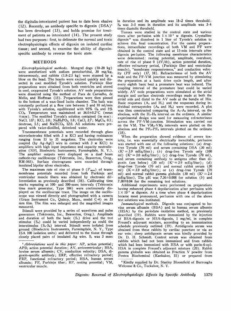
normal rabbit serum by a sodium sulfate precipitationmethod (21). The titers of DSA were determined by thedextran-coated charcoal method as described elsewhere(22, 23). DSA titers are expressed as the highest serumdilutions, 1 ml of which is capable of binding 50% of theadded digoxin-'H (32 ng). Sera from nonimmunized rab-bits (normal rabbit sera) and sera obtained from rabbitsimmunized with antigens unrelated to digoxin exhibited nobinding of digoxin-'H at a 1:20 dilution. In contrast, thetiters of sera containing DSA ranged from 1: 800 to 1: 3200(Fig. 1).
RESULTS
Normal electrophysiologic effectsMembrane characteristics. Isolated canine papillary
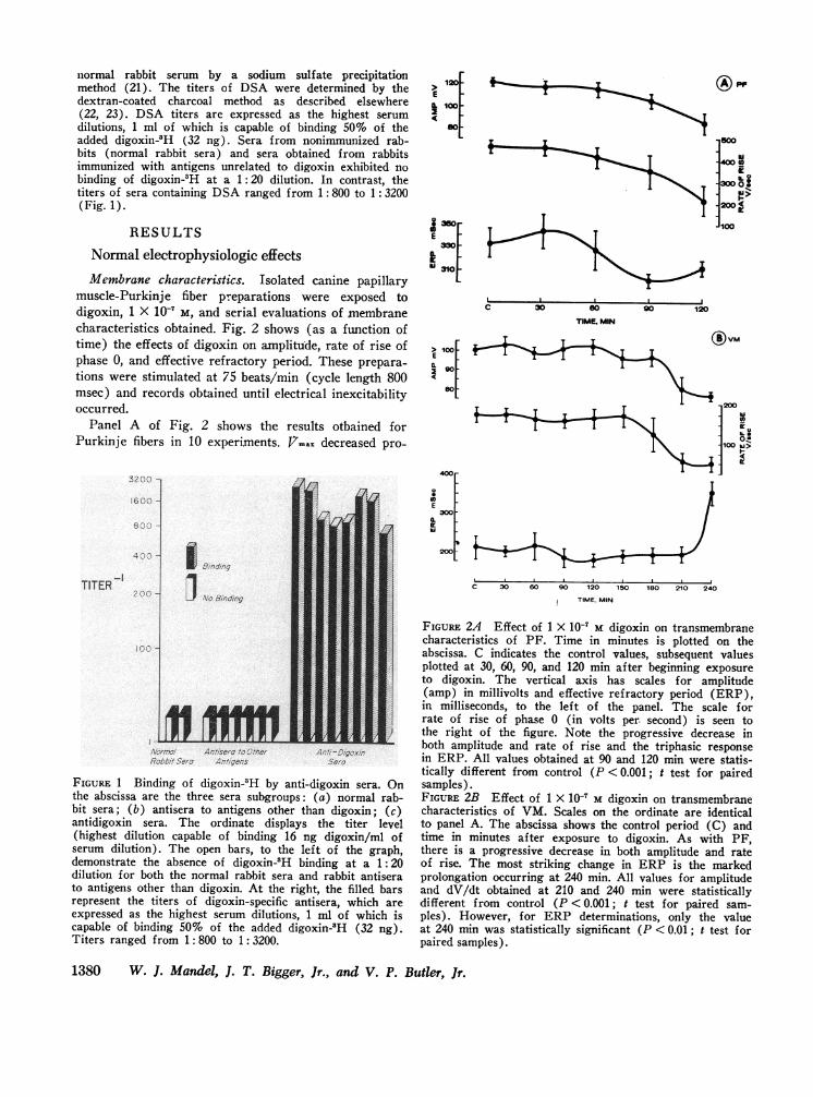
muscle-Purkinje fiber preparations were exposed todigoxin, 1 X 10-' M, and serial evaluations of membranecharacteristics obtained. Fig. 2 shows (as a function oftime) the effects of digoxin on amplitude, rate of rise ofphase 0, and effective refractory period. These prepara-tions were stimulated at 75 beats/min (cycle length 800msec) and records obtained until electrical inexcitabilityoccurred.
Panel A of Fig. 2 shows the results otbained forPurkinje fibers in 10 experiments. i7max decreased pro-
32 -
160, -
TITERI
J ',0o 3wdwrg
.1nIIcarsc 4risero 'C 2"ter
Fr-ort I ser. _r'v-
IFIGURE 1 Binding of digoxin-'H by anti-digoxin sera. Onthe abscissa are the three sera subgroups: (a) normal rab-bit sera; (b) antisera to antigens other than digoxin; (c)antidigoxin sera. The ordinate displays the titer level(highest dilution capable of binding 16 ng digoxin/ml ofserum dilution). The open bars, to the left of the graph,demonstrate the absence of digoxin-'H binding at a 1:20dilution for both the normal rabbit sera and rabbit antiserato antigens other than digoxin. At the right, the filled barsrepresent the titers of digoxin-specific antisera, which areexpressed as the highest serum dilutions, 1 ml of which iscapable of binding 50% of the added digoxin-'H (32 ng).Titers ranged from 1: 800 to 1: 3200.
> 1204E [X 1001-ac
so .~~~~~~~~~~~~~~~~530
c 30 60
TIME. MIN
90
(A) PF
_-500
-4w00.300 0 Y
- m>-200(
-100
120
(®) VM
Ea.
01-
80
E300-
200[
200AlU,0
I100 w>4I
C 30 60 90 120 150 180 210 240
TIME, MIN
FIGURE 2A Effect of 1 X 10' M digoxin on transmembranecharacteristics of PF. Time in minutes is plotted on theabscissa. C indicates the control values, subsequent valuesplotted at 30, 60, 90, and 120 min after beginning exposureto digoxin. The vertical axis has scales for amplitude(amp) in millivolts and effective refractory period (ERP),in milliseconds, to the left of the panel. The scale forrate of rise of phase 0 (in volts per. second) is seen tothe right of the figure. Note the progressive decrease inboth amplitude and rate of rise and the triphasic responsein ERP. All values obtained at 90 and 120 min were statis-tically different from control (P <0.001; t test for pairedsamples).FIGURE 2B Effect of 1 X 10' M digoxin on transmembranecharacteristics of VM. Scales on the ordinate are identicalto panel A. The abscissa shows the control period (C) andtime in minutes after exposure to digoxin. As with PF,there is a progressive decrease in both amplitude and rateof rise. The most striking change in ERP is the markedprolongation occurring at 240 min. All values for amplitudeand dV/dt obtained at 210 and 240 min were statisticallydifferent from control (P < 0.001; t test for paired sam-ples). However, for ERP determinations, only the valueat 240 min was statistically significant (P < 0.01 ; t test forpaired samples).
1380 W. J. Mandel, J. T. Bigger, Jr., and V. P. Butler, Jr.
VENTRICULAR MUSCLE
CL 2000
100mV
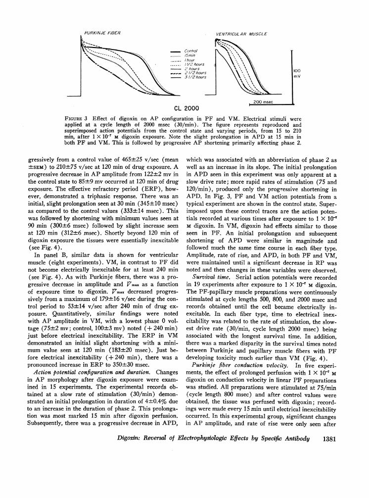
FIGURE 3 Effect of digoxin on AP configuration in PF and VM. Electrical stimuli wereapplied at a cycle length of 2000 msec (30/min). The figure represents reproduced andsuperimposed action potentials from the control state and varying periods, from 15 to 210min, after 1 X 10⁻⁷ M digoxin exposure. Note the slight prolongation in APD at 15 min inboth PF and VM. This is followed by progressive AP shortening primarily affecting phase 2.
gressively from a control value of 465+25 v/sec (mean+SEM) to 210±75 v/sec at 120 min of drug exposure. Aprogressive decrease in AP amplitude from 122±2 mv inthe control state to 85±9 mv occurred at 120 min of drugexposure. The effective refractory period (ERP), how-ever, demonstrated a triphasic response. There was aninitial, slight prolongation seen at 30 min (345±10 msec)as compared to the control values (333±+14 msec). Thiswas followed by shortening with minimum values seen at90 min (300±6 msec) followed by slight increase seenat 120 min (312±6 xnsec). Shortly beyond 120 min ofdigoxin exposure the tissues were essentially inexcitable(see Fig. 4).
In panel B, similar data is shown for ventricularmuscle (eight experiments). VM, in contrast to PF didnot become electrically inexcitable for at least 240 min(see Fig. 4). As with Purkinje fibers, there was a pro-gressive decrease in amplitude and J7max as a functionof exposure time to digoxin. J7.ra decreased progres-sively from a maximum of 179±16 v/sec during the con-trol period to 53±14 v/sec after 240 min of drug ex-posure. Quantitatively, similar findings were notedwith AP amplitude in VM, with a lowest phase 0 vol-tage (75±2 mv; control, 100±3 mv) noted (+ 240 min)just before electrical inexcitability. The ERP in VMdemonstrated an initial slight shortening with a mini-mumvalue seen at 120 min (183±20 msec). Just be-fore electrical inexcitability (+ 240 min), there was apronounced increase in ERP to 350±30 msec.
Action potential configuration and duration. Changesin AP morphology after digoxin exposure were exam-ined in 15 experiments. The experimental records ob-tained at a slow rate of stimulation (30/min) demon-strated an initial prolongation in duration of 4±0.4% dueto an increase in the duration of phase 2. This prolonga-tion was most marked 15 min after digoxin perfusion.Subsequently, there was a progressive decrease in APD,
which was associated with an abbreviation of phase 2 aswell as an increase in its slope. The initial prolongationin APD seen in this experiment was only apparent at aslow drive rate; more rapid rates of stimulation (75 and120/min), produced only the progressive shortening inAPD. In Fig. 3, PF and VMaction potentials from atypical experiment are shown in the control state. Super-imposed upon these control traces are the action poten-tials recorded at various times after exposure to 1 X 10'M digoxin. In VM, digoxin had effects similar to thoseseen in PF. An initial prolongation and subsequentshortening of APD were similar in magnitude andfollowed much the same time course in each fiber type.Amplitude, rate of rise, and APD, in both PF and VM,were maintained until a significant decrease in RP wasnoted and then changes in these variables were observed.
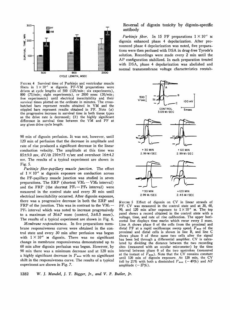
Survival time. Serial action potentials were recordedin 19 experiments after exposure to 1 X 10' M digoxin.The PF-papillary muscle preparations were continuouslystimulated at cycle lengths 500, 800, and 2000 msec andrecords obtained until the cell became electrically in-excitable. In each fiber type, time to electrical inex-citability was related to the rate of stimulation, the slow-est drive rate (30/min, cycle length 2000 msec) beingassociated with the longest survival time. In addition,there was a marked disparity in the survival times notedbetween Purkinje and papillary muscle fibers with PFdeveloping toxicity much earlier than VM (Fig. 4).
Purkinje fiber conduction velocity. In five experi-ments, the effect of prolonged perfusion with 1 X 10⁷ Mdigoxin on conduction velocity in linear PF preparationswas studied. All preparations were stimulated at 75/min(cycle length 800 msec) and after control values wereobtained, the tissue was perfused with digoxin; record-ings were made every 15 min until electrical inexcitabilityoccurred. In this experimental group, significant changesin AP amplitude, and rate of rise were only seen after
Digoxin: Reversal of Electrophysiologic Effects by Specific Antibody
PURKINJE FIBER
. 200 msec
1381
CPF*VM
T
800CYCLE LENGTH, MSEC
Reversal of digoxin toxicity by digoxin-specificantibodyPurkinje fiber. In 15 PF preparations 1 X 10' M
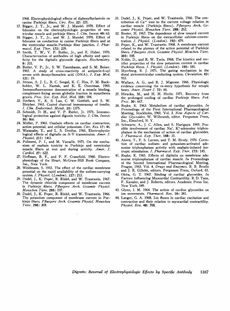
digoxin enhanced phase 4 depolarization. After pro-nounced phase 4 depolarization was noted, five prepara-tions were then perfused with DSAin drug-free Tyrode'ssolution. Recordings were made every 2 min until theAP configuration stabilized. In each preparation treatedwith DSA, phase 4 depolarization was abolished andnormal transmembrane voltage characteristics reestab-
FIGURE 4 Survival time of Purkinje and ventricular musclefibers in 1 X 10' M digoxin. PF-VM preparations weredriven at cycle lengths of 500 (120/min; six experiments),800 (75/min; eight experiments), or 2000 msec (30/min;five experiments) until electrical inexcitability and theirsurvival times plotted on the ordinate in minutes. The cross-hatched bars represent results obtained in VM and thestippled bars represent results obtained in PF. Note (a)the progressive increase in survival time in both tissue typesas the drive rate is decreased; (b) the highly significantdifference in survival time between the VM and PF atany given drive cycle length.
90 min of digoxin perfusion. It was not, however, until120 min of perfusion that the decrease in amplitude andrate of rise produced a significant decrease in the linearconduction velocity. The amplitude at this time was
85+9.0 ;mv, dV/dt 210±75 v/sec and overshoot 16±4.2mv. The results of a typical experiment are shown inFig. 5.
Purkinje fiber-papillary muscle junction. The effectof 1 X 10' M digoxin exposure on conduction across
the PF-papillary muscle junction was studied in seven
preparations. The ERP (shortest VM1- VM2 interval)and the FRP (the shortest PF1 - PF2 interval) were
measured in the control state and every 30 min untilelectrical inexcitability occurred. After digoxin exposure,
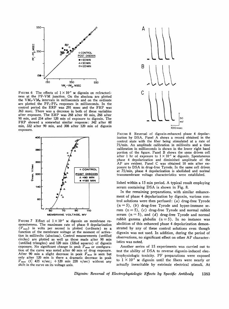
there was a progressive decrease in both the ERP andFRP of the junction. This was in contrast to the VM1-PF1 interval which was noted to increase progressivelyto a maximum of 36±7 msec (control, 3+0.5 msec).The results of a typical experiment are shown in Fig. 6.
Membrane responsiveness. In five preparations mem-
brane responsiveness curves were obtained in the con-
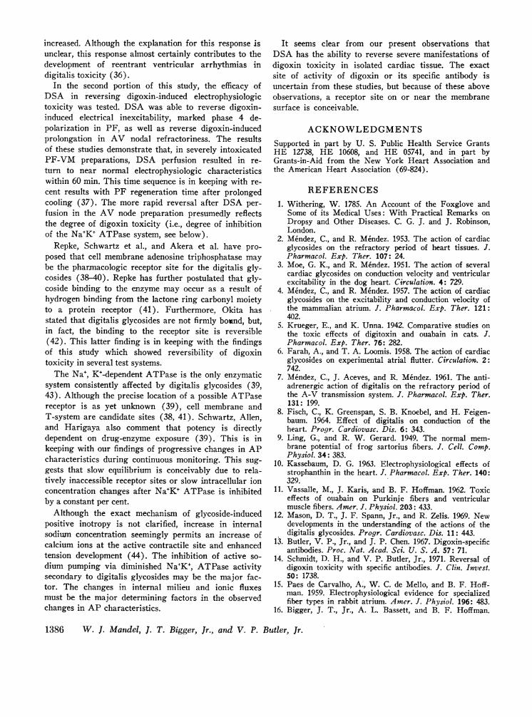
trol state and every 30 min after perfusion was begunwith 1 X 10- M digoxin. There was no significantchange in membrane responsiveness demonstrated up to60 min after digoxin perfusion was begun. However, by90 min there was a minimum decrease and at 120 mina highly significant decrease in i7ma. with no significantshift in the responsiveness curve. The results of a typicalexperiment are shown in Fig. 7.
nA
5001 B
V/secl 100 mV
CONTROL 5 msec
3.09 M/SEC
+ 30 MIN + 60 MIN2.99 M/SEC 2.99 M/SEC
+90 MIN2.99 M/SEC
+120 MIN2.45 M/SEC
FIGURE 5 Effect of digoxin on CV in linear strands ofPF. CV was measured in the control state and at 30, 60,90, and 120 min after exposure to 1 X 10' M. The toppanel shows a record obtained in the control state with a
voltage, time, and rate of rise calibration. The upper hori-zontal line displays time marks which recur every 5 msec.Line A shows phase 0 of the cells from the proximal anddistal PF at a rapid oscilloscope sweep speed. V.ma of theproximal and distal cells is shown in line B, and line Cshows phase 0 of these same two cells after the signalhas been led through a differential amplifier. CV is calcu-lated by dividing the distance between the two recordingsites (measured with an occular micrometer) by the timeinterval between phase 0 of the two upstrokes (measuredat the instant of lma.). Note that the CV remains constantuntil 120 min of digoxin exposure. At 120 min, the CVfell by 21% with both a diminished Vmax (-49%) and APamplitude (-27%).
1382 W. J. Mandel, J. T. Bigger, Jr., and V. P. Butler, Jr.
300-
Z
L, 200-
, 100-(n
r-11r, (: pr-.I . r,,-. &
C,)
Ma-LL7a.
A
VM1-VM2, MSEC
FIGURE 6 The effects of 1 X 10' M digoxin on ref ractori-ness at the PF-VM junction. On the abscissa are plottedthe VM1-VM2 intervals in milliseconds and on the ordinateare plotted the PF,-PFl responses in milliseconds. In thecontrol period the ERP was 295 msec and the FRP was363 msec. There was a decrease in both of these variablesafter exposure. The ERP was 268 after 60 min, 266 after90 min, and 254 after 120 min of exposure to digoxin. TheFRP showed a somewhat similar response: 342 after 60min, 332 after 90 min, and 308 after 120 min of digoxinexposure.
400
380 A
00
300-_ A;
0 CONTROL
j220 A POST DIGOXINE A+9 MINA>:̂ *+120 MIN
140
60-
, *.
50 70 90 110MEMBRANEVOLTAGE, MV
FIGURE 7 Effect of 1 X 10' M digoxin on membrane re-sponsiveness. The maximum rate of phase 0 depolarization(Jfmax) in volts per second is plotted (ordinate) as afunction of the membrane voltage at the moment of activa-tion in millivolts (abscissa). Control measurements (unfilledcircles) are plotted as well as those made after 90 min(unfilled triangles) and 120 min (filled squares) of digoxinexposure. No significant change in peak J7max or configura-tion of the curve was noted after 60 min of drug exposure.After 90 min a slight decrease in peak Pmax is seen butonly after 120 min is there a dramatic decrease in peak17ma,: (C 435 v/sec; + 120 min 220 v/sec) without anyshift in the curve on its voltage axis.
B
'A
50
I- E CmV
1000 msec
FIGURE 8 Reversal of digoxin-enhanced phase 4 depolar-ization by DSA. Panel A shows a record obtained in thecontrol state with the fiber being stimulated at a rate of75/min. An amplitude calibration in millivolts and a timecalibration in milliseconds is shown in the lower right handportion of the figure. Panel B shows the same driven cellafter 1 hr of exposure to 1 X 10' M digoxin. Spontaneousphase 4 depolarization and diminished amplitude of theAP are evident. Panel C was obtained 10 min after ex-posure to DSA in drug-free Tyrode. In the same cell drivenat 75/min, phase 4 depolarization is abolished and normaltransmembrane voltage characteristics were established.
lished within a 15 min period. A typical result employingserum containing DSA is shown in Fig. 8.
In the remaining preparations, with similar enhance-ment of phase 4 depolarization by digoxin, various con-
trol solutions were then perfused: (a) drug-free Tyrode(n = 5), (b) drug-free Tyrode and hyper-immune se-
rum (n = 5), (c) drug-free Tyrode and normal rabbitserum (n - 5), and (d) drug-free Tyrode and normalrabbit gamma globulin (n = 5). In no instance was
abolition of this enhanced phase 4 depolarization demon-strated by any of these control solutions even thoughdigoxin was not used. In addition, during the period ofobservations, no significant effect on other AP character-istics was noted.
Another series of 15 experiments was carried out totest the ability of DSA to reverse digoxin-induced elec-trophysiologic toxicity. PF preparations were exposedto 1 X 10- M digoxin until the fibers were nearly or
actually inexcitable by extrinsic electrical stimuli. At
Digoxin: Reversal of Electrophysiologic Effects by Specific Antibody 1383

this point in time, DSAin drug-free Tyrode or one of thecontrol solutions was used to perfuse the preparationand events followed every 5 min until stable. In all in-stances, perfusion with DSA-Tyrode's solution was as-sociated with slow return to or near control AP charac-teristics. None of the other control solutions demon-strated any significant restorative effect. The time-courseof events of a typical experiment using DSA-Tyrode'ssolution are shown in Fig. 9.
AV Node. The final group of experiments were de-signed to test the ability of DSA to reverse digoxin-in-duced AV conduction delay. AV node preparations from25 rabbits were exposed to 1 X 10' M digoxin after theERP (shortest A1- A2 interval conducted to the Hisbundle) and FRP (shortest H1 -H2 interval) were de-termined in the control state. At a time when A, - Hiinterval had increased by at least 20% above control,the preparation was then perfused with serum containingDSA or one of the various control solutions. In the fivepreparations exposed to the DSA-Tyrode's solution, therewas prompt (15±2 min) return of AV conduction tonormal. In similar preparations, studied at a time when asimilar prolongation of AV conduction was induced bydigoxin, no such decrease in AV conduction time, ERP,or FRPwas noted when the tissue was perfused with anyof the previously described test solutions (Table I).
AC KI4~~~~~~~~~~~~~~~~~~~~~~~~
c ai H
Li
B
Dl k N.I
150~~~~5mV
1000 msec
FIGURE 9 Reversal of digoxin-induced toxicity in PF.Panel A displays the AP and, below, J7 of phase 0 and acalibration (500 v/sec) obtained by electronic differentia-tion. Amplitude time calibrations are shown in the lowerright hand portion of the figure. Panel B shows the samecell 120 min after exposure to 1 X 10- M digoxin. At thispoint in time, DSA in drug-free Tyrode was applied.RP has decreased and the cell is essentially electricallyinexcitable. Panel C shows the same cell 15 min after DSAperfusion. There is now a return toward normal of thecell's electrical characteristics. Panel C shows the samecell, 60 min after DSA exposure. The cell shows evenfurther return toward normal in its electrical character-istics.
Hi H2MSEC 260- * A
220 -
180- 0o0 Control
A - After tx/C 7M Digoxin140 0 - After DSA
100-
100 140 180 220 260 300 340
Al A2 MSEC
FIGURE 10. Effect of digoxin on refractoriness of the AVnode, and its reversal by DSA. The A1-A2 interval is plottedon the abscissa and the H1-H2 response is plotted on theordinate. The basic cycle length is 500 msec. The controlobservations (unfilled circles) demonstrate an ERP of 125msec and FRP of 166 msec. After 45 min exposure to 1 X10- M digoxin (filled triangles), the ERP was 203 msecand FRP 229 msec. DSA was applied and after 10 minrefractory periods measured (filled circles) ; the ERP (118msec) and FRP (172 msec) shortened to values whichwere not significantly different from the control.
The results of a typical experiment utilizing DSA areshown in Fig. 10.
DISCUSSION
Previous studies have described the effects of severaldigitalis compounds on the electrophysiologic character-istics of cardiac tissue (2-8, 10, 11, 24-26). There has,however, been no detailed electrophysiologic investiga-tion of the effects of digoxin on isolated cardiac tissue.The results described in the initial portion of this studydemonstrate that the electrophysiologic properties ofdigoxin in isolated cardiac tissue are qualitatively similarto previously studied glycosides. Specifically, digoxininduced a time-dependent decrease in AP, amplitude, andrate of rise which was noted after 60 min in PF and 180min in VM. The progressive decrease in these variableswas paralleled by a progressive decrease in APD. Thetime-course of these changes cannot, in absolute terms,be compared with previous studies with ouabain andstrophanthin (10, 11) because of differences in molarconcentrations. However, the time-course differentialobtained with digoxin between the more sensitive PFand the less sensitive VM is similar to that noted forouabain (11). In addition, survival time after ouabainand strophanthin exposure has been found to be inverselyrelated to the stimulation frequency. This study hasshown that a quantitatively similar relationship was
1384 W. J. Mandel, J. T. Bigger, Jr., and V. P. Butler, Jr.

TABLE I
Test solutions
Hyper- Normal RabbitDSA- Drug-free immune rabbit gamma
AV refractory period Tyrode tyrode serum serum globulin
msec
Control ERP 13244.8 132±4.8 132±4.8 132±4.8 13244.8FRP 170±8.4 17048.4 170±8.4 170±8.4 170±8.4
n = 25 n = 25 n = 25 n = 25 n = 25
Post-digoxin ERP 199±9.7 19949.7 19949.7 199±9.7 199±9.7FRP 2354±12.2 235± 12.2 235± 12.2 235± 12.2 235± 12.2
n = 25 n = 25 n = 25 n = 25 n = 25P < 0.001* P < 0.001 P < 0.001 P < 0.001 P < 0.001
15 min after test serum ERP 130±6.7 203±6.7 207±7.6 200±t9.9 204±7.9FRP 175±9.6 235±15.1 240±8.9 242±12.8 242±16.1
n=5 n=5 n=5 n=5 n=5P < 0.001 NS NS NS NS
* t test for paired samples, mean ASEM.
found with digoxin (10, 11, 26). Serial studies on therelationship of peak Vnmax to digoxin exposure initiallydemonstrated little change in either PF or VM. How-ever, with continued exposure, there eventually was adecrease in peak Vmax. Associated with this finding wasa corresponding decrease in amplitude and conductionvelocity. The CV decrease would be anticipated as acorresponding significant decrease in its major deter-minants; AP amplitude and rate of rise of phase 0 wereobserved (27). In addition, serial measurements of mem-brane responsiveness were also done. This determination,which is felt to represent the availability of the sodium-carrier system (28), was initially little altered by digoxinexposure. Slight change was noted in Ima. at 90 mnin,and by 120 min a marked decrease in D7max was seenwithout any shift in the curve on its voltage axis.Therefore, digoxin has no specific effect on the sodium-carrier system.
As previously described for strophanthin, at slowstimulation rates (30/min), APD in VM initially in-creased followed by progressive decrease; with a morerapid rate of stimulation (60/min) only AP shorteningwas observed (10). The present study showed similarfindings for digoxin in both VMand PF. Configurationalchanges associated with shortening of APD demon-strated an initial decrease in the plateau or phase 2 fol-lowed by associated change in the slope of phase 3. It isas yet unclear what ionic species are responsible for theplateau phase of the AP. Prior studies have commentedon the contribution of Na+, K+, Cl, Ca++ ions to themaintenance of phase 2 (29-32). Repolarization of phase3 has, in large part, been related to a change in gK(30, 33, 34). The findings in this study suggest that
an increase in gK may be the predominant factor fordigoxin-induced changes in AP configuration.
Studies by Muller have offered partial confirmationby showing that even low concentrations of ouabain areassociated with a decrease in intracellular K+ (24). Re-cent studies by Polimeni and Vassalle have suggestedthat the toxic effects of ouabain are related to its abilityto compete with K+ at the outer layer of the cell mem-brane (26). This would result in inhibition of K+ influxand Na+ efflux. These authors, in addition, demonstratedthat the apparent reason for the time disparity betweenouabain toxicity in VMas compared to PF is that it re-quires 3 times as much ouabain to reduce K+ influx inVMas compared to PF.
Studies on the ERP of VM demonstrated minimalshortening except during overt toxicity when markedprolongation was noted. In contrast, PF ERPwas notedinitially to prolong slightly, followed by pronouncedshortening. Just before inexcitability, the ERP began toincrease slightly.
Studies undertaken to show effects of digoxin on therefractiveness across the PF-VM junction demonstratedprogressive shortening of the effective and functionalrefractory periods as drug perfusion was continued overa 120 min period. These findings are in sharp contrastto the prolongation of the effective and functional re-fractory period of the AV node by digitalis glycosides.These observations demonstrate that, in spite of recentstudies suggesting a similarity between the physiologicsignificance of VM-PF junction and the AV node, theresponse of these two sites to digoxin is clearly dissimilar(35). In our studies refractoriness decreased across theVM-PF junction after digoxin, while conduction time
Digoxin: Reversal of Electrophysiologic Effects by Specific Antibody 1385
increased. Although the explanation for this response isunclear, this response almost certainly contributes to thedevelopment of reentrant ventricular arrhythmias indigitalis toxicity (36).
In the second portion of this study, the efficacy ofDSA in reversing digoxin-induced electrophysiologictoxicity was tested. DSA was able to reverse digoxin-induced electrical inexcitability, marked phase 4 de-polarization in PF, as well as reverse digoxin-inducedprolongation in AV nodal refractoriness. The resultsof these studies demonstrate that, in severely intoxicatedPF-VM preparations, DSA perfusion resulted in re-turn to near normal electrophysiologic characteristicswithin 60 min. This time sequence is in keeping with re-cent results with PF regeneration time after prolongedcooling (37). The more rapid reversal after DSA per-fusion in the AV node preparation presumedly reflectsthe degree of digoxin toxicity (i.e., degree of inhibitionof the Na+K' ATPase system, see below).
Repke, Schwartz et al., and Akera et al. have pro-posed that cell membrane adenosine triphosphatase maybe the pharmacologic receptor site for the digitalis gly-cosides (38-40). Repke has further postulated that gly-coside binding to the enzyme may occur as a result ofhydrogen binding from the lactone ring carbonyl moietyto a protein receptor (41). Furthermore, Okita hasstated that digitalis glycosides are not firmly bound, but,in fact, the binding to the receptor site is reversible(42). This latter finding is in keeping with the findingsof this study which showed reversibility of digoxintoxicity in several test systems.
The Na', K+-dependent ATPase is the only enzymaticsystem consistently affected by digitalis glycosides (39,43). Although the precise location of a possible ATPasereceptor is as yet unknown (39), cell membrane andT-system are candidate sites (38, 41). Schwartz, Allen,and Harigaya also comment that potency is directlydependent on drug-enzyme exposure (39). This is inkeeping with our findings of progressive changes in APcharacteristics during continuous monitoring. This sug-gests that slow equilibrium is conceivably due to rela-tively inaccessible receptor sites or slow intracellular ionconcentration changes after Na+K' ATPase is inhibitedby a constant per cent.
Although the exact mechanism of glycoside-inducedpositive inotropy is not clarified, increase in internalsodium concentration seemingly permits an increase ofcalcium ions at the active contractile site and enhancedtension development (44). The inhibition of active so-dium pumping via diminished Na+K+, ATPase activitysecondary to digitalis glycosides may be the major fac-tor. The changes in internal milieu and ionic fluxesmust be the major determining factors in the observedchanges in AP characteristics.
It seems clear from our present observations thatDSA has the ability to reverse severe manifestations ofdigoxin toxicity in isolated cardiac tissue. The exactsite of activity of digoxin or its specific antibody isuncertain from these studies, but because of these aboveobservations, a receptor site on or near the membranesurface is conceivable.
ACKNOWLEDGMENTSSupported in part by U. S. Public Health Service GrantsHE 12738, HE 10608, and HE 05741, and in part byGrants-in-Aid from the New York Heart Association andthe American Heart Association (69-824).
REFERENCES1. Withering, W. 1785. An Account of the Foxglove and
Some of its Medical Uses: With Practical Remarks onDropsy and Other Diseases. C. G. J. and J. Robinson,London.
2. Mendez, C., and R. Mendez. 1953. The action of cardiacglycosides on the refractory period of heart tissues. J.Pharmacol. Exp. Ther. 107: 24.
3. Moe, G. K., and R. Mendez. 1951. The action of severalcardiac glycosides on conduction velocity and ventricularexcitability in the dog heart. Circulation. 4: 729.
4. Mendez, C., and R. Mendez. 1957. The action of cardiacglycosides on the excitability and conduction velocity ofthe mammalian atrium. J. Pharmacol. Exp. Ther. 121:402.
5. Krueger, E., and K. Unna. 1942. Comparative studies onthe toxic effects of digitoxin and ouabain in cats. J.Pharmacol. Exp. Ther. 76: 282.
6. Farah, A., and T. A. Loomis. 1958. The action of cardiacglycosides on experimental atrial flutter. Circulation. 2:742.
7. Mendez, C., J. Aceves, and R. Mendez. 1961. The anti-adrenergic action of digitalis on the refractory period ofthe A-V transmission system. J. Pharmacol. Exp. Ther.131: 199.
8. Fisch, C., K. Greenspan, S. B. Knoebel, and H. Feigen-baum. 1964. Effect of digitalis on conduction of theheart. Progr. Cardiovasc. Dis. 6: 343.
9. Ling, G., and R. W. Gerard. 1949. The normal mem-brane potential of frog sartorius fibers. J. Cell. Comp.Physiol. 34: 383.
10. Kassebaum, D. G. 1963. Electrophysiological effects ofstrophanthin in the heart. J. Pharmacol. Exp. Ther. 140:329.
11. Vassalle, M., J. Karis, and B. F. Hoffman. 1962. Toxiceffects of ouabain on Purkinje fibers and ventricularmuscle fibers. Amer. J. Physiol. 203: 433.
12. Mason, D. T., J. F. Spann, Jr., and R. Zelis. 1969. Newdevelopments in the understanding of the actions of thedigitalis glycosides. Progr. Cardiovasc. Dis. 11: 443.
13. Butler, V. P., Jr., and J. P. Chen. 1967. Digoxin-specificantibodies. Proc. Nat. Acad. Sci. U. S. A. 57: 71.
14. Schmidt, D. H., and V. P. Butler, Jr., 1971. Reversal ofdigoxin toxicity with specific antibodies. J. Clin. Invest.50: 1738.
15. Paes de Carvalho, A., W. C. de Mello, and B. F. Hoff-man. 1959. Electrophysiological evidence for specializedfiber types in rabbit atrium. Amer. J. Physiol. 196: 483.
16. Bigger, J. T., Jr., A. L. Bassett, and B. F. Hoffman.
1386 W. J. Mandel, J. T. Bigger, Jr., and V. P. Butler, Jr.
1968. Electrophysiological effects of diphenylhydantoin oncanine Purkinje fibers. Circ. Res. 22: 221.
17. Bigger, J. T., Jr., and W. J. Mandel. 1970. Effect oflidocaine on the electrophysiologic properties of ven-tricular muscle and purkinje fibers. J. Clin. Invest. 49: 63.
18. Bigger, J. T., Jr., and W. J. Mandel. 1970. Effect oflidocaine on conduction in canine Purkinje fibers and atthe ventricular muscle-Purkinje fiber junction. J. Phar-macol. Exp. Ther. 172: 239.
19. Smith, T. W., V. P. Butler, Jr., and E. Haber. 1970.Characterization of antibodies of high affinity and speci-ficity for the digitalis glycoside digoxin. Biochemistry.9: 331.
20. Butler, V. P., Jr., S. W. Tanenbaum, and S. M. Beiser.1965. A study of cross-reactivity of antipurin-6-oylserum with deoxyribonucleic acid (DNA). J. Exp. Med.121: 19.
21. Straus, A. J. L., B. C. Seegal, K. C. Hus, P. M. Buck-holder, W. L. Nastuk, and K. E. Osserman. 1960.Immunofluorescence demonstration of a muscle binding,complement-fixing serum globulin fraction in myastheniagravis. Proc. Soc. Exp. Biol. Med. 105: 184.
22. Herbert, V., K. S. Lau, C. W. Gottlieb, and S. W.Bleicher. 1965. Coated charcoal immunoassay of insulin.J. Clin. Endocrinol. Metab. 25: 1375.
23. Schmidt, D. H., and V. P. Butler, Jr. 1971. Immuno-logical protection against digoxin toxicity. J. Clin. Invest.50: 866.
24. Muller, P. 1965. Ouabain effects on cardiac contraction,action potential, and cellular potassium. Circ. Res. 17: 46.
25. Watanabe, Y., and L. S. Dreifus. 1966. Electrophysio-logical effects of digitalis on A-V transmission. Amer. J.Physiol. 211: 1461.
26. Polimeni, P. I., and M. Vassalle. 1971. On the mecha-nism of ouabain toxicity in Purkinje and ventricularmuscle fibers at rest and during activity. Amer. J.Cardiol. 27: 622.
27. Hoffman, B. F., and P. F. Cranefield. 1960. Electro-physiology of the Heart. McGraw-Hill Book Company,Inc., New York.
28. Weidmann, S. 1955. The effect of the cardiac membranepotential on the rapid availability of the sodium-carryingsystem. J. Physiol. (London). 127: 213.
29. Dudel, J., K. Peper, R. Rfidel, and W. Trautwein. 1967.The dynamic chloride component of membrane currentin Purkinje fibers. Pfluegers Arch. Gesamte Physiol.Menschen Tiere. 295: 197.
30. Dudel, J., K. Peper, R. Riidel, and W. Trautwein. 1966.The potassium component of membrane current in Pur-kinje fibers. Pfluegers Arch. Gesamte Physiol. MenschenTiere. 296: 308.
31. Dudel, J., K. Peper, and W. Trautwein. 1966. The con-tribution of Ca++ ions to the current voltage relation incardiac muscle (Purkinje fibers). Pfluegers Arch. Ge-samte Phvsiol. Menschen Tiere. 288: 262.
32. Reuter, H. 1967. The dependence of slow inward currentin Purkinje fibres on the extracellular calcium-concen-tration. J. Physiol. (London). 192: 479.
33. Peper, K., and W. Trautwein. 1968. A membrane currentrelated to the plateau of the action potential of Purkinjefibers. Pfluegers Arch. Gesamte Physiol. Menchen Tiere.303: 108.
34. Noble, D., and R. W. Tsein. 1968. The kinetics and rec-tifier properties of the slow potassium current in cardiacPurkinje fibres. J. Physiol. (London). 195: 185.
35. Myerburg, R. J. 1971. The gating mechanism in thedistal atrioventricular conducting system. Circulation. 43:955.
36. Wallace, A. G., and R. J. Mignone. 1966. Physiologicevidence concerning the re-entry hypothesis for ectopicbeats. Amer. Heart J. 72: 60.
37. Hiraoka, M., and H. H. Hecht. 1971. Recovery fromthe prolonged cooling of cardiac Purkinje fibers. Fed.Proc. 30: 667.
38. Repke, K. 1963. Metabolism of cardiac glycosides. InProceedings of the First International PharmacologicalMeeting, Stockholm, 1961. Vol. 3. New Aspects of Car-diac Glycosides. W. Wilbrandt, editor. Pergamon Press,Inc., Elmsford, N. Y.
39. Schwartz, A., J. C. Allen, and S. Harigaya. 1969. Pos-sible involvement of cardiac Na+, K+-adenosine triphos-phatase in the mechanism of action of cardiac glycosides.J. Pharmacol. Exp. Ther. 168: 31.
40. Akera, T., F. S. Larsen, and T. M. Brody. 1970. Corre-tion of cardiac sodium- and potassium-activated ade-nosine triphosphatase activity with ouabain-induced ino-tropic stimulation. J. Pharmacol. Exp. Ther. 173: 145.
41. Repke, K. 1965. Effects of digitalis on membrane ade-nosine triphosphatase of cardiac muscle. In Proceedingsof the Second International Pharmacological Meeting,Prague, 1963. Vol. 4. Drugs and Enzymes. B. B. Brodieand J. R. Gillette, editors. Pergamon Press, Oxford. 65.
42. Okita, G. T. 1967. Binding of cardiac glycosides. InFactors Influencing Myocardial Contractility. R. D. Tanz,F. Kavaler, and J. Roberts, editors. Academic Press Inc.New York. 549.
43. Glynn, I. M. 1964. The action of cardiac glycosides onion movements. Pharmacol. Rev. 16: 381.
44. Langer, G. A. 1968. Ion fluxes in cardiac excitation andcontraction and their relation to myocardial contractility.Phvsiol. Rev. 48: 708.
Digoxin: Reversal of Electrophysiologic Effects by Specific Antibody 1387