electrophysiologic assessment of neurologic injury -...
TRANSCRIPT

4/9/16
1
Electrophysiologic assessment of neurologic injuryGregory A Kinney, PhD
Dept of Rehabilitation MedicineUniversity of Washington
Seattle, WA
Electrophysiologic Monitoring of Spinal Cord Function
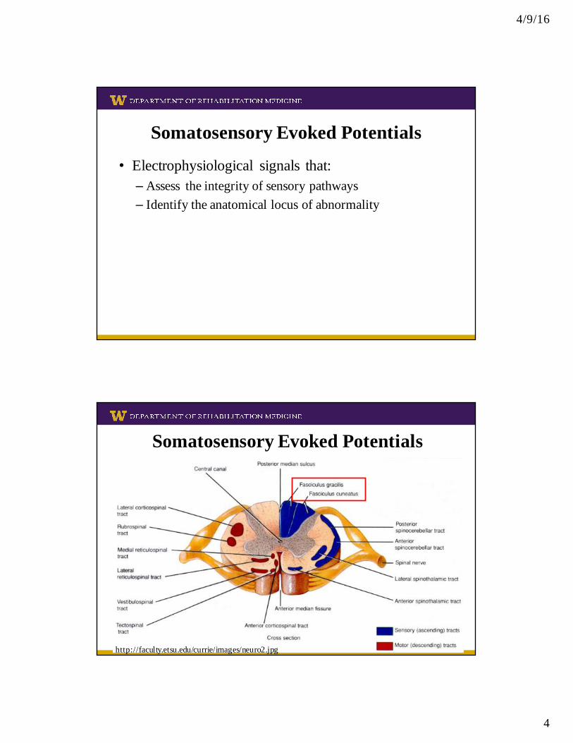
http://faculty.etsu.edu/currie/images/neuro2.jpg

4/9/16
2
Preserving Nervous System Function During Spine Surgery
Preserving Nervous System Function During Spine Surgery
• Somatosensory Evoked Potentials (SEPs)

4/9/16
3
Preserving Nervous System Function During Spine Surgery
• Somatosensory Evoked Potentials (SEPs)• Motor Evoked Potentials (MEPs)
Preserving Nervous System Function During Spine Surgery
• Somatosensory Evoked Potentials (SEPs)• Motor Evoked Potentials (MEPs)• EMG – spontaneous and triggered
4/9/16
4
Somatosensory Evoked Potentials
• Electrophysiological signals that:– Assess the integrity of sensory pathways– Identify the anatomical locus of abnormality
Somatosensory Evoked Potentials
http://faculty.etsu.edu/currie/images/neuro2.jpg
4/9/16
5
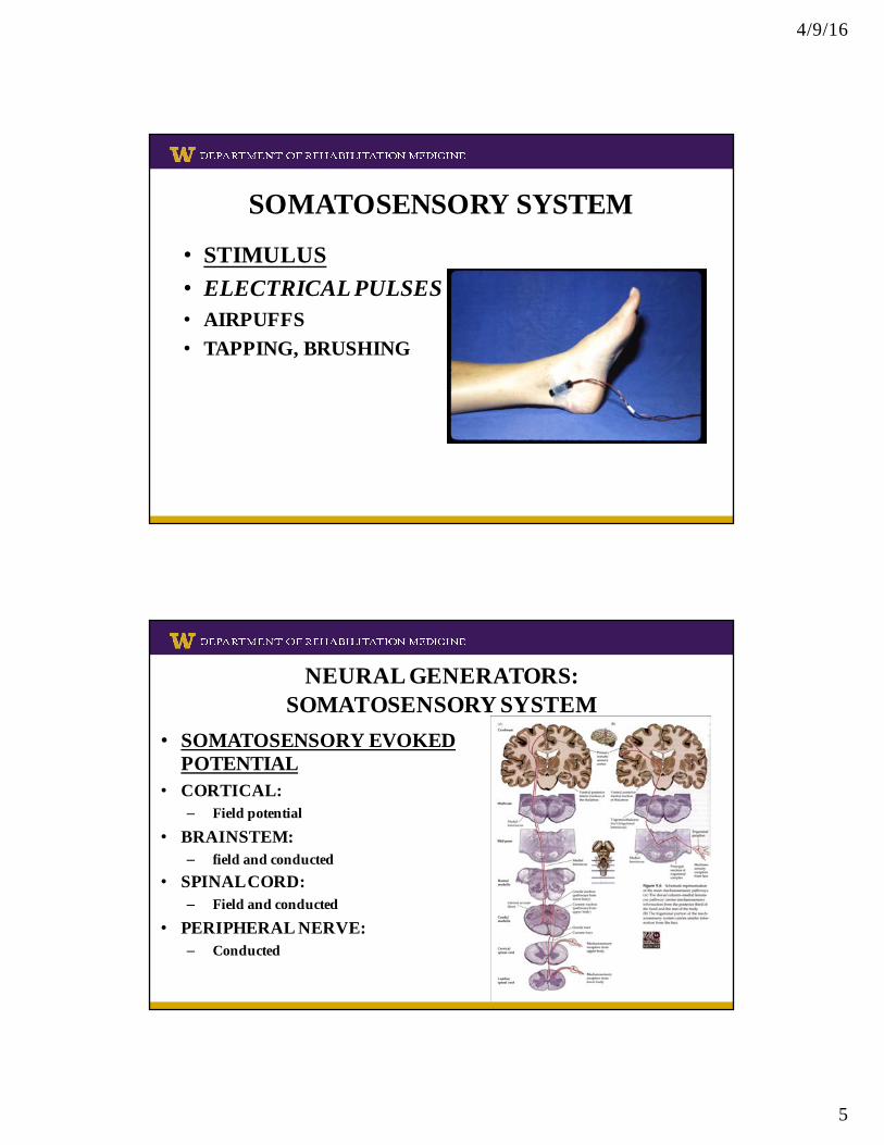
SOMATOSENSORY SYSTEM
• STIMULUS• ELECTRICAL PULSES• AIRPUFFS• TAPPING, BRUSHING
NEURAL GENERATORS: SOMATOSENSORY SYSTEM
• SOMATOSENSORY EVOKED POTENTIAL
• CORTICAL:– Field potential
• BRAINSTEM:– field and conducted
• SPINAL CORD:– Field and conducted
• PERIPHERAL NERVE:– Conducted

4/9/16
6
TIBIAL NERVE SEPsCORTICAL
T10 SPINE
T12 SPINE
L2 SPINE
L4 SPINE
POPLITEAL FOSSA
50 MSSTIM
100 MS
Cz’-Fz
MEDIAN/ULNAR NERVE SEPs
50 MS
STIM
CORTICAL
BRAINSTEM
CERVICAL
ERBS POINT
UPPER ARM
C3’-Fz, C4’-Fz
N20
P25

4/9/16
7
COMPLICATION ASSOCIATED WITH REDUCTION OF KYPHOSIS
BASELINE POSTINSTRUM. - PREREDUCTON
5 MIN POSTREDUCT. TO CLOSING BASELINES TAKEN 5 DAYS LATER 2457
Effectiveness of SEPs:Neurological Deficits With (Solid Bars) And Without (Hashed Bars)
Neuromonitoring During Scoliosis Repair
Nuwer et al, 1995

4/9/16
8
REGIONS OF THE SPINAL CORD
POSTERIOR COLUMNS
CORTICOSPINAL PATHWAY
ANTERIOR HORN
POSTERIOR HORN
SENSORY (PROPRIOCEPTION)
MOTOR:
SPINOTHALAMIC (PAIN/TEMPERATURE)
SEPs
MEPs
VASCULAR SUPPLY OF SPINAL CORD

4/9/16
9
http://www.meditouch.co.il/f/lib13-1.jpg
Anterior Cord Syndrome
Motor Evoked Potentials
• Electrical signals:– Elicited by transcranial stimulation– Directly evaluate the motor columns of the spinal cord– Evaluate the function of specific motor nerve roots of the
spinal cord

4/9/16
10
Motor Evoked Potentials
http://faculty.etsu.edu/currie/images/neuro2.jpg
Transcranial Electrical Motor Evoked MEPs
-Stimulate at the scalpoverlying the motor cortex
-RecordCompound Muscle ActionPotential (CMAP) in handsand legsSpinal cord
Reflects activity in corticospinal pathway
Relatively non-invasive
Allows bilateral analysis and evaluation of motor nerve roots

4/9/16
11
Typical Myogenic MEPs
10 ms/Div500 µV/Div
LN1th
LP1th
(21)LTh-LHy
LN1ta
LP 1ta
(21)LTA-Lpf
MEP-Right Cranium - Average
10 ms/Div
RN1th
RP1th
(22)RTh-RHy
RN1ta
RP 1ta
(22)RTA-Rpf
MEP-Left Cranium - Average
Monitoring findings:Tibial and peroneal SEPs were absent throughout the case.
MEPs were lost bilaterally after instrumentation implanted.Waited for recovery.When no recovery, changed head positioning. Signals recovered.

4/9/16
12
Stimulus Parameters• Pulse Duration: 0.05 msec• Train of pulses: 2-9• Stimulus Amplitude: 100-800 V
• Parameters and responses may vary considerably between patients, and even within the same procedure
Motor Evoked Potentials
• Electrical signals:– Elicited by transcranial stimulation– Directly evaluate the motor columns of the spinal cord– Evaluate the function of specific motor nerve roots of the
spinal cord
• Used with SSEPs, provide a relatively complete monitoring of spinal cord function

4/9/16
13
Advantages of MEPs• Rapid feedback • Directly tests descending motor pathways• Detection in the absence of SEP changes• Highly sensitive to spinal cord blood flow changes • Earlier detection than SSEPs*• For neuromonitoring, MEPs should reduce the
complication of paraplegic/motor impairment– Recent studies have shown combination SEP/MEP monitoring is more
effective at preventing injury/improving outcomes than SEP alone
* Neurophysiological detection of impending spinal cord injury during scoliosis surgery.Schwartz DM, Auerbach JD, Dormans JP, Flynn J, Drummond DS, Bowe JA, Laufer S, Shah SA, Bowen JR, Pizzutillo PD, Jones KJ, Drummond DS.J Bone Joint Surg Am. 2007 Nov;89(11):2440-9.
Limitations of Combined SEP+MEP Monitoring (multimodal IONM)
• False positives– Not uncommon with MEPs

4/9/16
14
Limitations of Combined SEP+MEP Monitoring (multimodal IONM)
• False positives– Not uncommon with MEPs
• False negatives
Limitations of Combined SEP+MEP Monitoring (multimodal IONM)
• False positives– Not uncommon with MEPs
• False negatives
• Nerve Root Complications

4/9/16
15
Spontaneous/Triggered EMG Monitoring
IATROGENIC NERVE STIMULATION
RECORD MUSCLE
STIMULATE NERVE
OR PEDICLE SCREW
RECORD MUSCLE
Use of sEMG in the Operating Room
• Protection and identification, not diagnosis

4/9/16
16
Peripheral Nerve/Muscle Innervation
• Single motor unit not desirable• Large muscle groups innervated by multiple nerve
fibers/fascicles– Potential injury site unknown
• Comprehensiveness with limited specificity• EMG activity not well correlated with outcome
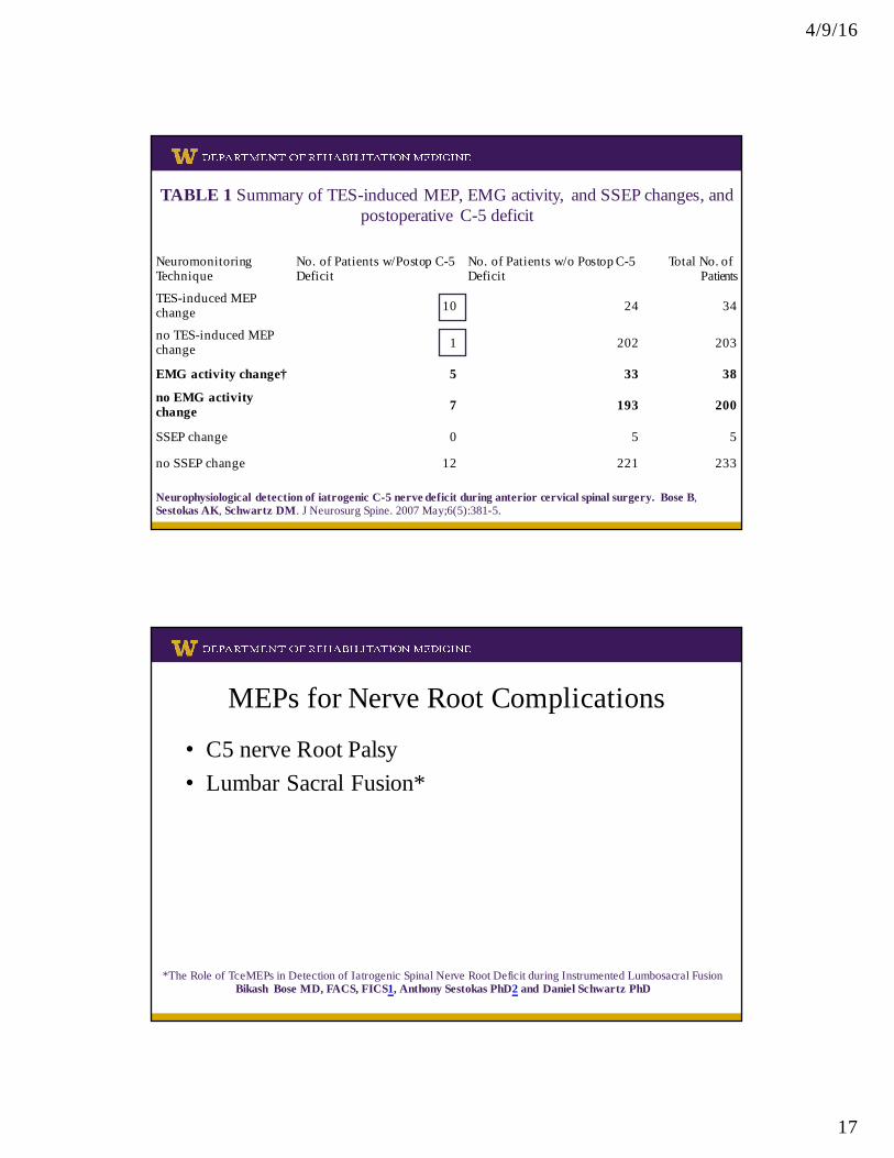
TABLE 1 Summary of TES-induced MEP, EMG activity, and SSEP changes, and postoperative C-5 deficit
Neuromonitoring Technique
No. of Patients w/Postop C-5 Deficit
No. of Patients w/o Postop C-5 Deficit
Total No. of _______Patients
TES-induced MEP change 10 24 34
no TES-induced MEP change 1 202 203
EMG activity change 5 33 38
no EMG activity change 7 193 200
SSEP change 0 5 5
no SSEP change 12 221 233
Neurophysiological detection of iatrogenic C-5 nerve deficit during anterior cervical spinal surgery. Bose B, Sestokas AK, Schwartz DM. J Neurosurg Spine. 2007 May;6(5):381-5.
4/9/16
17
TABLE 1 Summary of TES-induced MEP, EMG activity, and SSEP changes, and postoperative C-5 deficit
Neuromonitoring Technique
No. of Patients w/Postop C-5 Deficit
No. of Patients w/o Postop C-5 Deficit
Total No. of _______Patients
TES-induced MEP change 10 24 34
no TES-induced MEP change 1 202 203
EMG activity change† 5 33 38
no EMG activity change 7 193 200
SSEP change 0 5 5
no SSEP change 12 221 233
Neurophysiological detection of iatrogenic C-5 nerve deficit during anterior cervical spinal surgery. Bose B, Sestokas AK, Schwartz DM. J Neurosurg Spine. 2007 May;6(5):381-5.
MEPs for Nerve Root Complications
• C5 nerve Root Palsy• Lumbar Sacral Fusion*
*The Role of TceMEPs in Detection of Iatrogenic Spinal Nerve Root Deficit during Instrumented Lumbosacral Fusion Bikash Bose MD, FACS, FICS1, Anthony Sestokas PhD2 and Daniel Schwartz PhD