the discovery and characterization of mpf. yoshio masui(toronto university) and clement market(yale...
TRANSCRIPT

The Discovery and Characterization
of MPF

Yoshio Masui(Toronto University) and Clement Market(Yale
University)I
• The processes of germinal vesicle breakdown and first meiotic division are referred to as maturation and can be induced in fully grown oocytes by treatment with the steroid hormone progesterone.
• The first sign of maturation in the hormone treated amphibian oocyte is seen 13-18 hours following progesterone treatment as the germinal vesicle moves near the oocyte surface.
• Germinal vesicle breakdown soon follows,and the oocyte reaches metaphase of the second meiotic division by about 36 hours after hormone treatment.

• Progesterone induces maturation only if it is applied to the external medium surrounding the oocyte;if the hormone is injected into the oocyte,the oocyte shows no response.
• It appears that the hormone acts at the cell surface to trigger secondary changes in the cytoplasm of the oocyte that lead to germinal vesicle breakdown and the other changes associated with maturation

• Yoshio Masui and Clement Markert
• a series of experiments:
• They removed cytoplasm from isolated frog oocytes at various stages following progesterone treatment and injected 40-60 nm of the donor cytoplasm into fully grown, immature oocytes that had not been treated with the hormone.
II
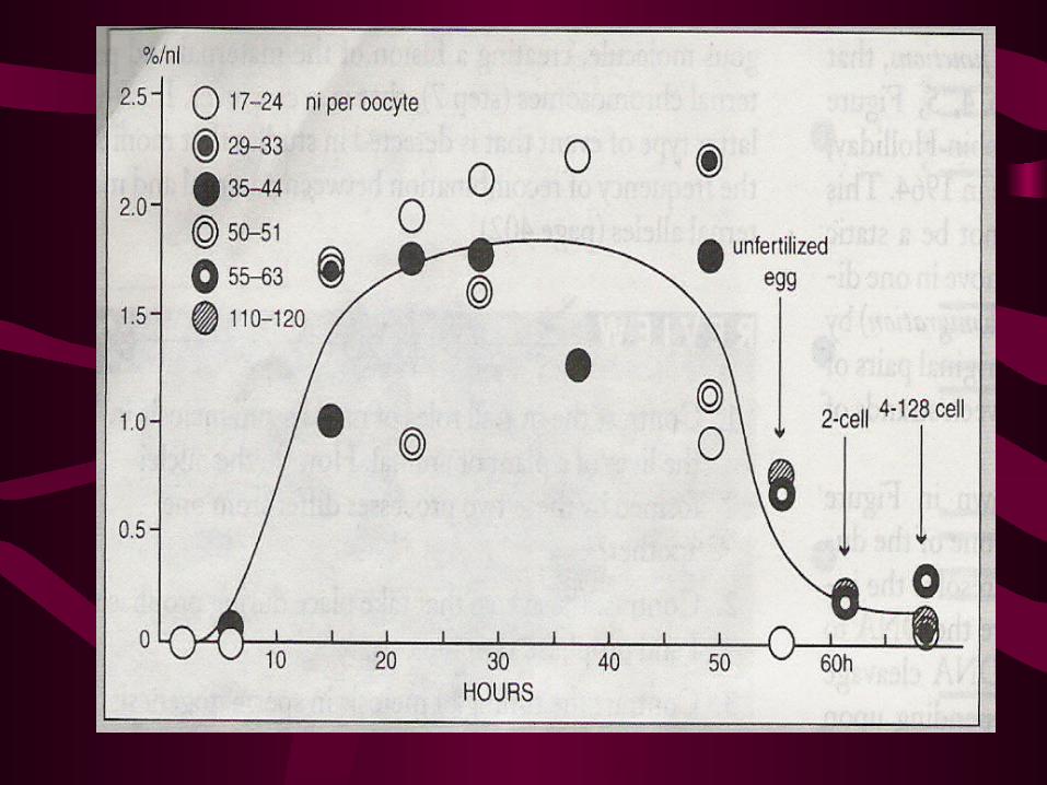
• However, cytoplasm taken from early embryos continued to show some ability to induce oocyte maturation.
• Masui and Markert referred to the cytoplasmic substance(s) that induce maturation in recipient oocytes as “maturation promoting factor,” which become known as MPF.

III
• In 1978,William Wasserman and Dennis Smith of Purdue University published a report on the behavior of MPF during early amphibian development.
• It had been assumed that MPF activity present in early embryos was simply a residue of activity that had been present in the oocyte.
• But they discovered that MPF activity undergoes dramatic fluctuations in cleaving eggs that correlate with changes in the cell cycle.

• It was found, for example, that cytoplasm taken from cleaving frog eggs within 30-60 minutes after fertilization contains little or no detectable MPF activity, as assayed by injection into immature oocytes.
• However, if cytoplasm is taken from an egg at 90 minutes after fertilization, MPF activity can again be demonstrated.
• MPF activity reaches a peak at 120 minutes after fertilization and starts to decline again at 150 minutes.
• At the time the eggs undergo their first cytokinesis at 180 minutes, no activity is detected in the eggs.
• Then, as the second cleavage cycle gets underway, MPF activity once again reappears, reaching a peak at 225 minutes postfertilization, and then declines again to a very low level.

• MPF activity disappears and reappears with the length of the cell cycle.
• In both species, the peak of MPF activity corresponds to the time of nuclear membrane breakdown and the entry of the cells into mitosis.
• These findings suggested that MPF does more than simply control the time of oocyte maturation and, in fact, may play a key role in regulating the cell cycle of dividing cells.

• mammalian cells
• cultured HeLa cells
IV. It became apparent about this same time that MPF activity is not limited to amphibian
eggs and oocytes but is present in a wide variety of organisms.

V. Another element of the machinery that regulates the cell cycle was discovered un studies on sea urchin embryos.
• If sea urchin eggs are fertilized in seawater containing an inhibitor of protein synthesis, the eggs fail to undergo the first mitotic division, arresting at a stage prior to chromosome compaction and breakdown of the nuclear envelope.
• Similarly, each of the subsequent mitotic divisions can also be blocked if an inhibitor of protein synthesis is added to the medium at a time well before the division would normally occur.
• This finding had suggested that one or more proteins must be synthesized during each if the early cell cycles if the ensuing mitotic division is to occur.

VI. In1983, Tim Hunt and his colleagues at the Marine Biological Laboratory at Woods Hole reported on several proteins that are synthesized in fertilized sea urchin eggs but not unfertilized eggs.
R. Timothy (Tim) Hunt
Imperial Cancer Research Fund London, United Kingdom
b. 1943

•To study these proteins further, they incubated fertilized eggs in seawater containing [35S] methionine and withdrew samples at 10-minute intervals beginning at 16 minutes after fertilization.
•Crude protein extracts were prepared from the samples and subjected to polyacrylamide gel electrophoresis,and the labeled proteins were located autoradiographically.
•Several prominent bands were labeled in gels from fertilized egg extracts that were not evident in comparable extracts made from unfertilized eggs.

• One of the bands that appeared strongly labeled at early stages after fertilization virtually disappeared from the gel by 85 minutes after fertilization, suggesting that the protein had been selectively degraded.
• This same band then reappeared in gels from eggs sampled at later times and disappeared once again in a sample taken at 127 minutes after fertilization.

• The fluctuations in the amount of this protein are putted in Figure 4(protein band A)together with the cleavage index,which indicates the time course of the first two cell divisions.
• The degradation of the protein occurs at about the same time that the cells undergo the first and second division.
Hunt and colleagues named the protein “cyclin” .

• Subsequent studies showed that there were two distinct cyclins,A and B,which are degraded at different times during the cell cycle.
• Cyclin A is degraded during a 5-6 minute period beginning just before the metaphase-anaphase transition,and cyclin B is degraded a few minutes after this transition.

Thank you for your attention !