the diagnosis and outcomes of persistent diarrhea in infants aged 0
TRANSCRIPT

Manuscript received: 02.05.2010 Accepted: 22.11.2010
Turk J Gastroenterol 2011; 22 (3): 260-269doi: 10.4318/tjg.2011.0211
Address for correspondence: Ödül E⁄R‹TAfiGazi University School of Medicine,Department of Pediatric Gastroenterology, Ankara, TurkeyPhone: + 90 312 202 51 46 • Fax: + 90 312 215 01 43E-mail: [email protected]
The diagnosis and outcomes of persistent diarrheain infants aged 0-24 months - A Turkish cohort study
Ödül E⁄R‹TAfi1, Sinan SARI1, Buket DALGIÇ1, Aylar POYRAZ2, Arzu ENSAR‹3
Departments of 1Pediatric Gastroenterology and 2Pathology Gazi University School of Medicine, AnkaraDepartment of 3Pathology, Ankara University School of Medicine, Ankara
Amaç: ‹nfantil persistan ishal, literatürde çok iyi dökümante edilmemifltir. Bu çal›flman›n amac›; klini¤imizde izlenen infant dö-nemde ortaya ç›kan persistan ishalli olgular›n, literatür bilgileri eflli¤inde gözden geçirilerek, pediatrik gastroenterologlar için pra-tik bir algoritma oluflturmakt›r. Yöntem: Ondört günden daha uzun süren ishal olgular› persistan ishal olarak kabul edilmifltir.Yafllar› 0-24 ay aras› olan 41 persistan ishal olgusu çal›flmaya dâhil edilmifltir. Hastalar, gaitalar›nda mukus ve/veya kan bulu-nup bulunmamas›na göre kolitler ve enteropatiler olmak üzere iki ana gruba bölünmüfltür. Kolit grubunda ay›r›c› tan› amac›yla;gaita kültürleri, diyet k›s›tlamalar›, tam kan say›m›, akut faz reaktantlar›, Paterji testi, Ailevi Akdeniz atefli ve Behçet hastal›¤›için gen analizleri, kolonoskopi ve biyopsileri yap›lm›flt›r. Enteropati grubunda ise ay›r›c› tan› amac›yla serum ve gaita elektrolit-leri, arterieyel kan gazlar›, serum albümin seviyesi, lipit profilleri, gaita alfa-1 antitripsin seviyeleri, gaita pH’lar›, gaitada ya¤ veredüktan madde varl›¤›na bak›lm›fl, endoskopi ve biyopsileri yap›lm›flt›r. Bulgular: Toplam 41 persistan ishalli olgu içerisinde; 7çölyak hastal›¤›, 5 intestinal lenfanjiektazi, 2 mikrovillus inklüzyon cisimci¤i hastal›¤›, 2 abetalipoproteinemi vakas› ve 11 inek sü-tü protein alerjisi, 27 vakal›k enteropati grubunu oluflturmufltur. Toplam 13 hastadan oluflan kolit grubunda ise; 1 Behçet hasta-l›¤›, 1 kolitis ülseroza ve 11 inek sütü protein alerjisi olgusu mevcuttu. Pankreatik yetmezlik olarak kendini gösteren 2 hasta kistikfibrozis tan›s› ald›lar. Bir hastada ise, kistik fibrozis ve inek sütü protein alerjisi mevcuttu. Sonuç: Literatür incelendi¤inde, bu ol-gu serisi enfeksiyon kaynakl› olmayan en genifl persistan ishal grubunu oluflturmaktad›r.
Anahtar kelimeler: Persistan ishal, infant, inatç› ishal
Background/aims: Infantile persistent diarrhea series are not well documented in the literature. Evaluating the literature, the aimof this study was to document persistent diarrhea cases followed in our center and to constitute a practical diagnostic algorithm forthe pediatrician by means of surveying methods. Methods: Diarrheas lasting more than 14 days were accepted as persistent diarr-hea. Forty-one persistent diarrhea cases aged 0-24 months were investigated in this study. The cases were evaluated for the presen-ce of mucus and/or leukocytes in stool and were thus divided into two major groups as colitis or enteropathies. For the differentialdiagnosis of the persistent colitis group, stool cultures, dietary restrictions, complete blood count, acute phase reactants, pathergytest, gene analysis for familial Mediterranean fever and Behçet’s disease, colonoscopy, and biopsies were performed. In the persis-tent enteropathy group, differential diagnosis was made with the following tests: serum and stool electrolytes, arterial blood gases,serum albumin, total protein, lipid profile, stool alpha-1 antitrypsin level, stool pH, presence of stool fat and reducing substance,endoscopy, and biopsies. Results: The 27 persistent enteropathy cases included 7 celiac disease, 5 intestinal lymphangiectasia, 2microvillus inclusion disease, 2 abetalipoproteinemia, and 11 cow’s milk allergy. The 13 cases of the infantile colitis group included1 Behçet’s disease, 1 colitis ulcerosa and 11 cow’s milk allergy. Two cases presenting as pancreatic insufficiency were diagnosed ascystic fibrosis. One case was diagnosed as cystic fibrosis plus cow’s milk allergy. Conclusions: Reviewing the literature, these ca-ses represent the largest non-infectious infantile group of persistent diarrheas. A practical diagnostic algorithm for persistent diarr-heas has been constituted.
Key words: Persistent diarrhea, infant, intractable diarrhea
ORIGINAL ARTICLE
‹nfantil (0-24 ay) persistan ishallerde tan› ve takip: Türk kohort çal›flmas›
INTRODUCTION
Diarrheal disease is a major cause of childhoodmorbidity and mortality worldwide. Major advan-ces in the understanding of the pathophysiology ininfantile persistent and intractable diarrheas al-low a new conceptual view of this heterogeneousgroup of disorders. Several enteral feeding pro-ducts may be required. If enteral therapy fails, pa-renteral nutrition becomes necessary. Sometimes,none of these is sufficient for this heterogeneousand difficult group. Diarrheas lasting more thantwo weeks must be investigated for diagnosis andtherapy. Chronic diarrhea etiology may differ dueto epidemiologic and risk factors between develo-ped and developing countries. Risk factors for de-veloping countries may be listed as: protein ener-gy malnutrition, vitamin A deficiency, zinc (Zn)deficiency, inadequate mother-fed babies, and ac-quired immune deficiencies. Exploring the Eng-lish and Turkish literature, we found no data abo-ut infantile chronic diarrhea in Turkey. This workis not a multicenter study and may not reflect a vi-ew on Turkey; however, as being the first publis-hed data, we believe it is worthy. The aim of thisstudy was to constitute an easily applicable, andin addition, developable diagnostic guideline thro-ughout the laboratory tests and results phase (1-3).
MATERIALS AND METHODS
Gazi University School of Medicine Pediatric Gas-troenterology Clinic in Ankara is a major center inTurkey with an average of 5000 outpatients and1200 endoscopies per year. This Pediatric Gastro-enterology Clinic is a referral center especially inchronic diarrhea and celiac disease (CD). None ofthe chronic diarrhea cases detailed below includedany infectious-originated states. We believe per-sistent and chronic diarrheas of infectious originare treated generally in primary and secondarycenters without referral. Diarrheas lasting morethan 14 days were accepted as persistent diarrhe-a. Forty-eight patients aged 0-24 months who ad-mitted to our clinic with persistent diarrhea bet-ween 2003 and 2007 were analyzed retrospecti-vely. Seven patients were excluded as they wereseen once as an outpatient and did not complywith follow-ups. Diarrheal symptoms were recor-ded as time of onset, frequency and presence ofmucus or blood in stool. Weight and height measu-rements, degree of dehydration and presence ofedema were also noted. The primary step is eva-
luating the cases with macroscopic (mucus and/orblood) plus microscopic stool examination, and thecases are grouped into either the enteropathy orcolitis group. This step facilitates further specificdiagnosis. Laboratory tests to be performed forspecific diagnosis were as follows: complete bloodcount, serum electrolytes, liver function tests, li-pid profile, apolipoprotein B, total protein, albu-min, acute phase reactants, arterial blood gases,immunoglobulins, skin prick test for food allergy,celiac antibodies, ferritin, vitamin B12, folic acid,sweat test, and cystic fibrosis (CF) and familialMediterranean fever (FMF) disease gene analysis.Stool samples were examined for microscopy andculture, pH, presence of fat and reducing substan-ce, occult blood, alpha-1 antitrypsin level, andelectrolytes. Abdominal ultrasonography (USG)and small and large bowel radiographs were per-formed. Endoscopic and/or colonoscopic examinati-ons were done, and endoscopic biopsies were ta-ken. Endoscopic biopsies were performed with anOlympus GIF P230 videogastroscope (OlympusOptical Corporation, Tokyo, Japan), and at leasttwo samples were obtained under direct visualiza-tion. Biopsy specimens were evaluated in lightmicroscopy (LM) and/or electron microscopy (EM).Patients were initially classified as enteropathyincluding protein-losing diarrhea, colitis and pan-creatic insufficiency based on history, physicalexamination and laboratory tests. Twenty-sevenpatients (65.8%) were diagnosed as enteropathy [7CD, 5 primary intestinal lymphangiectasia (PIL),2 microvillus inclusion disease (MID), 2 abetali-poproteinemia (ABL), 11 cow’s milk protein al-lergy, enteropathy type]. The colitis group (31.7%)was composed of 11 cow’s milk allergy, 1 infantilecolitis and 1 Behçet’s disease patients. Two pati-ents (4.8%) were diagnosed as CF. One case wasdiagnosed as cow’s milk protein allergy plus CF.Twenty-seven patients (65.8%) were outpatients.Fourteen patients (34.1%) were hospitalized. Thediagnosis and treatment, complications and fol-low-up of cases are as follows.
I- ENTEROPATHY GROUP
IA- Celiac Disease
Seven cases under the age of 2 were diagnosed asCD during the period 2003-2007 in our clinic. Se-ven patients admitted with typical symptoms ofCD like diarrhea and abdominal distention. Stoolswere watery and shapeless. Biopsy specimens we-re classified according to Marsh classification. No-
Persistent diarrhea in infants
261
ne of the cases had IgA deficiency. The delay bet-ween onset of symptoms and diagnosis was an ap-proximate average period of 6.3 months. The soci-ocultural status, being a rural population, and dif-ficulties in reaching a specialist or an urban hos-pital partly explain this diagnostic delay. Demog-raphic and clinical features of CD patients areshown in Table 1. Once the diagnosis was made,other family members were also screened for CD.Cases were evaluated for iron (Fe), Zn, folic acidlevels, and bone mineral densitometry. Symptoms,serologies, weight and height measurements, andclinical follow-ups were scheduled at 3-month in-tervals.
IB- Primary Intestinal Lymphangiectasia
Intestinal lymphangiectasia (IL) was observed in5 of 41 cases (12.2%). These cases were followed aschronic diarrhea during the infantile period. Allpatients presented with hypoalbuminemia andedema, except Case 3, who had consanguineousparents. Hepatic and renal functions and urineprotein of the cases were all in normal ranges. Se-condary IL was excluded with echocardiographyand abdominal USG evaluation. Cases 3 and 5 we-
re considered as having ascites formation by abdo-minal ultrasound. Ascites fluids were in chylousstate and triglycerides were 156 and 144 mg/dl inthese cases, respectively. The demographic and la-boratory characteristics of the 5 cases are summa-rized in Table 2. Follow-up periods of IL cases we-re a minimum of 6 months and maximum of 6 ye-ars (average: 33 months). None of the cases wascontrolled with a high-protein diet and usage ofmedium-chain triglycerides (MCT) alone; all ofthem required octreotide treatment. The youngestpatient (Case 5) received a single octreotide doseof 25 μg/day and symptoms were controlled withthis dose. In Case 3, 25 μg/day octreotide dosageswere started after diagnosis of IL. During the hos-pital follow-up, diarrhea continued and serum al-bumin levels were further reduced to 1.8 g/dl. Al-bumin was replaced and octreotide dosage waselevated during the follow-up according to the deg-ree of edema and albumin requirement periods. Inthe last year, octreotide dosage was a total of 125μg/day and albumin requirement period was 4months in each. Case 1 and Case 4 are today 7 and8 years old, respectively, with no symptoms, andwe managed to cut-off octreotide treatment in
E⁄R‹TAfi et al.
262
Age of Onset of Clinical features Serology Endoscopic Marshdiagnosis symptoms Sex Abdominal Diarrhea tT tT AGA AGA visualization classification(Months) (Months) distention IgA IgG IgA IgG
23 12 F + - + + + + Mucosal Cracks Type 3c
18 12 M + + + + - - Normal Type 3a
22 15 M + + + - + + Normal Type 3c
22 20 F + + + - + + Mucosal cracks Type 3c
21 12 M + + + + + + Mucosal cracks Type 3c
23 18 M + - + + - - Mucosal cracks Type 3a
14 10 M + + + + + + Mucosal cracks Type 3c
Table 1. Demographic and clinical features of celiac disease patients
Age Onset of Physical Examination Immunoglobulin Albumin Fecal alpha 1 Endoscopic Dilated(Months) symptoms Ascites Edema levels (mg/dl) (gr/dl) antitrypsin findings lymphatic
Sex symmetric IgA IgG IgM levels ducts in LM
Case 1 23, F Since birth - + 60 300* 55 2,5 300 mg/dl Multiple +(N:147-245) whitish spots
Case 2 20, M 19 months - + 45 345* 125 2,6 350 mg/dl Multiple +(N: 147-245) whitish spots
Case 3 4, F Since birth + + 18 180* 100 2,5 - Multiple -(Chylous) whitish spots
Case 4 18, M 15 months - + 25 389* 67 2,5 - Multiple +whitish spots
Case 5 2, F Since birth + + 10 125* 76 2,0 - Multiple +(Chylous) whitish spots
*Immunoglobulin levels indicated as bold characters are low according to age group
Table 2. Demographic and laboratory characteristics of intestinal lymphangiectasia patients
both cases. Headache was the only side effect pre-sented in Case 4 and it disappeared with disconti-nuation of octreotide. Case 2 has had no compla-ints for the last year and attained a normalgrowth pattern, so octreotide dosage was reduced.Diuretics were administered to the patient withascites.
IC- Microvillus Inclusion Disease
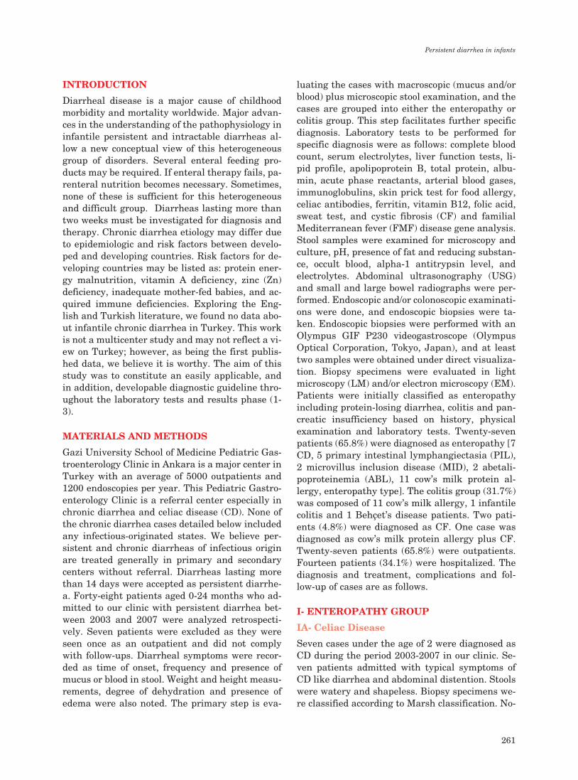
Two male patients aged 45 and 56 days were diag-nosed with MID. Onset symptoms were watery di-arrhea without any blood or mucus beginning from3 and 15 days of life, respectively. Diarrhea resul-ted in serious weight loss and dehydration. Bothcases were admitted to our clinic with criticaldehydration and acidotic state. Consanguineousparents, polyhydramnios and admittance weightbelow birth weight were the common features ofthe two cases. Mucosal findings in the endoscopicevaluation of both cases after general health condi-tion improvement were considered as normal. Vil-lous atrophy was noted in LM examination in bothcases. EM examination of Case 1 showed normalfinding at the first examination. The patient washospitalized for 156 days, and in the 12th month,endoscopic evaluation was repeated during a well-ness period. After one year, in the second EM exa-mination, typical intracytoplasmic inclusion bodieswere present, and the diagnosis of MID was made.Case 1 died from nosocomial sepsis. In Case 2, di-agnosis became definite due to the first EM exami-nation (Figure 1) that showed a typical intracytop-lasmic inclusion bodies pattern concordant withMID. Case 2 also died from a nosocomial sepsis af-ter a 68-day hospitalization period.
ID- Abetalipoproteinemia
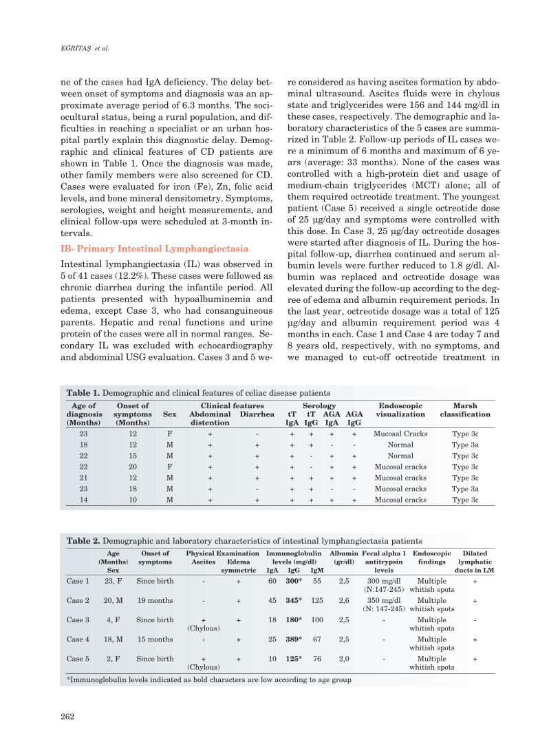
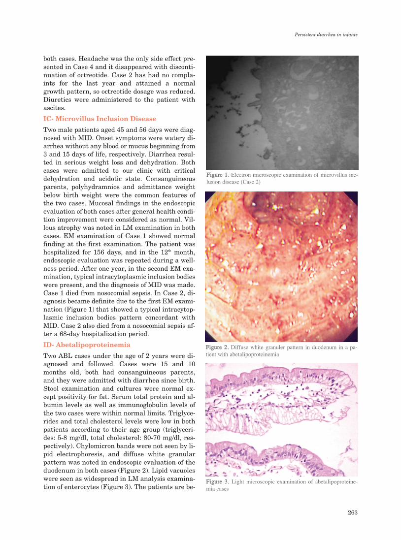
Two ABL cases under the age of 2 years were di-agnosed and followed. Cases were 15 and 10months old, both had consanguineous parents,and they were admitted with diarrhea since birth.Stool examination and cultures were normal ex-cept positivity for fat. Serum total protein and al-bumin levels as well as immunoglobulin levels ofthe two cases were within normal limits. Triglyce-rides and total cholesterol levels were low in bothpatients according to their age group (triglyceri-des: 5-8 mg/dl, total cholesterol: 80-70 mg/dl, res-pectively). Chylomicron bands were not seen by li-pid electrophoresis, and diffuse white granularpattern was noted in endoscopic evaluation of theduodenum in both cases (Figure 2). Lipid vacuoleswere seen as widespread in LM analysis examina-tion of enterocytes (Figure 3). The patients are be-
Persistent diarrhea in infants
263
FFiigguurree 11.. Electron microscopic examination of microvillus inc-lusion disease (Case 2)
FFiigguurree 22.. Diffuse white granuler pattern in duodenum in a pa-tient with abetalipoproteinemia
FFiigguurree 33.. Light microscopic examination of abetalipoproteine-mia cases
ing followed at 3-month intervals and supportedwith a diet rich with MCT and fat-soluble vita-mins. Ophthalmic and neurologic consultationsare done periodically.
IE- Pancreatic Insufficiency (Cystic Fibrosis)
Two cases representing pancreatic insufficiencyand chronic diarrhea were followed between 2003and 2007. Case 1 was 54 days old and admittedwith vomiting and diarrhea since birth. Growthretardation was present due to excessive diarrheawithout mucus or blood. In physical examination,lower extremity and eyelid edema was significant.Stool fat was positive, pH: 5, and reducing subs-tance was negative. Biochemistry values were asfollows: total protein: 4.2 g/dl, albumin: 2.0 g/dl,kidney functions: normal, aspartate aminotransfe-rase (AST): 58 U/L, alanine aminotransferase(ALT): 82 U/L, and gamma glutamyl transpeptida-se (GGT): 170 U/L. Sweat test result was perfor-med twice with a result of 100 mEq/L, so the pati-ent was accepted as CF. Genetic analysis showedN1303K/N1303K homozygote mutation. Case 2was a 4-month-old girl that had been followed ascow’s milk protein allergy disease (enteropathytype), and increasing diarrheal symptoms led toreevaluation of the patient. There was no adapta-tion problem of the mother to the given dietary re-commendations and the patient was only breast-fed. Stool microscopy was clear and cultures werenegative for pathogens. Stool pH was 5, fat waspositive and reducing substance was negative.Sweat test resulted in 90 mEq/L twice and geneti-cal analysis for CF revealed class 2 mutation. Thepatient was diagnosed as cow’s milk protein al-lergy and CF. The 2 patients are under treatmentwith pancreatic enzymes.
IF- Cow’s Milk Protein Allergy (EnteropathyType)
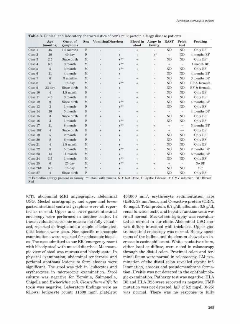
During the period 2003-2007, a total of 35 caseswere diagnosed as enteropathy type cow’s milkprotein allergy. Eleven of them had diarrhea las-ting more than 14 days without mucus or blood.Stool counts were increased and stools were wa-tery and shapeless. No leukocytes or erythrocyteswere seen in microscopic evaluation. Fat and re-ducing substance were negative and pH was nor-mal. Demographic features are summarized inTable 3. Fully hydrolyzed cow’s milk formula andadditional foods were started. All of the patientsexcept one benefitted from the diet. Diarrhea per-sisted despite dietary regulations and this patient
showed hypoalbuminemia during breastfeeding.Suspected diagnosis was CF, sweat test was posi-tive twice, and Class 2 mutation was detected.This case was followed as cow’s milk protein al-lergy plus CF. The CF-associated patient had exa-cerbation in symptoms during restart of cow’smilk, and despite reaching 2 years of age is stillunder dietary restrictions.
II- COLITIS GROUP
IIA- Cow’s Milk Protein Allergy
Between 2003 and 2007, 38 patients were diagno-sed as colitis type cow’s milk protein allergy disea-se in our clinic. Eleven of them had diarrhea las-ting more than 14 days with mucus or blood. The-ir characteristics are summarized in Table 3. No-ne of them had malnutrition or perianal fissureand all patients seemed to be healthy. All patientswith mucoid diarrhea had leukocytes in microsco-pic examination. Stool cultures were negative forall patients. Vomiting was the additional symp-tom of 3 patients, 2 of them with gastroesophage-al reflux disease. Cow’s milk protein allergy is anon-IgE-associated state, and allergic screeningwas not performed in most of the patients as seenin our follow-up scale. After the diagnosis phase,cow’s milk elimination was started and prohibiti-on of milk products was implemented to mothersof mother-fed babies. The earliest response to die-tary restrictions was noted at the end of the firstweek and the late response was in 3 weeks. Die-tary restrictions were gradually reduced in follow-ups after 1 year of age.
IIB- Behçet’s Disease
A single infant case with colitic manifestationswas diagnosed as Behçet’s disease. The case ad-mitted at the age of 10 months. The onset of the di-sease was at the age of 45 days with episodic ab-dominal pain and fever followed by rectal bleedingand mucoid diarrhea. History revealed recurrentperianal, oral aphthous lesions and complicatedafebrile convulsion. Two male siblings had died inthe infantile period due to afebrile convulsion, rec-tal bleeding and mucoid diarrhea. The parents we-re consanguineous. None of the family membershad Behçet’s disease diagnosis. Before referral toour clinic, afebrile convulsion investigation resul-ted in normal neurologic and ophthalmic examina-tion. Electroencephalogram, cranial magnetic re-sonance imaging (MRI), computerized tomography
E⁄R‹TAfi et al.
264
(CT), abdominal MRI angiography, abdominalUSG, Meckel scintigraphy, and upper and lowergastrointestinal contrast graphies were all repor-ted as normal. Upper and lower gastrointestinalendoscopy were performed in another center. Inthese evaluations, colonic mucosa not fully visuali-zed, reported as fragile and a couple of telangiec-tatic lesions were seen. Non-specific microscopicexaminations were reported for endoscopic biopsi-es. The case admitted to our ER (emergency room)with bloody stool with mucoid diarrhea. Macrosco-pic view of stool was mucous and bloody state. Inphysical examination, abdominal tenderness andperianal aphthous lesions to form abscess weresignificant. The stool was rich in leukocytes anderythrocytes in microscopic examination. Stoolculture was negative for Yersinia, Salmonella,Shigella and Escherichia coli. Clostridium difficiletoxin was negative. Laboratory findings were asfollows: leukocyte count: 11800 mm3, platelets:
464000 mm3, erythrocyte sedimentation rate(ESR): 38 mm/hour, and C-reactive protein (CRP):40 mg/dl. Total protein: 6.7 g/dl, albumin: 3.8 g/dl,renal function tests, and hepatic function tests we-re all normal. Meckel scintigraphy was reevalua-ted as normal in our clinic. Abdominal USG sho-wed diffuse intestinal wall thickness. Upper gas-trointestinal endoscopy was normal. Biopsy speci-mens of the bulbus and duodenum showed an in-crease in eosinophil count. White exudative ulcers,either local or diffuse, were noted in colonoscopythrough the distal colon. Proximal colon and ter-minal ileum were normal in colonoscopy. LM exa-mination of the distal colon revealed cryptic inf-lammation, abscess and pseudomembrane forma-tion. Uveitis was not detected in the ophthalmolo-gic examination. Pathergy test was negative. HLAB5 and HLA B25 were reported as negative. FMFmutation was not detected. IgD of 5.2 mg/dl (0-25)was normal. There was no response to fully
Persistent diarrhea in infants
265
Age Onset of Sex VomitingDiarrhea Blood in Atopy in RAST Prick Feeding(months) symptoms stool family test
Case 1 45 1,5 months F - - + - ND ND Only BF
Case 2 20 40 day F - + + +* + ND 4 months BF
Case 3 2,5 Since birth M - +** + - ND ND Only BF
Case 4 6,5 3 month M - +** + - + - 1 month BF
Case 5 5 3 month M - +** + - ND ND Only BF
Case 6 11 4 month M - + - - ND ND 4 months BF
Case 7 6 3 months M - - + - ND ND 3 months BF
Case 8 6 15 day M - +** + - ND ND BF & formula
Case 9 33 day Since birth M - + - - ND ND BF & formula
Case 10 4 1,5 month F - - + - ND ND Only BF
Case 11 4,5 3 month F - - + - ND ND Only BF
Case 12 9 Since birth M + +** + - ND ND 4 months BF
Case 13 3 1 month F - +** - - ND ND Only BF
Case 14 10 5 month F - - + - - - 4 months BF
Case 15 3 Since birth F + + - - ND ND Only BF
Case 16 3 1 month F - +** + - ND ND Only BF
Case 17 11 8 month F - +** + + + + 5 months BF
Case 18¥ 4 Since birth F + + + - + ++ Only BF
Case 19 5 2 month F - + + - ND ND Only BF
Case 20 8 6 month F - + + - ND ND Only BF
Case 21 4 2,5 month M - + + - ND ND Only BF
Case 22 8 5 month M - +** + - ND ND 2 months BF
Case 23 14 11 month M - + + - ND ND 6 months BF
Case 24 3,5 1 month M - +** + - ND ND Only BF
Case 25 6 25 day M - +** + + + - No BF
Case 26# 6,5 15 day M - + + - ND ND BF
Case 27 4 Since birth F - + + - ND ND Only BF
*: Penicillin allergy present in family, **: stool with mucus, ND: Not Done, ¥: Cystic Fibrosis, #: CMV infection, BF: BreastFed
Table 3. Clinical and laboratory characteristics of cow’s milk protein allergy disease patients
hydrolyzed cow’s milk formula. The patient wasdiagnosed as inflammatory bowel disease (IBD)and/or intractable colitis. Under steroid adminis-tration, his symptoms diminished and 5-acetylsa-licylic acid (ASA) treatment was started. The pati-ent returned to the ER with mucoid and bloodystool one month after discharge. A second colonos-copy was similar to the first except for regressionin white ulcerative lesions. Repeat microscopicexamination was parallel to the first. The pati-ent’s condition worsened after the second colonos-copy. Findings were as follows: fever, leukocytosis(24000 mm3), thrombocytosis (617000 mm3), highESR: 80 mm/hour, high CRP: 134 mg/dl, and ex-cessive bowel dilatation in abdominal X-ray. Thepatient was considered as toxic megacolon, anti-biotics were started, and the condition was takenunder control. Finally, the patient was consideredas Behçet’s disease. Colchicine treatment wasstarted. The case is now 4 years old, has attaineda normal growth pattern and all symptoms are un-der control.
IIC- Infantile Ulcerative Colitis
A 21-month-old girl admitted to our clinic. Shehad diarrhea since birth, which turned to mucoidand bloody stool state after 6 months. The patientreceived amebiasis treatment before admittance,her parents were consanguineous, and her sisterhad FMF diagnosis. In physical examination, thepatient seemed pale and weak. Body weight was5880 g (<3 p) and height was 64 cm (<3 p). Stoolexamination was rich in leukocytes and erythrocy-tes, Clostridium difficile toxin was negative, andstool cultures for Salmonella, Shigella, Yersinia,and E. coli were negative. Laboratory findings we-re as follows: WBC: 22000 mm3, Hb: 7.4 g/dl, me-an corpuscular volume (MCV): 56, RBC: 4.23 x 10⁶,PLT: 800000/mm3, total protein: 5.2 g/dl, and albu-min: 3.2 g/dl; hepatic and renal function tests we-re all normal. ESR: 45 mm/hour, CRP: 75 mg/dland immunoglobulin levels were normal, and sero-logy for CD was negative. During colonoscopy, theterminal ileum was considered as normal; colonicmucosa showed hyperemia, fragility, edema andulcerative lesions. Biopsy specimens of colonic mu-cosa revealed inflammation with cryptitis andcrypt abscess formation. Infectious reasons wereruled out. Cow’s milk elimination was not helpful;thus, the patient was considered as an infantilecolitis, and steroid treatment was started at a do-sage of 1 mg/kg/day. While steroid dosage was be-ing reduced, 5-ASA was started. After 2 months,
the patient returned with increased symptoms.The reendoscopy and biopsies showed no regressi-on in colonic mucosa. Thus, a steroid dosage of 2mg/kg/day was administered. FMF mutation wasfound heterozygote for M694V. We thought thatthe FMF association might worsen the clinical sta-te in IBD so colchicine was given. The patient isstill under colchicine, 5-ASA and azathioprine tre-atment. The patient reached 10 p for body weightand height and has had no symptoms for last 24months. Follow-up schedule is arranged for every3 months to evaluate long-term steroid usage sideeffects like osteoporosis, hypertension and diabe-tes.
DISCUSSION
Persistent diarrhea in infancy may be caused by amultitude of diseases concerning different sys-tems. Specific diagnosis can be attained with bioc-hemical and genetic assays, endoscopic evaluationand biopsies, which can be safely employed in in-fants. Yet, sound clinical assessment of the casesand simple laboratory measures such as stool tes-ting allow proper selection of specific diagnosticoptions.
A) The first step in a patient with persistent diarr-hea may be macroscopic and microscopic stool exa-mination in order to differentiate colitis and ente-ropathy.
Presence of mucoid stool / leukocyte in stool
(-) Enteropathy (+) Colitis
Infantile colitis has to be considered in infantspresenting with mucoid or bloody diarrhea. Cow’smilk allergy, infectious diseases, vasculitis, IBDs,ischemic colitis, microscopic colitis, eosinophiliccolitis, occasionally syndromic diarrhea, and Beh-çet’s disease have to be considered in the evaluati-on of an infant with colitic state (3).
Cow’s milk allergy represents the major group inour series presenting with colitic diarrhea. In in-fants with cow’s milk allergy, growth and develop-ment are usually not impaired. In our series, the-re was no numerical difference between the colitis-and enteropathy-type cow’s milk protein allergy,with each group consisting of 11 patients. In suchpatients, response to cow’s milk elimination for 2-3 weeks should be considered before further, moreinvasive diagnostic testing is attempted. Ulcerati-
E⁄R‹TAfi et al.
266
ve colitis patient with infantile onset and infanti-le Behçet’s disease are rare reasons for infantilecolitis. Each of these two diseases was representedby one case in our series. Infantile ulcerative coli-tis presented with bloody diarrhea, anemia,thrombocytosis, and increased acute phase reac-tants. Ruling out dietary hypersensitivity and in-fectious etiologies is important for the diagnosis ofthis colitis. IBD and FMF share common clinicaland biologic features, such as recurrent and perio-dic symptoms and an abnormal regulation of apop-tosis (4,5). An increasing number of studies havebeen published recently indicating that IBD is mo-re common and severe in patients with FMF. Cat-tan et al. (6) reported that the FMF and IBD asso-ciation in non-Ashkenazi Jews is 8-to-14-fold hig-her than expected. These authors suggested thatthe genes responsible for one disorder could havea modifying effect on the other inflammatory di-sease (6). FMF is reported as a possible modifyingfactor in treatment-resistant and early-onset ulce-rative colitis in infancy and childhood. FMF hete-rozygosity was detected in our patient and colchi-cine treatment helped in the control of the disease(7,8).
The etiology of Behçet’s disease is unknown andappears in genetically sensitive individuals withtriggering of environmental factors. Unfortuna-tely, a specific diagnostic test is not available. Pe-diatric Behçet’s disease patients with gastrointes-tinal manifestations are diagnosed with clinicalconclusion after exclusion of similar situations. Fi-nally, our patient was considered as Behçet’s di-sease according to two major findings (recurrentoral aphthous lesions and perianal ulcers) and 1minor finding (gastrointestinal manifestation)described in the revised Behçet’s disease diagnosiscriteria in 2005 (9). Behçet’s disease diagnosis ismore difficult in childhood because findings likeuveitis and positive pathergy test are rare in com-parison to adults. Although the major relevant zo-ne presenting lesions in Behçet’s disease is the ile-ocecal area, in our case, the distal colon was invol-ved. Several agents have been tested in the treat-ment of Behçet colitis, but none has been shown tobe convincingly effective. Colchicine may be usedwith modest success to treat the other organ invol-vement but is insufficient for the treatment of co-litis. However, our patient is in remission statuswith colchicine treatment alone and is now 4 yearsold. The case is interesting with its features likedistal type colitis, response to colchicine treatment
and infantile onset (10). Differential diagnosis ofinfantile colitis type persistent diarrhea requirescolonoscopy and biopsy after exclusion of infectio-us states and food allergies.
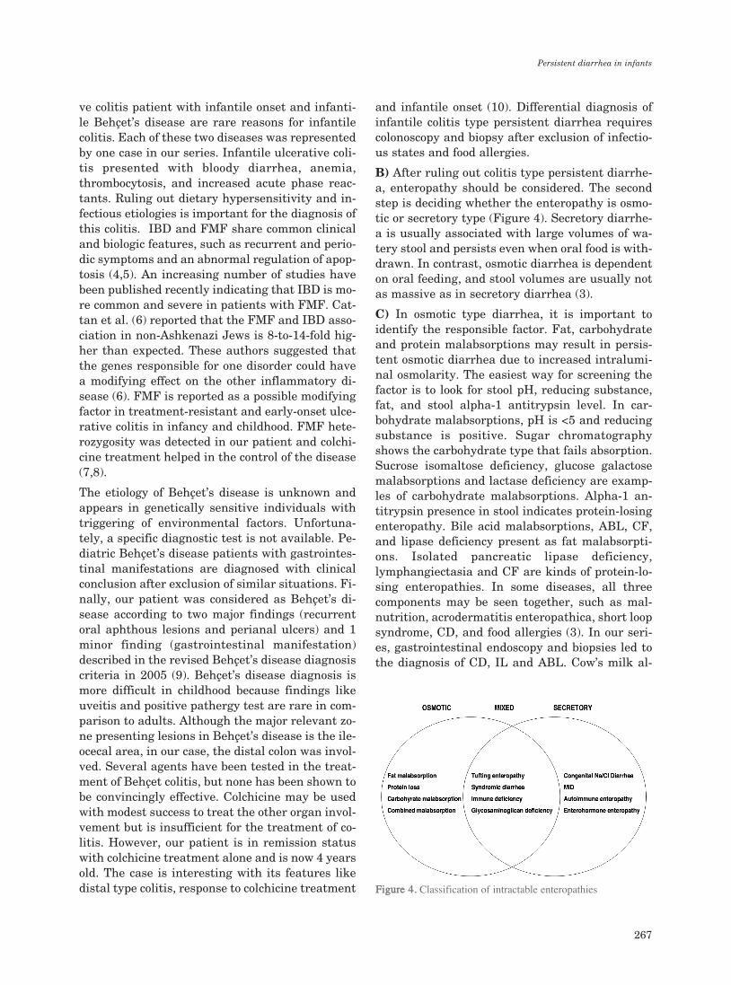
B) After ruling out colitis type persistent diarrhe-a, enteropathy should be considered. The secondstep is deciding whether the enteropathy is osmo-tic or secretory type (Figure 4). Secretory diarrhe-a is usually associated with large volumes of wa-tery stool and persists even when oral food is with-drawn. In contrast, osmotic diarrhea is dependenton oral feeding, and stool volumes are usually notas massive as in secretory diarrhea (3).
C) In osmotic type diarrhea, it is important toidentify the responsible factor. Fat, carbohydrateand protein malabsorptions may result in persis-tent osmotic diarrhea due to increased intralumi-nal osmolarity. The easiest way for screening thefactor is to look for stool pH, reducing substance,fat, and stool alpha-1 antitrypsin level. In car-bohydrate malabsorptions, pH is <5 and reducingsubstance is positive. Sugar chromatographyshows the carbohydrate type that fails absorption.Sucrose isomaltose deficiency, glucose galactosemalabsorptions and lactase deficiency are examp-les of carbohydrate malabsorptions. Alpha-1 an-titrypsin presence in stool indicates protein-losingenteropathy. Bile acid malabsorptions, ABL, CF,and lipase deficiency present as fat malabsorpti-ons. Isolated pancreatic lipase deficiency,lymphangiectasia and CF are kinds of protein-lo-sing enteropathies. In some diseases, all threecomponents may be seen together, such as mal-nutrition, acrodermatitis enteropathica, short loopsyndrome, CD, and food allergies (3). In our seri-es, gastrointestinal endoscopy and biopsies led tothe diagnosis of CD, IL and ABL. Cow’s milk al-
Persistent diarrhea in infants
267
FFiigguurree 44.. Classification of intractable enteropathies
lergy was the most relevant disease in our entero-pathy group and colitis group. A single case wasfurther investigated after being diagnosed as ente-ropathy type cow’s milk protein allergy due to re-latively poor response to dietary regulations. Du-ring the follow-up period, albumin levels diminis-hed under dietary treatment and breastfeeding.As seen in this case, as one of the major reasonsfor hypoalbuminemia during breastfeeding in thecase of consanguinity, CF has to be kept in mind.
There is no data about celiac prevalence in Tur-key; however, a survey about celiac prevalence inprimary school aged children has been carried outby our clinic. The presence of chronic diarrhea, po-or weight gain and abdominal distention after in-troduction of gluten to the infant diet are the ma-jor characteristics of all our cases. Endoscopic bi-opsies led to diagnosis of the disease and all pati-ents showed improvement with gluten-free diet.
Intestinal lymphangiectasia has to be consideredin the differential diagnosis when the hypoprote-inemia, edema and diarrhea triad is present in apatient. In our cases with IL, Cases 1, 2, 4, and 5had dilated lymphatic channels. In Case 3, signsand symptoms, laboratory findings and endoscopicevaluation led to the diagnosis of the case as IL,but histopathologically dilated lymphatic chan-nels were not visualized in LM. The golden stan-dard is to show dilated lymphatics histopathologi-cally. LM is not always sufficient to show dilatedlymphatic ducts. EM evaluation makes the diag-nosis definite. Exclusion of other protein-losingenteropathies and endoscopic findings led us tothe diagnosis of IL (11). Abdominal imaging tech-niques such as USG and CT can help identify theunderlying cause and can demonstrate PIL. Maz-zie et al. (12) reported CT findings of an IL patientfor the first time in 2003. In our cases, abdominalUSG was performed in all 4 patients. Cases 3 and5 had ascites, Case 4 had a thickened intestinalwall, and Cases 1 and 2 had normal sonographicevaluation. IL treatment is dietary interventionwith high protein, low fat and use of MCT. In ad-dition to this treatment, octreotide might be use-ful. There is no consensus regarding the dosage ofoctreotide in IL treatment. Unfortunately, follow-up studies on this point are not present in the lite-rature but some case reports have been noted.Thus, start-up dosage for octreotide treatment andintervals are not clear. Octreotide dosage was bet-ween 50-200 micrograms in our cases. Treatmentdosage was assayed according to the patient’s cli-
nical condition and demand for albumin replace-ment. No complications were observed throughoutthe treatment period. Despite the relatively short-term follow-ups and small number of cases, wemay declare the fact that early diagnosis of IL re-sulted in extended usage of octreotide (11,12).Malnutrition due to chronic diarrhea and vitamindeficiencies is observed in ABL patients, resemb-ling CD. However, low lipid profile can be addres-sed to lipid transport abnormalities. Fat-solublevitamin deficiencies due to absorption defect arealso observed in these patients. Endoscopic fin-dings are not specific; however, diffuse white gra-nular pattern is representative. In LM examinati-on, small bowel epithelial cells show lipid accumu-lation. These findings are seen in both ABL andhypobetalipoproteinemia. ABL and homozygotehypobetalipoproteinemia are hard to differentiatedue to clinical and laboratory findings. Geneticalanalysis is necessary, and our two cases are stillunder genetic investigation (13).
D) Persistent enteropathies, whether osmotic orsecretory type, necessitate endoscopic evaluationand biopsy in the early stages. LM and EM exami-nations are helpful for diagnosis of MID and auto-immune enteropathy. The most common diseaseamong the neonatal enteropathy group is MID.EM examination of small bowel biopsy specimensis the gold standard for diagnosis of MID. All LMand EM findings including villous atrophy may be-come evident in a period. In Case 2, the first bi-opsy was efficient for the diagnosis; however, Ca-se 1 needed a full year and the diagnosis was ma-de after the second biopsy. The case is being follo-wed with a prediagnosis of congenital diarrhea.After exclusion of immune deficiencies, transportdefects, short loop syndrome, cow’s milk proteinallergy, metabolic diseases, and CF, although ini-tial pathological signs did not point to it, the diag-nosis of MID was not excluded. Late manifestati-on of pathological findings in a patient surveyedfor congenital diarrhea may delay the definite di-agnosis (14).
Cystic fibrosis is the most common etiologic factorfor pancreatic failure in the infantile age group.Genotype and phenotype association is mostly do-ne in pancreatic manifestations of CF. Pancreaticenzyme activity has to drop below 10% for the ap-pearance of pancreatic insufficiency clinical state.CF infants have nearly 65% pancreatic insuffici-ency at birth. Cases with pancreatic insufficiencyare distinguished by weight gain difficulties in the
E⁄R‹TAfi et al.
268
first 6 months of life, diarrhea, hypoalbuminemia,and clinical state of malabsorption. Pancreaticenzymes could be assayed from stool for the pan-creatic insufficiency diagnosis; however, the easymethod is stool fat analysis. Both of our cases inthis series were chronic diarrhea patients recogni-zed by the presence of fat in stool and pancreaticinsufficiency in the clinical foreground (2).
In conclusion, in all age groups, diarrheas lastingmore than 14 days have to be accepted as persis-tent diarrhea apart from diarrheal type and areworth investigating. Diarrheas occurring in in-fants aged 0-24 months, in other words the infan-tile period, result in growth retardation and mal-nutrition. Congenital type diarrheas are also pre-sent among this group, and fatal results may occurwithout bowel transplantation.
Persistent diarrhea in infants
269
REFERENCES1. Bakirtas A, Turktas I, Dalgic B. Cow milk allergy presen-
ting as colitis. Eur J Pediatr 2003; 162: 55-6. 2. Gaskin KJ. Cystic fibrosis. In: Kleinman ER, Sanderson
RI, Goulet O, eds. Walker’s pediatric gastrointestinal di-sease. Hamilton, Ontario: BC Decker, 2008; 1227-39.
3. Guarino A, Marco GD. Persistent diarrhea. In: KleinmanER, Sanderson RI, Goulet O, eds. Walker’s pediatric gas-trointestinal disease. Hamilton, Ontario: BC Decker, 2008;265-75.
4. Lichtenberger GS, Flavell RA, Alexopoulou L. Innate im-munity and apoptosis in IBD. Inflamm Bowel Dis 2004; 10(Suppl 1): S58-62.
5. McDermott MF. A common pathway in periodic feversyndromes. Trends Immunol 2004; 25: 457-60.
6. Cattan D, Notarnicola C, Molinari N, et al. Inflammatorybowel disease in non-Ashkenazi Jews with familial Medi-terranean fever. Lancet 2000; 355: 378-9.
7. Heyman MB, Kirschner BS, Gold BD, et al. Children withearly-onset inflammatory bowel disease (IBD): analysis ofa pediatric IBD consortium registry. J Pediatr 2005; 146:35-40.
8. Booth DR, Lachmann HJ, Gillmore JD, et al. Prevalenceand significance of the familial Mediterranean fever genemutation encoding pyrin Q148. QJM 2001; 94: 527-31.
9. Majeed HA. Differential diagnosis of fever of unknown ori-gin in children. Curr Opin Rheumatol 2000; 12(5): 439-44.
10. Marshall SE. Behcet’s disease. Best Pract Res Clin Rhe-umatol 2004; 18: 291-311.
11. Vignes S, Bellanger J. Primary intestinal lymphangiectasi-a (Waldmann’s disease). Orphanet J Rare Dis 2008; 3: 5.
12. Mazzie JP, Maslin PI, Moy L, et al. Congenital intestinallymphangiectasia: CT demonstration in a young child. ClinImaging 2003; 27: 330-2.
13. Zamel R, Khan R, Pollex RL, et al. Abetalipoproteinemia:two case reports and literature review. Orphanet J RareDis 2008; 3: 19.
14. Ruemmele FM, Schmitz J, Goulet O. Microvillous inclusiondisease (microvillous atrophy). Orphanet J Rare Dis 2006;1: 22.