thallium-201 chloride scintigraphy in soft tissue...
TRANSCRIPT
INTRODUCTION
Thallium-201 chloride (201TlCl) scintigraphy is aconventional nuclear medicine technique to evaluate
the nature of tumors. Some studies have reportedthe utility of 201TlCl scintigraphy in the differentiationof malignant from benign lesions in various organs,including soft tissue tumors (1-4). Malignant tumorstypically demonstrate intense uptake, whereas be-nign lesions show less uptake. However, the differ-entiation between malignant and benign soft tissuetumors by the degree of 201TlCl uptake is sometimesdifficult due to the histological variability. 201TlCluptake reflects not only a malignant character but
ORIGINAL
Thallium-201 chloride scintigraphy in soft tissue tumors
Hideki Otsuka, Kaori Terazawa, Naomi Morita, Yoichi Otomi, Shoichiro Takao,
Seiji Iwamoto, Kyosuke Osaki, Masafumi Harada, and Hiromu Nishitani
Department of Radiology, Institute of Health Biosciences, the University of Tokushima Graduate School,
Tokushima, Japan
Abstract : Objective We report the findings of Thallium-201 chloride (201TlCl) scintigraphyand consider how to use this technique to evaluate the character of soft tissue lesions.Patients and Methods We enrolled 91 consecutive patients (45 males and 46 females, agerange 8-91-years-old). Nineteen patients were malignant and 72 were benign. Patientswere scanned 15 minutes (early phase) and 3 hours (delayed phase) after 201TlCl injection.More intense uptake in the lesion compared to the normal side was considered as ‘high’,the same degree of uptake was considered ‘iso’, and decreased uptake was ‘low’. The re-tention index (RI) was calculated in 9 patients in the malignant group and in 16 patientsin the benign group. Results In malignant tumors, 15 of 19 patients showed high uptakein both the early and delayed phases. One malignant fibrous histiocytoma patient was highonly in the delayed phase and 1 liposarcoma patient was high only in the early phase. Twoliposarcoma patients showed an iso uptake in both phases. One of these patients was pa-thologically diagnosed as a myxoid type. In benign lesions, no lipoma showed increaseduptake. All neurogenic tumors except for 2 demonstrated high uptake. All 3 ganglions ofthe lower extremities showed iso uptake. Most inflammatory diseases showed increaseduptake. Clinically-considered benign patients consisted of tumorous lesions or inflamma-tory disease. Only 2 patients were considered ‘low’, and these were diagnosed as intramus-cular hematoma and cyst. RI was variable in both malignant and benign lesions and nostatistically significant difference was seen between malignant and benign lesions by t-test (p=0.72). Conclusions A high 201TlCl uptake lesion is more frequently seen in malignanttumors, but regardless of whether the tumor is benign or malignant, according to the histo-pathological variety, the 201TlCl uptake pattern can not be the only indicator to differen-tiate malignant from benign tumors. We ultimately need to evaluate the nature of tumorsby a combination of several imaging techniques. J. Med. Invest. 56 : 136-141, August, 2009
Keywords :201TlCl scintigraphy, soft tissue tumors, retention index, malignant, benign
Received for publication March 13, 2009 ; accepted April 18,2009.
Address correspondence and reprint requests to Hideki Otsuka,Department of Radiology, Institute of Health Biosciences, theUniversity of Tokushima Graduate School, Kuramoto - cho,Tokushima 770-8503, Japan and Fax : +81-88-633-7174.
The Journal of Medical Investigation Vol. 56 2009
136
other factors such as vascularity, cell densities or tis-sue viability regardless of whether the tumor is be-nign or malignant.
We report here the findings of 201TlCl scintigraphyand consider how to use this technique to evaluatethe character of soft tissue lesions.
PATIENTS AND METHODS
We enrolled 91 consecutive patients who were ex-amined for 201TlCl scintigraphy (45 males and 46 fe-males, age range 8-91-years-old) in this study. Nine-teen patients were malignant and 72 were benign.All 19 patients with malignant lesions were pathologi-cally proven ; 46 of 72 patients with benign lesionswere pathologically proven, and the other 26 pa-tients were clinically diagnosed as benign with atleast 6 months of follow up with physical examina-tions and magnetic resonance imaging. Patients had111 MBq of 201TlCl, and were scanned 15 minutes(early phase) and 3 hours (delayed phase) after theinjection. In the early phase, whole body images anda spot scan of the lesion were obtained and a spot
scan was performed in the delayed phase. For visualanalysis, more intense uptake compared to the nor-mal side was considered as ‘high’, the same degree(could not be identified as a tumor) of uptake wasconsidered ‘iso’, and decreased uptake was ‘low’.The retention index (RI) was calculated in 9 patientsin the malignant group and in 16 patients in the be-nign group as follows : RI=(DR-ER)/ER�100 (%).ER (early ratio) is the ratio of lesion count to contra-lateral soft tissue count in the early phase and DR(delayed ratio) is the ratio of lesion count to contra-lateral soft tissue count in the delayed phase.
RESULTS
A summary of the patients is shown in Tables 1and 2. In malignant tumors, 15 of 19 patients showedhigh uptake in both the early and delayed phases(Figure 1). One malignant fibrous histiocytoma pa-tient was high only in the delayed phase and 1liposarcoma patient was high only in the early phase.Two liposarcoma patients showed an iso uptake inboth phases. One of these patients was pathologically
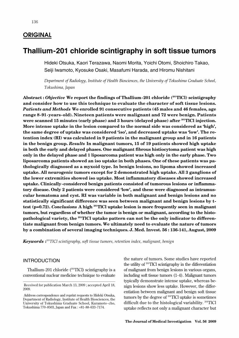
Table 1 A summary of the patients and the lesions.
The Journal of Medical Investigation Vol. 56 August 2009 137

diagnosed as a myxoid type (Figure 2) ; while theother had fluoro-deoxy-glucose positron emissioncomputed tomography/computed tomography(FDG-PET/CT) for follow up of uterine cervicalcancer and was incidentally noticed to have in-creased nodular uptake in her leg. The tumorshowed iso 201TlCl uptake and was considered
benign at the time, but in the follow up period, a ma-lignant component was suspected and it was surgi-cally resected 1.5 years after 201TlCl scintigraphy, andit was confirmed as liposarcoma pathologically. Inbenign lesions, no lipoma showed high uptake (Fig-ure 3). All neurogenic tumors except for 2 demon-strated high uptake (Figure 4). All 3 ganglions of the
Table 2 Uptake pattern of 201TlCl scintigraphy. Patients were scanned 15 minutes (early phase) and 3 hours (delayed phase) after201TlCl injection. More intense uptake in the lesion compared to the normal side was considered as ‘high’, the same degree of uptakewas considered ‘iso’, and decreased uptake was ‘low’.
aa dd
cc
bb ee
Figure 1 Malignant peripheral nerve sheath tumor (MPNST) in the left femur of patient with neurofiromatosis type I. a : early phaseimage of 201TlCl scintigraphy, b : delayed phase image of 201TlCl scintigraphy. MPNST showed high uptake in both early and delayedphase. c : FIESTA (fast imaging Employing Steady-state Acquisition) image of MRI of coronal view. This tumor demonstrated themixed intensity, separated by septum, measured in size of 8�7.5�1.5 cm. Another neurogenic tumor is located in the caudal side,which cannot be detected by 201TlCl scintigraphy. d : Axial T1-weighted SE MR image with fat suppression. Parts of the tumor showedhigh intensity, while others showed low intensity compared with the muscle. No fat component was evident in the tumor. e : Axialgadolinium-enhanced T1-weighted SE MR image with fat suppression showed strongly enhancement.
H. Otsuka, et al. Thallium-201 chloride scintigraphy in soft tissue tumors138

aa bb
cc dd ee ff
Figure 3 Lipoma in the right femur. a : early phase image of 201TlCl scintigraphy, b : delayed phase image of 201TlCl scintigraphy.The tumor showed iso uptake in both phases of 201TlCl scintigraphy. c : Coronal T2-weighted SE MR image. d : Coronal T1-weightedSE MR image. e : Coronal T1 weighted SE MR image with fat suppression. f : Coronal gadolinium-enhanced T1-weighted SE MRimage with fat suppression. The tumor was spindle-shaped and showed uniform high intensity on both T1-WI and T2-WI, measur-ing 10�3�3 cm, and well suppressed on fat -sat T1-WI. Only septum was enhanced and no other solid component was evident.
aa bb
cc dd ee ff
Figure 2 Liposarcoma (myxoid type) in the left femur. Iso uptake pattern in both early and delayed phase in malignant lesion. a :early phase image of 201TlCl scintigraphy. b : delayed phase image. The tumor showed iso uptake in both phases of 201TlCl scintigra-phy. c : Sagittal T1-weighted SE MR image. d : Sagittal T2-weighted SE MR image. e : Axial T1 weighted SE MR image with fat sup-pression. f : Sagittal gadolinium-enhanced T1-weighted SE MR image. The tumor, measuring 30�17�45 mm, showed iso intensityon T1-WI and high intensity on T2-WI compared to the muscle. The tumor intensity was suppressed on fat -suppressed T1-WI andthe tumor showed strong enhancement.
The Journal of Medical Investigation Vol. 56 August 2009 139
lower extremities showed iso uptake. Most inflam-matory diseases showed high uptake. Clinically - con-sidered benign patients (no pathology) consisted oftumorous lesions (mainly lipoma and neurogenictumor) or inflammatory disease. These tumors arestable or remitted clinically. Only 2 patients wereconsidered ‘low’, and these were diagnosed as
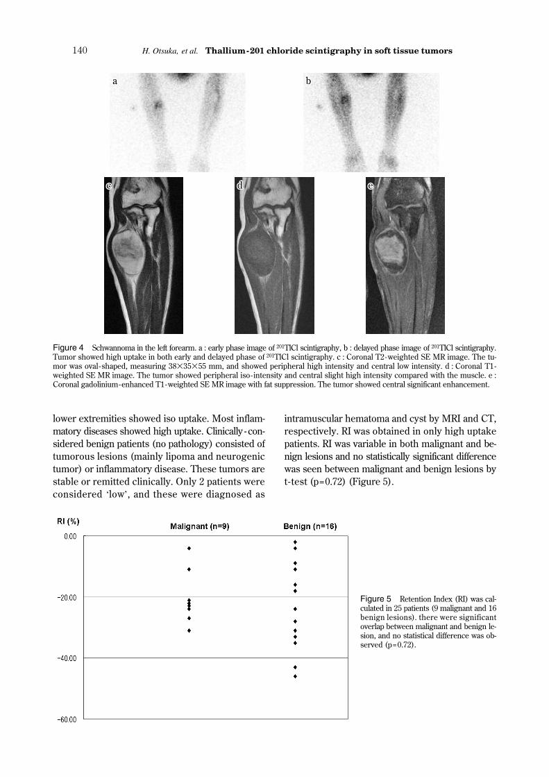
intramuscular hematoma and cyst by MRI and CT,respectively. RI was obtained in only high uptakepatients. RI was variable in both malignant and be-nign lesions and no statistically significant differencewas seen between malignant and benign lesions byt-test (p=0.72) (Figure 5).
aa bb
cc dd ee
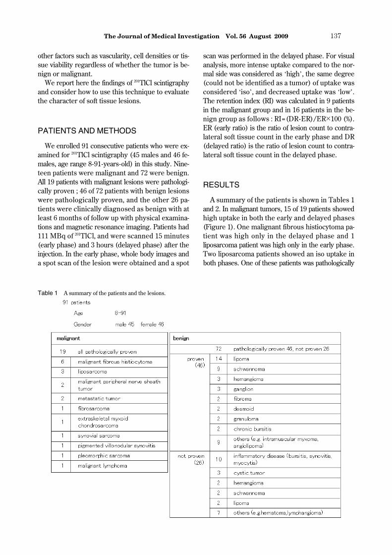
Figure 4 Schwannoma in the left forearm. a : early phase image of 201TlCl scintigraphy, b : delayed phase image of 201TlCl scintigraphy.Tumor showed high uptake in both early and delayed phase of 201TlCl scintigraphy. c : Coronal T2-weighted SE MR image. The tu-mor was oval -shaped, measuring 38�35�55 mm, and showed peripheral high intensity and central low intensity. d : Coronal T1-weighted SE MR image. The tumor showed peripheral iso- intensity and central slight high intensity compared with the muscle. e :Coronal gadolinium-enhanced T1-weighted SE MR image with fat suppression. The tumor showed central significant enhancement.
Figure 5 Retention Index (RI) was cal-culated in 25 patients (9 malignant and 16benign lesions). there were significantoverlap between malignant and benign le-sion, and no statistical difference was ob-served (p=0.72).
H. Otsuka, et al. Thallium-201 chloride scintigraphy in soft tissue tumors140

DISCUSSION
It is important to evaluate the nature of a lesionand differentiate malignant from benign in the man-agement of patients with soft tissue tumors. Imagingtechniques are essential and 201TlCl scintigraphy hasplayed an important role. FDG-PET is newly devel-oped in tumor imaging but the clinical indication forsoft tissue tumor is limited and not so widely usedcompared with 201TlCl scintigraphy (5). Thallium isa potassium analogue and accumulates in tumors re-flecting many factors. There are some reports thathave described the utility of 201TlCl scintigraphy inthe differentiation of malignant from benign lesionsbased on the degree of 201TlCl uptake. In dual timescanning (early and delayed phase), the early phaseuptake mainly reflects tumor vascularity and thedelayed phase uptake substantially reflects tumor vi-ability. In our study, most of the malignant lesionsdemonstrated high uptake in both the early anddelayed phases. Two of the malignant tumors thatshowed iso uptake were liposarcoma. In these cases,Gadolinium-enhanced MRI was useful to detect ill-defined malignant soft tissue components. It is re-ported that a low 201TlCl uptake is shown in well-differentiated or myxoid liposarcoma (6). One of ourpatients was incidentally detected by FDG-PET/CTfor follow up of uterine cervical cancer, and the le-sion showed high FDG uptake. The tumor was re-sected 1.5 years after 201TlCl scintigraphy and wasproven as liposarcoma. We were unable to deter-mine the existence of a malignant component at thetime of 201TlCl scintigraphy. The RI is reportedly use-ful for differentiating benign from malignant tumors.In our study, we calculated RI in high uptake pa-tients. The RI was variable in both malignant and be-nign lesions, and no statistically significant differencewas observed. We believe that this is due to histo-logical variability, variable lesion size and normalphysiological muscle uptake. We evaluated RI usingplanar images that demonstrated an overlapped up-take in the lesion and normal muscle. A small lesionis sometimes masked by surrounding tissue and
may not show high uptake.A high 201TlCl uptake is more frequently seen in
malignant tumors, but regardless of whether the tu-mor is benign or malignant, according to the histo-pathological variety, the 201TlCl uptake pattern cannot be the only indicator to differentiate malignantfrom benign tumors. We ultimately need to evaluatethe nature of tumors by a combination of several im-aging techniques.
REFERENCES
1. Otsuka H, Shinbata H, Hieda M, Yamashita K,Kitamura H, Senba T, Kashihara K, TagashiraH : The retention indices of 201Tl - SPECT inbrain tumors. Ann Nucl Med 16(7) : 455-459,2002
2. Sugawara Y, Kikuchi T, Kajihara M, Senba T,Ochi T, Fujii T, Mochizuki T, Sakayama K,Nakata S : Thallium-201 scintigraphy in boneand soft tissue tumors : a comparison of dy-namic, early and delayed scans. Ann Nucl Med19(6) : 461-468, 2005
3. Nishiyama Y, Yamamoto Y, Yokoe K,Kawaguchi Y, Toyama Y, Satoh K, OhkawaM : A comparative study of 201Tl scintigraphyand three-phase bone scintigraphy followingtherapy in patients with bone and soft-tissuetumors. Ann Nucl Med 18(3) : 235-241, 2004
4. Higuchi T, Taki J, Nakajima K, Kinuya S,Nonomura A, Tsuchiya H, Bunko H, NamuraM, Tonami N : Differentiation of soft tissue hae-mangioma by 201Tl scintigraphy. Nucl MedCommun 24 : 327-330, 2003
5. Otsuka H, Graham MM, Kubo A, Nishitani H :FDG-PET/CT findings of sarcomatous trans-formation in neurofibromatosis : a case report.Ann Nucl Med 19(1) : 55-58, 2005
6. Sato O, Kawai A, Ozaki T, Kunisada T, DanuraT, Inoue H : Value of thallium-201 scintigraphyin bone and soft tissue tumors. J Orthop Sci 3 :297-303, 1998
The Journal of Medical Investigation Vol. 56 August 2009 141