ternary complex factor-serum response factor complex-regulated gene activity is required
TRANSCRIPT
MOLECULAR AND CELLULAR BIOLOGY, Dec. 2004, p. 10340–10351 Vol. 24, No. 230270-7306/04/$08.00�0 DOI: 10.1128/MCB.24.23.10340–10351.2004Copyright © 2004, American Society for Microbiology. All Rights Reserved.
Ternary Complex Factor-Serum Response Factor Complex-RegulatedGene Activity Is Required for Cellular Proliferation and Inhibition
of Apoptotic Cell DeathElaine R. Vickers, Aneta Kasza, Isil Aksan Kurnaz,† Anne Seifert, Leo A. H. Zeef,
Amanda O’Donnell, Andy Hayes, and Andrew D. Sharrocks*Faculty of Life Sciences, University of Manchester, Manchester, United Kingdom
Received 30 June 2004/Returned for modification 19 August 2004/Accepted 3 September 2004
Members of the ternary complex factor (TCF) subfamily of the ETS-domain transcription factors areactivated through phosphorylation by mitogen-activated protein kinases (MAPKs) in response to a variety ofmitogenic and stress stimuli. The TCFs bind and activate serum response elements (SREs) in the promotersof target genes in a ternary complex with a second transcription factor, serum response factor (SRF). Theassociation of TCFs with SREs within immediate-early gene promoters is suggestive of a role for the ternaryTCF-SRF complex in promoting cell cycle entry and proliferation in response to mitogenic signaling. Here wehave investigated the downstream gene regulatory and phenotypic effects of inhibiting the activity of genesregulated by TCFs by expressing a dominantly acting repressive form of the TCF, Elk-1. Inhibition of ternarycomplex activity leads to the downregulation of several immediate-early genes. Furthermore, blocking TCF-mediated gene expression leads to growth arrest and triggers apoptosis. By using mutant Elk-1 alleles, wedemonstrated that these effects are via an SRF-dependent mechanism. The antiapoptotic gene Mcl-1 isidentified as a key target for the TCF-SRF complex in this system. Thus, our data confirm a role forTCF-SRF-regulated gene activity in regulating proliferation and provide further evidence to indicate a role inprotecting cells from apoptotic cell death.
Elk-1 is a member of the ternary complex factor (TCF)subfamily of ETS-domain transcription factors (reviewed inreferences 46 and 50). In mammals there are two other TCFs,SAP-1 and SAP-2/ERP/Net. These proteins are characterizedby their ability to form ternary complexes on target promotersin conjunction with the MADS-box protein serum responsefactor (SRF). The TCFs share four domains, the ETS DNA-binding domain, the B-box, the D-domain, and the C-domain.SAP-2/Net contains additional regions that impart repressiveproperties (9, 29). The D- and C-domains constitute the reg-ulatory part of Elk-1 and other TCFs. The D-domain acts as adocking site for mitogen-activated protein kinases (MAPKs)(reviewed in references 15 and 49). These docked kinases canthen phosphorylate residues in the C-domain, which consti-tutes the transcriptional activation domain (TAD). Phosphor-ylation of the TAD leads to elevation of the transactivationpotential of the TCFs and also enhances ternary complex for-mation (reviewed in references 46, 50, 54, and 58). The TCFscan be phosphorylated by members of all three of the majorMAPK pathways present in mammals: ERK, JNK, and p38(reviewed in references 46, 50, and 58). The ERK MAPKpathway predominantly transmits mitogenic and differentia-tion stimuli, whereas the JNK and p38 MAPK pathways pri-marily transduce stress and cytokine stimuli to the nucleus
(reviewed in reference 41). The TCFs therefore play a pivotalrole in transducing extracellular stimuli into alterations in geneexpression in the nucleus.
The B-box of the TCFs is required for ternary complexformation (11, 23, 55) and mediates protein-protein interac-tions between TCFs and SRF (28, 52). Single point mutationswithin the B-box in Elk-1 (e.g., leucine 158 to proline) aresufficient to severely disrupt interactions with SRF and henceblock ternary complex formation (28). Structural studies of theSRF-SAP-1 complex demonstrate that the B-box binds to asurface-exposed hydrophobic groove on SRF (see Fig. 4A) andconsists of two short 310 helices that flank a short �-strand (19).Recently, an additional role for the Elk-1 B-box in inhibitingRhoA signaling to SRF has been uncovered, thereby providingsignaling specificity to the TCF-SRF complex (35).
The primary action of the TCFs is thought to be the activa-tion of immediate-early genes by forming ternary complexeswith SRF at serum response elements (SREs) found in thepromoters of these genes. The best-characterized TCF target isthe proto-oncogene c-fos (17, 21, 22). c-fos is an immediate-early gene, and c-fos levels are known to increase rapidly uponstimulation of cells with various mitogens. SRF has been foundto be constitutively bound to the SRE in the c-fos promoter(21) and is able to recruit Elk-1 despite the suboptimal Elk-1binding site that makes up part of this promoter element. SREsare also known to be located in the promoters of a variety ofother genes, such as egr-1 (early growth response gene-1) andNur77, the murine homologue of the human TR3 gene. SRE-dependent activation is important for the activation of theNur77/TR3 gene (26) and the egr-1 gene in response to avariety of signals (1, 6, 12, 33, 38, 39). In vitro studies haveimplicated the TCFs as an important part of the regulatory
* Corresponding author. Mailing address: Faculty of Life Sciences,University of Manchester, Michael Smith Building, Oxford Rd.,Manchester M13 9PT, United Kingdom. Phone: 0044-161-275-5979.Fax: 0044-161-275-5082. E-mail: [email protected].
† Present address: Department of Genetics and Bioengineering,Faculty of Engineering and Architecture, Yeditepe University, Kayis-dagi, Istanbul, Turkey.
10340
on April 5, 2019 by guest
http://mcb.asm
.org/D
ownloaded from
complex that forms on these SREs (7, 26). A further targetgene identified for the TCFs is Mcl-1, thereby suggesting apotential role in regulating apoptosis (53). Although binding ofTCFs to SREs usually requires recruitment by SRF, they canalso bind autonomously to high-affinity ets DNA-binding mo-tifs. Additionally, it has been proposed that TCFs are able torecruit SRF to suboptimal SRF-binding sites such as thosefound in the pip92 and nur77 SREs (26). Indeed, this has beenshown to be the case for nur77, where cells deficient in SAP-1show reduced SRF recruitment to this promoter in vivo (8).The TCF Elk-1 has also been implicated in the regulation ofTNF-� (56), 9E3/cCAF (27), and �(1,3)-fucosyltransferase IV(59), where promoter binding apparently occurs independentlyof SRF.
Recently, mouse knockout studies on individual TCFs havebeen completed, but minimal phenotypes were observed, sug-gesting that functional redundancy might exist. SAP-2/Net hasa role in egr-1 repression and vascular development (2). SAP-1is important in the positive selection of thymocytes (8),whereas Elk-1 plays a role in the neuronal expression of im-mediate-early genes like c-fos in the brain (5). Due to thepossible functional redundancy of the TCFs, we have used arepressive form of Elk-1 to probe the potential role of TCFs inregulating cell growth and gene expression. We demonstratethat inhibition of TCF-regulated gene expression leads togrowth arrest and apoptosis. These apoptotic effects are de-pendent on the downregulation of the key antiapoptotic reg-ulator Mcl-1, which has been previously implicated as a TCFtarget gene (53). By using mutant Elk-1 alleles, we furtherdemonstrate that these effects are via an SRF-dependentmechanism. A large body of literature links the activation ofthe Ras/ERK pathway in controlling cell proliferation andsurvival. Our data demonstrate that members of one class ofnuclear target of this pathway, the TCFs, act through ternaryTCF-SRF-SRE complexes to regulate genes whose activity isrequired for proliferation and also for protecting cells fromapoptotic death.
MATERIALS AND METHODS
Plasmid constructs. pSRE-Luc (pAS821) contains two copies of the c-fos SRE(nucleotides �357 to �275, containing both an SRF binding site and an adjacentets motif) upstream of a minimal tk promoter and the luciferase gene (44).pE74-Luc (pNC101) contains four E74 ets binding sites upstream of a minimaltk promoter and the firefly luciferase gene (kindly provided by Erik Jansen).pMcl-1-Luc (pAS2156) contains the human Mcl-1 promoter (�3893 to �25)upstream from the firefly luciferase gene (kindly provided by Steve Edwards),pCH110 (Pharmacia) contains a simian virus 40-driven �-galactosidase (LacZ)gene and is used to monitor transfection efficiency. pRSV-Elk-1-VP16 (pAS348),pRSV-SAP-1-VP16, and pRSV-SAP-2-VP16 are Rous sarcoma virus (RSV)promoter-driven vectors encoding full-length wild-type human Elk-1, SAP-1, andSAP-2 fused to residues 410 to 490 of a VP16 C-terminal sequence (37). pCMV-MEK1 encodes constitutively active MEK-1(�NS218E-S222D) (30). pAS515encodes full-length zFli-1 fused to the engrailed (En) repressor domain (pro-vided by Dave Hickleton). pAS728 (encoding full-length His-FLAG-taggedElk-1) was created by ligating a NcoI-BamHI fragment from pAS278 (60) intothe same sites in pAS38 (47). pEGFP-N1 (Clontech) encodes enhanced greenfluorescent protein (EGFP).
The following plasmids were constructed for use in in vitro transcription-translation of proteins. pAS1407, encoding full-length Elk-1 fused to the Enrepressor domain and the FLAG epitope (Elk-En), was produced by ligating theHindIII-XbaI fragment from pAS1406 (described below) into pAS728. pAS1409,encoding Elk-1(L158P) fused to the En repressor domain and FLAG epitope,[Elk-En(L158P)], was constructed by ligating an NcoI-StuI fragment frompAS853 (61) into the same sites in pAS1407.
The following vectors were used for in vivo expression in mammalian cells.pAS1402 (encoding RSV-driven Elk-VP16{L158P})was created by ligating anNcoI/HindIII fragment from pAS1403 into the same sites in pAS348. pAS1403was created by inserting a StuI/NcoI fragment from pAS853 into the same sitesin pRSETB-Elk-VP16. pRSETB-Elk-VP16 was created by inserting an NcoI/HindIII fragment from pRSV-Elk-VP16 into pRSETB. To create stable cell lineswhich can be induced with ponasterone A (PA) to express FLAG-tagged Elk-Enand Elk-En(L158P), pAS1406 and pAS1410 were constructed. pAS1406, encod-ing Elk-En driven by ecdysone response elements, was constructed by ligating aHindIII/XhoI fragment from pAS1404 into the same sites of pAS728. pAS1404,encoding the En repressor domain (amino acids 2 to 298) fused to the FLAGepitope, was constructed by cloning an XhoI/XbaI-cleaved PCR fragment(primer pair ADS731-ADS732; template pAS515) into the same sites inpIND(SP1) (Invitrogen). The FLAG epitope was created through the use ofprimer ADS738, which was designed to include the FLAG sequence. pAS1410,encoding Elk-1(L158P)-En-FLAG, was created by ligating the HindIII/XbaIfragment of pAS1409 into the same sites in pAS1406. pAS1408 and pAS1411,encoding Elk-En-FLAG wild type (WT) and L158P, were created by ligatingHindIII/XbaI fragments of pAS1406 and pAS1409 into the same sites inpCDNA3. pexpMcl-1 (pAS2157) encodes full-length FLAG-tagged humanMcl-1 in a cytomegalovirus (CMV)-driven vector (kindly provided by X. Wang)(36).
Tissue culture, cell transfection, and reporter gene assays. EcR293 cells (In-vitrogen) were maintained in Dulbecco’s modified Eagle’s medium (DMEM)supplemented with 10% fetal bovine serum (FBS) (Gibco-BRL) and 400 �g ofZeocin (Invitrogen)/ml. HeLa cells were grown in DMEM–10% FBS. Whereindicated, the caspase inhibitor Z-VAD-fmk (Calbiochem) was added togetherwith PA. Transient transfection experiments were carried out using Superfecttransfection reagent (QIAGEN). Stable cell lines exhibiting inducible expressionof Elk-En (57) or Elk-En(L158P) were created by transfecting EcR293 cells withSspI-linearized pAS1406 or pAS1410 to create EcR293(Elk-En) andEcR293(Elk-En{L158P}) lines. Stably transfected clones were selected as de-scribed previously (57). Western blotting and reporter assays were used to lookfor inducible expression of FLAG-tagged Elk-En or Elk-En(L158P) and to checkfor leaky transcription from the vectors. A total of 30 separate clones wereexpanded for each cell line and assayed by Western blotting; 5 clones were foundto be positive for inducible Elk-En expression and 9 were found to be positive forinducible Elk-En(L158P). These were then used in reporter gene analysis exper-iments.
For reporter gene assays, SRE- and E74-driven vectors containing two tandemSREs and four E74 response elements, respectively, upstream of a minimalpromoter element or a Mcl-1 promoter fragment, followed by the firefly lucif-erase gene, were transfected into EcR293 cells or into the EcR293(Elk-En)stable cell lines. The cells were also transfected with pCH110 to monitor trans-fection efficiency and where indicated, with either pCMV-MEK or pRSV-Elk-VP16 to stimulate reporter activity. Following transfection, cells were left inDMEM containing 0.05% FBS for 24 h. In some experiments the cells werepretreated with 5 �M PA (Invitrogen) for 6 h prior to transfection to stimulateElk-En expression and then maintained in 0.05% FBS containing PA followingtransfection for a further 24 h. Cells were then solubilized, and luciferase assayswere carried out using a dual light reporter gene assay system (Tropix). Lightemissions were measured for 10 s with a Turner TD-20/20 luminometer.
Western blot analysis. Cells were harvested by direct lysis or lysed followingtrypsinization in lysis buffer (Tropix) containing phenylmethylsulfonyl fluorideand aprotinin. Anti-p53, p21cip1 (Santa Cruz), and a mouse monoclonal anti-M2FLAG antibody (Sigma) were used along with horseradish peroxidase-conju-gated secondary antibodies followed by detection using Supersignal West DuraExtended Duration substrate (Pierce) and visualization using a Bio-Rad Fluor-SMultiImager. Quantification of proteins was carried out using Quantity Onesoftware (Bio-Rad).
Gel retardation assays. Gel retardation assays were carried out with a 32P-labeled 49-bp fragment of the c-fos promoter, containing the SRE, the E74 site,or an 88-bp fragment of the Mcl-1 promoter (nucleotides �79 to �127; ADS1226[5�-AGCAACCCTCCGGAAGCTGCCGCCCCTTTCCCCTTTTATGGGAA-3�] and ADS1225 [5�-AGTATTCCCATAAAAGGGGAAAGGGGCGGCAGCTTCCGGAGGGT-3�]) as described previously (48, 52). Elk-1 was produced asa His-tagged protein in bacteria (see Fig. 7) (61) or as FLAG-tagged derivativesby in vitro transcription and translation using a TNT-coupled reticulocyte lysatesystem (Promega) (see Fig. 4). 35S-labeled proteins were analyzed by electro-phoresis through use of a 0.1% sodium dodecyl sulfate (SDS)–12% polyacryl-amide gel before visualization and quantification using a PhosphorImager andQuantity One software (Bio-Rad). Proteins were incubated with labeled probeand coreSRF purified from bacteria where indicated (52). Protein-DNA com-
VOL. 24, 2004 REGULATION OF GROWTH AND APOPTOSIS BY TCFs 10341
on April 5, 2019 by guest
http://mcb.asm
.org/D
ownloaded from
plexes were analyzed on nondenaturing 5% polyacrylamide gels cast in 0.5�Tris-borate-EDTA and visualized by autoradiography and phosphorimaging.
RT-PCR, Northern and microarray analysis. Cells were maintained in 0.05%FBS for 24 h and then treated with 5 �M PA in ethanol or with ethanol alone for24 h. Epidermal growth factor (EGF) (Sigma) was then added to the cells to afinal concentration of 50 nM for 15, 30, or 60 min where indicated. Cells werethen harvested, and total RNA was extracted using an RNeasy kit (QIAGEN).For reverse transcriptase PCR (RT-PCR), 1 �g of total RNA from each samplewas reverse transcribed using Moloney murine leukemia virus RT and randomhexanucleotide primers (Roche) according to the supplier’s protocol. cDNA (1�l) and Taq polymerase (Bioline) were used in each subsequent PCR withprimers to detect expression of GAPDH (ADS733 [5�-GCATTGCTGATGATCTTGAGG-3�] and ADS734 [5�-TCGGAGTCAACGGATTTG-3�]), c-fos(ADS735 [5�-CAGTGCCAACTTCATTCC-3�] and ADS736 [5�-GAACTCTAGTTTTTCCTTCTCC-3�]), egr-1 (ADS770 [5�-CCCTTCCCCAATTACTATTCC-3�] and ADS771 [5�-CCCCCAAATCATCACTCC-3�]), TR3 (ADS740 [5�-ATACACCCGTGACCTCAACC-3�] and ADS741 [5�-TACATCCCCAGCATCTTCC-3�]), and vinculin (ADS1029 [5�-CCAAGGTCAGAGAAGCCTTCC-3�] andADS1030 [5�-CGTAGCTGTTCAAGGTCTGGT-3�]) (42). PCR products weredetected using 2% agarose gels, and ethidium bromide-stained bands were vi-sualized using a Bio-Rad Gel Doc system. For Northern analysis, 5 �g of RNAwas subjected to formaldehyde gel electrophoresis and transferred to Hy-bond-XL membranes (Pharmacia Biotech) according to the manufacturer’s in-structions. The filters were prehybridized at 68°C for 3 h in 0.5 M sodiumphosphate buffer (pH 7.2)–7% sodium dodecyl sulfate (SDS)–1 mM EDTA andhybridized in the same solution with a human Mcl-1 full-length cDNA fragmentlabeled by random priming (Roche). After the hybridization, nonspecificallybound radioactivity was removed by four washes in 40 mM phosphate buffer–1%SDS–1 mM EDTA at 68°C for 20 min.
Microarray experiments were performed using human focus and humanU133A GeneChip oligonucleotide arrays (Affymetrix, Inc.) according to themanufacturer’s instructions. Briefly, 15 �g of total RNA was used to preparefirst-strand cDNA by use of an oligo(dT)-T7 primer. Following second-strandsynthesis, biotinylated cRNA targets were generated using an Enzo BioArrayHighYield RNA transcript-labeling kit (Affymetrix, Inc.) by in vitro transcriptionwith biotinylated UTP and CTP. The fragmented cRNA targets together withlabeled controls were hybridized to the arrays at 45°C with rotation at 60 rpm for16 h according to the manufacturer’s instruction. Following hybridization thearrays were processed using Affymetrix fluidics protocol Midi-euk2 for the Focusarrays and EukGE-WS2 for the U133A arrays and stained with R-phycoerythrinconjugated to streptavidin (Molecular Probes, Inc.). Images of the arrays werethen acquired using Microarray Suite (MAS) version 5.0 software and an Af-fymetrix 2500 GeneChip scanner. Normalization and further analyses were car-ried out using RMAExpress software (http://stat-www.berkeley.edu/users/bolstad/RMAExpress/RMAExpress.html). Microarray data were generated using threeindependent samples from PA-stimulated and two independent samples fromunstimulated EcR293(Elk-En)#1.3 cells.
Chromatin immunoprecipitation (IP). Cells were plated on 10-cm-diameterdishes, serum starved for 24 h, and than stimulated with 10 nM phorbol miristateacetate (PMA) for 15 min. Proteins were cross-linked to DNA by incubation with1% formaldehyde for 10 min at 37°C. The cells were washed twice with ice-cold0.125 M glycine in 1� PBS and scraped into 1 ml of PBS containing proteaseinhibitor cocktail (Boehringer) and 1 mM PMA. Cells were pelleted and lysedwith lysis buffer (1% SDS, 10 mM EDTA, 50 mM Tris-HCl, pH 8.0). Lysateswere sonicated to produce DNA sizes between 200 to 500 bp. Samples werediluted 1 in 10 with IP buffer (0.01% SDS, 1.1% Triton X-100, 1.2 mM EDTA,16.7 mM Tris-HCl [pH 8.0], 16.7 mM NaCl, protease inhibitor cocktail) andimmunoprecipitated with anti-Elk-1 (Santa Cruz) or nonspecific immunoglobu-lin G (IgG) (Upstate) overnight at 4°C. After 1 h of incubation with salmonsperm DNA, 30 �l of protein A dynal beads (Dynal Biotech) was added to thesamples. After 1 h the beads were washed with the following buffers: TSE I (20mM Tris [pH 8.1], 2 mM EDTA, 150 mM NaCl, 1% Triton, 0.1% SDS), TSE II(20 mM Tris [pH 8.1], 2 mM EDTA, 500 mM NaCl, 1% Triton, 0.1% SDS),buffer III (10 mM Tris [pH 8.1], 0.25 M LiCl, 1 mM EDTA, 1% NP-40, 1%sodium deoxycholate), and TE (10 mM Tris [pH 8.0], 1 mM EDTA). DNA waseluted with elution buffer (0.1 M NaHCO3, 1% SDS). NaCl (0.2 M) was addedto the eluted samples and to the input, and cross-linking was reversed by over-night incubation at 65°C. After digestion with proteinase K, DNA was cleanedusing a QIAGEN PCR cleanup kit. Promoter-specific primers used to amplifythe DNA by PCR were as follows: for human Egr-1 promoter, ADS1269 (TGCAGGATGGAGGTGCC) and ADS1270 (AGTTCTGCGCGCTGGGATCTCTC); for human Mcl-1 promoter, ADS1271 (ACTCAGAGCCTCCGAAGACC)and ADS1272 (CGCCAGCGAACTCTTTTT); and for SRF intronic sequence,
ADS1273 (GCCACAGGGCAGTAGATGTT) and ADS1274 (TCAGGCCCAAGTATCCACTC).
Hoechst staining and fluorescence microscopy. Cells were harvested and fixedon ice with 1% formaldehyde in PBS. Cells were then spun down and resus-pended in a solution of 10 ng of Hoechst stain (Sigma)/ml in PBS. Visualizationof staining was carried out using a pseudoconfocal fluorescence microscope.Pictures were generated using picture publisher software. EGFP was detectedusing a fluorescent microscope (oil immersion lens).
BrdU incorporation and fluorescence-activated cell sorting (FACS). Cellswere plated out onto 10-cm-diameter dishes, and at the required time pointsBrdU (Sigma) was added to the cells at a final concentration of 10 �M. The cellswere then incubated at 37°C for 1 h (times stated in figures are times without the1 h of BrdU incubation). For flow cytometry, cells were harvested and fixed bythe dropwise addition of 5 ml of ice-cold 70% ethanol. Cellular DNA wasdenatured with 2 M HCl, and the cells were stained with a mouse anti-BrdUantibody and a secondary fluorescein isothiocyanate (FITC)-conjugated antibody(Dako). Cells were then incubated with 10 mg of propidium iodide/ml to allowexamination of the DNA content of cells. Analyses were carried out on a Becton-Dickinson flow cytometer for both BrdU uptake and cell cycle profile.
RESULTS
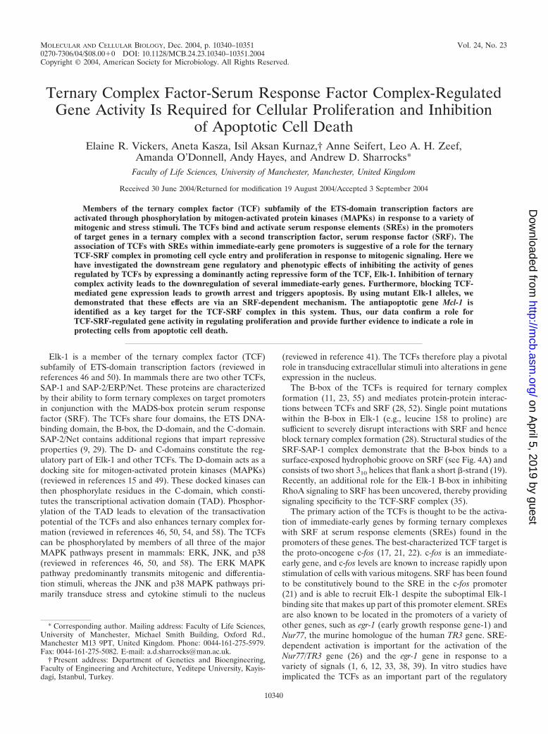
Elk-En fusion proteins repress SRE-dependent gene regu-lation. The TCFs have been implicated in regulating the ex-pression of immediate-early genes such as c-fos in response tostress and mitogenic signaling (Fig. 1A). In particular, theirassociation with the ERK MAPK cascade and transduction ofmitogenic signals suggests a role for the TCFs in regulating cellcycle entry and controlling cellular proliferation. To investigatesuch a potential role and the phenotypic consequences of dis-rupting TCF activity, we created a dominant-negative form ofElk-1 that consists of full-length Elk-1 fused to the DrosophilaEn repression domain (Elk-En). The expression of this fusionprotein, Elk-En, is predicted to repress the activity of TCFtarget genes and hence disrupt downstream cellular processes.Due to the conservation of the TCF structure, Elk-En is likelyto inhibit the activity of SAP-1- and SAP-2-regulated genes inaddition to Elk-1 targets. Thus, this approach should overcomeproblems associated with redundancy of function between theTCFs that is suggested by the phenotypes associated withknockout studies (2, 5, 8). Fusions of other transcription fac-tors to the En repression domain have been successfully usedto study their biological functions (3, 24). Stable cell lines werecreated in human EcR293 cells that inducibly express Elk-Enin response to stimulation with PA (Fig. 1B). One cell line,EcR293(Elk-En)#1.3, that exhibits undetectable levels ofElk-En in the absence of PA but high levels following PAinduction (Fig. 1C) was selected for further study.
To assess whether Elk-En can repress transcription throughSREs, EcR293(Elk-En)#1.3 cells were transfected with a lu-ciferase reporter construct driven by two tandem SREs. Co-transfection with MEK, a constitutively active form of the ERKactivator, led to a large increase in reporter activity (Fig. 1D).However, when the cells were treated with PA to induceElk-En expression, no reporter activation was observed (Fig.1D, right panel). In contrast, PA treatment had no effect onSRE-luciferase activity in the parental EcR293 cells (Fig. 1D,left panel). Thus, induction of Elk-En expression leads to po-tent repression of SRE-driven reporters in EcR293(Elk-En)#1.3 cells. Due to the structural homology shared by theTCFs, it is predicted that Elk-En would repress all TCF-me-diated gene regulation. To demonstrate that this is indeed thecase, we investigated whether Elk-En could compete with con-
10342 VICKERS ET AL. MOL. CELL. BIOL.
on April 5, 2019 by guest
http://mcb.asm
.org/D
ownloaded from
stitutively active Elk-1, SAP-1, and SAP-2 fusions to the VP16transcriptional activation domain in regulating SRE-driven re-porter constructs (Fig. 1E). Upon induction with PA, Elk-Enwas able to efficiently inhibit transcriptional activation by eachof the TCF-VP16 fusion proteins.
The TCFs are known to activate the transcription of theimmediate-early genes such as c-fos through SREs in responseto a variety of stimuli, including epidermal growth factor(EGF). To verify that Elk-En represses transcription of endog-enous SRE-regulated genes, we analyzed the effect of Elk-Enexpression on the putative target genes c-fos, egr-1, and TR3.EcR293(Elk-En)#1.3 cells were treated with PA and thenstimulated with EGF. In the absence of PA, EGF leads torapid induction of both c-fos and egr-1 expression followed byrapid shutoff. The kinetics of TR3 induction are somewhatslower (Fig. 1F, lanes 1 to 4). However, in the presence of PA,
gene induction is severely inhibited in all three cases (Fig. 1F,lanes 5 and 6). Elk-1 and SRF can also interact in solution invitro in the absence of DNA (52). We therefore tested whetherElk-1-independent SRF target genes such as vinculin (18) wererepressed by Elk-En through protein-protein interactions.However, vinculin was not repressed by Elk-En either in thepresence (Fig. 1G) or the absence (data not shown) of serum.Thus, the induction of Elk-En in EcR293(Elk-En)#1.3 cellsleads specifically to the repression of endogenous TCF targetgenes in addition to repressing TCF-responsive reporter con-structs.
Induction of Elk-En causes growth arrest. To study theeffect of Elk-En expression on cell growth properties, we firstmonitored cell growth following administration of PA toEcR293(Elk-En)#1.3 cells. In the absence of PA, the cellsshowed a continual increase in numbers over 5 days. However,
FIG. 1. Induction of Elk-En represses SRE-dependent gene regulation. (A) Schematic indicating the role of the TCFs as modulators oftranscriptional effects in response to signals from MAPK pathways. Elk-En fusion proteins block signaling through Elk-1 to downstream targetgenes. En represents the En repression domain. (B) The PA-inducible expression system contained in EcR293(Elk-En) cell lines. EcR and RXRrepresent the ecdysone and retinoid X receptors that, upon binding to PA, activate the expression of Elk-En. Elk-En contains a C-terminal FLAGtag. (C) Western blot of FLAG-tagged Elk-En in EcR293(Elk-En)#1.3 cells before and after PA treatment for 24 h. (D) Repression of SRE-Lucreporter genes in response to PA-induction of Elk-En in EcR293(Elk-En)#1.3 cells. Parental EcR293 or EcR293(Elk-En)#1.3 cells weretransfected with SRE-luciferase reporter constructs (0.25 �g) in the presence or absence of a vector encoding constitutively active MEK (0.5 �g)where indicated. Cells were incubated for 24 h in 10% FBS in the presence or absence of 5 �M PA. Data are presented as means and standarddeviations (n 3) normalized for �-galactosidase activity from a cotransfected pCH110 reporter construct. (E) Reporter gene analysis usingSRE-Luc reporter in EcR293(Elk-En)#1.3 cells. Where indicated, plasmids encoding 0.5 �g of Elk-1, SAP-1, or SAP-2-VP16 fusions weretransiently transfected to activate the reporters. Elk-En expression was induced by adding 5 �M PA. (F) Repression of EGFP induction ofendogenous TCF target genes by Elk-En following PA induction in EcR293(Elk-En)#1.3 cells. Cells were synchronized by incubation in 0.05%FBS for 48 h; where indicated, 5 �M PA was added for the final 12 h. Cells were then stimulated with 50 nM EGFP, and the expression of GAPDH,c-fos, egr-1, and TR3 was analyzed by RT-PCR at the indicated times. For clarity, the color on the original image has been inverted. The asterisksindicate the locations of the 600-bp bands in the marker (M) lane. (G) RT-PCR analysis of the SRF-regulated gene vinculin. EcR293(Elk-En)#1.3cells were serum starved for 24 h and incubated with PA for 7 h (where indicated) before stimulation with 10% serum for 1 h. GAPDH is shownas a control.
VOL. 24, 2004 REGULATION OF GROWTH AND APOPTOSIS BY TCFs 10343
on April 5, 2019 by guest
http://mcb.asm
.org/D
ownloaded from
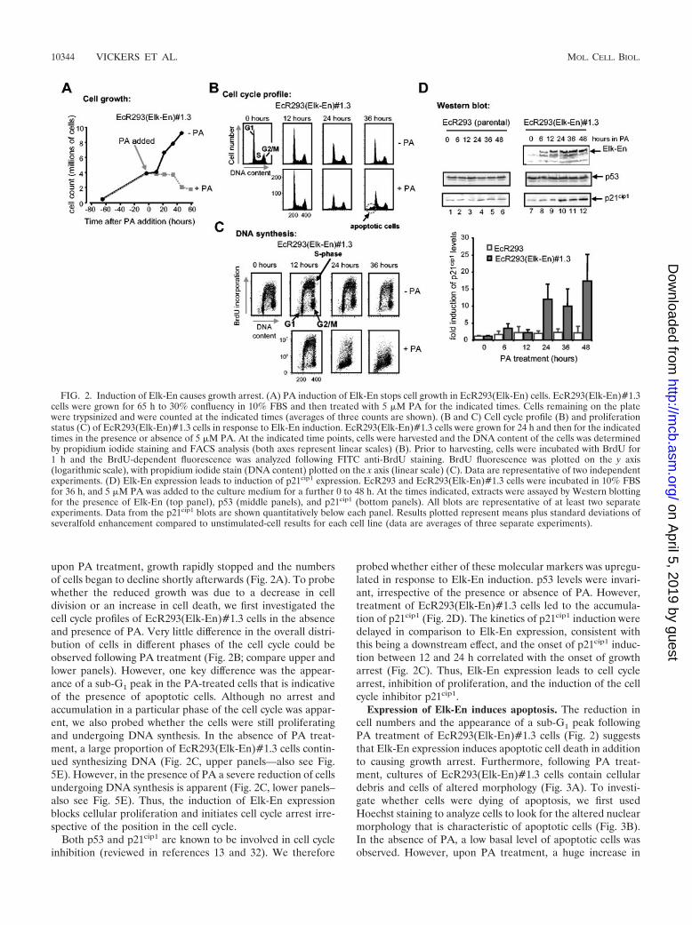
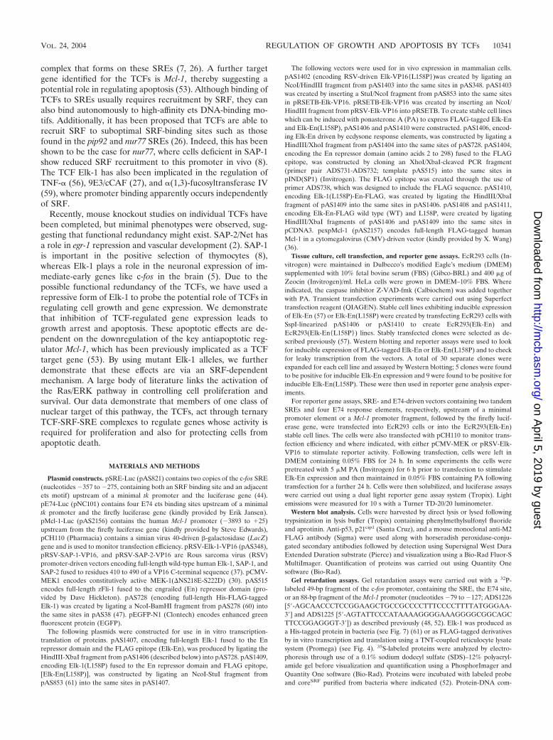
upon PA treatment, growth rapidly stopped and the numbersof cells began to decline shortly afterwards (Fig. 2A). To probewhether the reduced growth was due to a decrease in celldivision or an increase in cell death, we first investigated thecell cycle profiles of EcR293(Elk-En)#1.3 cells in the absenceand presence of PA. Very little difference in the overall distri-bution of cells in different phases of the cell cycle could beobserved following PA treatment (Fig. 2B; compare upper andlower panels). However, one key difference was the appear-ance of a sub-G1 peak in the PA-treated cells that is indicativeof the presence of apoptotic cells. Although no arrest andaccumulation in a particular phase of the cell cycle was appar-ent, we also probed whether the cells were still proliferatingand undergoing DNA synthesis. In the absence of PA treat-ment, a large proportion of EcR293(Elk-En)#1.3 cells contin-ued synthesizing DNA (Fig. 2C, upper panels—also see Fig.5E). However, in the presence of PA a severe reduction of cellsundergoing DNA synthesis is apparent (Fig. 2C, lower panels–also see Fig. 5E). Thus, the induction of Elk-En expressionblocks cellular proliferation and initiates cell cycle arrest irre-spective of the position in the cell cycle.
Both p53 and p21cip1 are known to be involved in cell cycleinhibition (reviewed in references 13 and 32). We therefore
probed whether either of these molecular markers was upregu-lated in response to Elk-En induction. p53 levels were invari-ant, irrespective of the presence or absence of PA. However,treatment of EcR293(Elk-En)#1.3 cells led to the accumula-tion of p21cip1 (Fig. 2D). The kinetics of p21cip1 induction weredelayed in comparison to Elk-En expression, consistent withthis being a downstream effect, and the onset of p21cip1 induc-tion between 12 and 24 h correlated with the onset of growtharrest (Fig. 2C). Thus, Elk-En expression leads to cell cyclearrest, inhibition of proliferation, and the induction of the cellcycle inhibitor p21cip1.
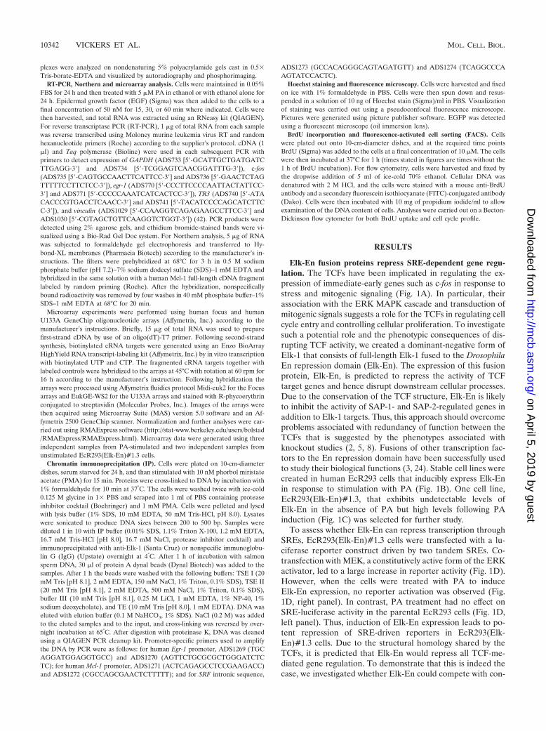
Expression of Elk-En induces apoptosis. The reduction incell numbers and the appearance of a sub-G1 peak followingPA treatment of EcR293(Elk-En)#1.3 cells (Fig. 2) suggeststhat Elk-En expression induces apoptotic cell death in additionto causing growth arrest. Furthermore, following PA treat-ment, cultures of EcR293(Elk-En)#1.3 cells contain cellulardebris and cells of altered morphology (Fig. 3A). To investi-gate whether cells were dying of apoptosis, we first usedHoechst staining to analyze cells to look for the altered nuclearmorphology that is characteristic of apoptotic cells (Fig. 3B).In the absence of PA, a low basal level of apoptotic cells wasobserved. However, upon PA treatment, a huge increase in
FIG. 2. Induction of Elk-En causes growth arrest. (A) PA induction of Elk-En stops cell growth in EcR293(Elk-En) cells. EcR293(Elk-En)#1.3cells were grown for 65 h to 30% confluency in 10% FBS and then treated with 5 �M PA for the indicated times. Cells remaining on the platewere trypsinized and were counted at the indicated times (averages of three counts are shown). (B and C) Cell cycle profile (B) and proliferationstatus (C) of EcR293(Elk-En)#1.3 cells in response to Elk-En induction. EcR293(Elk-En)#1.3 cells were grown for 24 h and then for the indicatedtimes in the presence or absence of 5 �M PA. At the indicated time points, cells were harvested and the DNA content of the cells was determinedby propidium iodide staining and FACS analysis (both axes represent linear scales) (B). Prior to harvesting, cells were incubated with BrdU for1 h and the BrdU-dependent fluorescence was analyzed following FITC anti-BrdU staining. BrdU fluorescence was plotted on the y axis(logarithmic scale), with propidium iodide stain (DNA content) plotted on the x axis (linear scale) (C). Data are representative of two independentexperiments. (D) Elk-En expression leads to induction of p21cip1 expression. EcR293 and EcR293(Elk-En)#1.3 cells were incubated in 10% FBSfor 36 h, and 5 �M PA was added to the culture medium for a further 0 to 48 h. At the times indicated, extracts were assayed by Western blottingfor the presence of Elk-En (top panel), p53 (middle panels), and p21cip1 (bottom panels). All blots are representative of at least two separateexperiments. Data from the p21cip1 blots are shown quantitatively below each panel. Results plotted represent means plus standard deviations ofseveralfold enhancement compared to unstimulated-cell results for each cell line (data are averages of three separate experiments).
10344 VICKERS ET AL. MOL. CELL. BIOL.
on April 5, 2019 by guest
http://mcb.asm
.org/D
ownloaded from
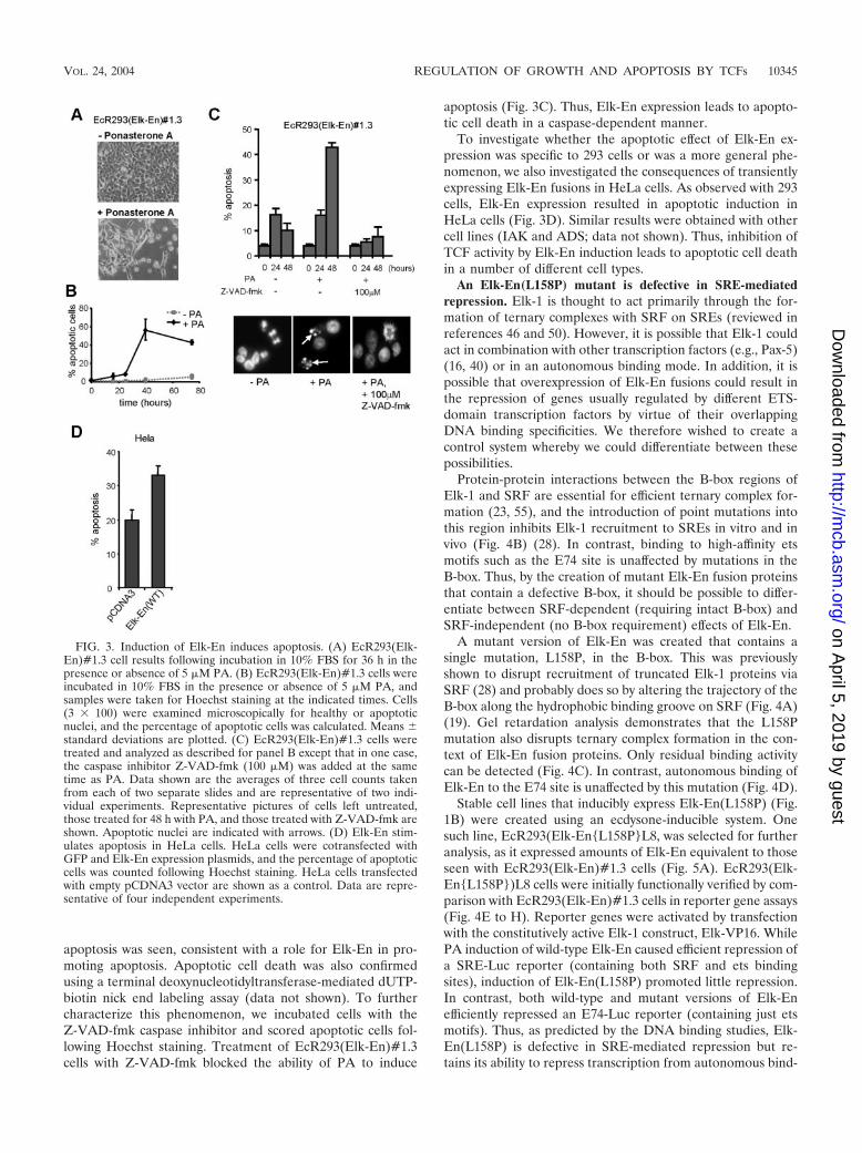
apoptosis was seen, consistent with a role for Elk-En in pro-moting apoptosis. Apoptotic cell death was also confirmedusing a terminal deoxynucleotidyltransferase-mediated dUTP-biotin nick end labeling assay (data not shown). To furthercharacterize this phenomenon, we incubated cells with theZ-VAD-fmk caspase inhibitor and scored apoptotic cells fol-lowing Hoechst staining. Treatment of EcR293(Elk-En)#1.3cells with Z-VAD-fmk blocked the ability of PA to induce
apoptosis (Fig. 3C). Thus, Elk-En expression leads to apopto-tic cell death in a caspase-dependent manner.
To investigate whether the apoptotic effect of Elk-En ex-pression was specific to 293 cells or was a more general phe-nomenon, we also investigated the consequences of transientlyexpressing Elk-En fusions in HeLa cells. As observed with 293cells, Elk-En expression resulted in apoptotic induction inHeLa cells (Fig. 3D). Similar results were obtained with othercell lines (IAK and ADS; data not shown). Thus, inhibition ofTCF activity by Elk-En induction leads to apoptotic cell deathin a number of different cell types.
An Elk-En(L158P) mutant is defective in SRE-mediatedrepression. Elk-1 is thought to act primarily through the for-mation of ternary complexes with SRF on SREs (reviewed inreferences 46 and 50). However, it is possible that Elk-1 couldact in combination with other transcription factors (e.g., Pax-5)(16, 40) or in an autonomous binding mode. In addition, it ispossible that overexpression of Elk-En fusions could result inthe repression of genes usually regulated by different ETS-domain transcription factors by virtue of their overlappingDNA binding specificities. We therefore wished to create acontrol system whereby we could differentiate between thesepossibilities.
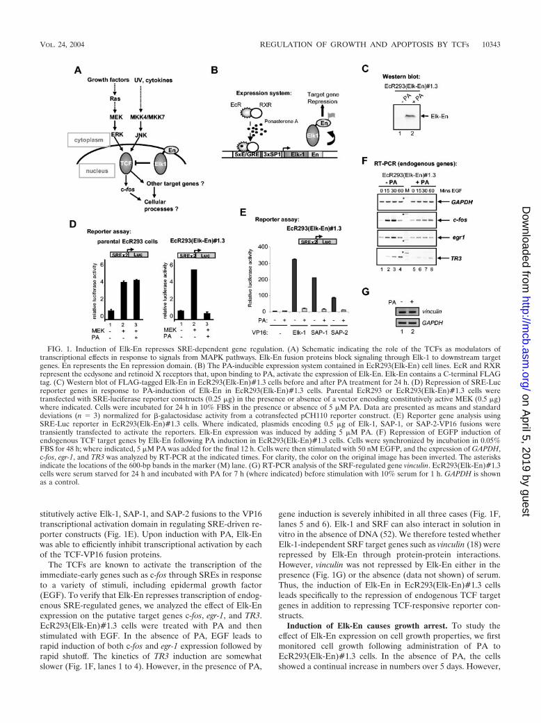
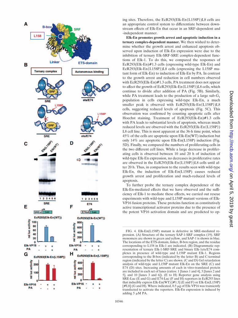
Protein-protein interactions between the B-box regions ofElk-1 and SRF are essential for efficient ternary complex for-mation (23, 55), and the introduction of point mutations intothis region inhibits Elk-1 recruitment to SREs in vitro and invivo (Fig. 4B) (28). In contrast, binding to high-affinity etsmotifs such as the E74 site is unaffected by mutations in theB-box. Thus, by the creation of mutant Elk-En fusion proteinsthat contain a defective B-box, it should be possible to differ-entiate between SRF-dependent (requiring intact B-box) andSRF-independent (no B-box requirement) effects of Elk-En.
A mutant version of Elk-En was created that contains asingle mutation, L158P, in the B-box. This was previouslyshown to disrupt recruitment of truncated Elk-1 proteins viaSRF (28) and probably does so by altering the trajectory of theB-box along the hydrophobic binding groove on SRF (Fig. 4A)(19). Gel retardation analysis demonstrates that the L158Pmutation also disrupts ternary complex formation in the con-text of Elk-En fusion proteins. Only residual binding activitycan be detected (Fig. 4C). In contrast, autonomous binding ofElk-En to the E74 site is unaffected by this mutation (Fig. 4D).
Stable cell lines that inducibly express Elk-En(L158P) (Fig.1B) were created using an ecdysone-inducible system. Onesuch line, EcR293(Elk-En{L158P}L8, was selected for furtheranalysis, as it expressed amounts of Elk-En equivalent to thoseseen with EcR293(Elk-En)#1.3 cells (Fig. 5A). EcR293(Elk-En{L158P})L8 cells were initially functionally verified by com-parison with EcR293(Elk-En)#1.3 cells in reporter gene assays(Fig. 4E to H). Reporter genes were activated by transfectionwith the constitutively active Elk-1 construct, Elk-VP16. WhilePA induction of wild-type Elk-En caused efficient repression ofa SRE-Luc reporter (containing both SRF and ets bindingsites), induction of Elk-En(L158P) promoted little repression.In contrast, both wild-type and mutant versions of Elk-Enefficiently repressed an E74-Luc reporter (containing just etsmotifs). Thus, as predicted by the DNA binding studies, Elk-En(L158P) is defective in SRE-mediated repression but re-tains its ability to repress transcription from autonomous bind-
FIG. 3. Induction of Elk-En induces apoptosis. (A) EcR293(Elk-En)#1.3 cell results following incubation in 10% FBS for 36 h in thepresence or absence of 5 �M PA. (B) EcR293(Elk-En)#1.3 cells wereincubated in 10% FBS in the presence or absence of 5 �M PA, andsamples were taken for Hoechst staining at the indicated times. Cells(3 � 100) were examined microscopically for healthy or apoptoticnuclei, and the percentage of apoptotic cells was calculated. Means standard deviations are plotted. (C) EcR293(Elk-En)#1.3 cells weretreated and analyzed as described for panel B except that in one case,the caspase inhibitor Z-VAD-fmk (100 �M) was added at the sametime as PA. Data shown are the averages of three cell counts takenfrom each of two separate slides and are representative of two indi-vidual experiments. Representative pictures of cells left untreated,those treated for 48 h with PA, and those treated with Z-VAD-fmk areshown. Apoptotic nuclei are indicated with arrows. (D) Elk-En stim-ulates apoptosis in HeLa cells. HeLa cells were cotransfected withGFP and Elk-En expression plasmids, and the percentage of apoptoticcells was counted following Hoechst staining. HeLa cells transfectedwith empty pCDNA3 vector are shown as a control. Data are repre-sentative of four independent experiments.
VOL. 24, 2004 REGULATION OF GROWTH AND APOPTOSIS BY TCFs 10345
on April 5, 2019 by guest
http://mcb.asm
.org/D
ownloaded from
ing sites. Therefore, the EcR293(Elk-En{L158P})L8 cells arean appropriate control system to differentiate between down-stream effects of Elk-En that occur in an SRF-dependent and-independent manner.
Elk-En promotes growth arrest and apoptotic induction in aternary complex-dependent manner. We then wished to deter-mine whether the growth arrest and enhanced apoptosis ob-served upon induction of Elk-En expression were due to theinhibition of ternary Elk-SRF-SRE complex-dependent func-tions of Elk-1. To do this, we compared the responses ofEcR293(Elk-En)#1.3 cells (expressing wild-type Elk-En) andEcR293(Elk-En{L158P})L8 cells (expressing the L158P mu-tant form of Elk-En) to induction of Elk-En by PA. In contrastto the growth arrest and reduction in cell numbers observedwith EcR293(Elk-En)#1.3 cells, PA treatment does not appearto affect the growth of EcR293(Elk-En{L158P})L8 cells, whichcontinue to divide after addition of PA (Fig. 5B). Similarly,while PA treatment leads to the production of a large sub-G1
population in cells expressing wild-type Elk-En, a muchsmaller peak is observed with EcR293(Elk-En{L158P})L8cells, suggesting reduced levels of apoptosis (Fig. 5C). Thisobservation was confirmed by counting apoptotic cells afterHoechst staining. Treatment of EcR293(Elk-En)#1.3 cellswith PA leads to substantial levels of apoptosis, whereas muchreduced levels are observed with the EcR293(Elk-En{L158P})L8 cell line. This is most apparent at the 36-h time point, when45% of the cells are apoptotic upon Elk-En(WT) induction butonly 14% are apoptotic upon Elk-En(L158P) induction (Fig.5D). Finally, we compared the numbers of proliferating cells inthe two different cell lines. While a large decrease in prolifer-ating cells is observed between 10 and 20 h of induction ofwild-type Elk-En expression, no decreases in proliferative ratesare observed in the EcR293(Elk-En{L158P})L8 cells until af-ter 20 h. Thus, in comparison to the results seen with wild-typeElk-En, the induction of Elk-En(L158P) causes reducedgrowth arrest and proliferation and much-reduced levels ofapoptosis.
To further probe the ternary complex dependence of theElk-En-mediated effects that we have observed and the suffi-ciency of Elk-1 to mediate these effects, we carried out rescueexperiments with wild-type and L158P mutant versions of Elk-VP16 fusion proteins. These proteins function as constitutivelyactive transcriptional activator proteins due to the presence ofthe potent VP16 activation domain and are predicted to op-
FIG. 4. Elk-En(L158P) mutant is defective in SRE-mediated re-pression. (A) Structure of the ternary SAP-1-SRF complex (19). SRFmonomers are shown in green and yellow, and SAP-1 is shown in blue.The locations of the ETS-domain, linker, B-box region, and the residuecorresponding to L158 in Elk-1 are indicated. (B) Diagrammatic rep-resentation of ternary Elk-1-SRF-SRE and binary Elk-1ets/E74 com-plexes in presence of wild-type and L158P mutant Elk-1. Regionscorresponding to the B-box (indicated by the letter B) and C-terminalregion (indicated by the letter C) are shown. (C and D) Gel retardationanalysis of wild-type and L158P mutant Elk-En on the SRE (C) andE74 (D) sites. Increasing amounts of each in vitro-translated proteinare included in each set of lanes (ratios: 1 [lanes 1 and 4], 3 [lanes 2 and5], and 10 [lanes 3 and 6]). (E to H) Reporter gene analysis usingSRE-Luc (E and G) and E74-Luc (F and H) reporters in EcR293 linesthat inducibly express Elk-En(WT) [#1.3] (E and F) or Elk-En(L158P)[#L8] (G and H). Where indicated, 0.5 �g of Elk-VP16 was transientlytransfected to activate the reporters. Elk-En expression is induced byadding 5 �M PA.
10346
on April 5, 2019 by guest
http://mcb.asm
.org/D
ownloaded from

pose the repressive function of Elk-En fusion proteins. If TCFactivity is specifically being altered by Elk-En expression, thenElk-VP16 should block apoptotic induction by Elk-En (Fig.6A). In addition, if Elk-En is working through a ternary-com-plex-dependent manner, then Elk-VP16(L158P) should be un-able to block apoptotic induction. EcR293(Elk-En)#1.3 cellswere transfected with vectors encoding either wild-type Elk-VP16 or Elk-VP16(L158P) along with an EGFP marker andthen stimulated with PA to induce Elk-En expression (Fig.6A). The apoptotic cells were then scored by Hoechst staining,and transfected cells were observed by EGFP fluorescence.Cells transfected with Elk-VP16 appeared to be rescued fromapoptotic death caused by Elk-En expression (Fig. 6B). Quan-tification of the data demonstrated that transfection ofEcR293(Elk-En)#1.3 cells with Elk-VP16 resulted in almostcomplete rescue of cells from apoptosis (Fig. 6C). In contrast,no apoptotic rescue was observed upon transfection with Elk-VP16(L158P).
Collectively, these data demonstrate that Elk-En inducesgrowth arrest and apoptosis in a ternary complex-dependentmanner.
The antiapoptotic gene Mcl-1 is a key TCF-SRF target. Thedata presented above indicate that the activity of genes regu-lated through ternary TCF-SRF complexes is required to pro-mote proliferation and to block apoptotic cell death. To iden-tify these target genes, we used Affymetrix microarray analysis
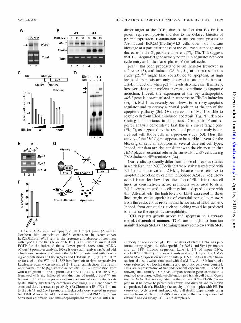
to compare the mRNA expression profiles of unstimulatedEcR293(Elk-En)#1.3 cells with those of the same cells stimu-lated with PA for 6 h to induce the expression of Elk-En. Oneof the most downregulated genes was Mcl-1 (ranked seventh;expression reduced to 56%). Northern analysis confirmed themicroarray data, demonstrating a clear reduction in Mcl-1mRNA levels following PA induction (Fig. 7A). Furthermore,in addition to inhibiting the basal levels of Mcl-1 expression,the induction of Elk-En blocks the increase in Mcl-1 expressioncaused by EGF stimulation (Fig. 7B). Mcl-1 has recently beenshown to be a key antiapoptotic protein that acts at the top ofthe apoptotic cascade (36) and a key cellular target in adeno-viral-mediated apoptosis (10), making it a likely target forElk-En-mediated apoptosis. Moreover, a previous study dem-onstrated the functional importance of an SRE-like sequencein the Mcl-1 promoter (53).
To demonstrate a direct effect of Elk-En on Mcl-1 expres-sion, we investigated the effect of Elk-En on the activity of aMcl-1 promoter-driven luciferase reporter construct. Wild-typeElk-En caused efficient repression of the Mcl-1 promoter in adose-dependent manner (Fig. 7C). However, in comparison,the mutant Elk-En(L158P) protein was much less efficient inrepressing this promoter (Fig. 7C). Conversely, an Elk-VP16fusion protein efficiently activated the Mcl-1 promoter (datanot shown). To investigate whether SRF and Elk-1 could formternary complexes on the Mcl-1 promoter in vitro, we carried
FIG. 5. EcR393 cells expressing the Elk-En(L158P) mutant exhibit reduced growth arrest and apoptotic induction. (A) Western blot analysisof wild-type Elk-En and the Elk-En(L158P) following PA induction for 24 h in the EcR293-Elk-En cell lines #1.3 and L8, respectively. (B) PAinduction of Elk-En does not stop cell growth in EcR293(Elk-En{L158P}) cells. EcR293(Elk-En{L158P})#L8 cells were grown and treated withPA as described for Fig. 2A. The data from the EcR293(Elk-En)#1.3 cell line results shown for Fig. 2A are shown in the background for comparison.(C) Cell cycle profile of EcR293(Elk-En{L158P})#L8 cells in response to Elk-En induction. EcR293(Elk-En{WT})#1.3 and EcR293(Elk-En{L158P})L8 cells were grown and treated with PA and analyzed by FACS as described for Fig. 2B. The apoptotic sub-G1 peak is highlighted.(D) Reduced levels of apoptotic cell death occur in EcR293-Elk-En(L158P)#L8 cells. Apoptotic cells were visualized by Hoechst staining followingtreatment of EcR293(Elk-En{WT})#1.3 and EcR293(Elk-En{L158P})L8 cells with PA as described for Fig. 3B. (E) The percentage of EcR293(Elk-En{WT})#1.3 and EcR293(Elk-En{L158P})L8 cells undergoing DNA synthesis following PA induction of Elk-En was analyzed by studyingBrdU incorporation by FACS analysis (Fig. 2C). Results shown represent the averages for two dishes for each time point and are representativeof at least two separate experiments. The dotted boxes highlight the differences during early time points between untreated and PA-treated cells.
VOL. 24, 2004 REGULATION OF GROWTH AND APOPTOSIS BY TCFs 10347
on April 5, 2019 by guest
http://mcb.asm
.org/D
ownloaded from

out gel retardation assays on fragments containing this SRE.Both SRF and Elk-1 can bind this promoter independently(Fig. 7D, lanes 2 and 3). However, in the presence of SRF,Elk-1 is efficiently recruited into a ternary complex (Fig. 7D,lane 4). This is consistent with previous observations that Elk-1can be found in complexes formed by nuclear extracts on thissite (53). To verify these findings in vivo, we investigatedwhether endogenous Elk-1 could be found on the Mcl-1 pro-moter in vivo by chromatin IP analysis. Elk-1 was found to beassociated with the promoters of both Mcl-1 and the knowntarget gene Egr-1 (Fig. 7E, top two panels). No association withan SRF intronic fragment could be seen, demonstrating thespecificity of binding (Fig. 7E, top two panels). Finally, weinvestigated whether Elk-En-mediated apoptosis could be res-cued by transiently overexpressing Mcl-1. In comparison tocells transfected with empty expression vector, Mcl-1 expres-sion was able to inhibit Elk-En-mediated apoptosis (Fig. 7F),
thereby demonstrating the importance of this TCF target genein protecting cells from apoptosis.
Together, these data therefore identify Mcl-1 as an impor-tant TCF target gene that protects cells from apoptosis inEcR293 cells.
DISCUSSION
The TCFs can be phosphorylated and activated in responseto activation of all three of the major MAPK cascades inmammalian cells (reviewed in references 46 and 54). As such,they represent an important point of convergence for cell sig-naling pathways. Due to their association with target genessuch as c-fos, it has been presumed that TCFs act to promotecell cycle entry and proliferation by activating the expression ofthis and other immediate-early genes. Here we demonstratethat the transcriptional activity of genes regulated by the ter-nary TCF-SRF-SRE complex is essential for cell growth andsurvival (Fig. 7G).
Immediate-early gene induction by TCFs. To block TCF-mediated transcription, we created cell lines that contain anintegrated inducible expression cassette that contains a geneencoding full-length Elk-1 fused to the En repression domain.Full-length Elk-1 was used to ensure that the correct interac-tion surfaces were present to permit promoter recruitment.The Drosophila En repression domain has been previouslyshown to convert other mammalian transcription factors intorepressors (3). In reporter gene assays, the Elk-En fusion pro-tein could inhibit SRE-dependent transcription and therebyblock ERK MAPK signaling through these elements (Fig. 1D).This effect was SRE specific, as no effect was observed on otherpromoters that lack TCF binding motifs, including the simianvirus 40 promoter contained in the pCH110 reporter vector(data not shown). Known TCF-dependent genes such as c-fosand egr-1 were shown to be repressed, and genes previouslyshown to be capable of forming ternary complexes in vitro suchas TR3 were also identified as targets for Elk-1 in vivo (Fig.1E). In contrast, the expression of GAPDH and the TCF-independent SRF target gene vinculin was unaffected. This isconsistent with the observation that TCF targeting to the vin-culin promoter is incapable of conferring TCF-specific signal-ing properties to SRF bound at this promoter (35). Microarrayanalysis identified a number of additional genes that weredownregulated by Elk-En, including Mcl-1 (see below). Thus,the Elk-En fusion protein acts as a specific inhibitor of SRE-mediated transcription and can be used to probe the biologicalfunctions of Elk-1 and to identify novel target genes.
Inhibition of TCF-regulated gene activity results in growtharrest and apoptosis. The expression of Elk-En inEcR293(Elk-En)#1.3 cells results in growth arrest and theinduction of apoptosis. This is not a clonal effect, as five inde-pendent cell lines died of apoptosis upon induction of Elk-Enexpression (data not shown). Furthermore, this effect is not celltype specific, as apoptosis is also induced in both HeLa cells(Fig. 3D) and PC12 cells (data not shown). The onset of theblock in cellular proliferation is between 12 and 24 h post–Elk-En induction (Fig. 2C). This correlates with the inductionof the cell cycle inhibitor p21cip1 (Fig. 2D) and indicates thatinduction of p21cip1 expression is one of the likely molecularcauses of growth arrest. However, p21cip1 is unlikely to be a
FIG. 6. Elk-VP16 rescues cells from death induced by Elk-En.(A) Schematic of the experimental protocol. �GFP, EGFP. (B and C)Rescue of apoptosis by transfection of Elk-VP16 following inductionof Elk-En with PA. EcR293(Elk-En)#1.3 cells were transfected withplasmids encoding EGFP (200 ng) and, where indicated, Elk-VP16 orElk-VP16(L158P) (50 ng). At 24 h posttransfection (time 0), 5 �M PAwas added where specified. Cells were analyzed by Hoechst staining atthe indicated time points. Slides were examined for transfection effi-ciency by observing green fluorescing cells and for percentages ofapoptosis by observing nuclear morphology. (B) Representative pic-tures are shown for cells transfected with 50 ng of Elk-VP16 andincubated with 5 �M PA for 48 h. The left panel shows Hoechststaining; the right panel shows GFP fluorescence. Arrows indicateapoptotic cells (Hoechst staining) and GFP-expressing cells (GFP flu-orescence). (C) Data shown are the averages of three cell counts(percentages of total cell population) taken from each of two separateslides.
10348 VICKERS ET AL. MOL. CELL. BIOL.
on April 5, 2019 by guest
http://mcb.asm
.org/D
ownloaded from
direct target of the TCFs, due to the fact that Elk-En is apotent repressor protein and due to the delayed kinetics ofp21cip1 expression. Examination of the cell cycle profiles ofPA-induced EcR293(Elk-En)#1.3 cells does not indicateblockage at a particular phase of the cell cycle, although slightdecreases in the G1 peak are apparent (Fig. 2B). This suggeststhat TCF-regulated gene activity potentially regulates both cellcycle entry and other later phases of the cell cycle.
p21cip1 has been proposed to be an inhibitor (reviewed inreference 13), and inducer (25, 31, 51) of apoptosis. In thisstudy, p21cip1 might have contributed to apoptosis, as highlevels of apoptosis are only observed at around 24 h post–Elk-En induction, when p21cip1 levels also increase. It is likely,however, that other molecular events contribute to apoptoticinduction. Indeed, the expression of the key antiapoptoticMcl-1 gene is downregulated in response to Elk-En induction(Fig. 7). Mcl-1 has recently been shown to be a key apoptoticregulator and to occupy a pivotal position at the top of theapoptotic pathway (36). Overexpression of Mcl-1 is able torescue cells from Elk-En-induced apoptosis (Fig. 7F), demon-strating its importance in this process. Chromatin IP and re-porter analysis demonstrate that this is a direct target gene(Fig. 7), as suggested by the results of promoter analysis car-ried out with K-562 cells in a previous study (53). Thus, theactivity of the Mcl-1 gene appears to be a critical event for theblocking of cellular apoptosis in several different cell types.Indeed, our data are also consistent with the observation thatMcl-1 plays an essential role in the survival of U937 cells duringPMA-induced differentiation (34).
Our results apparently differ from those of previous studiesin which Rat1 and MCF7 cells that were stably transfected withElk-1 or a splice variant, �Elk-1, became more sensitive toapoptotic induction by calcium ionophore A23187 (45). How-ever, it is not clear how direct the effect of Elk-1 is in these celllines, as constitutively active promoters were used to driveElk-1 expression, and the cells may have adapted to cope withthis. Alternatively, the high levels of Elk-1 expressed in theselines might cause squelching of essential coregulators awayfrom the endogenous proteins and hence loss of Elk-1 activity.Indeed, from our studies, such squelching would be predictedto enhance the apoptotic susceptibility.
TCFs regulate growth arrest and apoptosis in a ternarycomplex-dependent manner. TCFs are thought to functionmainly through SREs via forming ternary complexes with SRF.
FIG. 7. Mcl-1 is an antiapoptotic Elk-1 target gene. (A and B)Northern blot analysis of Mcl-1 expression in serum-starvedEcR293(Elk-En)#1.3 cells in the presence and absence of treatmentwith 5 �M PA for 18 h (A) or 2.5 h (B). (B) Cells were stimulated withEGFP for the indicated times. Lower panels show total mRNA.(C) Mcl-1 promoter analysis. 293 cells were transiently transfected witha luciferase construct containing the Mcl-1 promoter and with increas-ing concentrations of Elk-En(WT) and Elk-En(L158P) (0, 1, 5, 10, 25ng for each of the WT and L158P bars from left to right, respectively).Luciferase activity was measured 24 h after transfection. The resultswere normalized to �-galactosidase activity. (D) Gel retardation assaywith a fragment of Mcl-1 promoter (�79 to �127). The DNA wasincubated with the indicated combinations of purified coreSRF andfull-length Elk-1 in the presence of unprogrammed rabbit reticulocytelysate. Binary and ternary complexes containing Elk-1 are shown byopen and closed arrows, respectively. (E) Chromatin IP of Elk-1 boundto the Mcl-1 and Egr-1 promoters. HeLa cells were starved in serum-free DMEM for 48 h and then stimulated with 10 nM PMA for 15 min.Sonicated chromatin was immunoprecipitated with either anti-Elk-1
antibody or nonspecific IgG. PCR analysis of eluted DNA was per-formed using oligonucleotides specific for Mcl-1 and Egr-1 promotersand an SRF intronic sequence. Lane 1, 2% of input DNA.(F) EcR293(Elk-En) cells were transfected with 1.5 �g of a CMV-driven Mcl-1 expression vector or with pCDNA3. At 24 h after trans-fection, the cells were stimulated with 5 �M PA. At 48 h later, cellswere subjected to Hoechst staining and apoptotic cells were counted.Data are representative of two independent experiments. (G) Modelshowing that ternary TCF-SRF complex-specific gene expression isrequired to promote cellular proliferation and inhibit cell death. Genessuch as Mcl-1 that are regulated by the ternary TCF-SRF-SRE com-plex must be active to permit cell growth and division and to inhibitapoptotic cell death. Blocking the activity of this complex with Elk-Encauses cell cycle arrest and apoptotic cell death. Experiments usingmutant forms of Elk-En (L158P) demonstrated that the major route ofaction is not via binary TCF-DNA complexes.
VOL. 24, 2004 REGULATION OF GROWTH AND APOPTOSIS BY TCFs 10349
on April 5, 2019 by guest
http://mcb.asm
.org/D
ownloaded from
To investigate whether the biological effects we have observedare due to TCFs functioning in this manner, we used cell linesthat express a mutant allele of Elk-En [Elk-En(L158P)] thatcannot be recruited to SREs (Fig. 4). In contrast to the resultsseen with EcR293(Elk-En)#1.3 cells, EcR293(Elk-En{L158P})L8 cells exhibit reduced levels of apoptosis and growth arrest isnot as apparent (Fig. 5). Again, this is not a clonal effect, asnone of the independent cell lines that inducibly express Elk-En(L158P) underwent apoptosis at the levels observed withlines expressing wild-type Elk-En (data not shown). Further-more, Elk-VP16(L158P) was incapable of rescuing cells fromapoptosis whereas wild-type Elk-VP16 efficiently promotedcell survival. Interestingly, in addition to Elk-VP16, the rein-troduction of wild-type Elk-1 can rescue cells from apoptosis,demonstrating that the presence of Elk-1 is sufficient for rescue(data not shown). Thus, activation of Elk-1 either by fusion ofa potent activation domain or by endogenous signaling path-ways is sufficient to overcome the block induced by repressiveElk-1 constructs, indicating a delicate balance in regulating celldeath. Finally, a further advantage of using the mutant Elk-1allele is that it controls for nonspecific effects such as squelch-ing by the En repression domain. Thus, the use of the inhibi-tory Elk-En protein reveals a role for TCF-regulated geneactivity in promoting proliferation and cell survival in a ternarycomplex-specific manner (Fig. 7G).
Despite the clear differences compared to the results seenwith the EcR293(Elk-En)#1.3 cells, enhanced levels of apo-ptotic cell death and proliferative blockage are observed in theEcR293(Elk-En{L158P})L8 cells at later time points (Fig. 5).This could reflect the fact that residual levels of ternary com-plex formation can be observed with the Elk-1(L158P) mutant(Fig. 4C). At high expression levels, this would result in low-level inhibition of target genes; consequently, inhibitorythresholds would not be reached till a later time. Indeed, weobserve that Elk-En(L158P) can also inhibit the expression ofthe Mcl-1 promoter, albeit to a lesser extent than that seen withthe wild-type Elk-En (Fig. 7C), thereby providing a molecularexplanation for these phenotypic observations. Alternatively,several different TCF-regulated pathways might be operativethat guard against apoptosis. It is possible that an SRF-inde-pendent pathway becomes repressed at higher concentrationsof Elk-En or with reduced kinetics. Again, this would lead tothe toxicity that is revealed at the later time points. Such apossibility is suggested by the identification of tumor necrosisfactor alpha (56), 9E3/cCAF (27), and �(1,3)-fucosyltrans-ferase IV (59) as putative SRF-independent TCF target genes.Other SRF-independent target genes might exist that contrib-ute to the biological properties of the TCFs. However, what-ever the explanation, it is clear that we have identified a majorternary complex-dependent role for TCF-dependent gene reg-ulation in promoting both proliferation and cell survival.
SRF was recently shown to play a critical role in protectingcells from apoptosis (4, 14). SRF is cleaved by caspases duringapoptosis, and the reintroduction of a noncleavable SRF pro-tein protects cells from apoptotic cell death. This indicates thatSRF activity (and, by implication, ternary complex activity) isessential to block apoptosis and is consistent with our demon-stration that inhibition of the activity of the TCFs, the partnerproteins for SRF in the ternary complex, causes apoptotic celldeath. A recent study has revealed that bcl-2, a gene related to
Mcl-1 encoding an antiapoptotic protein, is an important targetfor SRF (43). However, this appears not to be regulated in aTCF-dependent manner, which is confirmed by the lack ofsignificant changes in bcl-2 expression observed in our microar-ray experiments (data not shown). Thus, both TCF-dependentand -independent modes of apoptotic regulation are controlledthrough SRF.
In summary, our results suggest how mitogenic and survivalpathways might trigger cell survival by utilizing the well-de-fined MAPK signaling route to the nucleus via the TCF com-ponent of the ternary complex (reviewed in reference 54). Asthe Ras-ERK MAPK pathway acts through the TCFs and hasbeen implicated in regulating both proliferation and cell sur-vival, it appears that the TCF-SRF complex is a key nuclearmediator of these processes. This has important implicationsfor how Ras-ERK pathway signaling might lead to tumorigen-esis, as blocking apoptosis is one of the prerequisites for tumorformation.
ACKNOWLEDGMENTS
We thank Linda Shore and Anne Clancy for excellent technicalsupport and Mike Jackson for help with the FACS analysis. We alsothank Richard Treisman, Steve Edwards, and Xiaodong Wang forreagents, Caroline Dive for helpful advice, and Shen-Hsi Yang, PaulShore, and Alan Whitmarsh for comments on the manuscript.
This work was funded by grants from the Wellcome Trust (WT), theAICR, and Cancer Research UK. E.R.V. was supported by a WilliamRoss CRC studentship. A.K. was supported by the WT and Founda-tion for Polish Science Fellowship. A.D.S. was supported by a Re-search Fellowship from the Lister Institute of Preventive Medicine.
REFERENCES
1. Alexandropoulos, K., S. A. Qureshi, M. Rim, V. P. Sukhatme, and D. A.Foster. 1992. v-Fps-responsiveness in the Egr-1 promoter is mediated byserum response elements. Nucleic Acids Res. 20:2355–2359.
2. Ayadi, A., H. Zheng, P. Sobieszczuk, G. Buchwalter, P. Moerman, K. Alitalo,and B. Wasylyk. 2001. Net-targeted mutant mice develop a vascular pheno-type and up-regulate egr-1. EMBO J. 20:5139–5152.
3. Badiani, P., P. Corbella, D. Kioussis, J. Marvel, and K. Weston. 1994.Dominant interfering alleles define a role for c-Myb in T-cell development.Genes Dev. 8:770–782.
4. Bertolotto, C., J. E. Ricci, F. Luciano, B. Mari, J. C. Chambard, and P.Auberger. 2000. Cleavage of the serum response factor during death recep-tor-induced apoptosis results in an inhibition of the c-FOS promoter tran-scriptional activity. J. Biol. Chem. 275:12941–12947.
5. Cesari, F., S. Brecht, K. Vintersten, L. G. Vuong, M. Hofmann, K. Klingel,J. J. Schnorr, S. Arsenian, H. Schild, T. Herdegen, F. F. Wiebel, and A.Nordheim. 2004. Mice deficient for the Ets transcription factor Elk-1 shownormal immune responses and mildly impaired neuronal gene activation.Mol. Cell. Biol. 24:294–305.
6. Christy, B., and D. Nathans. 1989. Functional serum response elementsupstream of the growth factor-inducible gene zif268. Mol. Cell. Biol. 9:4889–4895.
7. Clarkson, R. W., C. A. Shang, L. K. Levitt, T. Howard, and M. J. Waters.1999. Ternary complex factors Elk-1 and Sap-1a mediate growth hormone-induced transcription of egr-1 (early growth response factor-1) in 3T3-F442Apreadipocytes. Mol. Endocrinol. 13:619–631.
8. Costello, P. S., R. H. Nicolas, Y. Watanabe, I. Rosewell, and R. Treisman.2004. Ternary complex factor SAP-1 is required for Erk-mediated thymocytepositive selection. Nat. Immunol. 5:289–298.
9. Criqui-Filipe, P., C. Ducret, S. M. Maira, and B. Wasylyk. 1999. Net, anegative Ras-switchable TCF, contains a second inhibition domain, the CID,that mediates repression through interactions with CtBP and de-acetylation.EMBO J. 18:3392–3403.
10. Cuconati, A., C. Mukherjee, D. Perez, and E. White. 2003. DNA damageresponse and MCL-1 destruction initiate apoptosis in adenovirus-infectedcells. Genes Dev. 17:2922–2932.
11. Dalton, S., and R. Treisman. 1992. Characterization of SAP-1, a proteinrecruited by serum response factor to the c-fos serum response element. Cell68:597–612.
12. Datta, R., E. Rubin, V. Sukhatme, S. Qureshi, D. Hallahan, R. R. Weich-selbaum, and D. W. Kufe. 1992. Ionizing radiation activates transcription of
10350 VICKERS ET AL. MOL. CELL. BIOL.
on April 5, 2019 by guest
http://mcb.asm
.org/D
ownloaded from
the EGR1 gene via CArG elements. Proc. Natl. Acad. Sci. USA 89:10149–10153.
13. Dotto, G. P. 2000. p21(WAF1/Cip1): more than a break to the cell cycle?Biochim. Biophys. Acta 1471:M43–M56.
14. Drewett, V., A. Devitt, J. Saxton, N. Portman, P. Greaney, N. E. Cheong, T. F.Alnemri, E. Alnemri, and P. E. Shaw. 2001. Serum response factor cleavageby caspases 3 and 7 linked to apoptosis in human BJAB cells. J. Biol. Chem.276:33444–33451.
15. Ducret, C., S. M. Maira, Y. Lutz, and B. Wasylyk. 2000. The ternary complexfactor Net contains two distinct elements that mediate different responses toMAP kinase signalling cascades. Oncogene 19:5063–5072.
16. Fitzsimmons, D., W. Hodsdon, W. Wheat, S. M. Maira, B. Wasylyk, and J.Hagman. 1996. Pax-5 (BSAP) recruits Ets proto-oncogene family proteins toform functional ternary complexes on a B-cell-specific promoter. Genes Dev.10:2198–2211.
17. Gille, H., Sharrocks, A. D., and Shaw, P. E. 1992. Phosphorylation of tran-scription factor p62TCF by MAP kinase stimulates ternary complex formationat c-fos promoter. Nature 358:414–417.
18. Gineitis, D., and Treisman, R. 2001. Differential usage of signal transductionpathways defines two types of serum response factor target gene. J. Biol.Chem. 276:24531–24539.
19. Hassler, M., and T. J. Richmond. 2001. The B-box dominates SAP-1-SRFinteractions in the structure of the ternary complex. EMBO J. 20:3018–3028.
20. Herrera, R. E., P. E. Shaw, and A. Nordheim. 1989. Occupation of the c-fosserum response element in vivo by a multi-protein complex is unaltered bygrowth factor induction. Nature 340:68–70.
21. Hill, C. S., R. Marais, S. John, J. Wynne, S. Dalton, and R. Treisman. 1993.Functional analysis of a growth factor-responsive transcription factor com-plex. Cell 73:395–406.
22. Hipskind, R. A., V. N. Rao, C. G. Mueller, E. S. Reddy, and A. Nordheim.1991. Ets-related protein Elk-1 is homologous to the c-fos regulatory factorp62TCF. Nature 354:531–534.
23. Janknecht, R., and A. Nordheim, A. 1992. Elk-1 protein domains requiredfor direct and SRF-assisted DNA-binding. Nucleic Acids Res. 20:3317–3324.
24. Jaynes, J. B., and P. H. O’Farrell. 1991. Active repression of transcription bythe engrailed homeodomain protein. EMBO J. 10:1427–1433.
25. Kang, K. H., W. H. Kim, and K. H. Choi. 1999. p21 promotes ceramide-induced apoptosis and antagonizes the antideath effect of Bcl-2 in humanhepatocarcinoma cells. Exp. Cell Res. 253:403–412.
26. Latinkic, B. V., M. Zeremski, and L. F. Lau. 1996. Elk-1 can recruit SRF toform a ternary complex upon the serum response element. Nucleic AcidsRes. 24:1345–1351.
27. Li, Q. J., S. Vaingankar, F. M. Sladek, and M. Martins-Green. 2000. Novelnuclear target for thrombin: activation of the Elk1 transcription factor leadsto chemokine gene expression. Blood 96:3696–3706.
28. Ling, Y., J. H. Lakey, E. C. Roberts, and A. D. Sharrocks. 1997. Molecularcharacterization of the B-box protein-protein interaction motif of the ETS-domain transcription factor Elk-1. EMBO J. 16:2431–2440.
29. Maira, S. M., J. M. Wurtz, and B. Wasylyk. 1996. Net (ERP/SAP2), one ofthe Ras-inducible TCFs, has a novel inhibitory domain with resemblance tothe helix-loop-helix motif. EMBO J. 15:5849–5865.
30. Mansour, S. J., Matten, W. T., Hermann, A. S. Candia, J. M. Rong, S.Fukasawa, K. Vande, G. F. Woude, and N. G. Ahn. 1994. Transformation ofmammalian cells by constitutively active MAP kinase kinase. Science 265:966–970.
31. Matsushita, H., R. Morishita, I. Kida, M. Aoki, S. Hayashi, N. Tomita, K.Yamamoto, A. Moriguchi, A. Noda, Y. Kaneda, J. Higaki, and T. Ogihara.1998. Inhibition of growth of human vascular smooth muscle cells by over-expression of p21 gene through induction of apoptosis. Hypertension 31:493–498.
32. May, P., and E. May. 1999. Twenty years of p53 research: structural andfunctional aspects of the p53 protein. Oncogene 18:7621–7636.
33. Mora-Garcia, P., and K. M. Sakamoto. 2000. Granulocyte colony-stimulat-ing factor induces Egr-1 up-regulation through interaction of serum responseelement-binding proteins. J. Biol. Chem. 275:22418–22426.
34. Moulding, D. A., R. V. Giles, D. G. Spiller, M. R. White, D. M. Tidd, andS. W. Edwards. 2000. Apoptosis is rapidly triggered by antisense depletion ofMCL-1 in differentiating U937 cells. Blood 96:1756–1763.
35. Murai, K., and R. Treisman. 2002. Interaction of serum response factor(SRF) with the Elk-1 B box inhibits RhoA-actin signaling to SRF andpotentiates transcriptional activation by Elk-1. Mol. Cell. Biol. 22:7083–7092.
36. Nijhawan, D., M. Fang, E. Traer, Q. Zhong, W. Gao, F. Du, and X. Wang.2003. Elimination of Mcl-1 is required for the initiation of apoptosis follow-ing ultraviolet irradiation. Genes Dev. 17:1475–1486.
37. Price, M. A., A. E. Rogers, and R. Treisman. 1995. Comparative analysis ofthe ternary complex factors Elk-1, SAP-1a and SAP-2 (ERP/NET). EMBOJ. 14:2589–2601.
38. Qureshi, S. A., X. M. Cao, V. P. Sukhatme, and D. A. Foster. 1991. v-Srcactivates mitogen-responsive transcription factor Egr-1 via serum responseelements. J. Biol. Chem. 266:10802–10806.
39. Rim, M., S. A. Qureshi, D. Gius, J. Nho, V. P. Sukhatme, and D. A. Foster.1992. Evidence that activation of the Egr-1 promoter by v-Raf involvesserum response elements. Oncogene 7:2065–2068.
40. Roberts, E. C., R. W. Deed, J. D. Norton, and A. D. Sharrocks. 2001. Idhelix-loop-helix proteins antagonize the activity of Pax transcription factorsby inhibiting DNA binding. Mol. Cell. Biol. 21:524–533.
41. Robinson, M. J., and M. H. Cobb. 1997. Mitogen-activated protein kinasepathways. Curr. Opin. Cell Biol. 9:180–186.
42. Schratt, G., B. Weinhold, A. S. Lundberg, S. Schuck, J. Berger, H. Schwarz,R. Weinberg, U. Ruther, and A. Nordheim. 2001. Serum response factor isrequired for immediate-early gene activation yet is dispensable for prolifer-ation of embryonic stem cells. Mol. Cell. Biol. 21:2933–2943.
43. Schratt, G., U. Philippar, D. Hockemeyer, H. Schwarz, S. Alberti, and A.Nordheim. 2004. SRF regulates Bcl-2 expression and promotes cell survivalduring murine embryonic development. EMBO J. 23:1834–1844.
44. Seth, A., F. A. Gonzalez, S. Gupta, D. L. Raden, and R. J. Davis. 1992. Signaltransduction within the nucleus by mitogen-activated protein kinase. J. Biol.Chem. 267:24796–24804.
45. Shao, N., T. Chai, J. Q. Cui, N. Wang, K. Aysola, E. S. Reddy, and V. N. Rao.1998. Induction of apoptosis by Elk-1 and deltaElk-1 proteins. Oncogene17:527–532.
46. Sharrocks, A. D. 2002. Complexities in ETS-domain transcription factorfunction and regulation; lessons from the TCF subfamily. Biochem. Soc.Trans. 30:1–9.
47. Sharrocks, A. D., F. von Hesler, and P. E. Shaw. 1993. The identification ofelements determining the different DNA binding specificities of the MADSbox proteins p67SRF and RSRFC4. Nucleic Acids Res. 21:215–221.
48. Sharrocks, A. D., H. Gille, and P. E. Shaw. 1993. Identification of aminoacids essential for DNA binding and dimerization in p67SRF: implications fora novel DNA-binding motif. Mol. Cell. Biol. 13:123–132.
49. Sharrocks, A. D., A. Galanis, and S.-H. Yang. 2000. Docking domains andsubstrate specificity determination for the MAP kinases. Trends Biochem.Sci. 24:448–453.
50. Shaw, P. E., and J. Saxton. 2003. Ternary complex factors: prime nucleartargets for mitogen-activated protein kinases. Int. J. Biochem. Cell Biol.35:1210–1226.
51. Sheikh, M. S., M. Garcia, Q. Zhan, Y. Liu and A.J. Fornace, Jr. 1996. Cellcycle-independent regulation of p21Waf1/Cip1 and retinoblastoma proteinduring okadaic acid-induced apoptosis is coupled with induction of Baxprotein in human breast carcinoma cells. Cell Growth Differ. 7:1599–1607.
52. Shore, P., and A. D. Sharrocks. 1994. The transcription factors Elk-1 andserum response factor interact by direct protein-protein contacts mediatedby a short region of Elk-1. Mol. Cell. Biol. 14:3283–3291.
53. Townsend, K. J., P. Zhou, L. Qian, C. K. Bieszczad, C. H. Lowrey, A. Yen,and R. W. Craig. 1999. Regulation of MCL1 through a serum responsefactor/Elk-1-mediated mechanism links expression of a viability-promotingmember of the BCL2 family to the induction of hematopoietic cell differ-entiation. J. Biol. Chem. 274:1801–1813.
54. Treisman, R. 1996. Regulation of transcription by MAP kinase cascades.Curr. Opin. Cell Biol. 8:205–215.
55. Treisman, R., R. Marais, and J. Wynne. 1992. Spatial flexibility in complexesbetween SRF and its accessory proteins. EMBO J. 11:4631–4640.
56. Tsai, E., J. V. Falvo, A. V. Tsytsykova, A. K. Barczak, A. M. Reimold, L. H.Glimcher, M. J. Fenton, D. C. Gordon, I. F. Dunn, and A. E. Goldfeld. 2000.A lipopolysaccharide-specific enhancer complex involving Ets, Elk-1, Sp1,and CREB binding protein and p300 is recruited to the tumor necrosis factoralpha promoter in vivo. Mol. Cell. Biol. 20:6084–6094.
57. Vickers, E. R., and A. D. Sharrocks. 2002. The use of inducible engrailedfusion proteins to study the cellular functions of eukaryotic transcriptionfactors. Methods 26:270–280.
58. Wasylyk, B., J. Hagman, and A. Gutierrez-Hartmann. 1998. Ets transcriptionfactors: nuclear effectors of the Ras-MAP-kinase signalling pathway. TrendsBiochem. Sci. 23:213–216.
59. Withers, D. A., and S. Hakomori. 2000. Human alpha (1,3)-fucosyltrans-ferase IV (FUTIV) gene expression is regulated by elk-1 in the U937 cellline. J. Biol. Chem. 275:40588–40593.
60. Yang, S.-H., P. R. Yates, A. J. Whitmarsh, R. J. Davis, and A. D. Sharrocks.1998. The Elk-1 ETS-domain transcription factor contains a mitogen-acti-vated protein kinase targeting motif. Mol. Cell. Biol. 18:710–720.
61. Yang, S. H., P. Shore, N. Willingham, J. H. Lakey, and A. D. Sharrocks.1999. The mechanism of phosphorylation-inducible activation of the ETS-domain transcription factor Elk-1. EMBO J. 18:5666–5674.
VOL. 24, 2004 REGULATION OF GROWTH AND APOPTOSIS BY TCFs 10351
on April 5, 2019 by guest
http://mcb.asm
.org/D
ownloaded from