terms, definitions and measurements to describe ... · §§§department of obstetrics and...
TRANSCRIPT

Ultrasound Obstet Gynecol 2015Published online in Wiley Online Library (wileyonlinelibrary.com). DOI: 10.1002/uog.14806
Terms, definitions and measurements to describesonographic features of myometrium and uterine masses:a consensus opinion from the Morphological UterusSonographic Assessment (MUSA) group
T. VAN DEN BOSCH*#, M. DUEHOLM†#, F. P. G. LEONE‡, L. VALENTIN§, C. K. RASMUSSEN†,A. VOTINO¶, D. VAN SCHOUBROECK*, C. LANDOLFO**, A. J. F. INSTALLE††‡‡,S. GUERRIERO§§, C. EXACOUSTOS¶¶, S. GORDTS***, B. BENACERRAF†††, T. D’HOOGHE‡‡‡,B. DE MOOR††‡‡, H. BROLMANN§§§, S. GOLDSTEIN¶¶¶, E. EPSTEIN∧, T. BOURNE*∼ andD. TIMMERMAN**Department of Obstetrics and Gynecology, University Hospitals KU Leuven, Leuven, Belgium; †Department of Obstetrics andGynecology, Aarhus University Hospital, Aarhus, Denmark; ‡Department of Obstetrics and Gynecology, Clinical Sciences Institute LSacco, University of Milan, Milan, Italy; §Department of Obstetrics and Gynecology, Skane University Hospital, Lund University, Malmo,Sweden; ¶Department of Obstetrics and Gynecology, Brugmann University Hospital, Brussels, Belgium; **Department of Obstetrics andGynecology, Sant’ Orsola-Malpighi Hospital, University of Bologna, Bologna, Italy; ††KU Leuven, Department of Electrical Engineering(ESAT), STADIUS, Center for Dynamical Systems, Signal Processing and Data Analytics, Leuven, Belgium; ‡‡iMinds Medical IT, Leuven,Belgium; §§Department of Obstetrics and Gynaecology, Azienda Ospedaliera Universitaria of Cagliari and University of Cagliari, Cagliari,Italy; ¶¶Department of Biomedicine and Prevention, Obstetrics and Gynecological Clinic, University of Rome ‘Tor Vergata’, Rome, Italy;***L.I.F.E. (Leuven Institute for Fertility & Embryology), Leuven, Belgium; †††Departments of Radiology and Obstetrics & Gynecology,Harvard Medical School, Boston, MA, USA; ‡‡‡Leuven University Fertility Centre, University Hospitals KU Leuven, Leuven, Belgium;§§§Department of Obstetrics and Gynecology, VU University Medical Center, Amsterdam, The Netherlands; ¶¶¶Department of Obstetricsand Gynecology, New York University School of Medicine, New York, NY, USA; ∧Department of Obstetrics and Gynecology, KarolinskaUniversity Hospital, Stockholm, Sweden; ∼Queen Charlotte’s and Chelsea Hospital, Imperial College, London, UK
KEYWORDS: adenomyosis; consensus; fibroids; leiomyosarcoma; myometrium; ultrasonography; uterus
ABSTRACT
The MUSA (Morphological Uterus Sonographic Assess-ment) statement is a consensus statement on terms,definitions and measurements that may be used to describeand report the sonographic features of the myometriumusing gray-scale sonography, color/power Doppler andthree-dimensional ultrasound imaging. The terms anddefinitions described may form the basis for prospec-tive studies to predict the risk of different myometrialpathologies, based on their ultrasound appearance, andthus should be relevant for the clinician in daily practiceand for clinical research. The sonographic features anduse of terminology for describing the two most commonmyometrial lesions (fibroids and adenomyosis) and uter-ine smooth muscle tumors are presented. Copyright ©2015 ISUOG. Published by John Wiley & Sons Ltd.
INTRODUCTION
Ultrasonography is a first-stage imaging technique forassessing the myometrium and requires findings to be
Correspondence to: Dr T. Van den Bosch, Department of Obstetrics and Gynecology, University Hospitals KU Leuven, Herestraat 49, 3000Leuven, Belgium (e-mail: [email protected])
#T.V.d.B. and M.D. are joint first authors.
Accepted: 27 January 2015
reported consistently. Recently, the International Federa-tion of Gynecology and Obstetrics (FIGO) PALM-COEINsystem (polyp; adenomyosis; leiomyoma; malignancyand hyperplasia; coagulopathy; ovulatory dysfunction;endometrial; iatrogenic; not yet classified)1,2 was pub-lished, which classifies the etiology of abnormal uterinebleeding, including the myometrial pathologies adeno-myosis and fibroids. However, implementation of thisclassification system in daily clinical practice is hamperedby the lack of standardization of the terms and defini-tions used to describe ultrasound findings. Standardizedterms to be used when describing ultrasound images ofthe endometrium and uterine cavity have been suggestedby the IETA (International Endometrial Tumor Analysis)group3, but there remains no standardized terminology fordescribing ultrasound images of normal or pathologicalmyometrium, or uterine masses4.
In clinical practice and research, standardized reportingof ultrasound findings, with regard to the myometrium,is essential to reduce intra- and interobserver variabilityin the evaluation of pathology, to assess the effect ofmedical or surgical treatment and to compare ultrasound
Copyright © 2015 ISUOG. Published by John Wiley & Sons Ltd. CONSENSUS STATEMENT

2 Van den Bosch et al.
imaging with other imaging techniques. Moreover, com-mon terminology is necessary for comparison of studiesand when combining data in meta-analyses. Reliable pre-dictors of benign pathology are essential clinically to allowsafe use of minimally invasive techniques, such as selectiveuterine artery embolization, fibroid ablation or laparo-scopic morcellation5, for the treatment of uterine myomas.
The primary aim of this paper was to present aconsensus opinion on the terminology to be usedwhen describing the ultrasonographic features of themyometrium and myometrial lesions. These terms anddefinitions should be relevant both for clinicians reportingultrasound examinations in day-to-day practice and forclinical research. A secondary aim was to illustrate use ofthe terminology when describing the two most commonmyometrial lesions: fibroids and adenomyosis.
This Morphological Uterus Sonographic Assessment(MUSA) consensus paper is based on the opinion of apanel of clinicians with expertise that includes gyneco-logical ultrasonography, fertility treatment, hysteroscopy,general gynecology and clinical research. Amongst theauthors were members from the IOTA (InternationalOvarian Tumor Analysis) and IETA groups and, in orderto produce a consensus paper that includes opinions fromboth ultrasound and endoscopic interest groups, mem-bers of the ESGE (European Society of GynaecologicalEndoscopy) were also included.
EXAMINATION OF THE MYOMETRIUM
Ultrasound examination of the myometrium maybe performed using a transabdominal (TAS) ortransvaginal (TVS) approach. Although examination byhigh-resolution TVS is preferred generally, allowing fordetailed assessment of the myometrium within a limiteddepth of view, TAS may be necessary for imaging beyondthe small pelvis. For adequate visualization of the uterus,some bladder filling will be required to displace the smallbowel from the field of view. Image quality during TASmay be hampered by adiposity, scar tissue or uterineretroversion. Examination by TVS commences with adynamic two-dimensional (2D) scan of the uterus intwo perpendicular planes. Some gentle pressure appliedby either the probe or the examiner’s free hand maybe required to assess uterine mobility and to screen forsite-specific tenderness6.
On a sonographic cross-section through the uterus,the arcuate venous and arterial vessels can be seenin close proximity to the outer myometrial border.The junctional zone (JZ) (also referred to as innermyometrium, archimyometrium or stratum subvasculare)is visible as a hypoechogenic subendometrial halo. Thislayer is composed of longitudinal and circular closelypacked smooth-muscle fibers7.
Three-dimensional (3D) ultrasonography enables theoffline examination and manipulation of ultrasoundimages. In difficult cases this may facilitate access to asecond opinion from an expert examiner. To acquirethe 3D volume of the uterus, an adequately enlarged
midsagittal or transverse section of the uterine bodyis obtained. In optimal conditions, the midsagittalplane allows visualization of the entire length of theendometrium as well as the endocervical canal. Theacquisition angle chosen should include the entire uterinevolume of interest. Once the 3D volume has beenacquired, its examination is performed in the multiplanarview by scrolling in each sectional plane separately.
Coronal sections of the uterus provide information onthe external uterine contour and cavity shape.
Different features for image optimization and postpro-cessing are used. For example, rendering and volumecontrast imaging (VCI) modes provide details on the con-tinuity and thickness of the JZ8–10. Other postprocessingmodalities, such as TUI (also called multislice imaging),may also be helpful.
Uterine measurement, shape and external contour
The uterine corpus is measured as shown in Figure S1.If the purpose of the ultrasound scan is to evaluate themyometrium (e.g. in the diagnosis of adenomyosis), thenmeasurement of the uterine volume should exclude thecervix. If the length of the entire uterus (including thecervix) is required (e.g. at preoperative evaluation), thesum of the total length of the uterine corpus (d1) and thecervical length should be reported. d1 is calculated as thesum of the fundal length (from the fundal serosal surfaceof the uterus to the fundal tip of the endometrial cavity)and the endometrial cavity length (from the fundal tip ofthe endometrial cavity to the internal os of the cervix).Preferentially, each should be measured separately in thelongitudinal plane of the uterus. The longest anteroposte-rior diameter (d2) of the uterus is measured in the sagittalplane and the longest transverse diameter is measuredin the transverse plane. The formula for uterine volumecalculation based on these measurements is displayed inTable 1 and Figure S1. The serosal contour of the uterusis reported as either regular or lobulated (Figure S2).
The anterior and posterior myometrial walls aremeasured from the external uterine serosa to the externalendometrial contour and should include the JZ but not theendometrium. The myometrial walls are measured in thesagittal plane, perpendicular to the endometrium. Bothmeasurements are recorded from the same image, and themeasurements should be obtained from the thickest pointof the myometrial wall. The ratio between the anterior andposterior wall thickness is calculated. A ratio of around 1indicates that the myometrial walls are symmetrical and aratio well above or below 1 indicates asymmetry, althoughthis may also be estimated subjectively (Figure S3). Themyometrial walls can also be measured in the transverseor coronal planes if deemed necessary.
The junctional zone (JZ)
Although the JZ can often be visualized on 2D ultrasound,acquisition of a 3D volume enables a more completeassessment in the sagittal, transverse and coronal planes,
Copyright © 2015 ISUOG. Published by John Wiley & Sons Ltd. Ultrasound Obstet Gynecol 2015.

MUSA consensus 3
Table 1 Reporting the myometrium on ultrasound examination
Feature Description/term Quantification/measurement
Uterine corpus*† (Figure S1) Length, anteroposterior diameter,transverse diameter, volume*
Length (d1) = [fundus] + [cavity];anteroposterior diameter (d2);transverse diameter (d3); volume(cm3) = d1 (cm) × d2 (cm) × d3(cm) × 0.523†
Uterine corpus and cervix† (Figure S1) — Total length = [fundus] + [cavity] +[cervix] = d1 + c†
Serosal contour† (Figure S2) Regular/lobulated† —Myometrium
Myometrial walls* (Figure S3) Symmetrical/asymmetrical* Ratio or subjective impression ofasymmetry*
Overall echogenicity* Homogeneous/heterogeneous* —Myometrial lesions* Well-defined/ill-defined* —
Number* — Exact number (n)*Location*‡ Location: anterior, posterior, fundal,
right lateral or left lateral, global*—
Site (Figure 3)*‡ Site (for well-defined lesions):FIGO-classification 1–7*
—
Size*†‡ — Three perpendicular diameters (a1, a2,a3) and/or volume (cm3) = a1 (cm)× a2 (cm) × a3 (cm) × 0.523†
Outer lesion-free margin (OFM)†‡ (Figure S5) — Minimum distance between serosalsurface and outermost border oflesion†‡
Inner lesion-free margin (IFM)†‡ (Figure S5) — Minimum distance betweenendometrium and inner border oflesion†‡
Penetration of ill-defined lesions† (Figure S6) Ratio between thickness of lesion andthe total uterine wall thickness,measured on the same image†
Penetration = maximum diameter oflesion perpendicular toendometrium/maximum wallthickness perpendicular toendometrium†
Extent of ill-defined lesions† Localized (< 50% of total uterinevolume involved) or diffuse (≥ 50%of total uterine volume involved)†
Proportion (%) of myometrium volumeinvolved†
Echogenicity† (Figures 4 and S7) Uniform: hypo-, iso-, hyper-echogenic;non-uniform: mixed echogenicity,cystic areas (regular/irregular);anechogenic, low level, ground glass,mixed echogenicity of cyst fluid†
Very hypoechogenic (– –),hypoechogenic (–), isoechogenic,hyperechogenic (+), veryhyperechogenic (++)†
Rim† (Figure S8) Hypo- or hyperechogenic, or ill-defined† —Shape† (Figure S8) Round/not-round: oval, lobulated,
irregular†—
Shadowing (Figure 5a)Edge*† Present/absent* Degree of shadowing: slight, moderate,
strong†Internal*† Present/absent* Degree of shadowing: slight, moderate,
strong†Fan-shaped*† (Figure 5c) Present/absent* Degree of shadowing: slight, moderate,
strong†Cysts* (Figure 6a) Present/absent* —
Size† — Maximum diameter of largest cyst†Number of cysts† — Exact number (or single, 1–5, > 5) †Echogenicity† Cyst fluid: anechogenic, low level,
ground glass, mixed echogenicity;hyperechogenic rim: present/absent†
—
Hyperechogenic islands* (Figure 6b) Present/absent* —Outline† Regular, irregular or ill-defined† —Size† — Maximum diameter†Number† — Exact number (or single, 1–5, > 5)†
Subendometrial echogenic lines and buds* (Figure 7) Present/absent* —Number† — Exact number (or single, 1–5, > 5)
Location†
Definitions of terms and their quantifications are described in text and illustrated by ultrasound images and schematic diagrams.Measurements are reported in mm or cm (to tenths of a cm). *Items of importance in daily clinical practice. †Items of interest for researchpurposes. ‡If clinically relevant (e.g. preoperative work-up before myomectomy).
Copyright © 2015 ISUOG. Published by John Wiley & Sons Ltd. Ultrasound Obstet Gynecol 2015.

4 Van den Bosch et al.
Figure 1 Multiplanar view of the uterine corpus obtained by three-dimensional ultrasound. The junctional zone (JZ) can be seen as a darkline just beneath the endometrium (arrows and dashed lines). The JZ of the anterior and posterior wall is visualized in the A- and B-planesand the JZ of the left and right lateral walls and of the fundus in the C-plane.
Table 2 Reporting the junctional zone (JZ) on ultrasound examination
Structure Description Measurement
JZ*† Regular, irregular, interrupted, not visible, notassessable*
Maximum (JZmax) and minimum (JZmin) JZthickness in mm or ratio JZ/total myometrialwall thickness†
Irregular or interrupted JZ† Location: anterior, posterior, fundus, lateral right,lateral left, or global†
Magnitude of irregularity:(JZmax) – (JZmin) = JZdif; extent of irregularity:proportion (%) of JZ that is irregular
(< 50% or ≥ 50%)†Interrupted JZ† Location: anterior, posterior, fundus, lateral right,
lateral left, or global†Interruption of JZ: proportion (%) of JZ not
visualized (< 50% or ≥ 50%)†Irregularity in JZ† Cystic areas, hyperechogenic dots, hyperechogenic
buds and lines (in each location)†—
Definitions of terms and their quantifications are described in text and illustrated by ultrasound images and schematic diagrams (Figures 2and S4). *Items of importance in daily clinical practice. †Items of interest for research purposes.
as shown in a standardized multiplanar view11 (Figure 1).Using the standardized multiplanar view reduces interob-server variation in measurements, is used in general clini-cal practice for evaluation of the coronal view12 and maybe obtained by the z-rotation technique13. Imaging of theJZ may be optimized by using a postprocessing renderingmode, for example VCI. The thickness of the slices or ren-der box may be selected from between 1 mm and 4 mm9.
The JZ (Table 2 and Figure 2) may be reportedas regular, irregular, interrupted, not visible or notassessable3 or may manifest more than one feature (e.g.irregular and interrupted). For research purposes, anyirregularity in the JZ may be described (e.g. cystic areas,hyperechogenic dots, hyperechogenic buds and lines)in each location (anterior, posterior, lateral left, lateralright, fundus) according to the specific research protocol.Detailed morphological assessment and measurement ofthe JZ is generally relevant only in the context of researchprotocols. The JZ and the total uterine wall thickness are
measured perpendicular to the endometrium on the samesection through the uterus. The maximum thickness of theJZ (JZmax) is measured at the area in which it appears to bethickest, and the minimum thickness (JZmin) is measuredwhere it appears to be thinnest, after evaluation of thetotal 3D volume of the uterus (Figure S4). To define theratio between the JZ and the total uterine wall thickness,both measurements should be recorded from the sameimage. From where the measurement(s) should be takento calculate this ratio depends on the research protocol. Ifthe JZ is ill-defined or not visible, it should be reported as‘non-measurable’.
The magnitude and extent of any irregularity of the JZmay be reported and its JZ location (anterior, posterior,lateral left, lateral right, fundus) specified according to theresearch protocol. The magnitude of a JZ irregularityis expressed as the difference between the maximumand minimum JZ thickness: (JZdif) = JZmax – JZmin. Theextent of the irregularity is reported as the subjective
Copyright © 2015 ISUOG. Published by John Wiley & Sons Ltd. Ultrasound Obstet Gynecol 2015.

MUSA consensus 5
(a)
(b)
(c)
(d)
Figure 2 Schematic drawings illustrating a regular (a), irregular (b),interrupted (c) and not visible (d) junctional zone displayed in thecoronal plane (left) and in the sagittal plane (right).
estimation of the percentage of the JZ that is irregular(< 50% or ≥ 50%). This estimation can be made for theuterus as a whole or for each location. Interruption of theJZ may be caused by focal infiltration by endometrial tis-sue, but contractions and changes within the JZ may alsogive rise to apparent JZ irregularities or influence its thick-ness. The extent of interruptions is recorded as a subjectiveestimation of the percentage of the JZ that is interrupted(< 50% or ≥ 50%). Again, this may be calculated for theuterus as a whole or at each specific location.
Description of myometrial pathology (Table 1)
An evaluation of myometrial pathology includes anassessment of overall myometrial echogenicity, which isreported as homogeneous or heterogeneous. The reasonfor the heterogeneity (e.g. cysts, shadowing) should bespecified as outlined below.
72–5
5
6
40
1
23
Figure 3 The FIGO classification of myomas (adapted from Munroet al.2) should be used to report the site of well-defined localizedlesions: 0 = pedunculated intracavitary; 1 = submucosal, < 50%intramural; 2 = submucosal, ≥ 50% intramural; 3 = 100%intramural, but in contact with the endometrium; 4 = intramural;5 = subserosal, ≥ 50% intramural; 6 = subserosal, < 50%intramural; 7 = subserosal pedunculated; 8 = other (e.g. cervical,parasitic)1,14.
Myometrial pathology may be localized (one ormore lesions) or diffuse. A myometrial lesion may bewell-defined, as seen typically in fibroids, or ill-defined,as seen typically in adenomyosis. Each lesion shouldbe described according to its location, size and site(Table 1 and Figures 3, S5 and S6), but this may not bepossible for some ill-defined lesions. The lesion locationwithin the myometrium may be anterior or posterior,fundal, right lateral or left lateral. A lesion is global ifthe pathology involves the whole myometrium diffusely.The site of a well-defined lesion should be reported usingthe FIGO classification for fibroids: 0 = pedunculatedintracavitary; 1 = submucosal, < 50% intramural;2 = submucosal, ≥ 50% intramural; 3 = 100% intramu-ral, but in contact with the endometrium; 4 = intramural;5 = subserosal, ≥ 50% intramural; 6 = subserosal, < 50%intramural; 7 = subserosal pedunculated; 8 = other (e.g.cervical, parasitic)1,14 (Figure 3). Lesion size is estimatedby measuring the three largest orthogonal diameters. Theminimum distance from the lesion to the endometrium(inner lesion-free margin) and to the serosal surface(outer lesion-free margin) of the uterus15,16 is measuredas described in Figure S5.
Ill-defined lesions are, by definition, difficult to delineateand measurements may be inaccurate. The extent of anill-defined lesion can be estimated subjectively as the per-centage of the whole myometrial volume that is involved.If < 50% of the total myometrium is involved, the lesionis reported as localized, if ≥ 50% of the myometrium isinvolved it is reported as diffuse. For research purposesor in a preoperative setting, the percentage involved ineach location may need to be recorded. For ill-definedlesions, the penetration is defined as the ratio betweenthe maximum thickness of the lesion and the totaluterine wall thickness. The penetration is measuredwhere the lesion appears to be at its largest, as shown inFigure S6.
The echogenicity of a lesion is reported as uniform(homogeneous and/or having symmetrical patternof echogenicity) or non-uniform (heterogeneous)
Copyright © 2015 ISUOG. Published by John Wiley & Sons Ltd. Ultrasound Obstet Gynecol 2015.

6 Van den Bosch et al.
(a)
(b)
(c) (f)
(e)
(d)
Figure 4 Schematic drawings and ultrasound images illustrating different types of lesion echogenicity. The echogenicity of a lesion may beuniform (hypoechogenic (a), isoechogenic (b) or hyperechogenic (c)) or non-uniform (with mixed echogenicity (d), echogenic areas (e) orcystic areas (f)).
(Figure 4) and a uniform lesion may be hypo-, iso- orhyperechogenic. For research purposes, the echogenic-ity of the lesion may be compared to that of theadjacent myometrium and semi-quantified, as shownin Figure S7, as very hypoechogenic (– –), hypo-echogenic (–), isoechogenic, hyperechogenic (+) or veryhyperechogenic (++). A lesion may have non-uniformechogenicity due to mixed echogenicity or the presenceof echogenic areas or cystic areas (regular or irregular).If present, cystic contents may be anechoic, of low-levelechogenicity, of ground-glass appearance or of mixedechogenicity17. Anechoic areas can be differentiatedfrom large vessels by using power Doppler to confirmthe absence of blood flow. The rim of a lesion maybe ill-defined, hypoechogenic or hyperechogenic incomparison to the myometrium (Figure S8), and theshape of a lesion may be round or not round. A lesionthat is not round may be oval, lobulated or irregular(Figure S8).
Shadowing (Figure 5a) may arise from the edge of alesion, in which case they are reported as edge shadows, orfrom areas within the lesion, in which case they are termedinternal shadows. The degree of shadowing is reportedsubjectively as slight, moderate or strong. Fan-shapedshadowing (Figure 5c) is defined by the presence ofhypoechogenic linear stripes, sometimes alternating withlinear hyperechogenic stripes. This type of shadowingmay be caused by overlying (micro)cystic structure(s).The degree of shadowing is recorded subjectively as slight,moderate or strong.
Myometrial cysts (Figure 6a) are rounded lesions withinthe myometrium. The cystic contents may be anechoic,of low-level echogenicity, of ground-glass appearance orof mixed echogenicity. A cyst may be surrounded by ahyperechogenic rim. In the context of research studies,the number of cysts and the largest diameter of thelargest cyst or of a specified number of cysts, as wellas the echogenicity of the cystic fluid, may be reported.Some cysts are not measurable individually and may formaggregates of tiny, hypoechogenic microcysts (anechoiclacunae) within the myometrium. There are often severalaggregates of microcysts in an area.
Hyperechogenic islands (Figure 6b) are hyperechogenicareas within the myometrium and they may be regular,irregular or ill-defined. The number of hyperechogenicislands and the maximum diameter of the largest (or, ifapplicable, for example as part of a research protocol,maximum diameter of a specified number of hyper-echogenic islands) may be reported. Hyperechogenicislands should be distinguished from small hyperechogenicspots seen in the subendometrium (Figure 6c).
Hyperechogenic subendometrial lines or buds(Figure 7) may be observed disrupting the JZ. Hyper-echogenic subendometrial lines are (almost) perpendic-ular to the endometrial cavity and are in continuumwith the endometrium. These buds and lines should bedistinguished from small hyperechogenic spots seen inthe subendometrium (Figure 6c). For research purposes,the number and location of the subendometrial lines orbuds should be reported.
Copyright © 2015 ISUOG. Published by John Wiley & Sons Ltd. Ultrasound Obstet Gynecol 2015.

MUSA consensus 7
(b)
Shadowing
Acousticenhancement
Figure 5 Schematic diagrams and ultrasound images illustrating edge shadowing (a), internal shadowing (b) and fan-shaped shadowing (c).The lower image in (c) also shows an anechogenic myometrial cyst with a hyperechogenic rim surrounding the cyst and acousticenhancement posterior to the cyst.
Vascularization of the myometrium and myometriallesions
When using color or power Doppler ultrasound, thearcuate vessels of the uterus are often visible atthe periphery of the myometrium, running parallel tothe uterine serosa. Perpendicular to the arcuate vessels,the radial arteries and veins are usually detectable flowingthroughout the myometrium (Figure S9).
The use of power Doppler is preferred to that ofcolor Doppler, because, in general, it is superior for thedetection of small vessels with low blood-flow velocities.Color Doppler is used to assess the direction of bloodflow. Depending on the area of interest, the color orpower Doppler box should include the whole or aspecific part of the uterus, or be focused on a myometriallesion. Magnification and settings should be adjusted toensure maximum sensitivity, and the Doppler gain shouldbe reduced until all color artifacts disappear. Usually,settings allowing the detection of blood-flow velocitiesof 3–9 cm/s are optimal, but this may vary from oneultrasound machine to another.
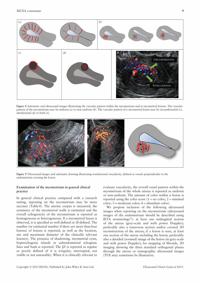
The vascular pattern within the myometrium maybe uniform or non-uniform (Figure 8) and the vascularpattern of a myometrial lesion may be circumfer-ential, intralesional or both (Figure 8). Some lesionsare associated with disruption of the normal uter-ine vasculature, while others are not. Translesional
vascularity (Figure 9) is characterized by the presence ofvessels perpendicular to the uterine cavity/serosa crossingthe lesion. The degree of vascularization should bereported using a subjective color score, with a color scoreof 1 representing no color and a score of 4 representingabundant color signals. This score is based on subjectiveevaluation of both the percentage of the lesion thatis vascularized and the color hue. The color score isassigned taking into account the lesion as a whole, but inlesions with uneven internal vascularization (e.g. becauseof cystic areas or central necrosis), the score reflectsthe degree of vascularization in the solid parts of thelesion. If there is an uneven spread of vascularizationin the solid components of the lesion, the score for themost vascularized solid component and the percentageof the solid components with color signals should berecorded. A color score may be assigned separately tocircumferential and intralesional vascularity (Figure S10).
When carrying out research studies, the vascularity oflesions may be reported as iso-, hypo- or hypervascularcompared with the vascularity of the surroundingmyometrium. Reporting a lesion’s vascularity mayinclude the number of vessels (single or multiple), vesselsize (small and equal, large and equal, unequal; or thevessel diameter may be measured), the direction of vessels(perpendicular or not perpendicular to endometrium),the vessel branching pattern (no branching, regular or
Copyright © 2015 ISUOG. Published by John Wiley & Sons Ltd. Ultrasound Obstet Gynecol 2015.

8 Van den Bosch et al.
Figure 6 Ultrasound images showing: (a) myometrial cysts (arrows); (b) hyperechogenic islands (surrounded by dotted lines); and(c) echogenic spots (arrows).
Figure 7 Ultrasound images illustrating echogenic subendometrial lines (a) and buds (b) (both encircled by dashed lines).
irregular branching) and may be further specified asoutlined in Table 3 and Figure S11. Irregular branchingvessels may be defined as abnormal tortuous vessels,irregular caliber vessels, a lack of hierarchy in branchingwith varying branching angles, vessel sprouts or anoverall impression of a chaotic vessel pattern. The termcircumferential vessels relates to vessels that surround alesion, whereas vessels located inside a lesion are calledintralesional.
In the context of research, color flow within a lesionmay be quantified using 3D ultrasound with virtual organcomputer-aided analysis (VOCALTM) in order to calculate3D power Doppler indices: the vascularity index (VI, thenumber of color voxels in the volume expressed as a
percentage of the total number of voxels in the volume,potentially reflecting vascularity); the flow index (FI, themean color value in the color voxels expressed as anumber from 0–100, potentially reflecting flow velocity;and the vascularization flow index (VFI, calculated asVI multiplied by FI, reflecting the mean color value inall the volumes’ voxels, expressed as a number from0–100, and potentially reflecting tissue perfusion)18,19.However, because 3D vascular indices depend on machinesettings, there remains doubt about their reproducibility,and their clinical use has yet to be explored adequately19.Until the limitations of these indices have been resolved,we recommend not using them outside the context of aspecific research project.
Copyright © 2015 ISUOG. Published by John Wiley & Sons Ltd. Ultrasound Obstet Gynecol 2015.
MUSA consensus 9
(a) (b)
(c) (d)
CIRCUMFERENTIAL
INTRA-LESIONAL
Figure 8 Schematic and ultrasound images illustrating the vascular pattern within the myometrium and in myometrial lesions. The vascularpattern of the myometrium may be uniform (a) or non-uniform (b). The vascular pattern of a myometrial lesion may be circumferential (c),intralesional (d) or both (e).
Figure 9 Ultrasound images and schematic drawing illustrating translesional vascularity, defined as vessels perpendicular to theendometrium crossing the lesion.
Examination of the myometrium in general clinicalpractice
In general clinical practice compared with a researchsetting, reporting on the myometrium may be moresuccinct (Table 4). The uterine corpus is measured, thesymmetry of the myometrial walls is estimated and theoverall echogenicity of the myometrium is reported ashomogeneous or heterogeneous. If a myometrial lesion isobserved, it is specified as well-defined or ill-defined. Thenumber (or estimated number if there are more than fourlesions) of lesions is reported, as well as the location,site and maximum diameter of the clinically relevantlesion(s). The presence of shadowing, myometrial cysts,hyperechogenic islands or subendometrial echogeniclines and buds is reported. The JZ is reported as regularor poorly defined (if it is irregular, interrupted, notvisible or not assessable). When it is clinically relevant to
evaluate vascularity, the overall vessel pattern within themyometrium of the whole uterus is reported as uniformor non-uniform. The amount of color within a lesion isreported using the color score (1 = no color; 2 = minimalcolor; 3 = moderate color; 4 = abundant color).
We propose inclusion of the following ultrasoundimages when reporting on the myometrium (ultrasoundimages of the endometrium should be described usingIETA terminology3): at least one midsagittal sectionof the uterus (gray-scale and with power Doppler);preferably also a transverse section and/or coronal 3Dreconstruction of the uterus; if a lesion is seen, at leastone section of the uterus including the lesion; preferablyalso a detailed (zoomed) image of the lesion (in gray-scaleand with power Doppler); for mapping of fibroids, 3Dimaging showing the three standard orthogonal planesthrough the uterus or tomographic ultrasound images(TUI) may sometimes be illustrative.
Copyright © 2015 ISUOG. Published by John Wiley & Sons Ltd. Ultrasound Obstet Gynecol 2015.

10 Van den Bosch et al.
Table 3 Reporting vascularity of the myometrium on ultrasound examination
Vascularization to be assessed Description Measurement
Whole uterusOverall vessel pattern* (Figure 8) Uniform, non-uniform* —
LesionsAmount of color (in a lesion)* (Figure S10) Color score (both percentage of lesion being
vascularized and color hue are taken into account)*No color (1); minimal color
(2); moderate color (3);abundant color (4)*
In case of uneven spread of vascularization† Color score in most vascularized part† No color (1); minimal color(2); moderate color (3);abundant color (4)†
Percent of solid tissue with color signal† 0–100%†Compared to adjacent myometrium† Iso-, hypo-, hypervascularity†
Location of vessels† (Figures 8 and 9) Circumferential, intralesional; uniform, non-uniform(areas with increased/decreased vascularity)†
—
Vessel morphology† (Figures 8 and S11) Number: single, multiple; size: large and equal, smalland equal, unequal; branching: regular, irregular,no branching; direction: perpendicular, notperpendicular†
—
*Items of importance in daily clinical practice. †Items of interest for research purposes.
Table 4 Reporting the myometrium in general clinical practice
Feature to be described Description/term
Uterine corpus Length, anteroposterior diameter, transverse diameterMyometrial walls Symmetrical/asymmetricalOverall echogenicity Homogeneous/heterogeneous
Myometrial lesions Well-defined/ill-definedNumber Number (1, 2, 3 or estimation in case > 4 lesions)Location Location of the largest/clinically relevant lesion(s): anterior, posterior, fundal, right
lateral or left lateral, globalSite Site (for well-defined lesions) of the largest/clinically relevant lesion(s): FIGO
classification 1–7Size Maximum diameter of the largest/clinically relevant lesion(s)Shadowing
Edge shadows Present/absentInternal shadows Present/absentFan-shaped shadowing Present/absent
Cysts Present/absentHyperechogenic islands Present/absentSubendometrial echogenic lines & buds Present/absent
Junctional zone Regular/poorly definedVascularity of myometrium
Overall vessel pattern (in whole uterus) Uniform/non-uniformAmount of color (in a lesion): color score (1) No color; (2) minimal color; (3) moderate color; (4) abundant color
FIGO, International Federation of Gynecology and Obstetrics2.
ULTRASOUND FINDINGS ASSOCIATEDWITH PATHOLOGY
In this section we describe ultrasound features that, inour opinion and on the basis of reports in the literature,are associated with pathology and in particular withadenomyosis and fibroids (Table 5). Further researchshould validate the importance of each of these features.
Adenomyosis
Adenomyosis is caused by a proliferation of endometrialglands and stroma leading to ill-defined lesions withinthe myometrium. Adenomyosis may be present at oneor more sites within the uterine wall or involve most
of the myometrium, and may often be dispersed withinthe myometrium rather than forming a confined lesion,i.e. diffuse adenomyosis. It may, on the other hand, bepresent in only one part of the myometrium, i.e. focaladenomyosis. In rare cases it may present as a largecyst (an adenomyotic cyst or cystic adenomyoma)20–24.On histological examination, adenomyosis is classifiedas diffuse when the endometrial glands or stroma aredistributed diffusely within the myometrium, and focalwhen circumscribed nodular aggregates are seen. Focaladenomyosis is not the same as an adenomyoma. Theseare defined by pathologists as focal adenomyosis withadditional compensatory hypertrophy of the surroundingmyometrium25.
Copyright © 2015 ISUOG. Published by John Wiley & Sons Ltd. Ultrasound Obstet Gynecol 2015.

MUSA consensus 11
Table 5 Features considered important in diagnosis of fibroids and adenomyosis
Feature Typical fibroid Adenomyosis
Serosal contour of uterus Lobulated or regular Often globally enlarged uterusDefinition of lesion Well-defined Ill-defined in diffuse adenomyosis
(adenomyoma may be well-defined)Symmetry of uterine walls Asymmetrical in presence of well-defined lesion(s) Myometrial anteroposterior asymmetryLesion
Outline Well-defined Ill-definedShape Round, oval, lobulated Ill-definedContour Smooth Irregular or ill-definedRim Hypo- or hyperechogenic No rimShadowing Edge shadows, internal shadows (often fan-shaped
shadowing)No edge shadows, fan-shaped
shadowing67
Echogenicity Uniform: hyper-, iso-, hypoechogenicNon-uniform: mixed echogenicity
Non-uniform: mixed echogenicity67,68
Cysts20–24,62, hyperechogenic islands,subendometrial lines and buds24,63
Vascularity Circumferential flow Translesional flow69
Junctional zone (JZ)JZ thickness, regularity Not-thickened; regular or not visible Thickened; irregular or ill-defined9,61–63
JZ interruption Interrupted or overstretched JZ in areas withlesions of FIGO types 1–3 (Figure 3)
Interrupted JZ (even in absence oflocalized lesions)9
FIGO, International Federation of Gynecology and Obstetrics2.
The ultrasound features of adenomyosis (Figure 10)should be reported and quantified (Tables 1–3). Theultrasound features of a globular uterus with ill-definedadenomyotic lesions may be explained by direct invasionof endometrial tissue from the endometrium, as seen in‘classic adenomyosis’, or by invasion from endometrioticimplants on the serosal surface of the uterus26. Rarely,diffuse adenomyosis may be localized as a solitaryfinding without direct continuation with the serosaor the endometrium22. The relative proportions ofendometrial glandular structures, endometrial stroma andhypertrophic muscle elements within a lesion probablyexplain the different ultrasound features reported to betypical of adenomyosis. The link between ultrasoundfeatures and histopathology has yet to be confirmed andrequires further research27.
Fibroid (leiomyoma)
A uterine fibroid is seen typically on ultrasound as awell-defined round lesion within the myometrium orattached to it, often showing shadows at the edge of thelesion and/or internal fan-shaped shadowing (Figure S12).The echogenicity varies and some hyperechogenicitymay be present internally. On color- or power-Dopplerimaging, circumferential flow around the lesion is oftenvisible. However, some fibroids do not exhibit suchtypical features. We suggest that such fibroids are labelledas sonographically atypical fibroids (Figure 11).
On histological examination, fibroids are composedof smooth muscle cells and connective tissue in denselypacked whorls. Acoustic shadowing may arise from theinterface between smooth muscle bundles, hyalinizedconnective tissue and normal myometrium28. Thesonographic appearance of a fibroid may depend on theproportion of muscle cells and fibrous stroma within thelesion.
Variants of fibroids and other uterine smooth muscletumors
Variants of fibroids
Fibroids may undergo degeneration, which may be spon-taneous or a result of induced infarction following uter-ine artery embolization. Coagulate necrosis is inducedafter high-intensity ultrasound or radiofrequency abla-tion. Types of degeneration are: a) red, b) hyaline, c) cystic/myxoid (myxoid leiomyoma) or d) hydropic. Spontaneousdegeneration may occur in pregnancy, and red degen-eration is an initial manifestation29 within days afterinfarction. The sonographic appearance of red degenera-tion may be unremarkable, although some cases have beenreported as homogeneous lesions with low echogenicity, ahyperechogenic rim and absent internal vascularity30–32.Hemorrhage and edema in these fibroids may give rise totumors of mixed echogenicity. Late manifestations afterinfarction are most commonly hyaline degeneration33,34,while some fibroids may show mixed echogenicity orhypoechogenic cystic areas.
Fibroids after induced infarction are often uniform,hypoechogenic with a hyperechogenic rim and acousticshadows35,36. There is usually no internal vascularityor, at most, a few disparate vessels are observed.Cystic or myxoid degeneration may develop, resulting inregular hypoechogenic cystic areas with fluid or myxoidcontent37,38. Degeneration may also occur in malignantuterine smooth muscle tumors39.
Uterine sarcomas and other uterine smooth-muscletumors
The prediction of malignancy is of utmost importance.However, data on the prediction of uterine sarcomaby ultrasound examination are scarce and based mainly
Copyright © 2015 ISUOG. Published by John Wiley & Sons Ltd. Ultrasound Obstet Gynecol 2015.

12 Van den Bosch et al.
(a) (b)
(e) (f) (g) (h)
(c) (d)
Figure 10 Schematic drawings illustrating the ultrasound features considered currently to be typical of adenomyosis: asymmetricalthickening (a), cysts (b), hyperechoic islands (c), fan-shaped shadowing (d), echogenic subendometrial lines and buds (e), translesionalvascularity (f), irregular junctional zone (g) and interrupted junctional zone (h).
CYSTIC DEGENERATION
UTERUS
RIGHT
FIBROID IN LEFT BROAD LIGAMENT FIGO 1 FIBROID
LEFT
Figure 11 Ultrasound images showing fibroids with atypical sonographic features. These fibroids have non-uniform echogenicity andintralesional anechoic cysts, and some have areas with hyperechogenicity. The FIGO type 1 fibroid (bottom right) has an irregular outline.
Figure 12 Gray-scale and color Doppler images of a sarcoma in the anterior wall of the uterus. The uterine corpus (solid arrows) is locatedposteriorly and contains clear fluid (open arrow).
Copyright © 2015 ISUOG. Published by John Wiley & Sons Ltd. Ultrasound Obstet Gynecol 2015.

MUSA consensus 13
on small retrospective case series, precluding definitiveguidelines. There are many rare uterine smooth-muscletumors other than benign leiomyomas40, but onlylimited information on their ultrasound features hasbeen reported to date. This issue has become increasinglyimportant in view of the debate about when, or if, fibroidsmay be morcellated during laparoscopic surgery.
Malignant sarcomas comprise leiomyosarcoma(Figure 12), endometrial stromal sarcoma, adenosarcomaand undifferentiated sarcoma. Uterine sarcomas presentas purely myometrial lesions and are typically single, largetumors41. Their ultrasound features may be indistinctfrom those of ordinary fibroids42 or they may appearas an irregularly vascularized mass, with a regular orirregular outline, often with irregular anechoic areas dueto necrosis43–49.
Uterine smooth-muscle tumor of uncertain malignantpotential (STUMP)
There are no specific ultrasound features describedfor STUMP. Intravenous leiomyomatosis, disseminatedperitoneal leiomyomatosis and benign metastasizingleiomyoma50–52 have the same ultrasound features asdo ordinary fibroids. There are often multiple fibroidsand they may be recognized by their location outsidethe uterine borders. These multiple fibroids should bedistinguished from diffuse leiomyomatosis53.
Fibroids with little or no recurrent and/or metastaticpotential
Ultrasound features of leiomyoma with bizarre nuclei(bizarre/symplastic/atypical leiomyoma), mitotically ac-tive leiomyoma, cellular and highly cellular leiomyoma,dissecting leiomyoma and leiomyoma with increasedcellularity, no atypia or mitotic figures and increasedvascularity40,54 may have the same macroscopicallypathological features as do fibroids40,54 and may haveincreased vascularity, as this feature seems to be related tocellularity55. A cotyledonoid leiomyoma or cotelydonoiddissecting leiomyoma56–58 is a nodular tumor withplacenta-like echogenicity on ultrasound, but may alsobe cystic. Ultrasonographic features of lipoleiomyoma59
comprise a hyperechogenic mass partly encased by ahypoechogenic rim. Ultrasonographic features for epithe-lioid leiomyoma40 and palisading/neurilemmoma-likeleiomyoma60 have not been described.
CONCLUSION
The terms and definitions presented in this paper aim tofacilitate consistent reporting of myometrial lesions whenusing ultrasonography in both daily clinical practiceand for research purposes. Clearly, the clinical relevanceof some of the terms that have been proposed hasnot yet been evaluated in prospective clinical studies.We acknowledge that some aspects of the systematic
reporting we have suggested may require a relativelyhigh level of ultrasound training. We also acknowledgethat some of the proposed terms and definitions aretoo detailed for use in general clinical practice and willinitially be suitable only for use in research settings.Future research should focus on the ability to predictspecific pathologies and on the clinical relevance of theultrasound features described in this paper. Althoughthe members of the panel involved in the writing of thisconsensus have different fields of expertise, includinggynecological ultrasonography, fertility treatment, hys-teroscopy, general gynecology and clinical research, weacknowledge that they all come from Europe and theUSA, leaving most areas of the world unrepresented.
The recent controversy about the safety of morcellationof lesions thought to be benign fibroids, but that werein fact malignant5, highlights the importance of reliablepreoperative characterization of myometrial lesions.Although recognizing a typical fibroid on ultrasoundis usually straightforward, differentiating betweenan atypical fibroid and a uterine sarcoma remainschallenging. The establishment of an internationaldatabase of ultrasound imaging and magnetic resonanceimaging (MRI) of uterine sarcomas and rare uterinetumors would be of great clinical value.
Adenomyosis may be difficult to diagnose with ultra-sound. Different ultrasound features have been suggestedto be associated with adenomyosis, but at present it is notclear which of the various ultrasound criteria are mostimportant for diagnosis. Some features may carry a greaterdiagnostic weight than others61 and the presence of morethan one ultrasound feature associated with adenomyosismight increase the likelihood of the diagnosis61–63.We have not included in our consensus statement theso called ‘question-mark sign’, suggested to be typicalof adenomyosis, because this sign occurs when thereis also deep infiltrating endometriosis in the posteriorcompartment64.
The terms that we suggest for characterization of theJZ are derived from MRI studies20,30. The JZ is bettervisualized by 3D9,65 than by 2D ultrasound, but theclinical implications of a thickened or disrupted JZ onultrasound needs to be established4,66.
The clinical relevance of myometrial lesions for abnor-mal uterine bleeding, pelvic pain, subfertility and preg-nancy outcome is an important topic for research. Certainultrasound features might prove to be more clinically rele-vant than others. The role of a systematic evaluation of thesonographic features of myometrial lesions when choosingmanagement (expectant management, medical therapy,selective embolization, high-intensity focused ultrasoundor surgical treatment) and in the follow-up during orafter treatment is another important topic for futureresearch.
To conclude, the terms and definitions in this consensusstatement should enable clinicians to produce a structuredreport when describing the sonographic appearance ofthe myometrium and myometrial lesions and harmonizenomenclature for future research.
Copyright © 2015 ISUOG. Published by John Wiley & Sons Ltd. Ultrasound Obstet Gynecol 2015.

14 Van den Bosch et al.
ACKNOWLEDGMENTS
T.B. is supported by the National Institute for HealthResearch (NIHR) Biomedical Research Centre based atImperial College Healthcare NHS Trust and ImperialCollege London. The views expressed are those of theauthor(s) and not necessarily those of the NHS, the NIHRor the Department of Health. D.T. is Senior ClinicalInvestigator of Scientific Research Fund (FWO) Flanders.
REFERENCES
1. Munro MG, Critchley HO, Fraser IS. The FIGO classification of causes of abnormaluterine bleeding in the reproductive years. Fertil Steril 2011; 95: 2204–2208.
2. Munro MG, Critchley HO, Broder MS, Fraser IS. FIGO classification system(PALM-COEIN) for causes of abnormal uterine bleeding in nongravid womenof reproductive age. Int J Gynaecol Obstet 2011; 113: 3–13.
3. Leone FP, Timmerman D, Bourne T, Valentin L, Epstein E, Goldstein SR, MarretH, Parsons AK, Gull B, Istre O, Sepulveda W, Ferrazzi E, Van den Bosch T.Terms, definitions and measurements to describe the sonographic features of theendometrium and intrauterine lesions: a consensus opinion from the InternationalEndometrial Tumor Analysis (IETA) group. Ultrasound Obstet Gynecol 2010; 35:103–112.
4. Gordts S, Brosens JJ, Fusi L, Benagiano G, Brosens I. Uterine adenomyosis: a needfor uniform terminology and consensus classification. Reprod Biomed Online 2008;17: 244–248.
5. Hampton T. Critics of fibroid removal procedure question risks it may pose forwomen with undetected uterine cancer. JAMA 2014; 311: 891–893.
6. Okaro E, Condous G, Khalid A, Timmerman D, Ameye L, Van Huffel SV, BourneT. The use of ultrasound-based ‘soft markers’ for the prediction of pelvic pathologyin women with chronic pelvic pain--can we reduce the need for laparoscopy? BJOG2006; 113: 251–256.
7. Tetlow RL, Richmond I, Manton DJ, Greenman J, Turnbull LW, Killick SR.Histological analysis of the uterine junctional zone as seen by transvaginal ultrasound.Ultrasound Obstet Gynecol 1999; 14: 188–193.
8. Naftalin J, Jurkovic D. The endometrial-myometrial junction: a fresh look at a busycrossing. Ultrasound Obstet Gynecol 2009; 34: 1–11.
9. Exacoustos C, Brienza L, Di GA, Szabolcs B, Romanini ME, Zupi E, Arduini D.Adenomyosis: three-dimensional sonographic findings of the junctional zone andcorrelation with histology. Ultrasound Obstet Gynecol 2011; 37: 471–479.
10. Exacoustos C, Luciano D, Corbett B, De FG, Di FM, Luciano A, Zupi E. The uterinejunctional zone: a 3-dimensional ultrasound study of patients with endometriosis.Am J Obstet Gynecol 2013; 209: 248.e1–7.
11. Martins WP, Raine-Fenning NJ, Leite SP, Ferriani RA, Nastri CO. A standardizedmeasurement technique may improve the reliability of measurements of endometrialthickness and volume. Ultrasound Obstet Gynecol 2011; 38: 107–115.
12. Woelfer B, Salim R, Banerjee S, Elson J, Regan L, Jurkovic D. Reproductiveoutcomes in women with congenital uterine anomalies detected by three-dimensionalultrasound screening. Obstet Gynecol 2001; 98: 1099–1103.
13. Abuhamad AZ, Singleton S, Zhao Y, Bocca S. The Z technique: an easy approachto the display of the mid-coronal plane of the uterus in volume sonography.J Ultrasound Med 2006; 25: 607–612.
14. Wamsteker K, Emanuel MH, de Kruif JH. Transcervical hysteroscopic resection ofsubmucous fibroids for abnormal uterine bleeding: results regarding the degree ofintramural extension. Obstet Gynecol 1993; 82: 736–740.
15. Casadio P, Youssef AM, Spagnolo E, Rizzo MA, Talamo MR, De AD, Marra E, GhiT, Savelli L, Farina A, Pelusi G, Mazzon I. Should the myometrial free margin stillbe considered a limiting factor for hysteroscopic resection of submucous fibroids? Apossible answer to an old question. Fertil Steril 2011; 95: 1764–1768.
16. Yang JH, Lin BL. Changes in myometrial thickness during hysteroscopic resectionof deeply invasive submucous myomas. J Am Assoc Gynecol Laparosc 2001; 8:501–505.
17. Timmerman D, Valentin L, Bourne TH, Collins WP, Verrelst H, Vergote I. Terms,definitions and measurements to describe the sonographic features of adnexal tumors:a consensus opinion from the International Ovarian Tumor Analysis (IOTA) Group.Ultrasound Obstet Gynecol 2000; 16: 500–505.
18. Alcazar JL. Three-dimensional power Doppler derived vascular indices: what are wemeasuring and how are we doing it? Ultrasound Obstet Gynecol 2008; 32: 485–487.
19. Raine-Fenning NJ, Campbell BK, Clewes JS, Kendall NR, Johnson IR. The reliabilityof virtual organ computer-aided analysis (VOCAL) for the semiquantification ofovarian, endometrial and subendometrial perfusion. Ultrasound Obstet Gynecol2003; 22: 633–639.
20. Cucinella G, Billone V, Pitruzzella I, Lo Monte AI, Palumbo VD, Perino A.Adenomyotic cyst in a 25-year-old woman: case report. J Minim Invasive Gynecol2013; 20: 894–898.
21. Ho ML, Ratts V, Merritt D. Adenomyotic cyst in an adolescent girl. J Pediatr AdolescGynecol 2009; 22: 33–38.
22. Protopapas A, Milingos S, Markaki S, Loutradis D, Haidopoulos D, SotiropoulouM, Antsaklis A. Cystic uterine tumors. Gynecol Obstet Invest 2008; 65:275–280.
23. Tahlan A, Nanda A, Mohan H. Uterine adenomyoma: a clinicopathologic review of26 cases and a review of the literature. Int J Gynecol Pathol 2006; 25: 361–365.
24. Reinhold C, Tafazoli F, Mehio A, Wang L, Atri M, Siegelman ES, Rohoman L.Uterine adenomyosis: endovaginal US and MR imaging features with histopathologiccorrelation. Radiographics 1999; 19: 147–160.
25. Haines, Taylor. Obstetrical and Gynaecological Pathology. (4th edn), Fox H, WellsM (eds). Churchill Livingstone, 1995.
26. Kishi Y, Suginami H, Kuramori R, Yabuta M, Suginami R, Taniguchi F. Four subtypesof adenomyosis assessed by magnetic resonance imaging and their specification. AmJ Obstet Gynecol 2012; 207: 114–117.
27. Ferenczy A. Pathophysiology of adenomyosis. Hum Reprod Update 1998; 4:312–322.
28. Kliewer MA, Hertzberg BS, George PY, McDonald JW, Bowie JD, Carroll BA.Acoustic shadowing from uterine leiomyomas: sonographic-pathologic correlation.Radiology 1995; 196: 99–102.
29. Ouyang DW, Economy KE, Norwitz ER. Obstetric complications of fibroids. ObstetGynecol Clin North Am 2006; 33: 153–169.
30. Lev-Toaff AS, Coleman BG, Arger PH, Mintz MC, Arenson RL, Toaff ME.Leiomyomas in pregnancy: sonographic study. Radiology 1987; 164: 375–380.
31. Cooper NP, Okolo S. Fibroids in pregnancy--common but poorly understood. ObstetGynecol Surv 2005; 60: 132–138.
32. Valentin L. Characterising acute gynaecological pathology with ultrasound: anoverview and case examples. Best Pract Res Clin Obstet Gynaecol 2009; 23:577–593.
33. McLucas B. Diagnosis, imaging and anatomical classification of uterine fibroids. BestPract Res Clin Obstet Gynaecol 2008; 22: 627–642.
34. Ueda H, Togashi K, Konishi I, Kataoka ML, Koyama T, Fujiwara T, Kobayashi H,Fujii S, Konishi J. Unusual appearances of uterine leiomyomas: MR imaging findingsand their histopathologic backgrounds. Radiographics 1999; 19: 131–145.
35. Nicholson TA, Pelage JP, Ettles DF. Fibroid calcification after uterine arteryembolization: ultrasonographic appearance and pathology. J Vasc Interv Radiol2001; 12: 443–446.
36. Allison SJ, Wolfman DJ. Sonographic evaluation of patients treated with uterineartery embolization. Ultrasound Clinics 2010; 5: 277–288.
37. Yarwood RL, Arroyo E. Cystic degeneration of a uterine leiomyoma masqueradingas a postmenopausal ovarian cyst. A case report. J Reprod Med 1999; 44:649–652.
38. Cohen JR, Luxman D, Sagi J, Jossiphov J, David MP. Ultrasonic ‘‘honeycomb”appearance of uterine submucous fibroids undergoing cystic degeneration. J ClinUltrasound 1995; 23: 293–296.
39. Karpathiou G, Sivridis E, Giatromanolaki A. Myxoid leiomyosarcoma of the uterus:a diagnostic challenge. Eur J Gynaecol Oncol 2010; 31: 446–448.
40. Ip PP, Tse KY, Tam KF. Uterine smooth muscle tumors other than the ordinaryleiomyomas and leiomyosarcomas: a review of selected variants with emphasis onrecent advances and unusual morphology that may cause concern for malignancy.Adv Anat Pathol 2010; 17: 91–112.
41. Bonneau C, Thomassin-Naggara I, Dechoux S, Cortez A, Darai E, Rouzier R.Value of ultrasonography and magnetic resonance imaging for the characterizationof uterine mesenchymal tumors. Acta Obstet Gynecol Scand 2013; 93:261–268.
42. Jayakrishnan K, Koshy AK, Manjula P, Nair AM, Ramachandran A, KattoorJ. Endometrial stromal sarcoma mimicking a myoma. Fertil Steril 2009; 92:1744–1746.
43. Exacoustos C, Romanini ME, Amadio A, Amoroso C, Szabolcs B, Zupi E, ArduiniD. Can gray-scale and color Doppler sonography differentiate between uterineleiomyosarcoma and leiomyoma? J Clin Ultrasound 2007; 35: 449–457.
44. Aviram R, Ochshorn Y, Markovitch O, Fishman A, Cohen I, Altaras MM, Tepper R.Uterine sarcomas versus leiomyomas: gray-scale and Doppler sonographic findings.J Clin Ultrasound 2005; 33: 10–13.
45. Hata K, Hata T, Maruyama R, Hirai M. Uterine sarcoma: can it be differentiated fromuterine leiomyoma with Doppler ultrasonography? A preliminary report. UltrasoundObstet Gynecol 1997; 9: 101–104.
46. Seki K, Hoshihara T, Nagata I. Leiomyosarcoma of the uterus: ultrasonography andserum lactate dehydrogenase level. Gynecol Obstet Invest 1992; 33: 114–118.
47. Hata K, Hata T, Makihara K, Aoki S, Takamiya O, Kitao M, Harada Y, NagaokaS. Sonographic findings of uterine leiomyosarcoma. Gynecol Obstet Invest 1990; 30:242–245.
48. Szabo I, Szantho A, Csabay L, Csapo Z, Szirmai K, Papp Z. Color Dopplerultrasonography in the differentiation of uterine sarcomas from uterine leiomyomas.Eur J Gynaecol Oncol 2002; 23: 29–34.
49. Amant F, Coosemans A, Debiec-Rychter M, Timmerman D, Vergote I. Clinicalmanagement of uterine sarcomas. Lancet Oncol 2009; 10: 1188–1198.
50. Fasih N, Prasad Shanbhogue AK, Macdonald DB, Fraser-Hill MA, Papadatos D,Kielar AZ, Doherty GP, Walsh C, McInnes M, Atri M. Leiomyomas beyond theuterus: unusual locations, rare manifestations. Radiographics 2008; 28: 1931–1948.
51. Cohen DT, Oliva E, Hahn PF, Fuller AF, Jr., Lee SI. Uterine smooth-muscle tumorswith unusual growth patterns: imaging with pathologic correlation. AJR Am JRoentgenol 2007; 188: 246–255.
52. Vaquero ME, Magrina JF, Leslie KO. Uterine smooth-muscle tumors with unusualgrowth patterns. J Minim Invasive Gynecol 2009; 16: 263–268.
53. Fedele L, Bianchi S, Zanconato G, Carinelli S, Berlanda N. Conservative treatmentof diffuse uterine leiomyomatosis. Fertil Steril 2004; 82: 450–453.
54. Robboy SJ, Bentley RC, Butnor K, Anderson MC. Pathology and pathophysiologyof uterine smooth-muscle tumors. Environ Health Perspect 2000; 108 Suppl 5:779–784.
55. Minsart AF, Ntoutoume SF, Vandenhoute K, Jani J, Van PC. Does three-dimensionalpower Doppler ultrasound predict histopathological findings of uterine fibroids? Apreliminary study. Ultrasound Obstet Gynecol 2012; 40: 714–720.
56. Smith CC, Gold MA, Wile G, Fadare O. Cotyledonoid dissecting leiomyoma of theuterus: a review of clinical, pathological, and radiological features. Int J Surg Pathol2012; 20: 330–341.
Copyright © 2015 ISUOG. Published by John Wiley & Sons Ltd. Ultrasound Obstet Gynecol 2015.

MUSA consensus 15
57. Raga F, Sanz-Cortes M, Casan EM, Burgues O, Bonilla-Musoles F. Cotyledonoiddissecting leiomyoma of the uterus. Fertil Steril 2009; 91: 1269–1270.
58. Gurbuz A, Karateke A, Kabaca C, Arik H, Bilgic R. A case of cotyledonoid leiomyomaand review of the literature. Int J Gynecol Cancer 2005; 15: 1218–1221.
59. Prieto A, Crespo C, Pardo A, Docal I, Calzada J, Alonso P. Uterine lipoleiomyomas:US and CT findings. Abdom Imaging 2000; 25: 655–657.
60. Gisser SD, Young I. Neurilemoma-like uterine myomas:an ultrastructural reaffirma-tion of their non-Schwannian nature. Am J Obstet Gynecol 1977; 129: 389–392.
61. Dueholm M, Lundorf E, Hansen ES, Sorensen JS, Ledertoug S, Olesen F.Magnetic resonance imaging and transvaginal ultrasonography for the diagnosisof adenomyosis. Fertil Steril 2001; 76: 588–594.
62. Bazot M, Cortez A, Darai E, Rouger J, Chopier J, Antoine JM, Uzan S.Ultrasonography compared with magnetic resonance imaging for the diagnosisof adenomyosis: correlation with histopathology. Hum Reprod 2001; 16:2427–2433.
63. Kepkep K, Tuncay YA, Goynumer G, Tutal E. Transvaginal sonography in thediagnosis of adenomyosis: which findings are most accurate? Ultrasound ObstetGynecol 2007; 30: 341–345.
64. Di Donato N, Bertoldo V, Montanari G, Zannoni L, Caprara G, Seracchioli R.Question mark form of uterus: a simple sonographic sign associated with thepresence of adenomyosis. Ultrasound Obstet Gynecol 2015; 46: 126–127.
65. Naftalin J, Hoo W, Nunes N, Mavrelos D, Nicks H, Jurkovic D. Inter-and intraobserver variability in three-dimensional ultrasound assessment of theendometrial-myometrial junction and factors affecting its visualization. UltrasoundObstet Gynecol 2012; 39: 587–591.
66. Tocci A, Greco E, Ubaldi FM. Adenomyosis and ‘endometrial-subendometrialmyometrium unit disruption disease’ are two different entities. Reprod BiomedOnline 2008; 17: 281–291.
67. Dueholm M. Transvaginal ultrasound for diagnosis of adenomyosis: a review. BestPract Res Clin Obstet Gynaecol 2006; 20: 569–582.
68. Reinhold C, Tafazoli F, Wang L. Imaging features of adenomyosis. Hum ReprodUpdate 1998; 4: 337–349.
69. Chiang CH, Chang MY, Hsu JJ, Chiu TH, Lee KF, Hsieh TT, Soong YK. Tumorvascular pattern and blood flow impedance in the differential diagnosis of leiomyomaand adenomyosis by color Doppler sonography. J Assist Reprod Genet 1999; 16:268–275.
SUPPORTING INFORMATION ON THE INTERNET
The following supporting information may be found in the online version of this article:
Figure S1 Schematic drawings illustrating how to measure the uterus.
Figure S2 Schematic drawings illustrating how to describe the serosal contour of the uterus.
Figure S3 Schematic drawings illustrating symmetry of the uterine walls.
Figure S4 Schematic drawings and ultrasound images illustrating measurement of the junctional zonethickness (for research purposes).
Figure S5 Schematic drawings illustrating measurement of the inner lesion-free margin and outer lesion-freemargin of a lesion.
Figure S6 Measurement of lesion penetration (for research purposes).
Figure S7 Schematic drawings illustrating echogenicity of a uniform lesion (for research purposes).
Figure S8 Schematic drawings illustrating the rim and shape of myometrial lesions.
Figure S9 Schematic and ultrasound images illustrating the normal vascular pattern of the myometrium.
Figure S10 Schematic images illustrating the color score (amount of color Doppler signals) in thecircumference of and inside myometrial lesions.
Figure S11 Schematic drawings illustrating how to describe the vascularization of a myometrial lesion inclinical research.
Figure S12 Schematic drawings illustrating the ultrasound features considered currently to be typical ofuterine fibroids.
Copyright © 2015 ISUOG. Published by John Wiley & Sons Ltd. Ultrasound Obstet Gynecol 2015.