syphilis the lung · syphilis of the lung by 1. m. librach syphilis of the lung is considered to be...
TRANSCRIPT

SYPHILIS OF THE LUNGBY
1. M. LIBRACH
Syphilis of the lung is considered to be veryuncommon. Osler (1912) found only twelve casesin 2,500 autopsies, and Babcock (1911) discoveredonly one in 6,000. However, if one considers theautopsies of syphilis alone, the frequency of lunginvolvement naturally increases. Thus, Netchaewfound 22 cases among 437 syphilitics, and Stolpersaw five cases in 61 syphilitics (both arequoted by Verse, 1931), whereas Carrera (1920)discovered twelve instances in 152 cases. Theaverage incidence, according to Tchertkoff andBerwick (1941), seems to be 8 per cent. of all lueticsbrought to autopsy. Karshner (1910) says thatpulmonary syphilis is twice as frequent amongstmales as amongst females, the peak of the ageincidence being in the early thirties. Most casesoccur five or more years after primary infection,though Dieulafoy (1889) put them at 10 to 11years later, i.e. later than late syphilitic visceralinvolvement.
Post-mortem reports of cases occur frequently inthe literature (De Jong (1936), Verse (1931), Prevot(1935), Dienst (1932), Kokawa (1906), Hammer(1931), Karshner (1920), etc.-all quoted byTchertkoff and Berwick, 1941), but reports of thiscondition in living patients seem to be rare.Thus Tchertkoff and Berwick (1941) report onecase in a coloured woman; De Navasquez(1942) cites another in a soldier aged 57 years;Romanus (1941) describes a further example in a42-year-old female; Pearson and De Navasquez(1938) report two cases; Lyons and others(1942) mention one case, and Kulchar and Windholz(1947) describe four cases, of whom one died andanother was treated successfully with penicillin.Wilson (1946) discusses with post-mortem findingsa case of bronchopneumonia in a Mexican maleaged 69 years, in whom a post-mortem revealedspirochaetes in the fibrotic lung areas.More recent articles have discussed the clinical
recognition of late-acquired cases without post-
mortem confirmation. Thus Loubeyre andGrangaud (1948) report a case in a North Africanmale patient aged 30 years with a putrid lungabscess and bronchiectasis whom they claim wascured by bismuth and arsenic. Findlay and others(1949) describe the case of a gumma of lung in aNegro aged 39 years, treated with penicillin andLugol's iodine, and later by lobectomy.
Case RecordA.F.P., a bachelor aged 49 years, a greengrocer,
first attended a chestclinic on 24 Sept., 1947, with a sixmonths' history of lassitude, tiredness, and loss of weightand appetite. He had brought up a moderate amountof mucopurulent sputum. During this time he had beentreated for " nerves ". A chest x ray on that dateshowed extensive mottling at the right apex withdiscrete heavy shadows at the bases and " peaking" ofboth leaves of the diaphragm (Fig. 1). A sputumexamination, also 24 Sept., 1947, was reported as positivefor acid-fast bacilli. There was no family history oftuberculosis, and past history was not noteworthy,except that in January 1945 he had been an in-patientin hospital for treatment of " duodenal ulcer "-thehistory then was nausea, vomiting, and abdominal painfor 10 days. Unfortunately I have been unable totrace the results of any investigation done at thattime, including a barium-meal examination, which thepatient volunteered was performed. (The symptoms,however, suggest a " gastric" crisis.) He was told togo home and rest, while awaiting admission to asanatorium. He was admitted to Ilford IsolationHospital on 30 June, 1948, when he repeated hisprevious history of loss of weight, strength, and appetite.He also complained of " tightness and fulness of thechest". He had been slightly deaf since the 1914-18 war.
Examination showed a thin, phthinoid, edentulouspatient. His temperature in the mouth was 990 F.pulse-rate 72 per minute, and respirations 20 per minute.The heart sounds were normal, but there was someimpairment of resonance with diminished breath soundsat the right apex. An E.S.R. was 12 mm. in 1 hour(Westergren method, at room temperature). A chestradiograph revealed a fibroid appearance at the right
126
copyright. on M
ay 27, 2020 by guest. Protected by
http://sti.bmj.com
/B
r J Vener D
is: first published as 10.1136/sti.26.3.126 on 1 Septem
ber 1950. Dow
nloaded from

SYPHILIS OF THE LUNG
FIG. 1.-Radiograph of chest, Sept. 24, 1947, showingextensive disease in both lungs before admission tohospital.
FIG. 2.-Radiograph of chest on admission to hospital,June 30, 1948, still showing extensive bilateral fibroticdisease, predominantly apical in distribution.
lung apex, hard shadows in both mid zones, exaggeratedhilar shadows, and ill-defined mottling in the left upperand mid zones (Fig. 2). Successive sputum examinationsfor acid-fast bacilli on six occasions were negative.Progress was equivocal. The pulse-rate varied from70 to 80 per minute and was of regular rhythm. Aslight evening rise of temperature to 99°F. occurredduring the first four days in hospital, but subsequentreadings were normal.On II Sept. he complained of pains in the knees, which
kept him awake at night, but no gross joint signs wereelicited. On 1 Oct. he had a sudden attack, consistingof pain in the neck and shoulders, giddiness, vomiting,and double vision. He complained that the wardseemed to be spinning round him and that people were" cut in half ". He was fully conscious but was flushed,the pulse rate being regular at 60 per minute. The bloodpressure in the arms was 145 80, the temperature was99.40 F. and the respiratory rate 20. Examination ofthe nervous system revealed the following findings:Both pupils showed no reaction to light, but reacted toaccommodation. The right was larger than the left,but both were regular and circular. The visual fieldsand retinae were normal. There was no nystagmus.He was slightly deaf in both ears but the drums wereintact. No other cranial nerves showed any abnormality.All deep reflexes were exaggerated in both arms and legs.The plantar reflexes were extensor. He was hypo-aesthetic to pin-prick over the whole body, but especiallyin both legs, where muscle sense was also diminished,but joint sense was present. Other forms of sensationwere not impaired. Two days later he felt better, thepulse rate rising to 80 per minute. Subsequentquestioning elicited a history of a penile chancre,contracted on war service in 1916, which had beentreated with mercury and salvarsan for six weeks.The following investigations were performed at the
time:Blood Wassermann Reaction: strongly positive.Lumbar Puncture: Cerebrospinal fluid deeply
xanthochromic and not under pressure. No evidenceof spinal block apparent.
Examination of cerebrospinal fluid: Red cells3,000. White cells 15 (Polymorphs 60 per cent.Protein 120 mg. per cent. Chlorides 730 mg.per cent.).
Wassermann Reaction: Strongly positive.Lange Curve: 5555543210Culture: Sterile.
Blood Examination: Hb 76 per cent. Red cells3,600,000. White cells 5,800 (polymorphs 64 percent. lymphocytes 30 per cent. monocytes 6per cent.).
X ray of skull (P.A. and Lateral): Normal.Urine: Normal.
He was transferred on 20 Oct. (after 4 months in thishospital) to a venereal diseases department. There thediagnosis of neurosyphilis was confirmed, and he wasgiven a course of 85 injections of40,000 units of penicillin2-hourly followed by a course of arsenic and bismuth.An E.S.R. at this time was 20 mm. in 1 hour (Westergren)and packed red cell volume 47 per cent.
127
copyright. on M
ay 27, 2020 by guest. Protected by
http://sti.bmj.com
/B
r J Vener D
is: first published as 10.1136/sti.26.3.126 on 1 Septem
ber 1950. Dow
nloaded from
BRITISH JOURNAL OF VENEREAL DISEASES
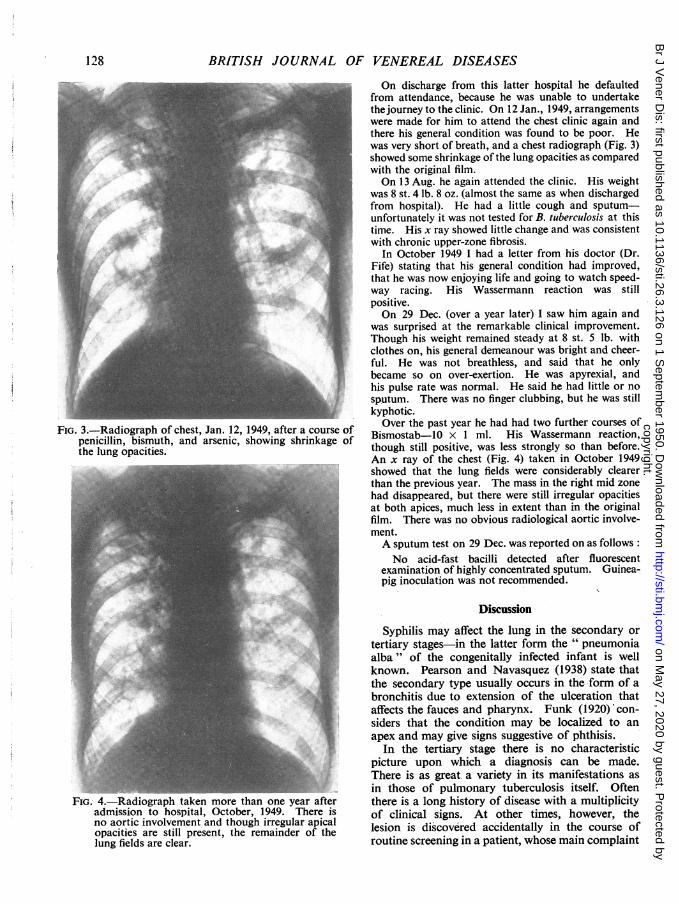
FIG. 3.-Radiograph of chest, Jan. 12, 1949, after a course ofpenicillin, bismuth, and arsenic, showing shrinkage ofthe lung opacities.
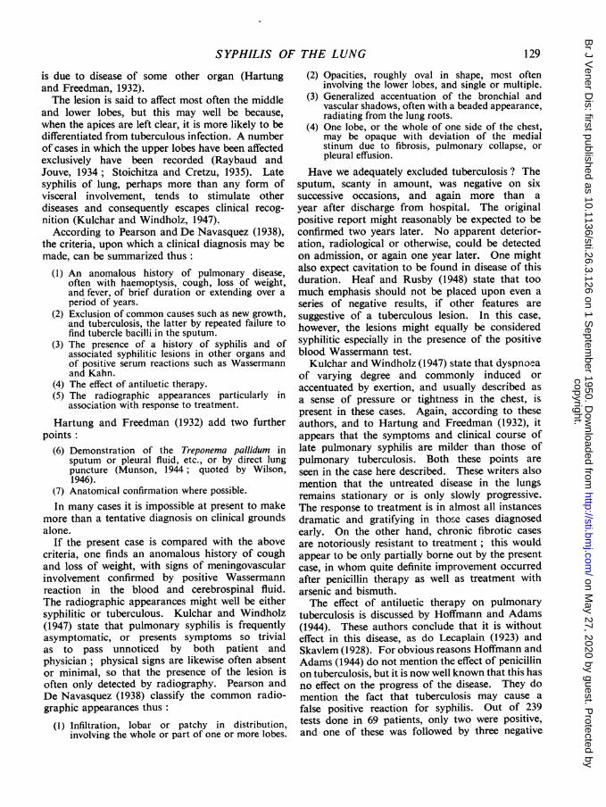
FIG. 4.-Radiograph taken more than one year afteradmission to hospital, October, 1949. There isno aortic involvement and though irregular apicalopacities are still present, the remainder of thelung fields are clear.
On discharge from this latter hospital he defaultedfrom attendance, because he was unable to undertakethejourney to the clinic. On 12 Jan., 1949, arrangementswere made for him to attend the chest clinic again andthere his general condition was found to be poor. Hewas very short of breath, and a chest radiograph (Fig. 3)showed some shrinkage of the lung opacities as comparedwith the original film.On 13 Aug. he again attended the clinic. His weight
was 8 st. 4 lb. 8 oz. (almost the same as when dischargedfrom hospital). He had a little cough and sputum-unfortunately it was not tested for B. tuberculosis at thistime. His x ray showed little change and was consistentwith chronic upper-zone fibrosis.
In October 1949 I had a letter from his doctor (Dr.Fife) stating that his general condition had improved,that he was now enjoying life and going to watch speed-way racing. His Wassermann reaction was stillpositive.On 29 Dec. (over a year later) I saw him again and
was surprised at the remarkable clinical improvement.Though his weight remained steady at 8 st. 5 lb. withclothes on, his general demeanour was bright and cheer-ful. He was not breathless, and said that he onlybecame so on over-exertion. He was apyrexial, andhis pulse rate was normal. He said he had little or nosputum. There was no finger clubbing, but he was stillkyphotic.Over the past year he had had two further courses of
Bismostab-10 x 1 ml. His Wassermann reaction,though still positive, was less strongly so than before.An x ray of the chest (Fig. 4) taken in October 1949showed that the lung fields were considerably clearerthan the previous year. The mass in the right mid zonehad disappeared, but there were still irregular opacitiesat both apices, much less in extent than in the originalfilm. There was no obvious radiological aortic involve-ment.A sputum test on 29 Dec. was reported on as follows:No acid-fast bacilli detected after fluorescent
examination of highly concentrated sputum. Guinea-pig inoculation was not recommended.
Discussion
Syphilis may affect the lung in the secondary ortertiary stages-in the latter form the "pneumoniaalba" of the congenitally infected infant is wellknown. Pearson and Navasquez (1938) state thatthe secondary type usually occurs in the form of abronchitis due to extension of the ulceration thataffects the fauces and pharynx. Funk (1920) con-siders that the condition may be localized to anapex and may give signs suggestive of phthisis.
In the tertiary stage there is no characteristicpicture upon which a diagnosis can be made.There is as great a variety in its manifestations asin those of pulmonary tuberculosis itself. Oftenthere is a long history of disease with a multiplicityof clinical signs. At other times, however, thelesion is discovered accidentally in the course ofroutine screening in a patient, whose main complaint
128
copyright. on M
ay 27, 2020 by guest. Protected by
http://sti.bmj.com
/B
r J Vener D
is: first published as 10.1136/sti.26.3.126 on 1 Septem
ber 1950. Dow
nloaded from
SYPHILIS OF THE LUNG
is due to disease of some other organ (Hartungand Freedman, 1932).The lesion is said to affect most often the middle
and lower lobes, but this may well be because,when the apices are left clear, it is more likely to bedifferentiated from tuberculous infection. A numberof cases in which the upper lobes have been affectedexclusively have been recorded (Raybaud andJouve, 1934; Stoichitza and Cretzu, 1935). Latesyphilis of lung, perhaps more than any form ofvisceral involvement, tends to stimulate otherdiseases and consequently escapes clinical recog-nition (Kulchar and Windholz, 1947).
According to Pearson and De Navasquez (1938),the criteria, upon which a clinical diagnosis may bemade, can be summarized thus:
(1) An anomalous history of pulmonary disease,often with haemoptysis, cough, loss of weight,and fever, of brief duration or extending over aperiod of years.
(2) Exclusion of common causes such as new growth,and tub_rculosis, the latter by repeated failure tofind tubercle bacilli in the sputum.
(3) The presence of a history of syphilis and ofassociated syphilitic lesions in other organs andof positive serum reactions such as Wassermannand Kahn.
(4) The effect of antiluetic therapy.(5) The radiographic appearances particularly in
association with response to treatment.
Hartung and Freedman (1932) add two furtherpoints:
(6) Demonstration of the Treponema pallidum insputum or pleural fluid, etc., or by direct lungpuncture (Munson, 1944; quoted by Wilson,1946).
(7) Anatomical confirmation where possible.in many cases it is impossible at present to make
more than a tentative diagnosis on clinical groundsalone.
If the present case is compared with the abovecriteria, one finds an anomalous history of coughand loss of weight, with signs of meningovascularinvolvement confirmed by positive Wassermannreaction in the blood and cerebrospinal fluid.The radiographic appearances might well be eithersyphilitic or tuberculous. Kulchar and Windholz(1947) state that pulmonary syphilis is frequentlyasymptomatic, or presents symptoms so trivialas to pass unnoticed by both patient andphysician ; physical signs are likewise often absentor minimal, so that the presence of the lesion isoften only detected by radiography. Pearson andDe Navasquez (1938) classify the common radio-graphic appearances thus:
(1) Infiltration, lobar or patchy in distribution,involving the whole or part of one or more lobes.
(2) Opacities, roughly oval in shape, most ofteninvolving the lower lobes, and single or multiple.
(3) Generalized accentuation of the bronchial andvascular shadows, often with a beaded appearance,radiating from the lung roots.
(4) One lobe, or the whole of one side of the chest,may be opaque with deviation of the medialstinum due to fibrosis, pulmonary collapse, orpleural effusion.
Have we adequately excluded tuberculosis ? Thesputum, scanty in amount, was negative on sixsuccessive occasions, and again more than ayear after discharge from hospital. The originalpositive report might reasonably be expected to beconfirmed two years later. No apparent deterior-ation, radiological or otherwise, could be detectedon admission, or again one year later. One mightalso expect cavitation to be found in disease of thisduration. Heaf and Rusby (1948) state that toomuch emphasis should not be placed upon even aseries of negative results, if other features aresuggestive of a tuberculous lesion. In this case,however, the lesions might equally be consideredsyphilitic especially in the presence of the positiveblood Wassermann test.
Kulchar and Windholz (1947) state that dyspnoeaof varying degree and commonly induced oraccentuated by exertion, and usually described asa sense of pressure or tightness in the chest, ispresent in these cases. Again, according to theseauthors, and to Hartung and Freedman (1932), itappears that the symptoms and clinical course oflate pulmonary syphilis are milder than those ofpulmonary tuberculosis. Both these points areseen in the case here described. These writers alsomention that the untreated disease in the lungsremains stationary or is only slowly progressive.The response to treatment is in alrnost all instancesdramatic and gratifying in those cases diagnosedearly. On the other hand, chronic fibrotic casesare notoriously resistant to treatment; this wouldappear to be only partially borne out by the presentcase, in whom quite definite improvement occurredafter penicillin therapy as well as treatment witharsenic and bismuth.The effect of antiluetic therapy on pulmonary
tuberculosis is discussed by Hoffmann and Adams(1944). These authors conclude that it is withouteffect in this disease, as do Lecaplain (1923) andSkavlem (1928). For obvious reasons Hoffmann andAdams (1944) do not mention the effect of penicillinon tuberculosis, but it is now well known that this hasno effect on the progress of the disease. They domention the fact that tuberculosis may cause a
false positive reaction for syphilis. Out of 239tests done in 69 patients, only two were positive,and one of these was followed by three negative
129
copyright. on M
ay 27, 2020 by guest. Protected by
http://sti.bmj.com
/B
r J Vener D
is: first published as 10.1136/sti.26.3.126 on 1 Septem
ber 1950. Dow
nloaded from

BRITISH JOURNAL OF VENEREAL DISEASES
results, while the other died before confirmationcould be obtained. In the present case, the factthat there are definite signs of meningovascularinvolvement must be held as confirming the presenceof syphilis.
Kulchar and Windholz (1947) consider radio-graphy of the chest to be by far the most importantfactor in the detection of the disease and in thejudging of the effect of the therapeutic test upon it.While the results of therapy are by no meansinfallible, this still remains the only method ofconfirming the diagnosis during life, until methodsof demonstrating the spirochaete are improved.By this yardstick the present case may be consideredto show some improvement, because the latestradiograph (Fig. 4) demonstrates a reduction in theapical opacities, disappearance of the right mid-zonal mass, and a general clearing of the lung fields.However, it behoves one to be cautious of the effectof antiluetic therapy on pulmonary syphilis. AsFindlay and others (1949) state, many of the reputedlesions which have shown dramatic x-ray responseto medical treatment have never been proven orexamined pathologically, and this certainly appliesto the case here recorded.
In conclusion, it might be of interest to discuss inbrief the conditions which determine the onset ofthe pulmonary lesions in a given case. Pearsonand De Navasquez (1938) hold that the earliestchanges are interstitial, i.e. they are confined to thevessels, and peribronchial and alveolar tissues.The respiratory passage is excluded as a portal ofentry because of the late involvement, if any, of thebronchial epithelium. The fan-shaped distributionof the lesion suggests an initial spread from hilumto periphery, thus supporting a preceding media-stinal infection such as aortitis or aneurysm. Theythink that aortic involvement is invariably presentin pulmonary syphilis. Vivoli (1935) noted aorticinvolvement in 75 per cent. of 25 cases. Symmers(1916) found aortic changes in 55 7 per cent. of314 cases and lung involvement in 10-5 per cent. ofthese, while Carrera (1920) described twelve casesall showing cardio-aortic changes. No clinical orradiological signs of aortitis were apparent in thecase described above, though, of course, latentchanges might be present.
SummaryA case of meningovascular syphilis with possible
pulmonary involvement in a man aged 49 years isdescribed, together with a discussion on the aetiology,pathology, and treatment of the condition. Thedifficulty in differentiating the lung involvementfrom pulmonary tuberculosis is stressed.
I should like to thank Dr. J. H. Weir (former MedicalOfficer of Health, Ilford) for his most helpful criticismof this paper, and for permission to publish it. I amgrateful to Dr. Thompson (Chest Physician, Romford)for his help and for permission to use his radiographs;to Dr. Elliott (Venereologist, Oldchurch County Hos-pital, Romford) for the clinical findings while the casewas under his care; to my colleague, Dr. G. L. Brown,for his helpful advice and assistance; and to Dr.Atkinson of the Department of Pathology, OldchurchHospital, for the pathological findings.
REFERENCESBabcock, R. H. (1911). Interst. med. J., 18, 85.Carrera, J. L. (1920). Amer. J. Syph., 4, 1.Dienst, C. (1932). R&ntgenpra.Ais, 4, 703.Dieulafoy, A. (1889). Gaz. hebd. Mid. Chir., 26, 285.Findlay, C. W., Lehman, W. J., and Rottenberg, L. A.
(1949). Ann. Surg., 129, 274.Funk, E. H. (1920). Amer. Rev. Tuberc., 3, 754.Hammer, H. (1931). Rontgenpraxis, 3, 301.Hartung, A., and Freedman, J. (1932). J. Amer. med.
Ass., 98, 1969.Heaf, F., and Rusby, N. L. (1948). " Recent Advances
in Respiratory Tuberculosis," 4th ed., p. 74. Churchill,London.
Hoffmann, R., and Adams, G. (1944). Amer. Rev.Tuberc., 50, 85.
Howard, C. P. (1924). Amer. J. Syph., 8, 1.De Jong, S. I. (1926). Ann. Anat. path. med-chir., 3, 193.Karshner, R. G., and Karshner, C. F. (1920). Ann.
Med., Hag-erstown, 1, 371.Kokawa, I. (1906). Arch. Derm. Syph., Wien, 78, 319.Kulchar, G. V., and Windholz, F. (1947). Amer. J.
Syph., 31, 166.Lecaplain, J. (1923). J. Amer. med. Ass., 81, 256.Loubeyre, J., and Grangaud (1948). Bull. Soc. mid.Hop. Paris, 64, 331.
Lyons, C. G., Brogan, A. J., and Sawyer, J. G. (1942).Amer. J. Roentgenol., 47, 877.
Munson, L. (1944). Med. Bull. Veterans' Adm., Wash.,20, 305.
De Navasquez, S. (1942). J. Path. Bact., 54, 315.Netschaew, A., and Eward, B. (1926). Arch. Derm.
Syph., Wien, 150, 213.Osler, W. (1912). " The Principles and Practice of
Medicine," 8th ed. Appleton, New York.Pearson, R. S. B., and De Navasquez, S. (1938). British
Journal of Venereal Diseases, 14, 243.Prev6t, R. (1935). Riintgenpraxis, 7, 686.Raybaud, A., and Jouve, A. (1934). Arch. m6d-chir.
Appar. resp., 9, 337.Romanus, T. (1941). Acta derm.-venereol., Stockh.,
22, 565.Skavlem, J. H. (1928). Amer. J. Syph., 12, 355.Stoichitza, N. N., and Cretzu, V. (1935). Arch. med.-
chir. Appar. resp., 10, 1.Stolper, P. (1896). " Beitrage zur Syphilis visceralis,
Magen-, Lungen- und Herz-syphilis." Fischer, Cassel.Symmers, D. (1916). J. Amer. med. Ass., 66, 1457.Tchertkoff, I. G., and Berwick, P. (1941). Quart. Bull.
Sea View Hosp., 7, 324.Verse, M. (1931). Henke and Lubarsch's " Handbuch
der Speziellen Pathologischen Anatomie und Histo-logie ", vol. 3, pt. 3, p. 164.
Vivoli, D. (1935). Prensa mid. argent., 22, 1569, 1627,1669, 1736, 1770, 1811, 1875.
Wilson, J. M. (1946). Ann. intern. Med., 25, 134.
130
copyright. on M
ay 27, 2020 by guest. Protected by
http://sti.bmj.com
/B
r J Vener D
is: first published as 10.1136/sti.26.3.126 on 1 Septem
ber 1950. Dow
nloaded from