synthetic strategies to improve the cytotoxicity of
TRANSCRIPT

1
Synthetic Strategies to Improve the Cytotoxicity of
Platinum-Based Cancer Therapeutics
by
Caroline Thalia Saouma
Submitted to the Department of Chemistry as supplement to the requirements for the degree of
Bachelor of Science in Chemistry
at the
MASSACHUSETTS INSTITUTE OF TECHNOLOGY
June 2005
© Caroline Thalia Saouma. All rights reserved.
The author hereby grants to MIT permission to reproduce and distribute publicly paper
and electronic copies of this thesis document in whole or in part.
Author……………………………………………………………………………………… Department of Chemistry
April 2005 Certified by…………………………………………………………………………………………
Stephen J. Lippard Arthur Amos Noyes Professor of Chemistry
Thesis Supervisor Accepted by………………………………………………………………………………………...
Rick L. Danheiser Chairman, Department Committee on Undergraduate Students
2
Synthetic Strategies to Improve the Cytotoxicity of Platinum-Based Cancer Therapeutics
By Caroline T Saouma
Submitted to the Department of Chemistry on 30 April 2005
In Fulfillment of the Requirements for 5.UTh (undergraduate thesis) Abstract
The purpose of this thesis is to explore the conjugation of biomolecules to platinum(IV) compounds. Ever since the serendipitous discovery that cisplatin has cytotoxic properties in the 1970’s, research has focused on both understanding the mode of action and making new potential drugs that have more desirable properties than cisplatin. Oxidizing cisplatin to platinum(IV) allows for the tethering of amines that can be used to target cancer cells.
The first chapter is a brief introduction on the scope of platinum compounds that have been made. It also provides general background on the proposed mode of action.
The second chapter adapts the knowledge of making cisplatin derived platinum(IV) complexes that are able to conjugate biomolecules, and adapts it to a second generation platinum drug, carboplatin. Furthermore, a method to make mono- or bis- substituted conjugates is devised.
Chapter three describes the tethering of a series of estrogen linkers to oxidized derivatives of carboplatin, in hopes of seeing increased toxicity in ER(+) cells. This work mimics previous work in our lab which dealt with the tethering of estrogen to oxidized derivatives of cisplatin.
Finally, in chapter four platinum-folate conjugates are described. Because cancer cells grossly overexpress folate receptors, tethering folic acid to platinum(IV) is desirable as it will allow for increased uptake in cancer cells.

3
This thesis is dedicated to my family.
4
Acknowledgments
In my 2.5 years of undergraduate research, many people have helped me along the way, and I am greatly indebted to them. I would like to first thank Prof. Lippard for allowing me to join his lab over IAP of 2003. He is a great scientist who is very enthusiastic about all of science, and the time that he dedicates to his job is inspiring. He does not accept mediocrity from his students, and as a result I have been trained to always strive for excellence in all aspects of my scientific endeavors, be it giving group meeting or thoroughly analyzing my data. He has always been supportive and kind whenever he would take his daily walks throughout the labs. Second I would like to thank Katie Barnes, who was my UROP mentor. She has taught me essentially all that I know about chemistry research, and was a great teacher. She would always take the time to explain things to me and answer my endless questions, no matter how silly they were, and working with her was an enjoyable experience. She also showed me my very first bird (through binoculars), a Bard owl, and because of her, I will never be able to look at a bird without pausing to observe it again. I wish her all the luck with her post graduation plans. The other members of my subgroup have all helped me along the way, and have made working in lab an enjoyable experience. Christianna Zhang would answer my questions when I could not find Katie, and Sumi was always there to talk sports with, or answer crystallography questions on the rare occasions that I had them. Evan Guggenheim is a lot of fun to work next to, and Dong Xu has helped me in tissue culture. Dong Wang, Yongwon Jung, Kate Lovejoy and Datong Song have all helped me get a better understanding for cisplatin. The other UROPS in the lab have definitely made lab more of a social place, especially when we all shared two desks. Cindy, Annie, Maryann, Pam, Jyoti, Andrew, Sonya, Rachel, and Gita made lab a lot more fun- I’ll always remember sharing those two desks and that infamous night junior year. The entirety of Lippard lab has helped me learn and provided an enjoyable working environment. Laurence Beauvais would always fix the LCMS for me, and Leslie Murray was always there to fix ANYTHING that was broken. Mi Hee Lim always makes me smile, and Andy Tennyson is always there to listen to me vent about classes. Overall, I would like to thank everyone in our lab, Liz, Rhett, Emily, Viviana, etc. because they have all taught me about science and life in general Mark and Dave from NMR trained me on the instruments, and were always there to help me with experiments. Finally and most importantly, I want to give the biggest thanks to my family, who have always supported me. My father has always inspired me and taught me strive for the best. My mother taught me how to live and enjoy life, and to not let any of life’s hardships get to me, no matter how bad they may appear. My big brother Richard, although he’s out in the California sunshine, always was there to listen to me on the phone, and offer me advice. And my little sister Sophie- she always makes me laugh and knows how to make my day.

5
Table of Contents
Abstract………………………………………………………………………………..…..2 Dedication…………….……………………………………………………………..…….3 Acknowledgements………………………………………………………………………..4 Table of Contents………………………………………………………………………….5 List of Figures………………………………………………………………………….….7 List of Tables…………………………………………………………………………..….8
Chapter 1. Introduction……………………………………………………………………………....9
The Biological Target of Platinum………………………………………………10 Proposed Mode of Action…………………………………………………..……10 Cisplatin Resistance……………………………………………………………...11 Second and Third Generation Platinum Design……………………………...…..11
References………………………………………………………………………………..13
Chapter 2. Synthesis and Characterization of Carboplatin Derived Platinum(IV) Compounds Capable of Conjugating Biomolecules……………………...……...........................….16 Introduction……………………………………………………………………………....17 Experimental Procedures……………………………………………………………...…18 Synthesis of hydroxy carboplatin……….………………………………….……18 Synthesis of carboplatin succinate……………………………………………….18 Synthesis of carboplatin acetate……………………………………………….....19 Synthesis of carboplatin monoacetate……………………………………………19 Synthesis of carboplatin monosuccinate…………………………………...…….20 Crystal Structure Determination of hydroxy carboplatin……………………...…20 Results and Discussion…………………………………………………………………..20 Synthesis and characterization of hydroxy carboplatin…………………………20 Synthesis and characterization of carboplatin succinate……….………………..21 Synthesis of platinum(IV) mono-substituted compounds………………………..22 Characterization of mono-substituted platinum(IV) compounds…………..……23 Conclusions and Future Plans……………………………………………………………25 References……………………………………………………………………….……….27 Chapter 3. Synthesis, Characterization, and Cytotoxicity of a Series of Estrogen-Tethered Platinum(IV) Compounds that Reduce to Carboplatin………………….…………..34 Introduction……….34 The interaction between HMGB1 and estrogen……………………………….....35 Sensitization of ER(+) cells to platinum(II) by estradiol………………...………36 Experimental Procedures……………………………………………………………...…37 Synthesis of BOC-TDA………………….…………………………………..…..37 Synthesis of BOC-TDA-succinate………………………………………….……37 Synthesis of EL17……………………………………………………………..…38

6
Synthesis of BECP1……………………………………………………………...38 Synthesis of BECP2…………………………………………………………...…39 Synthesis of BECP3………………………………………………………..…….40 Synthesis of BECP4………………………………………………………..…….40 Synthesis of BECP5………………………………………………………..…….41 Synthesis of BECP17…………………………………………………………….41 Overexpression of HMGB1 induced by BECPn Complexes…………………....42 Cytotoxicity of BECPn…………………………………………………………..43 Results and Discussion…………………………………………………………………..43 Synthesis and characterization of BECPn………………………….…………….43 Overexpression of HMGB1 in MCF-7 cells……………………………………..44 Cytotoxic behavior of BECPn………………………………………………...…45 References………………………………………………………………………………..49 Chapter 4 Targetting Cancer: Synthesis, Characterization, and Cytotoxicity of Folate-Tethered Platinum(IV) Conjugates………………………………...……………….…61 Introduction…………………………………………………………………………...….62 Folic acid and cancer………………………………………………...…………..62
Uptake of folic acid……………………………………………………………....63 Folic acid anti-cancer conjugates……………………………………...…………63
Experimental Procedures………………………………………………………………...64 Synthesis of BOC-PEG…………………………………………………………..64 Synthesis of PEG-Folate………………………………………………..………..65 Synthesis of BOC-TDA………………………………………………….………65 Synthesis of TDA-Folate………………………………………………..……….65 HPLC monitoring of CPG-mediated cleavage……………………………..……66 FITC conjugation of TDA folate……………………………………………...…66 Synthesis of Pt-PEG-Folate…………………………………………………...…66 Synthesis of PMF………………………………………………………………..67 Synthesis of PBF……………………………………………………..………….67 Synthesis of cPMF……………………………………………………………….68 Cytotoxicity of Pt-PEG-Folate and PMF………………………………..………68 Results and Discussion………………………………………….……………………….69 Synthesis and characterization of folate linkers………………………………….69 FITC conjugation to TDA-Folate……………………………………………..…70 Synthesis of platinum(IV) folates………………………………………………..71 Cytotoxicity of Pt-PEG-Folate and PMF………………………………...………72 Future Plans…………………………………………………………………………...…72 References…………………………………………………………………………….….73

7
List of Figures Figure 1.1 Structures of some platinum compounds……………………………...…15 Figure 2.1 Structure of cisplatin succinate…………………………………………..28 Figure 2.2 Synthetic scheme of carboplatin derivatives……………………………..29 Figure 2.3 X-ray structure of hydroxy carboplatin…………………………………..30 Figure 2.4 ESI-MS of carboplatin monosuccinato………………………………..…31 Figure 2.5 NMR spectra of carboplatin acetate and carboplatin monoacetate……....32 Figure 2.6 Chemical shift of ammines in carboplatin acetate
and carboplatin monoacetate……………………………………………..33 Figure 3.1 Proposed mechanism of action of platinum(IV) estradiol
tethered conjugates…………………………………………………….…52 Figure 3.2 Structure of BEP3……………………………………………………..…53 Figure 3.3 Synthesis of BECPn and structure of linkers…………………………….54 Figure 3.4 Synthesis of EL17………………………………………………………..55 Figure 3.5 Immunofluorescence of MCF-7 cells treated with BECPn……………...56 Figure 3.6 Cytotoxicity of BECPn…………………………………………………..57 Figure 3.7 Future linkers……………………………………………………….……58 Figure 4.1 Uptake and reduction of platinum(IV) folate conjugates……………..…76 Figure 4.2 Synthetic scheme of platinum(IV) folate conjugates…………………….77 Figure 4.3 Kill Curves of Pt-PEG-Folate and PMF………………………………....78

8
List of Tables Table 3.1 Proton NMR chemical shifts for BECPn………………………….……..59 Table 3.2 IC50 values of BECPn…………………………………………..………..60

9
Chapter One:
Introduction

10
Cisplatin (cis-diamminedichloroplatinum(II)) (Figure 1.1) was first synthesized in
1845 (1, 2), but the serendipitous discovery that it has anti-tumor properties was not made
until 1970 (2-4). By 1978 it was approved by the FDA (5), and today it is one of the most
widely used anti-tumor drugs in the world (6, 7). Testicular cancer, once a death letter,
now has a cure rate of over 90% (8); cisplatin has also had success in treating ovarian,
cervical, head and neck, esophageal, and nonsmall lung cancers (9). Despite this success,
nephrotoxicity, neurotoxicity, and emetogensis have limited the administrable dose of the
highly potent drug (10). Furthermore, the limited water solubility of cisplatin, the
relatively narrow range of treatable cancers, and the ability of carcinomas to develop
resistance have fuelled researchers to develop second generation platinum-based drugs
(10). Carboplatin (cis-diamminecyclobutanedicarboxylatoplatinum(II)) is a second
generation drug that has enjoyed success. It is less toxic than cisplatin, allowing for
higher dosages. Unfortunately, it is only active in treating the same type of tumors as
cisplatin (10).
The Biological Target of Cisplatin. Almost immediately, it was realized that DNA
is the biological target of cisplatin (5, 11). Although platinum can bind to other
biomolecules including proteins, RNA, phospholipids, and microfilaments (11), DNA is
its primary biological target.
Proposed Mode of Action. Cisplatin can enter cells via passive diffusion (12).
Because the chloride concentration in the cytoplasm is much lower than serum levels (20
mM verses 100 mM respectively), cisplatin undergoes aquation to form cis-
[Pt(NH3)2Cl(OH2)]+. The positively charged species is trapped inside the cell and can
subsequently bind to the N7 of purine bases. It predominantly binds to two adjacent

11
guanine bases, forming 1,2 intrastrand cross-links (5). Such a motif distorts the DNA
double helix, and blocks both replication and transcription (5, 13). Certain proteins
including high mobility group (HMG) domain proteins recognize and bind the kinked
DNA, block nucleotide excision repair (NER), and drive the cell towards apoptosis (5).
Cisplatin Resistance. Exposing cells to cisplatin results in resistance to the drug.
Certain modes of resistance that have been identified include a decrease in cellular
accumulation, an increase in DNA repair by NER, and increased detoxification in the
cytoplasm by glutathione (GSH) (14-16). The tripeptide GSH is the most prevalent
intracellular non-protein thiol which is used as a defense against toxins and oxidants.
Coordination of GSH to platinum via thiols leads to the deactivation of the platinum (16).
Interestingly enough, GSH is a good reducing agent for reducing inactive platinum(IV)
pro-drugs to active platinum(II) species (17).
Second and Third Generation Platinum Design. Over 3000 cisplatin analogues
have been prepared, and their cytotoxicities evaluated (18) (Figure 1.1). trans-
Diamminedichloroplatinum(II), the geome tric isomer of cisplatin, is clinically inactive
(5). Most compounds follow the structure-activity relationships that Cleare and
Hoeschele summarized in the 1970’s. These state that for a platinum(II) or platinum(IV)
analogue to be effective it must follow the general form cis-[PtX2(Am) 2] or cis-
[PtX2Y2(Am) 2], whereby X is a leaving group with intermediate binding strength, and
Am is an inert amine with one or more NH moieties (19, 20). Carboplatin and Oxaliplatin
are two drugs that follow this strategy and have been approved by the FDA.
With the increase in understanding of how cisplatin kills, different strategies
emerged to try and increase cytotoxicity. One such strategy is to use a sterically hindered

12
amine. The idea is that if the axial position above the platinum(II) plane is blocked,
substitution reactions would be suppressed and cellular detoxification by GSH would be
limited (10). Another strategy is to make platinum(IV) derivatives which would be more
stable, and in general will have a greater water solubility. Platinum(IV) is reduced to
platinum(II) in the cell cytoplasm, followed by aquation and DNA binding (10).
Alternatively, biomolecules can be tethered to platinum(II) or platinum(IV) to directly
target or increase the potency of the platinum to cancer cells. DNA intercalators, amino
acids, sugars, and hormones are all examples of biomolecules that have thus far been
tethered to platinum (10, 21).
Herein we discuss the synthesis of novel platinum(IV) compounds capable of
tethering biomolecules and the linking of two biomolecules to platinum(IV). Carboplatin
has different kinetic properties than cisplatin. Platinum(IV) derivatives that are capable of
conjugating biomolecules and reduce to give carboplatin in the cell cytoplasm were
developed. We then tethered estrogen to a novel carboplatin derivative to sensitize certain
breast cancer cells towards the platinum. Recent work in our lab has shown that the
linkage of estrogen to an oxidized derivative of cisplatin can sensitize certain cells
towards platinum treatment (21). Finally, we conjugated folic acid to platinum(IV)
cisplatin and carboplatin derivatives to increase cellular targeting of platinum. Folic acid
is an essential biomolecule for cell replication and, not surprisingly, cancer cells
overexpress folic acid receptors.
13
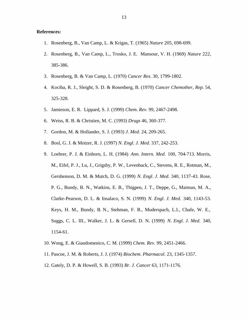
References:
1. Rosenberg, B., Van Camp, L. & Krigas, T. (1965) Nature 205, 698-699.
2. Rosenberg, B., Van Camp, L., Trosko, J. E. Mansour, V. H. (1969) Nature 222,
385-386.
3. Rosenberg, B. & Van Camp, L. (1970) Cancer Res. 30, 1799-1802.
4. Kociba, R. J., Sleight, S. D. & Rosenberg, B. (1970) Cancer Chemother, Rep. 54,
325-328.
5. Jamieson, E. R. Lippard, S. J. (1999) Chem. Rev. 99, 2467-2498.
6. Weiss, R. B. & Christien, M. C. (1993) Drugs 46, 360-377.
7. Gordon, M. & Hollander, S. J. (1993) J. Med. 24, 209-265.
8. Bosl, G. J. & Motzer, R. J. (1997) N. Engl. J. Med. 337, 242-253.
9. Loehrer, P. J. & Einhorn, L. H. (1984) Ann. Intern. Med. 100, 704-713. Morris,
M., Eifel, P. J., Lu, J., Grigsby, P. W., Levenback, C., Stevens, R. E., Rotman, M.,
Gershenson, D. M. & Mutch, D. G. (1999) N. Engl. J. Med. 340, 1137-43. Rose,
P. G., Bundy, B. N., Watkins, E. B., Thigpen, J. T., Deppe, G., Maiman, M. A.,
Clarke-Pearson, D. L. & Insalaco, S. N. (1999) N. Engl. J. Med. 340, 1143-53.
Keys, H. M., Bundy, B. N., Stehman, F. B., Muderspach, L.I., Chafe, W. E.,
Suggs, C. L. III., Walker, J. L. & Gersell, D. N. (1999) N. Engl. J. Med. 340,
1154-61.
10. Wong, E. & Giandomenico, C. M. (1999) Chem. Rev. 99, 2451-2466.
11. Pascoe, J. M. & Roberts, J. J. (1974) Biochem. Pharmacol. 23, 1345-1357.
12. Gately, D. P. & Howell, S. B. (1993) Br. J. Cancer 63, 1171-1176.

14
13. Johnson, N. P., Hoeschele, J. D., Kuemmerle, N. B., Masker, W. E. & Rahn, R.
O. (1978) Chem. Biol. Interact. 23, 267-271.
14. Kelland, L. R. (1994) Eur. J. Cancer 30, 725-727.
15. Perez, R. P. (1998) Eur. J. Cancer 34, 1535-1542.
16. Hall, M. D. & Hambley, T. W. (2002). Coordination Chemistry Reviews 232, 49-
67.
17. Iakovidis, A. & Hadjiliadis, N. (1994) Coord. Chem. Rev. 17, 135-136.
18. Kelland, L. R., Barnard, C. F. J., Mellish, K. J., Jones, M., Goddard, P. M.,
Valenti, M., Bryant, A., Murrer, B. A. & Harrap, K. R. (1994) Cancer Res. 54,
5618-5622.
19. Cleare, M. J. & Hoeschele, J. D. (1973) Plat. Met. Rev. 17, 2-13.
20. Cleare, M. J. & Hoeschele, J. D. (1973) Bioinorg. Chem. 2, 187-210.
21. Barnes, K. R., Kutikov, A. & Lippard, S. J. (2004) Chemistry & Biology 11, 557-
564.

15
PtCl NH3
Cl NH3
PtCl O
Cl O
O
O
PtNH2
O
H2N O
O
O
PtCl NH3
H3N Cl
PtCl N
Cl NH3
Me
PtCl N
H2
Cl NH3Pt
Cl NH2
Cl NH2
OCH2Me
OCH2Me
O
OH
OH
cisplatin trans-DDP
carboplatin oxaliplatin
ZD0473
JM216 iproplatin
PtCl NH3
Cl NH3
cisplatin succinato
O
O
OH
HO
O
O
O O
O
Figure 1.1. Structures of some platinum compounds, including cisplatin and its clinically inactive isomer, trans-DDP. Carboplatin and oxaliplatin are second generation drugs that have been approved by the FDA. JM216 and iproplatin are platinum(IV) compounds that have received much attention, and ZD0473 was designed to sterically cap the platinum. Cisplatin succinate is currently used to link biomolecules.

16
Chapter Two:
Synthesis and Characterization of Carboplatin Derived Platinum(IV) Compounds Capable of Conjugating Biomolecules

17
Introduction
Recent work in our lab has focused on the attachment of biomolecules to an
oxidized derivative of cisplatin to improve platinum-based chemotherapy by selectively
targeting cancer cells (1, 2). Carboplatin, cis-
diamminecyclobutanedicarboxylatoplatinum(II), is a second generation drug that has
different substitution kinetics than cisplatin (3). Having a platinum(IV) compound with
tethered biomolecules that will be reduced to give carboplatin is desirable. The slower
kinetics of carboplatin could be exploited to allow certain slow-acting biomolecules to
react, and use of the carboplatin platform expands the options for development of
platinum(IV) conjugates.
Current methodology used to tether biomolecules to cisplatin does not afford
much flexibility in the number of targeting moieties attached to the platinum(IV).
Because cis,cis,trans[PtCl2(NH3)2(succinate)2] (Figure 2.1) has two carboxyl groups
trans to one another, coupling to an amine results in either the mono- or bis- substituted
product. Modification of the coupling reagents and conditions allows for some selectivity
for either mono- or bis- substituted products, however, a mixture of products is often
obtained. Synthesis of a mono- conjugate is desirable because it might have better water
solubility, and would decrease the steric bulk of the platinum compound, facilitating
diffusion into the cell and subsequent reactions that cleave the biomolecule from the
platinum center.
Herein we report the synthesis and characterization of carboplatin derived
platinum(IV) compounds that can bind either one or two biomolecules selectively (Figure
2.2).

18
Experimental Procedures
General Considerations. Potassium tetrachloroplatinate(II) was a gift from
Engelhard. Carboplatin was prepared as described in the literature (4). All chemicals and
solvents were purchased from commercial sources. 1H and 195Pt NMR spectra were
collected on a Varian 300 and Varian 500 MHz spectrometers at the MIT Department of
Instrumentation Facility (DCIF). All 195Pt NMR was referenced to a standard of K2PtCl4
in water at d = -1631. Reverse phase HPLC was carried out on a Waters pump and
absorbance detector, and peaks were detected at 254 nm unless otherwise noted. LCMS
and ESI-MS was performed on an Agilent 1100 Series LC/MSD Trap. X-ray
crystallography was carried out on a Siemens diffractometer in our lab.
Synthesis of cis, cis, trans-diammine-cyclobutanedicarboxylato-
dihydroxyplatinum(IV) (hydroxy carboplatin). Carboplatin (2.36 g, 6.36 mmol) was
slowly added to 22 mL of 15% hydrogen peroxide in water. The reaction was stirred in
the dark for 2 h at 70 oC, and for an additional 30 min at 60 oC. The pale yellow
suspension that formed was cooled to room temperature and centrifuged to afford a white
solid (yield: 0.74 g, 1.8 mmol, 28.7%). The remaining solution was lyophilized, giving a
second crop (yield: 1.10 g, 2.7 mmol, 42.7%). 1H NMR (d6-DMSO, 300 MHz): δ 1.7 (m,
2H, CH2), 2.5 (m, 4H, CH2), 5.5 (bs, 6H, NH3), 10.2 (s, 2H, OH).
Synthesis of cis, cis, trans-diamminecyclobutanedicarboxylate-
disuccinateplatinum(IV) (carboplatin succinate). Hydroxy carboplatin (1.75 g, 4.32
mmol) and succinic anhydride (1.73 g, 17.3 mmol) were added to 25 mL of DMSO. The
solution was heated to 70 oC and stirred for 2.5 h, giving an amber solution.

19
Lyophilization removed most of the DMSO, resulting in a soft, yellow solid. The product
was precipitated out of the remaining DMSO by rinsing 3 times with 50% ether in
acetone (20 mL total). The off-white powder was dried in vacuo (yield: 1.60 g, 2.6 mmol,
61.2%). The ether and acetone supernatants were combined and cooled to –20 oC, upon
which more product precipitated out. The solid was isolated by centrifugation, and dried
in vacuo (yield: 0.35 g, 0.58 mmol, 13.4%). 1H NMR (d6-DMSO, 300 MHz): δ 1.8 (m,
2H, CH2), 2.2-2.6 (m, 12 H, CH2), 6.4 (bt, JH-N = 50.25 Hz, 6H, NH3), 12.1 (s, 2H, OH).
195Pt NMR (H2O, 500 MHz): δ 1893 (m, JPt-N = 250 Hz). MS (ESI) calculated for [M +
Na]+ 628.3, found 628.0 amu.
Synthesis of cis, cis, trans-Diammin-cyclubutanedicarboxylato-
diacetateplatinum(IV) (carboplatin acetate). A suspension of hydroxy carboplatin (0.10
g, 0.25 mmol) in acetic anhydride 2.5 mL was stirred at room temperature for 1 h. The
reaction was diluted with an additional 6 mL of acetic anhydride and heated to 50 oC.
After 3 h, the solution was cooled to –20 oC, rinsed with ether, and the white solid
isolated by centrifugation was dried in vacuo (yield: 0.08 g, 0.163 mmol, 66.2%). The
mother liquor was diluted with ether, and cooled to –20 oC overnight, and centrifugation
gave a second crop (0.03 g, 0.061 mmol, 24.8%). 1H NMR (d7-DMF, 300 MHz): δ 1.8 (s,
6H, CH3), 1.9 (m, 2H, CH2), 2.6 (t, 4H, CH2), 6.8 (m, JH-N = 41.25 Hz, JH-Pt = 66.7 Hz,
6H, NH3). 195Pt NMR (DMF, 500 MHz): δ 1923.
Synthesis of cis, cis, trans-
diamminecyclobutanedicarboxylatehydroxyacetateplatinum(IV) (carboplatin
monoacetate). A suspension of hydroxy carboplatin (0.04 g, 0.099 mmol) in 8 mL of
methanol was prepared, and acetic anhydride (10.0 µL, 0.099 mmol) added. The reaction
20
was allowed to stir overnight, before centrifugation isolated the precipitate. The solid was
rinsed with ether and dried in vacuo. 1H NMR (d7-DMF, 300 MHz): δ 1.8 (s, 6H, CH3),
1.9 (m, 2H, CH2), 2.5 (t, 2H, CH2), 2.6 (t, 2H, CH2), 6.3 (m, JH-N = 40.5 Hz, JH-Pt = 55.0
Hz, 6H, NH3). MS (ESI) calculated for [M-H]- 446.4, found 446.0 amu.
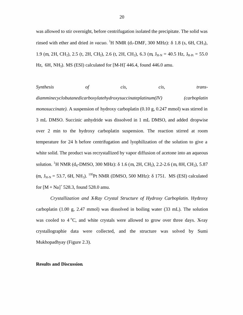
Synthesis of cis, cis, trans-
diamminecyclobutanedicarboxylatehydroxysuccinateplatinum(IV) (carboplatin
monosuccinate). A suspension of hydroxy carboplatin (0.10 g, 0.247 mmol) was stirred in
3 mL DMSO. Succinic anhydride was dissolved in 1 mL DMSO, and added dropwise
over 2 min to the hydroxy carboplatin suspension. The reaction stirred at room
temperature for 24 h before centrifugation and lyophilization of the solution to give a
white solid. The product was recrystallized by vapor diffusion of acetone into an aqueous
solution. 1H NMR (d6-DMSO, 300 MHz): δ 1.6 (m, 2H, CH2), 2.2-2.6 (m, 8H, CH2), 5.87
(m, JH-N = 53.7, 6H, NH3). 195Pt NMR (DMSO, 500 MHz): δ 1751. MS (ESI) calculated
for [M + Na]+ 528.3, found 528.0 amu.
Crystallization and X-Ray Crystal Structure of Hydroxy Carboplatin. Hydroxy
carboplatin (1.00 g, 2.47 mmol) was dissolved in boiling water (33 mL). The solution
was cooled to 4 oC, and white crystals were allowed to grow over three days. X-ray
crystallographie data were collected, and the structure was solved by Sumi
Mukhopadhyay (Figure 2.3).
Results and Discussion.
21
Synthesis of Characterization of Hydroxy Carboplatin. Carboplatin is readily
oxidized by hydrogen peroxide to give a platinum(IV) species. Because
cyclobutanedicarboxylic acid (CBDCA) is a better donor of electron density than
chloride ions, the reaction is more vigorous than the oxidation of cisplatin. The reaction is
not clean, with roughly 5% of the platinum forming a byproduct. Because the impurity is
only detectable in the chemical shift of the ammines in the proton NMR, it is believed to
be the aquated species of carboplatin, whereby the CBDCA is replaced by two
equivalents of water or OH. Although the half life for carboplatin in water is 8 hours (5),
the heat that forms in the reaction could increase the rate of substitution. Hydroxy
carboplatin is isolated by recrystalization from a supersaturated aqueous solution, or by
vapor diffusion of acetone into an aqueous solution to give colorless needle crystals. The
structure of hydroxy carboplatin was determined by X-ray crystallography, which shows
that the two hydroxo groups are trans to each other in axial positions. The Pt-O-C-C-C-O
ring of the CBDCA ligand lies flat in the equatorial plane, with the cyclobutane ring lying
in a plane perpendicular to the ring containing the platinum. Although the oxidized
product is not readily soluble in any organic solvent, a low quality proton NMR was
obtained which revelaed and shows that the ammines are shifted downfield relative to
those in carboplatin, reflecting the lower electron density around the platinum center.
Synthesis and Characterization of Carboplatin Succinate. Hydroxy carboplatin
readily reacts with two equivalents of succinic anhydride, giving carboplatin succinate
cleanly. The product has two acid moieties, which can be used to conjugate the platinum
to amines. Several attempts were made to grow crystals, including vapor diffusion of
acetone, methanol, and ethanol into an aqueous solution. All trials resulted in either no

22
solid formation or rapid formation of a fine powder. In order to slow the nucleation,
evaporation from a saturated aqueous solution and cooling of a supersaturated solution
were also attempted. Both of these trials resulted in no crystal growth, despite taking up
to a week to begin precipitating out of solution. Despite the lack of structural evidence for
carboplatin succinate, mass spectroscopy showed the product peak, and 195Pt and 1H
NMR showed that the carboplatin succinate was pure. The ammine shift in the proton
NMR is very sensitive to the environment of the platinum thus reflects purity. The
ammine protons are coupled to the nuclear spins of both 14N (I = 1) and 195Pt (I = ½, 33
% abundance) resulting in an overlapping triplet and quartet. When the platinum(IV)
species is pure, this pattern is clearly visible and symmetric. Impurities result in the
merging of the peaks into a broad singlet. The splitting is more readily seen in DMF than
DMSO, possibly because DMSO can bind to the platinum center.
Synthesis of Platinum(IV) Mono-Substituted Compounds. The neat conversion of
hydroxy carboplatin to carboplatin succinate inspired the exploration of the scope of this
reaction, and mono-substituted platinum(IV) compounds were prepared. Carboplatin
monosuccinate was prepared using more dilute reaction mixtures, stirring at room
temperature, and adding the anhydride dropwise. The insolubility of hydroxy carboplatin
in DMSO afforded a way to separate it from the product solution. When carried out on a
small scale, LCMS and proton NMR confirmed that no dicarboxylate product was
formed.
Carboplatin monoacetate was prepared under similar conditions as carboplatin
monosuccinate with the intent of being able to react it with one equivalent of succinic
anhydride to give a “capped” platinum(IV) carboplatin derivative that could link one

23
biomolecule (3 in Figure 4.3). Because reduction potentials are very sensitive to the trans
axial ligands, a “capped” monosuccinate will have different reduction properties than the
carboplatin monosuccinate prepared (6). As a comparison, carboplatin acetate was also
prepared.
Despite confirmation of pure carboplatin monosuccinate and carboplatin
monoacetate by LCMS, when the reactions were scaled up only 80-95 % of the isolated
product was mono-substituted. HPLC was attempted to purify the compounds, and
unfortunately, our species decomposed on the column. Currently, recrystallization trials
are underway for both carboplatin monosuccinate and carboplatin monoacetate; moderate
success at purification of the monosuccinate has been achieved by vapor diffusion of
acetone into an aqueous solution of carboplatin monosuccinate.
Characterization of the Mono-substituted Platinum(IV) Compounds. Evidence for
the synthesis of the mono substituted platinum(IV) compounds comes from MS and
NMR spectroscopy. ESI-MS gave the molecular ion peak with an isotope pattern that
matches the calculated pattern (Figure 2.4). Furthermore, protonation of the hydroxyl
groups followed by fragmentation gives a distinct [M-OH]+ fragment that is visible only
in the monosubstituted compounds. 195Pt NMR spectroscopy of carboplatin
monosuccinate revealed that it is shifted ~150 ppm downfield when compared to
carboplatin succinate. The ammine shift in the proton NMR is also between hydroxy
carboplatin and carboplatin succinate. Both of these observations are consistent with the
notion that hydroxy carboplatin has the greatest electron density about the platinum
center, and the platinum(IV) di- carboxylates have the lowest electron density about the
platinum center. The trend in proton NMR ammine shifts is observed in both the acetate

24
and succinate series of platinum(IV) compounds. Additional 195Pt NMR needs to be done
to confirm the trend in the other platinum(IV) derivatives.
The 1H NMR of carboplatin monoacetate is interesting, for it reveals that certain
protons that are equivalent in both hydroxy carboplatin and carboplatin acetate are in
different chemical environments in the carboplatin monoacetate. This effect is most
pronounced in the CH2 protons of the cyclobutane ring that are a- to the di- carboxylate.
The crystal structure reveals that the cyclobutane ring is perpendicular to the N-Pt-N
plane (Figure 2.3). This feature results in one methylene pointing towards the hydroxo
ligand, and the other pointing towards the carboxylate. Because of this asymmetry, the
two a- methylene groups have distinct chemical shifts; the increased symmetry in
carboplatin acetate gives rise to a single triplet (Figure 2.5).
In both carboplatin succinate and carboplatin acetate, the ammine signal in the
proton NMR is shifted significantly downfield compared to the ammine of hydroxy
carboplatin. This result indicates that the ammine protons are more deshielded in the
carboxylate complexes. The change in signal can be a probe for the electron density
around the platinum(IV) center. Because water has a higher pKa than carboxylic acids,
the hydroxide anion is less stable and donates more electron density to the platinum(IV).
This effect makes the platinum(IV) center more electron rich, and through inductive
effects, shields the ammine hydrogens. Because carboxylic acids have a lower pKa, and
are more electron withdrawing due to resonance, the carboxylate is more stable and
donates less electron density to the platinum center. The chemical shift for the amines in
the monocarboxylates lies between that of the dicarboxylates and hydroxy carboplatin.

25
The result is that the platinum takes more electron density from the ammines, deshielding
the proton NMR signal (Figure 2.6).
This insight gives us an indication of the reduction potentials of the mono-
carboxylates prepared. It is well documented in the literature that the reduction potentials
of platinum(IV) species depend on the axial ligands (7) and that they obey the following
trend: -Cl > -OOCR > -OH (8). Thus, a platinum(IV) with trans chlorides is most easily
reduced, and trans hydroxo complexes are the hardest to reduce. Because the ammine
shift of the mono- species is between that of the platinum(IV) bis- carboxylate and the
hydroxy carboplatin, the reduction potential should lie between that of the dicarboxylates
and hydroxy carboplatin.
To our knowledge the synthesis of a monocarboxylate trans to a hydroxy has been
achieved once as a side product to a reaction (9). Although it has generally been viewed
that trans hydroxy platinum(IV) complexes are too difficult to be reduced in the
cytoplasm (10), recent work using XANES to determine the oxidation state of platinum
in cells has shown that within 24 hours all of trans hydroxy platinum(IV) compounds are
reduced to platinum(II) (7). This result suggests to us that carboplatin monosuccinate
could be used successfully to deliver our molecule, and the lower reduction potential may
make it less prone to reduction in the blood.
Conclusion and Future Plans
Carboplatin succinate was successfully synthesized, and has been fully characterized.
Carboplatin monosuccinate was also prepared, although mixed products were obtained
upon scaling up. Further attempts will be made to purify both carboplatin monoacetate

26
and carboplatin monosuccinate and subsequentially derivative them. Cytotoxicity and
reduction potentials of the mono-carboxylates will be carried out, and compounds 1, 2,
and 3 will be synthesized (refer to Figure 2.2). Furthermore, the analogous complexes
using cisplatin will be prepared. This strategy will allow for the conjugation of a single
biomolecule that has similar reduction kinetics as carboplatin succinate, and to make a
platinum(IV) center that is conjugated with two different types of molecules. This
approach will give our lab more flexibility in conjugating biomolecules to platinum(IV).

27
References:
1. Barnes, K. R., Kutikov, A. & Lippard, S. J. (2004) Chemistry & Biology 11,
557-564.
2. Mukhopadhyay, S., Barnes, C. M., Haskel, A., Levine, D., Barnes, K. R.
Lippard, S. J. (2004) In preparation.
3. He, Q., Liang, C. H. & Lippard, S. J. (2000) PNAS 97, 5768-5772.
4. Harrison, R.C.; McAuliffe, C.A. (1980) Inorganic Chimica Actu, 46, L15-L16.
5. Hongo, A., Seki, S., Akiyama, K., & Kudo, T. (1994) Int. J. Biochem. 26, 1009-
1016.
6. Hall, M. D. & Hambley, T. W. (2002) Coord. Chem. Rev. 232, 49-67.
7. Ellis, L. T., Er, H. M. & Hambley, T. W. (1995) Aus. J. Chem. 48, 793-806.
8. Hall, M. D., Amjadi, S., Zhang, M., Beale, P. J. & Hambley, T. W. (2004)
Journal of Inorganic Biochemistry 98, 1614-1624.
9. Giandomenico, C.M., Abrams, M.J., Murrer, B.A., Vollano, J.F., Rheinheimer,
M.I., Wyer, S.B., Bossard, G.E. & Higgins III, J.D. (1995) Inorg. Chem. 34,
1015-1021.
10. Hall, M. D., Foran, G. J., Zhang, M., Beale, P. J. & Hambley, T. W. (2003) J.
Am. Chem. Soc. 125, 7524-7525.

28
Figures
PtNH3Cl
Cl NH3
O
O
OHO
O
HOO
O
cisplatin succinato
Figure 2.1. Structure of cis,cis,trans-PtCl2(NH3)2(succinate)2.

29
Pt NH3OO NH3
O
O
Pt NH3OO NH3
O
O
OH
OH
Pt NH3OO NH3
O
O
O
O
Pt NH3OO NH3
O
O
O
OH
OH
O
O
O
Pt NH3OO NH3
O
O
O
O
OH
O
O
Pt NH3OO NH3
O
O
O
OH
O
OHOO
O
Pt NH3OO NH3
O
O
O
O
OH
O
O
O
Pt NH3OO NH3
O
O
O
O
HN
O
O
NH2
OH
O
Pt NH3OO NH3
O
O
O
OH
HN
O
O
NH2
carboplatin
hydroxy carboplatin
carboplatin succinato carboplatin acetate
carboplatin monoacetatecarboplatin monosuccinato
3.1.
2.
O Figure 2.2. Synthetic scheme of carboplatin derivatives. Named complexes have been synthesized, and the numbered compounds are works in progress.

30
Figure 2.3. X-ray structure of hydroxy carboplatin.

31
Figure 2.4. ESI-MS of carboplatin monosuccinato showing the Pt isotope pattern.
[M + Na]+

32
Figure 2.5. 1H NMR spectra showing the chemical shift of Ha and Hb. The increased symmetry in carboplatin acetate results in a single triplet, whereas the loss of a mirror plane gives rise to two triplets in carboplatin monoacetate.
PtNH3O
O NH3
O
O
O
OH
O
carboplatin monoacetate
Ha
Hb
Ha
Hb
PtNH3O
O NH3
O
O
O
O
O
carboplatin acetate
Ha
Ha
Ha
Ha
O

33
Figure 2.6. The ammine region of the 1H NMR spectra of a. carboplatin monoacetate and 2. carboplatin acetate.
b.
a.

34
Chapter 3:
Synthesis, Characterization, and Cytotoxicity of a Series of Estrogen-Tethered Platinum(IV) Compounds that Reduce to Carboplatin

35
Introduction
From research delineating to the mechanism by which cisplatin may exert its
cytotoxicity, new platinum compounds can be developed that exploit the proposed mode
of killing. Cisplatin targets and binds DNA, forming mostly 1,2-intrastrand cross-links (1,
2). The resulting distortions in the double helix affect DNA replication and transcription,
and are recognized by several structure specific DNA-binding proteins including DNA
repair and high mobility group (HMG) domain proteins (3). HMG proteins are
architectural proteins that facilitate cellular functions involving chromosomal DNA (4),
and there is increasing evidence that the protein plays a key role in cisplatin induced
apoptosis. HMGB1 binds to platinum-DNA adducts, blocks nucleotide excision repair (5,
6) and drives apoptosis. When HMG proteins are removed from cell extracts, the amount
of repair is enhanced (7). Furthermore, overexpressing HMGB1 protein results in more
cell death when the protein concentrations are elevated at the time of platinum damage
(8). Thus there is a strong correlation between HMGB1 levels and DNA repair.
The Interaction Between HMGB1 and Estrogen. HMGB1 is a structure-specific
protein that is present in all tissues and species (9). It binds to deformed DNA structures
that include platinum damaged sites and cruciform DNA (4), and has several functions
(10-14) including the facilitation of binding steroid receptors to their DNA binding sites
(15). When estrogen receptors (ER) bind to the estrogen-responsive element (ERE), the
DNA bends (16), increasing the HMGB1 affinity. HMGB1 then binds, further altering
the DNA structure and making a more stable receptor-DNA complex (17-19). When
estrogen receptor positive (ER(+)) cells are treated with estrogen, an increase in HMGB1
level is observed (20).

36
Sensitization of ER(+) Cells to Platinum(II) by Estradiol. Previous work in our
lab has shown that cotreatment of MCF-7 cells with estrogen and cisplatin can sensitize
these ER(+) cells by a factor of 2 (8). Repeating the experiment in HCC-1937 cells which
lack the estrogen receptor (ER(-)) gave no heightened cytotoxicity. This finding
prompted the synthesis of a series of estrogen tethered platinum(IV) pro-drugs that are
reduced upon entering the cell, releasing cisplatin and two equivalents of a modified
estrogen. The estrogen has a linker of varying length attached at the 17ß position and
must be hydrolyzed by esterase before recognition by estrogen receptors (Figure 3.1).
Because the kinetics of esterase depend on linker length (21), and because HMGB1
upregulation must be optimized at the time of platinum damage, linkers of varying length
will sensitize ER(+) cells by different amounts. Indeed, only one of the five conjugates
made, BEP3 (Figure 3.2), showed a two-fold increase in cytotoxicity in ER(+) cells
compared to ER(-) cells (22). Despite the success, the IC50 values for the tethered drugs
were higher than the values obtained for cisplatin/estradiol cotreatment.
Estrogen can also sensitize ER(+) cells to carboplatin, a second generation
platinum(II) drug. Carboplatin has a slower rate of aquation and cells must be pretreated
for 24 hours before estradiol treatment to show any sensitization (8). Because carboplatin
has fewer side effects and is more widely used, and because the IC50 of BEP3 was higher
than that of cisplatin in ER(+) cells, carboplatin analogues were synthesized with the goal
of achieving kinetic optimization that would result in increased cell death. Herein we
report the synthesis, characterization, and cytotoxicity of a series of estrogen-tethered
platinum(IV) compounds that reduce to give carboplatin.

37
Experimental Procedures
General Considerations. Potassium tetrachloroplatinate(II) was a gift from Engelhard.
The preparation of EL1-EL5 compounds (Figure 3.3) were synthesized by methodology
developed in our lab (22), and carboplatin succinate was made as described elsewhere in
this work (Chapter 2). All chemicals and solvents were purchased from commercial
sources. 1H NMR spectra were collected on a Varian 300 or Varian 500 MHz
spectrometers at the MIT Department of Instrumentation Facility (DCIF). Mass
spectrometry was carried out at the MIT DCIF (HRMS). Atomic absorption spectra were
obtained on a Perkin Elmer AAnalyst 300 fitted with a graphite furnace autosampler.
Reverse phased HPLC was carried out on a Waters pump and absorbance detector, and
peaks were detected at 254 nm unless otherwise noted. LCMS and ESI-MS were
performed on an Agilent 1100 Series LC/MSD Trap instrument.
Synthesis of Mono-BOC Protected Trioxatridecanediamine (BOC-TDA). A
solution of trioxatridecanediamine (14.11 mL, 64.2 mmol) and BOC2O (7.00 g, 32.1
mmol) was stirred in 140 mL dioxane overnight. The solvent was removed via rotorary
evaporation to give a colorless oil that was extracted into ethyl acetate from water. The
organic fractions were collected and a colorless oil was isolated via rotorary evaporation
(yield: 8.39 g, 41%). ESI-MS [M + H]+ = 321.4 (calculated), [M + H]+ = 321.4
(observed).
Synthesis of BOC trioxadecanediaminesuccinate (BOC-TDA-succinate) (Figure
3.4). BOC-TDA (2.00 g, 6.23 mmol) and succinic anhydride (0.78 g, 7.78 mmol) were
dissolved in 24 mL of 1:1:1 pyridene: dichloromethane: acetonitrile and stirred at room
temperature for 4 h. The solvent was removed by rotorary evaporation, and the resulting

38
oil dried in vacuo (yield: 0.25 g, 100%). 1H NMR (d6-DMSO, 300 MHz): δ 1.4 (s, 9H,
CH3), 1.6 (m, 4H, CH2), 2.2-2.5 (m, 4H, CH2), 2.9-3.1 (m, 4H, CH2), 3.4-3.6 (m, 12H,
CH2), 6.8 (t, 1H, NH), 8.6 (t, 1H, NH), 12.05 (s, 1H, COOH).
Synthesis of TDA-succinate-estradiol linker (EL17). BOC-TDA-succinate (0.75 g,
1.80 mmol) and 4-DMAP (0.12 g, 1.0 mmol) were dissolved in 4 mL DMF, and DIPC
(.016 mL, 1.0 mmol) added. After 15 min of stirring, the estradiol (0.17 g, 0.45 mmol)
was added, and the reaction stirred at RT overnight. Dilution with 15 mL water and
cooling to 4 oC for an hour facilitated precipitation of BOC-protected product which was
filtered and dried in vacuo. The solid was dissolved in 12 mL of dichloromethane and
stirred with 1.2 mL trifluoroacetic acid for 2 h. The solvent was removed, and 10 mL
water was added to the resulting oil. Ammonium hydroxide was added to adjust the pH to
10.5, and the solution stirred overnight. Centrifugation afforded the isolation of a pellet
that was dried in vacuo (yield: 0.24 g, 80%). 1H NMR (d6-DMSO, 300 MHz): δ 0.80 (s,
3H, CH3), 1.2-2.0 (m, 12H, CH/CH2/NH2), 2.0-2.45 (m, 9H, CH/CH2), 2.82 (m, 4H,
CH2), 3.0-3.6 (m, 16H, CH2), 4.61 (t, 1H, CH), 6.95 (s, 1H, ArH), 7.0 (d, 1H, ArH), 7.35
(d, 1H, ArH), 7.60 (t, 2H, ArH), 7.75 (t, 1H, ArH), 7.85 (m, 1H, NH), 8.15 (d, 2H, ArH).
ESI-HRMS calculated for [M + H]+ 679.3953, found 679.3965 amu.
Synthesis of cis, cis, trans-Diamminecyclobutanedicarboxylatebis-(17-(N-
carbonylmethylsuccinate)-estradiol-3-benzoate)platinum(IV) (BECP1).
Diisopropylcarbodiimide (DIPC) (0.10 mL, 0.634 mmol) was added to a solution of 4-
DMAP (0.08 g, 0.634 mmol) and carboplatin succinate (0.15 g, 0.254 mmol) in 5 mL
DMF. After stirring for 10 min at room temperature, EL1 (0.22 g, 0.507 mmol) was
added and the reaction stirred overnight. The reaction mixture was diluted with 50 mL of

39
ether, and cooled to –20 oC for an hour to facilitate precipitation. The crude solid was
isolated by decanting off the ether, and the product was rinsed and sonicated for 5 min in
5 mL ethanol (3 times), 5 mL water (3 times), and methanol (1 time). After each rinsing,
the product was centrifuged for 3-5 min at 5000 rpm. The BECP1 was dried in vacuo to
give a white solid (yield: 0.1717 g, 0.12 mmol, 47.0%). 1H NMR (d6-DMSO, 500 MHz):
δ 0.783 (s, 6H, CH3), 1.2-1.6 (m, 4H, CH/CH2), 1.60-1.95 (m, 10H, CH/CH2), 2.0-2.65
(m, 14H, CH/CH2), 2.83 (m, 4H, CH2), 3.81 (d, 4H, CH2), 4.65 (t, 2H, CH), 6.32 (bt, 6H,
NH3), 6.95 (s, 2H, ArH), 7.01 (d, 2H, ArH), 7.35 (d, 2H, ArH), 7.60 (t, 4H, ArH), 7.73 (t,
2H, ArH), 8.12 (d, 4H, ArH), 8.31 (t, 2H, NH). ESI-HRMS calculated for [M + H]+
1437.5211 amu, found 1437.5208 amu.
Synthesis of cis, cis, trans-Diamminecyclobutanedicarboxylatebis-(17-(N-(2-
carboxy-ethyl)-succinate)-estradiol-3-benzoate)platinum(IV) (BECP2). This compound
was prepared as described for BECP1 with carboplatin succinate (0.17 g, 0.28 mmol), 4-
DMAP (0.09 g, 0.70 mmol), DIPC (0.11 mL, 0.70 mmol), and EL2 (0.25 g, 0.56 mmol)
in 5 mL DMF. The crude BECP2 was precipitated in 50 mL ether for 1 h at –20 oC. The
orange solid was rinsed and sonicated for 5-15 min with 10 mL ethanol (3 times), and 10
mL boiling water (3 times). Between washes, the solid was centrifuged at 3000 rpm for
3-5 min. BECP2 was dried in vacuo to give a white powder (yield: 0.2440 g, 0.167
mmol, 59.3%). 1H NMR (d6-DMSO, 500 MHz): δ 0.794 (s, 6H, CH3), 1.2-2.6 (m, 44H,
CH/CH2), 2.84 (m, 4H, CH2), 3.27 (t, 4H, CH2), 4.64 (t, 2H, CH), 6.42 (bt, 6H, NH3),
6.96 (s, 2H, ArH), 7.01 (d, 2H, ArH), 7.36 (d, 2H, ArH), 7.60 (t, 4H, ArH), 7.75 (t, 2H,
ArH), 7.95 (m, 2H, NH), 8.11 (d, 4H, ArH). ESI-HRMS calculated for [M + Na]+
1487.5344 amu, found 1487.5308 amu.

40
Synthesis of cis, cis, trans-Diammine-cyclobutanedicarboxylatebis-(17-(N-(3-
carboxy-propyl)-succinate)-estradiol-3-benzoate)platinum(IV) (BECP3). This compound
was prepared as described for BECP1 with carboplatin succinate (0.10 g, 0.165 mmol), 4-
DMAP (0.05 g, 0.413 mmol), DIPC (0.065 mL, 0.413 mmol), and EL3 (0.12 g, 0.260
mmol) in 3 mL DMF. The crude BECP3 was precipitated in 50 mL ether, and cooled to
–20 oC for 1 h. The soft solid was rinsed and sonicated for 5 min with 5 mL ethanol (3
times), and 5 mL water (3 times). Like with BECP1, the product was centrifuged between
washes. BECP3 was dried in vacuo, affording a white powder (yield: 0.0443 g, 0.03
mmol, 23.0%). 1H NMR (d6-DMSO, 300 MHz): δ 0.801 (s, 6H, CH3), 1.0-2.0 (m, 26H,
CH/CH2), 2.0-2.2 (m, 2H, CH), 2.2-2.6 (m, 16H, CH/CH2), 2.83 (m, 4H, CH2), 3.04 (m,
4H, CH2), 3.26 (t, 4H, CH2), 4.64 (t, 2H, CH), 6.36 (bt, 6H, NH3). 6.96 (s, 2H, ArH), 7.01
(d, 2H, ArH), 7.47 (d, 2H, ArH), 7.60 (t, 4H, ArH), 7.74 (t, 2H, ArH), 7.85 (t, 2H, NH),
8.11 9d, 4H, ArH). MS (ESI) calculated for [M + H]+ 1493.5838 amu, found 1493.5856
amu.
Synthesis of cis, cis, trans-Diammine-cyclobutanedicarboxylatebis-(17-(N-(4-
carboxy-butyl)-succinate)-estradiol-3-benzoate)platinum(IV) (BECP4). This compound
was prepared as described for BECP1 with carboplatin succinate (0.17 g, 0.281 mmol), 4-
DMAP (0.09 g, 0.702 mmol), DIPC (0.11 mL, 0.702 mmol), and EL4 (0.27 g, 0.562
mmol) in 5 mL DMF. The product was purified as described for BECP2, giving a white
powder (yield: 0.1997 g, 0.131 mmol, 46.7%). 1H NMR (d6-DMSO, 500 MHz): δ 0.800
(s, 6H, CH3), 1.2-2.6 (m, 52H, CH/CH2), 2.84 (m, 4H, CH2), 3.03 (m, 4H, CH2), 4.64 (t,
2H, CH), 6.34 (bt, 6H, NH3), 6.96 (s, 2H, ArH), 7.01 (d, 2H, ArH), 7.34 (d, 2H, ArH),

41
7.61 (t, 4H, ArH), 7.74 (t, 2H, ArH), 7.84 (m, 2H, NH), 8.10 (d, 4H, ArH). MS (ESI)
calculated for [M + Na]+ 1543.5970 amu, found 1543.5960 amu.
Synthesis of cis, cis, trans-Diammine-cyclobutanedicarboxylatebis-(17-(N-(5-
carboxy-pentyl)-succinate)-estradiol-3-benzoate)platinum(IV) (BECP5). This compound
was prepared in a similar manner as BECP1 with carboplatin succinate (0.17 g, 0.281
mmol), 4-DMAP (0.09 g, 0.702 mmol), DIPC (0.11 mL, 0.702 mmol), and EL5 (0.27 g,
0.562 mmol) in 5 mL DMF. The reaction was diluted with 45 mL water and cooled to 4
oC for 1 h, before centrifugation. The remaining solid was washed with 10 mL ethanol (3
times), and 5 mL boiling water (3 times), and dried in vacuo to give a white powder
(yield: 0.0993 g, 0.064 mmol, 22.8%). 1H NMR (d6-DMSO, 500 MHz): δ 0.801 (s, 6H,
CH3), 1.2-2.6 (m, 56H, CH/CH2), 2.84 (m, 4H, CH2), 3.00 (m, 4H, CH2), 4.64 (t, 2H,
CH), 6.33 (bt, 6H, NH3), 6.96 (s, 2H, ArH), 7.01 (d, 2H, ArH), 7.35 (d, 2H, ArH), 7.61 (t,
4H, ArH), 7.75 (t, 2H, ArH), 7.81 (m, 2H, NH), 8.11 (d, 4H, ArH). ESI-MS calculated
for [M – H]- 1547.67 amu, found 1547.0 amu.
Synthesis of cis, cis, trans-Diammine-cyclobutanedicarboxylatebis-17-(N-
(Trioxatridecanediamine-succinic)-succinate)-estradiol-3-benzoate)platinum(IV)
(BECP17). This compound was synthesized in a manner analogous to BECP1 using
carboplatin succinate (0.11 g, 0.184 mmol), 4-DMAP (0.06 g, 0.460 mmol), DIPC (0.07
mL, 0.460 mmol), and EL17 (0.25 g, 0.368 mmol) in 5 mL DMF. The reaction was
diluted with 50 mL ether after stirring for 24 h, and cooled to -20o C for 1.5 h. The
resulting precipitate was filtered, and rinsed three times with 4.5 mL boiling water. The
white powder was dried in vacuo (yield: 0.15 g, 46.0%). 1H NMR (d6-DMSO, 300 MHz):
δ 0.793 (s, 6H, CH3), 1.2-2.0 (m, 24H, CH/CH2), 2.0-2.45 (m, 18H, CH/CH2), 2.45-2.50

42
(m, 12H, CH2), 2.82 (m, 8H, CH2), 3.0-3.6 (m, 32H, CH2), 4.61 (t, 2H, CH), 6.3 (bs, 6H,
NH3), 6.97 (s, 2H, ArH), 7.0 (d, 2H, ArH), 7.35 (d, 2H, ArH), 7.62 (t, 4H, ArH), 7.75 (t,
2H, ArH), 7.86 (m, 4H, NH), 8.15 (d, 4H, ArH). ESI-HRMS calculated for [M + Na]+
1949.8444, found 1949.9074 amu.
Cell Culture Studies. MCF-7 cells were grown in DMEM (GIBCO/BRL) with
10% FBS, 1x antibiotic/antimycotic solution (GIBCO), and 2 mM L-glutamine. HCC-
1937 cells were grown in RPMI-1640 (ATCC) containing 10% FBS and 1x
antibiotic/antimycotic solution, 2.0 mM L-glutamine, 10 mM HEPES, 1.0 mM sodium
pyruvate, and 2 mM glucose. All cells were incubated at 37 oC in a 5% CO2 environment.
Absorbance were recorded on a Molecular Devices SpectraMax 340 PC at 550 nm. The
instrument was calibrated with 0.4% sulforhodamine B in 1% acetic acid at 550 nm, and
the linear region found to be between absorbance of 0.10 and 2.2 absorbance units.
Overexpression of HMGB1 Induced by BECPn (n=1-5, 17) Complexes. MCF-7
cells were grown to 70% confluence on 12 mm glass cover slips in 24 well plates. The
cells were treated with either estradiol or BECPn in 200 nM solutions, and incubated for
various lengths of time. The cells were then rinsed 3 x with PBS and permeabilized in 25
% acetic acid in methanol for 10 min at room temperature, and washed 3 more times in
PBS before treatment with a 1:100 dilution of anti-HMGB1 polyclonal antibody
(PharMingen) for 1 h at 37 oC. The cells were subsequently washed with PBS (3 times)
and incubated with a 1: 200 dilution of goat anti-rabbit IgG conjugated to FITC
(Biosource International) for 1 h at 37 oC. The cover slips were washed in PBS (3 times)
and water (2 times) before being fixed with gelvatol on microscope slides. The slides
were incubated at 4 oC for 15 h in the dark. HMGB1 levels were visualized under a

43
fluorescent-light microscope (Diagnostic Instruments) equipped with a digital camera
(Nikon).
Cytotoxicity of BECPn (n=1-5, 17). The cytotoxicities of BECPn compounds
were evaluated using the MTT assay. Briefly, 1000 cells (MCF-7) or 1,500 cells (HCC-
1937) were on a 96 well plate in 100 µL media, and incubated for 24 h. The cells were
then treated with BECPn compound at concentrations ranging from 0 to 25 µM, and
incubated for 96 h (MCF-7) or 144 h (HCC-1937). Cells were then treated with 20 µL of
MTT (5 mg / mL PBS), and incubated for 5 hr. The media was removed, and the cells
solubilized in 50-150 µL DMSO, and shaken for 5 min before reading the plates at 550
nm.
Results and Discussion
Synthesis and characterization of BECPn (n = 1-5, 17). The preparation of carboplatin
succinate is discussed elsewhere in this report (Chapter 2), and the carboplatin analogues
to BECPn were prepared in a similar fashion as that described in the literature (22). Two
equivalents of modified estrogen were allowed to react with carboplatin succinate along
with 4-DMAP and DIPC, which are standard peptide coupling reagents. Although a
mixture of mono- and bis- substituted platinum(IV) could be expected, proton NMR and
mass spectrometry showed only bis- product. BECPn was purified by rinsing with hot
water several times, followed by cold ethanol. Because ethanol removes only excess
estrogen, and because the product has a slight ethanol solubility, a slight excess of
carboplatin succinate was employed to ensure full reaction of estrogen. The integration of
the amide proton H22 (Figure 3.3) to protons on the estrogen (H17) and carboplatin
44
succinate (H23) revealed the expected ratio, further indicating that no mono substituted
product formed (Table 3.1). HPLC resulted in product decomposition to free estrogen
linker; the decomposition was observed by collecting the product peak, rinsing the
column, and reinjection. Proton NMR of the five compounds showed excellent
integration of the ammine protons to those found on the estrogen, and good integration
between the amide and succinate protons.
Estrogen was also attached to carboplatin via a TDA-succinate linker to give an
estrogen linker with a length of 17 atoms (Figure 3.4). TDA was first mono BOC
protected and reacted with succinic anhydride, which was then reacted with estradiol to
give EL17. EL17 was reacted with carboplatin as described for the other estrogen linkers,
but had increased water solubility because of the polyether nature of the linker.
Overexpression of HMGB1 in MCF-7 Cells Following treatment with BECPn
(n=1-5, 17). The ability of compounds BECPn to upregulate HMGB1 was monitored via
immunofluorescence. After cells are treated with BECPn they are incubated with a rabbit
anti-HMGB1 antibody, followed by incubation with a FITC conjugated anti-rabbit
antibody. This procedure allows for the visualization of HMGB1 in the cells. Because a
24 hour pretreatment of carboplatin was needed before treatment with estradiol to see any
effect on the cytotoxicity, we chose to look at the upregulation after 24 hours. Cells that
were not treated serve as a negative control, while cells that are treated with estradiol for
1.5 hours serve as a positive control. Estradiol induces the overexpression of HMGB1
0.5-2 hours after treatment (8), and the levels remain elevated before tapering off after 24
hours. As seen in figure 3.5, all BECPn compounds upregulate HMGB1. The degree of
upregulation cannot be quantified via immunofluorescence, but it is clear from the

45
brightness of the cells that HMGB1 levels are still elevated after 24 hours. The behavior
of BECPn should be analogous to BEPn, because the reduction kinetics for both should
be very similar, giving the same modified estrogens. Incubation of BECPn for 4 hours
showed 2-fold increase in HMGB1 levels; the same results are expected for BECPn.
Preliminary results with shorter incubation times with BECPn show elevated levels of
HMGB1. The ability of BECPn to induce HMGB1 overexpression indicates that the
compounds are being taken up into the cells and that the estradiol is formed. The estradiol
alcohol that results upon hydrolysis at the 17ß position is essential for estrogen receptor
recognition. Assuming that the platinum center is not sterically protected, from the
known kinetics of both platinum(IV) in vitro reduction and ester hydrolysis it is inferred
that reduction precedes hydrolysis (21, 23).
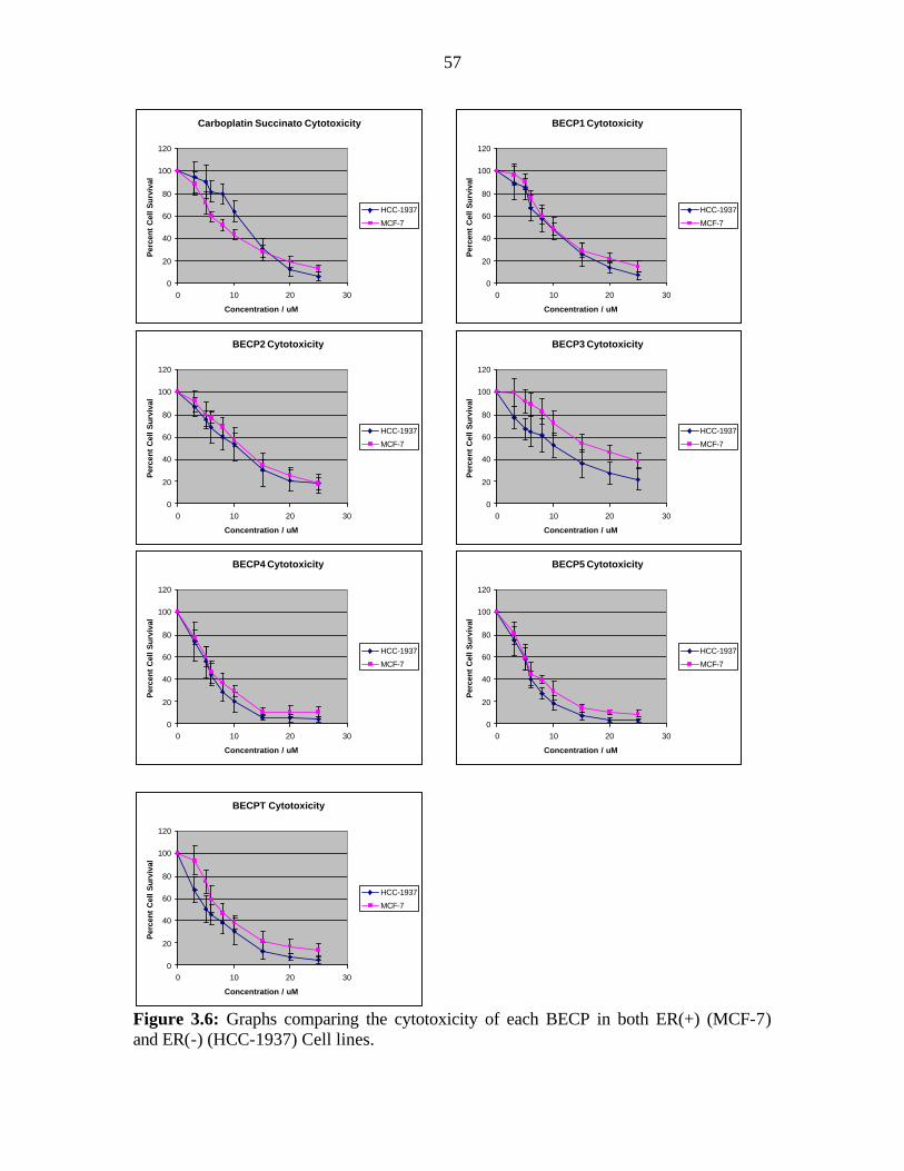
Cytotoxic Behavior of BECPn (n=1-5, 17) in ER(+) and ER(-) Cells. The
cytotoxic properties of BECP1-T were evaluated in MCF-7 and HCC-1937 cell lines, two
human breast cancer carcinoma that are ER(+) and ER(-) respectively. Cell survival was
evaluated with the MTT assay. In control experiments the cytotoxicity of carboplatin
succinate was evaluated in both cell lines and was lower in MCF-7 cells, most likely due
to increased uptake. The IC50 in MCF-7 cells was 8.45 µM and in HCC-1937 cells, 12.06
µM. Any increase in cytotoxicity will be due to the tethered estrogen, and discrepancies
between the cytotoxicities of the two cell lines will have to take the difference in IC50 of
the carboplatin succinate control into consideration. As shown in table 3.2, the
overexpression of HMGB1 in MCF-7 cells did not sensitize the cells to carboplatin.
BECP1, BECP2, BECP4 and BECP5 all had very similar IC50 values in both cell lines
(Figure 3.6). The cytotoxicity was increased by a factor of two for BECP4 and BECP5

46
compared to BECP1. Interestingly enough, BECP3, which had optimized kinetics for the
cisplatin system had a significantly larger IC50 value for MCF-7 cells. BECP17 also had a
larger IC50 value in ER(+) cells. Both of these compounds were 1.5x more toxic in ER(-)
cells than in ER(+) cells.
These results indicate that the kinetics of ester hydrolysis followed by HMGB1
upregulation do not overlap with that of carboplatin aquation and DNA binding. Because
carboplatin forms both 1,2 and 1,3 intrastrand crosslinks, and HMGB1 only recognizes
1,2 intrastrand crosslinks (24-26), the overlap of the kinetics is more important than in
cisplatin because there are fewer recognizable damage sites. Estradiol increases cell
proliferation, and increases the IC50 of non-platinum-based therapeutics such as paclitaxel
and adriamycin in ER(+) cell lines (27). It is plausible that BECPn conjugates induced
cell proliferation. BECP3 is expected to have the fastest hydrolysis kinetics because
BEP3 showed increased sensitivity to cisplatin in ER(+) cells. Cisplatin requires
cotreatement with estradiol or immediate release of free estradiol to see increased
toxicity. Although BECP17 has a longer linker and hence slower kinetics are expected,
the more hydrophilic nature of this linker compared to the hydrophobic nature of the
other linkers may result in a higher affinity for the esterase active site. The kinetics of
estrogen induced cell proliferation are unknown, yet it is reasonable to assume that is it
on the same time scale as HMGB1 upregulation because both are the result of signaling
to the DNA.
In both HCC-1937 and MCF-7 lines, the cytotoxicities for BECP4 and BECP5
were significantly lower than the cytotoxicities of the shorter linkers. This effect may be
due to increased cellular uptake; the increased lipophilicity of BECP4 and BECP4 would

47
increase passive uptake through the cell membrane; the result is similar IC50 values
between BECP4, BECP5, and carboplatin (8). Several groups have shown in
platinum(IV) carboxylates that the carboxylate length had a strong effect on cell uptake
and cytotoxicity; the addition or removal of a single methylene greatly affects the
cytotoxic properties of the compound (28-30). BECP17 had a slightly lower cytotoxicity
in ER(-) cells compared to BECP4 and BECP5, indicating that the polyether linker may
actually aid in cellular uptake. The increased IC50 in ER(+) cells of BECP4 hints that the
kinetics of cell proliferation are faster than those of HMGB1 upregulation. This result is
promising because it suggests that, if a longer linker with similar lipophilic properties can
be prepared, ER(+) cells can be sensitized to carboplatin and be more potent than
carboplatin alone. Work on TDA and poly(ethylene glycol) (PEG) derived longer linkers
is underway (Figure 3.7). Sterics along with increased entropy may affect both the
kinetics of hydrolysis and reduction for the PEG linker, as it can fold up on itself and
shield the platinum center.
The failure of carboplatin estrogen conjugates to increase sensitivity of ER(+)
cells to platinum(II) is most likely due to the failure in kinetic overlap, but other
possibilities should be explored. Because carboplatin is hydrolyzed much more slowly
than cisplatin, carboplatin can diffuse out of the cell more readily. Although this effect
has been shown to be significant in other systems (31), the fact that estrogen pretreatment
sensitized ER(+) cells to carboplatin do not make this a plausible explanation for our
system.
Despite the failure of BECP to heighten carboplatin sensitivity, the results of this
study are significant in supporting the hypothesis that the kinetics of HMGB1

48
upregulation is important to platinum induced apoptosis. The discrepancy between the
results obtained and those obtained for cisplatin show the fine line between HMGB1
upregulation kinetics and cell proliferation kinetics. The ability of BECP4 and BECP5 to
achieve similar IC50 to carboplatin is promising; if linkers with similar lipophilicities that
are longer and can thus slow the kinetics of estradiol release are prepared, it might be
possible to observe carboplatin sensitization that will result in lower IC50 than carboplatin
treatment alone in ER(+) cells.

49
References:
1. Yang, D. & Wang, A.J. (1996) Prog. Biophys. Mol. Biol. 66, 81-111.
2. Gelasco, A. & Lippard, S. J. (1998) in Topics in Biological Inorganic Chemistry,
eds. Clarke, M. J. & Sadler, P. J. (Springer, Berlin), Vol. 1, pp. 1-43.
3. Jamieson, E. R. & Lippard, S. J. (1999) Chem. Rev. 99, 2467-2498.
4. Bustin, M. & Reeves, R. (1996) Prog. Nucleic Acid Res. Mol. Biol. 54, 35-97.
5. McA’Nulty, M. M. & Lippard, S. J. (1995) in Nucleic Acids and Molecular
Biology, eds. Eckstein, F. & Lilley, D. M. J. (Springer, Berlin), Vol. 9, pp. 264-
284.
6. Whitehead, J. P. & Lippard, S. J. (1996) in Metal Ions in Biological Systems, eds.
Sigel, A. & Sigel, H. (Dekker, New York), Vol. 32, pp. 687-725.
7. Li, L., Liu, X., Glassman, A. B., Keating, M. J., Stros, M., Plunkett, W. & Yang,
L. (1997) Cancer Res. 57, 1487-1494.
8. He, Q., Liang, C. H. & Lippard, S. J. (2000) PNAS 97, 5768-5772.
9. Zlatanove, J. & van Holde, K. (1998) Am. J. Hum. Genet. 63, 1573-1577.
10. Landsman, D. & Bustin, M. (1993) BioEssays 20, 584-588.
11. Bianchi, M. E. & Beltrame, M. (1998) Am. J. Hum. Genet. 63m 1573-1577.
12. Falciola, L., Spada, F., Calogero, S., Langst, G., Voit, R., Grummt, I., & Bianchi,
M. E. (1997) J. Cell. Biol. 137, 19-26.
13. Grosschedl, R., Giese, K. & Pagel, J. (1994) Trends Genet, 10, 94-100.
14. Grosschedl, R. (1995) Curr. Opin. Cell Biol. 7, 362-370.
15. Ciocca, D. R. & Fanelli, M. A. (1997) Trends Endocrinol Metab. 8, 313-321.

50
16. Nardulli, A. M., Greene, G. L. & Shapiro, D. J. (1993) Mol. Endocrinol. 2, 331-
340.
17. Zhang, C. C., Krieg, S. & Shapiro, D. J. (1999) Mol. Endocrinol. 13, 632-643.
18. Onate, S. A., Prendergast, P., Wagner, J. P., Nissen, M., Reeves, R., Pettijohn, D.
E. & Edwards, D. P. (1994) Mol. Cell. Biol. 14, 3376-3391.
19. Melvin, V. S. & Edwards, D. P. (1999) Steroids 64, 576-586.
20. Chau, K. Y., Lam, H. Y. P. & Lee, K. L. D. (1998) Exp. Cell Res. 241, 269-272.
21. Larner, J. M., MacLusky, N. J. & Hochberg, R. B. (1985) J. Steroid Biochem. 22,
407-413.
22. Barnes, K. R., Kutikov, A. & Lippard, S. J. (2004) Chemistry & Biology 11, 557-
564.
23. Hambley, T. W., Battle, A. R., Deacon, G. B., Lawrenz, E. T., Fallon, G. D.,
Gatehouse, B. M., Webster, L. K. & Rainone, S. (1999) J. Inorg. Biochem. 77, 3-
12.
24. Los, G., Verdegaal, E., Noteborn, H. P. J. M., Ruevekamp, M., Graeff, A.<.,
Meesters, E. W., Huinink, D. T. B. & McVie, J. G. (1991) Biochem. Pharmacol.
42, 357-363.
25. Knox, R. J., Friedlos, F., Lydall, D. A. & Roberts, J. J. (1986) Cancer Res. 46,
1972-1979.
26. Hongo, A., Seki, S., Akiyama, K., & Kudo, T. (1994) Int. J. Biochem. 26, 1009-
1016.
27. Kim, R., Tanabe, K., Emi, M., Uchida, Y., Osaki, A. & Toge, T. (2005)
International Journal of Oncology, 26, 1025-1031.

51
28. Khan, S. R. A., Huang, A., Shamsuddin, S., Inutsuka, A., Whitmire, K. H.,
Siddik, Z. H. & Khokhar, A. R. (2000) Bioorganic & Medicinal Chemistry 8,
515-521.
29. Hall, M. D. & Hambley, T. W. (2002) Coordination Chemistry Reviews 232, 49-
67.
30. Hall, M. D., Amjadi, S., Zhang, M., Beale, P. J. & Hambley, T. W. (2004)
Journal of Inorganic Biochemistry 98, 1614-1624.
31. Aranov, O., Horowitz, A. T., Gabizon, A. & Gibson, D. (2003) Bioconjugate
Chem. 14, 563-574.

52
PtO
O NH3
NH3
O
O
O
O
OBz
BzO
OH
O
BzO
cell membrane
1) Aquation
2) DNA Binding
1) Ester Hydrolysis
2) ER Binding
1,2-(GpG)/1,3-(GpXpG)Intrastrand Cross-links
HMGB1 Upregulation
Carboplatin
Linker-modified-estradiol-3-benzoate
PtO
O NH3
NH3
BECPn
O
O
O
O
Figure 3.1. Proposed mechanism of action of platinum(IV) estradiol tethered conjugates.

53
PtCl NH3
Cl NH3O
O
HN
NH
O
OO
O
O
O
Me O
O
O
O
MeOO
Figure 3.2. Structure of BEP3.

54
PtO NH3
O NH3
O
O
OH
HO
O
O
O
OO
O
O
O
Me ONH2
O
n
PtO NH3
O NH3
O
O
H22N
NH
O
O
O
OO
O
O
O
Me O
O
n
O
O
MeO
O
n
O
HN
O NH O
HN O N
H
O
HN
O
HN O
OO
HN
O O O O
O O
O
+ DIPC, 4-DMAP, DMF
Linkers:
EL1/BECP1 EL2/BECP2 EL3/BECP3 EL4/BECP4
EL5/BECP5 EL17/BECP17
H23
H17 18
H6H6H4
H1
H2
H19
H20
H21
H20
H19
Ha Ha Ha Ha
Ha
Hb
Hb
Hb
Hb
17 position
Figure 3.3. Synthesis of BECPn and structure of linkers.

55
H2N OO
O NH2
HN OO
O NHBOC
H2N OO
O NHBOC
O
O
O
NH OO
O NH2
O
O
O
HO
O
BOC2O, 1,4-dioxane
succinic anhydride, pyridine, CH2Cl2, acetonit
1. 4-DMAP, DIPC, estradiol-3-benzoate, DM2. 10 % TFA in CH2Cl2
Figure 3.4. Synthesis of EL17.

56
Figure 3.5. Immunofluorescence of BECPn after 24 hour incubation. Estradiol was incubated for 1.5 hours prior to treatment with antibodies. Fluorescent and phase contrast photographs are shown.
control BECP1 BECP2 BECP3
BECP4 BECP5 BECP17 estradiol
57
Carboplatin Succinato Cytotoxicity
0
20
40
60
80
100
120
0 10 20 30
Concentration / uM
Per
cent
Cel
l Sur
viva
l
HCC-1937
MCF-7
BECP1 Cytotoxicity
0
20
40
60
80
100
120
0 10 20 30
Concentration / uM
Per
cent
Cel
l Sur
viva
l
HCC-1937
MCF-7
BECP2 Cytotoxicity
0
20
40
60
80
100
120
0 10 20 30
Concentration / uM
Per
cent
Cel
l Sur
viva
l
HCC-1937
MCF-7
BECP3 Cytotoxicity
0
20
40
60
80
100
120
0 10 20 30
Concentration / uM
Per
cent
Cel
l Sur
viva
l
HCC-1937
MCF-7
BECP4 Cytotoxicity
0
20
40
60
80
100
120
0 10 20 30
Concentration / uM
Per
cent
Cel
l Sur
viva
l
HCC-1937
MCF-7
BECP5 Cytotoxicity
0
20
40
60
80
100
120
0 10 20 30
Concentration / uM
Per
cent
Cel
l Sur
viva
l
HCC-1937
MCF-7
BECPT Cytotoxicity
0
20
40
60
80
100
120
0 10 20 30
Concentration / uM
Per
cent
Cel
l Sur
viva
l
HCC-1937
MCF-7
Figure 3.6: Graphs comparing the cytotoxicity of each BECP in both ER(+) (MCF-7) and ER(-) (HCC-1937) Cell lines.

58
NH
HN O
O
OHN
O
O O
O
n
n=1-5
HN O
O
OHN
NH
O
O
O
O
n
n=1-5
O
O
O
HNPEG
HN
HN O
O
OHN
NH
O
O
K O
O
O NH
O
O
O
NH
O estradiolPtPlace of linker in conjugate:
Figure 3.7. Linkers to be prepared and employed to tether estradiol to carboplatin succinate.

59
BECP1 BECP2 BECP3 BECP4 BECP5 BECP17
H1 7.35 d, 2H
7.36 d, 2H
7.47 d, 2H
7.34 d, 2H
7.35 d, 2H
7.35 d, 2H
H2 7.01 d, 2H
7.01 d, 2H
7.01 d, 2H
7.01 d, 2H
7.01 d, 2H
7.00 d, 2H
H4 6.95 s, 2H
6.96 s, 2H
6.96 s, 2H
6.96 s, 2H
6.96 s, 2H
6.97 s, 2H
H6 2.84 m, 4H
2.84 m, 4H
2.83 m, 4H
2.84 m, 4H
2.84 m, 4H
2.82 m, 4H
H17 4.65 t, 2H
4.64 t, 2H
4.64 t, 2H
4.64 t, 2H
4.64 t, 2H
4.61 t, 2H
H18 0.783 s, 6H
0.794 s, 6H
0.801 s, 6H
0.800 s, 6H
0.801 s, 6H
0.793 s, 6H
H19 8.12 d, 4H
8.11 d, 4H
8.11 d, 4H
8.11 d, 4H
8.11 d, 4H
8.15 d, 4H
H20 7.60 t, 4H
7.60 t, 4H
7.60 t, 4H
7.61 t, 4H
7.61 t, 4H
7.62 t, 4H
H21 7.73 t, 2H
7.75 t, 2H
7.74 t, 2H
7.74 t, 2H
7.75 t, 2H
7.75 t, 2H
H22 8.31 t, 2H
7.95 t, 2H
7.85 t, 2H
7.84 m, 2H
7.81 m, 2H
7.86 m, 2H
H23 6.32 bs, 6H
6.42 bs, 6H
6.36 bs, 6H
6.34 bs, 6H
6.33 bs, 6H
6.30 bs, 6H
Ha 3.81 d, 4H
3.27 d, 4H
3.26 m, 4H
3.03 m, 4H
3.00 m, 4H
--
Hb
--
2.84 m, 4H
3.04 m, 4H
2.84 m, 4H
2.84 m, 4H
--
Table 3.1. 1H NMR Chemical Shifts for Platinum Complexes in d6-DMSO at 25 oC.

60
MCF-7 HCC-1937 Cytotoxicity Ratio
(HCC-1937:MCF-7)
Carboplatin succinate
8.45 (0.80) 12.06 (2.83)
BECP1 9.50 (1.81) 9.73 (1.13) 1.02 BECP2 11.61 (1.36) 10.78 (4.17) 0.93 BECP3 17.67 (2.47) 10.84 (2.99) 0.65 BECP4 5.72 (1.04) 5.49 (1.29) 0.96 BECP5 5.64 (1.18) 5.45 (0.98) 0.97 BECPT 7.44 (1.49) 4.77 (1.01) 0.64
Table 3.2. IC50 values in µM, for platinum(IV) Complexes.

61
Chapter 4:
Targetting Cancer: Synthesis, Characterization, and Cytotoxicity of Folate-Tethered Platinum(IV) Conjugates

62
Introduction
A novel strategy for exploiting the cytotoxicity of cancer therapeutics is the
tethering of a cell-targeting moiety to a cytotoxic agent. Steroids, peptides, antibodies,
and growth factors are examples of molecules that have thus far been attached to
antineoplastic drugs (1-3) to increase cellular targeting while lessening the undesirable
side effects that accompany treatment. Recent studies in our lab have shown that certain
peptides and steroids can successfully be tethered to an oxidized derivative of cisplatin to
give a more potent platinum compound than the parent drug (1, 2). Here we focus
attention on folic acid, which has been used as a tumor targeting agent.
Folic Acid and Cancer. Folic acid is an essential component in the biosynthesis of
nucleotide bases and is also required for 1-carbon atom transfer reactions in certain
metabolic pathways (4). It is taken into healthy cells primarily via the reduced folate
carrier, but certain cells also express folate receptors (FR) (5, 6). Two different
phenomena occur in cancer cells that allow us to exploit internalization by folic acid
receptors. First, certain cancers including ovarian, uterus, brain, kidney, head and neck,
mesothelium, and endometrium tumors (7-11) overexpress the FR by up to two orders of
magnitude. Interestingly enough, tumors that have survived standard chemotherapy
regimes also have increased FR levels (12). Second, the folate receptors of malignant
growths, unlike healthy tissue, are expressed on the cells that come into contact with
circulating blood (4). Combined, these two factors allow us to capitalize on using folic
acid tethered conjugates to deliver a cytotoxic agent to cancer cells. Furthermore, the
high binding affinity of folic acid (Kd ~10-10), its ease of modification, stability, and low
immunogenicity, make it an even more appealing targeting agent (13).

63
Uptake of Folic Acid. There are four different FR isoforms, of which only the –a
and the –ß forms are expressed on the cell membrane. Although the details remain
unknown, folate conjugates are taken up via receptor-mediated endocytosis (11, 14). The
resulting endosomes are acidified (pH fluctuates between 6.9 and 4.3), prompting the
release of the folate from the receptor (15). Opening of the cavity in the cytoplasm
releases only 15-25% of the receptor bound folates; the remaining folate stays bound and
is recycled back to the surface. Nonetheless, cancer cells can take up 1-2 x 10⁵
molecules/cell/hour (3).
Folic Acid Anti-Cancer Conjugates. Several research groups have exploited the
properties of folic acid to increase cytotoxicity. Manipulating folic acid by tethering
compounds at the ? or a carboxyl allows for retention of receptor binding, although the
latter offers reduced affinity (16). Folate coated liposomes that encapsulate anti-tumor
drugs show increased cytotoxicity (17) and more recently conjugation to a tetraphenyl
porphyrin afforded a more cytotoxic photosensitizer (18), both due to increased uptake.
Folic acid has also been tethered to a platinum(II) carboplatin analogue via a
poly(ethelyene-glycol) (PEG) linker. Despite enhanced cellular uptake, the Pt-PEG-folate
compound showed a decrease in cytotoxicity when compared to carboplatin. The authors
indicate that the platinum may be inactivated during the endocytosis, or that the complex
is not readily released from the endosome due to the high molecular weight of PEG (19).
The use of platinum(IV) in the design of novel folate tethered agents is more
appealing than the use of platinum(II) because of greater kinetic inertness, which should
increase the resilience of the compound throughout receptor mediated endocytosis. Since
cancer cells have a reducing environment (21, 22), both folate and platinum(II) will be

64
released once the prodrug enters the cell (Figure 4.1). Here we describe the synthesis and
early characterization and cytotoxic profile of such conjugates.
Experimental Procedures
General Considerations. Potassium tetrachloroplatinate(II) was a gift from Engelhard.
Carboplatin and cisplatin succinate were prepared as described in the literature (1) and
elsewhere in this work. All chemicals and solvents were purchased from commercial
sources. 1H NMR spectra were collected on a Varian 300 MHz spectrometer at the MIT
Department of Instrumentation Facility (DCIF). ESI-HRMS was carried out at the MIT
DCIF. Reverse phase HPLC was run with a Waters pump and absorbance detector, and
peaks were detected at 254 nm unless otherwise noted. LCMS was performed on an
Agilent 1100 Series LC/MSD Trap instrument. Atomic Absoption spectra were obtained
on a Perkin Elmer AAnalyst 300 fitted with a graphite furnace autosampler. UV/VIS was
performed on a Hewlett Packard 8453 spectrometer. Unless otherwise noted, all reactions
involving folic acid or a folic acid derivative were run and worked up in the dark.
Mono-BOC protection of poly(ethylene glycol)-bis-amine (BOC-PEG) (Figure
4.2). Poly(oxyethelene)-bis-amine (2.53 g, 0.755 mmol) with an average molecular
weight of 3,350 was dissolved in 40 mL 50% aqueous 1,4-dioxane and di-tert-
butyldicarbonate (BOC2O) (0.0824 g, 0.378 mmol) was added. The reaction stirred
overnight and the solvent removed by rotorary evaporation to yield a white solid (yield:
2.61 g, 100%).
Synthesis of NH2-PEG-Folate. Folic acid (0.0524 g, 0.119 mmol) was dissolved
in 5 mL DMSO, and DCC (0.0221 g, 0.107 mmol) along with pyridine (8.0 µL, 0.101

65
mmol) was added, and the reaction allowed to stir for 5 min. This activated folic acid
solution was added dropwise to BOC-PEG (0.134 mmol) in 2 mL DMSO, and the
reaction was stirred overnight at room temperature in the dark. The product was isolated
by washing twice with 40 mL 30% acetone in ether, followed by centrifugation. The
resulting white solid was dissolved in water, and purified by reverse phase HPLC. The
product was then BOC deprotected by stirring in 2 N HCl overnight, before being dried
(yield: 0.0423 g, 0.011 mmol, 10.4%). 1H NMR (d6-DMSO, 300 MHz): δ 8.66 (s, 1H,
ArH), 8.20 (d, 1H, NH), 7.62 (d, 2H, ArH), 7.21 (t, 1H, NH), 6.62 (d, 2H, ArH), 4.45 (s,
2H), 4.30 (m, 1H, CH), 3.33 (bs, PEG), 2.20 (m, 4H, CH2).
Synthesis of Mono-BOC protected trioxatridecanediamine (BOC-TDA). A
solution of trioaxatridecanediamine (14.11 mL, 64.2 mmol) and BOC2O (7.00 g, 32.1
mmol) was stirred in 140 mL dioxane overnight. The solvent was removed via rotorary
evaporation to give a colorless oil that was extracted into ethyl acetate from water. The
organic fractions were collected and a colorless oil was isolated via rotorary evaporation
(yield: 8.39 g, 41%). ESI-MS [M + H]+ = 321.4 (calculated), [M + H]+ = 321.4
(observed).
Synthesis of Trioxatridecanediamine-folate (TDA-folate). DCC (0.52 g, 2.52
mmol) and pyridine (189 uL, 2.39 mmol) were added to a solution of folic acid (1.17 g,
2.65 mmol) in 100 mL DMSO. After stirring for 15 min, BOC-TDA (1.70 g, 5.30 mmol)
was added, and the reaction stirred 24 h in the dark. The solvent was removed in vacuo,
and the remaining orange solid rinsed with 30% acetone in ether, and collected by
centrifugation. The BOC protecting group was removed by stirring overnight in 100 mL
10% TFA in dichloromethane. The solvent was removed, and the remaining oil dissolved
66
and stirred in an aqueous solution of ammonium hydroxide (pH 9) for 2 hours, before
being dried in vacuo to give a yellow solid (yield: 0.75 g, 48.7%). 1H NMR (d6-DMSO,
300 MHz): δ 8.63 (s, 1H, ArH), 7.85 (m, 1H, NH), 7.6 (m, 3H, ArH and NH), 7.0 (bt, 1H,
NH), 6.63 (d, 2H, ArH), 4.49 (d, 2H, CH2), 4.2 (m, 1H, CH), 3.33 (m, 18H, CH2), 2.81 (t,
2H, CH2), 1.5-2.0 (m, 4H, CH2). ESI-HRMS [M + H]+ = 644.3151 (calculated), [M +
H]+= 644.3165 (observed).
HPLC Monitoring of CPG-Mediated Cleavage. Carboxypeptidase-G (CPG)
digestion was carried out as described in the literature (20). Briefly, 0.5 mL of a 0.1
mg/mL folic acid derivative solution in Tris buffer (pH = 7.3) was incubated with either
½ unit enzyme (in 250 µL) or 250 µL Tris for 24 h at 30 oC. The peak area of the
derivatives was monitored by HPLC (100% water to 50% water: 50% acetonitrile over 30
min) at both ? = 254 and ? = 280 nm.
FITC conjugation of TDA folate. TDA Folate (33 mg, 0.051 mmol) and FITC (20
mg, 0.051 mmol) were stirred in 5 mL DMF overnight in the dark. The reaction was
monitored by LCMS, and 75 mg of TDA folate was added to drive the reaction to
completion after the initial 24 h. The product was isolated by lyophilization to give a dark
amber solid (yield: 0.095 g, 100%). [folate] / [FITC] = 3.65 by UV/VIS (e282= 22,600 M-
1cm-1 (folate), e494= 68,000 M-1cm-1 (FITC)).
Synthesis of Platinum(IV) cisplatin bis PEG Folate (Pt-PEG-folate). Cisplatin
succinate (0.0069 g, 0.016 mmol), 4-DMAP (0.0040 g, 0.033 mmol), and DIPC (5.1 uL,
0.033 mmol) were dissolved in 1 mL of DMF, and allowed to stir for 15 min. A solution
of PEG Folate (0.1110 g, 0.029 mmol) in 1 mL DMF was added to the activated
succinate solution, and the reaction stirred overnight. Crude product was isolated by

67
adding 50 mL of ethyl ether and cooling the solution to -20 oC for 2 h. The yellow solid
was isolated via centrifugation and dialyzed several times in water. The resulting solution
was lyophilized, yielding a pale yellow solid (yield: 0.0032 g, 3%).
Synthesis of Platinum(IV) cisplatin monofolate (PMF). EDC (0.0851 g, 0.444
mmol), NHS (0.0511 g, 0.444 mmol), and cisplatin succinate (0.0963 g, 0.222 mmol)
stirred in 10 mL of water for 10 min before the addition of TDA folate (0.1070 g, 0.167
mmol) in 10 mL water. The reaction stirred overnight, and the reaction solution dialyzed
against 5 mM sodium bicarbonate buffer (pH 9.0) and water to remove unreacted starting
material and salt, respectively. The removal of both platinum and folate starting material
was monitored by reverse phase HPLC. The water was removed by lyophilization to give
a yellow powder that contained trapped water (yield: 0.177 g, 91 %). 1H NMR (d6-
DMSO, 300 MHz): 7.20 (m, NH, 1H). ESI-HRMS [M + H]+= 1160.2970 (calculated),
[M + H]+= 1160.2954 (observed).
Synthesis of Platinum(IV) cisplatin bis folate (PBF). EDC (0.064 g, 0.337 mmol),
HOBt (0.0455 g, 0.337 mmol), and cisplatin succinate (0.0487 g, 0.112 mmol) was
stirred for 10 min in 10 mL DMF before a solution of TDA folate (0.1082 g, 0.168 mmol)
in 15 mL DMF was added. The reaction stirred overnight. The solution was diluted with
50 mL water, and dialyzed as described for cisplatin monofolate. The product was dried
to give an insoluble, unionizeable yellow powder.
Synthesis of Platinum(IV) carboplatin monofolate (CPMF). Carboplatin succinate
(0.0408 g, 0.067 mmol), 4-DMAP (0.0247 g, 0.202 mmol), and DIPC (31.3 µL, 0.202
mmol) were dissolved in 10 mL of DMF at 0 oC. After stirring for 10 min, a solution of

68
TDA folate (0.13 g, 0.202 mmol) in 30 mL DMSO was added dropwise over 15 min, and
the reaction was allowed to warm up to room temperature. After stirring overnight, the
amber product was precipitated by adding 25% acetone in ether (200 mL) and cooling to
-20 oC for 2 hours. The product was dissolved in 5 mL DMSO and diluted with 20 mL
water, and dialyzed as described for cisplatin monofolate. The product was dried to give
an insoluble, unionizeable yellow powder.
Cell Culture Studies. HeLa cells were grown in RPMI-1640 (ATCC)
supplemented with 10% FBS, 1x antibiotic/antimycotic solution (GIBCO), 2 mM L-
glutamine, 10 mM HEPES, and 1.0 mM sodium pyruvate. Folate free media was
prepared with RPMI-1640 (folate free) (GIBCO) supplemented with charcoal-dextran
treated FBS, 1x antibiotic/antimycotic solution (GIBCO), 2 mM L-glutamine, 10 mM
HEPES, and 1.0 mM sodium pyruvate. Cells designated as HeLa FA- were HeLa cells
grown for one passage in folate-free media, and subsequent experiments carried out in
folate free media. All cells were incubated at 37o C in a 5% CO2 environment.
Absorbances were recorded on a Molecular Devices SpectraMax 340 PC at 550 nm.
Cytotoxic Profile of Platinum(IV) Tethered Folate Compounds. The cytotoxicity
of the made compounds were evaluated using the MTT assay. A quantity of 1000 HeLa
and HeLa FA- cells were seeded in 100 µL onto a 96 well plate and incubated for 24 h.
The cells were then treated with the platinum(IV) compound at concentrations ranging
from 0 to 25 µM and incubated for 96 h. Cells were then treated with 20 µL of MTT (5
mg / mL PBS), and incubated an additional 5 h. The media was removed, and the cells
solubalized in 200 µL DMSO, and shaken for 5 min before reading the plates at 550 nm.

69
Results and Discussion.
Synthesis and Characterization of Folate Linkers. In order to link folic acid to our
oxidized platinum(IV) succinate species, a diamine was needed. Initial trials in our lab to
tether platinum(IV) to folate via an ethylenediamine linker failed because of the low
solubility of the ethylenediamine-folate. Tethering a mono-BOC protected ethylene
diammine to the platinum(IV) was successful, but unfortunately the BOC deprotection in
hydrochloric acid resulted in decomposition. In order to increase the solubility, a linker
that had ether groups was desired. Synthesis of PEG-folate was reported in the literature,
and we decided to apply the methodology to our Pt(IV) conjugates. However, the steric
properties of PEG made purification difficult and, as a result, we also chose to focus on a
shorter linker, namely trioxatridecane-bis-diamine (TDA). This linker was successfully
employed to tether estrogen to platinum(IV) with increased solubility.
Following literature procedures (17, 19), PEG-folate was prepared, and analyzed
by HPLC. Several impurities with longer retention times than free folic acid and what we
presumed to be our product were present and because PEG does not absorb at 254 nm, it
is believed that these peaks correspond PEG-bis-folate, and small oligomers.
Unfortunately, our attempts to purify via dialysis and size exclusion chromatography did
not give clean results, and we resorted to HPLC as a mode of purification.
Synthesis of the TDA-folate was much cleaner than the PEG-folate, giving two
products. LCMS showed that both peaks had an m / z = 644.3, indicating that the a- and
?- conjugated product could be separated. Because conjugation at the a-carboxyl leads to
a T-shaped compound and conjugation at the ?-carboxyl gives a linear compound, these
different retention times are expected because of the different morphologies.

70
The isolated folate linkers were incubated with CPG which cleaves the glutamic
residue of folic acid only if the a-carboxyl is unconjugated. After 24 h of incubation, the
PEG-folate peak completely disappeared, indicating that the 100% of the isolated product
was ?-conjugated. Incubation with TDA-folate resulted in the complete disappearance of
the peak at tr = 11.8’, whereas the peak at tr = 13.4’ was unaffected by CPG, indicating
that 65% was ?-conjugated. Literature values for tethered folates range from 65-85 % ?-
conjugated (18, 19).
FITC-Conjugation to TDA-Folate. The fluorescent marker FITC was conjugated
to TDA-folate in order to evaluate the cellular uptake into cells by fluorescent-sensitive
techniques. Initially a 1:1 molar ration of FITC: TDA-folate was stirred in DMF, but the
failure for the reaction to go to completion resulted in the addition of excess TDA-folate.
The amount of FITC was compared to the amount of folate by comparing the absorbance
at 282 nm (folate) to that at 494 nm (FITC) and a folate / FITC = 3.65. The product was
not purified because no unreacted FITC was present. For the fluorescent-based cellular
uptake studies excess folate will not affect the outcome of the experiments.
Synthesis of Platinum(IV) Folates. Cisplatin succinate was employed to make
conjugates that release cisplatin upon intercellular reduction. Pt-PEG-folate was prepared
by using the standard peptide coupling reagents of 4-DMAP and DIPC. This combination
tends to favor the formation of bis-substituted platinum(IV) compounds, yet a mixture of
mono and bis substituted Pt-PEG-folate formed. Once the first PEG-folate is conjugated,
it most likely blocks the subsequent activation or reaction at the carboxyl group that is
attached to the platinum(IV). Size-exclusion chromatography and HPLC were both
unsuccessful at separating the two, and dialysis was employed. Unfortunately due to the

71
constraints of the available MW cut-offs, only the minor bis-Pt-PEG-folate could be
isolated. Concentration of bis-Pt-PEG-folate in solution was determined separately by
AA and UV/Vis, which were in good agreement with each other. ESI-HRMS showed no
molecular ion peak.
After the mixture of mono and bis- folate substituted PEG platinum(IV), several
coupling conditions were screened to determine if one form of Pt-TDA-folate could be
made cleanly. EDC and NHS gave a greater than 90 % preference for mono-substituted
platinum(IV), whereas DCC and HOBt gave bis-substituted product. Carboplatin
monofolate was also prepared, and all three platinum(IV) compounds were purified by
dialysis. Once dry, both carboplatin monofolate and cisplatin bis-folate had no solubility
in any solvents, and neither were ionizeable by ESI-HRMS. Fortunately, cisplatin
monofolate has solubility that is comparable to that of folic acid, and HRMS gave an m /
z in excellent agreement to the calculated value. Crude NMR showed the formation of an
amide at 7.2 ppm that corresponds to the successful coupling of TDA folate to the
cisplatin succinate.
Cytotoxicity of Pt-PEG-Folate and PMF. The cytotoxicity of Pt-PEG-Folate was
evaluated both in normal HeLa cells, and in HeLa FR+ cells. By comparing the IC50 in
both cell lines, we can directly see the role that the folate has on cytotoxicity (Figure
4.3a). Treating cells with concentrations varying from 0 to 23 µM, followed by treatment
with MTT after a 96 hour incubation resulted in no recognizable cell death in either cell
line. Because the IC50 of cisplatin is around 1.5 µM in HeLa cells, higher concentrations
were not evaluated because it was evident that the PEG folate was either not taken up or
hindering platinum reduction; indeed the encumbering steric properties of PEG in

72
biological systems is not unknown (23-25). This prompted us to study folate conjugates
with shorter linkers, namely using TDA.
Early results of the cytotoxicity of PMF are promising (Figure 4.3b). To our
surprise the IC50 appears to be around 0.4 µM, indicating that PMF is roughly 5 times
more toxic than cisplatin. HeLa FA- cells were cultured in hopes of seeing increased
uptake of PMF and thus higher toxicity. PMF must compete with folic acid for uptake,
and this competition should result in less uptake. This has thus far not been observed, yet
it is too early to draw conclusions. PMF is about 50 times as cytotoxic as cisplatin
succinate. Because both PMF and carboplatin succinato or doubly negative, the
membrane should be impermeable to both. The heightened toxicity of PMF supports the
FR uptake hypothesis for PMF.
Future Plans
Currently, cytotoxicity and uptake studies are set up to study the uptake and
cytotoxicity of the shorter PMF. At least ten different cell lines are going to be screened,
and immunofluorescence studies will be done to determine which cell lines express the
FR in excess. Because it takes at least 5 months to culture cells that overexpress folate
receptors (26), we are focusing on showing that adding excess folate (several order of
magnitude) will inhibit uptake and thus decrease cytotoxicity.
References:

73
1. Barnes, K. R., Kutikov, A. & Lippard, S. J. (2004) Chemistry & Biology 11, 557-
564.
2. Mukhopadhyay, S., Barnes, C. M., Haskel, A., Levine, D., Barnes, K. R. Lippard,
S. J. In preparation (2004).
3. Wong, E. & Giandomenico, C. M. (1999) Chem. Rev. 99, 2451-2466.
4. Lu, Y. & Low, P. S. (2002) Advanced Drug Delivery Reviews 54, 675-693.
5. Nakashima-Matsushita, N., Homma, T., Yu, S., Matsuda, T., Sunahara, N.,
Nakamura, T., Tsukano, M., Ratnam, M. & Matsuyama, T. (1999) Arthritis
Rheum. 42, 1609-1616.
6. Turk, M. J., Breur, G. J., Widmer, W. R., Paulos, C. M., Xu, L., Grote, L. A. &
Low, P. S. (2002) Arthritis and Rheumatism 46, 1947-1955.
7. Toffoli, G., Cernigoi, C., Russo, A., Gallo, A., Bagnoli, M. & Boiocchi, M.
(1997) Int. J. Cancer 74, 193-198.
8. Ross, J. F., Chaudhuri, P. K. & Ratnam, M. (1994) Cancer 73, 2432-2443.
9. Weitman, S. D., Frazier, K. M. & Kamen B. A. (1994) J. Newrooncol. 21, 107-
112.
10. Bueno, R., Appasani, K. Mercer, H., Lester, S. & Sugarbaker, D. (2001) J.
Thorac. Cardiovasc. Surg. 121, 225-233.
11. Turek, J. J., Leamon, C. P. & Low, P. S. (1993) J. Cell. Sci. 106, 423-430.
12. Toffoli, G., Russo, A., Gallo, A., Cernigoi, C., Miotti, S., Sorio, R., Tumolo, S. &
Boiocchi, M. (1998) Int. J. Cancer 79, 121-126.
13. Reddy, J. A. & Low, P. S. (1998) Crit. Rev. Ther. Drug. Carrier Syst. 15, 587-
627.

74
14. Leamon, C. P. & Low, P. S. (1991) Proc. Natl. Acad. Sci. USA 88, 5572-5576.
15. Kamen, B. A. &Caston, J. D. (1986) Biochem. Pharmacol. 35, 2323-2329.
16. Leamon, C. P., DePrince, R. B. Hendren, R. W. (1999) J. Drug Target. 7, 157-
169.
17. Lee, R. J. & Low, P. S. (1995) Biochimica et Biophysica Acta 1233, 134-144.
18. Schneider, R., Schmitt, F., Frochet, C., Fort, Y., Lourette, N., Guillemin, F.,
Muller, J-F. & Berberi-Heyob, M. (2005) Bioorganic & Medicinal Chemistry 13,
2799-2808.
19. Aronov, O., Horowitz, A. T., Gabizon, A. & Gibson, D. (2003) Bioconjugate
Chem. 14, 563-574.
20. Gabizon, A., Horowitz, A. T., Goren, D., Tzemach, D., Mandelbaum-Shavit, F.,
Qazen, M. M. & Zalipsky, S. (1999) Bioconjugate Chem. 10, 289-298.
21. Hall, M. D. & Hambley, T. W. (2002) Coordination Chemistry Reviews 232, 49-
67.
22. Hall, M. D., Foran, G. J., Zhang, M., Beale, P. & Hambley, T. W. (2003) J. Am.
Chem. Soc. 125, 7524-7525.
23. Torchilin, V. P., Klibanov, A. L. & Huang, L. (1992) FASEB J. 6, 2716-2719.
24. Klibanov, A. L., Maruyama, K., Beckerleg, A. M., Torchilin, V. P. & Huang, L.
(1991) Biochim. Biophys. Acta 1062, 142-148.
25. Mori, A., Klibanov, A. L., Tochilin, V. P. & Huang, L. (1991) FEBS Lett. 284,
263-266.

75
26. Miotti, S., Facheris, P., Tomassetti, A., Bottero, F., Bottini, C., Ottone, F.,
Colnaghi, M. I., Bunni, M. A., Priest, D. G. & Canevari, S. (1995) Int. J. Cancer
63, 395-401.

76
H+
H+
H+H+
H+
H+
H+
internalization
endosomeacidification
endosomal release
membranefusion
recyling endosome
to nucleus
folate receptor
folic acid
linker
platinum(IV)
platinum(II)
Figure 4.1. Uptake and reduction of platinum(IV) folate conjugates.

77
H2N ONH3
N
N N
N
HN
HN
NH
O
OH
H2N
COOH
O
ONH
PtCl NH3
NH3ClO
O
OH
O
O
O
O
N
N N
NNH
NH
HN
O
OH
H2N
COOH
O
ONH2
TDA NH2H2N TDA NHBOCH2N
TDA NH2
FA
PEG NH2H2N PEG NHBOCH2N PEG NH2FA
PtCl NH3
NH3ClO
O
O
O
O
O
TDA NHFA
TDAHN FA
PtCl NH3
NH3ClO
O
O
O
O
O
PEG NHFA
PEGHN FA
PtO NH3
NH3OO
O
O
O
O
O
TDA NHFA
OH
PEG = *O
*n
3
H2N ONHBOC
3
3
3
cPMFPBF
PMF
alpha gamma
Figure 4.2. Synthetic scheme of Pt(IV) folate conjugates.

78
a.
Pt-PEG-Folate Cytotoxicity
0
20
40
60
80
100
120
140
160
0 5 10 15 20 25
Concentration / uM
Per
cen
t C
ell S
urv
ival
HeLa
HeLa FR+
b.
PMF Cytotoxicity
0
20
40
60
80
100
120
0 1 2 3 4 5 6
Concentration / uM
Perc
en
t C
ell
Su
rviv
al
HeLa FA-HeLa
c.
Cisplatin Succinate Cytotoxicity
0
20
40
60
80
100
120
0 5 10 15 20 25 30
Concentration / uM
Perc
en
t C
ell
Su
rviv
al
HeLa FA-HeLa
Figure 4.3. a. Kill Curve of Pt-PEG-Folate, b. PMF, and c. carboplatin succinato. HeLa FA- is the same as HeLa FR+.