synthesis of silver doped hydroxyapatite nanospheres using ... 33 06.pdf · keywords: apatite,...
TRANSCRIPT
Processing and Application of Ceramics 10 [3] (2016) 169–174
DOI: 10.2298/PAC1603169P
Synthesis of silver doped hydroxyapatite nanospheres using Ouzo effect
Marija Prekajski1,∗, Bojan Jokic2, Ana Kalijadis1, Jelena Maletaškic1, Nadežda Stankovic1,Jelena Lukovic1, Branko Matovic1
1Vinca Institute of Nuclear Science, University of Belgrade, PO Box 522, 11001 Belgrade, Serbia2Faculty of Technology and Metallurgy, University of Belgrade, Karnegijeva 4, 11000 Belgrade, Serbia
Received 11 March 2016; Received in revised form 15 July 2016; Accepted 20 September 2016
Abstract
Nanoemulsion technique, based on Ouzo effect, was applied for synthesis of the pure and silver doped (2.5 and5 mol%) calcium hydroxyapatite (HAp). After calcination at 500 °C fully crystallized powders were obtained.X-ray powder diffraction analysis accompanied with Rietveld refinement revealed that the synthesized pow-ders were single-phase hydroxyapatite. Raman spectroscopy also confirmed that the synthesized powders weresingle-phase. The obtained HAp particles were spherical in shape and their sizes were in the nanometer rangewhich was revealed by field emission scanning electron microscopy analysis (FESEM). The successful synthe-sis of the single-phase Ag doped HAp showed that nanoemulsion method is a simple technique for obtainingpure and doped hydroxyapatite nanospheres.
Keywords: apatite, nanoparticles, nanoemulsion, Raman spectroscopy, X-ray diffraction
I. Introduction
Hydroxyapatite (denoted as HAp) belongs to a groupof phosphate minerals and it is the major componentof tooth enamel and bone mineral. HAp has attractedwide interest in science due to the possibility of its ap-plication as bioceramic material. Namely, hydroxyap-atite has been widely used as a bone substitute andsmart drug delivery due to its adequate mechanical andbioactive properties [1,2]. Properties of this material canbe improved by doping that can induce complex struc-tures at the unit cell level [3,4]. In pure CHAp Ca2+
ions can be replaced by various isovalent or aliovalentmetal ions like Ag+, Sr2+, Mg2+, Zn2+, Ce3+, La3+, Y3+,Gd3+, Tb3+, etc. [3–11]. Usually, this kind of substitu-tions leads to improvement of biological properties.
In recent years there is a constant need for develop-ment of new drugs and drug targets due to the ability ofmicroorganisms to rapidly adapt and become resistant.The pharmaceutical companies have introduced onlyfew new antibiotics, and none of them demonstratedimprovements against multidrug-resistant bacteria [12].That is why nanoparticles with antibacterial propertiesare nowadays on the top of the scientific research, asan alternative to classical antibiotics [13]. Having that
∗Corresponding author: tel: +381 11 3408224,fax: +381 11 3408224, e-mail: [email protected]
in mind, it would be interesting to develop smart drugdelivery system based on HAp with antibacterial prop-erties. Hydroxyapatite doped with small amounts of sil-ver ions showed a broad spectrum of antibacterial ac-tivity and the absence of cytotoxicity [14–17]. Silverions in particular show oligodynamic effect with a min-imal development of microorganism’s resistance [18].Hydroxyapatites are usually doped with Ag concentra-tions of 0.1 to 5 mol% [16]. Studies showed that the an-timicrobial effect is stronger as the concentration of Agis higher [19], but attention must be paid not to reachcytotoxic silver concentration for the surrounding tissue[17]. That is the reason why we decided to reach 5 mol%of Ag dopant.
HAp for smart drug delivery application is mostlyused as powder and its behaviour and usefulness de-pends on powder properties such as mean particle size,surface area and morphology. Among various methodsthat can be used for synthesis of Ag+ doped HAp, na-noemulsion technique can be ideal for the fabricationof nano-biomaterials, because it provides possibility tomanipulate the structure of biomaterials at the molecu-lar level and it can produce spherical nanoparticles withsmall size of droplets [20,21].
Nanoemulsions represent a special class of liquid dis-perse systems, with droplet diameter less than 100 nm[22]. Spontaneous emulsification occurs when strongly
169
M. Prekajski et al. / Processing and Application of Ceramics 10 [3] (2016) 169–174
hydrophobic oil is dissolved in a water-miscible solvent.This effect is nanoemulsification and it is usually calledthe Ouzo effect. Ouzo effect offers the possibility of ob-taining dispersions without the need to use any surfac-tant at all, because emulsification occurs almost simul-taneously in the entire volume [23].
The aim of this work was to synthesize themonophase fully crystallized silver doped hydroxyap-atite nanopowders. We have demonstrated a simple pro-cedure for the synthesis of nanospheres of the pure anddoped calcium hydroxyapatite in which 2.5 and 5 mol%of calcium is substituted with silver via a nanoemulsionroute. To the best of our knowledge, this is the veryfirst time that this method was used for the synthesisof doped HAp.
II. Experimental methods
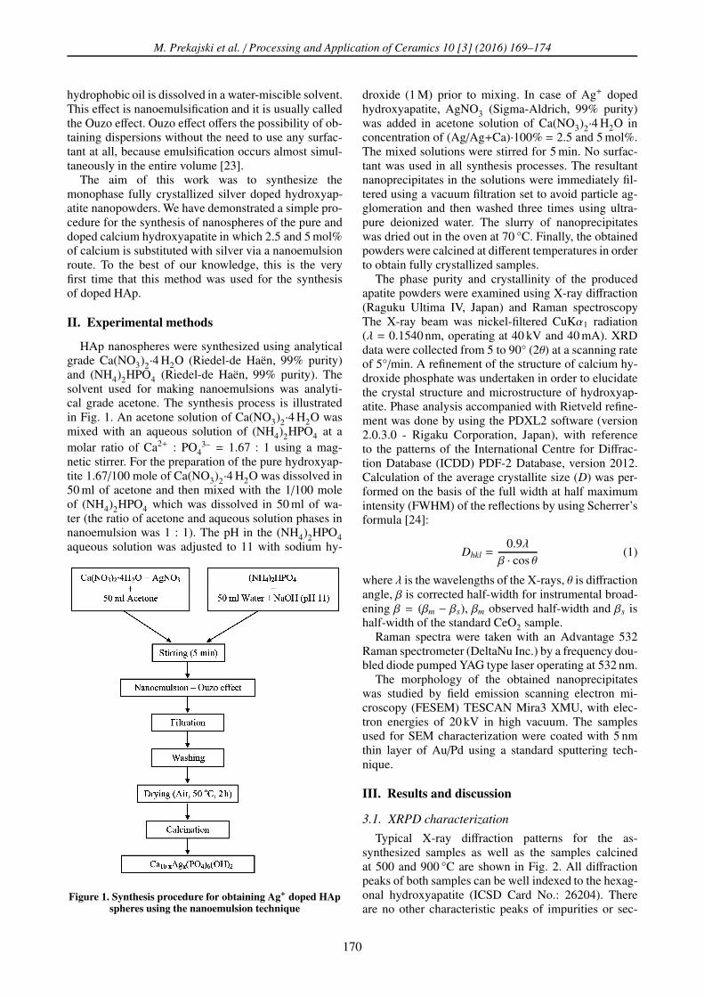
HAp nanospheres were synthesized using analyticalgrade Ca(NO3)2·4 H2O (Riedel-de Haën, 99% purity)and (NH4)2HPO4 (Riedel-de Haën, 99% purity). Thesolvent used for making nanoemulsions was analyti-cal grade acetone. The synthesis process is illustratedin Fig. 1. An acetone solution of Ca(NO3)2·4 H2O wasmixed with an aqueous solution of (NH4)2HPO4 at amolar ratio of Ca2+ : PO4
3– = 1.67 : 1 using a mag-netic stirrer. For the preparation of the pure hydroxyap-tite 1.67/100 mole of Ca(NO3)2·4 H2O was dissolved in50 ml of acetone and then mixed with the 1/100 moleof (NH4)2HPO4 which was dissolved in 50 ml of wa-ter (the ratio of acetone and aqueous solution phases innanoemulsion was 1 : 1). The pH in the (NH4)2HPO4aqueous solution was adjusted to 11 with sodium hy-
Figure 1. Synthesis procedure for obtaining Ag+ doped HApspheres using the nanoemulsion technique
droxide (1 M) prior to mixing. In case of Ag+ dopedhydroxyapatite, AgNO3 (Sigma-Aldrich, 99% purity)was added in acetone solution of Ca(NO3)2·4 H2O inconcentration of (Ag/Ag+Ca)·100% = 2.5 and 5 mol%.The mixed solutions were stirred for 5 min. No surfac-tant was used in all synthesis processes. The resultantnanoprecipitates in the solutions were immediately fil-tered using a vacuum filtration set to avoid particle ag-glomeration and then washed three times using ultra-pure deionized water. The slurry of nanoprecipitateswas dried out in the oven at 70 °C. Finally, the obtainedpowders were calcined at different temperatures in orderto obtain fully crystallized samples.
The phase purity and crystallinity of the producedapatite powders were examined using X-ray diffraction(Raguku Ultima IV, Japan) and Raman spectroscopyThe X-ray beam was nickel-filtered CuKα1 radiation(λ = 0.1540 nm, operating at 40 kV and 40 mA). XRDdata were collected from 5 to 90° (2θ) at a scanning rateof 5°/min. A refinement of the structure of calcium hy-droxide phosphate was undertaken in order to elucidatethe crystal structure and microstructure of hydroxyap-atite. Phase analysis accompanied with Rietveld refine-ment was done by using the PDXL2 software (version2.0.3.0 - Rigaku Corporation, Japan), with referenceto the patterns of the International Centre for Diffrac-tion Database (ICDD) PDF-2 Database, version 2012.Calculation of the average crystallite size (D) was per-formed on the basis of the full width at half maximumintensity (FWHM) of the reflections by using Scherrer’sformula [24]:
Dhkl =0.9λβ · cos θ
(1)
where λ is the wavelengths of the X-rays, θ is diffractionangle, β is corrected half-width for instrumental broad-ening β = (βm − βs), βm observed half-width and βs ishalf-width of the standard CeO2 sample.
Raman spectra were taken with an Advantage 532Raman spectrometer (DeltaNu Inc.) by a frequency dou-bled diode pumped YAG type laser operating at 532 nm.
The morphology of the obtained nanoprecipitateswas studied by field emission scanning electron mi-croscopy (FESEM) TESCAN Mira3 XMU, with elec-tron energies of 20 kV in high vacuum. The samplesused for SEM characterization were coated with 5 nmthin layer of Au/Pd using a standard sputtering tech-nique.
III. Results and discussion
3.1. XRPD characterization
Typical X-ray diffraction patterns for the as-synthesized samples as well as the samples calcinedat 500 and 900 °C are shown in Fig. 2. All diffractionpeaks of both samples can be well indexed to the hexag-onal hydroxyapatite (ICSD Card No.: 26204). Thereare no other characteristic peaks of impurities or sec-
170

M. Prekajski et al. / Processing and Application of Ceramics 10 [3] (2016) 169–174
(a)
(b)
(c)
Figure 2. XRD patterns of pure and Ag+ doped HApsamples, calcined at different temperatures: a) as-
synthesized (70 °C), b) 500 °C and c) 900 °C
ondary phases, such as CaO, Ag2O or silver phosphates.The absence of other phases in XRD pattern of Ag+
doped HAp samples demonstrates that the Ag+ ionshave successfully substituted Ca2+ ions without affect-ing the crystal structure of the original HAp. This re-sult is in agreement with previous studies conducted byShirkhanzadeh et al. [25] and Ravindran et al. [26].
The XRD pattern of the as-synthesized samples (Fig.2a) revealed that the HAp phase was already form-ing at low temperature. However peaks are significantlybroadened indicating poor crystallinity and small crys-tallite size. According to calculation based on the Scher-rer’s equitation crystallite size of the as-synthesizedsamples is about 4 nm (Table 1). Calcination at 500 °Cfor 2 hours promotes process of crystallization (Fig. 2b)and crystals grow to the size of 7 nm (Table 1). Sincethe peaks are still very broad we decided to increase cal-cination temperature to 900 °C. At this temperature allsamples are fully crystallized and reflections are welldefined by the sharp peaks (Fig. 2c). There is still no in-dications of any secondary phases in Ag doped sampleswhile the crystallites sizes grow to 19 nm (Table 1).
Refined unit cell parameters of all samples are pre-sented in Fig. 3. Doping of Ag+ ions into the ap-atite shows an increase in a and c lattice parameters(Fig. 3). An expansion of these lattice parameters arecaused by the substitution of smaller radius ion ofthe Ca2+ (0.099 nm) by the larger Ag+ (0.128 nm) ion.These results are in accordance with previous reportsby Rameshbabu et al. [27] which assumed that silversubstitute for calcium in the HAp lattice. On the otherhand, strain in crystal lattice remains almost the same atall temperatures, but it depends on the concentration ofAg+ in the samples. Namely, incorporation of larger ionprovokes additional stress and strain in crystal structure(Table 1).
3.2. Raman spectral studies
Raman analysis was used in order to verify the pres-ence of pure HAp phase in samples, because it is knownthat Raman spectroscopy method is more sensitive onthe presence of secondary phases than X-ray powderdiffraction. Raman spectrum of HAp sample doped withhigher concentration of Ag+ is shown in Fig. 4. Forcomparison Raman spectrum of the pure as-synthesizedsample is also represented at Fig. 4. The most intensivemode near 950 cm-1 is assigned to the internal modes ofthe PO4
3– tetrahedral ν1 frequency which correspondsto the symmetric stretching of P−O bonds [13,28]. Thebands present near 1027 cm-1, 1050 cm-1, and 1080 cm-1
can be assigned to the asymmetric ν3 (P−O) stretch-ing [19]. The other expected bands near 576, 590 and616 cm-1 which originate from ν4 PO4 and bands near430 (ν2) and 450 cm-1 (ν2) attributed to the O−P−Obending modes have weak intensities and were not de-tected [29,30]. The reason for such behaviour may liein water vibrational modes, which gives rise to weak in-tensity stretching and bending bands in Raman spectra
171

M. Prekajski et al. / Processing and Application of Ceramics 10 [3] (2016) 169–174
Table 1. Refined microstructural factors: crystallite size (D) and strain (ε)
Temperature0 mol% 2.5 mol% 5 mol%
D [nm] ε [%] D [nm] ε [%] D [nm] ε [%]70 °C 4.1 0.4 3.9 0.41 4.2 0.45500 °C 6.6 0.2 6.8 0.4 6.9 0.59900 °C 18.6 0 19.8 0.39 19.2 0.48
(a)
(b)
Figure 3. Refined lattice parameters a (a) and c (b) of pureand Ag+ doped HAp samples, calcined at different
temperatures
[31,32]. The OH– vibrational bands expected in the re-gion of 630 cm-1 are not clearly detected. This behaviouris in good agreement with the previous studies [33].
3.3. Morphology characterization
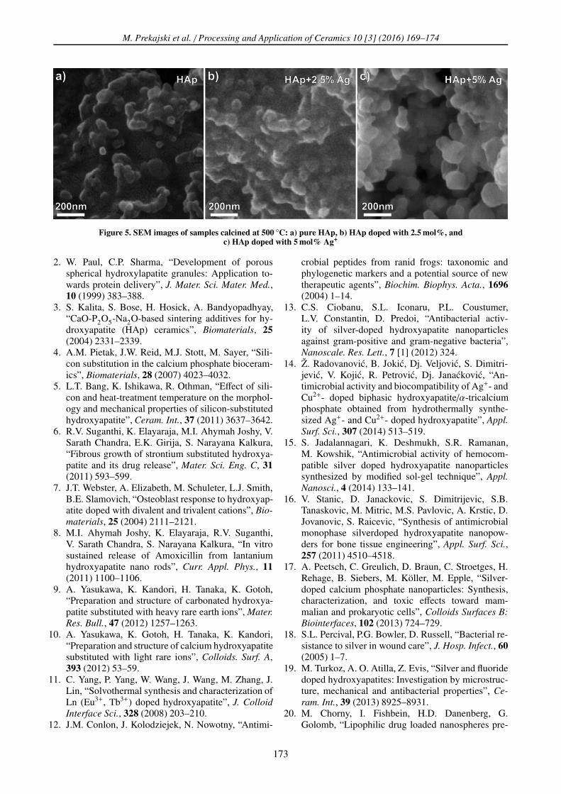
SEM micrographs of the pure and Ag-doped HAppowders calcined at 500 °C are shown in Fig. 5. Theresults showed that the obtained hydroxyapatite pow-ders were composed of nearly spherical particles of nar-row size distribution. The average particle size is about∼100 nm in all samples. Individual spheres are agglom-erated and a lot of porosity is present. The formationof spherical HAp nanoparticles is due to the Ouzo ef-
Figure 4. Raman spectra of pure and Ag+ doped HApsamples calcined at 500 °C
fect which occurs in present synthesis method [34]. Thematerial with this microstructural and texture propertieshave an advantage due to its application as bioceramicmaterials [20].
IV. Conclusions
The nanoemulsion method has been found to be verypromising for the synthesis of spherical nanoparticlesof the pure and Ag+ doped hydroxyapatite. Significantconcentration of Ag+ ions (5 mol%) were completely in-corporated in HAp lattice without formation of any im-purities or secondary phases which was confirmed bothby XRD and Raman analysis. Calcination at higher tem-peratures promotes crystallization process and crystal-lite growth. Incorporation of larger Ag+ ion instead ofCa2+ ion causes increase of lattice parameters. The sil-ver ions cause the increase in the size of the crystal-lites in the doped samples. The synthesized nanoparti-cles were spherical in shape and their sizes were in therange of ∼100 nm. The obtained results give new per-spective for possible application of smart drug deliveryin form of HAp nanospheres improved with Ag+ ionswhich has antibacterial properties.
Acknowledgements: Financial support from the Ser-bian Education and Science Ministry in the Frameworkof projects No. 45012 is gratefully acknowledged.
References
1. L.L. Hench, J. Wilson, “Surface active biomaterials”,Science, 226 (1984) 630–636.
172
M. Prekajski et al. / Processing and Application of Ceramics 10 [3] (2016) 169–174
Figure 5. SEM images of samples calcined at 500 °C: a) pure HAp, b) HAp doped with 2.5 mol%, andc) HAp doped with 5 mol% Ag+
2. W. Paul, C.P. Sharma, “Development of porousspherical hydroxylapatite granules: Application to-wards protein delivery”, J. Mater. Sci. Mater. Med.,10 (1999) 383–388.
3. S. Kalita, S. Bose, H. Hosick, A. Bandyopadhyay,“CaO-P2O5-Na2O-based sintering additives for hy-droxyapatite (HAp) ceramics”, Biomaterials, 25
(2004) 2331–2339.4. A.M. Pietak, J.W. Reid, M.J. Stott, M. Sayer, “Sili-
con substitution in the calcium phosphate bioceram-ics”, Biomaterials, 28 (2007) 4023–4032.
5. L.T. Bang, K. Ishikawa, R. Othman, “Effect of sili-con and heat-treatment temperature on the morphol-ogy and mechanical properties of silicon-substitutedhydroxyapatite”, Ceram. Int., 37 (2011) 3637–3642.
6. R.V. Suganthi, K. Elayaraja, M.I. Ahymah Joshy, V.Sarath Chandra, E.K. Girija, S. Narayana Kalkura,“Fibrous growth of strontium substituted hydroxya-patite and its drug release”, Mater. Sci. Eng. C, 31
(2011) 593–599.7. J.T. Webster, A. Elizabeth, M. Schuleter, L.J. Smith,
B.E. Slamovich, “Osteoblast response to hydroxyap-atite doped with divalent and trivalent cations”, Bio-
materials, 25 (2004) 2111–2121.8. M.I. Ahymah Joshy, K. Elayaraja, R.V. Suganthi,
V. Sarath Chandra, S. Narayana Kalkura, “In vitrosustained release of Amoxicillin from lantaniumhydroxyapatite nano rods”, Curr. Appl. Phys., 11
(2011) 1100–1106.9. A. Yasukawa, K. Kandori, H. Tanaka, K. Gotoh,
“Preparation and structure of carbonated hydroxya-patite substituted with heavy rare earth ions”, Mater.
Res. Bull., 47 (2012) 1257–1263.10. A. Yasukawa, K. Gotoh, H. Tanaka, K. Kandori,
“Preparation and structure of calcium hydroxyapatitesubstituted with light rare ions”, Colloids. Surf. A,393 (2012) 53–59.
11. C. Yang, P. Yang, W. Wang, J. Wang, M. Zhang, J.Lin, “Solvothermal synthesis and characterization ofLn (Eu3+, Tb3+) doped hydroxyapatite”, J. Colloid
Interface Sci., 328 (2008) 203–210.12. J.M. Conlon, J. Kolodziejek, N. Nowotny, “Antimi-
crobial peptides from ranid frogs: taxonomic andphylogenetic markers and a potential source of newtherapeutic agents”, Biochim. Biophys. Acta., 1696
(2004) 1–14.13. C.S. Ciobanu, S.L. Iconaru, P.L. Coustumer,
L.V. Constantin, D. Predoi, “Antibacterial activ-ity of silver-doped hydroxyapatite nanoparticlesagainst gram-positive and gram-negative bacteria”,Nanoscale. Res. Lett., 7 [1] (2012) 324.
14. Ž. Radovanovic, B. Jokic, Dj. Veljovic, S. Dimitri-jevic, V. Kojic, R. Petrovic, Dj. Janackovic, “An-timicrobial activity and biocompatibility of Ag+- andCu2+- doped biphasic hydroxyapatite/α-tricalciumphosphate obtained from hydrothermally synthe-sized Ag+- and Cu2+- doped hydroxyapatite”, Appl.
Surf. Sci., 307 (2014) 513–519.15. S. Jadalannagari, K. Deshmukh, S.R. Ramanan,
M. Kowshik, “Antimicrobial activity of hemocom-patible silver doped hydroxyapatite nanoparticlessynthesized by modified sol-gel technique”, Appl.
Nanosci., 4 (2014) 133–141.16. V. Stanic, D. Janackovic, S. Dimitrijevic, S.B.
Tanaskovic, M. Mitric, M.S. Pavlovic, A. Krstic, D.Jovanovic, S. Raicevic, “Synthesis of antimicrobialmonophase silverdoped hydroxyapatite nanopow-ders for bone tissue engineering”, Appl. Surf. Sci.,257 (2011) 4510–4518.
17. A. Peetsch, C. Greulich, D. Braun, C. Stroetges, H.Rehage, B. Siebers, M. Köller, M. Epple, “Silver-doped calcium phosphate nanoparticles: Synthesis,characterization, and toxic effects toward mam-malian and prokaryotic cells”, Colloids Surfaces B:
Biointerfaces, 102 (2013) 724–729.18. S.L. Percival, P.G. Bowler, D. Russell, “Bacterial re-
sistance to silver in wound care”, J. Hosp. Infect., 60
(2005) 1–7.19. M. Turkoz, A. O. Atilla, Z. Evis, “Silver and fluoride
doped hydroxyapatites: Investigation by microstruc-ture, mechanical and antibacterial properties”, Ce-
ram. Int., 39 (2013) 8925–8931.20. M. Chorny, I. Fishbein, H.D. Danenberg, G.
Golomb, “Lipophilic drug loaded nanospheres pre-
173
M. Prekajski et al. / Processing and Application of Ceramics 10 [3] (2016) 169–174
pared by nanoprecipitation: Effect of formulationvariables on size, drug recovery and release kinet-ics”. J. Control. Release., 83 (2002) 389–400.
21. W.Y. Zhou, M. Wang, W.L. Cheung, B.C. Guo,D.M. Jia, “Synthesis of carbonated hydroxyapatitenanospheres through nanoemulsion”, J. Mater. Sci.
Mater. Med., 19 (2008) 103–110.22. M. Yu Koroleva, E.V. Yurtov, “Nanoemulsions: the
properties, methods of preparation and promising ap-plications”, Russ. Chem. Rev., 81 (2012) 21–43.
23. M. Prekajski, M. Mirkovic, B. Todorovic, A.Matkovic, M. Marinovic-Cincovic, J. Lukovic, B.Matovic, “Ouzo effect - New simple nanoemulsionmethod for synthesis of strontium hydroxyapatitenanospheres”, J. Eur. Ceram. Soc., 36 [5] (2016)1293–1298.
24. P. Scherrer, “Bestimmung der Grösse und der in-neren Struktur von Kolloidteilchen mittels Röntgen-strahlen”, Gött Nachr, 2 (1918) 98–100.
25. M. Shirkhanzadeh, M. Azadegan, G.Q. Liu, “Bioac-tive delivery systems for the slow release of antibi-otics: incorporation of Ag+ ions into micro-poroushydroxyapatite coatings”, Mater. Lett., 24 (1995) 7–12.
26. A. Ravindran, A. Singh, A.M. Raichur, N. Chandra-sekaran, A. Mukherjee, “Studies on interaction ofcolloidal Ag nanoparticles with bovine serum albu-min (BSA)”, Colloid. Surface. B, 76 (2010) 32–37.
27. N. Rameshbabu, T.S.S. Kumar, T.G. Prabhakar, V.S.Sastry, K.V.G.K. Murty, K.P. Rao, “Antibacterialnanosized silver substituted hydroxyapatite: synthe-sis and characterization”, J. Biomed. Mater. Res.,
80A (2007) 581–591.28. C.S. Ciobanu, S.L. Iconaru, P.L. Coustumer, D. Pre-
doi, “Vibrational investigations of silver-doped hy-droxyapatite with antibacterial properties”, J. Spec-
troscopy, 2013 (2013) 471061.29. A. Mortier, J. Lemaitre, P.G. Rouxhet, “Temperature
programmed characterization of synthetic calcium-deficient phosphate apatites”, Thermochim. Acta.,143 (1989) 265–282.
30. J. Elliot, Structural and Chemistry of Apatites and
other Calcium Ortophosphates, Amsterdam, Else-vier, 1994.
31. A. Costescu, C.S. Ciobanu, S.L. Iconaru, R.V. Ghita,C.M. Chifiriuc, L.G. Marutescu, D. Predoi, “Fab-rication, characterization, and antimicrobial activ-ity, evaluation of low silver concentrations in silver-doped hydroxyapatite nanoparticles”, J. Nanomater.,2013 (2013) 194854.
32. C.L. Popa, C.S. Ciobanu, G. Voicu, E. Vasile, M.C.Chifiriuc, S.L. Iconaru, D. Predoi, “Influence of ther-mal treatment on the antimicrobial activity of silver-doped biological apatite”, Nanoscale Res. Lett., 10
(2015) 502.33. B.O. Fowler, “Infrared studies of apatites. I. Vibra-
tional assignments for calcium, strontium, and bar-ium hydroxyapatites utilizing isotopic substitution”,Inorg. Chem., 13 [1] (1974) 194–207.
34. F. Ganachaud, J.L. Katz, “Nanoparticles andnanocapsules created using the Ouzo effect: Spon-taneous emulsification as an alternative to ultra-sonic and high-shear devices”, Chem. Phys. Chem.,6 (2005) 209–216.
174