syntheses and structures of metal cyanamide compounds
TRANSCRIPT

Syntheses and Structures of Metal
Cyanamide Compounds
Xiaohui Liu


Syntheses and Structures of Metal
Cyanamide Compounds
Von der Fakultät für Mathematik, Informatik und Naturwissenschaften
der Rheinisch-Westfälischen Technischen Hochschule Aachen
zur Erlangung des akademischen Grades eines
Doktors der Naturwissenschaften
genehmigte Dissertation
vorgelegt von
M. Sc. Xiaohui Liu
aus Yanan (China)
Berichter: Universitätsprofessor Dr. R. Dronskowski
Universitätsprofessor Dr. U. Simon
Tag der mündlichen Prüfung: 24. April 2002
Diese Dissertation ist auf den Internetseiten der Hochschule online verfügbar.


To my husband Bing Wang, and our son Yi Wang


Acknowledgments
I am deeply grateful to Prof. Dr. Richard Dronskowski for his enthusiastic guidance and care.
He taught me not only the chemistry, but more importantly the new methodologies for solving
the problems. I thank him for his trust and giving me a chance to realize my doctoral dream.
I thank Dr. Jürgen Huster for his constant help in the solution of the experimental and
structural problems, fruitful discussions and performing the nice STOE X-ray powder
diffraction. Dr. Peter Kroll was involved in this project in many ways. I thank him for his
advice, discussion. I also thank Dr. Schmitz and Dr. Müller for their kindly help. I am also
grateful to Dr. Bernhard Eck for his aid in the computer technique.
I thank Dr. Michael Scholten, Dr. Stephen Irsen, Andreas Prange, Uwe Couhorn, Philipp
Kölle and other members of the laboratory for their help, friendship and good atmosphere in
the group. It is a pleasure to thank Klaus Kruse and Dr. Chunhua Hu for the collection of the
single crystal X-ray intensities and Dr. Felix Hüning for the susceptibility measurement.
I am also grateful to other my friends and colleagues in the Institute für Anorganisch Chemie,
RWTH, for their friendship and help.
I also thank Family Westhovens for taking care of my family and their encouragement. It was
their kindness and help made my promotion possible.
My husband, Bing Wang, always stands by me, although in most cases I stand behind him.
Our son Yi Wang, as the world’s greatest kid, is proud of his academic mother and has never
made some trouble to me during this work. My parents are my continuous source of
inspiration and give me constant encouragement. Thanks to them for their importantly and
notably love and supports.


Parts of this work are already published:
Xiaohui Liu, Andrea Decker, Dieter Schmitz, and Richard Dronskowski
Crystal Structure Refinement of Lead Cyanamide and the Stiffness of the Cyanamide Anion
Z. Anorg. Allg. Chem. 2000, 626, 103.
Xiaohui Liu, Peter Kroll, and Richard Dronskowski
Crystal Structure, Magnetic Properties, and Electronic Structure of
Co(NCNH2)4Cl2 and Ni(NCNH2)4Cl2
Z. Anorg. Allg. Chem. 2001, 627, 1682.
R. Dronskowski and X. Liu
Crystal Structure of copper(II) tetracyanamide dichloride, Cu(NCNH2)4Cl2
Z. Kristallogr. NCS, 2002, 217, 118.
Xiaohui Liu, Paul Müller, Peter Kroll, and Richard Dronskowski
Synthesis, Structure Determination, and Quantum-Chemical Characterization of
an Alternate HgNCN Polymorph
Inorg. Chem. 2002, 41, 4259.
Xiaohui Liu and Richard Dronskowski
Mercury Cyanamide/Carbodiimide Networks: Synthesis and Crystal Structures of
Hg2(NCN)Cl2 and Hg3(NCN)2Cl2
Z. Naturforsch. 2002, 57b, 1108.


I
Contents
1 Introduction………………………………...……………………………….................
1.1 General introduction ……...…………………………...………………………......
1.2 Scope of this thesis …………...……………………...……………………….....…
2 Experimental section ...................................................................................................
2.1 General procedures ..................................................................................................
2.2 X-ray powder diffraction .........................................................................................
2.3 X-ray single crystal measurement ...........................................................................
2.4 Other measurements .............................................................................................…
2.5 Programs ..................................................................................................................
3 The crystal structure of PbNCN and the stiffness of the cyanamide anion ............
3.1 Introduction …….….....…………...…….……………….…..……….….....….....
3.2 Experimental ……...………….….....………………….....………………....….....
3.3 Crystal structure refinement of PbNCN ……......……...………..………...............
3.4 Stiffness of cyanamide anion ......……....…….……………...………….…...........
4 Mercury cyanamide and related complexes ..............................................................
4.1 Synthesis and characterization of mercury cyanamide ..........................................
4.1.1 Introduction .......................................................….........................................
4.1.2 Experimental ..............................................................................................….
4.1.3 Structure determination from powder data ...........................…......................
4.1.4 Results and discussion ..........................................................…......................
4.2 Structures of Hg2(NCN)Cl2 and Hg3(NCN)2Cl2 .....................................................
4.2.1 Introduction ...........................................................................…......................
4.2.2 Synthesis ..................................................................................…................…
4.2.3 Single crystal structure determination and discussion ....................................
4.2.4 The crystal structure of Hg2(NCN)Cl2 ...................................….....................
4.2.5 The crystal structure of Hg3(NCN)2Cl2 ...........................…...........................
4.2.6 Discussion ......................................................................................................
5 Syntheses and structures of Ag2NCN and Ag(H2NCN)3NO3 ...................................
5.1 Synthesis and structure determination of Ag2NCN ................................................
5.1.1 Introduction .............................................................................................…....
1
1
6
7
7
7
8
9
9
12
12
12
14
16
18
18
18
19
20
30
33
33
33
34
34
38
44
47
47
47

II
5.1.2 Synthesis ..................................................................................................…...
5.1.3 The single crystal structure determination and discussion ................….........
5.1.4 PXRD measurement and structure discussion ...........................................….
5.2 Synthesis and structure of Ag(H2NCN)3NO3 .........................................................
5.2.1 Introduction .......................................................................................….....….
5.2.2 Synthesis .............................................................................................…........
5.2.3 X-ray structure determination ....................................................................….
5.2.4 The structure of Ag(H2NCN)3NO3 and discussion .................................…....
6 Syntheses, crystal structures, magnetic properties, and electronic structures of
Co(NCNH2)4Cl2, Ni(NCNH2)4Cl2 and Cu(NCNH2)4Cl2 ...........................................
6.1 Introduction .............................................................................................................
6.2 Synthesis .................................................................................................................
6.3 X-ray structure determination .................................................................................
6.4 Structure and Packing .............................................................................................
6.5 Magnetism and Electronic Structure .......................................................................
7 A novel synthesis of Li2NCN .......................................................................................
7.1 Introduction ..............................................................................................................
7.2 Synthesis .............................................................................................................….
7.3 X-ray powder diffraction ............................................................................….........
8 Summary .......................................................................................................................
9 References .....................................................................................................................
47
48
52
56
56
56
56
60
64
64
64
65
67
70
74
74
74
75
77
81

1
1. Introduction
1.1. General introduction
Cyanamide (H2NCN), which is a well known compound, m.p. 45 - 46 °C and prepared by
hydrolysis of CaNCN [1], is a weak acid and exceedingly soluble in water, alcohol and ether.
It may spontaneously polymerize to form the dimer, dicyanodiamide (cyanoguanidine), or
even the trimer, melamine. In fact, during syntheses of metal cyanamides we have produced
all of these polymers. The interatomic distances of cyanamide are found to be 115 pm and
131 pm, the N(1)-C-N(2) angle is 178.6° [2, 3].
While metal cyanamides have been typically prepared via high-temperature routes (600 –
1300 °C), in the laboratory they can be made from the reaction between carbonates and HCN
gas, sometimes followed by a calcination process in a nitrogen atmosphere [4]. CaNCN is a
typical metal cyamamide and also largely used for fertilizing. On the industrial scale [5],
CaNCN is manufactured in an impure form from calcium carbide and molecular nitrogen.
CaC2 + N2 ca. 1100 °C CaNCN + C ∆H = -297 KJ⋅mol-1
In fact, numerous synthetic routes have been proposed for metal cyanamides, however, their
preparation in a pure and well-defined state has not been unambiguously achieved before
1994. Additionally, reliable structural data were only available for CaNCN by Vannerberg
[4]; here, the cyanamide anion is linear, C-N = 122 pm, and is isostructural and isoelectronic
with CO2.
Pulham succeeded in synthesizing lithium cyanamide from a typical solid-state reaction of
Li2C2 with Li3N at 600 °C, finally crystallizing it from liquid lithium [6]. When the reaction
was carried out in molten lithium, colorless single crystals of the salt formed upon
evaporation of the metal. The lattice is composed of Li+ and centrosymmetric NCN2- ions (C-
N = 123 pm). The symmetry of NCN2- belongs to D∞h point group. Each Li+ ion is at the center
of a squashed tetrahedron of nitrogen atoms.
Sodium cyanamide can be generated by the reaction of sodium hydrogen cyanamide
(NaHCN2) and an excess of sodium at 300 °C [7]. The single crystals were recently obtained

2
by reaction of sodium amide with sodium hydrogen cyanamide at 200 °C in vacuum, then by
heating the product (500 °C, 8 days) in silver crucibles. The NCN2- units are linear exhibiting
a C-N bond length of 123.6 pm, while sodium is coordinated by five nitrogen atoms forming a
square pyramid [8]. K2NCN [9] characterized by powder data has the same structure as
Na2NCN.
Strid and co-workers obtained SrNCN [10] and BaNCN [11] by the reaction of the respective
carbonates with hydrogen cyanide at temperatures between 600 and 650 °C, but they were
unable to solve the crystal structures completely. Later, Schnick’s group succeeded to
synthesize the alkaline-earth metal cyanamides MgNCN, SrNCN, and BaNCN by the reaction
of melamine C3N3(NH2)3 with the metal nitrides (Mg3N2, Sr2N, and Ba3N2) at temperatures
between 740 and 850 °C [12]. All three alkaline-earth metal cyanamides investigated contain
almost linear NCN2- anions, although the authors suggested a reduction of the anionic
symmetry from D∞h to C2v since the observed N-C-N angle is 177.2(18)° in BaNCN. Though
both cation and anion are coordinated by six counter ions in all three crystal structures, the
crystal structures are not directly related to each other. In MgNCN alternating layers of
cations and anions are found. In SrNCN the layers contain both ions, and in BaNCN it is no
longer a layer structure, but a three-dimensional arrangement of the ions in the solid.
Dronskowski found two unexpected indium cyanamides in crystalline form, namely
In2.24(NCN)3 and NaIn(NCN)2, by a reaction of indium monobromide InBr and sodium
cyanide NaCN at 400 °C [13]. In2.24(NCN)3 is a mixed valence compound, containing both
trivalent and mono-valent indium cations. Their crystal structures reveal ionic layer
structures. They are built from sheets of cationic and anionic (cyanamide) motifs. The crystal
structure of NaIn(NCN)2 displays strong similarities with the In2.24(NCN)3. The NCN2- of
both crystals reflects D∞h symmetry.
The crystal structure of the PbNCN was previously studied by Cooper [14]. The bonding of
PbNCN is covalent with an asymmetric N-C-N group (117 and 125 pm) that is more strongly
bonded to lead atoms at one end. Nevertheless, a center of symmetry had been overlooked in
the structure determination, carried out in space group Pna21, and important structure details
such as the angle of N-C-N and the bond lengths of C-N were of limited accuracy.

3
In the course of this thesis, Jansen’s group reported the crystal structures of ZnNCN [15] and
HgNCN [16]. Zinc cyanamide was precipitated from aqueous solution of ZnSO4 and
Na2NCN. The single crystals were grown from compacted ZnNCN powder at 570 °C. The
crystal structure is composed of corner-linked ZnN4/2 tetrehedra. Zinc is tetrahedrally
coordinated by N atoms, with strong covalent bonds (Zn-N are 198.5 and 203.5 pm,
respectively) and each nitrogen atom is bonded by two Zn atoms. Carbon and nitrogen atoms
form (NCN)2- dumb-bells (N-C = 123 pm) and N-C-N is slightly bent (176.3°). Mercury
cyanamide was prepared by double conversion of HgCl2 with sodium cyanamide in aqueous
solution, and the crystal structure had been determined using X-ray powder and neutron
diffraction data. It shows that mercury atoms coordinate N atoms from both sides of the NCN
unit, and infinitive Hg-NCN-Hg zigzag chains are formed. Two C-N distances are almost
equal (122.8 pm and 121.6 pm) and cyanamide is bent (172.4°). Here, if the bond difference
of N-C is ignored, the symmetry of the NCN unit is reduced from D∞h to C2v. Surprisingly, the
total N-C-N distance in ZnNCN or HgNCN does not enlarge although the NCN units form
strong covalent bonds to metal atoms from both sides.
Silver cyanamide was prepared by mixing aqueous solutions of H2NCN and silver nitrate, and
the single crystals were obtained by re-crystallization of Ag2NCN precipitation in ammonia.
The NCN2- anion is slightly bent (177.1°) and exhibits two significantly different bond
lengths (N(1)-C: 119.4; C-N(2): 126.6 pm) [17]. Here, if the bending of the NCN unit is
ignored, the symmetry of the NCN unit tends to reduce from D∞h to C∞v. In fact, Ag2NCN is a
well known compound, and used for surface decomposition studies [18, 19]. The structure
had been examined in 1962 [19], and only the unit cell, the space group and the positions of
the silver atoms (wrong!) were determined.
The single phase K5H(CN2)3 was synthesized by the reaction of KHCN2 with melamine
C3N3(NH2)3 at 320 °C. K5H(CN2)3 crystallizes in the cubic space group Im-3m with Z = 2,
determined by X-ray powder and single crystal data [20]. In the compound the NCN2- anion
exhibits D∞h symmetry. The protons are loosely bonded to the NCN2- anions according to 1H
and 13C solid state MAS-NMR investigations, temperature dependent impedance spectro-
scopy, and FTIR spectroscopy.

4
The mixed cyanamide-cyanides M2(CN2)(CN)2 (M = Ba, Sr) were synthesized by the reaction
of Ba2N and SrCO3, respectively, with HCN at 630 °C [21]. The crystal structure is a partially
filled defect variant of the anti-NiAs structure type with a distorted hexagonal close-packed
arrangement of M2+-ions. All NCN2- and a quarter of the CN- ions occupy 3/4 of the
octahedrally coordinated interstices; the remaining cyanide anions are located at 3/8 of the
tetrahedral sites. All NCN2- anions exhibit D∞h symmetry. Ca11N6(NCN)2 and Ca4N2(NCN)
were synthesized from Ca3N2 and C under nitrogen at 1300 °C [22]. Both compounds can be
described as open three-dimensional frameworks. The NCN2- guest species are hosted in
channels formed by linking distorted Ca6N octahedra through shared edges and corners. The
NCN2- is linear and two C-N bonds are slightly different.
Table 1.1 Crystal data of cyanamide and metal-cyanamides.
Lattice constant
Compound a (Å)
b (Å)
c (Å)
β ( o )
Volume
(Å3), Z
Space group (NCN)2-
H2NCN [3] 6.856 6.628 9.147 - 415.7, 8 Pbca (No. 61) (N-C≡N)2-
Li2NCN [6] 3.687 3.687 8.668 - 117.8, 2 I4/mmm (No. 139) (N=C=N)2-
Na2NCN [8] 5.046 5.001 5.536 110.08 131.2, 2 C2/m (No. 12) (N=C=N)2-
K2NCN [9] 5.788 5.703 5.786 109.02 180.6, 2 C2/m (No. 12) (N=C=N)2-
CaNCN [4] 5.347 5.347 5.347 40.4 58.0, 1 R -3mR (No. 166) (N=C=N)2-
MgNCN [12] 3.273 3.273 14.128 - 131.1, 3 R-3mH (No. 166) (N=C=N)2-
SrNCN [12] 12.410 3.963 5.389 - 265.0, 4 Pnma (No. 62) (N=C=N)2-
BaNCN [12] 15.282 15.282 7.013 - 1418.4, 18 R-3cH (No. 167) (N=C=N)2-
In2.24(NCN)3 [13] 6.061 6.061 28.844 - 917.7, 6 R-3c (No. 161) (N=C=N)2-
NaIn(NCN)2 [13] 9.613 7.168 6.037 - 416.0, 4 Cmcm (No. 63) (N=C=N)2-
PbNCN [14b] 5.553 11.732 3.867 - 251.9, 4 Pna21 (No. 33) (N-C≡N)2-
ZnNCN [15] 8.804 8.804 5.433 - 421.2, 8 I-42d (No. 122) (N=C=N)2-
HgNCN [16] 10.485 6.514 6.8929 - 470.8, 8 Pbca (No. 61) N=C=N)2-
Tl2NCN [14a] ? ? ? ?
Ag2NCN [17] 7.315 6.010 6.684 102.3 287.1, 4 P21/c (No. 14) (N-C≡N)2-
NaHNCN [24a] 3.531 10.358 6.486 - 237.2, 4 Pbcm (No. 57) (N-C≡N)2-
KHNCN [23] 7.087 9.090 9.014 - 580.7, 8 P212121 (No. 19) (N-C≡N)2-
RbHNCN [24b] 7.299 9.435 9.420 - 628.7, 8 P212121 (No. 19) (N-C≡N)2-
Si(NCN)2 [25] 6.188 6.188 6.188 - 237.0, 2 Pn3m (No. 224) N=C=N)2-

5
For the preparation of KHNCN, melamine has been reacted with potassium amide in liquid
ammonia. After evaporation of the solvent solid KHNCN crystallized at 210 °C [23]. Using
the same method, RbHNCN was prepared [24b]. In the solids K+ or Rb+ and the HCN2- ions
occur. As expected, two significantly differing bond-distances of C-N have been found in the
anion. NaHNCN [24a] is isostructural with KHNCN.
The reaction of SiCl4 with bis(trimethylsily)carbodiimide directly provides silicon cyanamide
Si(NCN)2 when pyridine acts as catalyst at temperatures between 25 and 100 °C [25]. Above
900 °C, Si(NCN)2 decomposes to Si2N2(NCN), cyanogen (C2N2) and nitrogen. The powder
diffraction analysis shows that Si(NCN)2 reveals a cubic unit cell with Pn3m at high
temperature (250 - 650 °C) and Si2N2(NCN) reveals an orthorhombic unit cell with Aba2 at
room temperature. These two cyanamide compounds contain linear N-C-N groups with very
short C-N bond lengths (110 pm for Si(NCN)2 and 104 pm for Si2N2(NCN)).
Table 1.2 The N-C bond lengths for a number of important cyanamide compounds.
CaNCN: C=N: 1.22 Å - - N=C=N: 2.44 Å
Li2NCN: C=N: 1.23 Å - - N=C=N: 2.46 Å
MgNCN:
ZnNCN:
HgNCN:
C=N:
C=N:
C≅N:
1.24 Å
1.23 Å
1.22 Å
-
-
C-N:
-
-
1.23 Å
N=C=N:
N=C=N:
N=C=N:
2.48 Å
2.46 Å
2.45 Å
Ag2NCN:
PbNCN:
C≡N:
C≡N:
1.19 Å
1.17 Å
C-N:
C-N:
1.26 Å
1.25 Å
N≡C-N:
N≡C-N:
2.45 Å
2.42 Å
H2NCN: C≡N: 1.14 Å C-N: 1.30 Å N≡C-N: 2.44 Å
We notice that, up to now, metal cyanamides have been mostly but not exclusively limited to
main group metals, e.g. alkaline and alkaline earth compounds. Normally, alkaline and
alkaline-earth metal cyanamide compounds are synthesized by high temperature routes. The
crystal data of metal cyanamide compounds are summarized in Table 1.1 and the N-C bond
lengths of cyanamides summarized in Table 1.2. In most cases, the metal cyanamides
manifest that practically all accurate crystal structure refinements show linear, symmetrical
(D∞h) cyanamide groups within instrumental error limits, regardless of whether or not the
carbon atom coincides with a crystallographic symmetry element. Only in PbNCN and
Ag2NCN, the NCN group is slightly bent and exhibits two significantly different bond
lengths.

6
1.2. Scope of this thesis
As mentioned above, most structures of the alkali metal and alkali-earth metal cyanamides are
well known. The question is what the structures of “soft” cation cyanamide compounds will
be and in which symmetry the NCN unit will exist in such compounds. We expect that metal
cyanamides present an interesting transition metal chemistry. In the present work, we
therefore aim at chemical synthetic routes of new metal cyanamides, especially transition
metal cyanamides, and the structure determination of those compounds using X-ray powder
data and single crystal data.
Owing to flowing electrons from the lone pair of nitrile in H2NCN into empty transition metal
d orbitals, we expect that H2NCN should have similar ligand properties as ammonia. It should
therefore be possible to synthesize transition metal-cyanamide complexes and discover some
novel structures.

7
2. Experimental Section
2.1. General Procedures
All of the chemicals used for the syntheses were of reagent grade from Aldrich, Fluka or
Merck and used without further purification.
To exclude moisture and oxygen, some parts of our operations had been performed using
standard schlenk glassware and vacuum techniques under a purified nitrogen or argon
atmosphere or in an argon glovebox.
Organic solvents were dried by standard methods and distilled under a nitrogen atmosphere
prior to use.
2.2. X-ray powder diffraction
X-ray diffraction is used for identification, quantitative phase analysis and structure
refinement of phases in synthesized powders. We mostly relied on an Imaging Plate Guinier
Camera 670 (Huber) to check reaction products and a Stoe STADI2/PL powder
diffractometer for the crystal structure determination in case no single crystals of the
compounds can been obtained. Most diffraction data were processed by the WinXPOW
software package.
Imaging Plate Guinier Camera 670 (Huber): powder diffractometer with digital Guinier
Camera and focussing a primary Cu-Kα1 radiation monochromator. The camera covers the
range from 0 to 100° in 2 theta. Digital data acquisition is a matter of a couple of minutes
when a sample is very well crystallized. We normally took 2 hours exposure time for both
capillary (φ = 0.2 - 1.0 mm) and flat sample holders. The data files were available in the most
usual data formats for further handling such as Rietveld refinement.
Stoe STADI2/PL powder diffractometer: The diffractometer with a Deby-Scherrer geometry
is equipped with a curved Germanium (111) primary monochromator yielding a convergent
primary beam of Cu-Kα1 radiation and a scintillation counter or a linear position sensitive

8
detector (PSD). The diffraction investigations were carried out in Debye-Scherrer geometry
and the samples were enclosed in glass capillaries with 0.3 mm diameter or using a
transmission sample holder (as very thin films of samples can be prepared on STOE´s zero
scattering foils, this technique is preferable for highly absorbing materials). X-ray powder
diffraction (XRD) patterns were collected using a θ/2θ scan with Cu-Kα1 radiation, operating
at 40 kV and 30 mA at room temperature. For the structure refinement, the diagrams were
obtained by step scanning from 10 to 100° in 2 theta with a step size of 0.01°. The typical
sample amount we used was around 1.0 mg.
The obtained diffraction patterns were mostly indexed by the WinXPOW program package
with Visser [26] or Werner [27] methods, and a suitable space group was derived from the
systematic extinctions. The determination of angles and intensities produced by diffraction of
X-ray radiation by lattices provides information which is characteristic for their crystalline
structures. The intensities extracted by the program EXTRA were used as input for the direct
methods program SIRPOW, which reveals the position of all atoms in the unit cell and
generates a starting model for Rietveld refinement. The structure was refined by the Rietveld
method using the program FULLPROF [32].
2.3. X-ray single crystal measurement
Single crystal X-ray Diffraction is an analytical technique for applications in which X-rays
are employed to determine with certainty the actual atom arrangements within a crystalline
specimen. X-ray crystallographic systems generally include dedicated computers with
associated hardware and software for instrument control, data reduction, solution and
refinement of molecular structures, and display and plotting of final results.
Enraf-Nonius CAD-4: Four-circle diffractometer with Mo-Kα or Cu-Kα radiation, graphite
monochromator and scintillation counter. The software package SHELXTL Plus (Version
5.1) [37] was used to solve and refine structures.
SMART APEX CCD (Bruker-axs): Three-circle diffractometer with Mo-Kα radiation,
graphite monochromator and CCD detector. Data collection: SMART-NT (Bruker, 1998); cell
refinement: SMART-NT; Data reduction: SAINT-NT (Bruker, 1998); program(s) used to

9
solve structure: SHELXS97 (Sheldrick 1990); program(s) used to refine structure:
SHELXL97 (Sheldrick, 1997) [38].
Some of the calculations were performed on a Silicon Graphics computer using the program
package SHELXTL Plus.
2.4. Other measurements
The metal compositions were determined with a Shimadzu AA6200 atomic absorption/flame
emission spectrophotometer.
Elemental analysis (C, H, N) was performed using a Perkin-Elmer 240B elemental analyzer.
IR spectra were recorded on a Nicolet FT-IR 360 E.S.P spectrophotometer measuring a range
from 400 to 4000 cm-1 with KBr windows (500 mg KBr/ 0.5 mg sample).
Raman spectra were recorded on a Bruker IFS 66v/S spectrophotometer with FRA 106/S, Nd-
YAG laser, λ = 1064 nm, 75 – 80 mW with scanning, and samples were run as powders in
spinning capillary tubes.
Thermal Analysis was carried out using a DTA/TG device (STA 409, Netzsch, Selb).
Conductivity measurements were performed by means of a PPM system (Quantum Design) at
temperatures between 2 – 400 K.
We used a SQUID magnetometer (Quantum Design) in order to measure magnetic properties
of crystal and powders.
2.5. Programs
1. WinXPOW [28]: Raw Data – is used for handling and displaying of raw data , format
conversion, peak picking, profile fitting and evaluation of internal
standards.

10
Cell – contains a collection of routines that index powder patterns,
refine lattice constants, or generate patterns from user input.
Phase Analysis – comprises the (optional) phase identification routine.
2. SIRPOW-92 [29]: A package for full pattern decomposition and for solving crystal
structures by direct methods.
3. EXPO [30] : A package for full pattern decomposition and for solving crystal structures
by direct methods. EXPO is the integration of the EXTRA [31] package with
SIRPOW.
4. FULLPROF [32]: The program is mainly developed for Rietveld analysis [33] (structure
profile refinement) of neutron (nuclear and magnetic scattering) or X-
ray powder diffraction data collected at constant step in scattering angle
2θ. The program can also be used as a profile matching tool, without
the knowledge of the structure.
5. BACKPOW [34]: A program for background correction and filtering of powder data.
Backpow can automatically convert the data to FullProf style, and
allows you to define your own background curve interactively by linear
interpolation for powder diagram.
6. TOPAS [35]: A profile analysis program built around a general nonlinear least squares
fitting system, specifically designed for powder diffraction line profile
analysis. TOPAS integrates various types of X-ray and neutron diffraction
analyses by supporting all profile fit methods currently employed in powder
diffractometry:
• Single line fitting up to whole powder pattern fitting.
• Whole powder pattern decomposition (Pawley and LeBail method).
• Rietveld structure refinement and quantitative Rietveld analysis.
• Ab-initio structure solution from powder data in direct space.
TOPAS P is designed for profile analysis of powder data without reference
to a crystal structure model. TOPAS R is designed for profile analysis of

11
powder data with reference to a crystal structure model. The whole powder
pattern decomposition method is implemented in both TOPAS P and
TOPAS R.
7. ATOMS [36]: A program to display crystallographic structures.
8. SHELXTL Plus (Version 5.1) [37]: A software package on a Silicon Graphics workstation
for the determination of the structure from single crystal diffraction data.
XPREP – Automatic space group determination, absorption corrections,
scaling and merging of different datasets, index transformations,
reflection statistics, reciprocal space plots and contoured
Patterson sections.
XS – Structure solution by ‘phase annealing’ direct methods or
automated Patterson interpretation based on superposition minimum
functions [38].
XL – Least-squares structure refinement [38].
XP – Interactive molecular graphics and publication quality diagrams.

12
3. The crystal structure of PbNCN and the stiffness of
the cyanamide anion
3.1. Introduction
With the exception of lead cyanamide, PbNCN, all structural studies of main group metal
cyanamides were consistent with the existence of a linear, symmetrical (D∞h) cyanamide
anion in any of the whole class of compounds. In addition, it became clear that a center of
symmetry had been overlooked in the 1964 structure determination of PbNCN, carried out in
space group Pna21 [14]. However, structural details such as the C-N bond lengths and angle
were suffering from a too limited accuracy, making a definite answer for the shape of
PbNCN's cyanamide group admittedly difficult. Here we describe the crystal structure of
PbNCN using a new, independent structure refinement and related theoretical studies.
3.2. Experimental
Lead cyanamide powder is prepared by double decomposition, and in this case equivalent
amounts (0.1 mmol/l) of cyanamide and lead acetate were mixed in aqueous solution under
adding diluted ammonia solution. The reaction is as follows:
Pb(CH3COO)2 + H2NCN NH3⋅H2O PbNCN + 2CH3COOH
The bulky yellow precipitation was filtered off, washed with water and dried in air. Single
crystals of lead cyanamide were picked from the precipitate. For high quality PbNCN powder
for X-ray powder diffraction analysis, the reaction should be performed as quickly as possible
since Pb(OH)2 may also be produced during the PbNCN crystal growing. PbNCN decomposes
to lead metal and unknown carbon-nitrogen compounds at 350 °C.
A careful X-ray scan using an automated powder diffractometer and strictly
monochromatized radiation allowed for the precise determination of the lattice constants,
derived from a profile matching refinement of 167 reflections [32]. Selected crystals
(dimensions: 13 µm × 50 µm × 7 µm) were then transferred into sealed glass capillaries and
mounted on a single crystal diffractometer. A complete set of intensities was measured at
13
room temperature and numerically corrected with respect to absorption [39]. As predicted, the
structure refinement [37] was significantly more stable in the centrosymmetric space group
Pnma. All important numerical details of the refinement may be found in Table 3.1. Positional
and displacement parameters are listed in Table 3.2.
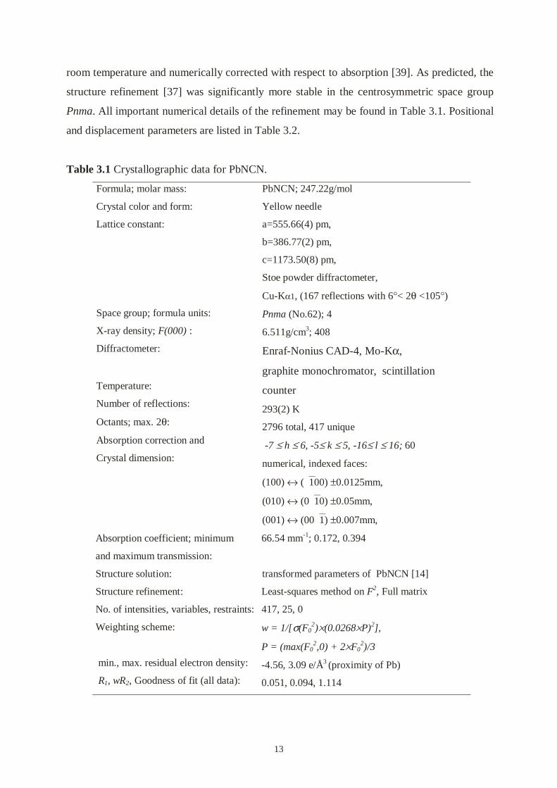
Table 3.1 Crystallographic data for PbNCN.
Formula; molar mass:
Crystal color and form:
Lattice constant:
Space group; formula units:
X-ray density; F(000) :
Diffractometer:
Temperature:
Number of reflections:
Octants; max. 2θ:
Absorption correction and
Crystal dimension:
PbNCN; 247.22g/mol
Yellow needle
a=555.66(4) pm,
b=386.77(2) pm,
c=1173.50(8) pm,
Stoe powder diffractometer,
Cu-Kα1, (167 reflections with 6°< 2θ <105°)
Pnma (No.62); 4
6.511g/cm3; 408
Enraf-Nonius CAD-4, Mo-Kα,
graphite monochromator, scintillation
counter
293(2) K
2796 total, 417 unique
-7 ≤ h ≤ 6, -5≤ k ≤ 5, -16≤ l ≤ 16; 60
numerical, indexed faces:
(100) ↔ (100) ±0.0125mm,
(010) ↔ (010) ±0.05mm,
(001) ↔ (001) ±0.007mm,
Absorption coefficient; minimum
and maximum transmission:
Structure solution:
Structure refinement:
No. of intensities, variables, restraints:
66.54 mm-1; 0.172, 0.394
transformed parameters of PbNCN [14]
Least-squares method on F2, Full matrix
417, 25, 0
Weighting scheme:
min., max. residual electron density:
R1, wR2, Goodness of fit (all data):
w = 1/[σ(F02)×(0.0268×P)2],
P = (max(F02,0) + 2×F0
2)/3
-4.56, 3.09 e/Å3 (proximity of Pb)
0.051, 0.094, 1.114

14
Table 3.2 Positional parameters (all atoms on 4c, y ≡ 1/4) and isotropic a) as well
as anisotropic b) (U23 ≡ U12 ≡ 0) displacement parameters (pm2) for
PbNCN (standard deviations in parentheses).
Atom x z Ueq U11 U22 U33 U13
Pb 0.3929(1) 0.63560(7) 157(3) 126(3) 187(5) 156(4) -5(4)
N(1) 0.333(3) 0.441(2) 235(54) 121(86) 264(158) 321(112) 21(77)
N(2) 0.925(3) 0.361(2) 189(43) 125(83) 32(101) 408(106) 44(99)
C 0.114(4) 0.402(2) 145(59) 148(95) 258(178) 327(114) 25(105)
a) Ueq is a third of the trace of the orthogonalized Uij tensor b) The components Uij refer to a displacement factor of the form exp{-2π2(U11h
2a*2+ ⋅⋅⋅ +2U23 klb*c*)}
3.3. Crystal structure refinement of PbNCN
PbNCN crystallizes in space group Pnma (Z = 4) with a = 555.66(4) pm, b = 386.77(2) pm,
and c = 1173.50(8) pm. The crystal packing of PbNCN shows in Figure 3.1. The crystals form
as needles parallel to the b axis, which is the distance between lead atoms (386.77(2) pm).
The shortest Pb-N(1) contact is nearly parallel to the c axis and forms closely packed sheets.
Identical spirals of slightly asymmetric N-C-N anions pack closely together along the a axis,
being linked by two weaker Pb-N(1) and two weaker Pb-N(2) bonds. There are very weak
bondings outside the closely packed sheets.
Pb
Pb
Pb
N2
N1
Pb
C
N2
N1
N1
C
N2
C
Pb
N1
C
N2
Pb
N2
C
N1
C
N2
C
N1
N1
N2
C
Pb
N1
N2
Pb
Pb
N2
C
N1
Pb
Pb
N2
C
N1
Pb
Pb
N2
N1
Pb
C
N2
N1
N1
C
N2
C
Pb
N1
C
N2
Pb
N2
C
N1
C
N2
C
N1
N1
N2
C
Pb
N1
N2
Pb
Pb
N2
C
N1
Pb
N2
C
N1
cab
Figure 3.1 The crystal packing of PbNCN

15
Figure 3.2 shows a perspective view of the Pb atoms coordination in lead cyanamide. The
lead atom is bonded to one close N(1) atom at 231(2) pm (solid stick), and four second-
nearest N(1) and N(2) atoms at distances of 262(1) pm (open sticks). Two more N(2) atoms
augment the coordination hole at nonbonding distances of 343(1) pm (dashed lines). These
interatomic distances are in very good agreement with the earlier study [14]. It is interesting
to note that when evaluating the five nearest Pb-N distances with the tabulated bond valence
parameter of 222 pm [40], the lead atom already acquires a total bond strength sum (empirical
valence) of 2.14.
Concerning the cyanamide group itself, it is clear that the two C-N bonds differ significantly
in length. While the N(2)-C bond amounts to 115.6(28) pm, the N(1)-C bond length is
129.7(29) pm; although carried out in the centrosymmetric space group, the differentiation
now is even somewhat more pronounced than in the earlier study (117(8) and 125(6) pm
[14]). The C-N(2) distance is shorter than the C-N(1) distance, which is consistent with the
stronger bonding between Pb and N(1). The N-C-N angle lies at 175.6(27)°, linear within
instrumental resolution. Nevertheless, the lowered anionic symmetry is corroborated by the
complex infrared spectrum of PbNCN [41].
N(1)
N(1)
C
N(2)
N(1) N(2) N(2)
N(2)N(2)
Pb
N(1)-C = 129.7 (3) pm
N(2)-C = 115.6(3) pm
N(1)-C-N(2) = 175.6(3)°
Pb-N(1) = 231(2) pm
2 x Pb-N(1) = 262(1) pm
2 x Pb-N(2) = 262(1) pm
2 x Pb-N(2) = 343(1) pm
Pb-N(1)-Pb = 104.8(1)°
Figure 3.2 Perspective view of the Pb coordination in PbNCN using 70% probability
ellipsoids and selected interatomic distances.

16
Although the carbon-nitrogen double bond in neutral organic molecules can be expected to be
about 130 pm [42], they show C-N bond lengths between 119 and 125 pm in the literature
data on all of the D∞h cyanamide anions. The average value is 122 pm, coinciding with the
one in the most common cyanamide, CaNCN. Using this 122 pm as the default length for the
cyanamide double bond, the bond strengths for the shorter and longer C-N bonds in PbNCN
arrive at values of 2.38 and 1.62, perfectly summing up to two double bond strengths (4.00).
Thus, C-N bonding seems to be nicely adjusted in this asymmetrical cyanamide species.
The symmetry reduction of the cyanamide anion obviously goes back to the one short Pb-
N(1) bond which most certainly is polar covalent in nature. In the spirit of Pearson´s acid-base
language, the comparatively soft lead atom (absolute hardness: 8.46 eV) is much better suited
for such bond formation than the respective alkali or alkaline-earth metals (twice as hard or
harder) [43], which interact more ionically. Thus, the cyanamide bonding situation in PbNCN
very much resembles the situation of the singly protonated cyanamide anion in KHNCN [23],
with C-N bond lengths of 117 and 129 pm and an N-C-N angle of 174°.
3.4. Stiffness of cyanamide anion
To investigate the stiffness of the cyanamide anion in more detail, parameter-free (DFT)
quantum-chemical calculations on the NCN2- unit were performed, assuming an N-C bond
length of 122 pm and a linear geometry at the very beginning. The local-density
approximation (VWN parameterization) [44] together with the BLYP gradient correction [45]
was used, and the basis set was of triple-ζ (+2 polarization functions) quality [46], with a 1s
frozen core orbital (program ADF [47]). Figure 3.3 surprisingly reveals that a local symmetry
lowering introduced by a small shift of the central C atom away from the center of the anion
is uncritical. In fact, the observed bond length asymmetry (about 7 pm) in the cyanamide unit
costs about 30 kJ/mol, and this energy loss must be more than counterbalanced by the buildup
of the one short Pb-N bond.
Changes in the N-C-N bond angle are even less important. In Figure 3.4 we show how the
total energy of the experimentally observed cyanamide geometry, with N-C bond lengths of
116 and 130 pm, is affected by a varying N-C-N angle. Less than 2.5 kJ/mol (the room
temperature thermal energy) is needed to bend the linear unit to 175°. The small deviation
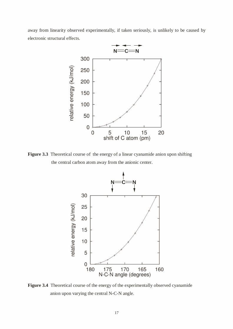
17
away from linearity observed experimentally, if taken seriously, is unlikely to be caused by
electronic structural effects.
Figure 3.3 Theoretical course of the energy of a linear cyanamide anion upon shifting
the central carbon atom away from the anionic center.
Figure 3.4 Theoretical course of the energy of the experimentally observed cyanamide
anion upon varying the central N-C-N angle.
N NC
N NC

18
4. Mercury cyanamide and related complexes
4.1. Synthesis and characterization of mercury cyanamide
4.1.1. Introduction
The group 12 elements, Zn, Cd, and Hg, each have a filled (N-1)d shell plus two Ns electrons.
Because the d electrons are fairly contracted, many chemists approve that these elements are
not classified as typical transition elements. The M2+ ions with their d10 configurations show
no stereo-chemical preferences arising from ligand field stabilization effects. Therefore, they
display a variety of coordination numbers and geometries based on the interplay of
electrostatic forces, covalence and the size factor.
The mercuric cation Hg2+ has a considerably greater polarizing power, which is a distinctly
“soft” cation (absolute hardness: 7.7 eV), showing a strong preference for O, Cl, Br, I, P, S,
Se and certain N-type ligands. It displays coordination numbers of 2 through 6, with a
preference for the lower ones. Its marked preference for linear 2-coordination is a distinctive
feature. An immense number of crystal structures have been reported in recent years,
however, we do not find any non-linear 2-coordination mercury compounds which have been
reported. The short distances between Hg and O, C and N show that there are very strong
covalent bonds formed [48].
The group 12 metal cyanamides can, in fact, very easily be prepared by a solution of M2+ (M=
Zn, Cd, and Hg) and H2NCN in a base condition. We used this method to prepare the
powderous ZnNCN, whose structure agrees with that reported from single crystal [15];
however, polymorphism is a big problem for the structure determination by powder data.
Recently, the Jansen group reported the structure of mercury cyanamide, which was prepared
by double conversion of HgCl2 with sodium cyanamide in aqueous solution [16]. The crystal
structure has been determined using X-ray powder data and neutron diffraction data. It
crystallizes in an orthorhombic space group Pbca with lattice parameters a = 1058.5 pm, b =
651.4 pm, c = 689.3 pm (see Figure 4.1.1). It shows that cyanamide anions are slightly bent
(172.4(7)°) and the two N-C distances are almost equal (N(1)-C =122.8(9) pm, C-N(2) =

19
121.6(8) pm). Cyanamide units are coordinated to mercury atoms at both ends, and form
infinitive zigzag chains. The two Hg-N bond lengths from the “left” and “right” side of
cyanamide unit are very similar (205.6(5) and 207.4(6) pm), and the N-Hg-N angle, 170(3)°,
is close to linearity. Here, we call this phase of mercury cyanamide HgNCN(I).
abc
N2
N2
N2
C
N1
C
N1
C
N1
Hg
Hg
Hg
Hg
Hg
N1
Hg C
Hg
Hg
N2
N1
N1
C
C
N1
C
N2
N2
N2
Figure 4.1.1 The crystal structure of HgNCN(I).
During working on mercury cyanamide, we found another phase of mercury cyanamide. Here,
we call it HgNCN(II). HgNCN(II) was structurally characterized by X-ray analysis using
powder methods with a monoclinic unit cell. Two N-C distances reveal two significantly
different bond lengths in the nearly linear NCN unit, which has been confirmed by vibrational
spectroscopy.
4.1.2. Experimental
HgNCN(II): 5 ml 0.1 M Hg2+ of Hg(NO3)2 or HgCl2 and 5 ml 0.1 M H2NCN were mixed by
stirring at room temperature; a small mount of white precipitate was immediately formed, and
the solution appeared strong acidic. Low concentration of NH3⋅H2O (about 0.1 M) was slowly
and carefully added until pH ≈ 6, a large bulky white precipitate was obtained, filtered,
washed and dried in air. We did not find Hg(NH3)2Cl2, although a white precipitate
Hg(NH3)2Cl2 could be formed during adding aqueous ammonia to HgCl2 solution.
HgNCN(I): 5.5 ml 0.1 M H2NCN was added to 10 ml 0.2 NaOH aqueous solution, then 5 ml
0.1 M Hg2+ of Hg(NO3)2 or HgCl2 was added by stirring at room temperature. A bulky white
precipitation of HgNCN(I) was formed immediately. After keeping the precipitation in the

20
base mother solution for 4 hours, it was filtered off, washed with water and dried in air. For a
high resolution X-ray powder pattern, HgNCN(I) was dried at 120 °C in vacuum.
Elemental analytical data are presented in Table 4.1.1. H and Cl contaminations were not
present in both compounds. These observations provide a first hint that the both compounds
show identical composition.
Table 4.1.1 Elemental analytical data.
Found Calc.
N (%) C (%) N (%) C (%)
HgNCN(I) 11.52(5) 4.91(5) 11.645 4.992
HgNCN(II) 11.64(5) 4.92(5) 11.645 4.994
All attempts, however, to grow good-quality single crystals failed up to now, so that only
powder diffraction data have been recorded. For the unit cell indexing and structure solving,
the XRD pattern was obtained with an Imaging Plate Guinier Camera 670 (Huber). The data
were taken by transmission through a flat powder after 2 hours exposure. For the refinement,
the XRD powder data were collected on a STOE STADI-P diffractometer with a flat sample
holder. The data collection 2θ range: 15-100°, step size: 0.01. All of data was collected at
room temperature.
A FT-IR Avatar 360 E.S.P spectrometer (Nicolet) was used for the determination of the IR
spectra of two phases of HgNCN in KBr discs over the range 400-4000 cm-1. Raman spectra
were recorded on a Bruker IFS 66v/S spectrophotometer with FRA 106/S, Nd-YAG laser, λ =
1064 nm, 75–80 mW, 256 scans, and samples were run as powders in spinning capillary
tubes.
4.1.3. Structure determination from powder data
Structure solving
A peak marked pattern of the HgNCN(II) collected by the Imaging Plate Guinier Camera 670
(Huber) is shown in Figure 4.1.2, and it was indexed on the 33 strong reflections using the

21
auto-indexing program STOE WinXPow Version 1.04 [28]. The program gave a monoclinic
solution with a figure of merit M = 46.9. The solution from WinXPow was refined using the
program Fullprof [32]. The unit-cell parameters are a = 685.22(4), b = 349.08(2), c =
555.31(2) pm and β = 113.21(8)°. The systematic absences [hkl, h + k = 2n; h0l, h = 2n; 0kl, k
= 2n; hk0, h + k = 2n; 0k0, k = 2n; h00, h = 2n] were consistent with space groups Cm (No. 8)
and C2/m (No. 12).
Figure 4.1.2 X-ray diffraction pattern of HgNCN(II) measured by Imaging Plate Guinier
camera, and indexed in C2/m.
The extracted intensities were used as input for the direct-methods program SIRPOW92 [29].
We used C2/m as input space group for the structure solving. An E-map computed for the
solution with the best R (3.8%) revealed the position of the Hg, C and N atoms. Carbon and
nitrogen atoms form NCN dumb-bells with the C atom on a twofold axis. This structural
model was used as a starting model for Rietveld refinement using Fullprof.
Refinement of HgNCN(II) in space group Cm
For the refinement we used the data collected by a STOE STADI-P diffractometer. The
background was corrected by the program BACKPOW [34]. The data were re-indexed. From
the X-ray powder diffraction pattern it shows there is a partial contribution of the

22
orthorhombic phase. We tried to separate this two phases by control of pH value of the
reaction, without success. Besides the peaks of HgNCN(II), the all rest peaks can be indexed
with the orthorhombic cell of HgNCN(I). We used the orthorhombic phase result that the
Jansen group reported as phase I, then determined the structure of monoclinic of HgNCN as
phase II.
Recognizing that the unit-cell volume was consistent with the presence of two HgNCN
molecules, the space group was taken as Cm on the working assumption that the atoms were
unlikely to be all on special positions in the asymmetric unit. Once the appropriate structure
had been known, we used it as a starting point to refine the structure of HgNCN(II). All atoms
were refined isotropically. Neutral atomic scattering factors, as stored in Fullprof, were used
for all atoms. No corrections were made for absorption. The final Rietveld refinement of the
complete structure converged with Rwp = 0.10 and Rp = 0.08. Experimental details, refined
atomic coordinates and selected geometric parameters are given in Table 4.1.2, 4.1.3 and
4.1.4, respectively. The final Rietveld difference plot is shown in Figure 4.1.3.
Figure 4.1.3 X-ray diffraction pattern of HgNCN(II) measured by STOE STADI-P
diffractometer. Observed, calculated and difference profile for the final Rietveld
refinement. The position of the Bragg reflections: HgNCN(II) in Cm (up),
HgNCN(I) (down).

23
Table 4.1.2 Experimental details for data collection and Rietveld refinement for Cm.
Crystal data:
Chemical formula; molar mass; Color:
Lattice constant:
Space group; formula units:
Temperature:
Instrument:
Scan range; step:
Refinement:
Program:
Zero point:
Profile function:
u, v, w parameters:
No. of reflections used in refinement:
No. of parameters used:
Content
Rp; Rwp:
RBragg
HgNCN, 240.614 g/mol, White fine powder
a = 685.337(1) pm,
b = 349.181(1) pm,
c = 555.269(1) pm,
β = 113.21(8)°.
Cm (No. 8); 2
293(2)K
STOE STADI-P diffractometer with Cu-Kα1
radiation
15°< 2θ < 99.8°; 0.01
FULLPROF
0.0054(4)
pseudo-Voigt
0.210(1)
-0.030(6)
0.0239(1)
80 (phase II), 243 (phase I)
20
94.51% HgNCN(I), 5.49% HgNCN(II)
0.076; 0.096
0.053
RBragg = ∑ Ii(obs)2 –Ii(calc)2/ Ii(obs)2;
Rwp = {∑ wi [yi(obs)2 –yi(calc)]2 / ∑ wi[yi(obs)]2 }1/2;
Rp = ∑ yi(obs) –yi(clac) / ∑ yi(obs).
Table 4.1.3 Fractional atomic coordinates and equivalent isotropic displacement
parameters (Å2) for HgNCN (II).
Atom x y Z Biso
Hg 0 0 0 2.67(5)
N(1) 0.550(2) 0 0.851(3) 1.6(5)
C 0.525(2) 0 0.618(3) 1.5(5)
N(2) 0.498(6) 0 0.401(3) 0.2(5)

24
Table 4.1.4 Selected bond distances (pm) and bond angles (o) for HgNCN (II)
Hg-N(1) 204(3) × 2
Hg-N(2) 283 (3)
Hg-C 273(3)
N(1)-C 130(3)
C-N(2) 114 (3)
N(1)-C-N(2) 173.1(15)
Hg-N(1)-Hg 117.5(15)
N(1)-Hg-N(1) 117(1)
c
a
b
Hg Hg
Hg
Hg
N1 C N2
Hg
Hg
N1 C N2
c ab
Figure 4.1.4 View of the crystal structure of HgNCN(II) in Cm. The large circles represent
mercury atoms, and the small circle units represent NCN.
The structure is shown in Figure 4.1.4. Hg atoms and cyanamide units are forming a layer-like
structure along the a direction. Cyanamide units are slightly bent out of the bc plane. The
distance of Hg-N(1) is 204 pm. Each N(1) bonds to two Hg atoms and each Hg atom bonds to

25
two N(1) from one side. These distances are a good agreement with related 2-coordineted
mercury and nitrogen compounds [49, 50, 51]. But, the unusual one-side Hg-N(1)-Hg zigzag
chains (N(1)-Hg-N(1) = 114°) is hard to be accepted. The NCN unit has two different bond
lengths (N(1)-C = 130, C-N(2) = 114 pm) and the angle of N(1)-C-N(2) is 173°.
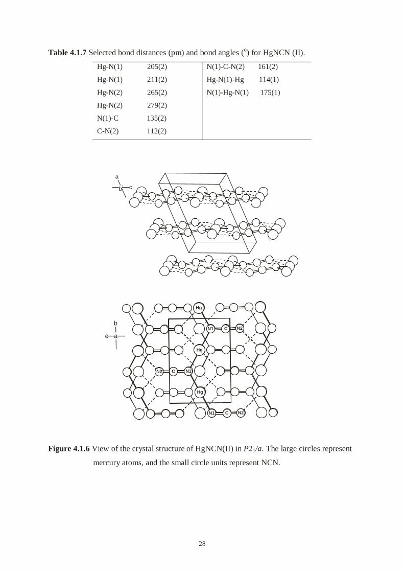
Refinement of HgNCN(II) in space group P21/a
The angle of N(1)-Hg-N(1) in HgNCN(II) is 114° in Cm space group. It may be unusual. We
simply doubled the b axis of the unit cell in order to result out linear 2-coordinated mercury
atoms. The ideal effect of unit cell doubling (in bc plane) should be:
N2
N2
C
C
N1
N1
Hg
Hg
Hg
N2
N2
C
C
N1
N1
Hg
Hg
Hg
doubling in b direction
Hg
Hg
Hg
N1CN2
N2CN1
Hg
Hg
Hg
N1CN2
N2CN1
Hg
Hg
Hg
Cm P21/a
The refinement proceeded as before. The final Rietveld refinement of the complete structure
converged with Rwp = 0.10 and Rp = 0.08. Experimental details, refined atomic coordinates
and selected geometric parameters are given in Table 4.1.5, 4.1.6 and 4.1.7, respectively. The
final Rietveld difference plot is shown in Figure 4.1.5.
The structure is shown Figure 4.1.6. As expected, the arrangement of Hg atoms keeps almost
the same as that in Cm, and the structure is still layer-like along the a direction. Each mercury
is attached diagonally to two N(1) by the N(1)-Hg-N(1) angle of 175(1)°, and the distances of
Hg-N(1) are 205(2) and 211(2) pm. The distances of Hg-N(2) (265(2), 279(2) pm) belong to
the nonbonding region. In fact, only one end of the NCN unit is covalently bonded to mercury
atoms, and mercury atoms and cyanamide units built up of infinite zigzag chains Hg-N(1)-
Hg-N(1) running parallel to the b axis. NCN unit exhibits two strongly different bond lengths
(N(1)-C = 135(2), C-N(2) = 112(2) pm) and the angle of N(1)-C-N(2) is 161(1)°, which is
significantly more bent than in other cyanamide compounds.

26
Figure 4.1.5 X-ray diffraction pattern of HgNCN(II) measured by STOE STADI-P
diffractometer. Observed, calculated and difference profile for the final Rietveld
refinement. The position of the Bragg reflections: HgNCN(II) in P21/a (up),
HgNCN(I) (down).

27
Table 4.1.5 Experimental details for data collection and Rietveld refinement for P21/a.
Crystal data:
Chemical formula; molar mass; Color :
Lattice constants:
Space group; formula units:
Temperature:
Instrument:
Scan range; step:
Refinement:
Program:
Zero point:
Profile function:
u, v, w parameters:
No. of reflections used in refinement:
No. of parameters used:
Content
Rp; Rwp:
RBragg
HgNCN; 240.614 g/mol; White fine powder
a = 685.337(1) pm,
b = 698.362(1) pm,
c = 555.269(1) pm,
β = 113.21(8)°.
P21/a (No.14); 4
293(2)K
STOE STADI-P diffractometer with Cu-
Kα1 radiation
15°< 2θ <99.8°; 0.01
FULLPROF
0.0055(3)
pseudo-Voigt
0.298(10)
-0.090(8)
0.0415(1)
253(phase II), 243(phase I)
23
96.51% HgNCN(I), 3.49% HgNCN(II)
0.077; 0.098
0.068
RBragg = ∑ Ihkl(obs)2 –Ihkl(clac)2/ Ihkl(obs)2;
Rwp = {∑ wi [yi(obs)2 –yi(clac)]2 / ∑ wi[yi(obs)]2 }1/2;
Rp = ∑ yi(obs) –yi(clac) / ∑ yi(obs).
Table 4.1.6 Fractional atomic coordinates (all atoms at 4e) and equivalent isotropic
displacement parameters (Å2) for HgNCN (II).
Atom x y z Biso
Hg 0.758(1) 0.134(1) 0.995(1) 1.33(2)
N(1) 0.218(1) 0.127(3) 0.185(1) 1.14(18)
C 0.231(3) 0.120(4) 0.434(2) 2.89(18)
N(2) 0.230(1) 0.122(3) 0.654(1) 1.66(18)
28
Table 4.1.7 Selected bond distances (pm) and bond angles (o) for HgNCN (II).
Hg-N(1) 205(2)
Hg-N(1) 211(2)
Hg-N(2) 265(2)
Hg-N(2) 279(2)
N(1)-C 135(2)
C-N(2) 112(2)
N(1)-C-N(2) 161(2)
Hg-N(1)-Hg 114(1)
N(1)-Hg-N(1) 175(1)
a
b c
N2 C N1
Hg
Hg
Hg
N1
N1
C
C
N2
N2
N2 C N1
Hg
Hg
Hg
N1
N1
C
C
N2
N2
c a
b
Figure 4.1.6 View of the crystal structure of HgNCN(II) in P21/a. The large circles represent
mercury atoms, and the small circle units represent NCN.

29
The Structure Discussion
Before starting discussion of the structure of HgNCN(II), I would like to tell a story. In 1956,
Roth reported the crystal structure of HgO [52]. HgO was refined in an orthorhombic unit cell
(a = 329.6, b = 351.3 and c = 550.4 pm) with space group Imm2. The structure was described
that Hg-O-Hg-O formed infinite zigzag chains. The chains packed to form planar layers. The
distance of Hg and O was 201.5 pm and the angle of O-Hg-O was about 110°. Half a year
later, Anrivillins published another paper to describe the structure of HgO [53]. He simply
doubled the a axis and refined the structure with space group Pnma. The mercury atoms have
the same arrangement as before. Hg and O form also infinite zigzag chains and the Hg-O
distance is 203 pm, but O-Hg-O is 179 ± 3°, linear. The relative compounds show also linear
O-Hg-O [54]. Most of chemists would like to accept the structure of Anrivillin.
From crystallographic view, both refinements of HgNCN(II) are practically identical and
acceptable, but not perfect. As a chemist, I would rather believe the structure of HgNCN(II) in
P21/a. Until now we have not found a 2-coordinated Hg compound that has a non-linear
structure. The electronic structure calculations (LDA as well GGA) show that HgNCN(II) in
P21/a has a lower energy and is not stable in Cm.
There may be another possibility. The arrangement of mercury atoms is well stochastic.
Therefore, we can observe nice X-ray diffraction. The NCN units are slightly disordered,
although they do bond to mercury from one side. That may be a reason that “soft” cation
metal cyanamides are so difficult to determine the structures. So far no structure of CuNCN
has been reported.
A comparison of the coherent scattering amplitudes of mercury and nitrogen for neutrons and
X-rays shows that neutron diffraction should be more suited for the structure determination
(scatting lengths: Hg/N ≈1.35 in neutron diffraction; Hg/N ≈ 10 in X-ray diffraction). It is
difficult to locate the positions of light like atoms as N and C by X-ray diffraction techniques.
Recently, we collected the neutron diffraction data, and the subsequent time-of-flight (TOF)
neutron data show that the whole structure does not possess a C-centering but must be
described with a primitive unit cell (P21/a). The result of neutron data refinement is a good
agreement with of X-ray data refinement.

30
4.1.4. Results and discussion
Generally, the preparations of HgNCN can be described as:
Hg2+ + H2NCN OH¯ HgNCN + 2H+
Here, if we did not add NH3·H2O, only a small amount of precipitate was obtained, which is
badly crystallized, since H2NCN hydrolyzes to form urea in the presence of a strong or mild
acid. If we added a small mount of NH3·H2O in H2NCN solution before mixing, we could
obtain different percentages of two HgNCN phase mixtures.
If we first add a base to H2NCN solution, the NCN2- anion is formed and the NCN2- unit tends
to D∞h symmetry. Hg2+ can bond with N (1) and N(2) from both sides of NCN unit and
mercury cyanamide crystallizes in an orthorhombic phase. If we mix the Hg2+ and the H2NCN
at first, Hg2+ prefers to bond with N(1), and an intermediate “Hg(HNCN)2” may be formed.
Upon adding a base to the solution, like as NH3⋅H2O, the other hydrogen is removed from
N(1), and the Hg2+ bonds directly with N(1) again and the compound adopts a monoclinic
phase. The NCN unit shows asymmetric.
It seems to be impossible to transfer one phase to another phase of HgNCN by only varying
temperature (-170 ∼ 220 °C). Before they acquire enough energy to phase transition, they
already decompose. We found that thermal decomposition temperatures of these two phases
are the same, about 230 °C. We used a long glass ampoule carefully to heat one side at 240 °C
and kept the other side at room temperature. In the colder side we obtained metallic mercury,
and a white carbon nitrogen polymer was obtained in the hotter side. It will be very
interesting further to study this nitrogen rich polymer.
If we treated the HgNCN(II) powder in a 1 M NaOH solution at 60 °C for about 8 hours,
most of HgNCN(II) changed to HgNCN(I), and we did not observe that HgNCN(II) dissolved
in the solution in this transfer process. Here, OH- may act as a catalyst. This finding points
into direction of HgNCN(I) being the thermodynamically stable phase. The theoretical
calculations prove that HgNCN(I) is more stable by about 7 kJ/mol than HgNCN(II) based
on either LDA or the GGA.

31
In fact, we can not synthesize pure HgNCN(II) phase since there is always a trace amounts (
about 5 %) of HgNCN(I). The crystallographic density of HgNCN(I), 6.789 g/cm3, is 3.5%
higher than that of HgNCN(II), 6.540 g/cm3; usually - but not always – higher stability goes
with better packing. Both phases of HgNCN are electrical insulators.
Figure 4.1.7 Infrared spectra of HgNCN(I) and HgNCN(II).
Figure 4.1.8 Raman spectra of HgNCN(I) and HgNCN(II).
HgNCN(II)
HgNCN(I)
20
30
40
50
60
70
T%
500 1000 1500 2000 2500 3000 3500
Wavenumber (cm-1)

32
IR and Raman spectra of both phases are shown in Figure 4.1.7 and Figure 4.1.8. Both
compounds show strong absorption in the region 2100-1900 cm-1, the typical band sequence
of the three-atomic arrangement (whether symmetric or asymmetric). The asymmetric
stretching frequency is already split for HgNCN(I) (2031 and 1948 cm-1), and the splitting is
even larger in HgNCN(II) (2097 and 1949 cm-1). Maybe, in HgNCN(I) the absorption of the
νas is splitted as a result of the two crystallographically independent nitrogen atoms in the
NCN unit, and the larger splitting in HgNCN(II) causes by more asymmetry of two C-N
bonds in NCN unit. As expected by the selection principle for molecules containing an
inversion center, the asymmetric stretching and the deformation vibration of NCN unit are
allowed in IR spectra, whereas the symmetric stretching mode is Raman active. If the
symmetry of a three-atomic unit is lowered from D∞h to C2v or Cs, the symmetric stretching
vibration can be observed in the IR. The symmetric stretching mode is already IR-observable
for HgNCN(I) at 1219 cm-1 but stronger for HgNCN(II), also at 1219 cm-1, because of the
lower symmetry; in the meantime the symmetric stretching vibration of HgNCN(I) is also
observable in Raman spectra. The vibration frequencies of related metal cyanamide
compounds are given in Table 4.1.8. It is obvious that the NCN unit exits in a form of
N=C=N in HgNCN(I) and N≡C-N in HgNCN(II).
Table 4.1.8 Vibration frequencies of metal cyanamide compound.
Frequency in cm-1 ReferenceCompound
νs(NCN) νas(NCN) δ(NCN)
MgNCN 1301(Raman) 2114 681 12
ZnNCN not IR active 2148 660 this work
HgNCN(I) 1219 (Raman) 2031, 1948 667 this work, 16
HgNCN(II) 1219 (IR) 2097,1949 749(?), 667 this work
PbNCN 1307 (IR) 1929 626 this work, 14
Ag2NCN 1281 (IR) 2002 631 this work, 17

33
4.2. Structures of Hg2(NCN)Cl2 and Hg3(NCN)2Cl2
4.2.1. Introduction
The synthesis of lower dimension frameworks is currently of great interest as a method for the
construction of new supramolecular architectures [55]. The many important properties of 2D
frameworks arise from their structures and topology. The various organic or inorganic spacer
ligands coordinating to metal cations have been explored; here, N-containing spacers such as
pyridine and cyano ligands were typically and successfully used [56]. On the other side, the
probably smallest N, N’-type ligand the NCN2- anion, either in carbodiimide N=C=N2- or
in cyanamide N-C≡N2- form had not been expected to form pore or channel structures. It is
also clear that the metal cation plays a decisive role for the total structure since its preferences
for coordination number, coordination geometry and chemically matching ligands must be
taken into account. For Hg(II), twofold linear coordination is observed throughout [48] but
there are also some examples of 4- and 6-coordinated Hg(II) centers with longer interatomic
distances [57]. To the best of our knowledge, no metal-cyanamide polymeric networks have
been reported so far.
In an attempt to grow high-quality HgNCN single crystals by slow diffusion, we unexpectedly
found two such inorganic polymers involving Hg2+ and NCN2- in crystalline form. Here, we
describe both synthesis and crystal structures of Hg2(NCN)Cl2 and Hg3(NCN)Cl2 and further
discuss the coordination situation as well as the two-dimensional frameworks observed in the
two compounds. The seemingly rather flexible coordination of the NCN2- anion and Hg2+
cation allows for differing capacities to host guest species within the framework.
4.2.2. Synthesis
A Ø2 cm 4A pottery filter (the finest filter) was fixed in the middle of a “U form” glass. 5 ml
0.1 mol/l HgCl2 solution and 5 ml 0.1 mol/l H2NCN solution were carefully separated by the
filter from left and right. After one day there was white powder observed at both sides. The
white powder is HgNCN(II). During washing of the HgNCN(II), we found two kinds of
differently shaped colorless crystals. The regular hexagon crystals are Hg2(NCN)Cl2 and
found mostly at the HgCl2 side. The other rectangular plate crystals are Hg3(NCN)2Cl2 and
34
found mostly at the H2NCN side. Despite several repeated preparations, we could not get pure
products. After drying in air, the selected crystals were well suitable for X-ray single crystal
measurement.
4.2.3. Single crystal structure determination
Selected crystals of Hg2(NCN)Cl2 (size: 0.033 × 0.042 × 0.097 mm3) and Hg3(NCN)2Cl2
(size: 0.07 × 0.04 × 0.02 mm3) were mounted on the tips of glass fibers and transferred to a
Bruker SMART CCD diffractometer with graphite monochromatized Mo-Kα radiation. The
complete sets of intensities were measured at room temperature. Unfortunately, after
inspection of a number of crystals, all Hg3(NCN)2Cl2 crystals turned out to be twinned.
The structures were solved by direct methods and refined by alternating cycles of difference
Fourier syntheses and full-matrix least-squares refinements [38].
4.2.4. The crystal structure of Hg2(NCN)Cl2
The crystallographic data of the final refinement of Hg2(NCN)Cl2 are summarized in Table
4.2.1, whereas Tables 4.2.2 and 4.2.3 give positional and isotropic as well as anisotropic
displacement parameters.

35
Table 4.2.1 Crystallographic data for Hg2(NCN)Cl2 [62].
Formula; molar mass:
Crystal color and form:
Lattice constant:
Hg2(NCN)Cl2; 512.11 g/mol
Colorless hexagon
a = 806.7(1) pm
b = 907.1(2) pm
c = 788.0(1) pm
β = 106.446(3)°
Cell volume:
Space group; formula units:
X-ray density; F(000) :
Diffractometer:
Temperature:
553.0(2) ×106 pm3
P21/c (No.14); 4
6.151 g/cm3; 856
Bruker SMART CCD diffractometer with
Mo-Kα radiation, graphite monochromator
293(1) K
Number of reflections:
Octants; max. θ:
Absorption coefficient; absorption
correction and crystal dimension:
Structure solution:
Structure refinement:
No. of intensities, variables, restraints:
4569 (Req = 0.0557)
1374 independent reflections
-10≤ h ≤10, -11≤ k ≤ 12, -10≤ l ≤ 10; 28.30°
56.280 mm-1;
numerical, indexed faces:
(100) ↔ (100) -0.014 ↔ 0.019 mm,
(010) ↔ (010) -0.022 ↔ 0.020 mm,
(001) ↔ (001) -0.042 ↔ 0.055 mm.
Direct Methods (SHELXS-97)
Least-squares method on F2, Full matrix
1374, 67, 0
Weighting scheme: w = 1/[σ(F02) × (0.0581× P)2],
where P = (max(F02,0) + 2× F0
2)/3
Min., max. residual electron density:
R1, wR2, Goodness of fit(all data):
-1.478, 6.961 e/Å3 (proximity of Hg)
0.0463 , 0.1103, 0.888

36
Table 4.2.2 Positional parameters and isotropic displacement parameters (Ueq in pm2, a third
of the trace of the orthogonalized Uij tensor) for Hg2(NCN)Cl2 (standard
deviations in parentheses).
Atom Wyckoff-Site x y z Ueq
Hg(1) 4e 0.1093(1) 0.82183(8) 0.2337(1) 259(2)
Hg(2) 2d 1/2 0 1/2 239(3)
Hg(3) 2b 1/2 0 0 541(5)
Cl(1) 4e 0.4907(7) 0.7527(6) 0.2527(7) 306(11)
Cl(2) 4e 0.8009(6) 0.0217(6) 0.0761(7) 319(12)
N(1) 4e 0.234(2) 0.990(2) 0.399(2) 225(36)
C 4e 0.135(3) 0.086(2) 0.424(2) 228(46)
N(2) 4e 0.035(2) 0.176(2) 0.453(2) 271(38)
Table 4.2.3 Anisotropic displacement parameters (pm2) for Hg2(NCN)Cl2 (standard
deviations in parentheses). The components Uij refer to a displacement factor
of the form exp{-2π2(U11h2a*2+ ⋅⋅⋅ +2U23 klb*c*)}.
Atom U11 U22 U33 U23 U13 U12
Hg(1) 258(4) 207(4) 283(4)) -35(4) 28(3) -47(4)
Hg(2) 145(5) 270(6) 278(6) -36(5) 22(4) -3(5)
Hg(3) 196(6) 931(14) 478(9) -57(9) 65(6) -140(8)
Cl(1) 362(28) 258(28) 292(26) -19(22) 84(22) 32(24)
Cl(2)
N(1)
C
N(2)
236(26)
180(80)
358(123)
184(82)
294(29)
166(87)
178(104)
291(97)
384(30)
358(99)
125(100)
280(90)
36(24)
-10(73)
53(77)
51(84)
17(23)
124(73)
34(92)
29(71)
-10(23)
-27(72)
7(93)
52(83)
Table 4.2.4 Selected interatomic distances (pm) and bond angles (o) for Hg2(NCN)Cl2.
Hg(1)-N(1) 207.0(16)
Hg(1)-N(2) 207.4(16)
Hg(2)-N(1) 206.6(16) (2×)
Hg(2)-Cl(1) 295.9(5) (2×)
Hg(3)-Cl(2) 233.8(5) (2×)
N(1)-C 124(3)
C-N(2) 122(3)
N(1)-Hg(1)-N(2) 172.4(7)
N(1)-Hg(2)-N(1) 180
C-N(1)-Hg(1) 113.5(14)
C-N(1)-Hg(2) 123.5(15)
C-N(2)-Hg(1) 124.4(15)
Hg(2)-N(1)-Hg(1) 123.0(8)
N(1)-C-N(2) 177(2)
Cl(2)-Hg(3)-Cl(2) 180
Cl(1)-Hg(2)-Cl(1) 180

37
Figure 4.2.1 ORTEP view of Hg2(NCN)Cl2 showing the atom labeling scheme. The thermal
ellipsoids are drawn at the 90% probability level; the incorporated HgCl2
molecule has been omitted for clarity.
The coordination structure of Hg2(NCN)Cl2 with labeling is shown in Figure 4.2.1. Bond
distances and angles are given in Table 4.3.4. Generally, X-ray structural determination
reveals that mercury atoms and cyanamide units form an unprecedented two-dimensional
framework (Figure 4.2.2). Each Hg(1) bonds to two nitrogen atoms from two different NCN
units (Hg(1)-N(1) = 207.0(16) pm, Hg(1)-N(2) = 207.4(16) pm) in a nearly linear
arrangement (N(1)-Hg(1)-N(2) = 172.4(7)°). Hg(1)s and NCN units form infinite zigzag
chains along the b axis, NCN unit acts as a µ-bridging ligand. Each Hg(2) bonds linearly to
two nitrogen atoms also, but only with N(1) from two NCN units (Hg(2)-N(1) = 206.6(16)
pm, N(1)-Hg(2)-N(1) = 180°). All distances of these mercury and nitrogen are almost
identical (207 pm) and very good agree with those found in related materials (Hg-N: 205-211
pm) [16, 50, 51].

38
(a)
abc
(b)
abc
(c)
cab
Figure 4.2.2 Packing of Hg2(NCN)Cl2. Perspective view of the sheet packing down the a, b
and c axis. The circles represent in order of decreasing size Hg, Cl and N, C.
Black, gray and white circles represent Hg(1), Hg(2) and Hg(3), respectively.

39
In Hg2(NCN)Cl2, the distance between Hg(3) and Cl(2) is 233.8 pm and the angle of Cl(2)-
Hg(3)-Cl(2) is 180°, which is very similar to that of HgCl2 (Hg-Cl: 227 and 229 pm, Cl-Hg-
Cl: 178.6°) [58]. We may simply consider that Hg(3) and Cl(2) just forms a molecular HgCl2.
Cl(2)s have very weak interaction with Hg(1) (Hg(1)-Cl(2): 305.5(5) pm). The Cl(1) anions
have very week inter-action to all mercury atoms, in which the distances of Cl(1) and each of
Hg(2) or Hg(3) are from 295.9(5) to 309.(5) pm. Thus, Cl(1) can be considered as free
chloride ions, because mercury atoms prefer to covalently bond to nitrogen atoms. If all these
weak interactions are considered, each Hg is coordinated with four chlorides.
Within the NCN unit, the distances of N(1)-C (124(3) pm) and C-N(2) (122(3) pm) are
practically equal and N(1)-C-N(2) is nearly linear (177(2)°); however, a N(1) bonds to two
mercury atoms and a N(2) bonds only to one mercury atom. These N-C distances agree with
those in HgNCN(I) which arrive at 122 and 123 pm [16].
It is interesting to note that a Hg(2) links to two nitrogen atoms from two chains formed by
Hg(1) and NCN units (Figure 4.2.1 and 2(a)). Cl(1)s locate between the sheets formed by two
times crossed Hg(1)-NCN-Hg(1) chains and Hg(2)s (Figure 4.2.2(b)). Remarkably, it is
shown 20-membered rings by sharing edges and the HgCl2 guest species occupy in the middle
of the ring along the a axis channels (Figure 4.2.2(c)).
4.2.5. The crystal structure of Hg3(NCN)2Cl2
The structure of Hg3(NCN)2Cl2 was solved and refined in the acentric space group Pca21. We
followed step by step the SHELX-97 program for the refinement. The program suggested a
possible racemic twin and a twin refinement. We tried to give all atoms inverted positions, but
the program still gave the same suggestion and no higher symmetry space group was found.
Then we refined it as a twin structure by use of the corresponding twin law (SHELXL-97)
[38]; the BASF scale factor was 0.47(2) and R1 was not significantly reduced from 0.0433 to
0.0380. This is not too surprising since the heavy atom positions (like Hg) keep in the same
position.
Because of the strong absorption of mercury (µ = 55.463 mm-1 in Hg3(NCN)2Cl2), we also
tried face-index absorption correction besides the SADABS absorption correction [59], but it
gave no better results. First, we tried to refine all of atoms anisotropically, but the
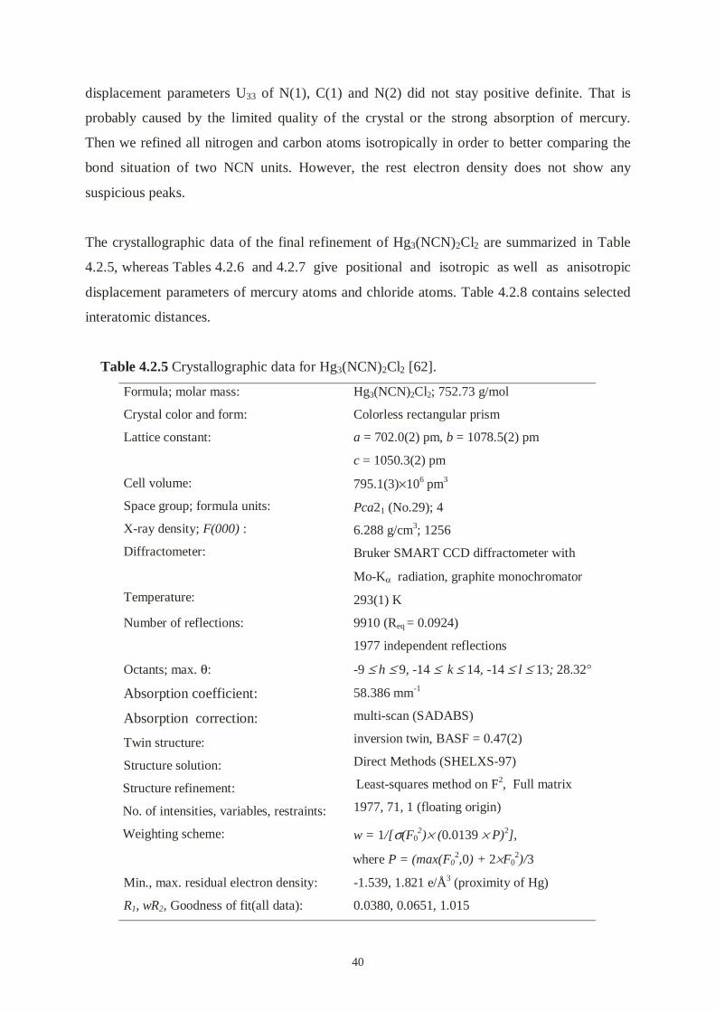
40
displacement parameters U33 of N(1), C(1) and N(2) did not stay positive definite. That is
probably caused by the limited quality of the crystal or the strong absorption of mercury.
Then we refined all nitrogen and carbon atoms isotropically in order to better comparing the
bond situation of two NCN units. However, the rest electron density does not show any
suspicious peaks.
The crystallographic data of the final refinement of Hg3(NCN)2Cl2 are summarized in Table
4.2.5, whereas Tables 4.2.6 and 4.2.7 give positional and isotropic as well as anisotropic
displacement parameters of mercury atoms and chloride atoms. Table 4.2.8 contains selected
interatomic distances.
Table 4.2.5 Crystallographic data for Hg3(NCN)2Cl2 [62].
Formula; molar mass:
Crystal color and form:
Lattice constant:
Hg3(NCN)2Cl2; 752.73 g/mol
Colorless rectangular prism
a = 702.0(2) pm, b = 1078.5(2) pm
c = 1050.3(2) pm
Cell volume:
Space group; formula units:
X-ray density; F(000) :
Diffractometer:
Temperature:
795.1(3)×106 pm3
Pca21 (No.29); 4
6.288 g/cm3; 1256
Bruker SMART CCD diffractometer with
Mo-Kα radiation, graphite monochromator
293(1) K
Number of reflections:
Octants; max. θ:
Absorption coefficient:
Absorption correction:
Twin structure:
Structure solution:
Structure refinement:
No. of intensities, variables, restraints:
9910 (Req = 0.0924)
1977 independent reflections
-9 ≤ h ≤ 9, -14 ≤ k ≤ 14, -14 ≤ l ≤ 13; 28.32°
58.386 mm-1
multi-scan (SADABS)
inversion twin, BASF = 0.47(2)
Direct Methods (SHELXS-97)
Least-squares method on F2, Full matrix
1977, 71, 1 (floating origin)
Weighting scheme: w = 1/[σ(F02)× (0.0139 × P)2],
where P = (max(F02,0) + 2×F0
2)/3
Min., max. residual electron density:
R1, wR2, Goodness of fit(all data):
-1.539, 1.821 e/Å3 (proximity of Hg)
0.0380, 0.0651, 1.015

41
Table 4.2.6 Positional parameters (all atoms on 4a) and isotropic displacement parameters
(Ueq in pm2 , a third of the trace of the orthogonalized Uij tensor) for
Hg3(NCN)2Cl2 (standard deviations in parentheses).
Atom x y Z Ueq
Hg(1) 0.7065(1) 0.08577(7) 0.95711(7) 296(2)
Hg(2) 0.98652(9) 0.44557(6) 0.00562(8) 236(2)
Hg(3) 0.23031(9) 0.20550(6) 0.64448(7) 247(2)
Cl(1) 0.0697(6) 0.2006(4) 0.8980(5) 275(9)
Cl(2) 0.6336(6) 0.2972(4) 0.0809(5) 324(11)
N(1) 0.155(2) 0.017(1) 0.603(1) 193(31)
C(1) 0.105(2) 0.956(2) 0.702(2) 209(38)
N(2) 0.044(2) 0.906(2) 0.788(1) 231(34)
N(3) 0.150(2) 0.385(1) 0.158(1) 164(28)
C(2) 0.159(2) 0.440(2) 0.260(2) 255(41)
N(4) 0.182(2) 0.498(2) 0.357(2) 316(40)
Table 4.2.7 Anisotropic displacement parameters (pm2) for Hg3(NCN)2Cl2 (standard
deviations in parentheses). The components Uij refer to a displacement factor
of the form exp{-2π2(U11h2a*2+ ⋅⋅⋅ +2U23 klb*c*)}.
Atom U11 U22 U33 U23 U13 U12
Hg(1) 365(4) 285(4) 239(4) 13(4) -38(3) 26(3)Hg(2) 280(3) 207(3) 220(3) 37(3) -18(3) 41(3)Hg(3) 321(3) 114(3) 305(4) -13(3) 11(3) -33(3)Cl(1) 350(23) 244(23) 231(21) -76(20) 25(19) 8(19)Cl(2) 396(25) 133(22) 442(28) -40(23) 99(23) -49(21)
Table 4.2.8 Selected interatomic distances (pm) and bond angles (o) for Hg3(NCN)2Cl2.
Hg(1)-N(2)
Hg(1)-N(1)
Hg(1)-Cl(2)
Hg(1)-Cl(1)
Hg(2)-N(4)
Hg(2)-N(3)
Hg(2)-Cl(1)
Hg(3)-N(3)
Hg(3)-N(1)
Hg(3)-Cl(2)
Hg(3)-Cl(1)
Hg(3)-Cl(1)
211.4(15)
213.0(14)
267.4(5)
290.2(5)
205.2(16)
207.3(13)
293.2(5)
211.5(13)
214.4(14)
282.0(4)
289.2(5)
294.5(5)
C(1)-N(2)
C(1)-N(1)
N(3)-C(2)
C(2)-N(4)
113(2)
128(2)
123(2)
120(2)
N(1)-Hg(3)-N(3)
N(3)-Hg(2)-N(4)
N(1)-Hg(1)-N(2)
Hg(1)-N(1)-Hg(3)
Hg(2)-N(3)-Hg(3)
C(1)-N(1)-Hg(1)
C(1)-N(1)-Hg(3)
C(1)-N(2)-Hg(1)
C(2)-N(3)-Hg(2)
C(2)-N(3)-Hg(3)
C(2)-N(4)-Hg(2)
N(1)-C(1)-N(2)
N(3)-C(2)-N(4)
168.2(5)
178.2(6)
151.1(6)
121.7(6)
117.2(6)
116.2(12)
113.0(12)
116.3(13)
123.5(13)
118.6(13)
136.2(14)
173.5(19)
175(2)

42
Figure 4.2.3 Thermal ellipsoid plot (90% probability) of the atomic coordination in
Hg3(NCN)2Cl2 with the atom labeling scheme.
Figure 4.2.3 shows the coordination situation of Hg3(NCN)2Cl2. There are two NCN units in
Hg3(NCN)2Cl2. In the N(1)-C(1)-N(2) unit, the two bond distances of nitrogen and carbon are
definitely different (N(1)-C(1): 128(2) pm, C(1)-N(2): 113(2) pm), comparable with those in
PbNCN and HgNCN(II). However, in the N(3)-C(2)-N(4) unit, the two bond distances of
nitrogen and carbon are almost equal (N(3)-C(2): 123(2) pm, C(2)-N(4): 120(2) pm), close to
those found in HgNCN(I) [16]. Both NCN units are close to linear (N(1)-C(1)-N(2):
173.5(19)°, N(3)-C(2)-N(4): 175(2)°).
Hg(1) bonds to a N(1) and a N(2) and the N-Hg-N bond angle significantly deviates from
linearity (Hg(1)-N(1): 213.0(14) pm, Hg(1)-N(2): 211.4(15) pm, N(1)-Hg(1)-N(2):
151.1(6)°). Hg(1)s and NCN units form zigzag chains along the c axis. Hg(2) bonds to a N(3)
and a N(4) and the N-Hg-N bond angle is linear (Hg(2)-N(3): 207.3(13) pm, Hg(2)-N(4):
205.2(16) pm, N(3)-Hg(2)-N(4): 178.2(6)°). Hg(2)s and NCN units also form zigzag chains
along the c axis. Hg(3) bonds to a N(1) and a N(3) and the N-Hg-N bond angle is slightly
bent (Hg(3)-N(1): 214.4(14) pm, Hg(3)-N(3): 211.4(14) pm, N(1)-Hg(3)-N(3): 168.2(6)). In
Hg3(NCN)2Cl2 the shortest distance between mercury and chloride atom is 267.4(5) pm,
corresponding to very weak interaction between mercury and chlorine existed. The two
chloride atoms can be simply considered as free chloride anions.

43
(a)
a b
c
(b)
abc
(c)
abc
Figure 4.2.4 The crystal packing of Hg3(NCN)2Cl2 viewed down the a, b and c axis. The
circles represent in order of decreasing size Hg, Cl and N, C. Black, gray and
white circles represent Hg(1), Hg(2) and Hg(3), respectively.

44
We can easily find that the mercury atoms and NCN units have the almost same arrangement
with HgNCN(I) (see Figure 4.1.1) if Hg(3)s and chloride anions are neglected (Figure 4.2.4
(a)). The distances of Hg(1)-Hg(1) and Hg(2)-Hg(2) are 397(1) and 371(1) pm, slightly longer
than that of Hg-Hg (367(1) pm) in HgNCN(I). Because Hg(3)s insert between each two
chains and join the chains together, a 2D framework is formed, in the mean time Hg(3)s make
enough space for chloride ions as guests to hold in the compound (figure 4.2.4 (b)). Mercury
atoms and the NCN units form also 20-membered rings, but the cavities are much smaller
than those in Hg(NCN)Cl2 and allow only chloride ions to host in the ring channels Figure
4.2.4 (c)).
4.2.6. Discussion
The structural chemistry of system with NCN2- and Hg2+ units has been characterized so far
by two binding modes, schematically given in Scheme 4.2.1: mode (a) is found in mercury
carbodiimide, HgNCN(I), mode (b) in mercury cyanamide, HgNCN(II). To generate the
novel structural networks observed in Hg2(NCN)Cl2 and Hg3(NCN)2Cl2, the new binding
mode (c) is necessary. The binding mode (d) has, so far, never been observed experimentally,
but quantum-chemical calculations from first principles suggest this coordinative scenario as
one possible (and energetically high-lying) transition structure when going from (a) to (b)
[60].
(a) (b) (c) (d)
Scheme 4.2.1 The coordination modes between NCN2- and Hg 2+.
The structural analysis makes it clear that the title compounds are characterized by a
stoichiometrically identical [Hg3(NCN)2]2+ cationic backbone which is built up from 20-
membered rings. Thus, the chemical formulas could be rewritten as Hg2(NCN)Cl2 =
1/2×{[Hg3(NCN)2]Cl2⋅HgCl2} and as Hg3(NCN)2Cl2 = [Hg3(NCN)2]Cl2. While the N-C-N
Hg
NCNHg Hg
NCNHg
Hg
HgNCN
Hg
HgHgNCN
Hg Hg

45
and -N-Hg-N- bond distances and angles are relatively constant for both compounds, the
cavities (and channels) thus formed seem to be remarkably flexible; flexible enough, at least,
to adapt to either Cl-, HgCl2, or a combination of the two. It remains to be seen whether the
backbone can host other species such as Br-, HgBr2 etc. as well.
The generation of the backbone itself is schematically depicted in Scheme 4.2.2:
NCN Hg NCN HgNCN HgNCN Hg NCN
NCN Hg NCN HgNCN HgNCN Hg NCN
NCN Hg NCN HgNCN HgNCN Hg NCN
Hg Hg
Hg Hg
Hg Hg
NCN Hg NCN HgNCN HgNCN Hg NCNn HgCl2
2n+
2n Cl-
Scheme 4.2.2 The [Hg3(NCN)2]2+ framework.
As stated before, the new binding mode (c) is needed to set up the 20-membered rings
composed of NCN2- and Hg2+. While the sketch alludes to the notion of an open material, an
inspection of the packing efficiency yields an astonishing result. Based on the tabulated [61]
volume increment of Hg2+ (8 cm3/mol), it is possible to calculate an average volume
increment, on the basis of the crystal structures of HgNCN(I) and HgNCN(II), for the NCN2-
unit, namely 28.1 cm3/mol; thus, NCN2- is similar in spatial requirement to S2- (29 cm3/mol).
Including the value for Cl- (20 cm3/mol), the theoretical molar volume of Hg2(NCN)Cl2
should be 84.1 cm3/mol – the experimental volume is even smaller by 1%, though. Likewise,
Hg3(NCN)2Cl2 should require 120.2 cm3/mol but is more dense by 0.4%; as it seems, the
packing efficiency is clearly optimised.
It is not known, up to now, whether compounds of the above kind may serve useful in the
synthesis of functional materials exhibiting cavities of diverse shapes and sizes. The rational
design of such coordination networks continues to be a challenging goal for crystal engineers
[55, 56]. Samples of Hg2(NCN)Cl2 might nonetheless be subjected to the attempt of

46
exchanging the hosted HgCl2 molecular species (48 cm3/mol) against (substituted)
hydrocarbons of similar size. In any case, the existence of Hg2(NCN)Cl2 and Hg3(NCN)2Cl2
shows that it is possible to generate new channels and cavities using NCN2-.

47
5. Syntheses and structures of Ag2NCN and Ag(H2NCN)3NO3
5.1. Synthesis and structure determination of Ag2NCN
5.1.1. Introduction
Most transition metal cyanamides tend to form polymers and the determinations of the
structures are difficult. However, silver cyanamide is a well-known compound. A hundred
years ago its bright yellow color was used to test the existence of cyanamide. In 1961
Montagu-Pollock wrote an article entitled “ slow decomposition of explosive crystals and
their damage by fission fragments” published in Nature [18], in which the crystals of silver
cyanamide were chosen for their work. They reported that silver cyanamide has a monoclinic
unit cell, space group P21/c, with a = 726 pm, b = 592 pm, c = 661 pm and β = 102°20',
containing four molecules of Ag2NCN. In addition, they described the possible position of
silver [19], unfortunately erroneous. Forty years passed, but there are no further reports of the
structure of silver cyanamide.
We focussed on the true structure of silver cyanamide as an example of a transition metal
cyanamide. We successfully obtained single crystals of silver cyanamide and determined the
structure. Recently, Jansen’s group published the single crystal structure of silver cyanamide
[20], and it agrees with our results. Besides determining the single crystal structure of silver
cyanamide, we measured a series of powder diffraction patterns. We tried to use powder
diffraction to describe the structure of Ag2NCN. We found that some of powder diffraction
patterns do not agree with the result of the single crystal structure determination, and there
exists probably another phase of Ag2NCN.
5.1.2. Synthesis
Silver cyanamide was synthesized from cyanamide and silver nitrate in ammonia solution.
The powder was prepared by equivalent amounts (1mmol) of H2NCN and AgNO3 in
ammoniac aqueous solution. The reaction is as follows:
Ag(NH3)2+ + H2NCN Ag2NCN + 2NH4
+

48
The bulky yellow precipitate was filtered off, washed and dried. The elementary analysis
confirmed that the preparation was correct (calculated, C: 4.70% N: 10.95% and Ag: 84.35%;
found, C: 4.75% N: 10.89% and Ag: 83.55%). This fine powder was used for x-ray powder
diffraction measurement. The infrared (IR) spectrum of Ag2NCN shows 2002 (νas), 1281 (νs)
and 631(δ) cm-1 and is similar to that of PbNCN.
50 mg Ag2NCN were dissolved in 25 ml of 7 mol/l NH3⋅H2O for the re-crystallization. The
Ag2NCN solution was kept in a beaker with a constant acetate atmosphere at room
temperature; yellow rectangle crystals were obtained after three days which were suitable for
X-ray single crystal measurement.
5.1.3. The single crystal structure determination and discussion
Table 5.1.1 Crystallographic Data for Ag2NCN.
Formula; molar mass:Crystal color and form:Lattice constant:
Ag2NCN; 255.77 g/molyellow rectangular prisma = 730.8(3) pmb = 601.1(2) pmc = 668.6(3) pmβ = 102.124(9)°
Cell volumeSpace group; formula units:X-ray density; F(000) :Diffractometer:
Temperature:
287.14(18)×106 pm3
P21/c (No.14); 45.916 mg/m3; 456Bruker SMART CCD diffractometer withMo Kα radiation, graphite monochromator293(2) K
Number of reflections:
Octants; max. θ:Absorption correctionAbsorption coefficient; minimum andMaximum transmission:Structure solution:
Structure refinement: No. of intensities, variables, Restraints:
1985 (Req = 0.0668)714 independent reflections-9 ≤ h ≤ 5, -8 ≤ k ≤ 7, -8 ≤ l ≤ 8; 28.33°multi-scan SADABS [56],13.332 mm-1; 0.7264, 1.00
Direct Methods (SHELXS-97) Least-squares method on F2, Full matrix714, 34, 0
Weighting scheme: w = 1/[σ(F02)×(0.0517×P)2],
where P = (max(F02,0) + 2×F0
2)/3Min., max. residual electron density:R1, wR2, Goodness of fit(all data):
-1.628, 3.773 e/Å3 (proximity of Ag)0.0535 , 0.1156, 0.7542

49
Table 5.1.2 Positional parameters and isotropic displacement parameters (Ueq in pm2 , a third
of the trace of the orthogonalized Uij tensor) for Ag2NCN (standard deviations
in parentheses).
Atom Wyckoff-Site x y z Ueq
Ag(1) 4e 0.6677(3) 0.1959(2) 0.1726(3) 320(5)Ag(2) 2a 0 0 0 228(6)Ag(3) 2c 0 0 0.5 229(6)N(1) 4e 0.181(2) 0.015(2) 0.293(2) 200(30)C 4e 0.292(2) 0.178(3) 0.313(2) 80(40)N(2) 4e 0.608(2) 0.839(2) 0.177(2) 170(40)
Table 5.1.3 Anisotropic displacement parameters (pm2) of silver atoms in Ag2NCN
standard deviations in parentheses). The components Uij refer to a
displacement factor of the form exp{-2π2(U11h2a*2+ ⋅⋅⋅ +2U23 klb*c*)}.
Atom U11 U22 U33 U23 U13 U12
Ag(1) 224(8) 245(8) 489(11) -1(8) 71(8) -17(8)Ag(2) 173(13) 164(9) 335(14) -40(11) 29(11) -17(11)Ag(3) 173(13) 166(9) 354(14) -10(11) 66(11) -17(11)
A selected crystal was transferred into a sealed glass capillary and mounted on a Bruker
SMART CCD diffractometer with graphite monochromatized Mo-Kα radiation. A complete
set of intensities was measured at room temperature.
For the data collection only thin needles were available. This caused some particular
concerning of absorption correction. In fact, only the silver atoms could be refined
anisotropically and structural details such as the C-N bond lengths and angles were of limited
accuracy. Fortunately, Jansen’s group reported more accurate results that agree with our
results[17]. The crystallographic data of the final refinement is summarized in Table 5.1.1,
whereas Tables 5.1.2 and 5.1.3 give positional and isotropic as well as anisotropic
displacement parameters of silver atoms.
The coordination structure of the atoms with labeling is shown in Figure 5.1.1 and the crystal
packing is shown in Figure 5.1.2. The distances of Ag(2)-Ag(3) are 300.5 pm (=1/2b) in the b
direction and the distances of Ag(2)-Ag(3) are 334.2 pm in the a direction. The distances of
Ag(1)-Ag(2) and Ag(1)-Ag(3) are 312.9 pm and 313.7 pm, respectively. All of these
distances are shorter than the van der Waals distance of silver-silver (340 pm) [63]. The

50
Ag(2)s and Ag(3)s lie on the bc face and Ag(1)s are out of the bc face to form triangle silver
clusters.
Table 5.1.4 Selected interatomic distances (pm) and bond angles (o) for Ag2NCN.
N(1)-CC-N(2)
Ag(1)-N(2)Ag(1)-N(1)Ag(1)-N(2)Ag(2)-N(1)Ag(3)-N(1)
Ag(1)-Ag(2)Ag(1)-Ag(3)Ag(2)-Ag(3)Ag(2(-Ag(3)
127(2)120.4(19)
219.1(13)220.4(15)257.7(13)212.2(15) (2×)210.5(14) (2×)
312.9(2)313.7(2)300.5(1)334.1(2)
N(2)-C-N(1)
N(2)-Ag(1)-N(1)N(1)-Ag(2)-N(1)N(1)-Ag(3)-N(1)
Ag(2)-Ag(1)-Ag(3)Ag(3)-Ag(2)-Ag(3)Ag(2)-Ag(3)-Ag(2)
Ag(1)-N(1)-Ag(2)Ag(1)-N(1)-Ag(3)Ag(2)-N(1)-Ag(3)Ag(1)-N(2)-C
175.6(19)
161.1(6)180.0180.0
64.46(4)180.090.0
105.1(6)105.4(6)104.5(7)103.0(10)
Ag1N1CN2
N1C
N2
Ag1
Ag1
Ag2
Ag3
N1C
N2 N1 C N2Ag1
Ag2
Ag3
Ag3
Figure 5.1.1 The crystal structure of Ag2NCN with atoms labeling.
In the NCN unit, each N(1) is bonded to three silver atoms (Ag(1), Ag(2) and Ag(3)) and each
N(2) is bonded to only one silver atom (Ag(1)). Each Ag(2) or Ag(3) is bonded in a linear

51
fashion to two N(1) atoms, and there results infinite zigzag chains N(1)-Ag-N(1)-Ag-N(1).
The distances of N(1)-Ag(2) and N(1)-Ag(3) are 211 pm and 210 pm, respectively. The bond
situation of Ag(1) to nitrogen atoms is quite different. The distances of Ag(1)-N(1) and
Ag(1)-N(2) are 220.4 and 219.1 pm, respectively. They are almost equal. They form infinite
Ag(1)-N(1)-C-N(2)-Ag(1)-N(1)-C-N(2) zigzag chains. The angle of N(1)-Ag(1)-N(2) is
161.1°. Ag2NCN can be also considered as a layer structure formed by these two kinds twined
bonding chains, parallel to the bc face (Figure 5.1.2). The next Ag-N(2) distance is 257.7 pm
between these layers, obviously too long for a chemical bonding.
a bc
abc
Figure 5.1.2 The crystal packing of Ag2NCN. Large circles represent silver atoms and triple
units represent NCN units.
Generally, in NCN unit there are two different bonding situations possible. In most cases
NCN is linear and symmetrical (D∞h), e.g. in alkaline metal, alkaline-earth metal and zinc
cyanamide etc. In other cases it shows asymmetrical NCN unit, e.g. PbNCN and KHNCN etc.
In the NCN unit of Ag2NCN, the distances of N(1)-C (128 pm) and N(2)-C (120 pm) are
definitely different and the NCN unit is nearly linear (175.6(19)°). However, it is clear that the
difference between two N-C bonds is smaller than that in PbNCN. Maybe it is caused by the
Ag(1)s bonding to both of N(1) and N(2) from both sides of NCN units, and the Ag(2)s and
Ag(3)s bonding to only N(1)s from one side of NCN units. Here, we assume that NCN unit
has mixed character of “N=C=N” and “N≡C-N” in Ag2NCN.

52
5.1.4. PXRD measurement and structure discussion
For powder X-ray data collection, 0.1 mg Ag2NCN was loaded in a Stoe transmission sample
holder. The measurement was performed on a diffractometer (Stadi P, Stoe, Darmstadt,
Germany, Cu Kα1 radiation; Germanium monochromator; position sensitive detector).
According to an evaluation of the powder pattern, silver cyanamide can be easily indexed by a
monoclinic cell with the lattice constants with a = 1470.137 pm, b = 598.163 pm, c = 666.564
pm and β = 102.505° with space group I2/a (No.15). It shows that only the a axis is doubled
by comparing the single crystal lattice constant. There are several peaks which can not be
indexed in this pattern if we use the parameters of the single crystal. Inspection of the peak
width, FWHM’s did not show significant different. For distinguishing these two cells, we
address that the single crystal result is Ag2NCN(S) and the powder result is Ag2NCN(P).
Figure 5.1.3 Observed (crosses) and calculated (line) X-ray powder diffraction as well as
difference profile of the Rietveld refinement of Ag2NCN(P). The position of
the Bragg reflections are given.
The direct methods program SIRPOW was used to generate a suitable starting model for the
Rietveld refinement. The positions as well as the unit cell could be refined using the program
Fullprof. The refinement converged with application of isotropic temperature factors, to Rp =
7.4%, Rwp = 10.7%. Final plots of observed and calculated X-ray powder patterns and of the

53
difference are shown in Figure 5.1.3. Further details on data collection and the results of the
X-ray structure determination are given in Table 5.1.5. The atomic parameters are given in
Table 5.1.6. Selected bond distances and bond angles are shown in Table 5.1.7.
Table 5.1.5 Experimental details for data collection and Rietveld refinement.
Crystal data:Chemical formula; molar mass :Color :Lattice constant:
Cell volume:Instrument:
Sample dimension:Scan range; step:Space group; formula units:Temperature:
Refinement:ProgramProfile function:No. of reflections used in refinement:No. of parameters used, restraints:Rp
Rwp
RBragg
Ag2NCN; 240.614 g/molBright yellow powdera =1470.13(7) pm ,b = 598.22(4) pm,c = 666.45(4) pm,β = 102.51(1)°.572.18(7)×106 pm3
STOE STADI-P diffractometer withCu-Kα1 radiationFlat sample8°< 2θ < 99.9°; 0.01I2/a (No.15); 8293(2)K
FullprofPseudo-Voigt29327, 07.410.714.1
Table 5.1.6 Fractional atomic coordinates (all atoms at 8e) and equivalent isotropic
displacement parameters (Å2) for Ag2NCN(P).
Atom x y z Biso
Ag(1) 0.2561(2) 0.9979(3) 0.2340(4) 2.57(5)
Ag(2) 0 4217(2) 0.7869(7) 0.585(1) 10.4(1)
N(1) 0.3659(9) 0.062(2) 0.070(3) 0
C 0.411(1) 0.340(4) 0.546(3) 0
N(2) 0.445(1) 0.169(2) 0.586(3) 0

54
Table 5.1.4 selected interatomic distances (pm) and bond angles (o) for Ag2NCN(P).
Ag(1)-N(1) 216.9(12)
Ag(2)-N(1) 223.9(15)
Ag(2)-N(2) 225.4(16)
N(1)-C 126(5)
C-N(2) 106(2)
N(2)-C-N(1) 144.6(19)
Ag2
Ag2Ag2
N1
Ag1
Ag2
C
N2
Ag1
N2
N2
N1
C
C
Ag1
CN1
N1
Ag2
N2
Ag1
Ag2Ag2
Ag2
Figure 5.1.4 The crystal structure of Ag2NCN(P) with atoms labeling.
abc
abc
Figure 5.1.5 The crystal packing of Ag2NCN(P).
The coordination structure of the atoms with labeling is shown in Figure 5.1.4 and the crystal
packing is shown in Figure 5.1.5. The crystal structures of Ag2NCN(S) and Ag2NCN(P) are
very similar. Ag(2)s and Ag(3)s of Ag2NCN(S) atoms are positioned at the special Wyckoff
positions, and Ag(1)s of Ag2NCN(P) are sitting at general Wyckoff positions and seem to be
slightly disordered comparing the single crystal. The observed bond distances of Ag(1)-Ag(1)

55
shows a small deviation from the distances of Ag(2)-Ag(3) in single crystal. Because of the
different arrangement of Ag(2)s with Ag(1)s of Ag2NCN(S), the half of Ag(2)s have moved
b/2 along the b axis and form edge-sharing octahedral silver clusters. The Ag(1) bonds to only
one N(1). The Ag(2) bonds to both N(1) and N(2), and the NCN-Ag-NCN-Ag zigzag chain is
formed.
We do not discuss the structure of Ag2NCN(P) any further because of the limited resolution.
The N-C distance and the angle of N-C-N are unreasonably short and bent.
In fact, we repeated the experiment many times and observed a series of Ag2NCN powder
diffraction patterns. In some cases the patterns agree more with the single crystal result and in
some cases the patterns agree more with the powder result (Figure 5.1.6). We do not yet know
the precise experimental condition to get the different phases of Ag2NCN.
20 40 60 80
(b)Inte
nsity
2 Theta
(a)
Fig 5.1.6 Two observed PXRD patterns of Ag2NCN. The patter (a) agrees more
with single crystal result. The pattern (b) agrees more with powder result.

56
5.2. Synthesis and structure of Ag(H4N4C2)3NO3
5.2.1. Introduction
Dicyanodiamide (cyanoguanidine), which is the dimeric form of cyanamide and a
commercially important compound, is a versatile precursor for the syntheses of
organonitrogen compounds. Dicyanodiamide readily coordinates the later transition metals to
form the complexes such as copper(II) [64, 65], copper(I) [66], zinc (II) [67], cadmium (II),
and mercury (II) [68]. Recently, the nickel-dicyanodiamide complexes have been reported
[69]. However, so far there is no silver-dicyanodiamide complex has been reported in the
literature.
5.2.2. Synthesis
Ag(H4N4C2)3NO3 was prepared by mixing AgNO3 (0.2 mmol) and H2NCN (1.2 mmol) in 10
ml diethylether, yielding a colorless solution. During slowly adding hexane the complex
crystallized as colorless long needles. The product was separated by filtration, washed with
diethylether and hexane, and then dried in a desiccator over P2O5.
6H2NCN 3H4N4C2
+
AgNO3 Ag(H4N4C2)3NO3
An IR spectrum indicated a nitrile unit (major absorption ν(C≡N) at 2150(s) cm-1) as well as a
series of N-H groups in the range 3200-3450 cm-1, and the elemental analysis agreed with
Ag(H4N4C2)3NO3.
5.2.3. X-ray structure determination
A crystal (0.22×0.2×0.04 mm3), mounted in a glass capillary in air, was used for the data
collection. Data were collected on an Enraf-Nonius CAD-4 diffracometer equipped with a
graphite monochromator. 25 reflections with 16 < 2θ < 34 were used to determine the cell
parameters, concurring with indexed result of an X-ray powder diffraction pattern (XRD). Cu

57
radiation was used for the data collection in view of the very small size of the crystal; the
greater intensity obtainable with the Cu radiation was felt to outweight the greater absorption
errors. Data were collected in the range of 7 < 2θ < 130° for one quadrant (-18 < h < 18, -4 <
k < 4, –30 < l < 30), and 7455 reflections were measured. An empirical ψ scan absorption
correction was employed. Systematic extinctions (h0l, h + l = 2n; 0k0, k = 2n) indicated the
space group P21/n (No.14). The structure was solved by Direct Methods. After 2θ > 80° the
reflections were so weak that they only increased the background. The reflections were used
until 2θ = 80° (2704 reflections) for refinement.
Table 5.2.1 Crystallographic data for Ag(H4N4C2)3NO3 .
Formula; molar mass:Crystal color and form:Lattice constant:
Ag(H4N4C2)3NO3; 422.12 g/molColorless needlesa = 1542.1(3) pmb = 363.60(7) pmc = 2568.4(5) pmβ = 92.49(3)°
Cell volumeSpace group; formula units:X-ray density; F(000) :Diffractometer:
Temperature:
1438.8(5)×106 pm 3
P21/n, (No.14); 41.949 g/cm3; 840Enraf-Nonius CAD-4, Cu-Kα, graphitemonochromator, scintillation counter293(2) K
Number of reflections:Octants; 2θmax:Absorption correction andAbsorption coefficient; min. andmax. transmission:Structure solution:
Structure refinement: No. of intensities, variables, Restraints:
2704 total, 857 unique-12 ≤ h ≤ 12, -3≤ k ≤ 3, -21≤ l ≤ 21; 80.ψ scan
11.651 mm-1; 0.0447, 0.2359
Direct Methods Least-squares method on F2, Full matrix 857, 209, 0
Weighting scheme: w = 1/[σ(F02)×( 0.1226×P)2],
P = (max(F02,0) + 2×F0
2)/3max., min. residual electron density:R1, wR2, Goodness of fit(all data):
0.765, -0.922 e/Å3 (proximity of Ag)0.0746, 0.1904, 1.061
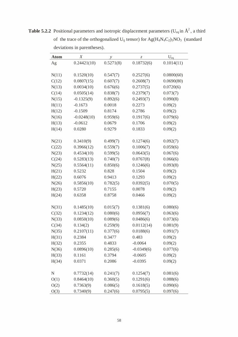
58
Table 5.2.2 Positional parameters and isotropic displacement parameters (Ueq in Å2 , a third
of the trace of the orthogonalized Uij tensor) for Ag(H4N4C2)3NO3 (standard
deviations in parentheses).
Atom X y z Ueq
Ag
N(11)C(12)N(13)C(14)N(15)H(11)H(12)N(16)H(13)H(14)
N(21)C(22)N(23)C(24)N(25)H(21)H(22)N(26)H(23)H(24)
N(31)C(32)N(33)C(34)N(35)H(31)H(32)N(36)H(33)H(34)
NO(1)O(2)O(3)
0.24421(10)
0.1520(10)0.0807(15)0.0034(10)0.0505(14)-0.1325(9)-0.1673-0.1509-0.0248(10)-0.06120.0280
0.3410(9)0.3966(12)0.4534(10)0.5283(13)0.5564(11)0.52320.60760.5856(10)0.57200.6358
0.1485(10)0.1234(12)0.0850(10)0.134(2)0.2107(11)0.23840.23550.0896(10)0.11610.0371
0.7732(14)0.8464(10)0.7363(9)0.7340(9)
0.5271(8)
0.547(7)0.607(7)0.676(6)0.838(7)0.892(6)0.00180.81740.959(6)0.06790.9279
0.499(7)0.559(7)0.599(5)0.740(7)0.850(6)0.8280.94130.782(5)0.71550.8758
0.015(7)0.080(6)0.089(6)0.259(9)0.377(6)0.34770.48330.285(6)0.37940.2086
0.241(7)0.360(5)0.086(5)0.247(6)
0.18732(6)
0.2527(6)0.2608(7)0.2737(5)0.2379(7)0.2493(7)0.22730.27860.1917(6)0.17060.1833
0.1274(6)0.1006(7)0.0643(5)0.0767(8)0.1246(6)0.15040.12930.0392(5)0.00780.0466
0.1381(6)0.0956(7)0.0486(6)0.0112(14)0.0188(6)0.483 -0.0064-0.0349(6)-0.0605-0.0395
0.1254(7)0.1291(6)0.1618(5)0.0795(5)
0.1014(11)
0.0800(60)0.0690(80)0.0720(6)0.073(7)0.090(8)0.09(2)0.09(2)0.079(6)0.09(2)0.09(2)
0.092(7)0.059(6)0.067(6)0.066(6)0.093(8)0.09(2)0.09(2)0.070(5)0.09(2)0.09(2)
0.080(6)0.063(6)0.073(6)0.081(9)0.091(7)0.09(2)0.09(2)0.077(6)0.09(2)0.09(2)
0.081(6)0.088(6)0.090(6)0.097(6)

59
Table 5.2.3 Anisotropic displacement parameters(Å2) for Ag(H4N4C2)3NO3 (standard
deviations in parentheses). The components Uij refer to a displacement factor
of the form exp{-2π2(U11h2a*2+ ⋅⋅⋅ +2U23 klb*c*)}.
Atom U11 U22 U33 U23 U13 U12
Ag 0.0767(11) 0.180(2) 0.0477(8) 0.0012(15) 0.0102(6) 0.0084(18)
N(11) 0.051(9) 0.121(19) 0.067(10) -0.001(13) 0.008(8) 0.012(15)
C(12) 0.079(14) 0.09(2) 0.041(9) 0.032(11) 0.021(10) -0.013(15)
N(13) 0.053(9) 0.125(19) 0.037(8) 0.021(9) -0.002(7) 0.015(11)
C(14) 0.068(12) 0.11(2) 0.043(10) 0.003(12) 0.003(9) -0.011(14)
N(15) 0.042(8) 0.16(2) 0.067(10) 0.007(13) -0.009(7) 0.000(12)
N(16) 0.065(9) 0.121(17) 0.050(8) -0.006(12) 0.002(7) -0.009(13)
N(21) 0.059(9) 0.16(2) 0.059(9) -0.003(15) 0.022(8) -0.034(17)
C(22) 0.052(10) 0.065(17) 0.058(10) 0.006(13) -0.021(9) 0.009(14)
N(23) 0.062(9) 0.103(18) 0.036(7) -0.014(9) 0.013(7) -0.010(11)
C(24) 0.060(13) 0.081(18) 0.056(12) 0.000(13) 0.008(9) 0.001(14)
N(25) 0.083(12) 0.16(2) 0.033(8) -0.020(11) -0.002(8) -0.007(13)
N(26) 0.068(10) 0.107(16) 0.035(7) -0.020(9) -0.001(7) -0.017(11)
N(31) 0.080(11) 0.100(16) 0.058(9) 0.022(13) 0.004(8) -0.011(14)
C(32) 0.060(10) 0.079(19) 0.049(10) 0.026(12) 0.005(8) 0.002(13)
N(33) 0.064(9) 0.114(19) 0.042(8) 0.014(10) -0.006(7) -0.007(12)
C(34) 0.055(12) 0.12(2) 0.051(11) -0.005(13) 0.002(9) -0.006(14)
N(35) 0.085(12) 0.15(2) 0.035(7) 0.019(11) -0.003(8) -0.010(14)
N(36) 0.071(11) 0.114(16) 0.046(9) 0.019(10) -0.006(8) -0.016(12)
N 0.086(13) 0.103(18) 0.052(10) 0.004(11) -0.005(10) -0.007(14)
O(1) 0.076(9) 0.121(17) 0.065(9) 0.014(9) -0.018(8) -0.014(11)
O(2) 0.073(8) 0.149(18) 0.048(7) 0.009(10) 0.012(6) -0.002(11)
O(3) 0.088(10) 0.151(16) 0.049(8) 0.005(10) -0.013(7) -0.037(12)
The structure was refined with full-matrix least-squares refinement on F2. The hydrogen
atoms were placed at calculated positions and allowed to ride on the atoms which they were
attached to. The non-H atoms were given anisotropic thermal parameters while the H atoms
were refined isotropically. All calculations were performed using the SHELXL PLUS
software package.
Numerical details of the refinement are shown in Table 5.2.1. Positional and isotropic as well
as anisotropic displacement parameters are listed in Table 5.2.2 and Table 5.2.3. The thermal
ellipsoids of all light atoms are quite large in the b direction; this is almost certainly a
consequence of the poor quality of the crystal.
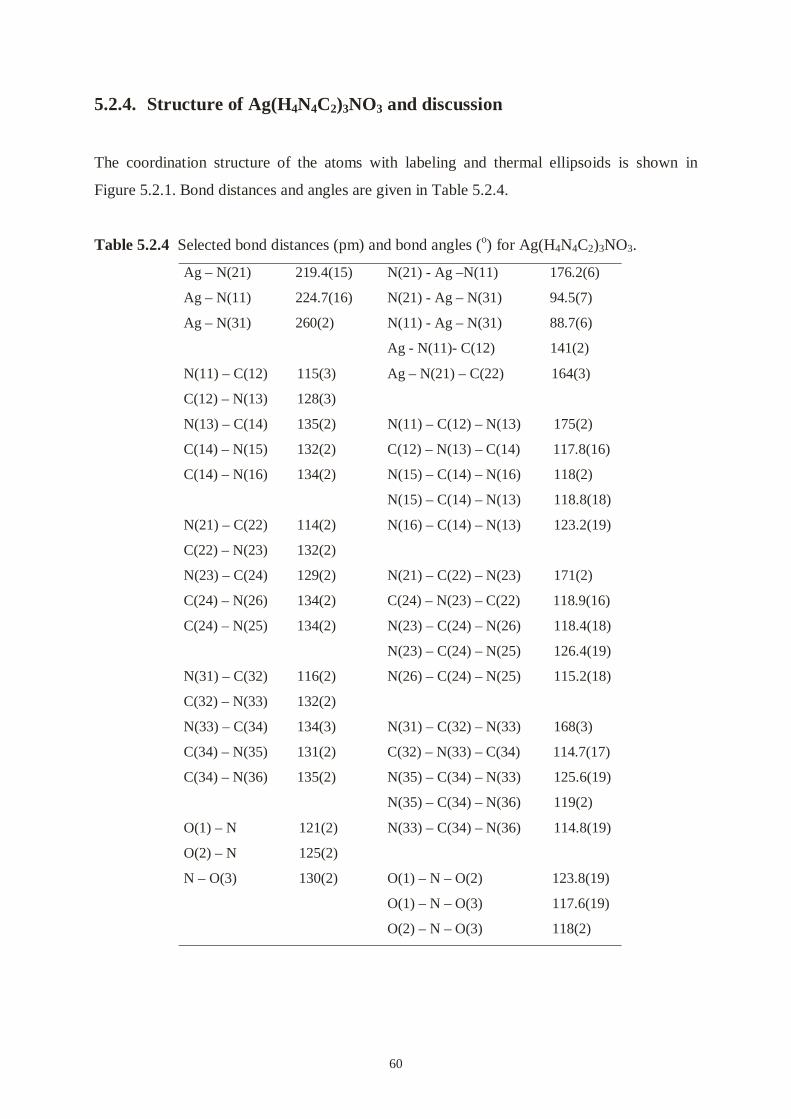
60
5.2.4. Structure of Ag(H4N4C2)3NO3 and discussion
The coordination structure of the atoms with labeling and thermal ellipsoids is shown in
Figure 5.2.1. Bond distances and angles are given in Table 5.2.4.
Table 5.2.4 Selected bond distances (pm) and bond angles (o) for Ag(H4N4C2)3NO3.
Ag – N(21) 219.4(15)
Ag – N(11) 224.7(16)
Ag – N(31) 260(2)
N(11) – C(12) 115(3)
C(12) – N(13) 128(3)
N(13) – C(14) 135(2)
C(14) – N(15) 132(2)
C(14) – N(16) 134(2)
N(21) – C(22) 114(2)
C(22) – N(23) 132(2)
N(23) – C(24) 129(2)
C(24) – N(26) 134(2)
C(24) – N(25) 134(2)
N(31) – C(32) 116(2)
C(32) – N(33) 132(2)
N(33) – C(34) 134(3)
C(34) – N(35) 131(2)
C(34) – N(36) 135(2)
O(1) – N 121(2)
O(2) – N 125(2)
N – O(3) 130(2)
N(21) - Ag –N(11) 176.2(6)
N(21) - Ag – N(31) 94.5(7)
N(11) - Ag – N(31) 88.7(6)
Ag - N(11)- C(12) 141(2)
Ag – N(21) – C(22) 164(3)
N(11) – C(12) – N(13) 175(2)
C(12) – N(13) – C(14) 117.8(16)
N(15) – C(14) – N(16) 118(2)
N(15) – C(14) – N(13) 118.8(18)
N(16) – C(14) – N(13) 123.2(19)
N(21) – C(22) – N(23) 171(2)
C(24) – N(23) – C(22) 118.9(16)
N(23) – C(24) – N(26) 118.4(18)
N(23) – C(24) – N(25) 126.4(19)
N(26) – C(24) – N(25) 115.2(18)
N(31) – C(32) – N(33) 168(3)
C(32) – N(33) – C(34) 114.7(17)
N(35) – C(34) – N(33) 125.6(19)
N(35) – C(34) – N(36) 119(2)
N(33) – C(34) – N(36) 114.8(19)
O(1) – N – O(2) 123.8(19)
O(1) – N – O(3) 117.6(19)
O(2) – N – O(3) 118(2)

61
Figure 5.2.1 ORTEP drawing of the Ag(H4N4C2)3NO3 with thermal ellipsoids scaled at 30%
probability level.
The Ag(I) is surrounded by three nitrogen atoms from three dicyanodiamide molecules. Two
dicyanodiamides behave as a mono-dentate ligand through nitrile nitrogen atoms. The
distances of Ag-N(21) and Ag-N(11) are 219.4(5) and 224.7(16) pm respectively, and the
angle of N(11)-Ag(1)-N(12) is 176.2(6)°, close to linear. It is a very common phenomenon in
2-coordinate Ag(I) complexes. The interaction of Ag(I) and N(31) (Ag-N(31) = 260(2) pm)
appears to be very weak because it is beyond the covalent distance between Ag and N. If it
was a 3-coordinate Ag complex, the average distance of Ag-N would be 234 pm, longer than
that in AgN(CN)2 with 2-coordinate Ag (211.1 pm) [70], AgC(CN)3 with 3-coordinate Ag
(222.0 pm) [71] and AgC(CN)2NO with 4-coordinate Ag (223 pm) [72]. The third
dicyanodiamide can be simply considered as locating in the vacant site of the crystal by weak
interactions of silver atom and nitrile nitrogen atom and hydrogen bonds.
The shortest Ag⋅⋅⋅Ag distance is 363.7 pm which is along the b axis. The arrangement of
silver atoms falls a 21 fold along the b axis (Figure 5.2.2). Those close-packed silver
atoms form the backbone of the framework.
b
Figure 5.2.2 The arrangement of silver atoms
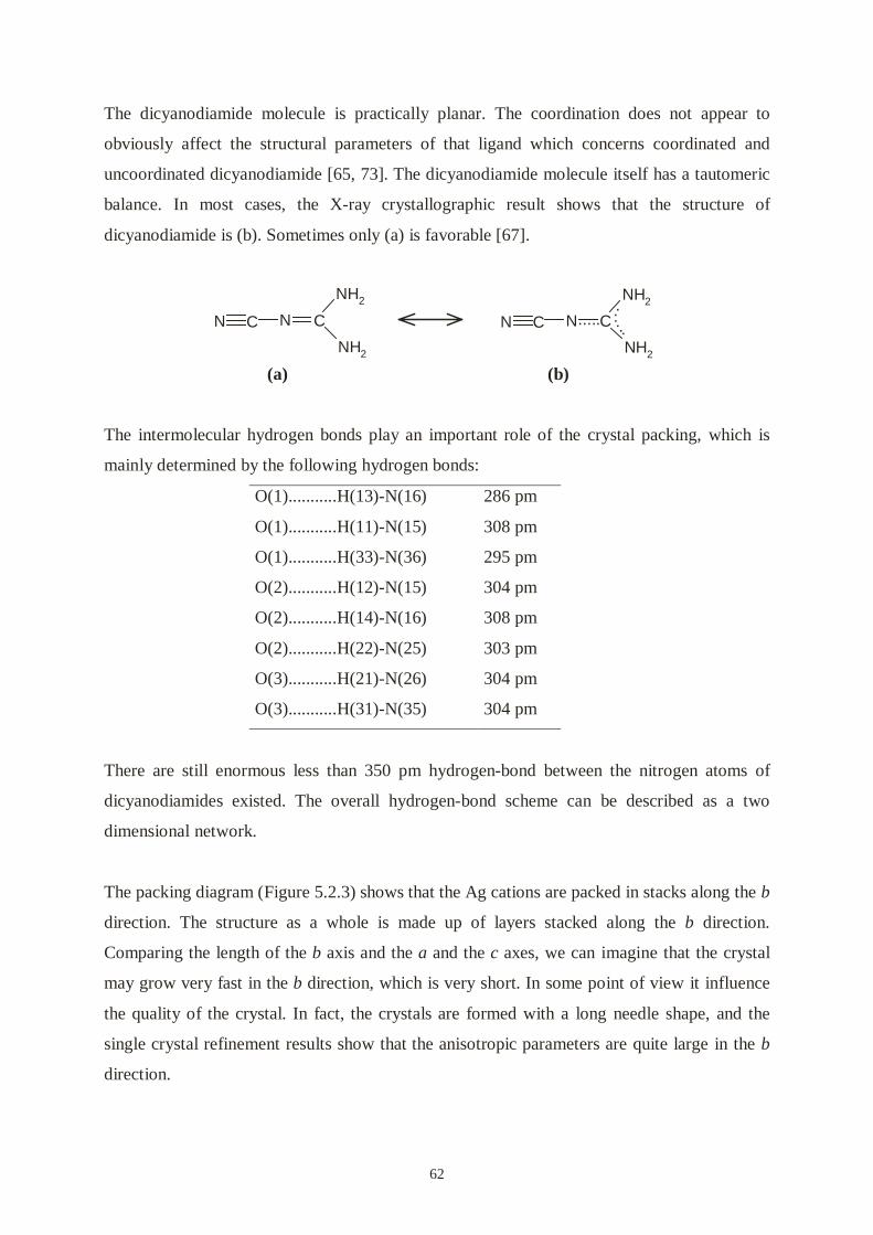
62
The dicyanodiamide molecule is practically planar. The coordination does not appear to
obviously affect the structural parameters of that ligand which concerns coordinated and
uncoordinated dicyanodiamide [65, 73]. The dicyanodiamide molecule itself has a tautomeric
balance. In most cases, the X-ray crystallographic result shows that the structure of
dicyanodiamide is (b). Sometimes only (a) is favorable [67].
(a) (b)
The intermolecular hydrogen bonds play an important role of the crystal packing, which is
mainly determined by the following hydrogen bonds:
O(1)...........H(13)-N(16) 286 pm
O(1)...........H(11)-N(15) 308 pm
O(1)...........H(33)-N(36) 295 pm
O(2)...........H(12)-N(15) 304 pm
O(2)...........H(14)-N(16) 308 pm
O(2)...........H(22)-N(25) 303 pm
O(3)...........H(21)-N(26) 304 pm
O(3)...........H(31)-N(35) 304 pm
There are still enormous less than 350 pm hydrogen-bond between the nitrogen atoms of
dicyanodiamides existed. The overall hydrogen-bond scheme can be described as a two
dimensional network.
The packing diagram (Figure 5.2.3) shows that the Ag cations are packed in stacks along the b
direction. The structure as a whole is made up of layers stacked along the b direction.
Comparing the length of the b axis and the a and the c axes, we can imagine that the crystal
may grow very fast in the b direction, which is very short. In some point of view it influence
the quality of the crystal. In fact, the crystals are formed with a long needle shape, and the
single crystal refinement results show that the anisotropic parameters are quite large in the b
direction.
N C N C
NH2
NH2
N C N C
NH2
NH2
...........

63
a
b c
Figure 5.4.3 Packing of Ag(H4N4C2)3NO3 viewed down the b axis. Large circles
represent silver atoms, small circle units represent H4N4C2 and triangle
units represent NO3-.

64
6. Syntheses, crystal structures, magnetic properties, and
electronic structures of Co(NCNH2)4Cl2, Ni(NCNH2)4Cl2
and Cu(NCNH2)4Cl2
6.1. Introduction
With respect to the complexing ability of molecular cyanamide through the nitrile nitrogen,
H2NCN can be expected to show similar coordination properties such as ammonia. In fact, the
color change upon mixing aqueous solutions of transition metal ions and cyanamide lets us
believe that H2NCN can form a number of transition metal cyanamide complexes
M(NCNH2)xn+.
Divalent nickel, cobalt and copper exhibit a large number of complexes with coordination
numbers of 4, 5, and maximum 6. On the other hand, a considerable number of neutral
ligands, especially amines, replaces some or all of the water molecules in octahedral
[M(H2O)6]2+ to result in complexes such as [Ni(NH3)6]Cl2, [Co(NH3)6]Cl2 and [Cu(NH3)4]Cl2.
The structure around Ni2+ in NiCl2⋅4H2O [74], for example, is a distorted octahedron with a
cis configuration of the chlorine ligands.
6.2. Synthesis
An aqueous solution of 0.01 mol/l CoCl2 or NiCl2 was added to an aqueous solution of 0.04
mol/l H2NCN, and the mixture was maintained at room temperature. Green cubic crystals
Ni(NCNH2)4Cl2 or red cubic crystals Co(NCNH2)4Cl2 were obtained by a very slow
evaporation of the solution, i.e., 5 ml solution within one month. The green-blue crystals of
Cu(NCNH2)4Cl2 were obtained from 0.05 mol/l CuCl2 and 0.5 mol/l H2NCN by evaporation
of the aqueous solution at room temperature. The solution became wax-like due to a large
amount of H2NCN, and the crystals grew in the solution.
These compounds have a strong IR absorption of the ν(C≡N) stretch at about 2150 cm-1(s)
and a broad single ν(H-N) band at 3200 cm-1.

65
The resulting crystals (typical dimensions: 300 µm × 300 µm × 300 µm for Co(NCNH2)4Cl2
and Ni(NCNH2)4Cl2, 900 µm × 900 µm × 900 µm for Cu(NCNH2)4Cl2) were stable in the
mother solutions and well-suited for single crystal X-ray diffraction. All of these three
compounds tend to decompose in dry air and change to black-brown powders with unknown
composition.
6.3. X-ray structure determination
Table 6.1 Crystallographic data for Co(NCNH2)4Cl2, Ni(NCNH2)4Cl2 and Cu(NCNH2)4Cl2.
Formula; molar mass:
Crystal color and form:
Lattice constant:
Space group, formula units:
X-ray density; F(000):
Instrument:
Temperature:
Reflections collected / unique:
Octants; max. 2θ:
Absorption correction:
Absorption coefficient; min.
and max. transmission:
Rint, Rσ
Structure solution:
Structure refinement:
No. of intensities, variables,
restraints:
Co(NCNH2)4Cl2; 298.01 g/mol
red cubes
a = 1266.3(2) pm
I m-3m (No. 229); 6
1.462 g/cm3; 894
Enraf-Nonius CAD-4, Mo-Kα,
graphite monochromator,
scintillation counter
293(2) K
6286/ 329
0≤ h ≤17, -17≤ k ≤ 17, -17≤ l
≤ 17; 60°
none
1.647 mm-1
0.0364, 0.0136
Direct Methods
Least-squares method on F2,
Full matrix
329, 23, 0
Ni(NCNH2)4Cl2; 297.79 g/mol
green cubes
a = 1259.3(2) pm
I m-3m (No. 229); 6
1.486 g/cm3; 900
Enraf-Nonius CAD-4, Mo-Kα,
graphite monochromator,
scintillation counter
293(2) K
3431 / 329
-2 ≤ h ≤17, -4≤ k ≤ 17, -17≤ l
≤ 17; 60°
ψ scan
1.843 mm-1; 0.2020, 0.2837
0.0798, 0.0314
Direct Methods
Least-squares method on F2,
Full matrix
329, 23, 0
Cu(NCNH2)4Cl2; 302.62 g/mol
green blue cubes
a = 1244.3(2) pm
I m-3m (No. 229); 6
1.564 g/cm3; 906
Enraf-Nonius CAD-4, Mo-Kα,
graphite monochromator,
scintillation counter
293(2) K
4612 / 329
0 ≤ h ≤20, -1≤ k ≤ 20, -20≤ l
≤ 20; 70°
none
2.099 mm-1
0.0864, 0.0464
Direct Methods
Least-squares method on F2,
Full matrix
327, 23, 0
Weighting scheme: w = 1/[σ2(Fo2)+ (0.0321×
P)2 + 1.0007 × P],
P = (max(Fo2,0) + 2 ×Fo
2)/3
w = 1/[σ2(Fo2)+ (0.0209 ×
P)2 + 0.6923 × P],
P = (max(Fo2,0) + 2 ×Fo
2)/3
w = 1/[σ2(Fo2)+ (0.0896 ×
P)2 + 1.0561 × P],
P = (max(Fo2,0) + 2 ×Fo
2)/3
Goodness of fit (all data):
Final R indices [I >4 σ(I)] :
R indices (all data):
max., min. residual electrondensity:
1.070
R1 = 0.0241, wR2 = 0.0646
R1 = 0.0327, wR2 = 0.0695
0.153, -0.265 e/Å3
1.066
R1 = 0.0245, wR2 = 0.0560
R1 = 0.0430, wR2 = 0.0633
0.290, -0.392 e/Å3
1.199
R1 = 0.0508, wR2 = 0.1627
R1 = 0.0670, wR2 = 0.1787
1.170, -0.900 e/Å3

66
A selected crystal was transferred into a sealed glass capillary and mounted on an Enraf-
Nonius CAD-4 four-circle diffractometer with graphite monochromatized Mo-Kα radiation.
The lattice parameters were determined by a least-squares fit to the positive and negative θ
angles of 25 independent reflections. A complete set of intensities was measured at room
temperature.
The lattice symmetry and the systematic absences were consistent with space group I m-3m
(No. 229). The crystal structure was solved by Direct Methods (SHELXS-97) [38], giving the
two heavy atom positions, Ni2+, Co2+ or Cu2+ on 6b and Cl- on 12e. The location of the
cyanamide group was found from a subsequent Fourier synthesis. When placed onto the
octahedron's basal mirror plane, the N(2) atom of the cyanamide molecule exhibited an
unphysically large thermal parameter perpendicular to that plane. A careful analysis revealed
that N(2) does not sit on Wyckoff position 24h but on 48k with half occupancy, i.e., roughly
30 pm above and below the mirror plane. The crystallographic data of the final refinement
[38] are summarized in Table 6.1, whereas Tables 6.2 and 6.3 give positional and isotropic
as well as anisotropic displacement parameters.
Table 6.2 Positional parameters and isotropic displacement parameters (Ueq in pm2 , a third
of the trace of the orthogonalized Uij tensor) for Co(NCNH2)4Cl2, Ni(NCNH2)4Cl2
and Cu(NCNH2)4Cl2 (standard deviations in parentheses) [75].
Atom Site SOF X y Z Ueq
Co 6b 1 0 1/2 0 559(3)Cl 12e 1 0 0.30131(11) 0 657(3)N(1) 24h 1 0.38331(14) 0 X 666(7)C 24h 1 0.32074(17) 0 X 612(8)N(2) 48k 0.5 0.2486(2) 0.0252(9) X 850(40)H 48j 1 0.187(2) 0 0.266(2) 960(100)
Ni 6b 1 0 1/2 0 539(3)Cl 12e 1 0 0.30281(11) 0 639(3)N(1) 24h 1 0.38499(15) 0 X 635(7)C 24h 1 0.32203(17) 0 X 598(8)N(2) 48k 0.5 0.2497(2) 0.0252(7) X 790(30)H 48j 1 0.183(2) 0 0.266(2) 890(90)
Cu 6b 1 0 1/2 0 538(5)Cl 12e 1 0 0.2764(2) 0 760(7)N(1) 24h 1 0.38343(3) 0 X 609(11)C 24h 1 0.32048(3) 0 X 687(14)N(2) 48k 0.5 0.2503(4) 0.024(3) X 920(120)H 48j 1 0.187(2) 0 0.266(2) 620(110)

67
Table 6.3 Anisotropic displacement parameters (pm2) for Co(NCNH2)4Cl2, Co(NCNH2)4Cl2
and Cu(NCNH2)4Cl2 (standard deviations in parentheses). The components Uij
refer to a displacement factor of the form exp{-2π2(U11h2a*2+ ⋅⋅⋅ +2U23klb*c*)}.
Atom U11 U22 U33 U23 U13 U12
Co 405(3) 867(6) U11 0 0 0Cl 577(4) 818(8) U11 0 0 0N(1) 487(8) 1030(20) U11 0 -37(12) 0C 454(9) 930(20) U11 0 26(12) 0N(2) 511(10) 1510(120) U11 40(20) -78(15) U23
Ni 389(3) 840(6) U11 0 0 0Cl 557(4) 802(8) U11 0 0 0N(1)C
466(8)451(9)
980(20)890(20)
U11
U11
00
-27(12)45(13)
00
N(2) 498(9) 1390(100) U11 60(20) -73(16) U23
Cu 376(4) 863(9) U11 0 0 0Cl 639(7) 1001(16) U11 0 0 0N(1)C
473(12)479(14)
880(30)1100(40)
U11
U11
00
57(16)48(19)
00
N(2) 536(19) 1700(400) U11 20(40) -80(16) U23
6.4. Structure and packing
Figure 6.1 ORTEP drawing of Ni(NCNH2)4Cl2 with atom labelling scheme; thermal
ellipsoids are drawn at 30% probability level. The alternative N(2) split
positions are indicated with small isotropic ellipsoids.

68
Table 6.4 Selected intramolecular distances (pm) and bond angles (o) for Co(NCNH2)4Cl2 ,
Ni(NCNH2)4Cl2 and Cu(NCNH2)4Cl2.
Co-N(1)
Co-Cl
N(1)-C
C-N(2)
N(2)-H
Co-N(1)-C
N(1)-C-N(2)
C-N(2)-H
209.0(3) (4×)
251.6(1) (2×)
112.1(4)
133.1(5)
87(3)
180
166.2(5)
111(2)
Ni-N(1)
Ni-Cl
N(1)-C
C-N(2)
N(2)-H
Ni-N(1)-C
N(1)-C-N(2)
C-N(2)-H
204.8(3) (4×)
248.3(1) (2×)
112.1(4)
132.7(5)
93(3)
180
166.2(4)
113(2)
Cu-N(1)
Cu-Cl
N(1)-C
C-N(2)
N(2)-H
Ni-N(1)-C
N(1)-C-N(2)
C-N(2)-H
203.7(4) (4×)
278.3(1) (2×)
104.7(7)
134.3(12)
98(5)
180
167.4(16)
120(3)
The coordination of the central Ni2+ in the crystal structure of Ni(NCNH2)4Cl2 is given as a
thermal ellipsoid plot in Figure 6.1 while the atomic numbering refers to Table 6.4. Figure 6.2
offers a more schematic view into the crystal packing of the complex units and their
intermolecular connectivities.
Divalent nickel is octahedrally coordinated by four equatorial cyanamide molecules and two
axial chlorine ions. The interatomic distances amount to 204.8(3) pm for the Ni-N and to
248.3(1) pm for the Ni-Cl combination. Slightly larger Ni-N distances are known from the
crystal structures of Ni(NCS)2(NH3)4 [76], namely 208 pm (Ni-NCS) and 210 pm (Ni-NH3),
and from the structure of Ni(NH3)4(NO2)2 [77], 210 pm (Ni-NH3). The present Ni-Cl distance,
on the other hand, turns out to be somewhat longer than those in related compounds such as
NiCl2⋅4H2O [70], 237-240 pm, or Ni[SC(NH2)2]4Cl2 [78], 240-250 pm.
The interatomic distances are 209.0(3) pm for the Co-N and 251.6(1) pm for the Co-Cl
combination for Co(NCNH2)4Cl2. These may be compared with reported distances like
217.0(2) pm (Co-N) in Co(NH3)6Cl2 [79] and 240.7(7) pm (Co-Cl) in Co(H2O)4Cl2 [80].
The interatomic distances are 203.7(4) pm for the Cu-N and 278.3(1) pm for the Cu-Cl
combination for Cu(NCNH2)4Cl2. The Cu⋅⋅⋅Cl distance in Cu(NCNH2)4Cl2 is much longer
than the sum of the ionic radii. The axially elongated octahedron is due to the Jahn-Teller
effect. That is a common phenomenon of Cu2+(d9). The typical distortion of the octahedron is
an elongation along one 4-fold axis, so that there is a planar array of four short Cu-L bonds
and two long ones in trans position.

69
The C-N distances within the cyanamide ligands are 112.1(4) pm for N(1)-C, consistent with
a formal triple bond, and 132.7(5) pm / 133.1(5) pm for C-N(2), consistent with a formal
single bond in the Ni / Co complex. These values compare very well with those in pure
cyanamide which arrive at 115 and 132 pm at a temperature of 108 K [3] and at 114 and 130
pm at a temperature of 237 K [81], also. The crystal quality of Cu(NCNH2)4Cl2 is not as good
as the other compounds. The C-N distances within the cyanamide ligands are 104.7(7) pm for
the formal triple bond N(1)-C and 134.3(12) pm for the formal single bond C-N(2). There is a
difference with regard to the N-C-N bond angle. Within pure cyanamide, the molecule can be
satisfying regarded as being almost linear (N-C-N around 178-179°) [3, 81], whereas the
present refinement gives an N-C-N angle of 166-167° in all three cases. Nevertheless, we
have described that the N-C-N angle is quite soft and can be easily distorted without
significant energy losses (see quantum chemical calculations in chapter 3). The energy losses
of distortion may be compensated by the formation of hydrogen bonds. The hydrogen atom
attached to the N(2) atom was easily detectable, and the refined distances of 87(3) pm in
Co(NCNH2)4Cl2, 93(3) pm in Ni(NCNH2)4Cl2 and 98(3) pm in Cu(NCNH2)4Cl2 reflect the
expected, unphysical X-ray shortening.
Figure 6.2 Crystal packing of Ni(NCNH2)4Cl2, Co(NCNH2)4Cl2 and Cu(NCNH2)4Cl2.
Ni, Co or Cu ions are in the centers of the octahedra. Large white circles represent
chloride ions, triple gray units represent N(1)-C-N(2) units and small white
circles represent hydrogen atoms.

70
For establishing the intermolecular connectivity, each chlorine is linked to four disordered
cyanamide molecules via hydrogen bridges Cl⋅⋅⋅H–N, and the calculated Cl⋅⋅⋅H distance is on
the order of 235 pm. As a consequence, there results a large cavity in the center of the cubic
unit cell (0see Figure 6.2). Taking into account the shortest Cl-Cl distance between opposite
octahedral units (762 pm) and the ionic radius of Cl- (181 pm), the central cavity can
accommodate a sphere of radius 200 pm. The difference electron density in this region,
however, is flat and does not show any suspicious peaks.
6.5. Magnetic Properties and Electronic structure
From the point of view of coordination chemistry, Ni(NCNH2)4Cl2 and Co(NCNH2)4Cl2 are
uncommon 20 and 19 electron species. Octahedral Ni2+ complexes show a relatively simple
magnetic behavior. If we neglect the orbital contribution for reasons of simplicity, the
effective number of Bohr magnetons, neff, can be calculated according to
neff = g √S(S+1) ,
where S is the spin quantum number and g is about 2.
Taking into account the approximate octahedral coordination and the corresponding ligand
field splitting of Ni2+ and Co2+(high spin), one expects two/three unpaired electrons (S=1 and
S=3/2), thus, neff around 2.8 and 3.9; indeed, most octahedral Ni2+ exhibit magnetic moments
between 2.9 and 3.4, whereas magnetic moments of most high spin octahedral Co2+ are
between 4.7 and 5.2 [82, 83], i.e., according to three unpaired electrons, and exhibit a strong
temperature dependency.
Figure 6.3 offers the measured number of effective Bohr magnetons, derived from the square
root of the product between magnetic susceptibility and temperature. Between room
temperature and about 20 K, Ni(NCNH2)4Cl2 is a Curie paramagnet, as expected, and the neff
equals to about 3. The neff of Co(NCNH2)4Cl2 equals to about 4.8 at room temperature. The
enlarged g value indicates a significant orbital contribution since the ground state results from
a symmetry-lowering of the 4T1 term in the case of perfect Oh symmetry.

71
Figure 6.3 Magnetic measurement of Co(NCNH2)4Cl2 ( triangles, H = 10000 Oe) and
Ni(NCNH2)4Cl2 (circles, H = 5000 Oe).
For the electronic structure calculations we used the Amsterdam Density-Functional program
(ADF99) [47, 84, 85]. The valence atomic orbitals were expanded in a set of Slater type
orbitals (STOs) and the core shell was treated using the frozen core approximation. For N, C,
and H atoms a triple-ζ basis set with two polarization functions (type V) was chosen. For Ni
and Co, the 3p electrons were treated as core electrons and a triple-ζ basis set with one
polarization function (type IV) was used for the remaining valence orbitals. Exchange and
correlation were taken care of by the local spin density approximation (LSDA) as
parameterized by Vosko, Wilk and Nusair [44]. Further gradient corrections were applied
using the approach of Becke for exchange [86, 87] and of Perdew for correlation [88].
The geometry of the complex Ni(H2NCN)4Cl2 was first optimized in D2d symmetry. This
choice reflects the packing in the crystal structure most appropriately and it allows for a
bending within the cyanamide group and for pyramidalization of the N(2) atoms. The
calculated bond lengths and bond angles match the experimental values very well; Ni-Cl =
242 pm, Ni-N(1) = 209 pm, N(1)-C =116 pm, C-N(2) = 134 pm. The angles Ni-N(1)-C and
N(1)-C-N(2) are approximately linear, with 175° and 179°, respectively. However, we note
that the energy surface corresponding to a bending of the N(1)-C-N(2) angle is very shallow
and that we do not model the hydrogen connectivity in the quantum-chemical approach.
0
1
2
3
4
5
0 50 100 150 200 250 300 350
T (K)
neff

72
The electronic structure of the Ni complex in the vicinity of the topmost orbitals shows the
resemblance with the orbital splitting of ML6 in an octahedral crystal field [89]. Since we
have Ni2+, thus 8e- in the d orbitals of the central Ni atom, one expects the well-known 3-
below-2 splitting of an octahedral complex with a completely filled t2g set and an eg set with
2 electrons only. A simplified outline of the orbital pattern is given in Figure 6.4 on the left.
Things are, however, slightly more complicated since the idealized octahedral symmetry is
broken by the difference between axial chlorine ions and equatorial cyanamide groups. Within
our choice of D2d, the t2g set splits into b2 + e and the eg set splits into a1 and b1. Furthermore,
a spin-polarized calculation yields different orbital patterns for majority (α) and minority (β)
electrons as shown in Figure 6.4 on the right.
α β
a1
a1b1
b1
b2
b2e
e
t2g
eg
Octahedral ML6 (high-spin) Ni(NCNH2)4Cl2 (D2d)
E (eV)-2
-5
-3
-4
-6
Figure 6.4 Left side: The orbital splitting of a high-spin octahedral ML6 fragment with 8
electrons. Right side: Orbital energies for the Ni(NCNH2)4Cl2 complex (in D2d
symmetry) within a spin-polarized calculation.
The energy splitting between the highest filled orbital (a1 of α) and the lowest unfilled orbital
(b1 of β) located on the central Ni is 1.1 eV. The difference between a1 and b1 (both of α) is
1.5 eV. With one electron less the corresponding Co compound might either be "low-spin",
with one unpaired electron, or "high-spin", with three unpaired electrons. Experimentally, the
“high-spin” complex is verified. Tracing the appropriate electronic structure of the Co
compound is laborious, however. Based on the calculated bonding energies alone it is not

73
possible to distinguish between the two different spin-states, since they both come out within
0.05 eV difference for the whole molecule.
The decisive factor in favor of the “high-spin” complex is found in a comparison of the
calculated bond distances Co-Cl (237 pm) and Co-N(1) (210 pm) with the experimental data.
The corresponding distances calculated for the "low-spin" state turn out to be about 4%
shorter.
The complete orbital pattern of the Co-complex is considerably more complicated than that of
the Ni-complex, since the depleted electron of the minority spin component is taken from the
degenerate e-set of orbitals. Thus the system might undergo a first-order Jahn-Teller
distortion as to lower its total energy, with the consequence of increasing the HOMO-LUMO
gap. The calculated bonding energy gain (using C2v as an approximate symmetry of the
molecule), however, is not significant (∼0.05 eV) and the calculated bond distances are almost
identical.
In addition, in each case of high-spin octahedral and tetrahedral of cobalt(II) there is a quartet
ground state and three spin-allowed electronic transitions to the excited quartet states. The
visible spectrum is dominated by the energy transition 4A2→ 4T1(P) for tetrahedral and 4T1g(F)
→ 4T1g(P) for octahedral; normally the energy differences of 4A2 and 4T1(P) in the tetrahedral
case are smaller than that of 4T1g(F) and 4T1g(P) in the octahedral case, as might be inferred
from the simple observation that octahedral complexes are typical pale red or purple whereas
many common tetrahedral ones show an intense blue.

74
7. A novel synthesis of Lithium cyanamide
7.1. Introduction
Many years ago, the salt Li2NCN had been synthesized by a traditional solid-state reaction.
Li2NCN has been prepared by heating Li2CO3 with CO(NH2)2 at temperatures up to 800 °C
and a more pure form (96-98%) results from thermal decomposition at 870-900 °C of
LiHNCN [90, 91]. Later, it was reported that Li2NCN was prepared by a reaction of Li2C2
with Li3N at 600 °C, and the crystal structure has been determined from single crystal [6].
Here, we present a novel simple procedure with standard Schlenk techniques leading to pure
lithium cyanamide by the reaction of H2NCN and LiBu. The purity of the product was
checked by X-ray powder diffraction. Normally, since the deprotonation of H2NCN is not
complete, the strong alkali metal base react with cyanamide to only form MHNCN (M = Na,
K or Rb) [20, 24, 23]; only Li2NCN is an exception.
7.2. Synthesis
1.0 g H2NCN (23.8 mmol) in a 250 ml round bottom flask under an ice bath was dried by a
dynamic vacuum and then solved in 100 ml ether. 2 g anhydrous MgSO4 (using anhydrous
Na2SO4 is also possible) was added to the solution of H2NCN and was stirred for 2 hours to
remove trace water; then MgSO4 was filtered off. 39.7 ml LiBu (1.6 M LiBu in hexane, 47.5
mmol) was dropwise added to the solution of H2NCN at 0 °C. Immediately a large bulky
white precipitate were obtained (Notice: keep the protecting gas at low pressure and let the
volatile easily get out!).
H2NCN + 2LiBu Li2NCN + 2HBu↑
When the addition was completed, the mixture was stirred for another 2 hours. The precipitate
was filtered and washed with Et2O (3 × 5 ml) and dried at high vacuum (1.5×10⁻² Pa).
Continually, the precipitate was heated and remained at 150 °C under dynamic vacuum for
about 10 hours, then naturally cooling down to room temperature. Well crystallized 1.15 g
white powder Li2NCN was obtained. Yield : ~90%.

75
Li2NCN is white fine powder and very moisture sensitive.
Li2NCN + H2O 2LiOH + H2NCN
IR: 2000(s), 690(m) (under protecting gas).
7.3. X-ray powder diffraction
Li2NCN powder was sealed under nitrogen in a ∅ 0.5 or 0.7 mm glass capillary. The intensity
data of Li2NCN were collected on an ImagePlate with digital Guinier Cameras (Huber) using
monochromatized Cu Kα1 radiation. The obtained diffraction pattern was indexed by the
winXPOW program Package.
The X-ray diffraction pattern of the powder crystals agrees with that of the single crystal data
of R. J. Pulham [6]. The structure of lithium cyanamide, Li2NCN, is tetragonal, space group
I4/mmm (No. 139) with unit-cell dimensions a = b = 368.7(3) pm, c = 866.8(5) pm, and Z = 2.
The lattice is composed of Li+ and centrosymmetric [NCN]2+ ions (C-N = 123 pm). Each Li+
ion is at the centre of a squashed tetrahedron of N atoms (Li-N = 207 pm). The NCN units are
vertical with respect to the layers of Lithium ions (Figure 7.2).
Figure 7.1 Experimental and Calculated PXRD patterns of Li2NCN.
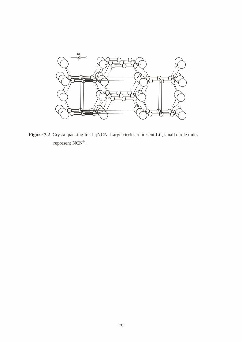
76
a1a2 c
Figure 7.2 Crystal packing for Li2NCN. Large circles represent Li+, small circle units
represent NCN2-.

77
8. Summary
This thesis describes syntheses and structures of some metal cyanamide compounds, based on
“soft cations” of metal elements. According to the concepts of hard and soft acids and bases
of R. G. Pearson, the bonding between hard acids and bases can be described approximately
in terms of ionic or dipole-dipole interactions. Soft acids and bases are more polarizable than
hard acids and bases, and the acid-base interaction has a more pronounced covalent character.
The NCN2- anion is positioned at the borderline of hard and soft base. Thus, soft cations can
form strong covalent bonds with the nitrogen atoms of cyanamide, and lead the NCN unit to
show different symmetries (from D∞h, C2v and C∞v, even if Cs). On the other hand, NCN2-
anion shows D∞h symmetry in all alkaline and alkaline earth metal cyanamide compounds
(some of them slightly tend to C2v).
Furthermore, by subtle alterations of the reaction condition transition metal-cyanamide
complexes have been synthesized, in which neutral cyanamide acts as one of the ligands.
In chapter 3, the single crystal structure of PbNCN is presented, which has been refined on the
basis of a single crystal grown from solution. PbNCN crystallizes in space group Pnma (Z =
4) with a = 555.66(4) pm, b = 386.77(2) pm, and c = 1173.50(8) pm. The cyanamide anion
exhibits two C-N bond lengths of 116 and 130 pm, and the N-C-N angle is 176°.
Quantum-chemical DFT calculations prove that the cyanamide anion is comparatively easy to
distort. In fact, the observed bond length asymmetry (about 7 pm) in the cyanamide unit costs
about 30 kJ/mol, and this energy loss is probably counterbalanced by the formation of one
short Pb-N bond which is 231 pm. Changes in the N-C-N bond angle are even less important
than in the N-C bond length. Less than 2.5 kJ/mol (the room temperature thermal energy) is
needed to bend the linear unit to 175o. The comparatively soft lead atom is better suited for
such bond formation than the alkali or alkaline-earth metals, which interact more ionically.
In chapter 4, a new phase of mercury cyanamide and related complexes are described. We
found a monoclinic phase of mercury cyanamide, namely HgNCN(II) by controlling the pH
value of the reaction, which has significantly different X-ray powder pattern with the reported
orthorhombic phase, HgNCN(I). HgNCN(II) was structurally characterized by X-ray analysis

78
using powder methods. It adopts a monoclinic unit cell with parameters a = 685.223(1) pm, b
= 698.362(1) pm, c = 555.269(1) pm and β = 113.21(8)° in space group P21/a, although two
competing structure models (one in space group Cm with a half of the b lattice; the other one
in P21/a) at the very beginning. The refinement result shows that Hg atoms and NCN units
form planar Hg-N(1)-Hg-N(1) zigzag chains running parallel to the b axis and lying in the bc
plane. Each mercury atom is attached diagonally to two N(1) and the N(1)-Hg-N(1) angle of
175(1)°, and the distances of Hg-N(1) are 205(2) and 211(2) pm. The distances of Hg-N(2)
(265(2), 279(2) pm) belong to the nonbonding region. The NCN unit exhibits two strongly
different bond lengths (N(1)-C = 135(2), C-N(2) = 112(2) pm) and the angle of N(1)-C-N(2)
is 161(1)°, which is significantly more bent than that in other cyanamide compounds.
Nevertheless, the two N-C distances reveal significantly different bond lengths, which has
been confirmed by vibration spectroscopy and different with the symmetry of the NCN unit
(C2v) in HgNCN(I). This phenomenon may explain by a gradually de-protonation process.
The experimental shows that HgNCN(I) shows higher density than HgNCN(II) and can
transfer to HgNCN(II) in a strong base condition. The theoretical calculations prove that
HgNCN(I) is slightly more stable by about 7 kJ/mol than HgNCN(II).
The coordinational behavior of NCN anion to mercury is investigated by two complexes.
Hg2(NCN)Cl2 and Hg3(NCN)2Cl2 were prepared from HgCl2 and H2NCN solution by a slow
diffusion method. Hg2(NCN)Cl2 crystallizes in a monoclinic cell with a = 806.67(13) pm, b
= 907.05(15) pm, c = 788.02(13) pm and β = 106.446(3)° in space group P21/c. Hg(2)s are
linked to the chains formed by Hg(1)s and nitrogens from both sides of NCN units. Cl(1)s as
free anion locate between the sheets formed by two times crossed Hg(1)-NCN-Hg(1) chains
and Hg(2)s. Remarkably, it is shown 20-membered rings by sharing edges and a HgCl2 guest
specie locate in the middle of the ring along the a axis channels.
Hg3(NCN)2Cl2 crystallizes in an orthorhombic cell with a = 701.95(16) pm, b = 1078.5(2) pm,
c = 1050.3(2) pm in space group Pca21. Mercury atoms (Hg(1) and Hg(2)) and NCN units
form infinite Hg-NCN-Hg zigzag chains. Because Hg(3)s insert between each two of the
chains and connect the chains together resulting in a 2D framework. Mercury atoms and NCN
units form also 20-membered rings, but the cavities are smaller than that in Hg(NCN)Cl2 and
allow only chloride ions host in the ring channels. It is interesting that the backbone for this
two complexes is the same as [Hg3(NCN)2]2+. The cavities of the 20-membered rings seem to
be depended on the guest species. All mercury atoms bond to neighboring nitrogen in a close
linear fashion in these two complexes. The geometry of NCN units in these two crystals
79
changes only very little from orthorhombic phase HgNCN upon complexation. While
Hg2(NCN)Cl2 exclusively contains N=C=N2- carbodiimide species, Hg3(NCN)2Cl2 is the first
structural example of a crystalline material built up from N=C=N2-carbodiimide and N-C ≡N2-
cyanamide groups at the same time.
In chapter 5, we focussed on the true structure of silver cyanamide as an example of a
transition metal cyanamide. Ag2NCN was precipitated by the reaction of equivalent amounts
of H2NCN and AgNO3 via double decomposition in ammonia aqueous solution and re-
crystallized by control of the pH value. The single crystal refinement shows that Ag2NCN
crystallizes in a monoclinic cell with a = 730.8(3) pm, b = 601.1(2) pm, c = 668.6(3) pm and
β = 102.124(9)° in space group P21/c. There are two kinds of infinite chains in Ag2NCN. One
is Ag-N(1)-Ag-N(1) zigzag chains and the other is Ag(1)-N(1)-C-N(2)-Ag(1)-N(1)-C-N(2)
zigzag chains. The two N-C bond lengths are significantly different (N(1)-C = 127, C-N(2) =
120 pm), but the difference is smaller than that in PbNCN and larger than that in
orthorhombic HgNCN. Here, we assume that NCN unit has mixed character of “N=C=N” and
“N≡C-N”. Besides determining the single crystal structure of silver cyanamide, we measured
a series of powder diffraction patterns. We found that some of powder diffraction patterns do
not agree with the result of the single crystal. We interpret this result in a way, it is probably
another phase of Ag2NCN exists.
A silver-dicyanodiamide nitrate complex, Ag(H4N4C2)3NO3, was prepared by mixing 1 : 6
amounts of AgNO3 and H2NCN in diethylether. During slowly adding hexane, the complex
crystallizes as colorless long needle crystals with a monoclinic cell, a = 1542.1(3) pm, b =
363.74(7) pm, c = 2568.4(5) pm and β = 92.49(3)° in space group P21/n. The structure of
Ag(H4N4C2)3NO3 as a whole is made up of layers stacked along the b direction. One Ag(I) is
surrounded by three nitrogen atoms from three dicyanodiamide molecules. Two
dicyanodiamides behave as a mono-dentate ligand through nitrile-nitrogen atoms. The third
dicyanodiamide can be simply considered as located in the vacant site of the crystal by weak
interaction of silver atom. All dicyanodiamide molecules are practically planar. The
intermolecular hydrogen bonds between oxygen atoms from NO3- and protons from
dicyanodiamide play an important role of the crystal packing.
In chapter 6, the crystal structures of Ni(NCNH2)4Cl2, Co(NCNH2)4Cl2 and Cu(NCNH2)4Cl2,
the first complexes with cyanamide as a neutral ligand, have been determined from single

80
crystal data (Im-3m, Z = 6, a = 1259.3(2) pm for Ni(NCNH2)4Cl2, a = 1266.3(2) pm for
Co(NCNH2)4Cl2 and a =1244.6(4) pm for Cu(NCNH2)4Cl2). Ni2+, Co2+ and Cu2+ are
octahedrally coordinated by four equatorial H2NCN molecules and two axial chloride ions,
and the octahedral complexes are connected by a network of hydrogen bonds. The cyanamide
ligands are slightly bent (166°). The two N-C distances are 112 and 133 pm in
Ni(NCNH2)4Cl2 and Co(NCNH2)4Cl2. The two N-C distances are 105 and 134 pm in
Cu(NCNH2)4Cl2. Ni(NCNH2)4Cl2/Co(NCNH2)4Cl2 are Curie paramagnets with two/three
unpaired electrons. The axially elongated (Cu-Cl) octahedron of Cu(NCNH2)4Cl2 shows
strong Jahn-Teller effect.
In chapter 7, a novel simple procedure leading to pure lithium cyanamide is presented. The
compound can be prepared with standard Schlenk techniques via the reaction of H2NCN and
LiBu.

81
9. References
1. L. A. Pinck, J. M. Salisbury, Inorganic Syntheses, 1950, 3, 39.
2. Z. V. Zvonkova, A. N. Khvatkina, Sov. Phys. Crystallogr. 1961, 6, 147.
3. L. Denner, P. Luger, J. Buschmann, Acta Crystallogr. 1988, C44, 1979.
4. N.-G. Vannerberg, Acta Chem. Scand. 1962, 16, 2263.
5. W. Büchner, R. Schliebs, G.Winter, K. H. Büchel: Industrielle Anorganische Chemie,
2Ed., VCH, Weinheim, New York, 1986.
6. M. G. Down, M. J. Haley, P. Hubberstey, R. J. Pulham, A. E. Thunder, J. Chem. Soc.
Dalton Trans. 1978, 1407.
7. A. Harper, P. Hubberstey, J. Chem. Res. (S), 1989, 7, 194.
8. M. Becker, J. Nuss, M. Jansen, Z. Anorg. Allg. Chem. 2000, 626, 2505.
9. M. Becker, M. Jansen, Solid State Sciences, 2000, 2, 711.
10. K.-G. Strid, N.-G. Vannerberg, Acta Chem. Scand. 1966, 20, 1064.
11. K.-G. Strid, Z. Anorg. Allg. Chem. 1968, 360, 205.
12. U. Berger, W. Schnick, J. Alloys Comp. 1994, 206, 179.
13. R. Dronskowski, Z. Naturforsch. B, 1995, 50, 1245.
14. a). M. J. Cooper, Acta Crystallogr. 1964, 17, 1449.
b). M. J. Cooper, Acta Crystallogr. 1964, 17, 1452.
15. M. Becker, M. Jansen, Acta Crystallgr. 2001, C57, 347.
16. M. Becker, M. Jansen, Z. Anorg. Allg. Chem. 2000, 626, 1639.
17. M. Becker, J. Nuss, M. Jansen, Z. Naturforsch. 2000, 55b, 383.
18. F. P. Bowden, H. M. Montagu-Pollock, Nature, 1961, 191, 556.
19. H. M. Monagu-Pollock, Proc. Roy. Soc. 1962, 269A, 219.
20. M. Becker, M. Jansen, A. Lieb, W. Milius, W. Schnick, Z. Anorg. Allg. Chem. 1998,
624, 113.
21. U. Berger, W. Milius, W. Schnick, Z. Anorg. Allg. Chem. 1995, 621, 2075.
22. O. Reckeweg, F. J. DiSalvo, Angew. Chem. 2000, 112, 397.
23. W. Schnick, H. Huppertz, Z. Anorg. Allg. Chem. 1995, 621, 1703.
24. a). A. Harper, P. Hubberstey, J. Chem. Res. (L), 1989, 1452.
b). M. Becker, M. Jansen, Z. Naturforsch. 1999, 54b, 1375.

82
25. R. Riedel, A. Greiner, G. Miehe, W. Dressler, H. Fuess, J. Bill, F. Aldinger, Angew.
Chem. 1997, 109, 657.
26. J. W. Visser, J. Appl. Cryst. 1969, 2, 89.
27. P. E. Werner, Z. Kristallogr. 1964, 120, 375.
28. WinXPOW Software Manual, Stoe & CIE GmbH 1999.
29. A. Altomare, G. Cascarano, C. Giacovazzo, A. Guagliardi, M. C. Burla, G. Polidori,
M. Camalli, J. Appl. Cryst. 1994, 27, 435.
30. EXPO user’s manual, http//www.irmec.ba.cnr.it/
31. A.Altomare, M. C. Burla, G. Cascarano, C. Giacovazzo, A. Guagliardi, A.G.G Moliterni,
G. Polidori, J. Appl. Cryst. 1995, 28, 842.
32. J. Rodriguez-Caravajal, Fullprof (Version 3.2) Laboratoire Léon Brillouin, Januar 1997.
33. R. A. Young (Editor), The Rietveld Method, Oxford University Press, New York, 1993.
34. R. Dronskowski, Program BACKPOW version 1.14, Institut fur Anorganische Chemie
der RWTH Aachen, 1997.
35. TOPAS General profile and structure analysis software for powder diffraction data
Version 2.0 ( User Manual, Bruker AXS, Karlsruhe, Germany).
36. Eric Dowty, ATOMS, Version 4.0, Shape Software, Copyright 1997.
37. SHELXTL Version 5.1 Software Manual, 1997.
38. G. M. Sheldrick, SHELXS97 and SHELXL97, University of Göttingen, Germany.
39. W. R. Busing, H.A. Levy, Acta Crystllogr. 1957, 10, 180.
40. N. E. Brese, M. O’Keeffe, Acta Crystallogr. 1991, B47, 192.
41. R. A. Nyquist, R. O. Kagel, Infrared Spectra of Inorganic Compounds, Academic Press,
New York and London, 1971, p. 73.
42. J. Emsley, The Elements, 2nd Ed., Oxford University Press, Oxford, 1995, p. 46.
43. R. G. Pearson, Inorg. Chem. 1988, 27, 734.
44. S. H. Vosko, L. Wilk, M. Nusair, Can. J. Phys. 1980, 58, 1200.
45. A. D. Becke, Phys. Rev. A 1988, 38, 3098; C. Lee, W. Yang, R. G. Parr, Phys. Rev. B,
1988, 37, 785.
46. J. G. Snijders, E. J. Baerends, P. Vernoojis, At. Nucl. Data Tables 1982, 26, 483.
47. E. J. Baerends, D. E. Ellis, P. Ros, Chem. Phys. 1973, 2, 41; G. te Velde, E. J. Baerends,
J. comp. Phys. 1992, 99, 84; C. Fonsecca Guerra, O. Visser, J. G. Snijders, C. te Velde,
E. J. Baerends, in Methods and Techniques for Computational Chemistry, (Eds.: E.
Clementi, C. Corongui); STEF, Cagliari, 1995, p.305.
48. D. Breitinger, K. Brodesen, Angew. Chem. 1970, 82, 379.
83
49. C. H. MacGillavryu. J. M. Bijvoet, Z. Kristallogr. Kristallgeomtr. Kristallphysik
Kristallchem. 1936, 94, 231.
50. H. Schumann, J. Cottfriedsen, S. Dechert, F. Girgsdies, Z. Anorg. Allg. Chem. 2000,
626, 747.
51. N. Masciochi, G. A. Ardizzoia, A. Maspero, G. Lamonica, A. Sironi, Inorg. Chem. 1999,
38, 3657.
52. W. L. Roth, Acta Crystallogr. 1956, 9, 277.
53. a). K. Anrivillius, Acta Crystallogr. 1956, 9, 685.
b). K. Anrivillius, Acta Chem. Scand. 1956, 10, 852.
54. a). R. Hoppe, R. Baier, W. Carl, H. Glaum, H. Untenecker, Z. Anorg. Allg. Chem. 1988,
567, 69.
b). Th. Hansen, Hk. Mueller-Buschbaum, Z. Anorg. Allg. Chem. 1994, 620, 1137.
55. a). L. R. MacGillivray and J. L. Atwood, Nature 1997, 389, 469.
b). P. Holy, J. Závada, I. Císarová and J. Podlaha, Angew. Chem. Int. Ed. 1999, 38, 381.
56. a). M. S. Zaworotko, Chem. Commun. 2001, 1.
b). B. Holliday and C. Mirkin, Angew. Chem. Int. Ed. 2001, 40, 2022.
c). A. K. Cheetham, G. Férey and T. Loiseau, Angew. Chem. Int. Ed. 1999, 38, 3268.
57. D. Bebout, A. DeLanoy, D. Ehmann, M. Kasner, D. Parrish and R. Butcher, Inorg.
Chem. 1998, 37, 2952.
58. V. Subramanian, K. Seff, Acta. Crystallogr. 1980, B36, 2132.
59. Brucker (1999), SADABS. SMART and SAINT, Brucker AXS Inc. Madison Wisconsin,
USA.
60. X. Liu, P. Müller, P. Kroll and R. Dronskowski, Inorg. Chem. 2002, 41, 4259.
61. W. Biltz, Raumchemie der festen Stoffe, Leipzig, Verlag von Leopold Voss, 1934.
62. Details may be obtained from: Fachinformationszentrum Karlsruhe, D-76344
Eggenstein-Leopoldshafen (Germany), by quoting the registry numbers CSD-412608
(Hg2(NCN)Cl2) and CSD-412609 (Hg3(NCN)2Cl2).
63. A. Bondi, J .Phys. Chem. 1964, 68, 411.
64. a). A. Chiesi, L. Coghi, A. Mangia, M. Nardell, G. Pelizzi, Acta Crystallogr. 1971, B27,
192.
b). M. J. Begley, P. Hubberstey, P. H. Spittle and P. H. Walton, Acta. Crystallogr. 1993,
C49, 1047.
65. A. S. Batsanov, P. Hubberstey, C. E. Russell, P. Walton, J. Chem. Soc. Dalton Trans.
1997, 2667.
84
66. A. Batsanov, M. J. Begley, P. Hubberstey, J. Stroud, J. Chem. Soc. Dalton Trans. 1996,
1947.
67. J. Pickardt, B. Kühn, Z. Naturforsch. 1996, 51b, 1469.
68. J. Pickardt, B. Kühn, Z. Naturforsch. 1996, 51b, 1701.
69. F. Meyer, I. Hyla-Kryspin, E. Kaifer, P.Kircher, Eur. J. Chem. 2000, 771.
70. D. Bretton, Acta Crystallogr. 1990, C46, 2297.
71. J. Konnert, D. Britton, Inorg. Chem. 1966, 5, 1193.
72. Y. M. Chow, D. Britton, Acta. Crystallogr. 1974, B30, 1934.
73. M. Fernanda, N. N. Carvalho, J. L. Pombeiro, J. Organomet. Chem. 1993, 469, 179.
74. K. Waizumi, H. Masuda and H. Ohtaki, Inorg. Chim. Acta, 1992, 192,173.
75. Details may be obtained from: Fachinformationszentrum Karlsruhe, D-76344
Eggenstein-Leopoldshafen (Germany), by quoting the registry numbers CSD-411553
(Ni(H2NCN)4Cl2) and CSD-411554 (Co(H2NCN)4Cl2).
76. E. Durcanska, J. Kozisek, J. Durik and V. Kettmann, Acta Crystallogr. 1993, C49, 535.
77. B. N. Figgis, P. A. Reynolds, A. H. White, G. A. Williams and S. Wright, J. Chem. Soc.
Dalton Trans. 1981, 997.
78. B. N. Figgis and P. A. Reynolds, J. Chem. Soc. Dalton Trans. 1986, 125.
79. J. M. Newman, M. Binns, T. W. Hambley and H. C. Freeman, Inorg. Chem. 1991, 30,
3502.
80. K. Wa, K. Waizumi, H. Masuda, H. Ohtaki, I. Sunagawa, Bull. Chem. Soc. Japan, 1990,
63, 3426.
81. X. Liu and R. Dronskowski, unpublished results.
82. H. Lueken, Magnetochemie, (p. 256), B. G. Teubner, Stuttgart, Leipzig, 1999.
83. F. A. Cotton and G. Wilkinson, Advanced Inorganic Chemistry, (p. 771), John Wiley
& Sons Inc., 1980.
84. ADF99, Theoretical Chemistry, Vrije Universiteit, Amsterdam, 1997.
85. C. Fonseca Guerra, J. G. Snijders, G. te Velde, and E. J. Baerends, Theor. Chem. Acc.
1998, 99, 391.
86. A. D. Becke, J. Chem. Phys. 1986, 84, 4524.
87. A. D. Becke, Phys. Rev. 1988, A 38, 3098.
88. J. P. Perdew, Phys. Rev. 1986, B 33, 8822; ibid. 1986, 34, 7406(E).
89. T. A. Albright, J. K. Burdett, M-W. Whangbo, "Orbital Interactions in Chemistry",
Wiley, New York 1985.

85
90. A. M. Pavlov, N. A. Sokolov, Yu. I. Dergunov, and V. G. Golov, trudy khim. ikhim.
tekhnol. 1973, 2, 27.
91. N. A. Sokolov, M. K. Safonova, V. A. Shushunov, Yu. I. Dergunov, and V. G. Golov,
trudy khim. ikhim tekhnol. 1973, 2, 18.


CURRICULUM VITAE
Personal Details:
Name:
Date of Birth:
Place of Birth:
Parents:
Family Status:
Education History:
9. 1971 – 1.1977
2. 1977 – 7. 1983
9. 1983 – 7.1987
9. 1990 – 7. 1993
1. 1998
1. 2002
Working Experience:
7. 1987 – 8. 1990
7. 1993 – 12. 1995
Liu, Xiaohui
August 14, 1965
Yanan, shaanxi, CHINA
Prof. Liu, Jianbo and Liu, Zhilan
Married Dr. Wang, Bing
Yanan primary school, Shaanxi, China
Yanan middle and high school, Shaanxi, China
Department of Chemistry, Shaaxi Teacher’s University,
Xian, Shaanxi Province. B. Sc. (chemistry), received in
July 1987. Supervisor: Prof. Jiuru Lu.
Shanghai Institute of Metallurgy, Chinese Academy of
Sciences. M. Sc. (Analytical Chemistry), received in
July 1993. Supervisor: Prof. Sinru Lin.
Starting doctoral subject with Prof. Dr. R. Dronskowski,
RWTH, Aachen.
Finishing doctoral thesis.
Teacher of Analytical Chemistry, Department of
Chemistry, Yanan University, Shaanxi, China.
Researcher, Laboratory of Analytical Chemistry,
Shanghai Institute of Metallurgy, Chinese Academy of
Sciences.
