surgery of the bovine large intestine
TRANSCRIPT

Surgery of the BovineLarge Intestine
Mireille Meylan, Dr med vet, FVH, MS, PhD, Dr habil
KEYWORDS
� Cecal dilation � Atresia � Colon � Malformation� Field surgery � Cattle
This article consists of a short review of the anatomy of the bovine large intestine,and a more thorough description of the two most relevant diseases of the largeintestine amenable to field surgery: cecal dilatation and dislocation (CDD) incows and intestinal atresia in calves. Rectal prolapse is addressed in anotherarticle in this issue.
In disorders affecting the large intestine, as in any other disease, a complete historyand careful physical examination are prerequisites for successful treatment, whichincludes not only surgical intervention but also pre- and postoperative medical treat-ment. The clinical examination of cattle that have abdominal disease has beendescribed in detail.1–3
ANATOMYOF THE BOVINE LARGE INTESTINE
The bovine large intestine consists of the cecum, the ascending, transverse, anddescending colon, and the rectum (Fig. 1). The cecum usually lies in the supraomentalrecess with its blind end directed caudally toward the pelvis. It is connected ventrallyto the ileum by the ileocecal fold, which contains blood vessels and nerves, and dor-sally to the proximal loop of the ascending colon (PLAC) through the short cecocolicband. At the ileocecocolic junction, the cecum continues as the PLAC without visibletransition. After extending cranially to the level of the 12th rib, the PLAC turns backlaterally until it reaches the caudal flexure of the duodenum. There, it turns around cra-nially and continues as the spiral colon (spiral loop of the ascending colon), which isembedded in its mesentery in the form of a flat oval. The spiral colon consists of cen-tripetal and centrifugal coils (with 1.5 to 2 revolutions each). The distal loop of theascending colon runs first in a caudal, then in a cranial direction, and leads to thedescending colon over the short transverse colon, which crosses from the right tothe left side of the abdomen in front of the cranial mesenteric artery. The rectum liesmostly in the peritoneal part of the pelvic cavity.4–7
Clinic for Ruminants, Vetsuisse Faculty of the University of Berne, Bremgartenstrasse 109A,3012 Berne, SwitzerlandE-mail address: [email protected]
Vet Clin Food Anim 24 (2008) 479–496doi:10.1016/j.cvfa.2008.06.002 vetfood.theclinics.com0749-0720/08/$ – see front matter ª 2008 Elsevier Inc. All rights reserved.

Fig. 1. The bovine large intestine. A, ileum; B, cecum; C, proximal loop of the ascendingcolon; D, spiral loop of the ascending colon; E, distal loop of the ascending colon; F, trans-verse colon; G, descending colon; H, rectum; a, ileocecal fold. The cecocolic fold betweenthe cecum and the proximal loop of the ascending colon is not visible under the distalloop of the ascending colon.
Meylan480
CECAL DILATATION AND DISLOCATIONClinical Presentation and Diagnosis
CDD is a common and economically important abdominal disorder that affects mainlydairy cattle. In an epidemiologic study in Switzerland, similar prevalences (0.05% atindividual cow level) were found for CDD and abomasal displacement.8 The pathologicfeatures of the condition include distention, displacement, and, in some cases, retro-flexion or torsion of the cecum and proximal portion of the colon, leading to partial orcomplete obstruction of the passage of digesta. A retroflexion of the cecum is definedas a displacement of the cecal apex from its normal position toward the pelvic inlet ina cranial direction. A cecal torsion is present if a twist of the cecum occurs along itslongitudinal axis, leading to increased tension on the ileocecal fold, thus jeopardizingblood circulation and innervation of the organ. Cecal torsion occurs less frequentlythan CDD with or without retroflexion.7
The clinical presentation of the disease has been described in numerous clinicalreports.3,7,9–16 Typical clinical signs include acute onset of mild colic, a normal to mod-erately elevated heart rate, decreased appetite, reduced rumen motility, decreased toabsent fecal output, distention of the right flank, and positive succussion and percus-sion auscultation of the right flank (in the dorsal part of the paralumbar fossa, over anarea extending caudally to the tuber coxae). In a retrospective study of 111 cases,rectal examination was considered the most reliable diagnostic investigation forCDD, with swinging and percussion auscultation of the right flank.16 In that report,the distended, displaced, or twisted cecum could be palpated at rectal examinationin 95% of the affected animals. In the case of a simple dilatation, the cecal apex is eas-ily recognizable as a rounded, dome-shaped structure, with a diameter of approxi-mately 15 cm, extending into the pelvic inlet. If a retroflexion is present, the body ofthe cecum, but not the apex, can be palpated as a large, rounded, tense structurein front of the pelvis; rectal palpation alone does not always allow for differentiationof cecal dilatation with retroflexion from right abomasal displacement or volvulus.Cecal torsion can often be identified on rectal examination: in addition to the typicaldilated cecal apex, the ileocecal fold is palpable as a taut, twisted, and painfulband. Dilated loops of small intestine and of the spiral colon are often encounteredon rectal examination of animals that have CDD.15–17 The appearance of the dilatedcecum at ultrasonography from the right flank has been described.18,19 Blood
Surgery of the Bovine Large Intestine 481
chemistry values and acid-base status are mostly normal in cows that have CDD.Changes similar to those seen with obstruction of the small intestine (hypokalemic,hypochloremic metabolic alkalosis) can be seen in protracted cases, or if retroflexionor torsion of the cecum is present.15,20 In one study, hypocalcemia was observed in48% of 79 affected animals.20 In the same study, elevated packed cell volume valuesindicating circulatory compromise were present in only 13% of 86 patients. Theconcentration of chloride ions in rumen fluid was normal (<25 mmol/L) in 83% of93 animals that had CDD, whereas it was elevated in the remaining 17%.20
Although an association between the occurrence of CDD and season, age of theanimals, stage of pregnancy, or time after parturition has not been demonstrated,some investigators report a trend to more frequent occurrence of the disease duringthe fall and winter months.11,14–16
The main differential diagnoses for CDD include right displacement of the aboma-sum or abomasal volvulus, small intestinal ileus, complete or partial volvulus of themesentery, or other causes of abdominal pain (eg, pyelonephritis).11,16,21
Etiopathogenesis
Despite numerous clinical descriptions of CDD, the pathogenesis of the diseaseremains poorly understood. In the past, atony or dysmotility of the cecum andPLAC, leading to accumulation of gas and digesta, and followed by dilatation and sec-ondary displacement of the cecum, has been hypothesized to be of primary relevancein the pathogenesis of CDD.11,12,21,22 However, the patterns of myoelectric activityobserved in the cecum and PLAC of cows that showed delayed recovery or recur-rence of CDD after surgical correction presented a striking similarity to those recordedimmediately orad to a mechanical obstruction in the distal PLAC of an experimentalcow. These observations led to the hypothesis that decreased motility preventing nor-mal transit of ingesta might be present not in the cecum itself but in the spiralcolon.23,24
Alterations in mRNA expression of receptors involved in the regulation of gastroin-testinal motility, such as adrenergic,25 serotonergic,26 and muscarinic27 receptors,have been reported in the ileum and large intestine, especially the spiral colon, ofcows that have CDD, but it remained unclear whether the observed changes indicatethat these receptors are involved in the pathogenesis of the disease or if they area bare consequence of cecal dilatation.
Elevated concentrations of volatile fatty acids (VFA) in intestinal contents, asobserved in cattle fed carbohydrate-rich diets, have been suspected of inhibiting gas-trointestinal motility. Total VFA concentrations similar to those in the rumen have beenreported for the bovine large intestine.22,28 Cecal-colonic fermentation accounts for8.6% to 16.8% of total VFA production in sheep,29 and the efficiency of the largeintestine for VFA production is similar to that of the rumen.30 Starch resistant to micro-bial digestion in the rumen (bypass starch) is partially digested enzymatically in thesmall intestine and partially fermented in the large intestine, the rest being excretedin the feces. Corn starch, especially, is fermented much more slowly than starchfrom other cereals, thus resulting in large proportions of bypass starch and increasedpostruminal fermentation.22,30 When increased amounts of fermentable carbohy-drates reach the large intestine, more VFA are produced there by microbial fermenta-tion.30,31 Several investigators have reported the effects of increased VFAconcentrations or high-concentrate diets on ruminant gastrointestinal motility. How-ever, their results are contradictory, with five studies22,32–35 reporting inhibition ofmotility with increased VFA levels and three investigators36–38 being unable to detectan effect on motility parameters. Nevertheless, VFA concentrations were found to be

Meylan482
elevated in the contents of cecum, PLAC, and rectum in cows that had spontaneousCDD, as compared with healthy control cows, but it remained unclear if this elevationof VFA was the cause or the consequence of reduced motility and stasis of digesta.39
In contrast, comparison of the contractility of ex-vivo intestinal wall specimens fromthe cecum and spiral colon of healthy cows with or without preincubation with VFAdid not reveal significant differences.40 An abrupt increase of concentrates in thediet of healthy dairy cows (from 0% to 50% of the ration over 60 hours) did causea significant increase of VFA concentrations in large intestinal ingesta, but no relevantinhibition of the myoelectric activity of the spiral colon was observed.41 In conclusion,elevated VFA concentrations do not appear to play a predominant role in the regula-tion of bovine intestinal motility or in the pathogenesis of CDD.
Several cows with CDD have been referred to the author’s clinic for treatment duringoutbreaks occurring in herds after introduction of rumen buffers, but these casescould not be documented in detail and a possible role of rumen buffers in the patho-genesis of CDD remains speculative.
Therapy of Cecal Dilatation/Dislocation
Various approaches to the treatment of CDD have been reported. According to earlierreports, attempts at conservative therapy of CDD were first considered useless oreven contraindicated.10,11,21 It is now accepted that in the early stages of the disease,when the general condition of the affected animal remains good, when some feces arestill passed, and in cases where a torsion or retroflexion of the cecum can be ruled outby physical examination, conservative treatment with prokinetic drugs, purgatives, orintravenous infusions may be attempted.14,42 Administration of neostigmine (87.5 mgin 5 or 10 L of isotonic saline solution supplemented with 5% dextrose as a continuousintravenous infusion at a rate of 1–2 drops per second)42,43 or of bethanechol (0.07 mg/kgsubcutaneously, every 8 hours for 2 days)44 have been advocated. Paraffin oil (3 L) orsodium sulfate (300 g in 10 L water) administered per stomach tube have beendescribed as adequate purgatives for conservative or postoperative treatment ofCDD.14,42
In more advanced cases, if no improvement occurs on medical treatment, or whena retroflexion or torsion of the organ is suspected, surgical correction of the conditionby means of a right flank laparotomy, allowing drainage of the cecum and PLACcontents, is the therapy of choice (see Refs.7,11,13,15,21,42). The intervention is bestperformed under local anesthesia of the right flank (eg, using a proximal or distal para-vertebral block) with the animal standing. Positioning the animal in left recumbencywith or without general anesthesia, while preventing the risk of the patient lyingdown during surgery, renders manipulation of the dilated intestine more difficultbecause the weight of the filled cecum pulls it away from the incision to theleft (down) side of the abdomen. After surgical preparation and local anesthesia,a 25- to 30-cm incision is made either vertically in the middle of the right paralumbarfossa or slightly more caudally in a caudodorsal to cranioventral direction. The abdo-men is opened in a routine manner. After exploration of the abdominal cavity, anadditional plastic drape is added to minimize contamination of the sterile drapingaround the incision, and the apex of the cecum is exteriorized (Fig. 2), which is bestachieved by gently pushing the cecum from behind toward the body wall with thepalm of the hand. Grasping the cecal wall and pulling toward the incision should beavoided because it bears an increased risk for rupture of the distended cecum. Theapex is fixed on both sides by the assistant surgeon with two sterile gauzes, incisedover approximately 4 cm, and the contents of the cecum are drained. The ingesta inthe exteriorized part of the cecum normally flow freely out of the incision (unless

Fig. 2. The dilated cecum is exteriorized through a right flank incision for drainage of itscontents.
Surgery of the Bovine Large Intestine 483
they are especially dry) and additional material is massaged outside of the abdominalcavity from the rest of the cecal body and the PLAC (Fig. 3). When no additionalmaterial can be removed, the incision is closed with an inverting pattern (Cushing orLembert) using absorbable material (eg, 2-0 polyglyconate or polyglactin 910). Thececal apex is copiously flushed with warm saline solution and replaced in the abdo-men. In some cases, intestinal contents flow into the cecum from the distal smallintestine or from the colon after reposition. If the cecum fills again with a large volumeof ingesta, the apex is exteriorized and the suture is reopened to allow for further
Fig. 3. A stab incision of the cecal apex allows for drainage of its contents. Contents from thececal body and from the PLAC are massaged in the direction of the cecal apex.

Meylan484
removal of cecal contents. It may be necessary to repeat this procedure several times,but usually not more than once or twice. Contents of the spiral colon often do not flowback into the cecum and are eliminated as feces when large intestinal motility returnsafter decompression of the cecum. When the cecum remains empty after reposition inthe abdomen, a second layer of suture is added using the same pattern to close thececal apex definitively (Fig. 4). After extensive flushing, the cecum is repositioned inthe abdomen in its normal anatomic position with the apex directed caudally. Theabdominal cavity is closed routinely.45
Postoperative care includes antibiotics for 5 days (eg, 30’000 IU Na-penicillin intra-venously three times a day), anti-inflammatory drugs as needed (eg, flunixin meglu-mine, 2 mg/kg intravenously once daily for 1 or 2 days), and infusions of saline orpolyionic solutions according to the results of blood chemistry analysis or to the ex-pected changes (hypochloremia, hypokalemia, alkalosis). Under field conditions, theuse of a penicillin product with depot effect (eg, procaine penicillin) requiring onlyone daily application may be preferred, and fluid replacement can be performedthrough drenching of 20 to 30 L of salt solution into the rumen. Restrictive feedingas part of the therapy for CDD has been described.14,42 At the author’s clinic, nofeed is offered on the day of surgery for correction of CDD (day 1), 25% of the calcu-lated ration is offered on the second day, 75% on the third day, and 100% from thefourth day onwards. The animals should be monitored clinically for signs of recurrencefor several days after surgery, with normalization of fecal output and rumen activity to-gether with the absence of dilated intestines at rectal palpation indicating normal re-covery from CDD.14,42
Partial typhlectomy is recommended if the cecal wall is devitalized (eg, after cecaltorsion) or if CDD recurs after a first surgical intervention.7,12,15,43 In such cases, drain-age of cecal contents is performed as described earlier, but when the cecum is empty,it is exteriorized as far as possible through the flank incision and an amputation isperformed. First, the ileocecal fold is infiltrated with 2% lidocaine to minimize painat manipulation and thus the risk of the animal trying to lie down. Mass ligatures(eg, with overlapping simple interrupted sutures, using USP 2 chromic catgut orUSP 1 polydioxanone) are used to prevent hemorrhage from the vessels in theileocecal fold. The ligatures are placed near the cecum in order to preserve the vesselssupplying the ileum. The ileocecal fold is dissected from distal (cecal apex) to as far
Fig. 4. The cecal apex has been rinsed and closed with a double Cushing suture using 2-0polyglyconate.

Surgery of the Bovine Large Intestine 485
proximal as would be safe to remove the cecal apex without causing abdominalcontamination. In the chosen location, two intestinal clamps are placed from the mes-enteric and the antimesenteric side of the cecum, through the entire width of the cecalbody. The distal part of the cecum is resected 2 to 3 cm distally from the clamps, andthe incision is closed in two inverting layers (Cushing or Lembert, with 2-0 polyglyco-nate or polyglactin 910). The stump is flushed thoroughly with warm saline solution andreplaced in the abdominal cavity, which is closed in a routine manner.7,43 A methodwith staple closure of the stump was compared with double inverting suture, butthe results were not significantly different,46 and the use of stapling devices in food an-imals is mostly cost prohibitive. Postoperative care for typhlectomy is the same as thatdescribed for typhlotomy.42 In one study,43 the long-term prognosis after partialtyphlectomy was not better than after simple typhlotomy. Thus, the technically morecomplicated cecal amputation, resulting in prolonged surgery time and increaseddegree of contamination, is not recommended as a routine treatment of CDD.15,43
Total typhlectomy including removal of the ileocecocolic junction has been per-formed in experimental17 and clinical settings,47 and partial resection of the PLAChas been described;48 however, these complicated procedures normally requiregeneral anesthesia and are unsuitable for treatment of CDD under field conditions.7
Prognosis
Except for the risk for recurrence and for peritonitis due to devitalization of the intes-tinal wall or abdominal contamination at surgery, the prognosis of CDD is consideredto be reasonably good, with reports of approximately 90% of referred cases beingdischarged in good general condition after treatment.11,42,43 Up to 10% of treatedanimals will show recurrence of the disease within the first week after typhlotomyand another 12.5% within the first year after surgery.15,43
SURGERYOF THE BOVINE COLON
Difficult mobilization and exteriorization of the bovine colon preclude extensive surgi-cal interventions under field conditions. Intussusceptions involving the large intestine(colon or cecum) have been reported predominantly in young animals.49–52 This con-dition is presumed to be more common in calves than in adult animals because of thethin colonic mesentery of young animals that allows for more extensive movement ofadjacent intestinal segments, in contrast to more extensive mesenteric fat deposits,which stabilize the large intestine in adults.49,51,53
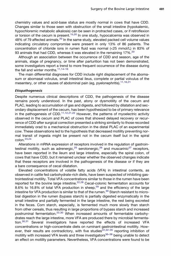
Trichobezoars, phytobezoars, and enteroliths, although they mostly lead to diseasein the rumen, abomasum, or small intestine, can also cause obstruction of the bovinespiral colon. Clinical signs are similar to those of CDD and a definitive diagnosis ismade on abdominal exploration through right flank celiotomy. In such cases, theaffected intestinal segment is exteriorized and opened, and the bezoars removed.The enterotomy site is closed with two inverting continuous sutures (Lembert or Cush-ing [eg, with 3-0 or 2-0 polyglyconate]) apposed transversally if necessary to maximizelumen and prevent stricture formation.54 Colonic obstruction may further be caused byextraluminal compression (eg, due to adhesions from prior surgery or to mesenteric fatnecrosis) (Fig. 5). Bypass of the obstruction by side-to-side anastomosis of the bowelproximal to the obstruction and the outer loop of the spiral colon has been described.7
Two cases of obstruction of the descending colon caused by cecal incarceration ina mesocolic defect in cows have also been described.55,56
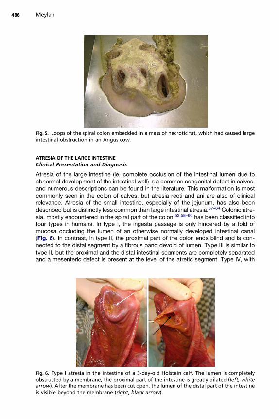
Fig. 5. Loops of the spiral colon embedded in a mass of necrotic fat, which had caused largeintestinal obstruction in an Angus cow.
Meylan486
ATRESIAOF THE LARGE INTESTINEClinical Presentation and Diagnosis
Atresia of the large intestine (ie, complete occlusion of the intestinal lumen due toabnormal development of the intestinal wall) is a common congenital defect in calves,and numerous descriptions can be found in the literature. This malformation is mostcommonly seen in the colon of calves, but atresia recti and ani are also of clinicalrelevance. Atresia of the small intestine, especially of the jejunum, has also beendescribed but is distinctly less common than large intestinal atresia.57–64 Colonic atre-sia, mostly encountered in the spiral part of the colon,53,58–60 has been classified intofour types in humans. In type I, the ingesta passage is only hindered by a fold ofmucosa occluding the lumen of an otherwise normally developed intestinal canal(Fig. 6). In contrast, in type II, the proximal part of the colon ends blind and is con-nected to the distal segment by a fibrous band devoid of lumen. Type III is similar totype II, but the proximal and the distal intestinal segments are completely separatedand a mesenteric defect is present at the level of the atretic segment. Type IV, with
Fig. 6. Type I atresia in the intestine of a 3-day-old Holstein calf. The lumen is completelyobstructed by a membrane, the proximal part of the intestine is greatly dilated (left, whitearrow). After the membrane has been cut open, the lumen of the distal part of the intestineis visible beyond the membrane (right, black arrow).

Surgery of the Bovine Large Intestine 487
several sites of atresia, is described by some investigators as occurring only inhumans.53,60,63,65–67
Calves that have large intestinal atresia are typically presented to a veterinarian atthe age of 2 or 3 days (although presentation of calves as old as 11 days has beenreported), with a history of having been alert and nursing well immediately after birth,but showing decreasing appetite, increasing abdominal discomfort and distention,and progressive weakness after 24 to 36 hours of life. The calves’ owners often reportthat defecation has not been observed since birth. At physical examination, theseanimals show signs of colic (restlessness, saw-bock stance, kicking toward the abdo-men), a distended abdomen, an elevated heart rate, positive percussion and succus-sion auscultation of the right flank, and neither spontaneous nor provoked defecation.Whereas the absence of an anal opening is immediately visible at physical examina-tion of calves that have atresia ani (Fig. 7), the rectum is empty at palpation or containsonly mucus in colonic atresia, or can be obstructed at a variable distance from theanus if rectal atresia is present.57,59–64,68 The dilated rectum protrudes in the perinealregion at abdominal palpation in the case of anal atresia alone.57,61,64 Ultrasono-graphic examination of the right flank, showing dilated loops of small and large intes-tine without contractions or movement of ingesta, confirms the diagnosis of ileus(Fig. 8). In protracted cases, dilated loops of intestine can be detected by adspectionand manual palpation of the right flank.62 Attempts to confirm a diagnosis of colonicatresia by introducing a catheter from the rectum into the colon to localize the obstruc-tion site should be discouraged because of the risk for perforating the fragile rectalwall of neonates.2,62,63 Abdominal radiography confirms gas distention of the intestinebut even contrast radiography usually does not allow one to identify the atreticarea.61,62,68 Definitive confirmation of the diagnosis occurs at exploratory celiotomy(Fig. 9).59,61,64,68 Because atresia ani is often observed in combination with a malfor-mation or malformations of other parts of the large intestine, a careful, complete phys-ical examination must be performed. Furthermore, if multiple defects are suspected(eg, if abdominal palpation does not induce perineal bulging, suggesting atresia rectiin addition to atresia ani), detailed examination of the intestine through celiotomy isindicated before surgery for anal reconstruction.57,61,64 In some cases of anal atresiain female animals, a rectovaginal fistula develops that allows for defecation throughthe vagina. In these cases, the animals may be presented much later to the veterinar-ian because the malformation does not become evident immediately after birth.57,61
Hematologic and blood chemistry panels may be normal or may indicate dehydra-tion (elevated packed cell volume or, in advanced cases, signs of prerenal uremia),
Fig. 7. Atresia ani in a Red Holstein calf.

Fig. 8. Typical ultrasonographic findings in the right flank of a calf that has atresia coli,showing dilated small intestines and increased abdominal fluid ventrally (left), and dilatedloops of large intestine with a cuboidal shape dorsally (right). (Courtesy of A. Steiner, Dr medvet, FVH, MS, Dr habil, Berne, Switzerland.)
Meylan488
intestinal obstruction (as manifested by hypochloremic, hypokalemic metabolic alka-losis), or in the case of cardiovascular shock or intestinal devitalization, signs of toxe-mia and metabolic acidosis. Leukocytosis with neutrophilia and left-shift is commonlyobserved in calves that have atresia coli, but no correlation is evident between whitecell count and outcome.62 Hypogammaglobulinemia is a common finding in calvesthat have atresia coli, because colostrum ingestion does not necessarily result inabsorption of immunoglobulins in calves that have intestinal obstruction.2,53,59–62
Differential diagnoses for large intestinal atresia include atresia of the small intestineand, at least in theory, other causes of ileus. However, when the typical combination ofanamnesis and clinical presentation described above is present, intestinal malforma-tion must be considered the most likely diagnosis until proved otherwise. A case for
Fig. 9. Atresia coli in a 4-day-old Red Holstein calf with the dilated cecum (white arrow), theblind end of the dilated proximal part of the spiral colon (black arrow), and dilated smallintestine loops (white arrowheads). (Courtesy of A. Steiner, Dr med vet, FVH, MS, Dr habil,Berne, Switzerland.)

Surgery of the Bovine Large Intestine 489
which another cause of ileus would have been found on explorative surgery has notbeen documented at the Clinic for Ruminants of the Vetsuisse Faculty of Berne duringthe last 20 years.
Etiopathogenesis
In humans, hereditary factors leading to failure in fetal intestinal development anddisruption of the blood supply to the affected portion of the intestine have beendiscussed as potential causes of intestinal atresia. Intestinal atresia has been repro-duced experimentally in dogs and rabbits through in utero ligation of mesentericvessels.65 In calves, however, the pathogenesis of intestinal atresia isunknown.61,64,69,70 The proximal blind end of the colon was examined histologicallyafter surgical resection in nine calves, but no consistent alteration of the intestinalwall was observed.60 Genetic and traumatic causes have been discussed for intestinalatresia in calves.58,63,69,71 A genetic component appears to play a role in certainbreeds,71–73 but a report of atresia coli in only one of two identical Simmental twinsdoes not support the hypothesis of a genetic cause, at least in that case.74 Plannedmating experiments with putative carrier dams and sires were suggestive of an auto-somal recessive mode of inheritance in the Holstein breed.72 On the other hand, earlypregnancy diagnosis after 35 to 42 days of gestation through palpation of the amnioticvesicle has been suspected of causing disruption of the embryonic vasculature, lead-ing to atresia of intestinal segments.61,62,69,71,72 Several investigators have observedhigher frequency rates of intestinal atresia in calves if amniotic palpation for pregnancydiagnosis had been performed on their dams;58,71,75 however, in one case, palpationhad taken place not before 40 days but between 55 and 90 days of gestation.62
Treatment
Anal reconstruction for atresia ani can be performed under epidural anesthesia, with orwithout sedation. After surgical preparation of the area, a 2.5- to 3-cm–diametercircular piece of skin is removed 4 to 6 cm ventral to the tail, where bulging of the per-ineum is observed at abdominal palpation. Subcutaneous tissues are prepared bluntlyuntil the meconium-filled rectum can be identified and mobilized. The rectal submu-cosa is first sutured to subcutaneous perineal tissue in a simple interrupted pattern(2-0 polyglactin 910 or polydioxanone), starting with 4 sutures at 180� and 90� toensure accurate circumferential alignment. Further adaptation sutures are set subcu-taneously as needed and finally, a circular piece of the rectal blind end is excisedapproximately 1 cm central from the sutures. Skin and full-thickness rectum wallare then apposed in a simple interrupted pattern (0 or 2-0 polypropylene).53,61,64
Skin incision using a cruciate pattern (instead of resection of a circular piece of skin)leads to anal stricture and is not recommended.61
A tentative diagnosis of atresia coli must be confirmed through explorativeceliotomy. If the owner refuses surgery, affected calves must be euthanized immedi-ately, before the condition leads to death through autointoxication, circulatory failure,or fecal peritonitis following intestinal rupture. Conservative treatment should not beattempted.
For surgery, the calves are positioned in left lateral recumbency and the right flank isprepared surgically. Depending on the planned intervention (exploratory laparotomyonly for diagnosis confirmation before euthanasia, colostomy, or anastomosis of theintestinal blind end to the descending colon), the right paralumbar fossa is anesthe-tized (eg, with an inverted L-block) or general anesthesia is performed. In the author’sopinion, sedation or general anesthesia are not necessary and not recommended forexploratory celiotomy in neonates that have pre-existing cardiovascular compromise,

Meylan490
but inhalation anesthesia has been described as the method of choice if anastomosisis to be attempted.61–63 Preoperative care includes the administration of antibiotics(eg, Na-penicillin, 30,000 IU/kg or a broad-spectrum antibiotic) and intravenous fluidsto correct acid-base and electrolyte abnormalities, and non-steroidal anti-inflamma-tory drugs (eg, flunixin meglumine, 2 mg/g intravenously) as needed. The abdominalcavity is opened routinely2 but with special caution because the abdominal wall ofneonatal calves is thin and, in the case of intestinal atresia, additionally distendedby the dilated intestine lying immediately underneath the peritoneum in the right flank.Puncture and gas aspiration for decompression of the intestine may be necessarybefore exploration of the abdomen can be performed.62 Colonic atresia is easily diag-nosed when a second dilated blind end (of the atretic colon) is found in addition to thedilated cecum (see Fig. 9). It is obviously of uppermost relevance to differentiatebetween the different types of colonic atresia (eg, between type II and type IV) beforeattempting surgical correction, because the type of malformation will impact not onlyon surgical options for a given case but also on prognosis for the animal. Furthermore,a thorough exploration of the abdominal cavity is indicated because malformations inother parts of the intestine or in other organs (eg, in the urogenital tract) have beenassociated with atresia coli.62
Four options can be considered after a diagnosis of colonic atresia is confirmed onexploratory surgery: end-to-side or side-to side anastomosis of the proximal blind endof the colon and the colon descendens, end-to-end anastomosis of the intestine prox-imal and distal to the atretic segment, colostomy, and euthanasia. End-to-end anas-tomosis is only possible in rare cases because the distal part of the colon is, in mostcases, atrophied and cannot be mobilized for end-to-end apposition and suture. Priorto anastomosis, an enterotomy must be performed at the apex of the cecum or theblind end of the proximal colon segment to drain intestinal contents, to reduce tensionbefore attempted anastomosis, and to minimize the possibility of postoperativeobstruction through impacted ingesta. The cecal apex or blind end of the colon isexteriorized, and meconium is milked out of the proximal intestine through a stabincision. The enterotomy site is flushed with warm saline solution and closed usinga double inverting suture pattern (Cushing or Lembert) and absorbable suture material(eg, 3-0 polyglyconate or polydioxanone). If apposition of the proximal and the distalpart of the colon is possible, intestinal clamps are placed across the proximal and dis-tal intestinal segments, the blind ends are removed, and end-to-end anastomosis isperformed with absorbable suture material (eg, 3-0 polyglyconate) by first apposingthe mesenteric and antimesenteric edges of the intestine with a single perforatingsimple suture each, and then closing the intestinal lumen either with a double layerof interrupted continuous inverting sutures (Cushing or Lembert) or with a single rowof simple perforating interrupted sutures. Surgery to create a bypass of the affectedintestinal segment through side-to-side or end-to-side anastomosis between the blindend of the proximal intestinal segment and the descending colon has been describedin numerous reports.2,53,60–62 It is a delicate operation that needs to be performedunder general anesthesia.2 For this procedure, a soft flexible tube can be passedwith caution 10 to 20 cm into the rectum by an assistant to allow intra-abdominal iden-tification of the descending colon. A low-volume enema may be administered carefullyto remove mucus and cellular debris.61–63 An intestinal clamp is placed on the proxi-mal colon segment and its blind end is resected. For end-to-side anastomosis, anadequate site is chosen on the descending colon and two simple interrupted Lemberttraction sutures are placed approximately 5 cm apart through the antimesentericseromuscular layer of the descending colon and at 180� intervals on the open proximalcolonic segment. The lumen of the descending colon is opened between the stay

Surgery of the Bovine Large Intestine 491
sutures and end-to-side anastomosis is performed with a single layer of suturesplaced in a simple interrupted pattern (with absorbable suture material such as 3-0 pol-ydioxanone, polyglyconate, or polyglactin 910). If necessary, a second layer of invert-ing sutures is added.63 If side-to-side anastomosis is to be performed, the transectedend of the spiral colon is sutured with a double inverting suture pattern and side-to-side anastomosis is performed in a manner similar to that described earlier for end-to-side anastomosis. The use of stapling devices for colocolic anastomosis has alsobeen described.53,60,61,63 Some investigators have recommended removing 6 to10 cm of the dilated proximal blind end of the intestine to prevent anastomotic aper-istalsis subsequently to chronic distention,60,61,63,65 but this procedure did not appearto improve the short-term results of surgery.63 After completion of the anastomosis,the intestine is flushed carefully with warm saline solution and repositioned in theabdomen. The body wall is closed in a routine manner.
If colostomy is chosen by the surgeon or the owner, or if this salvage procedure isthe only possible option because the proximal and distal parts of the intestine cannotbe apposed for anastomosis, the blind end of the proximal segment of the intestine issutured to the skin in 3 layers in the right flank distally to the surgical incision. Local-ization of the stoma dorsally in the flank results in severe fecal contamination of theabdominal wall, whereas a more ventral stoma bears an increased risk for intestinalprolapse. A circular piece of skin with a diameter of 3 to 4 cm is removed in the chosenlocation and the muscles of the abdominal wall are prepared by blunt dissection downto the peritoneum. After opening the abdominal cavity, the peritoneum and transverseabdominal muscle are sutured to the skin on both sides of the wound witha size 0 absorbable material to protect the abdominal wall. Then, the blind end ofthe intestine is pulled through the incision, and the wall of the intestine is sutured tothe skin with eight simple interrupted sutures (eg, 0 polyglyconate). The lumen isopened and the intestinal wall is sewn to the skin in a simple continuous pattern.2,57
As an alternative to the blind end of the proximal segment of the colon, the cecalapex can be sutured to the skin. In such cases, amputation of the colon is recommen-ded to limit impaction of its blind end.2,68
In all cases, postoperative care after correction of large intestinal atresia includesantibiotics for at least 5 days (eg, procaine penicillin, 30,000 IU/kg intramuscularlyor subcutaneously once daily, or a broad-spectrum antibiotic) and anti-inflammatorydrugs as needed. Correction of dehydration and acid-base and electrolyte imbalanceswith intravenous infusions of saline or polyionic solutions containing glucose has beendescribed by several investigators as an essential part of the clinical management ofcalves that have intestinal atresia. Plasma administration is indicated in the case offailure of passive transfer of immunity.2,60–64
Feces are usually passed within 24 hours of surgery, which are first of loose consis-tency and become more consistent within 4 to 5 days. Oral feeding can be restoredwithin the first 24 hours after surgery if the calf passed feces, with small amounts ofmilk or milk replacers fed four to six times daily.60,63
Prognosis
The prognosis for atresia ani after surgical correction is principally good if the animalsare presented for treatment before they become strongly debilitated and if no analstricture develops. Although this procedure can be used for salvage, it remains ques-tionable as to whether such animals should be further used for breeding becausea genetic component of the development of intestinal atresia is not excluded.57,61 Inaddition to combined atresia of multiple intestinal segments, congenital malformations

Meylan492
affecting the umbilicus, the urogenital system, the musculoskeletal system, or theeyes have been reported in 18% to 38% of calves that had intestinal atresia.60,61,63,76
Short-term survival rates as high as 71% but also as low as 27% have been reportedfor colonic atresia, and long-term success rates (ie, the percentage of calves born withcolonic atresia corrected surgically and subsequently reaching reproductive age) aredistinctly lower, ranging from 14.3% to 16.7%.61–63 In two case series, between 42%and 48% of calves that had atresia coli could be released from the clinic after treatment.The most common postoperative complications were peritonitis secondary to anasto-mosis failure and megacolon or impaction of the anastomosis despite patent lumen.60,62
Loose manure and poor growth have been reported in calves after surgery for correctionof colon atresia.60,63 Higher success rates (43%–72%) have been reported for thebypass procedure than for end-to-end anastomosis of the proximal and distal partsof the intestine (17%–40%).60,63 The prognosis for survival is better for calves that arealert and able to stand at presentation than in severely debilitated calves.62,64,68
In one study,64 creation of a stoma in the flank was unsuccessful in seven out ofseven cases; thus, this method should not be recommended. If it should be performeddespite these recommendations, calves that have fistulas must be slaughtered whenthey reach a weight of 130 to 150 kg, to avoid later complications.2,68
Because of issues related to animal welfare, because of the high frequency of mul-tiple malformations in calves that have atresia coli,57,60,63,64 and because the role ofgenetic inheritability in bovine intestinal atresia is unclear and therefore the impactof continued breeding of such animals on persistence of the malformation in the pop-ulation is unknown,73 the author does not recommend surgical correction of colonicatresia. When this diagnosis is confirmed on exploratory surgery at the author’s clinic,the calf is usually euthanized immediately after informed consent has been obtainedfrom the owner.
SUMMARY
Surgery for correction of CDD can be performed easily under field conditions, even ifcecal amputation needs to be performed. However, complicated cases requiringbypass procedures in the large intestine require general anesthesia and are bestperformed in hospital conditions.
In calves that have atresia coli, the author recommends celiotomy for confirmationof the diagnosis but not for surgical correction of the condition because of reasons ofanimal welfare and breeding hygiene. If, however, corrective surgery should beattempted, colostomy can be performed in the field, but the more complicated anas-tomosis procedures are best done in hospital settings. Anal reconstruction can beperformed easily under epidural anesthesia and may allow fattening and salvage ofcalves born with anal atresia.
REFERENCES
1. Van Metre DC, Callan RJ, Holt TN, et al. Abdominal emergencies in cattle. Vet ClinNorth Am Food Anim Pract 2005;21:655–96.
2. Mulon PY, Desrochers A. Surgical abdomen of the calf. Vet Clin North Am FoodAnim Pract 2005;21:101–32.
3. Meylan M, Eicher R, Steiner A. [Physical examination of ruminants showing signsof abdominal pain]. Schweiz Arch Tierheilkd 1999;141:391–5 [in German].
4. Nickel R, Schummer A, Seiferle E. Eingeweide. In: Schummer A, Nickel R, editors.Lehrbuch der Anatomie der Haustiere. 5th edition. Berlin und Hamburg(Germnay): Paul Parey; 1982. p. 169–77.

Surgery of the Bovine Large Intestine 493
5. Maala CP, Sack WO. The arterial supply to the ileum, cecum and proximal loop ofthe ascending colon in the ox. Anat Histol Embryol 1981;10:130–46.
6. Maala CP, Sack WO. The venous supply of the cecum, ileum, and the proximalloop of the ascending colon in the ox. Anat Histol Embryol 1983;12:154–66.
7. Fubini SL. Surgery of the bovine large intestine. Vet Clin North Am Food AnimPract 1990;6:461–71.
8. Eicher R, Audige L, Braun U, et al. [Epidemiology and risk factors of cecaldilatation/dislocation and abomasal displacement in dairy cows]. Schweiz ArchTierheilkd 1999;141:423–9 [in German].
9. Radostits OM. Torsion of the cecum in cattle. Can Vet J 1960;1:405–7.10. Espersen G. Cecal dilatation and dislocation. Mod Vet Pract 1961;25–7.11. Dirksen G. [Caecal dilatation and torsion in cattle]. Deutsche Tierarztliche
Wochenschrift 1962;15:409–16 [in German].12. Pearson H. Dilatation and torsion of the bovine caecum and colon. Vet Rec 1963;
75:961–4.13. Dietz O, Prietz G. [Experience as to the occurrence, diagnosis, and therapy of
right abomasal dislocation and cecal dilation and torsion in cattle]. Monatsheftefur Veterinarmedizin 1968;23:773–9 [in German].
14. Grunder HD. [Possibilities of conservative treatment of cecum dilatation andtorsion in cattle]. Deutsche Tierarztliche Wochenschrift 1971;78:317–9[in German].
15. Fubini SL, Erb HN, Rebhun WC, et al. Cecal dilatation and volvulus in dairy cows:84 cases (1977–1983). J Am Vet Med Assoc 1986;189:96–9.
16. Braun U, Eicher R, Hausammann K. Clinical findings in cattle with dilatation andtorsion of the caecum. Vet Rec 1989;125:265–7.
17. Maala CP, Smith DF, Hintz HF, et al. Removal of the cecum, including the ileoce-cocolic junction, and its effects on digestibility in cattle. Am J Vet Res 1983;44:2237–43.
18. Braun U, Amrein E, Koller U, et al. Ultrasonographic findings in cows with dilata-tion, torsion and retroflexion of the caecum. Vet Rec 2002;150:75–9.
19. Braun U. Ultrasound as a decision-making tool in abdominal surgery in cows. VetClin North Am Food Anim Pract 2005;21:33–53.
20. Braun U, Hermann M, Pabst B. Haematological and biochemical findings in cattlewith dilatation and torsion of the caecum. Vet Rec 1989;125:396–8.
21. Muller W. [Cattle cecal dilatation and torsion]. Schweiz Arch Tierheilkd 1970;112:117–24 [in German].
22. Svendsen P, Kristensen B. Cecal dilatation in cattle. Nord Vet Med 1970;22:578–83.
23. Stocker S, Steiner A, Geiser S, et al. Myoelectric activity of the cecum andproximal loop of the ascending colon in cows after spontaneous cecaldilatation/dislocation. Am J Vet Res 1997;58:961–8.
24. Steiner A, Roussel AJ, Brumbaugh GW, et al. Myoelectric activity of the cecumand proximal loop of the ascending colon in cows. Am J Vet Res 1994;55:1037–43.
25. Kobel B, Engel L, Ontsouka EC, et al. Quantitative mRNA analysis of adrenergicreceptor subtypes in the intestines of healthy dairy cows and dairy cows withcecal dilatation-dislocation. Am J Vet Res 2006;67:1367–76.
26. Engel L, Kobel B, Ontsouka EC, et al. Distribution of mRNA coding for 5-hydroxytryptamine receptor subtypes in the intestines of healthy dairy cowsand dairy cows with cecal dilatation-dislocation. Am J Vet Res 2006;67:95–101.

Meylan494
27. Ontsouka EC, Steiner A, Bruckmaier RM, et al. Quantitative mRNA analysis ofmuscarinic acetylcholine receptors in the intestine of dairy cows with spontane-ous caecal dilatation-dislocation. Vet J 2008; in press.
28. Ward JK, Richardson D, Tsien WS. Volatile fatty acid concentration and propor-tions in the gastrointestinal tract of full-fed beef heifers. J Anim Sci 1961;20:830–2.
29. Siciliano-Jones J, Murphy MR. Production of volatile fatty acids in the rumen andcecum-colon of steers as affected by forage:concentrate and forage physicalform. J Dairy Sci 1989;72:485–92.
30. Matth�e A, Lebzien P, Flachowski G. Zur Dedeutung von Bypass-Starke fur dieGlucoseversorgung von hochleistenden Milchkuhen. Ubersichten zur Tierernah-rung 2000;28:1–64.
31. Breves G, Diener M, Ehrlein HJ, et al. Physiologie des Magen-Darm-Kanals. In:von Engelhardt W, Breves B, editors. Physiologie der Haustiere. Stuttgart(Germany): Enke im Hyppokrates Verlag GmbH; 2000. p. 303–407.
32. Svendsen P. Inhibition of cecal motility in sheep by volatile fatty acids. Nord VetMed 1972;24:393–6.
33. Svendsen P. Abomasal displacement in cattle. Nord Vet Med 1970;22:571–7.34. Bolton JR, Merritt AM, Carlson GM, et al. Normal abomasal electromyography
and emptying in sheep and the effects of intraabomasal volatile fatty acid infu-sion. Am J Vet Res 1976;37:1387–92.
35. Gregory PC. Inhibition of reticulo-ruminal motility by volatile fatty acids and lacticacid in sheep. J Physiol 1987;382:355–71.
36. Fioramonti J, Ruckebusch Y. Diet and caecal motility in sheep. Ann Rech Vet1979;10:593–9.
37. Madison JB, Merritt AM, Rice B, et al. Influence of an abrupt change in diet onantroduodenal myoelectric activity in lactating cattle. Am J Vet Res 1993;54:793–7.
38. Lester GD, Bolton JR. Effect of dietary composition on abomasal and duodenalmyoelectrical activity. Res Vet Sci 1994;57:270–6.
39. Abegg R, Eicher R, Lis J, et al. Concentration of volatile fatty acids in digestasamples obtained from healthy cows and cows with cecal dilatation or disloca-tion. Am J Vet Res 1999;60:1540–5.
40. Allemann M, Eicher R, Mevissen M, et al. Effect of sodium butyric acid, sodiumvalerianic acid, and osmolarity on contractility of specimens of intestinal wallobtained from the cecum and spiral colon of healthy cows. Am J Vet Res 2000;61:678–83.
41. Meylan M, Eicher R, Blum JW, et al. Effects of an abrupt increase of starch-richconcentrates in the diet of dairy cows on concentrations of volatile fatty acidsin the rumen and large intestine and on myoelectric activity of the spiral colon.Am J Vet Res 2002;63:857–67.
42. Braun U, Steiner A, Bearth G. Therapy and clinical progress of cattle with dilata-tion and torsion of the caecum. Vet Rec 1989;125:430–3.
43. Steiner A, Braun U, Lischer C, et al. Surgical treatment of cecal dilatation/torsionin the cow 80 cases (1988–1990). [Die chirurgische Behandlung der Blinddarm-dilatation/-torsion bei der Kuh–80 Falle (1988-1990)]. Wien Tierarztl Monatsschr1992;79:41–6 [in German].
44. Steiner A, Meylan M, Eicher R. [New aspects on the etiopathogenesis and treat-ment of cecal dilatation/-dislocation in cows-a review]. Schweiz Arch Tierheilkd1999;141:419–22 [in German].

Surgery of the Bovine Large Intestine 495
45. Tulleners EP. Prevention and treatment of complications of bovine gastrointestinalsurgery. Vet Clin North Am Food Anim Pract 1990;6:495–514.
46. Steiner A, Braun U, Waldvogel A. Comparison of staple and suture techniques forpartial typhlectomy in the cow: a prospective clinical study of 40 cases. ZentralblVeterinarmed A 1992;39:26–37.
47. Green MJ, Husband JA. Sigmoid caecal volvulus in a dairy cow treated by totaltyphlectomy. Vet Rec 1996;139:233–5.
48. Pankowski RL, Fubini SL, Stehman S. Cecal volvulus in a dairy cow: partial resec-tion of the proximal portion of the ascending colon. J Am Vet Med Assoc 1987;191:435–6.
49. Horne MM. Colonic intussusception in a Holstein calf. Can Vet J 1991;32:493–5.50. Hamilton GF, Tulleners EP. Intussusception involving the spiral colon in a calf. Can
Vet J 1980;21:32.51. Constable PD, St Jean G, Hull BL, et al. Intussusception in cattle: 336 cases
(1964–1993). J Am Vet Med Assoc 1997;210:531–6.52. Doll K, Klee W, Dirksen G. [Cecal intussusception in calves]. Tierarztl Prax 1998;
26:247–53 [in German].53. Bristol DG, Fubini SL. Surgery of the neonatal bovine digestive tract. Vet Clin
North Am Food Anim Pract 1990;6:473–93.54. Anderson DE, Ewoldt JM. Intestinal surgery of adult cattle. Vet Clin North Am
Food Anim Pract 2005;21:133–54.55. Ross MW, Ducharme NG, Power HT. Torsion of the descending colon in a cow.
Can Vet J 1983;24:150–1.56. Diefenderfer DL, Tulleners EP. Obstruction of the descending colon due to torsion
of the cecum through a mesocolic defect in a cow. J Am Vet Med Assoc 1986;188:1440–1.
57. Steenhaut M, De Moor A, Verschooten F, et al. Intestinal malformations in calvesand their surgical correction. Vet Rec 1976;98:131–3.
58. Ness H, Leopold G, Muller W. Genesis of congenital ileus in calf (atresia coli etjejuni). [Zur Genese des angeborenen Darmverschlusses (Atresia coli et jejuni)des Kalbes]. Monatshefte fur Veterinarmedizin 1982;37:89–92 [in German].
59. Johnson R, Ames NK, Coy C. Congenital intestinal atresia of calves. J Am VetMed Assoc 1983;182:1387–9.
60. Ducharme NG, Arighi M, Horney FD, et al. Colonic atresia in cattle: a prospectivestudy of 43 cases. Can Vet J 1988;29:818–24.
61. Dreyfuss DJ, Tulleners EP. Intestinal atresia in calves: 22 cases (1978–1988).J Am Vet Med Assoc 1989;195:508–13.
62. Constable PD, Rings DM, Hull BL, et al. Atresia coli in calves: 26 cases (1977–1987). J Am Vet Med Assoc 1989;195:118–23.
63. Smith DF, Ducharme NG, Fubini SL, et al. Clinical management and surgicalrepair of atresia coli in calves: 66 cases (1977–1988). J Am Vet Med Assoc1991;199:1185–90.
64. Martens A, Gasthuys F, Steenhaut M, et al. Surgical aspects of intestinal atresia in58 calves. Vet Rec 1995;136:141–4.
65. Louw JH. Congenital intestinal atresia and stenosis in the newborn. Observationson its pathogenesis and treatment. Ann R Coll Surg Engl 1959;25:209–34.
66. van der Gaag I, Tibboel D. Intestinal atresia and stenosis in animals: a report of34 cases. Vet Pathol 1980;17:565–74.
67. Johnson R. Intestinal atresia and stenosis: a review comparing its morphology.Vet Res Commun 1986;10:105–11.

Meylan496
68. Berchtold M, Mittelholzer A, Camponovo L. [Colonic atresia in the calf]. DeutscheTierarztliche Wochenschrift 1985;92:395–8 [in German].
69. Johnson R. Intestinal atresia and stenosis: a review comparing its etiopathogen-esis. Vet Res Commun 1986;10:95–104.
70. Saperstein G. Congenital abnormalities of internal organs and body cavities. VetClin North Am Food Anim Pract 1993;9:115–25.
71. Syed M, Shanks RD. Incidence of atresia coli and relationships among theaffected calves born in one herd of Holstein cattle. J Dairy Sci 1992;75:1357–64.
72. Syed M, Shanks RD. Atresia coli inherited in Holstein cattle. J Dairy Sci 1992;75:1105–11.
73. Syed M, Shanks RD. What causes atresia coli in Holstein calves? Cornell Vet1993;83:261–3.
74. Hoffsis GF, Bruner RR Jr. Atresia coli in a twin calf. J Am Vet Med Assoc 1977;171:433–4.
75. Brenner J, Orgad U. Epidemiological investigations of an outbreak of intestinalatresia in two Israeli dairy herds. J Vet Med Sci 2003;65:141–3.
76. Sharratt RK. The surgical correction of a case of anorectal agenesis in a calf. VetRec 1966;79:108–10.