surface topology of the escherichia k-12 ferric ... · 2738 murphyet al. 2 h, washed, and developed...
TRANSCRIPT

Vol. 172, No. 5
Surface Topology of the Escherichia coli K-12 FerricEnterobactin Receptort
CHRISTOPHER K. MURPHY, VALDIS I. KALVE, AND PHILLIP E. KLEBBA*
Department of Microbiology, Medical College of Wisconsin, 8701 Watertown Plank Road, Milwaukee, Wisconsin 53226
Received 6 October 1989/Accepted 9 February 1990
Monoclonal antibodies (MAb) were raised to the Escherichia coli K-12 ferric enterobactin receptor, FepA,and used to identify regions of the polypeptide that are involved in interaction with its ligands ferricenterobactin and colicins B and D. A total of 11 distinct FepA epitopes were identified. The locations of theseepitopes within the primary sequence of FepA were mapped by screening MAb against a library ofFepA::PhoAfusion proteins, a FepA deletion mutant, and proteolytically modified FepA. These experiments localized the11 epitopes to seven different regions within the FepA polypeptide, including residues 2 to 24, 27 to 37, 100 to178, 204 to 227, 258 to 290, 290 to 339, and 382 to 400 of the mature protein. Cell surface-exposed epitopes ofFepA were identified and discriminated by cytofluorimetry and by the ability of MAb that recognize them toblock the interaction of FepA with its ligands. Seven surface epitopes were defined, including one each inregions 27 to 37, 204 to 227, and 258 to 290 and two each in regions 290 to 339 and 382 to 400. One of these,within region 290 to 339, was recognized by MAb in bacteria containing intact (rfa+) lipopolysaccharide (LPS);all other surface epitopes were susceptible to MAb binding only in a strain containing a truncated (rfaD) LPScore, suggesting that they are physically shielded by E. coli K-12 LPS core sugars. Antibody binding to FepAsurface epitopes within region 290 to 339 or 382 to 400 inhibited killing by colicin B or D and the uptake of ferricenterobactin. In addition to the FepA-specffic MAb, antibodies that recognized other outer membranecomponents, including Cir, OmpA, TonA, and LPS, were identified. Immunochemical and biochemicalcharacterization of the surface structures of FepA and analysis of its hydrophobicity and amphilicity were usedto generate a model of the ferric enterobactin receptor's transmembrane strands, surface peptides, andligand-binding domains.
In response to iron deprivation, Escherichia coli inducesthe expression of numerous outer membrane (OM) proteinsthat function in the transport of ferric siderophores (30).Among these is FepA, which facilitates the uptake of thenative E. coli siderophore, ferric enterobactin, across theOM. FepA also acts as the cognate OM receptor for colicinsB and D (12, 34). Passage of all three molecules through thecell envelope requires the function of TonB and ExbB (14,34, 47). The specific details of the interaction of FepA withits ligands are unknown, but the colicin- and siderophore-binding domains of the receptor have been separated genet-ically (23) and by their thermal denaturation properties (16).The physical characteristics of FepA are also unknown, butits predicted structural features (11, 20) indicate that it is aprotein dominated by ,-sheets, with highly antigenicstretches of hydrophilicity interspersed throughout its se-quence. In this sense, FepA resembles several other E. coliOM proteins, including OmpA (15, 37), OmpF (10, 27, 38),PhoE (17), and LamB (3, 9, 39). Models of bacterial OMprotein structure, which are largely based on analysis ofporins (3, 10, 17, 38, 39, 46) and OmpA (37, 46), emphasizethe likelihood of membrane-spanning 1-strands and -sheetsthat are perpendicular to the plane of the bilayer. Theseconclusions result from the study of bacteriophage resis-tance mutations (9), antibody-binding domains (39), genefusions (45), and spectrophotometric data (10, 17, 25, 27).
In this study we have used immunochemical analysis ofmonoclonal antibody (MAb)-binding sites and antibody in-hibition of colicin killing and siderophore uptake to identify
* Corresponding author.t This paper is dedicated to the memory of Donald R. Harris, who
was involved in many helpful discussions during its preparation.
cell surface-exposed peptides of the ferric enterobactinreceptor. Antibodies that recognize FepA surface epitopesfall into three distinct categories: those that block ligandinteraction with FepA and bind it in bacteria with a completelipopolysaccharide (LPS) core; those that inhibit ligandinteractions and react with their epitope only if the LPS corestructure of the host strain is truncated; and those thatrecognize their epitope if the LPS core is truncated butnevertheless do not inhibit ligand binding. The ferric en-terobactin- and colicin-binding domains of FepA are notdiscriminated by the antibody binding studies we reportherein and in fact seem identical to one another.
MATERIALS AND METHODS
Bacterial strains and media. Bacteria were grown in LBmedium (24) or T medium (18). Bacterial strains, plasmids,and phage used in this study are listed in Table 1. KDF29, aCL29 derivative (4), was made recA by transduction with aP1 lysate grown on E. coli K-12 strain JC10240 (5, 24).Tetracycline-resistant, UV-sensitive transductants weremade FepA- by selection for spontaneous colicin B resis-tance and subsequently immunoblotted with rabbit anti-FepA serum to confirm the absence of the protein. PhageK3hl, a host range mutant of K3, was used to selectKDF101, an OmpA- mutant of SA8, as described previously(22). Lack of OmpA in this strain was verified by immuno-blot with polyclonal anti-OmpA serum. pITS449RV8, anEcoRV deletion mutant of pITS449, was constructed byusing standard molecular genetic techniques (21). pFP24 wasconstructed as described previously (26) and encodes a
FepA::PhoA fusion protein containing the first 24 matureFepA amino acids.The level offepA expression in the bacteria was controlled
2736
JOURNAL OF BACTERIOLOGY, May 1990, p. 2736-27460021-9193/90/052736-11$02.00/0Copyright C 1990, American Society for Microbiology
on August 20, 2019 by guest
http://jb.asm.org/
Dow
nloaded from
ANTI-FepA MAb 2737
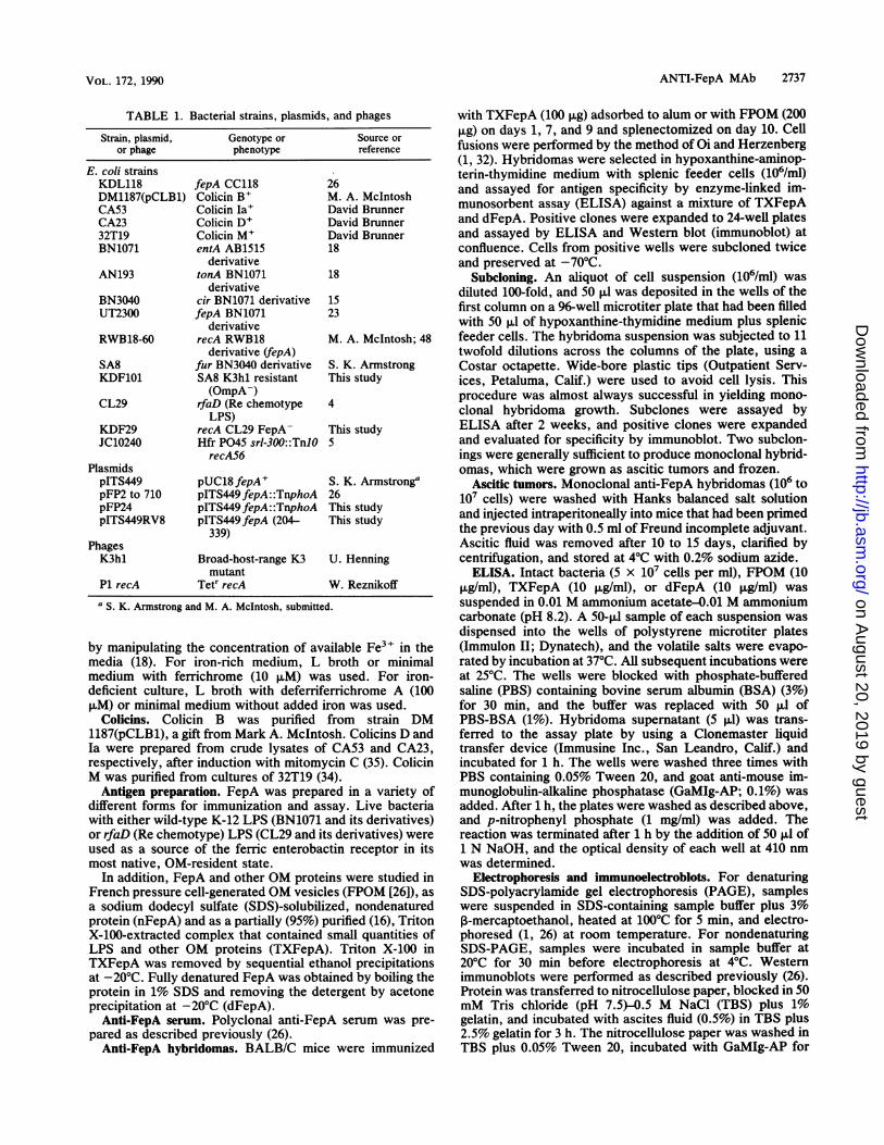
TABLE 1. Bacterial strains, plasmids, and phages
Strain, plasmid, Genotype or Source oror phage phenotype reference
E. coli strainsKDL118 fepA CC118 26DM1187(pCLB1) Colicin B+ M. A. McIntoshCA53 Colicin Ia+ David BrunnerCA23 Colicin D+ David Brunner32T19 Colicin M' David BrunnerBN1071 entA AB1515 18
derivativeAN193 tonA BN1071 18
derivativeBN3040 cir BN1071 derivative 15UT2300 fepA BN1071 23
derivativeRWB18-60 recA RWB18 M. A. McIntosh; 48
derivative (fepA)SA8 fur BN3040 derivative S. K. ArmstrongKDF101 SA8 K3hl resistant This study
(OmpA-)CL29 rfaD (Re chemotype 4
LPS)KDF29 recA CL29 FepA- This studyJC10240 Hfr P045 srl-300::TnlO 5
recAS6PlasmidspITS449 pUC18 fepA' S. K. ArmstrongapFP2 to 710 pITS449fepA::TnphoA 26pFP24 pITS449fepA::TnphoA This studypITS449RV8 pITS449fepA (204- This study
339)PhagesK3hl Broad-host-range K3 U. Henning
mutantP1 recA Tetr recA W. Reznikoffa S. K. Armstrong and M. A. McIntosh, submitted.
by manipulating the concentration of available Fe3" in themedia (18). For iron-rich medium, L broth or minimalmedium with ferrichrome (10 ,uM) was used. For iron-deficient culture, L broth with deferriferrichrome A (100p,M) or minimal medium without added iron was used.
Colicins. Colicin B was purified from strain DM1187(pCLB1), a gift from Mark A. McIntosh. Colicins D andIa were prepared from crude lysates of CA53 and CA23,respectively, after induction with mitomycin C (35). ColicinM was purified from cultures of 32T19 (34).
Antigen preparation. FepA was prepared in a variety ofdifferent forms for immunization and assay. Live bacteriawith either wild-type K-12 LPS (BN1071 and its derivatives)or rfaD (Re chemotype) LPS (CL29 and its derivatives) wereused as a source of the ferric enterobactin receptor in itsmost native, OM-resident state.
In addition, FepA and other OM proteins were studied inFrench pressure cell-generated OM vesicles (FPOM [26]), asa sodium dodecyl sulfate (SDS)-solubilized, nondenaturedprotein (nFepA) and as a partially (95%) purified (16), TritonX-100-extracted complex that contained small quantities ofLPS and other OM proteins (TXFepA). Triton X-100 inTXFepA was removed by sequential ethanol precipitationsat -20°C. Fully denatured FepA was obtained by boiling theprotein in 1% SDS and removing the detergent by acetoneprecipitation at -20°C (dFepA).
Anti-FepA serum. Polyclonal anti-FepA serum was pre-pared as described previously (26).
Anti-FepA hybridomas. BALB/C mice were immunized
with TXFepA (100 ,ug) adsorbed to alum or with FPOM (200,g) on days 1, 7, and 9 and splenectomized on day 10. Cellfusions were performed by the method of Oi and Herzenberg(1, 32). Hybridomas were selected in hypoxanthine-aminop-terin-thymidine medium with splenic feeder cells (106/ml)and assayed for antigen specificity by enzyme-linked im-munosorbent assay (ELISA) against a mixture of TXFepAand dFepA. Positive clones were expanded to 24-well platesand assayed by ELISA and Western blot (immunoblot) atconfluence. Cells from positive wells were subcloned twiceand preserved at -70°C.
Subcloning. An aliquot of cell suspension (106/ml) wasdiluted 100-fold, and 50 ,ul was deposited in the wells of thefirst column on a 96-well microtiter plate that had been filledwith 50 RI of hypoxanthine-thymidine medium plus splenicfeeder cells. The hybridoma suspension was subjected to 11twofold dilutions across the columns of the plate, using aCostar octapette. Wide-bore plastic tips (Outpatient Serv-ices, Petaluma, Calif.) were used to avoid cell lysis. Thisprocedure was almost always successful in yielding mono-clonal hybridoma growth. Subclones were assayed byELISA after 2 weeks, and positive clones were expandedand evaluated for specificity by immunoblot. Two subclon-ings were generally sufficient to produce monoclonal hybrid-omas, which were grown as ascitic tumors and frozen.
Ascitic tumors. Monoclonal anti-FepA hybridomas (106 to107 cells) were washed with Hanks balanced salt solutionand injected intraperitoneally into mice that had been primedthe previous day with 0.5 ml of Freund incomplete adjuvant.Ascitic fluid was removed after 10 to 15 days, clarified bycentrifugation, and stored at 4°C with 0.2% sodium azide.ELISA. Intact bacteria (5 x 10⁷ cells per ml), FPOM (10
,ug/ml), TXFepA (10 ,ug/ml), or dFepA (10 p,g/ml) wassuspended in 0.01 M ammonium acetate-0.01 M ammoniumcarbonate (pH 8.2). A 50-pAl sample of each suspension wasdispensed into the wells of polystyrene microtiter plates(Immulon II; Dynatech), and the volatile salts were evapo-rated by incubation at 37°C. All subsequent incubations wereat 25°C. The wells were blocked with phosphate-bufferedsaline (PBS) containing bovine serum albumin (BSA) (3%)for 30 min, and the buffer was replaced with 50 pul ofPBS-BSA (1%). Hybridoma supernatant (5 pA) was trans-ferred to the assay plate by using a Clonemaster liquidtransfer device (Immusine Inc., San Leandro, Calif.) andincubated for 1 h. The wells were washed three times withPBS containing 0.05% Tween 20, and goat anti-mouse im-munoglobulin-alkaline phosphatase (GaMIg-AP; 0.1%) wasadded. After 1 h, the plates were washed as described above,and p-nitrophenyl phosphate (1 mg/ml) was added. Thereaction was terminated after 1 h by the addition of 50 pA of1 N NaOH, and the optical density of each well at 410 nmwas determined.
Electrophoresis and immunoelectroblots. For denaturingSDS-polyacrylamide gel electrophoresis (PAGE), sampleswere suspended in SDS-containing sample buffer plus 3%P-mercaptoethanol, heated at 100°C for 5 min, and electro-phoresed (1, 26) at room temperature. For nondenaturingSDS-PAGE, samples were incubated in sample buffer at20°C for 30 min before electrophoresis at 4°C. Westernimmunoblots were performed as described previously (26).Protein was transferred to nitrocellulose paper, blocked in 50mM Tris chloride (pH 7.5)-0.5 M NaCl (TBS) plus 1%gelatin, and incubated with ascites fluid (0.5%) in TBS plus2.5% gelatin for 3 h. The nitrocellulose paper was washed inTBS plus 0.05% Tween 20, incubated with GaMIg-AP for
VOL. 172, 1990
on August 20, 2019 by guest
http://jb.asm.org/
Dow
nloaded from

2738 MURPHY ET AL.
2 h, washed, and developed in bromochloroindolyl phos-phate (2).
Antibody isotypes and concentrations. The immunoglobulinheavy-chain class of each MAb was determined by ELISA.Plates were coated with a mixture of TXFepA (10 ,ug/ml) anddFepA (10 ,ug/ml), blocked with 1% BSA for 1 h, andincubated with anti-FepA ascitic fluid (1%) for 1 h. Class-specific GaMIg (Zymed) was added, the preparation wasincubated for 1 h, and the assay was developed with rabbitanti-goat AP (Sigma).Antibody concentrations were also determined by ELISA.
Microtiter plates were coated with GaMIg (Fc specific) byincubation with a 0.1% solution of the sera in 0.01 MNaHPO4 (pH 7.5) at 4°C for 2 h. Serial 10-fold dilutions ofanti-FepA ascitic fluid were added to the washed plates andincubated for 1 h. The assay was developed with GaMIg-AP-p-nitrophenyl phosphate. Monoclonal mouse immuno-globulins of each heavy-chain class, quantitated by proteindetermination (19), were assayed as standards. For flowcytometry and assays of antibody inhibition of colicin orsiderophore binding, MAb were adjusted to approximately 1,ug/ml.Flow cytometry. Whole cells or OM vesicles were reacted
with diluted ascites fluid and fluorescein isothiocyanate(FITC)-conjugated GaMIg as described previously (26), ex-cept that vesicles were centrifuged at 11,000 x g for 30 min.Stained material was analyzed on a Coulter EPICS V flowcytometer/cell sorter for green fluorescence.
Ferric siderophores. Purified enterobactin (1.5 ,umol) wasdissolved in 0.5 ml of methanol, and FeCl3 (0.5 ,umol)dissolved in 0.4 ml of 0.05 N HCI was added. For prepara-tion of [59Fe]enterobactin, 59FeCl3 was used (0.5 ,umol; 5.5mCi/,umol; Dupont, NEN Research Products, Boston,Mass.). The mixture was incubated for 30 min, and 0.1 ml of0.5 M NaHPO4 (pH 7.0) was added to form the ironcomplex. The solution was analyzed spectrophotometricallyto determine its exact concentration of ferric enterobactin(eMM = 5.6) and adjusted to 0.5 mM if necessary by additionof distilled water.
Ferrichrome, purified from Ustilago sphaerogena (29),was reconstituted in 0.05 M NaHPO4 (pH 7.0) at 0.5 mM anddiluted as necessary. [59Fe]ferrichrome was prepared in thefollowing manner. Ferrichrome (25 mg in 1.25 ml of distilledwater) was deferrated by incubation with 1 N NaOH (1.25ml) for 30 min at 0°C. Insoluble Fe(OH)n was removed bycentrifugation, and the solution of deferriferrichrome wasadjusted to neutrality by addition of 1 N HCl at 0°C.[59Fe]ferrichrome was formed by the addition of 1.5 ,mol ofdeferriferrichrome to 0.5 ,umol of 59FeCl3 in 0.05 N HCl. Thesolution was analyzed spectrophotometrically (erm = 2.9) todetermine its exact concentration of ferrichrome and ad-justed to 0.5 mM if necessary by addition of distilled water.
Siderophore uptake experiments. Two methods were usedto assess the ability of MAbs to inhibit siderophore uptake.As an initial screen of the antibody test panel, a modificationof the siderophore nutrition assay (48) was used. A total of107 cells of the entA strain BN1071, grown previously innutrient broth containing 100 puM deferriferrichrome A, wereincubated with 0.01 pig of anti-FepA MAb for 30 min. Then1 ml of molten nutrient agar containing 100 ,uM deferriferri-chrome A was added, and the cells were plated in 45-mm-diameter dishes. A sterile disk containing 2 pI of 50 puMsiderophore (either ferric enterobactin or ferrichrome) wasapplied to the center of the dish, which was incubated at37°C. The presence or absence of a growth halo around thedisk was scored after 4, 6, and 8 h. To evaluate the
specificity of antibody inhibition of siderophore uptake,inhibition of ferrichrome transport was also tested.To quantitatively assess the ability of anti-FepA MAb to
inhibit ferric enterobactin transport, 59Fe-siderophore up-take experiments were performed (23). BN1071 or KDF29was cultured in L broth and subjected to iron stress at adensity of 8 x 10⁷ cells per ml by the addition of 100 puMdeferriferrichrome A (18). After 3 h, the bacteria werepelleted by centrifugation and then washed with and sus-pended in uptake medium (M63 minimal medium [24]) at 4 x10⁹/ml. Bacteria (108) were incubated with ascitic fluidcontaining individual MAb (10 ,ug in 25 ,ul) for 30 min at 0°C;1 ml of uptake medium was added, and the bacteria wereincubated for 10 min at 37°C with shaking (250 rpm).[59Fe]enterobactin or [59Fe]ferrichrome was added to 1 ,uM,and the bacteria were incubated for 15 min at 37°C withshaking. The cells were diluted to 4 ml with 10 mM EDTA,filtered onto 0.2-p.m-pore-size filters, and washed with 10 mlof 0.9% saline. The filters were counted in a Packard gammascintillation counter.
Colicin binding experiments. Anti-FepA MAb were testedfor their ability to block colicin killing by a modification ofthe method of Guterman (12, 13). BN1071 or its fepA, cir, ortonA derivatives were diluted in L broth to 104 cells per ml,and 100 ,ul was incubated with 0.01 p.g of MAb for 15 min. Adilution of appropriate colicin (in L broth) that gave approx-imately one hit per cell was added and incubated for 15 min.Soft agar (2.5 ml [24]) was added, and the mixture waspoured onto LB plates and incubated overnight at 37°C.Colonies were counted, and the number of hits per cell in thepresence or absence of the anti-FepA MAb was determinedby the relationship SISO = e-k, where SO is the number ofcells plated (colonies in the absence of colicin), S is thenumber of cells that survive exposure to colicin (colonies inthe presence of colicin), and k is the number of hits per cell(12, 13).
RESULTS
Generation and characterization of anti-FepA MAbs. Anti-FepA hybridomas were identified initially by ELISA, usingmicrotiter plates coated with a mixture of TXFepA anddFepA, even though these preparations contained minoramounts of other OM antigens, including TonA, OmpA, Cir,OmpF, and LPS. Positive hybridomas were expanded andimmediately assayed by Western immunoblot to determinetheir specificity. Roughly 80% of the hybridomas isolated inthis manner recognized FepA, and the remainder reactedwith the other antigens noted above. Western analysis at thisstage was essential to the ultimate isolation of monoclonalhybridomas and the understanding of MAb specificity, be-cause several of the initial isolates were not monoclonal andproduced multiple different antibodies that recognizedTonA, OmpA, OmpF, or LPS in addition to FepA. Eachhybridoma was subcloned (usually twice) until monoclonalspecificity could be demonstrated by immunoblot.Hybridomas were grown as ascitic tumors, and ascitic
fluids were collected, diluted, and assayed by ELISA andWestern blot. A test panel containing 36 antibodies ofinterest was assembled. MAb in ascitic fluids were charac-terized with respect to immunoglobulin isotype and concen-tration (Table 2); the antibodies in the panel were normalizedto 0.1 p.g/ml for cytofluorimetric studies.MAb specificity. The panel of 36 MAb was tested by
immunoblot for reactivity with FepA::PhoA fusion proteins(Fig. 1). These chimeric proteins, generated previously by
J. BACTEPJOL.
on August 20, 2019 by guest
http://jb.asm.org/
Dow
nloaded from

ANTI-FepA MAb 2739
TABLE 2. MAb characteristics
Heavy ~~~~~~~~~~FACSbEpitopel MAb Heavy 81K*chain SA8wc KDF29(pITS449) SA8FPOM
FepA2-24 29 G2b - - -27-37a 1 Gl - - -27-37b 6 Gl - - +27-37a 20 Gl - -
27-37a 26 G2b27-37a 47 G2b100-178 2 G2b + - -
3 G2a + - -4 NDC + - _5 G2b + - -7 G2b + - _10 Gl + - -11 G2a + -
17 Gl + - -27 ND + - -30 G2b + - -38 Gl + - -39 ND + - -41 G3 + - -
204-227 33 Gl + - +258-290a 16 G2b + -
258-290b 34 Gl + - +290-339a 31 Gl + + + +290-339a 35 G3 + + + +290-339b 37 G2b + + + +290-339a 44 G2b + + + +290-339a 45 G2b + + + +382-400a 23 G2b + +382-400b 24 G2a + +
OmpA 19 Gl - +dCir 9 G2b - _eTonA 32 G2b -
36 G3 -LPS 46 G2b - +f+
53 Gl - + + +
a FepA epitopes were defined by MAb by using immunoblot analysis, cytofluorimetry, and ligand inhibition experiments as described in Materials and Methods.b Flow cytometry and fluorescence-activated cell sorting (FACS) were carried out as described previously (26). Values were considered positive if the mean
peak fluorescence was significantly higher than for the background or control population (see Fig. 2). Control populations were as follows: for SA8 whole cells(SA8wc), RWB18-60; for KDF29(pITS449), plasmidless KDF29; for SA8FPOM, RWB18-6OFPOM.
c ND, Not done.d MAb19 was reactive with both SA8FPOM and RWB18-60FPOM by flow cytometry. The negative control population used for this MAb was KDF1O1FPOM
(OmpA-; see Fig. 2D).e MAb9 was reactive with RWB18-60 whole cells (Cir+) but not SA8 (Cir-).f MAb46 was reactive with whole cells of SA8 and RWB18-60 and with FPOM derived from both of these strains. It failed to react with both rfaD and galE
(KDL118) whole cells by flow cytometric analysis (data not shown).
transposition of TnphoA into the fepA structural gene ofpITS449, contain various lengths of the amino terminus ofthe FepA protein (26). Since the level of FepA expressionand its susceptibility to endogenous proteolysis variedamong the mutant strains (26), the amount of full-lengthmutant protein in each sample was initially estimated byimmunoblot with polyclonal anti-PhoA or anti-FepA serum,and sample concentrations were appropriately normalizedfor further experiments (Fig. 1). Immunoblot analysis of theFepA::PhoA fusion polypeptides revealed seven distinctdomains of the receptor (residues 2 to 24, 27 to 56, 100 to178, 178 to 227, 258 to 290, 290 to 352, and 382 to 400) thatwere recognized by antibodies within the test panel (Table2). An example of the immunoblot mapping procedure isshown in Fig. 1 for MAb reactive with regions 2 to 27, 27 to56, 258 to 290, 290 to 352, and 382 to 400; antibodies thatrecognized regions 100 to 178 and 178 to 227 were alsoidentified (not shown).MAb that reacted with regions 178 to 227 and 290 to 352
were assayed by immunoblot against a mutant FepA poly-peptide containing the internal, EcoRV-generated in-framedeletion of amino acids 204 to 339 to further refine theboundaries of their epitopes. All MAb in both groups wereunreactive with the FepA deletion (not shown), indicatingthat the epitopes recognized by these antibodies residewithin regions 204 to 227 and 290 to 339, respectively.Mature FepA contains four potential OmpT cleavage sites
(43), at positions 37, 147, 172, and 680. When FepA wastruncated by OmpT (8, 43), antibodies that reacted withepitopes within regions 2 to 24 (MAb 29) and 27 to 56 (MAb1, 6, 20, 26, and 47) did not bind the resultant 81K* (16)polypeptide (Table 2). MAb that recognized residues withinregion 100 to 178 or further downstream, on the other hand,bound both 81K* and FepA on immunoblots (Table 2).These results define epitopes that are upstream from or
include the OmpT cleavage site within FepA. They are
consistent with OmpT cleavage of FepA at position 37.
VOL. 172, 1990
on August 20, 2019 by guest
http://jb.asm.org/
Dow
nloaded from

2740 MURPHY ET AL.
A
5 6 8 Y
BMAb 29
3 4
34 44
5 6 7 8 1>0ci
FIG. 1. (A) Quantitation of FepA::PhoA fusion proteins by im-munoblot. KDL118 cells containing pFP fusion plasmids (26) wererun on SDS-PAGE and immunoblotted, using rabbit anti-AP sera toestimate the amount of full-length FepA::PhoA fusion protein ineach sample (indicated by arrowheads). FepA::PhoA fusion pro-teins from plasmids pFP2 (lane 1), pFP24 (lane 2), pFP27 (lane' 3),pFP56 (lane 4), pFP258 (lane 5), pFP290 (lane 6), pFP352 (lane 7),pFP382 (lane 8), and pFP400 (lane 9) were analyzed. (B) Immuno-blots of FepA::PhoA fusion proteins with anti-FepA MAb. Wholecells (-5 x 10⁷) containing fusion proteins were subjected toSDS-PAGE and then immunoblotted with different MAb, as shown.E. coli KDL118 contained plasmids pFP2 (lane 1), pFP24 (lane 2),pFP27 (lane 3); pFP56 (lane 4), pFP258 (lane 5), pFP290 (lanes 6 and7), pFP352 (lane 8), pFP382 (lane 9), and pFP400 (lane 10).
Additional' cleavage at 147, 172, or 680 is inconsistent withthe observed molecular mass of 81K* (74 kilodaltons [kDa]).Other hybridomas were isolated which produced antibod-
ies that reacted with Cir (MAb 9), TonA (MAb 32 and 36)OmpA (MAb 19), and LPS (MAb 46 and 53) (Table 2); thesewere used as controls in the 'analysis of anti-FepA MAbsurface reactivity and ligand-binding inhibition (see below).
Surface-reactive MAb. To identify MAb that recognizedsurface-exposed determinants of FepA, the test panel wasassayed cytofluorimetrically for reactivity'with intact bacte-ria. Cells of the rfa+ strain SA8, the rfaD strain KDF29, ortheir OM protein-deficient or plasmid-containing derivatives(Table 1) were incubated with MAb, stained with FITC-GaMIg, and analyzed for green fluorescence by flow cytom-etry. FepA expression in these bacteria was maximized bythe fur nmutation of SA8 and growth of KDF29(pITS449) iniron-deficient minimal media. The overexpression of'FepAin these strains was confirmed by immunoblot (data notshown).MAb 31, 35, 44, and 45 bound to SA8 (fepA+) cells but not
to RWB18-60 (fepA), indicating their specificity for outersurface-exposed epitopes of FepA (Table 2; Fig. 2A). Theseantibodies also exhibited FepA-dependent adsorption toKDF29(pITS449). MAb 37 was similar but distinct in that itsbinding to FepA in SA8 was only weakly positive and greatly
A
C
2
3
4
B
2
3
4
D
2
3
-4
log fluorescenceFIG. 2. Fluorescence histograms of cells or vesicles stained with
anti-FepA MAb and FITC-GaMIg. (A) Whole cells-of SA8 (panel 1),RWB18-60 (panel'2), KDF29(pITS449) (panel 3), or KDF29 (panel 4)were reacted with MAb 44 and GaMIg-FITC and analyzed for greenfluorescence on the flow cytometer. (B) Panels are the same as forpart A, but MAb 37 was used instead of MAb 44. (C) Panels are thesame as in parts A and B, but MAb 6 was used to stain the bacteria.(D) FPOM from SA8 (panel 1) and KDF101 (panel 2) and- whole cellsof SA8 (panel 3) or KDF29 (panel 4) were reacted with MAb 19 andstained with GaMIg-FITC.
enhanced in the rfaD strain (Fig. 2B). MAb 31, 35, 37, 44,and 45 all reacted with FepA within region 290 to 339.Another category of antibodies was found, including MAb 6,23, 24, 33, and'34, which showed FepA-dependent binding towhole cells of KDF29(pITS449) but not SA8 (Fig. 2C). MAbwithin this group recognized FepA eiptopes that wereshielded from antibody binding in SA8 by the- LPS core andbecame accessible to antibody binding only in its absence. Itis relevant that all of the antibodies in this category boundFepA epitopes that were distinct from the residues withinregion 290 to 339 (Table 2).The Cir-specific MAb 9 reacted cytofluorimetrically with
cir+ strains KDF29 and RWB18-60 but not the cir strain SA8(Table 2), indicating that it binds an outer surface epitope ofthe colicin I receptor. Numerous antibodies were found thatdid not recognize OM proteins in immunoblots but reacted
J. BACTERIOL.
..> 1. M., 1. [>..doww>"a
i
on August 20, 2019 by guest
http://jb.asm.org/
Dow
nloaded from
ANTI-FepA MAb 2741
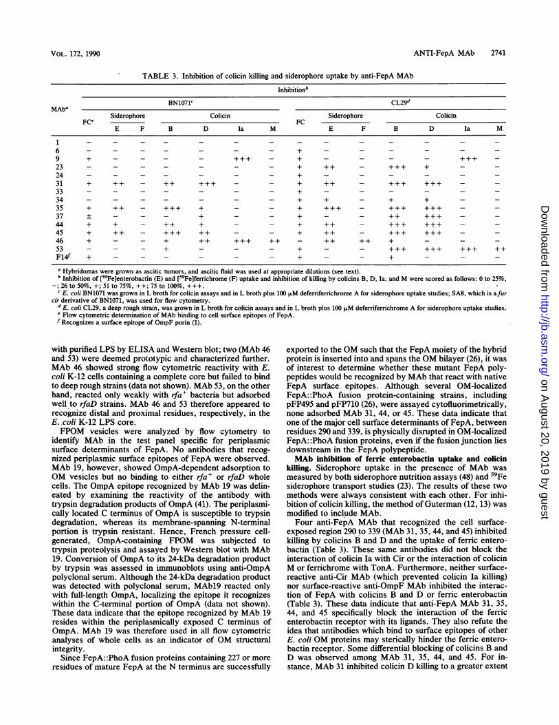
TABLE 3. Inhibition of colicin killing and siderophore uptake by anti-FepA MAb
Inhibitionb
BN1071c CL29dMAba
Siderophore Colicin Siderophore ColicinFCe FC
E F B D Ia M E F B D Ia M
1 - - - - - - - _ - _ _ _ _ _6 - - - - - - - + - _ - _ _ _9 + - - - - +++ - + _ _ +++23 - - - - - - - + ++ - +++ + - -
24 - - - - - - - + - - - - - -
31 + ++ - + +++ - - + ++ - +++ +++ - -33 - - - - - - - + - - - - - -
34 - - - - - - - + + - + + - -
35 + ++ - +++ + - - + +++ - +++ +++ - -37 ± - - - + - + - - +++ - -44 + + - ++ + - - + ++ - +++ +++ - -45 + ++ - +++ ++ - - + ++ - +++ +++ - -46 + - - + ++ ++++++ - + + +53 - - - + - - - + - - +++ +++ +++ ++F14f + - - - - - - + - - + - - -
a Hybridomas were grown as ascitic tumors, and ascitic fluid was used at appropriate dilutions (see text).b Inhibition of [59Fe]enterobactin (E) and [59Fe]ferrichrome (F) uptake and inhibition of killing by colicins B, D, Ia, and M were scored as follows: 0 to 25%,
-; 26 to 50%, +; 51 to 75%, ++; 75 to 100o, +++.c E. coli BN1071 was grown in L broth for colicin assays and in L broth plus 100 FLM deferriferichrome A for siderophore uptake studies; SA8, which is afur
cir derivative of BN1071, was used for flow cytometry.d E. coli CL29, a deep rough strain, was grown in L broth for colicin assays and in L broth plus 100 FM deferriferrichrome A for siderophore uptake studies.e Flow cytometric determination of MAb binding to cell surface epitopes of FepA.f Recognizes a surface epitope of OmpF porin (1).
with purified LPS by ELISA and Western blot; two (MAb 46and 53) were deemed prototypic and characterized further.MAb 46 showed strong flow cytometric reactivity with E.coli K-12 cells containing a complete core but failed to bindto deep rough strains (data not shown). MAb 53, on the otherhand, reacted only weakly with rfa+ bacteria but adsorbedwell to rfaD strains. MAb 46 and 53 therefore appeared torecognize distal and proximal residues, respectively, in theE..coli K-12 LPS core.FPOM vesicles were analyzed by flow cytometry to
identify MAb in the test panel specific for periplasmicsurface determinants of FepA. No antibodies that recog-nized periplasmic surface epitopes of FepA were observed.MAb 19, however, showed OmpA-dependent adsorption toOM vesicles but no binding to either rfa+ or rfaD wholecells. The OmpA epitope recognized by MAb 19 was delin-eated by examining the reactivity of the antibody withtrypsin degradation products of OmpA (41). The periplasmi-cally located C terminus of OmpA is susceptible to trypsindegradation, whereas its membrane-spanning N-terminalportion is trypsin resistant. Hence, French pressure cell-generated, OmpA-containing FPOM was subjected totrypsin proteolysis and assayed by Western blot with MAb19. Conversion of OmpA to its 24-kDa degradation productby trypsin was assessed in immunoblots using anti-OmpApolyclonal serum. Although the 24-kDa degradation productwas detected with polyclonal serum, MAb19 reacted onlywith full-length OmpA, localizing the epitope it recognizeswithin the C-terminal portion of OmpA (data not shown).These data indicate that the epitope recognized by MAb 19resides within the periplasmically exposed C terminus ofOmpA. MAb 19 was therefore used in all flow cytometricanalyses of whole cells as an indicator of OM structuralintegrity.
Since FepA: :PhoA fusion proteins containing 227 or moreresidues of mature FepA at the N terminus are successfully
exported to the OM such that the FepA moiety of the hybridprotein is inserted into and spans the OM bilayer (26), it wasof interest to determine whether these mutant FepA poly-peptides would be recognized by MAb that react with nativeFepA surface epitopes. Although several OM-localizedFepA: :PhoA fusion protein-containing strains, includingpFP495 and pFP710 (26), were assayed cytofluorimetrically,none adsorbed MAb 31, 44, or 45. These data indicate thatone of the major cell surface determinants of FepA, betweenresidues 290 and 339, is physically disrupted in OM-localizedFepA: :PhoA fusion proteins, even if the fusion junction liesdownstream in the FepA polypeptide.MAb inhibition of ferric enterobactin uptake and colicin
killing. Siderophore uptake in the presence of MAb wasmeasured by both siderophore nutrition assays (48) and 59Fesiderophore transport studies (23). The results of these twomethods were always consistent with each other. For inhi-bition of colicin killing, the method of Guterman (12, 13) wasmodified to include MAb.Four anti-FepA MAb that recognized the cell surface-
exposed region 290 to 339 (MAb 31, 35, 44, and 45) inhibitedkilling by colicins B and D and the uptake of ferric entero-bactin (Table 3). These same antibodies did not block theinteraction of colicin Ia with Cir or the interaction of colicinM or ferrichrome with TonA. Furthermore, neither surface-reactive anti-Cir MAb (which prevented colicin la killing)nor surface-reactive anti-OmpF MAb inhibited the interac-tion of FepA with colicins B and D or ferric enterobactin(Table 3). These data indicate that anti-FepA MAb 31, 35,44, and 45 specifically block the interaction of the ferricenterobactin receptor with its ligands. They also refute theidea that antibodies which bind to surface epitopes of otherE. coli OM proteins may sterically hinder the ferric entero-bactin receptor. Some differential blocking of colicins B andD was observed among MAb 31, 35, 44, and 45. For in-stance, MAb 31 inhibited colicin D killing to a greater extent
VOL. 172, 1990
on August 20, 2019 by guest
http://jb.asm.org/
Dow
nloaded from

2742 MURPHY ET AL.
than colicin B killing, whereas MAb 35, 44, and 45 blockedcolicin B more efficiently than colicin D. This phenomenonwas also observed for MAb 37, which did not significantlyinhibit colicin B killing of BN1071 but did weakly retardkilling of this strain by colicin D.The biochemical significance of antibody binding to pep-
tides within region 290 to 339 was emphasized by theinability of several other surface-reactive anti-FepA MAb toblock the interaction of colicins and ferric enterobactin withthe receptor. Antibodies that bound surface determinantswithin regions 27 to 37 (MAb 6), 204 to 227 (MAb 33), 258 to290 (MAb 34), and 382 to 400 (MAb 23 and 24) were analyzedin a deep rough, rfaD background [KDF29(pITS449)] be-cause, as described above, an intact E. coli K-12 LPS coreprevented their adsorption to FepA epitopes. When evalu-ated cytofluorimetrically, these antibodies exhibited FepA-dependent cell surface binding to KDF29(pITS449), butthree of the five (MAb 6, 33, and 24) showed no inhibition ofligand interaction with FepA in this strain. Only MAb 23 and34 blocked ferric enterobactin uptake into and colicin B andD killing of KDF29(pITS449); inhibition by MAb 34 wasquantitatively less than that caused by MAb 23 for all threeligands. MAb 31, 35, 44, and 45 showed greater ability toprotect the rfaD strain against colicin killing and inhibitferric enterobactin transport than did the rfa+ strain BN1071(Table 2). MAb 37, which recognized an epitope withinregion 290 to 339 but did not significantly inhibit colicin Bkilling or ferric enterobactin uptake in the rfa' strain,strongly blocked both colicins and siderophore inKDF29(pITS449) (Table 3). This inhibitory effect of theintact LPS core on the reactivity of MAb 37 differentiates itfrom the other antibodies that bind epitopes within region290 to 339.
Unlike the anti-FepA- or anti-Cir-mediated inhibition ofkilling by colicin B or Ia, respectively, anti-LPS MAb 46 and53 reacted with residues in the LPS core (see above) toprevent the killing by colicins B, D, Ia, and M. Thisphenomenon is illustrated by the fact that MAb 46, whichapparently recognizes residues in the distal portion of theLPS core (see above), inhibited colicin killing of rfa+ strains,whereas MAb 53, which reacts with LPS either in the2-keto-3-deoxyoctulosonic acid region of core or in the lipidA moiety, significantly inhibited colicin killing only in deeprough, rfaD strains (Table 3). These data indicate thatantibody binding to LPS may generally inhibit the killingactivity of group B colicins, ostensibly through steric hin-drance at the stage of adsorption to the OM.
Native FepA structure. FepA and Cir migrated with char-acteristic mobilities in SDS-PAGE after denaturation byheating in solutions of SDS to Rf values of 0.34 (81 kDa) and0.37 (74 kDa), respectively (Fig. 3). The availability ofanti-FepA and anti-Cir MAb allowed us to determine themobilities of these OM proteins in SDS-PAGE without heatdenaturation and, in the process, identify electrophoreticforms of FepA and Cir that were previously unrecognized. Ifsolubilized in SDS without heating and subjected to SDS-PAGE, both proteins exhibited multiple electrophoreticforms with increased mobilities relative to those of theSDS-denatured proteins (Fig. 3). When such gels weresubjected to Western immunoblotting, FepA showed a mi-nor band of Rf 0.45 (61 kDa) and a major band of Rf 0.47 (56kDa). The latter band could be resolved as a triplet onextended electrophoresis (data not shown). Cir had twomajor nondenatured forms (Fig. 3) with RA of 0.53 and 0.59(46 and 37 kDa, respectively) and a triplet of minor bandswith an approximate Rf of 0.56 (43 kDa).
d FepA
t_-~~~o_dCir
.4547 _ _ nFepA
.53
.56 _W _ nCir
.59 _ mm
1 2 3 4 5 6
FIG. 3. Western immunoblot of boiled (lanes 1 to 3) and unboiled(lanes 4 to 6) E. coli K-12 strain BN1071 FPOM, reacted withanti-FepA MAb 29 (lanes 1, 2, 4, and 5) or anti-Cir MAb 9 (lanes 1,3, 4, and 6). Note that lanes 1 and 4 contain both MAb 29 and MAb9. The positions of denatured (d) and nondenatured (n) FepA andCir, as well as the Rjs of the nondenatured bands, are indicated.
DISCUSSION
One of the unusual characteristics of enteric bacterial OMproteins is their ability to interact with several structurallydifferent nutrilites, toxins, or bacteriophage (30, 31). Al-though such multifunctionality is a common trait among OMproteins, the molecular mechanisms of these receptor-ligandinteractions are not understood. Our immunochemical anal-ysis of FepA addresses the nature of its ligand-binding sites:do ferric enterobactin, colicin B, and colicin D all interactwith FepA within a single structural domain of the protein,or does the receptor contain multiple ligand-binding regions?The results we report suggest that all three ligands bindFepA within a relatively short, surface-exposed sequence ofthe receptor that is bounded by residues 290 and 339.Antibody recognition of a surface-exposed epitope in thisregion inhibits both ferric enterobactin uptake into the celland killing by either colicin. By themselves, these data donot definitively implicate the epitope within region 290 to 339as a ligand-binding domain; other explanations are possible.For example, residues 290 to 339 may be physically distinctfrom the ligand-binding site but in such close proximity thatantibody binding to this region inhibits receptor-ligand con-tact. Alternatively, residues 290 to 339 may be distant fromthe siderophore- and colicin-binding domain, but antibodybinding to these residues may prevent ligand recognition ortransport by inducing or retarding conformational changes inFepA structure. Other results, nevertheless, support theidea that amino acids within region 290 to 339 participatedirectly in ligand binding. Residues within this region areaccessible to MAb binding in the presence of an rfa' LPScore, whereas other surface epitopes of FepA (within resi-dues 27 to 37, 204 to 227, 258 to 290, and 382 to 400) areobscured by rfa+ LPS. These data argue that colicins B andD, which approximate the molecular dimensions of antibod-ies (58 and 92 kDa, respectively [33, 44]), are likewise able tobind region 290 to 339 but are sterically inhibited by LPSfrom contact with regions 27 to 37, 204 to 227, 258 to 290,and 382 to 400.The LPS core may not constitute a barrier to ferric
enterobactin, which is physically much smaller (700 Da) thancolicins or antibodies and may diffuse through the coresugars to interact with other FepA domains. Hypothetically,
J. BACTERIOL.
on August 20, 2019 by guest
http://jb.asm.org/
Dow
nloaded from

ANTI-FepA MAb 2743
ferric enterobactin may bind to FepA regions that are
ensheathed in LPS. The inhibition of ferric enterobactinuptake by MAb 31, 35, 44, and 45 is inconsistent with thisidea, however, because their binding to the exposed epitopein region 290 to 339 is not expected to sterically hinder otherFepA domains that are buried within the LPS core. Further-more, region 290 to 339 is extremely hydrophilic (<h> =
-0.22; Fig. 4) and contains five basic residues that may
participate in electrostatic bonds with the acidic [3(-)] ferricenterobactin. We conclude that either residues 290 to 339 are
directly involved ferric enterobactin binding or MAb recog-
nition of this region allosterically inhibits interaction with thesiderophore at another site.The possibility of antibody-mediated inhibition of al-
lostery within the ferric enterobactin receptor appears un-
likely. Antibody binding to certain surface FepA epitopes(within regions 27 to 37, 204 to 227, and 382 to 400) had no
effect on either ferric enterobactin transport or colicin kill-ing. If conformational changes occur during physiologicalfunction of FepA, they are localized and unaffected byantibody binding within these sites. A final category ofsurface-reactive anti-FepA MAb (MAb 23 and 34) adsorbedonly to deep rough strains and blocked ligand binding(although the inhibition by MAb 34 was weak). This reactiv-ity suggests that residues within regions 258 to 290 and 382 to
400 either participate in or are in close proximity to thesiderophore- and colicin-binding site in region 290 to 339.The model of FepA structure that emerges from theseimmunochemical analyses contains at least five distinctpolypeptide domains that are located at the exterior surfaceof the OM and recognized by MAb. Four of these are
sterically masked by the LPS core, but antibody recognitionof the remaining epitope, between residues 290 and 339, isindependent of core structure. It is likely that FepA ligandsphysically bind to the receptor within this domain. Althoughthe molecular structure of region 290 to 339 cannot bededuced from the available data, the fact that surface-reactive antibodies which recognize this region show quan-
titative differences in the extent to which they inhibit thereceptor-ligand interaction suggests that colicins B and Dand ferric enterobactin may recognize distinct microdomainswithin this region. This assumption agrees with the geneticstudies of McIntosh et al. (23).
All of the anti-FepA MAb characterized in this study,including those that bind surface determinants and blockligand function, ostensibly recognize sequential determi-nants (42). For example, MAb 31, 35, 37, 44, and 45 bind a
site that is entirely contained within region 290 to 339,because they recognize their epitope even when the FepApolypeptide has been denatured by boiling in SDS. Theiraffinity for denatured FepA is not enhanced by the presence
of residues downstream from 290 to 339 and is completelyeliminated by truncations that delete residues 290 to 339.Hence, these antibodies react specifically and exclusivelywith residues within region 290 to 339. These argumentsapply equally well to all of the anti-FepA MAb that we haveisolated and therefore distinguish them from antibodiesraised to E. coli porin trimers. For OmpF, MAb have beenraised that specifically bind conformational determinants inthe cell surface-exposed regions of the trimer; they are
unreactive with denatured OmpF monomer (1). No suchconformation-dependent anti-FepA MAb were found in thetest panel. These differences in the immunochemistry ofporin and FepA probably arise from intrinsic differences inthe stability of their tertiary structures. Porin trimers are
resistant to both heat denaturation and protease degradation,
whereas FepA is both sensitive to denaturation and suscep-tible to proteolysis. Hence, during the process of antigenrecognition by the immune system, FepA may presentsequential determinants, while OmpF may present strictlyconformation epitopes. This interpretation is supported bythe fact that antibodies which recognize sequential OmpFdeterminants can be raised by immunizing mice with SDS-denatured OmpF monomer (1; P. E. Klebba, S. A. Benson,S. Bala, T. Abdullah, J. Reid, S. P. Singh, and H. Nikaido,J. Biol. Chem., in press).We attempted, unsuccessfully, to isolate MAb that recog-
nized periplasmic surface determinants of FepA. Our failureto find hybridomas specific for periplasmic epitopes of FepAmay indicate their lack of immunogenicity. Alternatively,antibodies specific for periplasm-exposed domains of FepAmay exist in the current panel, but FepA may be situated inthe OM such that these regions are inaccessible to antibody.Periplasmic epitopes of FepA could also be conformationaland reactive only with MAb to the native protein. Finally,the panel of anti-FepA MAb that we have compiled is notconsidered exhaustive; periplasmic FepA epitopes may beidentified upon further study.Numerous E. coli membrane proteins exhibit atypical
mobility in SDS-PAGE that depends on sample preparationconditions (25, 28, 38, 40). Such proteins contain extensive Pstructure that imparts a compactness to their form, increas-ing their electrophoretic mobility (40). Heating in SDSconverts them to more diffuse, unfolded structures thatmigrate with consistent Rfs characteristic of their true mo-lecular weights. FepA and Cir also have SDS-stable, com-pact forms that are denatured by heating. The multiplenative FepA and Cir electrophoretic forms may be caused byassociation of the receptors with LPS or may representpartially unfolded conformations of the native OM proteinthat are produced by the in vitro conditions.Using the accumulated data on the surface epitopes and
putative ligand-binding domains of FepA, as well as thepredicted hydrophobicity (6) and hydrophobic moment (7) ofthe receptor, we have constructed a theoretical model of itsstructure in the bacterial OM. We base this hypothesis onthe following facts. (i) FepA contains cell surface-exposedepitopes in regions 27 to 37, 204 to 227, 258 to 290, 290 to339, and 382 to 400. (ii) The predominant secondary struc-ture of FepA is probably P-sheet. Several other entericbacterial OM proteins, including OmpA, OmpF, LamB, andPhoE, consist almost exclusively of P structure (3, 10, 17,37). Although spectrophotometric data on the purified nativereceptor are not available, the heat modifiability of FepA inSDS-PAGE (Fig. 3) is consistent with a compact nativestructure dominated by P-strands (15). (iii) The FepA poly-peptide is not particularly hydrophobic (6), and in factcontains numerous strongly hydrophilic domains inter-spersed throughout its sequence. Examination of FepAhydrophobic moment (7), however, shows that the proteincontains many amphiphilic stretches of sufficient length tospan the OM lipid bilayer in P structure. The mean hydro-phobicities of the opposing faces of these putative P-strandsare consistent with the hypothesis that they interact with ahydrophobic surface on one side and a hydrophilic surfaceon the other (Fig. 4; 36). The hydrophobic faces of theseamphiphilic transmembrane strands are expected to interactwith the OM lipid bilayer; the hydrophilic faces may circum-scribe a water-filled pore (Klebba et al., in press) or may bejuxtaposed against one another. The model was created byidentifying the first potential transmembrane P-strand in thesequence of the mature protein. Since a surface epitope was
VOL. 172, 1990
on August 20, 2019 by guest
http://jb.asm.org/
Dow
nloaded from

2744 MURPHY ET AL. J. BACTERIOL.
0)U,
|1ola a ,o 4: 3 <D g >z
-UErJE D C
t-EiI!r-r*-- 3
-J0-im L J'
CD~~~~1 4-. 3 lE - -'- L - E J D S19 g C - "
CDCDU>F*DU.-I=a
~~~ ~~o
~*s,. g CDUX,
U;-J~~~~~-5 M
o,, ; > ~~~~~~~~~~~~~~~~~~~~~~~~~~P- 4- @ *
3o^>b tD> Sst* Ec
,!^ 2~~~~~~~~~~~~~~~~) .0 0 2t
<cD ACD
-I~~ ~ ~ ~ ~ ~ -
-O 0)4'0
A.fl*~~~~~~* U ~ ~ El 0
* E~~~~~E-
mmw * * I~~~~~~~~~E~~a O .
A. p.~~~~~'a44 WZ~~~U~~HE.E(K~~~~ U =....*:(...P~~~~~~~~~~~~~C
%S UVWol ;-( v~-.0 4 4
-
0 . ..&m 0'
on August 20, 2019 by guest
http://jb.asm.org/
Dow
nloaded from

ANTI-FepA MAb 2745
found immediately downstream from this site (amino acids27 to 37), residues preceding the transmembrane domainwere assumed to reside on the periplasmic face of the OM,and those immediately downstream (to the site of the nexttransmembrane (3-strand) were considered cell surface ex-posed. Succeeding downstream regions, separated by trans-membrane ,B-structures, were assumed to alternate betweenthe periplasmic and outer surfaces of the OM. Although wesearched for potential transmembrane or surface-seekinga-helical domains in FepA, none were found. Turns in thepolypeptide were assigned according to the algorithm ofWilmot and Thornton (49). This approach to the modeling ofFepA predicts several features that are consistent withempirical data. Foremost, each of the immunochemicallydefined cell surface-exposed regions of FepA is predicted bythe method to exist on the exterior surface of the OM.Second, for the complete mature FepA sequence, the meanhydrophobicity of residues proposed to associate with theOM bilayer lipids is 0.38, whereas that for residues proposedto associate with an aqueous interface is -0.17. Thesevalues agree with the average residue hydrophobicities ofbilayer-exposed and aqueous-exposed residues of othermembrane proteins (36). Other points of interest are thelocalization of the FepA proposed TonB consensus region(20) embedded within the OM bilayer at the periplasmicsurface and its OmpT cleavage site on the bacterial cellsurface. Finally, the predicted locations of the sites of PhoAfusion into FepA (26) are dispersed throughout the exteriorsurface, transmembrane strands, and periplasmic surfacedomains of the receptor. More significantly, the immuno-chemically defined surface epitopes of FepA, presentedherein, include sites of PhoA fusion (pFP178, -227, -258,-290, -382, and -400). These data establish that even thoughthe PhoA moiety of FepA::PhoA fusion proteins remainsperiplasmic (26), the sites of PhoA fusion need not beperiplasmic surface domains of FepA. This conclusion issupported by our finding that the immunochemical structureof the FepA portion of FepA::PhoA fusion proteins isdistorted relative to the native ferric enterobactin receptor.
ACKNOWLEDGMENTSWe thank David Eisenberg and Carrie Wilmot for their algorithms
and insights on protein structure prediction and Mark McIntosh,Sandy Armstrong, Ulf Henning, David Brunner, and Bill Reznikofffor bacterial strains and phage.
This work was supported by Public Health Service grant AI22608from the National Institutes of Health.
LITERATURE CITED1. Bentley, A. T., and P. E. Klebba. 1988. Effect of lipopolysac-
charide structure on the reactivity of anti-porin monoclonalantibodies with the bacterial cell surface. J. Bacteriol. 170:1063-1068.
2. Blake, M. S., K. H. Johnston, G. J. Russell-Jones, and E. C.Gotschlich. 1984. A rapid, sensitive method for detection ofalkaline phosphatase-conjugated anti-antibody on westernblots. Anal. Biochem. 1136:175-179.
3. Charbit, A., J. M. Clement, and M. Hofnung. 1984. Furthersequence analysis of the phage lambda receptor site: possibleimplications for the organization of the LamB protein in Esch-erichia coli K12. J. Mol. Biol. 175:395-401.
4. Coleman, W. G., Jr., and L. Leive. 1979. Two mutations whichaffect the barrier function of Escherichia coli K-12 outer mem-brane. J. Bacteriol. 139:899-910.
5. Csonka, L., and A. J. Clark. 1980. Construction of an Hfr strainuseful for transferring recA mutations between Escherichia colistrains. J. Bacteriol. 143:529-530.
6. Eisenberg, D. 1984. Three-dimensional structure of membrane
and surface proteins. Annu. Rev. Biochem. 53:595-623.7. Eisenberg, D., R. M. Weiss, and T. C. Terwilliger. 1984. The
hydrophobic moment detects periodicity in protein hydropho-bicity. Proc. Natl. Acad. Sci. USA 81:140-144.
8. Fiss, E. H., P. Stanley-Samuelson, and J. B. Neilands. 1982.Properties and proteolysis of the ferric enterobactin outermembrane receptor in Escherichia coli K-12. Biochemistry21:4517-4522.
9. Gabay, J., S. Benson, and M. Schwartz. 1983. Genetic mappingof antigenic determinants on a membrane protein. J. Biol.Chem. 258:2410-2414.
10. Garavito, R. M., J. Jenkins, J. N. Jansonius, R. Karlsson, andJ. P. Rosenbusch. 1983. X-ray diffraction analysis of matrixporin, an integral membrane protein from Escherichia coli outermembranes. J. Mol. Biol. 164:313-327.
11. Garnier, J., D. J. Osguthorpe, and B. Robson. 1978. Analysis ofthe accuracy and implications of simple methods for predictingthe secondary structure of globular proteins. J. Mol. Biol.120:97.
12. Guterman, S. 1971. Inhibition of colicin B by enterochelin.Biochem. Biophys. Res. Commun. 44:1149-1155.
13. Guterman, S. K. 1973. Colicin B: mode of action and inhibitionby enterochelin. J. Bacteriol. 114:1225-1230.
14. Guterman, S. K., and L. Dann. 1973. Excretion of enterochelinby exbA and exbB mutants of Escherichia coli. J. Bacteriol.114:1225-1230.
15. Heller, K. B. 1978. Apparent molecular weights of a heat-modifiable protein from the outer membrane of Escherichia coliin gels with different acrylamide concentrations. J. Bacteriol.134:1181-1183.
16. Hollifield, W. C., E. H. Fiss, and J. B. Neilands. 1978. Modifi-cation of a ferric enterobactin receptor protein from the outermembrane of Escherichia coli. Biochem. Biophys. Res. Com-mun. 83:739-746.
17. Jap, B. K. 1988. High-resolution electron diffraction of recon-stituted PhoE porin. J. Mol. Biol. 199:229-231.
18. Klebba, P. E., M. A. McIntosh, and J. B. Neilands. 1982.Kinetics of biosynthesis of iron-regulated membrane proteins inEscherichia coli. J. Bacteriol. 149:880-888.
19. Lowry, 0. H., N. J. Rosebrough, A. L. Farr, and R. J. Randall.1951. Protein measurement with the Folin phenol reagent. J.Biol. Chem. 193:265-275.
20. Lundrigan, M. D., and R. J. Kadner. 1986. Nucleotide sequenceof the gene for the ferrienterochelin receptor FepA in Esche-richia coli: homology among outer membrane receptors thatinteract with TonB. J. Biol. Chem. 261:10797-10801.
21. Maniatis, T., E. F. Fritsch, and J. Sambrook. 1982. Molecularcloning: a laboratory manual. Cold Spring Harbor Laboratory,Cold Spring Harbor, N.Y.
22. Manning, P. A., A. Puspurs, and P. Reeves. 1976. Outer mem-brane of Escherichia coli K-12: isolation of mutants with alteredprotein 3A by using host range mutants of bacteriophage K3. J.Bacteriol. 127:1080-1084.
23. McIntosh, M. A., S. S. Chenault, and C. F. Earhart. 1979.Genetic and physiological studies on the relationship betweencolicin B resistance and ferrienterochelin uptake in Escherichiacoli K-12. J. Bacteriol. 137:653-657.
24. Miller, J. H. 1972. Experiments in molecular genetics, p. 431.Cold Spring Harbor Laboratory, Cold Spring Harbor, N.Y.
25. Mizushima, S. 1974. Effect of sodium dodecylsulfate and heatingon protein conformation in outer and cytoplasmic membranesfrom Escherichia coli. Biochem. Biophys. Res. Commun. 61:1221-1226.
26. Murphy, C. K., and P. E. Klebba. 1989. Export of FepA::PhoAfusion proteins to the outer membrane of Escherichia coli K-12.J. Bacteriol. 171:5894-5900.
27. Nabedryk, E., R. M. Garavito, and J. Breton. 1988. The orien-tation of B-sheets in porin. A polarized Fourier transforminfrared spectroscopic investigation. Biophys. J. 53:671-676.
28. Nakamura, K., and S. Mizushima. 1976. Effects of heating indodecyl sulfate solution on the conformation and electropho-retic mobility of isolated major outer membrane proteins fromEscherichia coli K-12. J. Biochem. 80:1411-1422.
VOL. 172, 1990
on August 20, 2019 by guest
http://jb.asm.org/
Dow
nloaded from

2746 MURPHY ET AL.
29. Neilands, J. B. 1966. Naturally occurring non-porphyrin ironcompounds, p. 59-108. In C. K. Jorgenson, J. B. Neilands,R. S. Nyholm, D. Reinin, and R. J. P. Williams (ed.), Structureand bonding. Springer-Verlag, New York.
30. Neilands, J. B., K. Konopka, B. Schwyn, M. Coy, R. T. Francis,B. A. Paw, and A. Bagg. 1987. Comparative biochemistry ofmicrobial iron assimilation, p. 3-33. In G. Winkelman, D. vander Helm, and J. B. Neilands (ed.), Iron transport in microbes,plants, and animals. VCH Verlagsgesellschaft, Weinheim, Fed-eral Republic of Germany.
31. Nikaido, H., and M. Vaara. 1985. Molecular basis of outermembrane permeability. Microbiol. Rev. 49:1-32.
32. Oi, V. T., and L. A. Herzenberg. 1980. Immunoglobulin-pro-ducing hybrid cell lines, p. 351-372. In B. B. Mishell and S. M.Shiigi (ed.), Selected methods in cellular immunology. W. H.Freeman & Co., San Francisco.
33. Olschlager, T., E. Schramm, and V. Braun. 1984. Cloning andexpression of the activity and immunity genes of colicins B andM on ColBM plasmids. Mol. Gen. Genet. 196:482-487.
34. Pugsley, A. P., and P. Reeves. 1976. Characterization of group Bcolicin-resistant mutants of Escherichia coli K12: colicin resis-tance and the role of enterochelin. J. Bacteriol. 127:218-228.
35. Pugsley, A. P., and P. Reeves. 1977. Comparison of colicinsB-K260 and D-CA23: purification and characterization of thecolicins and examination of colicin immunity in the producingstrains. Antimicrob. Agents Chemother. 11:345-358.
36. Rees, D. C., L. DeAntonio, and D. Eisenberg. 1989. Hydropho-bic organization of membrane proteins. Science 245:510-513.
37. Reithmeier, R. A. F., and P. D. Bragg. 1977. Molecular charac-terization of a heat-modifiable protein from the outer membraneof Escherichia coli. Arch. Biochem. Biophys. 178:527-534.
38. Rosenbusch, J. P. 1974. Characterization of the major envelopeprotein from Escherichia coli. Regular arrangement on thepeptidoglycan and unusual dodecyl sulfate binding. J. Biol.Chem. 249:8019-8029.
39. Schenkman, S., E. Couture, and M. Schwartz. 1983. Monoclonal
antibodies reveal LamB antigenic determinants on both faces ofthe Escherichia coli outer membrane. J. Bacteriol. 155:1382-1392.
40. Schnaitman, C. A. 1973. Outer membrane proteins of Esche-richia coli. I. Effect of preparative conditions on the migrationof protein in polyacrylamide gels. Arch. Biochem. Biophys.157:541-552.
41. Schweizer, M., I. Hindennach, W. Garten, and U. Henning.1978. Major proteins of Escherichia coli outer cell envelopemembrane. Interaction of protein II* with lipopolysaccharide.Eur. J. Biochem. 82:211-217.
42. Shinnick, M. T., J. G. Sutcliffe, N. Green, and R. Lerner. 1983.Synthetic peptide immunogens as vaccines. Annu. Rev. Micro-biol. 37:425-446.
43. Sugimura, K., and N. Higashi. 1988. A novel outer-membrane-associated protease in Escherichia coli. J. Bacteriol. 170:3650-3654.
44. Timmis, K. 1972. Purification and characterization of colicin D.J. Bacteriol. 109:12-20.
45. Tommassen, J., and B. Lugtenberg. 1984. Amino terminus ofouter membrane protein PhoE protein: localization by use of abla-phoE hybrid gene. J. Bacteriol. 157:327-329.
46. Vogel, H., and F. Jahnig. 1986. Models for the structure ofouter-membrane proteins of Escherichia coli derived from Ra-man spectroscopy and prediction methods. J. Mol. Biol. 190:191-199.
47. Wang, C. C., and A. Newton. 1971. An additional step in thetransport of iron defined by the tonB locus of Escherichia coli.J. Biol. Chem. 246:2147-2151.
48. Wayne, R., K. Frick, and J. B. Neilands. 1976. Siderophoreprotection against colicins M, B, V, and Ia in Escherichia coli.J. Bacteriol. 126:7-12.
49. Wilmot, C. M., and J. M. Thornton. 1988. Analysis and predic-tion of the different types of P-turn in proteins. J. Mol. Biol.203:221-232.
J. BACTERIOL.
on August 20, 2019 by guest
http://jb.asm.org/
Dow
nloaded from