supporting information - pnas€¦ · supporting information ... (neun) and microglia (iba-1) or...
TRANSCRIPT

Supporting InformationCicchetti et al. 10.1073/pnas.0904239106SI TextSupplemental Clinical Outcome. Significant cortical atrophy wasnoted on the preoperative MRI scan of patient 1 (B.L.). At theend of surgery, brain shrinkage because of cerebrospinal f luidloss was noted. The surface of the brain was �8 mm from theinner cortex of the skull. No subdural hematoma was noted onthe immediate postoperative MRI scan. However, corticalshrinkage led to targeting error in the last needle tract (in theright caudate) (see Fig. S2). A 2-cm thick asymptomatic subacutesubdural hematoma was noted on the localizing MRI scan donebefore the second operation and was drained uneventfully aftercompletion of the contralateral transplant procedure. Follow-upMRI scans demonstrated complete resolution of the hematoma.The Unified Huntington’s Disease Rating Scale (UHDRS)scores improved from 42 at baseline to 30 to 32 at 3 and 9 monthspostoperatively, but returned to baseline (1) by 18 months aftersurgery. Preoperative gait instability with falling improved for2.5 years, whereupon a wheelchair was required and swallowingdifficulties began to emerge. UHDRS scores stabilized until 3.5years after surgery. Scores then progressively deteriorated to 52,65, and 78 at 4, 6, and 7 years postoperatively. Cognitive functionalso progressively declined with Mini Mental State Evaluation(MMSE) scores worsening from 30 at both baseline and at 21months postsurgery to 14 at 7 years postsurgery. She died at 67years of age of an aspiration pneumonia complicated by amyocardial infarction 9 years postoperatively.
Patient 3 (M.C.) had an asymptomatic 1-cm cortical hemor-rhage after her first operation, as well as a thin (3 mm) subduralhematoma without mass effect. She had postoperative confusionthat resolved in 2 weeks. A 4-mm subdural hematoma was notedafter the second operation, and the bilateral thin subduralhematomas both resolved on subsequent postoperative imagingwithout complications. Her preoperative UHDRS score of 33remained unchanged until 10 to 12 months postsurgery, whenscores improved to 28 and 21, respectively. Her preoperativebalance difficulties and falling (once per month) improvedpostoperatively, and she did not fall for at least 2 years aftersurgery. Symptoms then worsened to a UHDRS score of 37 at20 months after surgery and plateaued there until 3 yearspostoperatively. Cognitive function declined mildly with anMMSE score of 26 at baseline and 24 at 30 months postsurgery.Some depressive symptoms emerged at 2 years after surgery. Thepatient died at the age of 75 of a cardiorespiratory arrest ascomplication of end-stage HD, 10.5 years after transplantation.
Patient 5 (M.S.) was noted to have significant cortical andstriatal atrophy on the preoperative MRI scan. She had 7-mmthick bilateral hygromas postoperatively, which increased to 1 cmafter the second operation. She tripped at home, hitting her head2 weeks after her second operation, with a 1- to 2-min loss ofconsciousness. A CT evaluation demonstrated conversion of herhygromas into subdural hematomas that required surgical drain-age bilaterally. The hematomas were 2.5-cm thick on the left sideand 1.0-cm thick on the right side at the time of surgery. The CTscan 1 month later demonstrated complete resolution of thesubdural hematomas. She never improved back to baseline. HerUHDRS score was 27 before surgery and 31 after drainage of hersubdural hematomas. She deteriorated to a score of 39 by 9months postoperation that remained stable until 2.5 years aftersurgery, when she began a more rapid deterioration to a scoreof 53. By 5 years postoperatively, her UHDRS score was 64.MMSE scores declined from 27 at baseline to 16 at 5 years
postsurgery. She died at the age of 68 of a cardiorespiratoryarrest, secondary to end-stage HD, 9.5 years after surgery.
SI MethodsHistochemistry. Sections were washed 3 times in PBS 0.1M beforehistochemical staining for NADPH-d (marker for nitric oxidecontaining striatal interneurons), AChE (enzyme catalyzinghydrolysis of the neurotransmitter acetyl choline in cholinergicneurons), and H&E (brain cytoarchitecture). For NADPH-dstaining, sections were washed in PBS 0.1M pH 7.4, preincubatedin 0.25% Triton X-100 in PBS for 10 min and transferred in afresh solution of 0.25% Triton X-100, 0.05% of the reduced formof nicotinamide adenine dinucleotide phosphate (b-NADPH;Calbiochem), 0.02% nitro blue tetrazolium (Sigma) in PBS for5 min at room temperature and then at 37 °C for 8 h. Sectionswere rinsed in PBS, mounted on gelatin-coated slides, and keptat 37 °C overnight. They were subsequently dehydrated in as-cending grades of ethanol and coverslipped with DPX mountingmedia (Electron Microscopy Science).
For AChE staining, sections were washed 3 times in distilledwater and incubated in 0.2% acetylthiocholine iodide (Sigma) ina stock solution (copper sulfate, glycine, magnesium chloride,maleic acid in 4% NaOH, 40% sodium sulfate). They were thenrinsed 3 times in 40% sodium sulfate, and incubated in 10%ammonium sulfide for 2 min. After washes in distilled water,sections were counterstained with Kernechtrot red (J.T. Baker)for 1 min and rinsed in tap water. Slices were mounted ongelatin-coated slides, air-dried overnight, dehydrated in ascend-ing grades of ethanol, and cover-slipped with DPX mountingmedia.
For H&E staining, sections were first mounted on gelatin-coated slides, air-dried overnight, and hydrated in 50% ethanol.They were then stained with hematoxylin (Fisher Scientific) for40 sec, washed in running water for 5 min, and placed indifferentiator solution (0.5% pure glacial acetic acid in 95%ethanol) for 1 min. Sections were washed using distilled waterand counterstained with Eosin Y (Sigma) for 40 sec, dehydratedin ethanol and xylene baths (3 � 90% ethanol, 2 � 100% ethanol,2� xylene) and cover-slipped with DPX mounting media.
Immunohistochemistry. Before immunostaining procedures, free-floating sections were washed 3 times in PBS 0.1M pH 7.4 andplaced in 3% peroxide in 0.1M PB for 30 min at room temper-ature. For single immunostaining, sections were subsequentlywashed in PBS and then preincubated for 30 min at roomtemperature in a blocking solution containing, 0.1% TritonX-100 (Sigma) and 5% Normal Goat Serum (NGS, Wisent Inc.)diluted in PBS. Sections were incubated for 24 h at 4 °C in thesame solution to which either anti-GFAP (Dako Canada,;1:2,500) or anti-TH (Pel-Freez; 1:1,000), or for 48 h at 4 °C withanti-PV (Sigma; 1:1,000). Sections were then washed in PBS andincubated for 1 h at room temperature in the blocking solutionto which biotinylated goat anti-rabbit (for GFAP and TH) orbiotinylated goat anti-mouse (for PV) (Vector Laboratories;1:1,500) was added. Following 3 washes in PBS 0.1M, sectionswere placed in a solution of avidin-biotin peroxidase complex(ABC Elite kit, Vector Laboratories) for 1 h at room temper-ature. Antibodies were revealed by placing the sections in Trisbuffer solution containing 0.05% 3.3�-diaminobenzidine tetra-hydrochloride (DAB, Sigma) and 0.1% of 30% hydrogen per-oxidase at room temperature. Reaction was stopped by washingin 0.05M Tris buffer and subsequent PBS washes. Slices were
Cicchetti et al. www.pnas.org/cgi/content/short/0904239106 1 of 7

mounted on gelatin-coated slides, air-dried overnight, dehy-drated in ascending grades of ethanol, xylene, and cover-slippedwith DPX mounting media.
Other sections were immunohistochemically processed withnickel-intensification of DAB to enhance the chromogen signal.These sections were treated in a similar manner as describedabove except that the main buffer solution was composed of PBS0.2M pH 7.4 and 1% BSA (Sigma) was added as a blocking agentin the primary and secondary antibodies, as well as in the ABCsolutions. Sections were incubated 48 h at 4 °C with eitheranti-CB (Sigma; 1:2,500), CD4 (Serotec; 1:250), CD8 (Serotec;1:200), HLA-DR (Serotec; 1:200), ubiquitin (Calbiochem;1:250), synaptophysin (Calbiochem; 1:500), EM48 (provided byX.J. Li, Emory University; 1:2,000) or anti-CR (Swant; 1:2,500)and 1 h with biotinylated goat anti-mouse (for CB) or goatanti-rabbit (for CR) (Vector Laboratories; 1:1,500). After incu-bation with ABC, sections were washed twice in acetate imida-zole 0.2M pH 7.2 followed by Ni-DAB solution (dH2O, sodiumacetate 1M pH 7.2, imidazole 0.2M pH 9.2, nickel-sulfate 6.H2O,DAB, H2O2 30%). Immunohistochemical reaction was termi-nated by washes in acetate imidazole 0.2M (pH 7.2) followed by0.2M PBS rinses. Slices were mounted on gelatin-coated slides,air-dried overnight, dehydrated in ascending grades of ethanol,and cover-slipped with DPX mounting media. In these experi-ments, immunohistochemical controls included omission of theprimary or the secondary antibody, which completely abolishedthe immunostaining.
Other sections were processed for double immunohistochem-istry to visualize neuronal nuclei (NeuN) and microglia (Iba-1)or calcium binding protein (CB) and vGlut1. After overnightincubation at 4 °C with an antibody against Iba-1 (Wako Chem-icals; 1:1000) or 48 h at 4 °C with an antibody against vGlut1(Sigma; 1:500), the sections were extensively washed in PBS andincubated for 1 h at room temperature in a PBS solutioncontaining biotinylated goat anti-rabbit IgG (Vector Labs; di-lution 1:1,500), Triton X-100 (0.1%), BSA (1%), and NGS (5%).After further washing in PBS, the sections were placed in asolution containing ABC (Elite kit; Vector Labs) for 1 h at roomtemperature. The bound peroxidase was revealed with nickel-intensified DAB as the chromogen. After immunostaining forIba-1 or vGlut1, the sections were reincubated overnight at 4 °Cwith a NeuN antibody (Chemicon; 1:1,000) or 48h at 4 °C withan antibody against CB (Sigma; 1:2,500). The incubation pro-cedures were the same as above, except that the incubation timein secondary antibody, goat anti-mouse (Vector Labs; 1:1,500),was 2 h and sections were revealed using DAB (Sigma) and0.01% hydrogen peroxide in 0.05 M Tris-imidazole (pH 7.2) atroom temperature. The reaction was stopped after 10 to 15 minby extensive washing in PBS. Controls included omission of
either one of the primary antibodies to exclude cross-reactivityof the secondary antibodies.
Sections intended for electron microscopy were prepared asabove for vGlut1 immunohistochemistry, but for single immu-nostaining only, without Triton X-100 in all solutions and usingDAB as the chromogen. After revelation, these sections wereosmicated, dehydrated in ethanol and propylene oxide, andflat-embedded in Durcapan (Fluka). Rectangular pieces withinthe grafted P-zone were removed from the flat-embeddedvGlut1-immunostained sections, glued to the tip of resin blocks,and sectioned ultrathin (80 nm) with a Reichert Jung ultrami-crotome. These sections were collected on bare 150 mesh coppergrids, stained with lead citrate, and examined with a PhillipsCM100 electron microscope (60 kV, Philips Electronique).
Assessment of Graft Volume and Location. Nissl staining, as de-scribed previously (2), was used to perform the 3-dimensionalreconstruction of transplantation sites using Neurolucida mod-eling software (Microbrightfield) attached to a E800 Nikonmicroscope (Nikon Instruments) (see below). Two distinct setsof calculations were performed in relation to graft volume. Thefirst calculations, which are found in Table 1 of Fig. 1, depict thevolumes of the entire corpus striatum (putamen, caudate and theglobus pallidus). Representative serial sections (1 in 10) werecompared to equivalent sections from the corpus striatum of acontrol brain. The second set of volumetric measurements wasused to evaluate the degree of brain shrinkage of patients 1 and5. For this measurement, serial sections of the putamen of thetransplant recipient were compared to equivalent serial sectionsof the putamen in an age-matched control brain.
Estimation of the Striatal Zones (P-Zones) Within the Grafts. Volu-metric evaluation of graft size (Cavalieri method) as well asP-zone and non-P-zone areas for patients 1 and 5 were explicitlyperformed using the right hemisphere of both patients 1 and 5.Both grafts and P-zones were delineated using Nissl staining andthe Tracing Contours option in the Stereo Investigator software,version 5.0 (Microbrightfield). Areas for either P-zones ornon-P-zones were calculated using Contour Measurements op-tion.
Three-dimensional reconstruction. Three-dimensional reconstruc-tion was performed using the Serial Section Reconstructionmethod provided by the Neurolucida software, version 6.0(Microbrightfield). Briefly, the caudate, putamen, and trans-plant sites were traced using the Tracing Contours function foreach section. Subsequently, each tracing was imported into theNeuroExplorer software, where the drawings were aligned tocomplete the 3-dimensional reconstruction. This procedure al-lowed calculation of structure and graft volumes, which took intoaccount section thickness (40 �m).
1. Vonsattel JP, et al. (1985) Neuropathological classification of Huntington’s disease.J Neuropathol Exp Neurol 44:559–577.
2. Freeman TB, et al. (2000) Transplanted fetal striatum in Huntington’s disease: pheno-typic development and lack of pathology. Proc Natl Acad Sci USA 97:13877–13882.
Cicchetti et al. www.pnas.org/cgi/content/short/0904239106 2 of 7
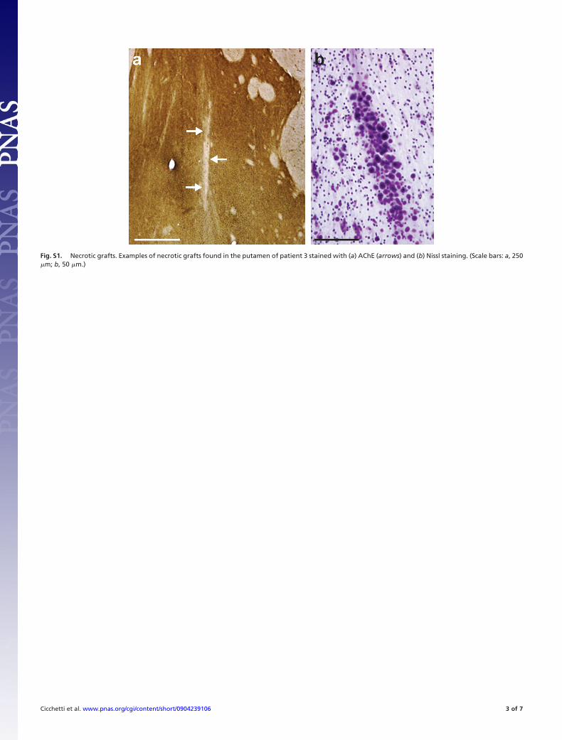
Fig. S1. Necrotic grafts. Examples of necrotic grafts found in the putamen of patient 3 stained with (a) AChE (arrows) and (b) Nissl staining. (Scale bars: a, 250�m; b, 50 �m.)
Cicchetti et al. www.pnas.org/cgi/content/short/0904239106 3 of 7
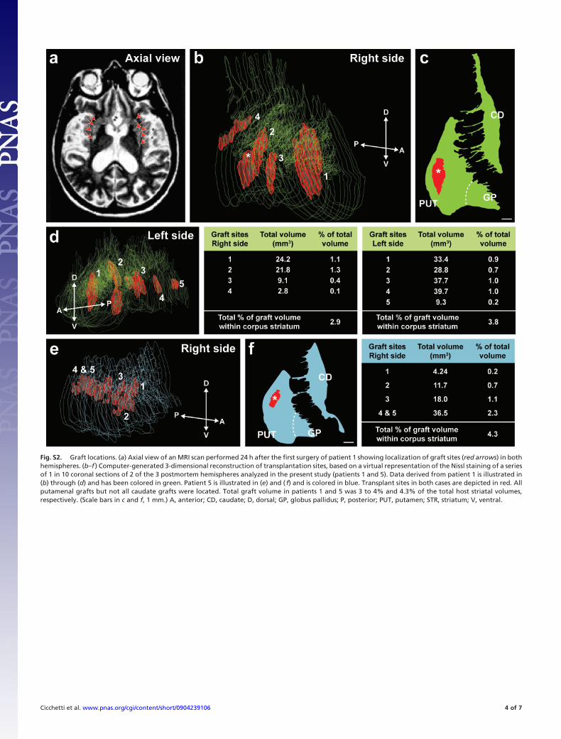
Fig. S2. Graft locations. (a) Axial view of an MRI scan performed 24 h after the first surgery of patient 1 showing localization of graft sites (red arrows) in bothhemispheres. (b–f ) Computer-generated 3-dimensional reconstruction of transplantation sites, based on a virtual representation of the Nissl staining of a seriesof 1 in 10 coronal sections of 2 of the 3 postmortem hemispheres analyzed in the present study (patients 1 and 5). Data derived from patient 1 is illustrated in(b) through (d) and has been colored in green. Patient 5 is illustrated in (e) and ( f) and is colored in blue. Transplant sites in both cases are depicted in red. Allputamenal grafts but not all caudate grafts were located. Total graft volume in patients 1 and 5 was 3 to 4% and 4.3% of the total host striatal volumes,respectively. (Scale bars in c and f, 1 mm.) A, anterior; CD, caudate; D, dorsal; GP, globus pallidus; P, posterior; PUT, putamen; STR, striatum; V, ventral.
Cicchetti et al. www.pnas.org/cgi/content/short/0904239106 4 of 7
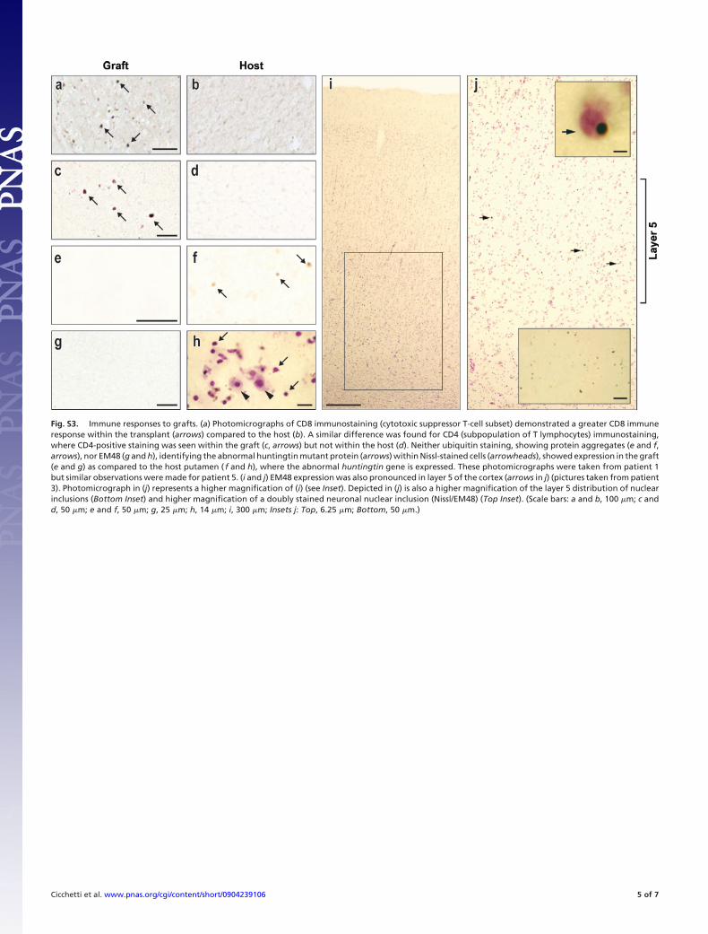
Fig. S3. Immune responses to grafts. (a) Photomicrographs of CD8 immunostaining (cytotoxic suppressor T-cell subset) demonstrated a greater CD8 immuneresponse within the transplant (arrows) compared to the host (b). A similar difference was found for CD4 (subpopulation of T lymphocytes) immunostaining,where CD4-positive staining was seen within the graft (c, arrows) but not within the host (d). Neither ubiquitin staining, showing protein aggregates (e and f,arrows), nor EM48 (g and h), identifying the abnormal huntingtin mutant protein (arrows) within Nissl-stained cells (arrowheads), showed expression in the graft(e and g) as compared to the host putamen ( f and h), where the abnormal huntingtin gene is expressed. These photomicrographs were taken from patient 1but similar observations were made for patient 5. (i and j) EM48 expression was also pronounced in layer 5 of the cortex (arrows in j) (pictures taken from patient3). Photomicrograph in (j) represents a higher magnification of (i) (see Inset). Depicted in (j) is also a higher magnification of the layer 5 distribution of nuclearinclusions (Bottom Inset) and higher magnification of a doubly stained neuronal nuclear inclusion (Nissl/EM48) (Top Inset). (Scale bars: a and b, 100 �m; c andd, 50 �m; e and f, 50 �m; g, 25 �m; h, 14 �m; i, 300 �m; Insets j: Top, 6.25 �m; Bottom, 50 �m.)
Cicchetti et al. www.pnas.org/cgi/content/short/0904239106 5 of 7

Table S1. Patient characteristics
Patient 1 (B.L.) Patient 3 (M.C.) Patient 5 (M.S.)
Gender Female Female FemaleCAG repeats 42 42 42Grades 3 3 2Time from diagnosis (years) 5 12 5Symptom duration (years) 8 17 9Age at transplantation (years) 58 64 59Post-operative latency (years) 9 10.5 9.5Causes of death Aspiration pneumonia
complicated by amyocardial infarction
Cardiorespiratory arrest as a complication of end-stage HD
Immunosuppression CsA 6 mg/kg/day 7 days before first operation continuing for 14 days after second operation, then 2mg/kg/day for 6 months
Number and location oftransplants
1 L caudate 2 L caudate 1 L caudate
4 L putamen 6 L putamen 4 L putamen1 R caudate 2 R caudate 1 R caudate4 R putamen 6 R putamen 5 R putamen
Surgical complications Asymptomatic subacute subduralhematoma after first operation
Asymptomatic corticalhemorrhage and thin subdural
hematoma after first and secondoperations
Bilateral hygromas post-operatively.Conversion of her hygromas intosubdural hematomas requiring
surgical drainage
Data from ref. 1 and subsequent clinical evaluation.
1. Hauser RA, Sandberg PR, Freeman TB, Stoessl AJ (2002) Bilateral human fetal striatal transplantation in Huntington’s disease. Neurology 58:1704, author reply 1704.
Cicchetti et al. www.pnas.org/cgi/content/short/0904239106 6 of 7

Table S2. Clinical outcomes
Patient 1 (B.L.) Patient 3 (M.C.) Patient 5 (M.S.)
UHDRS score pre-operation 42 33 27 (31 after subdural hematoma drainage)UHDRS score post-operation 30 (3 mo) 28 (10 mo) 39 (9 mo)
32 (9 mo) 21 (12 mo) 53 (2.5 yrs)40 (18 mo) 37 (20 mo) 64 (5 yrs)52 (4 yrs)65 (6 yrs)78 (7 yrs)
MMSE score pre-operation 30 26 27MMSE score post-operation 30 (21 mo) 24 (30 mo) 16 (5 yrs)
14 (7 yrs)
MMSE, Mini Mental State Evaluation; UHDRS, Unified Huntington’s Disease Rating Scale.Data from ref. 1 and subsequent clinical evaluation.
1. Hauser RA, Sandberg PR, Freeman TB, Stoessl AJ (2002) Bilateral human fetal striatal transplantation in Huntington’s disease. Neurology 58:1704, author reply 1704.
Cicchetti et al. www.pnas.org/cgi/content/short/0904239106 7 of 7