microglia and monocytes synergistically promote …wulab.cbn.rutgers.edu/_documents/2016 peng nat...
TRANSCRIPT
ARTICLE
Received 7 Oct 2015 | Accepted 23 May 2016 | Published 28 Jun 2016
Microglia and monocytes synergistically promotethe transition from acute to chronic pain afternerve injuryJiyun Peng1,*, Nan Gu1,2,*, Lijun Zhou1,3, Ukpong B Eyo1, Madhuvika Murugan1, Wen-Biao Gan4 & Long-Jun Wu1
Microglia and peripheral monocytes contribute to hypersensitivity in rodent models of
neuropathic pain. However, the precise respective function of microglia and peripheral
monocytes has not been investigated in these models. To address this question, here we
combined transgenic mice and pharmacological tools to specifically and temporally control
the depletion of microglia and monocytes in a mouse model of spinal nerve transection
(SNT). We found that although microglia and monocytes are required during the initiation of
mechanical allodynia or thermal hyperalgesia, these cells may not be as important for
the maintenance of hypersensitivity. Moreover, we demonstrated that either resident
microglia or peripheral monocytes are sufficient in gating neuropathic pain after SNT.
We propose that resident microglia and peripheral monocytes act synergistically to
initiate hypersensitivity and promote the transition from acute to chronic pain after peripheral
nerve injury.
DOI: 10.1038/ncomms12029 OPEN
1 Department of Cell Biology and Neuroscience, Rutgers University, Piscataway, New Jersey 08854, USA. 2 Department of Anesthesia, Xijing Hospital, FourthMilitary Medical University, Xi’an 710032, China. 3 Department of Physiology and Pain Research Center, Zhongshan School of Medicine, Sun Yet-SenUniversity, Guangzhou 510080, China. 4 Skirball Institute, Department of Neuroscience and Physiology, New York University School of Medicine, New York,New York 10016, USA. * These authors contributed equally to this work.. Correspondence and requests for materials should be addressed to L.-J.W.(email: [email protected]).
NATURE COMMUNICATIONS | 7:12029 | DOI: 10.1038/ncomms12029 | www.nature.com/naturecommunications 1
Neuropathic pain is a chronic pain state resulting fromperipheral or central nerve injury due to trauma(for example, amputation and nerve injury) or systemic
disease (for example, diabetes, viral infection and cancer)1,2.A key event in neuropathic pain is the transition from acuteto chronic pain; however, the underlying mechanisms ofthis transition remain largely unknown. Our understanding ofneuropathic pain mechanisms has expanded from being focusedlargely on neurocentric mechanisms to include neuro-glialinteractions3–5. Spinal glia including astrocytes and microgliabecome activated after peripheral nerve injury, subsequentlycontributing to chronic pain by releasing a number of glialmediators that sensitize spinal neurons6–9. In addition, peripheralnerve injury recruits circulating monocytes to the injury sites,which can also release proinflammatory mediators that causeneuronal hyperactivity in the periphery10. These studies raisethe intriguing possibility that central resident microglia andperipheral monocytes may control the transition from theacute to chronic pain.
Microglia comprise a unique subset of glial cells (5–10%) as theprincipal immune cells in the central nervous system (CNS).Resting microglia have highly dynamic processes by which theysurvey the microenvironment in the brain and spinal cord11–15.After peripheral nerve injury, spinal microglia transformfrom resting to reactive states, exhibiting marked changes incell surface protein expression, and releasing a varietyof proinflammatory mediators7,16–19. Inhibiting these micro-glia-derived molecules strongly suppresses pain-like hyper-sensitivity, suggesting that microglial response is a criticalcomponent of neuropathic pain. However, no evidencedirectly addresses the exact role of microglia in the initiation ormaintenance of neuropathic pain.
Peripheral nerve injury is also associated with the recruitment ofperipheral monocytes and their infiltration into the CNSparenchyma. Indeed, it was demonstrated that blood-bornecirculating monocytes infiltrate into the spinal cord and play anessential role in neuropathic pain development20. However, the roleof peripheral monocytes in neuropathic pain is still debated21,22.Nevertheless, the importance of peripheral monocytes in pain-likehypersensitivity is evident from the studies showing dissociationbetween activation of spinal microglia and chronic pain incertain animal models of peripheral nerve injury23,24 and inchemotherapy-induced pain25. So far, no report has pinpointed thespecific role of resident microglia and peripheral monocytes inneuropathic hypersensitivity.
Using transgenic mice that enabled us to ablate residentmicroglia and peripheral monocytes in a temporally controlledfashion, we delineated the time window during which microgliaand monocytes are required for the development of neuropathicpain in a mouse model of spinal nerve transection (SNT). Inaddition, we combined pharmacological and genetic tools to ablateresident microglia and peripheral monocytes, and determined theirrespective functions in neuropathic hypersensitivity. Our resultsindicate that depletion of both resident microglia and peripheralmonocytes completely prevented the development of neuropathicpain. However, either resident microglia or peripheral macro-phages are critical for the initiation of neuropathic pain, suggestingthat they act synergistically to promote the transition from acute tochronic pain after peripheral nerve injury.
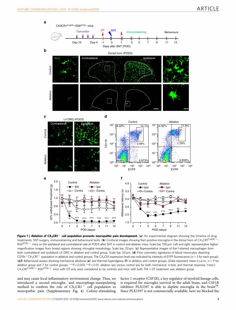
ResultsCX3CR1þ cell depletion prevents hypersensitivity after SNT.The chemokine receptor CX3CR1 is predominantly expressed bymicroglia in the CNS, but is also found in a subset of monocytes,macrophages, natural killer cells and dendritic cells in the
periphery26. To study the role of CX3CR1þ cells in neuropathicpain, we used a strategy to temporally and specifically express thediphtheria toxin receptor (DTR) in CX3CR1þ cells by crossingCX3CR1CreER mice with Rosa26-stop-DTR mice (CX3CR1CreER/þ :R26iDTR/þ ) and subsequent induction of cre recombinase bytamoxifen (TM) injection27. Therefore, we were able to control theablation of CX3CR1þ cells by diphtheria toxin (DT) applicationand directly investigated the temporal role of CX3CR1þ cells inchronic pain behaviours after L4 SNT, a well-established mousemodel of neuropathic pain. TM (150 mg kg� 1 in corn oil, 4 doseswith 2-day intervals) or corn oil control was intraperitoneally (i.p.)injected to CX3CR1CreER/þ :R26iDTR/þ mice before SNT and DT(50mg kg� 1, i.p., 3 days after last TM dose) was administered 1 daybefore and 1 day after SNT to deplete CX3CR1þ cells (Fig. 1a). Atpostoperative day 3 (POD3), we examined the CNS microglia anddorsal root ganglia (DRG) macrophages by immunostaining forIba1, as well as blood monocytes that express CX3CR1 by flowcytometry (Fig. 1b–d). CD11b, another marker for microglia andmacrophages, was also used to confirm the ablation efficiency inspinal dorsal horn (Supplementary Fig. 1a). In CX3CR1CreER/þ :R26iDTR/þ mice without TM-induced DTR expression(control), SNT markedly increased the number of microglia inthe ipsilateral dorsal horn and resident macrophages in DRGscompared with contralateral sides at POD3. However, inCX3CR1CreER/þ :R26iDTR/þ mice with both TM and DTinjection (ablation), spinal microglia and DRG macrophages inboth contralateral and ipsilateral sides were largely depleted(Fig. 1b,c and Supplementary Fig. 1a,c). In these CX3CR1CreER/þ
mice in which the CreER-encoding gene was followed by anIRES-EYFP element27, a subset of CD11bþ /EYFPþ þ -positiveblood cells that are monocytes with high CX3CR1 expression wasalso depleted (Fig. 1d and Supplementary Fig. 1d). In addition,microglia were depleted in most supraspinal brain regions, such asthe rostral ventromedial medulla, anterior cingulate cortex andhippocampus (Supplementary Fig. 1b,c). Together, these resultsindicate that our ablation strategy was able to successfully depleteCX3CR1þ cells, including microglia in the brain and spinal cord,DRG macrophages and CX3CR1þ monocytes.
Next, we wanted to know whether depletion of CX3CR1þ cellsaffected mouse pain behaviours. First, we measured acute painbehaviours in mice with CX3CR1þ cell depletion. We found thatCX3CR1þ cell depletion did not alter acute pain responses toeither mechanical or thermal stimulation (POD0, Fig. 1e,f). Also,there was no difference in tail flick tests between control andablation groups (Supplementary Fig. 2a). Motor coordination inthe rotarod test was similar between the two groups, althoughinter-session motor learning was impaired in mice withCX3CR1þ cell depletion (Supplementary Fig. 2b). Second, wecompared chronic pain behaviours after peripheral nerve injuryin mice with or without CX3CR1þ cell depletion, using the SNTmouse model of neuropathic pain. We found that mechanicaland thermal hypersensitivity following SNT were completelyabolished in mice with CX3CR1þ cell depletion compared withcontrol mice. Both mechanical allodynia and thermal hyper-algesia were prevented after SNT by such ablation and lasted atleast 2 weeks (Fig. 1e,f). A recent report showed a sex differencein the role of microglia in mechanical allodynia in male andfemale mice 7 days after nerve injury28. Interestingly, we foundthat mechanical and thermal hypersensitivity following SNT werecompletely abolished in both male and female mice withCX3CR1þ cell-depleted at POD3 compared with control mice(Supplementary Fig. 3). These results suggest that CX3CR1þ cellsequally participated in the neuropathic pain development in bothmale and female mice.
The CX3CR1þ cell depletion strategy using DT inCX3CR1CreER/þ :R26iDTR/þ mice induces microglial cell death
ARTICLE NATURE COMMUNICATIONS | DOI: 10.1038/ncomms12029
2 NATURE COMMUNICATIONS | 7:12029 | DOI: 10.1038/ncomms12029 | www.nature.com/naturecommunications
and may cause local inflammatory environment change. Thus, weintroduced a second microglia- and macrophage-manipulatingmethod to confirm the role of CX3CR1þ cell population inneuropathic pain (Supplementary Fig. 4). Colony-stimulating
factor 1 receptor (CSF1R), a key regulator of myeloid lineage cells,is required for microglia survival in the adult brain, and CSF1Rinhibitor PLX3397 is able to deplete microglia in the brain29.Since PLX3397 is not commercially available, here we blocked the
CX3CR1CreER/+:R26iDTR/+ mice
Behaviours
1411975310–1Day-4
Dorsal horn (POD3)
Contralateral
Contralateral
Ipsilateral
Ipsilateral
Iba1
Days after SNT (POD)Day-10
Con
trol
Abl
atio
n
Con
trol
Abl
atio
n
Control104
104
EYFP
4.22% 4.32% 11.8%
1.29%
0.659%
10.1%
3.49%
0.873%84.8% 83.2%
103
103
102
102
101
101100
104
103
102
101
100
100
2.0
2.0
1.50
2.5
3.0
3.5
4.0
1.6
1.2
0.8
0.4
0.00 1 3 5 7 9 11 14
*********
****** *** *** ** ***
*********
***
POD (days)1 3 5 7 9 11 14
POD (days)
With
draw
al la
tenc
y (s
)
Control
Ipsi IpsiContra Contra
Ablation ControlIpsi IpsiContra Contra
Ablation
50%
with
draw
al th
resh
old
(g)
104
EYFP103102101100
CD
11b
AblationL4 DRG (POD3)
Tamoxifen DT SNTImmunostaining
a
b
c d
e f
Figure 1 | Ablation of CX3CR1þ cell population prevents neuropathic pain development. (a) An experimental diagram showing the timeline of drug
treatments, SNT surgery, immunostaining and behavioural tests. (b) Confocal images showing Iba1-positive microglia in the dorsal horn of CX3CR1CreER/þ :
R26iDTR/þ mice on the ipsilateral and contralateral side at POD3 after SNT in control and ablation mice. Scale bar, 100mm. Left and right: representative higher-
magnification images from boxed regions showing microglial morphology. Scale bar, 20mm. (c) Representative images of Iba-1-stained macrophages from
both contralateral and ipsilateral L4 DRG in ablation and control group. Scale bar, 50mm. (d) Flow cytometry signatures of blood monocytes depicting
CD11bþCX3CR1þ population in ablation and control groups. The CX3CR1 expression level was indicated by intensity of EYFP fluorescence (n¼ 3 for each group).
(e,f) Behavioural assays showing mechanical allodynia (e) and thermal hyperalgesia (f) in ablation and control groups. (Data represent mean±s.e.m., n¼ 9 for
ablation group and 7 for control groups. ***Po0.001, **Po0.01, ablation ipsi versus control ipsi for both mechanical, U-test, and thermal response, t-test.)
CX3CR1CreER/þ :R26iDTR/þ mice with DT only were considered to be controls and mice with both TMþDT treatment was ablation group.
NATURE COMMUNICATIONS | DOI: 10.1038/ncomms12029 ARTICLE
NATURE COMMUNICATIONS | 7:12029 | DOI: 10.1038/ncomms12029 | www.nature.com/naturecommunications 3
CSF-1 pathway by neutralizing CSF-1 with antibody (200 ng in5 ml ACSF) through daily intrathecal injections from POD0 toPOD5 after SNT surgery (Supplementary Fig. 4a). We foundthat neutralizing CSF-1 antibody was not able to completelyablate microglia and macrophages, but it suppressed microgliaand macrophage numbers by reducing their proliferation, whichis consistent with a recent study showing that CSF-1 signalling iscritical for microglial activation after peripheral nerve injury30.The proliferation of spinal microglia and DRG macrophageswas examined by a proliferating marker Ki-67 staining at POD3after SNT. Compared with vehicle-treated mice (CX3CR1GFP/þ
mice), the number of Ki-67þ proliferating dorsal horn microgliaor DRG macrophages were markedly reduced in CSF-1 antibody-treated mice (Supplementary Fig. 4b,c). Consistently, wefound that although the vehicle-treated mice developed chronicneuropathic pain normally, both mechanical allodynia andthermal hyperalgesia were significantly reversed in the CSF-1-neutralizing antibody-treated mice (Supplementary Fig. 4d,e).These results confirmed that manipulation of the number ofspinal microglia and DRG macrophages is able to affectneuropathic pain development and replicate some aspects ofCX3CR1þ cell depletion strategy using CX3CR1CreER/þ :R26iDTR/þ mice.
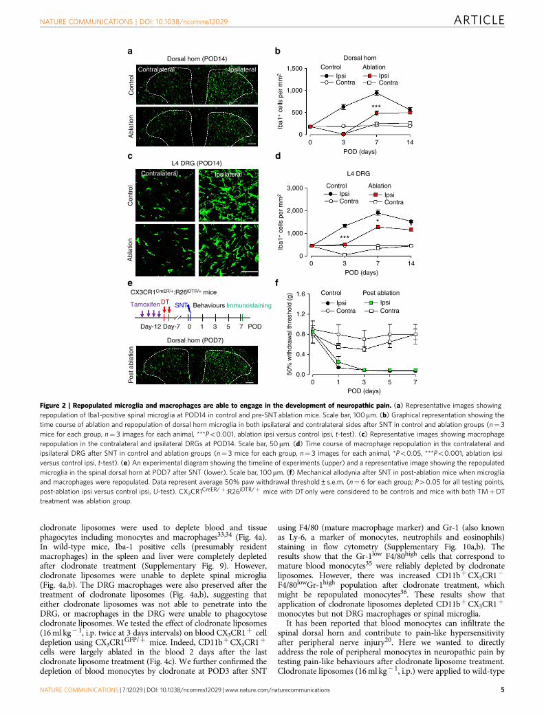
Repopulated CX3CR1þ cells cannot recover hypersensitivity.A unique property of microglia and macrophages is their capacityto quickly repopulate the CNS after ablation29,31. Here weallowed the repopulation of spinal microglia and DRGmacrophages and examined their function in neuropathic painusing CX3CR1CreER/þ :R26iDTR/þ mice after SNT. We foundthat microglia in the spinal dorsal horn repopulated rapidly aftertheir complete depletion at POD3. In both contralateral andipsilateral dorsal horn, spinal microglia repopulated quickly atPOD7 and POD14 in CX3CR1 cell-ablated mice (Fig. 2a,b). Themorphology of the newly repopulated spinal microglia differedfrom that in age-matched control mice, exhibiting shorterprocesses and fewer branches at POD7 (Supplementary Fig. 5).In addition, macrophages in the injured L4 DRG also showedrapid repopulation at POD7 and POD14 (Fig. 2c,d). These resultsindicate that both spinal microglia and DRG macrophagesundergo rapid repopulation after their depletion by DT inCX3CR1CreER/þ :R26iDTR/þ mice. Although microglia andmacrophages repopulated in the spinal cord and DRG within aweek, the reduction in pain-like behaviours in CX3CR1 cellablation mice was long-lasting (Fig. 1e,f). Therefore, these resultssuggest that repopulated microglia and macrophages after SNTare unable to re-establish neuropathic hypersensitivity.
To test whether repopulated microglia and macrophages arefunctional in the development of neuropathic pain, we depletedCX3CR1þ cells and allowed a week for their repopulation.Then SNT was performed in these mice (Fig. 2e). We confirmedthat the number of repopulated spinal microglia and DRGmacrophages in CX3CR1 cell-ablated mice was comparable tothat in control mice (Fig. 2e). In these mice with repopulatedmicroglia and macrophages (post ablation), we performed SNTand then neuropathic pain behaviours were tested. We found thatmechanical allodynia developed normally in these mice and therewas no difference in hypersensitivity between control and post-ablation groups (Fig. 2f). These results suggest that repopulatedmicroglia and macrophages are functional being able to initiateneuropathic pain after peripheral nerve injury. Thus, wedemonstrate that repopulated microglia and macrophages areable to engage in the development of neuropathic pain after denovo but not pre-existent nerve injury. These results suggest acritical period when signals derived from peripheral nerve injury,
such as CSF-1 or ATP30,32, are able to recruit microglia todevelop neuropathic pain-like hypersensitivity.
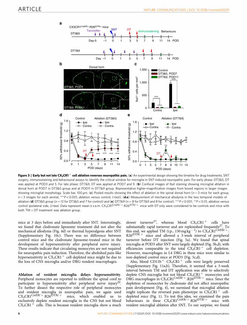
Early ablation of CX3CR1þ cells reverses hypersensitivity.Depletion of CX3CR1þ cells abolished neuropathic painbehaviours after peripheral nerve injury (Fig. 1). However, theprecise role of these cells at different stages of neuropathicpain development is still unknown. Taking advantage ofCX3CR1CreER/þ :R26iDTR/þ mice, we were able to temporallyablate CX3CR1þ cells by DT injection in the early (POD3–5) andlate (at POD7–9) stages after SNT. Neuropathic hypersensitivityafter SNT gradually developed from POD1–3 and was fullyestablished after POD7. Consistently, the number of microglia inthe ipsilateral spinal cord and L4 DRG increased duringneuropathic pain development and peaked at POD7 (Fig. 2a–d).
To directly assess the role of CX3CR1þ cells in thedevelopment and initiation of neuropathic pain, we administeredDT (i.p., 50 mg kg� 1) at POD3 and POD5 (DT3&5) after SNTand then examined the depletion of microglia and macrophages,as well as chronic pain behaviours (Fig. 3a). As expected, DT atPOD3 and 5 completely ablated spinal microglia (Fig. 3b,c) andmost DRG macrophages at POD7 (Supplementary Fig. 6).Interestingly, behavioural experiments revealed that mechanicalallodynia was significantly reversed at POD7 (Fig. 3d). Inaddition, the reduced mechanical allodynia was sustained, lastingup to POD14 (Fig. 3d), although microglia had largelyrepopulated the spinal cord by this stage (data not shown).Consistently, thermal hyperalgesia was also significantly reversedwhen CX3CR1 cells were ablated at POD3 and POD5 after SNT(Supplementary Fig. 7a,b). These results indicate that CX3CR1þ
cells are critical during the initiation of neuropathic pain.Next, we asked whether CX3CR1þ cells are required for the
maintenance of neuropathic hypersensitivity. To this end, weablated microglia and macrophages when neuropathic painwas fully developed. DT was administered (i.p., 50 mg kg� 1) atPOD7 and POD9 (DT7&9), which completely depleted spinalmicroglia and most DRG macrophages at POD11 (Fig. 3b,c andSupplementary Fig. 6). Surprisingly, when we examined thepain-like behaviours in these mice, we found that mechanicalallodynia was only transiently reversed but then maintained to acomparable level as those without CX3CR1þ cell ablation atPOD11 (Fig. 3e). In addition, the mechanical allodynia persistedat least to POD14 in those mice with CX3CR1þ cell ablation(Fig. 3e). Consistently, thermal hyperalgesia in mice, in whichCX3CR1þ cells were ablated at POD7 and 9, was also onlytransiently reversed compared with those in control micewithout cell ablation (Supplementary Fig. 7a,c). Interestingly, infemale mice, thermal hyperalgesia but not mechanical allodyniawas transiently reversed after DT injection at POD7 andPOD9 (Supplementary Fig. 8), suggesting that sexual dimorphismmay differentiate CX3CR1þ cell’s function in different modalitiesof neuropathic pain. Taken together, our results suggestthat CX3CR1þ cells participate in the initiation of theneuropathic pain state, but are only transiently required inthe maintenance of neuropathic pain. In particular, there during acritical time window of at POD0–POD5, when CX3CR1þ
cells promote the transition from acute to chronic pain afterperipheral nerve injury.
Depletion of monocytes did not alter hypersensitivity. CX3CR1is expressed in CNS microglia, DRG macrophages and circulatingmonocytes26. Our above results demonstrated the pivotal functionof CX3CR1þ cells in neuropathic pain. However, the respectiverole of peripheral monocytes and microglia in neuropathichypersensitivity remains unknown. To address this question,
ARTICLE NATURE COMMUNICATIONS | DOI: 10.1038/ncomms12029
4 NATURE COMMUNICATIONS | 7:12029 | DOI: 10.1038/ncomms12029 | www.nature.com/naturecommunications
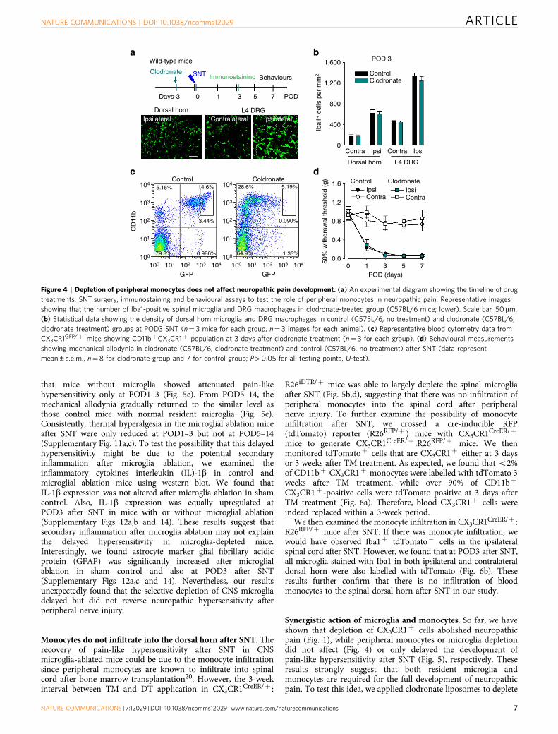
clodronate liposomes were used to deplete blood and tissuephagocytes including monocytes and macrophages33,34 (Fig. 4a).In wild-type mice, Iba-1 positive cells (presumably residentmacrophages) in the spleen and liver were completely depletedafter clodronate treatment (Supplementary Fig. 9). However,clodronate liposomes were unable to deplete spinal microglia(Fig. 4a,b). The DRG macrophages were also preserved after thetreatment of clodronate liposomes (Fig. 4a,b), suggesting thateither clodronate liposomes was not able to penetrate into theDRG, or macrophages in the DRG were unable to phagocytoseclodronate liposomes. We tested the effect of clodronate liposomes(16 ml kg� 1, i.p. twice at 3 days intervals) on blood CX3CR1þ celldepletion using CX3CR1GFP/þ mice. Indeed, CD11bþCX3CR1þ
cells were largely ablated in the blood 2 days after the lastclodronate liposome treatment (Fig. 4c). We further confirmed thedepletion of blood monocytes by clodronate at POD3 after SNT
using F4/80 (mature macrophage marker) and Gr-1 (also knownas Ly-6, a marker of monocytes, neutrophils and eosinophils)staining in flow cytometry (Supplementary Fig. 10a,b). Theresults show that the Gr-1low F4/80high cells that correspond tomature blood monocytes35 were reliably depleted by clodronateliposomes. However, there was increased CD11bþCX3CR1�
F4/80lowGr-1high population after clodronate treatment, whichmight be repopulated monocytes36. These results show thatapplication of clodronate liposomes depleted CD11bþCX3CR1þ
monocytes but not DRG macrophages or spinal microglia.It has been reported that blood monocytes can infiltrate the
spinal dorsal horn and contribute to pain-like hypersensitivityafter peripheral nerve injury20. Here we wanted to directlyaddress the role of peripheral monocytes in neuropathic pain bytesting pain-like behaviours after clodronate liposome treatment.Clodronate liposomes (16 ml kg� 1, i.p.) were applied to wild-type
Dorsal horn (POD14)
L4 DRG (POD14)
Con
trol
1,500
1,000
3,000
2,000
1,000
0
1.6
1.2
50%
with
draw
al th
resh
old
(g)
0.8Dorsal horn (POD7)
Day-12 Day-7 0
SNTDT
1 3 5 7 POD
Behaviours
CX3CR1CreER/+:R26iDTR/+ mice
Tamoxifen Immunostaining
0.4
0.00
0
500
00 3 7 14
POD (days)
3 7 14POD (days)
3 5 71POD (days)
***
***
* *
Control AblationDorsal horn
L4 DRG
Contra ContraIpsiIpsi
Control Ablation
Contra ContraIpsiIpsi
Control Post ablation
Pos
t abl
atio
n
Contra ContraIpsiIpsi
Iba1
+ c
ells
per
mm
2Ib
a1+ c
ells
per
mm
2
Abl
atio
nC
ontr
olA
blat
ion
Contralateral Ipsilateral
Contralateral Ipsilateral
a b
c d
e f
Figure 2 | Repopulated microglia and macrophages are able to engage in the development of neuropathic pain. (a) Representative images showing
repopulation of Iba1-positive spinal microglia at POD14 in control and pre-SNT ablation mice. Scale bar, 100mm. (b) Graphical representation showing the
time course of ablation and repopulation of dorsal horn microglia in both ipsilateral and contralateral sides after SNT in control and ablation groups (n¼ 3
mice for each group, n¼ 3 images for each animal, ***Po0.001, ablation ipsi versus control ipsi, t-test). (c) Representative images showing macrophage
repopulation in the contralateral and ipsilateral DRGs at POD14. Scale bar, 50mm. (d) Time course of macrophage repopulation in the contralateral and
ipsilateral DRG after SNT in control and ablation groups (n¼ 3 mice for each group, n¼ 3 images for each animal, *Po0.05, ***Po0.001, ablation ipsi
versus control ipsi, t-test). (e) An experimental diagram showing the timeline of experiments (upper) and a representative image showing the repopulated
microglia in the spinal dorsal horn at POD7 after SNT (lower). Scale bar, 100mm. (f) Mechanical allodynia after SNT in post-ablation mice when microglia
and macrophages were repopulated. Data represent average 50% paw withdrawal threshold±s.e.m. (n¼ 6 for each group; P40.05 for all testing points,
post-ablation ipsi versus control ipsi, U-test). CX3CR1CreER/þ :R26iDTR/þ mice with DT only were considered to be controls and mice with both TMþDT
treatment was ablation group.
NATURE COMMUNICATIONS | DOI: 10.1038/ncomms12029 ARTICLE
NATURE COMMUNICATIONS | 7:12029 | DOI: 10.1038/ncomms12029 | www.nature.com/naturecommunications 5
mice at 3 days before and immediately after SNT. Interestingly,we found that clodronate liposome treatment did not alter themechanical allodynia (Fig. 4d) or thermal hyperalgesia after SNT(Supplementary Fig. 10c). There was no difference betweencontrol mice and the clodronate liposome-treated mice in thedevelopment of hypersensitivity after peripheral nerve injury.These results indicate that circulating monocytes are not requiredfor neuropathic pain induction. Therefore, the abolished pain-likehypersensitivity in CX3CR1þ cell-depleted mice might be due tothe loss of CNS microglia and/or DRG resident macrophages.
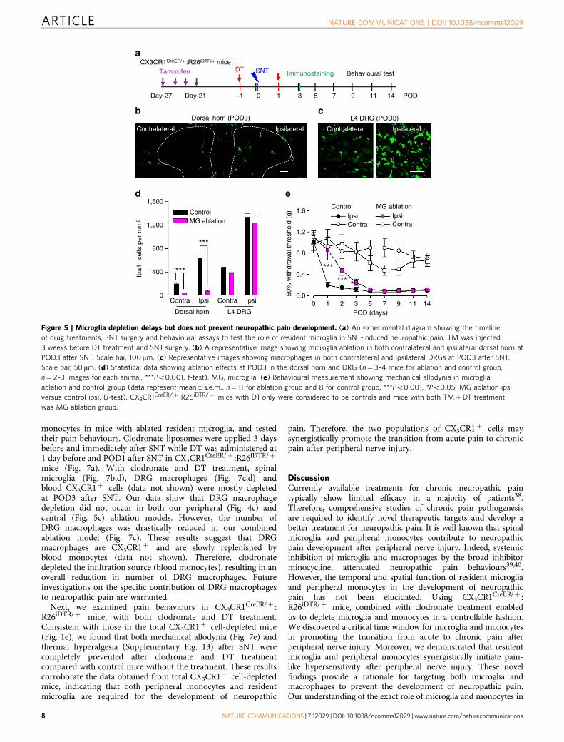
Ablation of resident microglia delays hypersensitivity.Peripheral monocytes are reported to infiltrate the spinal cord toparticipate in hypersensitivity after peripheral nerve injury20.To further dissect the respective role of peripheral monocytesand resident microglia in neuropathic pain, we again usedCX3CR1CreER/þ :R26iDTR/þ mice, which enabled us toexclusively deplete resident microglia in the CNS but not bloodCX3CR1þ cells. This is because resident microglia show a much
slower turnover37, whereas blood CX3CR1þ cells havesubstantially rapid turnover and are replenished frequently27. Tothis end, we applied TM (i.p., 150 mg kg� 1) to CX3CR1CreER/þ :R26iDTR/þ mice and allowed a 3-week interval of peripheralturnover before DT injection (Fig. 5a). We found that spinalmicroglia at POD3 after SNT were largely depleted (Fig. 5b,d), withefficiencies comparable to the total CX3CR1þ cell depletion.However, macrophages in L4 DRG in these mice were similar tonon-depleted control mice at POD3 (Fig. 5c,d).
Also, blood CD11bþ CX3CR1þ cells were largely preserved(Supplementary Fig. 11a,b). Therefore, it seemed that a 3-weekinterval between TM and DT application was able to selectivelydeplete CNS microglia but not blood CX3CR1þ monocytes andDRG macrophages in CX3CR1CreER/þ :R26iDTR/þ mice. Since thedepletion of monocytes by clodronate did not affect neuropathicpain development (Fig. 4), we surmised that microglial ablationmight replicate the reversed pain phenotype in CX3CR1þ cell-depleted mice (Fig. 1). To test this idea, we examined the painbehaviours in these CX3CR1CreER/þ :R26iDTR/þ mice withresident microglial ablation after SNT. To our surprise, we found
CX3CR1CreER/+:R26iDTR/+ mice
DT3&5
DT7&9
Day-5
Day
Dorsal horn
DT3&5, POD7
DT7&9, POD11
–1
–1 0
0
1 3 5 7 9 11 14 POD
1 3 5 7
1,500
1,000
500
0Contra
Control1.6
1.2
0.8
0.4
50%
with
draw
al th
resh
old
(g)
0.0
1.6
1.2
0.8
0.4
50%
with
draw
al th
resh
old
(g)
0.00 1 3 5 7 9 11 14
POD (days)0 1 3 5 7 8 9 10 11 14
POD (days)
*** *** *** ***
**
***DT
DT
Ablation (DT3&5)
Contra ContraIpsi Ipsi
Control Ablation (DT7&9)
Contra ContraIpsi Ipsi
Ipsi
***
***
ControlDT3&5, POD7DT7&9, POD11
Iba1
+ c
ells
per
mm
2
9 11 14 POD
BehavioursImmunostainingDTSNTTamoxifen
a
b c
d e
Figure 3 | Early but not late CX3CR1þ cell ablation reverses neuropathic pain. (a) An experimental design showing the timeline for drug treatments, SNT
surgery, immunostaining and behavioural assays to identify the critical window for microglia in SNT-induced neuropathic pain. For early phase: DT3&5, DT
was applied at POD3 and 5. For late phase: DT7&9, DT was applied at POD7 and 9. (b) Confocal images of Iba1 staining showing microglial ablation in
dorsal horn at POD7 in DT3&5 group and at POD11 in DT7&9 group. Representative higher-magnification images from boxed regions in larger images
showing microglial morphology. Scale bar, 100mm. (c) Pooled results showing the effect of ablation in the spinal dorsal horn (n¼ 3 mice for each group,
n¼ 3 images for each animal, ***Po0.001, ablation versus control, t-test). (d,e) Measurement of mechanical allodynia in the two temporal models of
ablation (d) DT3&5 group (n¼ 12 for DT3&5 and 7 for control) and (e) DT7&9 (n¼ 8 for DT7&9 and 8 for control). ***Po0.001, **Po0.01, ablation versus
control ipsilateral side, U-test. Data represent mean±s.e.m. CX3CR1CreER/þ :R26iDTR/þ mice with DT only were considered to be controls and mice with
both TMþDT treatment was ablation group.
ARTICLE NATURE COMMUNICATIONS | DOI: 10.1038/ncomms12029
6 NATURE COMMUNICATIONS | 7:12029 | DOI: 10.1038/ncomms12029 | www.nature.com/naturecommunications
that mice without microglia showed attenuated pain-likehypersensitivity only at POD1–3 (Fig. 5e). From POD5–14, themechanical allodynia gradually returned to the similar level asthose control mice with normal resident microglia (Fig. 5e).Consistently, thermal hyperalgesia in the microglial ablation miceafter SNT were only reduced at POD1–3 but not at POD5–14(Supplementary Fig. 11a,c). To test the possibility that this delayedhypersensitivity might be due to the potential secondaryinflammation after microglia ablation, we examined theinflammatory cytokines interleukin (IL)-1b in control andmicroglial ablation mice using western blot. We found thatIL-1b expression was not altered after microglia ablation in shamcontrol. Also, IL-1b expression was equally upregulated atPOD3 after SNT in mice with or without microglial ablation(Supplementary Figs 12a,b and 14). These results suggest thatsecondary inflammation after microglia ablation may not explainthe delayed hypersensitivity in microglia-depleted mice.Interestingly, we found astrocyte marker glial fibrillary acidicprotein (GFAP) was significantly increased after microglialablation in sham control and also at POD3 after SNT(Supplementary Figs 12a,c and 14). Nevertheless, our resultsunexpectedly found that the selective depletion of CNS microgliadelayed but did not reverse neuropathic hypersensitivity afterperipheral nerve injury.
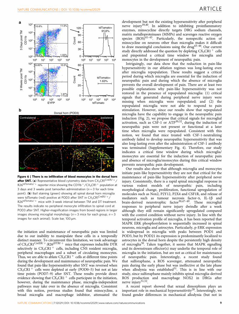
Monocytes do not infiltrate into the dorsal horn after SNT. Therecovery of pain-like hypersensitivity after SNT in CNSmicroglia-ablated mice could be due to the monocyte infiltrationsince peripheral monocytes are known to infiltrate into spinalcord after bone marrow transplantation20. However, the 3-weekinterval between TM and DT application in CX3CR1CreER/þ :
R26iDTR/þ mice was able to largely deplete the spinal microgliaafter SNT (Fig. 5b,d), suggesting that there was no infiltration ofperipheral monocytes into the spinal cord after peripheralnerve injury. To further examine the possibility of monocyteinfiltration after SNT, we crossed a cre-inducible RFP(tdTomato) reporter (R26RFP/þ ) mice with CX3CR1CreER/þ
mice to generate CX3CR1CreER/þ :R26RFP/þ mice. We thenmonitored tdTomatoþ cells that are CX3CR1þ either at 3 daysor 3 weeks after TM treatment. As expected, we found that o2%of CD11bþ CX3CR1þ monocytes were labelled with tdTomato 3weeks after TM treatment, while over 90% of CD11bþ
CX3CR1þ -positive cells were tdTomato positive at 3 days afterTM treatment (Fig. 6a). Therefore, blood CX3CR1þ cells wereindeed replaced within a 3-week period.
We then examined the monocyte infiltration in CX3CR1CreER/þ :R26RFP/þ mice after SNT. If there was monocyte infiltration, wewould have observed Iba1þ tdTomato� cells in the ipsilateralspinal cord after SNT. However, we found that at POD3 after SNT,all microglia stained with Iba1 in both ipsilateral and contralateraldorsal horn were also labelled with tdTomato (Fig. 6b). Theseresults further confirm that there is no infiltration of bloodmonocytes to the spinal dorsal horn after SNT in our study.
Synergistic action of microglia and monocytes. So far, we haveshown that depletion of CX3CR1þ cells abolished neuropathicpain (Fig. 1), while peripheral monocytes or microglia depletiondid not affect (Fig. 4) or only delayed the development ofpain-like hypersensitivity after SNT (Fig. 5), respectively. Theseresults strongly suggest that both resident microglia andmonocytes are required for the full development of neuropathicpain. To test this idea, we applied clodronate liposomes to deplete
Wild-type mice
Behaviours
1,600
1,200
800
400
0Contra Contra IpsiIpsi
Dorsal horn
POD (days)
Control ClodronateIpsi IpsiContra Contra
0 1 3 5 7
1.6
1.2
0.8
0.4
0.050%
with
draw
al th
resh
old
(g)
L4 DRG
ControlClodronate
POD 3
Iba1
+ c
ells
per
mm
2
Days-3 0 1 3 5 7 POD
L4 DRGDorsal horn
Control5.15% 14.6%
3.44%
0.986% 1.33%
5.19%28.6%
64.9%
0.090%
79.3%
104
103
102
101
100
100 101 102 103 104
GFP100 101 102 103 104
GFP
CD
11b
104
103
102
101
100
Coldronate
Ipsilateral IpsilateralContralateral
Clodronate SNT Immunostaining
a b
c d
Figure 4 | Depletion of peripheral monocytes does not affect neuropathic pain development. (a) An experimental diagram showing the timeline of drug
treatments, SNT surgery, immunostaining and behavioural assays to test the role of peripheral monocytes in neuropathic pain. Representative images
showing that the number of Iba1-positive spinal microglia and DRG macrophages in clodronate-treated group (C57BL/6 mice; lower). Scale bar, 50 mm.
(b) Statistical data showing the density of dorsal horn microglia and DRG macrophages in control (C57BL/6, no treatment) and clodronate (C57BL/6,
clodronate treatment) groups at POD3 SNT (n¼ 3 mice for each group, n¼ 3 images for each animal). (c) Representative blood cytometry data from
CX3CR1GFP/þ mice showing CD11bþCX3CR1þ population at 3 days after clodronate treatment (n¼ 3 for each group). (d) Behavioural measurements
showing mechanical allodynia in clodronate (C57BL/6, clodronate treatment) and control (C57BL/6, no treatment) after SNT (data represent
mean±s.e.m., n¼8 for clodronate group and 7 for control group; P40.05 for all testing points, U-test).
NATURE COMMUNICATIONS | DOI: 10.1038/ncomms12029 ARTICLE
NATURE COMMUNICATIONS | 7:12029 | DOI: 10.1038/ncomms12029 | www.nature.com/naturecommunications 7
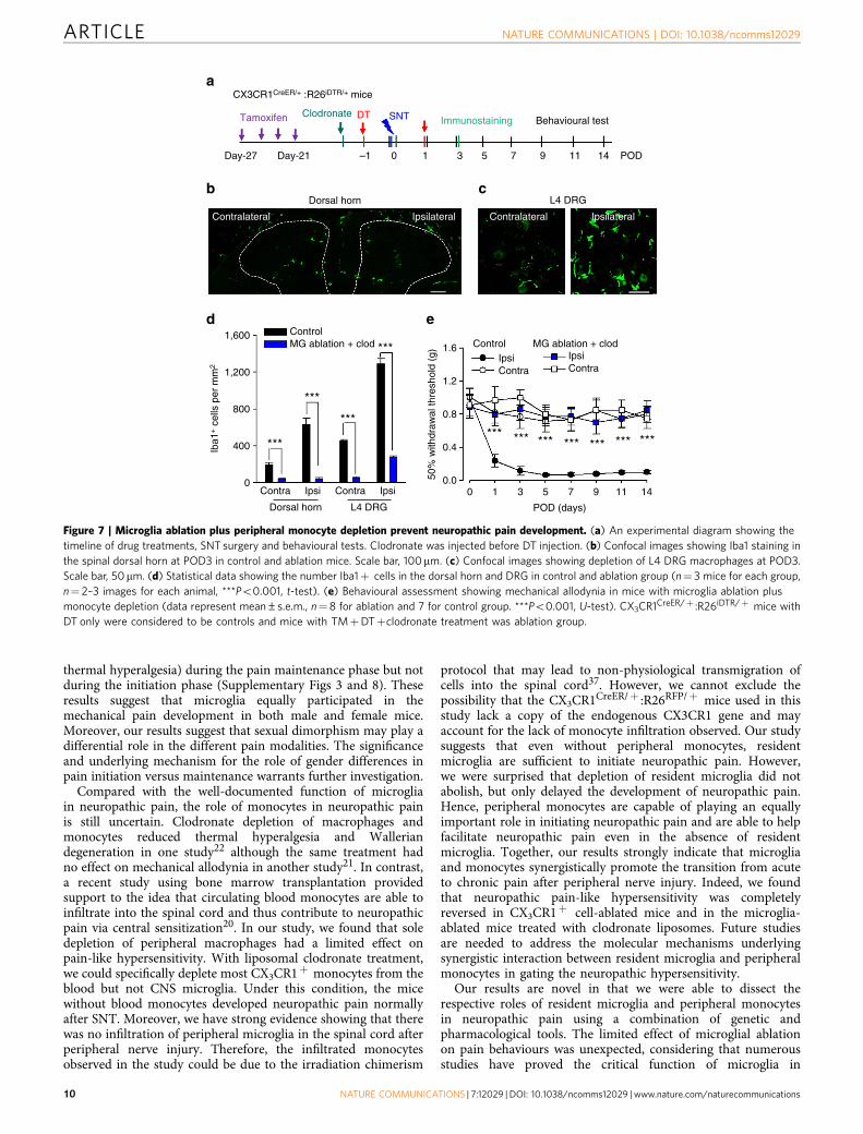
monocytes in mice with ablated resident microglia, and testedtheir pain behaviours. Clodronate liposomes were applied 3 daysbefore and immediately after SNT while DT was administered at1 day before and POD1 after SNT in CX3CR1CreER/þ :R26iDTR/þ
mice (Fig. 7a). With clodronate and DT treatment, spinalmicroglia (Fig. 7b,d), DRG macrophages (Fig. 7c,d) andblood CX3CR1þ cells (data not shown) were mostly depletedat POD3 after SNT. Our data show that DRG macrophagedepletion did not occur in both our peripheral (Fig. 4c) andcentral (Fig. 5c) ablation models. However, the number ofDRG macrophages was drastically reduced in our combinedablation model (Fig. 7c). These results suggest that DRGmacrophages are CX3CR1þ and are slowly replenished byblood monocytes (data not shown). Therefore, clodronatedepleted the infiltration source (blood monocytes), resulting in anoverall reduction in number of DRG macrophages. Futureinvestigations on the specific contribution of DRG macrophagesto neuropathic pain are warranted.
Next, we examined pain behaviours in CX3CR1CreER/þ :R26iDTR/þ mice, with both clodronate and DT treatment.Consistent with those in the total CX3CR1þ cell-depleted mice(Fig. 1e), we found that both mechanical allodynia (Fig. 7e) andthermal hyperalgesia (Supplementary Fig. 13) after SNT werecompletely prevented after clodronate and DT treatmentcompared with control mice without the treatment. These resultscorroborate the data obtained from total CX3CR1þ cell-depletedmice, indicating that both peripheral monocytes and residentmicroglia are required for the development of neuropathic
pain. Therefore, the two populations of CX3CR1þ cells maysynergistically promote the transition from acute pain to chronicpain after peripheral nerve injury.
DiscussionCurrently available treatments for chronic neuropathic paintypically show limited efficacy in a majority of patients38.Therefore, comprehensive studies of chronic pain pathogenesisare required to identify novel therapeutic targets and develop abetter treatment for neuropathic pain. It is well known that spinalmicroglia and peripheral monocytes contribute to neuropathicpain development after peripheral nerve injury. Indeed, systemicinhibition of microglia and macrophages by the broad inhibitorminocycline, attenuated neuropathic pain behaviours39,40.However, the temporal and spatial function of resident microgliaand peripheral monocytes in the development of neuropathicpain has not been elucidated. Using CX3CR1CreER/þ :R26iDTR/þ mice, combined with clodronate treatment enabledus to deplete microglia and monocytes in a controllable fashion.We discovered a critical time window for microglia and monocytesin promoting the transition from acute to chronic pain afterperipheral nerve injury. Moreover, we demonstrated that residentmicroglia and peripheral monocytes synergistically initiate pain-like hypersensitivity after peripheral nerve injury. These novelfindings provide a rationale for targeting both microglia andmacrophages to prevent the development of neuropathic pain.Our understanding of the exact role of microglia and monocytes in
CX3CR1CreER/+ :R26iDTR/+ mice
Day-27 Day-21 –1 0 1 3 5 7 9 11 14 POD
L4 DRG (POD3)Dorsal horn (POD3)
1,600
1,200
1.6
1.2
0.8
0.4
0.00 1 2 3 5 7 9 11 14
POD (days)
***
*** *50
% w
ithdr
awal
thre
shol
d (g
)
800
400
0Contra Contra
Contra Contra
Dorsal horn L4 DRG
***
ControlMG ablation
Control MG ablation
***Iba1
+ c
ells
per
mm
2
Ipsi Ipsi
IpsiIpsi
Contralateral ContralateralIpsilateral Ipsilateral
Behavioural testImmunostainingSNTDTTamoxifen
a
b c
d e
Figure 5 | Microglia depletion delays but does not prevent neuropathic pain development. (a) An experimental diagram showing the timeline
of drug treatments, SNT surgery and behavioural assays to test the role of resident microglia in SNT-induced neuropathic pain. TM was injected
3 weeks before DT treatment and SNT surgery. (b) A representative image showing microglia ablation in both contralateral and ipsilateral dorsal horn at
POD3 after SNT. Scale bar, 100 mm. (c) Representative images showing macrophages in both contralateral and ipsilateral DRGs at POD3 after SNT.
Scale bar, 50mm. (d) Statistical data showing ablation effects at POD3 in the dorsal horn and DRG (n¼ 3–4 mice for ablation and control group,
n¼ 2–3 images for each animal, ***Po0.001, t-test). MG, microglia. (e) Behavioural measurement showing mechanical allodynia in microglia
ablation and control group (data represent mean±s.e.m., n¼ 11 for ablation group and 8 for control group, ***Po0.001, *Po0.05, MG ablation ipsi
versus control ipsi, U-test). CX3CR1CreER/þ :R26iDTR/þ mice with DT only were considered to be controls and mice with both TMþDT treatment
was MG ablation group.
ARTICLE NATURE COMMUNICATIONS | DOI: 10.1038/ncomms12029
8 NATURE COMMUNICATIONS | 7:12029 | DOI: 10.1038/ncomms12029 | www.nature.com/naturecommunications
the initiation and maintenance of neuropathic pain was limiteddue to our inability to manipulate these cells in a temporallydistinct manner. To circumvent this limitation, we took advantageof CX3CR1CreER/þ :R26iDTR/þ mice that expresses inducible DTRselectively in CX3CR1þ cells, including CNS resident microglia,peripheral macrophages and a subset of circulating monocytes.Thus, we are able to ablate CX3CR1þ cells at different time pointsduring the development and maintenance of neuropathic pain. Wefound that pain-like hypersensitivity after SNT was reversed whenCX3CR1þ cells were depleted at early (POD0–5) but not at latetime points (POD7–9) after SNT. These results provide directevidence showing that CX3CR1þ cells participate in the initiation,however, during the maintenance phase, microglia-independentpathways may take over in the absence of microglia. Consistentwith this notion, previous studies found that minocycline, abroad microglia and macrophage inhibitor, attenuated the
development but not the existing hypersensitivity after peripheralnerve injury39,40. In addition to inhibiting proinflammatoryenzymes, minocycline directly targets DRG sodium channels,matrix metalloproteinases (MMPs) and scavenges reactive oxygenspecies (ROS)41–43. Particularly, the nonspecific action ofminocycline on neurons other than microglia makes it difficultto draw meaningful conclusions using the drug43–46. Our currentstudy directly addressed the question by depleting CX3CR1þ cellsand pinpointed a critical time window for microglia andmonocytes in the development of neuropathic pain.
Intriguingly, our data show that the reduction in pain-likehypersensitivity in our ablation regimes was long-lasting evenafter microglia repopulation. These results suggest a criticalperiod during which microglia are essential for the induction ofneuropathic pain and during which the absence of microgliaprevents the overall development of pain. There are at least twopossible explanations why pain-like hypersensitivity was notrestored in the presence of repopulated microglia: (1) criticalsignals that generated during peripheral nerve injury weremissing when microglia were repopulated; and (2) therepopulated microglia were not able to respond to painstimulation. However, since our results show that repopulatedmicroglia have the capability to engage in the neuropathic paininduction (Fig. 2), we propose that critical signals for microglialactivation, such as CSF-1 or ATP30,32, during the induction ofneuropathic pain were not present or functional at a latertime when microglia were repopulated. Consistent with thisnotion, we found that mice treated with CSF-1-neutralizingantibody failed to develop neuropathic hypersensitivity that wasalso long-lasting even after the administration of CSF-1 antibodywas terminated (Supplementary Fig. 4). Therefore, our studyindicates a critical time window during which microglia/monocytes are essential for the induction of neuropathic painand absence of microglia/monocytes during this critical windowprevents neuropathic pain development.
Our results also show that although microglia and monocytesinitiate pain-like hypersensitivity they are not that critical for themaintenance of pain-like hypersensitivity after peripheral neverinjury. Consistently, there is a rapid spinal microglial response invarious rodent models of neuropathic pain, includingmorphological change, proliferation, functional upregulation ofmolecules such as Nox2, P2Y12, P2X4 and CSF1R, and release ofmediators such as tumour necrosis factor-a, IL-1b andbrain-derived neurotrophic factor30,47–49. These microglialresponses to peripheral nerve injury dwindle after a weekalthough they still remain significantly upregulated comparedwith the control condition without nerve injury. In line with thetemporal activation profile of microglia, it has been reported thatMAPK ERK phosphorylation is sequentially increased in spinalneurons, microglia and astrocytes. Particularly, p-ERK expressionis widespread in microglia with peaks between POD1 andPOD3, but by POD21 its expression is predominantly localized toastrocytes in the dorsal horn despite the persistently high densityof microglia50. Taken together, it seems that MAPK signallingand its downstream effector(s) may underlie the temporal role ofmicroglia in the initiation, but are not as critical for maintenanceof neuropathic pain. Interestingly, a recent study foundthat sulforaphane, a ROS scavenger, attenuated neuropathicpain during the early phase but was ineffective at the late phasewhen allodynia was established51. This is in line with ourstudy, since sulforaphane mainly inhibits spinal microglia-derivedROS production and macrophage NOX2 in DRGs afternerve injury51,52.
A recent report showed that sexual dimorphism plays animportant role in mechanical hypersensitivity28. Interestingly, wefound gender differences in mechanical allodynia (but not in
104
103
102
101
100
100 101 102 103 104 100 101 102 103 104
100 101 102 103 104100 101 102 103 104
1.55%
11.5%120
90
Cou
ntC
ount
60
60
40
20
0
Dorsal horn (POD3)
Contralateral Ipsilateral
tdTomato
Iba1 tdTomato
30
0
10.6%
76.3%
6.83% 10.5%
1.97%80.7%
EYFP tdTomato
tdTomato+1.63%
tdTomato+91.6%
CD
11b
3 D
ays
post
TM
104
103
102
101
100
CD
11b
3 W
eeks
pos
t TM
a
b
Figure 6 | There is no infiltration of blood monocytes in the dorsal horn
after SNT. (a) Representative blood cytometry data from CX3CR1CreER/þ/
R26tdTomato/þ reporter mice showing the CD11bþ/CX3CR1þ population at
3 days and 3 weeks post tamoxifen administration (n¼ 3 for each time
point). (b) Iba1 staining (green) showing all spinal dorsal horn microglia
were tdTomato (red) positive at POD3 after SNT in CX3CR1CreER/þ/
R26tdTomato/þ mice with 3-week interval between TM and DT treatment.
The results indicate no peripheral monocyte infiltration to spinal cord at
POD3 after SNT. Higher-magnification images from boxed regions in larger
images showing microglial morphology (n¼ 3 mice for each group, n¼ 3
images for each animal). Scale bar, 100mm.
NATURE COMMUNICATIONS | DOI: 10.1038/ncomms12029 ARTICLE
NATURE COMMUNICATIONS | 7:12029 | DOI: 10.1038/ncomms12029 | www.nature.com/naturecommunications 9
thermal hyperalgesia) during the pain maintenance phase but notduring the initiation phase (Supplementary Figs 3 and 8). Theseresults suggest that microglia equally participated in themechanical pain development in both male and female mice.Moreover, our results suggest that sexual dimorphism may play adifferential role in the different pain modalities. The significanceand underlying mechanism for the role of gender differences inpain initiation versus maintenance warrants further investigation.
Compared with the well-documented function of microgliain neuropathic pain, the role of monocytes in neuropathic painis still uncertain. Clodronate depletion of macrophages andmonocytes reduced thermal hyperalgesia and Walleriandegeneration in one study22 although the same treatment hadno effect on mechanical allodynia in another study21. In contrast,a recent study using bone marrow transplantation providedsupport to the idea that circulating blood monocytes are able toinfiltrate into the spinal cord and thus contribute to neuropathicpain via central sensitization20. In our study, we found that soledepletion of peripheral macrophages had a limited effect onpain-like hypersensitivity. With liposomal clodronate treatment,we could specifically deplete most CX3CR1þ monocytes from theblood but not CNS microglia. Under this condition, the micewithout blood monocytes developed neuropathic pain normallyafter SNT. Moreover, we have strong evidence showing that therewas no infiltration of peripheral microglia in the spinal cord afterperipheral nerve injury. Therefore, the infiltrated monocytesobserved in the study could be due to the irradiation chimerism
protocol that may lead to non-physiological transmigration ofcells into the spinal cord37. However, we cannot exclude thepossibility that the CX3CR1CreER/þ :R26RFP/þ mice used in thisstudy lack a copy of the endogenous CX3CR1 gene and mayaccount for the lack of monocyte infiltration observed. Our studysuggests that even without peripheral monocytes, residentmicroglia are sufficient to initiate neuropathic pain. However,we were surprised that depletion of resident microglia did notabolish, but only delayed the development of neuropathic pain.Hence, peripheral monocytes are capable of playing an equallyimportant role in initiating neuropathic pain and are able to helpfacilitate neuropathic pain even in the absence of residentmicroglia. Together, our results strongly indicate that microgliaand monocytes synergistically promote the transition from acuteto chronic pain after peripheral nerve injury. Indeed, we foundthat neuropathic pain-like hypersensitivity was completelyreversed in CX3CR1þ cell-ablated mice and in the microglia-ablated mice treated with clodronate liposomes. Future studiesare needed to address the molecular mechanisms underlyingsynergistic interaction between resident microglia and peripheralmonocytes in gating the neuropathic hypersensitivity.
Our results are novel in that we were able to dissect therespective roles of resident microglia and peripheral monocytesin neuropathic pain using a combination of genetic andpharmacological tools. The limited effect of microglial ablationon pain behaviours was unexpected, considering that numerousstudies have proved the critical function of microglia in
Tamoxifen Clodronate DT SNT Immunostaining
Contralateral Ipsilateral Contralateral Ipsilateral
CX3CR1CreER/+ :R26iDTR/+ mice
Behavioural test
Day-27 Day-21 –1 0 1 3 5 7 9 11 14 POD
L4 DRGDorsal horn
1,600
1,200
800
400
0Contra Contra
Dorsal horn L4 DRG
0 1 3 5 7 9 11 14
POD (days)
*** *** *** *** *** *** ***
Control MG ablation + clodIpsiIpsiContraContra
1.6
1.2
0.8
0.4
0.050%
with
draw
al th
resh
old
(g)
***
***
***
ControlMG ablation + clod ***
Iba1
+ c
ells
per
mm
2
Ipsi Ipsi
a
b c
d e
Figure 7 | Microglia ablation plus peripheral monocyte depletion prevent neuropathic pain development. (a) An experimental diagram showing the
timeline of drug treatments, SNT surgery and behavioural tests. Clodronate was injected before DT injection. (b) Confocal images showing Iba1 staining in
the spinal dorsal horn at POD3 in control and ablation mice. Scale bar, 100mm. (c) Confocal images showing depletion of L4 DRG macrophages at POD3.
Scale bar, 50mm. (d) Statistical data showing the number Iba1þ cells in the dorsal horn and DRG in control and ablation group (n¼ 3 mice for each group,
n¼ 2–3 images for each animal, ***Po0.001, t-test). (e) Behavioural assessment showing mechanical allodynia in mice with microglia ablation plus
monocyte depletion (data represent mean±s.e.m., n¼8 for ablation and 7 for control group. ***Po0.001, U-test). CX3CR1CreER/þ :R26iDTR/þ mice with
DT only were considered to be controls and mice with TMþDTþclodronate treatment was ablation group.
ARTICLE NATURE COMMUNICATIONS | DOI: 10.1038/ncomms12029
10 NATURE COMMUNICATIONS | 7:12029 | DOI: 10.1038/ncomms12029 | www.nature.com/naturecommunications
neuropathic pain. However, since most studies were not able todistinguish the particular molecules in microglia versusmonocytes and macrophages, the interpretation of microglialfunction in those studies should be interpreted with caution.Although our monocyte depletion study indeed supports theimportant function of microglia, we believe that both microgliaand monocytes work in concert to initiate neuropathic pain afternerve injury. However, peripheral monocytes alone may becapable of initiating pain under conditions where microglialactivation is minimal23,25. A caveat is that we also found astrocyteactivation in the spinal cord after microglial depletion. This resultmay complicate the explanation for the delayed pain-likehypersensitivity caused by monocytes alone, considering thecritical role of astrocytes in neuropathic pain maintenance5,53.However, since there is no obvious pain-like hypersensitivitydespite the astrocyte activation in microglia ablation mice insham control, we suspect that the reactive astrocytes may notdirectly account for delayed hypersensitivity after microgliadepletion. In sum, our current study demonstrates thatmicroglia and monocytes participate in the initiation of pain-like hypersensitivity but may not be as crucial for its maintenanceafter peripheral nerve injury. In addition, either residentmicroglia or peripheral monocytes are sufficient to initiateneuropathic pain and thus they synergistically promote thetransition from acute to chronic neuropathic pain. Our resultsprovide a rationale for early intervention of pain developmenttargeting both resident microglia and peripheral monocytes.
MethodsAnimals. Mice (7–12 weeks old) were used in accordance with institutional guidelinesas approved by the animal care and use committee at Rutgers University. C57BL/6J(Charles River) and CX3CR1GFP/þ mice were used as wild-type control.CX3CR1CreER� EYFP/þ mice were obtained from Dr Wen-Biao Gan at New YorkUniversity. The mice were crossed with R26iDTR/þ or R26tdTomato/þ (purchased fromJax lab) to obtain CX3CR1CreER/þ :R26iDTR/þor CX3CR1CreER/þ :R26tdTomato/þ
mice, respectively. Male mice were used throughout the study, unless, the use of femalemice was specifically indicated. Mice were assigned to experimental groups randomlywithin a litter. Experimenters were blind to drug treatments.
Surgery. Lumbar 4 SNT was done in 7- to 9-week-old mice. SNT surgery wasperformed under 2% isoflurane anaesthesia. An incision was made along themid-line of the lumbar spine. The left paraspinal muscles in front of the pelvic bonewere separated to expose the L5 transverse process. The L5 transverse process wasremoved to expose L4 spinal nerve. The L4 spinal nerve was separated and trans-ected and removed 1–1.5 mm from the end to DRG. The wound was then irrigatedwith sterile PBS and closed with #6 silk sutures for the muscles and #5 silk sutures forthe skin. POD represents the post-operative day following SNT and all theexperimental timelines are in reference to POD0, which is the day of SNT surgery.
CX3CR1þ cell ablation. TM (Sigma) was administered as a solution in corn oil(Sigma) to mice over 6 weeks old by i.p. injection. Animals received four doses ofTM (150 mg kg� 1, 20 mg ml� 1 in corn oil) in 48-h intervals. For total CX3CR1þ
cell ablation, two doses of DT (Sigma, Catalogue #D0564, 50 mg kg� 1, 2.5 mg ml� 1
in PBS) were given at 3 and 5 days after the last TM treatment. For microgliaablation, the interval between the last TM and the first DT was 3 weeks. Miceadministered with DT only (without TM) were used as control for all ablationexperiments.
Monocyte depletion. Liposome-encapsulated clodronate was used to deplete pha-gocytic macrophages. Clodronate liposomes (15 ml kg� 1, ClodronateLiposomes.com)were i.p. injected 3 days before and immediately after the SNT surgery.
CSF-1-neutralizing antibody treatment. CSF-1 antibody (200 ng in 5 ml ACSF,R&D #AF416) or the vehicle (ACSF) were daily injected intrathecally by directlumbar puncture between L5 and L6 vertebrae of the spine, using a 10-ml Hamiltonsyringe (Hamilton Bonaduz AG) with a 31G needle. Successful lumbar puncturewas identified by tail reflex.
Behavioural measurement. Mechanical allodynia was assessed by measuring thepaw withdraw threshold, with a set of Von Frey filaments (0.04–2 g; North Coastmedical). Mice were placed on an elevated metal grid. The filament was applied
to the plantar surface at a vertical angle for up to 3 s from the bottom. Fiftyper cent withdraw threshold values were determined using the up–down method54.
Thermal hyperalgesia was assessed by measuring the paw withdraw latency toradiant heat stimuli. Mice were placed in elevated chambers with Plexiglas floorand allowed to habituate for 20 min. The radiant heat source (IITC Inc life science)was applied to the centre of the plantar surface of the hind paw four times with atleast 3-min intervals. The average withdrawal latency of the four trials was recordedas the response latency.
Tail flick was assessed using the same radiant heat source (IITC Inc life science).Mice were restrained in a cylindrical holder with the tail hung out. Heat wasapplied to the tail at B2 cm from the rear end. The machine automaticallydetects the flick latency as the light/heat beam travels unhindered after the tailmoves away.
The rotarod tests were performed using a four-lane Rotarod apparatus(Med Assocaites Inc). The rotarod speed started from 4 rounds per minute anduniformly accelerated to 40 rounds per minute in 5 min. Each mouse was tested for3 times with 5-min interval.
Fluorescent immunostaining. Mice were deeply anaesthetized with isoflurane(5% in O2) and perfused transcardially with 20 ml PBS followed by 20 ml of cold4% paraformaldehyde (PFA) in PBS containing 1.5% picric acid. The spinal cordand DRG were removed and post-fixed with the same 4% PFA for 4–6 h at 4 �C.The samples were then transferred to 30% sucrose in PBS overnight. Samplesections (14 mm in thickness) were prepared on gelatin-coated glass slide with acryostat (Leica). The sections were blocked with 5% goat serum and 0.3% TritonX-100 (Sigma) in TBS buffer for 60 min, and then incubated overnight at 4 �C withprimary antibody for rabbit-anti-Iba1 (1:1,000, Wako Chemicals, Catalogue#019-19741), rat-anti-CD11b (1:200, Biolegend, Catalogue #101202) andrabbit-anti-Ki-67 (1:500, Abcam, Catalogue #16667). The sections were thenincubated for 60 min at room temperature, with secondary antibodies (1:500, AlexaFluor 594, Life Technologies). The sections were mounted with Fluoromount-G(SouthernBiotech) and fluorescent images were obtained with a confocalmicroscope (LSM510, Zeiss). Cell counting and fluorescent signal intensity wasquantified using ImageJ software (National Institutes of Health, Bethesda, MD).Note that the EYFP signal in CX3CR1 cells in tissue obtained fromCX3CR1CreER/þ : R26iDTR/þ mice was too weak. Hence, Iba-1 staining wasperformed as described above. The Iba-1 staining images were represented in greenchannel (using Image J) for consistency.
Monocyte flow cytometry. Whole mouse blood was collected and monocyteswere separated from erythrocyte and granulocyte on a Ficoll (GE Healthcare)gradient. Separated monocytes were washed with Hank’s Balanced Salt Solutionand then incubated with 2% goat serum for 10 min, and single stained withallophycocyanin (APC)-conjugated CD11b antibody (1:200, Biolegend, Catalogue#101212) or double stained with CD11b plus PE-conjugated Gr-1antibody (1:600,Biolegend, Catalogue #108408) or PE-conjugated F4/80 antibody (1:100, Biolegend,Catalogue #123110) for 45 min. Cells were then fixed with 1% PFA for 10 minbefore flow cytometry. Cells population data were obtained on a FACS Caliburcytometer (Becton Dickinson) using the CellQuest software (Becton Dickinson).Data were analysed using FlowJo software (FlowJo, LLC). CX3CR1CreER/þ
:R26iDTR/þor CX3CR1CreER/þ :R26tdTomato/þ mice were used for flow cytometryexperiments. Both the EYFP and tdTomato signals were well detectable in theblood monocytes, hence no additional signal amplification strategy was used todetect these signals.
Western blot. Under isoflurane anaesthesia, lumbar 4–5 spinal dorsal horn inipsilateral were collected in different treatment groups of mice. The tissues werehomogenized and sonicated on ice in SDS lysis buffer with protease inhibitorcocktail (Roche Molecular Biochemicals) and phosphatase inhibitor, followed bycentrifugation at 13,000 r.p.m. for 20 min at 4 �C to obtain supernatant containingprotein. Equal concentration of protein from different supernatant were loaded andseparated by SDS–PAGE. After the transfer to a PVDF membrane (Bio-Rad), blotswere blocked and incubated at 4 �C overnight with primary antibodies, rabbit anti-IL-1b (Abcam, 1:2,000), mouse anti-GFAP (Cell Signalling Technology, 1:2,000)and mouse anti-b-actin (Cell Signalling Technology, 1:2,000). Following which, theblots were incubated with horseradish peroxidase-conjugated goat anti-rabbit orgoat anti-mouse IgG (secondary antibody, 1:5,000, Jackson Immune Laboratory)for 1 h at 25 �C and washed. The immune complex on the membrane was detectedby SuperSignal West Femto Maximum Sensitivity Substrate (34,095; ThermoScientific) and captured on ImageQuantLAS4000 (Fujifilm Life Science).Integrated optical density was determined using ImageJ 1.48 (NIH). Standardcurves were constructed to establish that we operated within the linear range of thedetection method.
Statistical analysis. Quantification of Iba1 cells was done with ImageJ software(NIH Image). Data were presented as mean±s.e.m. Student’s t-test and Wilcoxonrank-sum test (U-test) were used to establish significance. No statistical methodswere used to predetermine sample sizes.
NATURE COMMUNICATIONS | DOI: 10.1038/ncomms12029 ARTICLE
NATURE COMMUNICATIONS | 7:12029 | DOI: 10.1038/ncomms12029 | www.nature.com/naturecommunications 11
Data availability. All relevant data and analysis are available on request from thecorresponding author.
References1. Treede, R. D. et al. Neuropathic pain: redefinition and a grading system for
clinical and research purposes. Neurology 70, 1630–1635 (2008).2. Fillingim, R. B., King, C. D., Ribeiro-Dasilva, M. C., Rahim-Williams, B. &
Riley, 3rd J. L. Sex, gender, and pain: a review of recent clinical andexperimental findings. J. Pain 10, 447–485 (2009).
3. Milligan, E. D. & Watkins, L. R. Pathological and protective roles of glia inchronic pain. Nat. Rev. Neurosci. 10, 23–36 (2009).
4. Scholz, J. & Woolf, C. J. The neuropathic pain triad: neurons, immune cells andglia. Nat. Neurosci. 10, 1361–1368 (2007).
5. Watkins, L. R., Milligan, E. D. & Maier, S. F. Glial activation: a driving force forpathological pain. Trends Neurosci. 24, 450–455 (2001).
6. Taves, S., Berta, T., Chen, G. & Ji, R. R. Microglia and spinal cord synapticplasticity in persistent pain. Neural Plast. 2013, 753656 (2013).
7. Zhuo, M., Wu, G. & Wu, L. J. Neuronal and microglial mechanisms ofneuropathic pain. Mol. Brain 4, 31 (2011).
8. Abbadie, C. et al. Chemokines and pain mechanisms. Brain Res. Rev. 60,125–134 (2009).
9. Inoue, K. & Tsuda, M. Microglia and neuropathic pain. Glia 57, 1469–1479 (2009).10. Dubovy, P. Wallerian degeneration and peripheral nerve conditions for both axonal
regeneration and neuropathic pain induction. Annal Anat. 193, 267–275 (2011).11. Davalos, D. et al. ATP mediates rapid microglial response to local brain injury
in vivo. Nat. Neurosci. 8, 752–758 (2005).12. Nimmerjahn, A., Kirchhoff, F. & Helmchen, F. Resting microglial cells are
highly dynamic surveillants of brain parenchyma in vivo. Science 308,1314–1318 (2005).
13. Dibaj, P. et al. NO mediates microglial response to acute spinal cord injuryunder ATP control in vivo. Glia 58, 1133–1144 (2010).
14. Eyo, U. B. et al. Modulation of microglial process convergence toward neuronaldendrites by extracellular calcium. J. Neurosci. 35, 2417–2422 (2015).
15. Eyo, U. B. et al. Neuronal hyperactivity recruits microglial processes vianeuronal NMDA receptors and microglial P2Y12 receptors after statusepilepticus. J. Neurosci. 34, 10528–10540 (2014).
16. Tsuda, M., Beggs, S., Salter, M. W. & Inoue, K. Microglia and intractablechronic pain. Glia 61, 55–61 (2013).
17. Ji, R. R. & Suter, M. R. p38 MAPK, microglial signaling, and neuropathic pain.Mol. Pain 3, 33 (2007).
18. Salter, M. W. & Beggs, S. Sublime microglia: expanding roles for the guardiansof the CNS. Cell 158, 15–24 (2014).
19. Gu, N. et al. Microglial P2Y12 receptors regulate microglial activation andsurveillance during neuropathic pain. Brain Behav. Immun. 55, 82–92 (2016).
20. Zhang, J. et al. Expression of CCR2 in both resident and bone marrow-derivedmicroglia plays a critical role in neuropathic pain. J. Neurosci. 27, 12396–12406(2007).
21. Rutkowski, M. D., Pahl, J. L., Sweitzer, S., van Rooijen, N. & DeLeo, J. A.Limited role of macrophages in generation of nerve injury-induced mechanicalallodynia. Physiol. Behav. 71, 225–235 (2000).
22. Liu, T., van Rooijen, N. & Tracey, D. J. Depletion of macrophages reducesaxonal degeneration and hyperalgesia following nerve injury. Pain 86, 25–32(2000).
23. Colburn, R. W., Rickman, A. J. & DeLeo, J. A. The effect of site and type ofnerve injury on spinal glial activation and neuropathic pain behavior. Exp.Neurol. 157, 289–304 (1999).
24. Colburn, R. W. et al. Dissociation of microglial activation and neuropathic painbehaviors following peripheral nerve injury in the rat. J. Neuroimmunol. 79,163–175 (1997).
25. Old, E. A. et al. Monocytes expressing CX3CR1 orchestrate the development ofvincristine-induced pain. J. Clin. Invest. 124, 2023–2036 (2014).
26. Jung, S. et al. Analysis of fractalkine receptor CX(3)CR1 function by targeteddeletion and green fluorescent protein reporter gene insertion. Mol. Cell. Biol.20, 4106–4114 (2000).
27. Parkhurst, C. N. et al. Microglia promote learning-dependent synapseformation through brain-derived neurotrophic factor. Cell 155, 1596–1609(2013).
28. Sorge, R. E. et al. Different immune cells mediate mechanical painhypersensitivity in male and female mice. Nat. Neurosci. 18, 1081–1083 (2015).
29. Elmore, M. R. et al. Colony-stimulating factor 1 receptor signaling is necessaryfor microglia viability, unmasking a microglia progenitor cell in the adult brain.Neuron 82, 380–397 (2014).
30. Guan, Z. et al. Injured sensory neuron-derived CSF1 induces microglialproliferation and DAP12-dependent pain. Nat. Neurosci. 19, 94–101 (2016).
31. Varvel, N. H. et al. Microglial repopulation model reveals a robust homeostaticprocess for replacing CNS myeloid cells. Proc. Natl Acad. Sci. USA 109, 18150–18155 (2012).
32. Tsuda, M. et al. P2X4 receptors induced in spinal microglia gate tactileallodynia after nerve injury. Nature 424, 778–783 (2003).
33. Naito, M. et al. Liposome-encapsulated dichloromethylene diphosphonateinduces macrophage apoptosis in vivo and in vitro. J. Leukoc. Biol. 60, 337–344(1996).
34. Soesatyo, M., Biewenga, J., van Rooijen, N., Kors, N. & Sminia, T. The in situimmune response of the rat after intraperitoneal depletion of macrophages byliposome-encapsulated dichloromethylene diphosphonate. Res. Immunol. 142,533–540 (1991).
35. Strauss-Ayali, D., Conrad, S. M. & Mosser, D. M. Monocyte subpopulationsand their differentiation patterns during infection. J. Leukoc. Biol. 82, 244–252(2007).
36. Sunderkotter, C. et al. Subpopulations of mouse blood monocytes differ inmaturation stage and inflammatory response. J. Immunol. 172, 4410–4417(2004).
37. Ajami, B., Bennett, J. L., Krieger, C., Tetzlaff, W. & Rossi, F. M. Localself-renewal can sustain CNS microglia maintenance and function throughoutadult life. Nat. Neurosci. 10, 1538–1543 (2007).
38. Finnerup, N. B., Sindrup, S. H. & Jensen, T. S. The evidence forpharmacological treatment of neuropathic pain. Pain 150, 573–581 (2010).
39. Ledeboer, A. et al. Minocycline attenuates mechanical allodynia andproinflammatory cytokine expression in rat models of pain facilitation.Pain 115, 71–83 (2005).
40. Raghavendra, V., Tanga, F. & DeLeo, J. A. Inhibition of microglialactivation attenuates the development but not existing hypersensitivity in a ratmodel of neuropathy. J. Pharmacol. Exp. Ther. 306, 624–630 (2003).
41. Schildknecht, S. et al. Neuroprotection by minocycline caused bydirect and specific scavenging of peroxynitrite. J. Biol. Chem. 286, 4991–5002(2011).
42. Machado, L. S. et al. Delayed minocycline inhibits ischemia-activated matrixmetalloproteinases 2 and 9 after experimental stroke. BMC Neurosci. 7, 56(2006).
43. Kim, T. H., Kim, H. I., Kim, J., Park, M. & Song, J. H. Effects of minocycline onNaþ currents in rat dorsal root ganglion neurons. Brain Res. 1370, 34–42(2011).
44. Chen, Y. H. et al. Minocycline inhibits D-amphetamine-elicitedaction potential bursts in a central snail neuron. Neuroscience 223, 412–428(2012).
45. Huang, W. C. et al. Direct protection of cultured neurons from ischemia-likeinjury by minocycline. Anat. Cell Biol. 43, 325–331 (2010).
46. Liu, N., Zhang, D., Zhu, M., Luo, S. & Liu, T. Minocycline inhibitshyperpolarization-activated currents in rat substantia gelatinosa neurons.Neuropharmacology 95, 110–120 (2015).
47. Calvo, M. & Bennett, D. L. The mechanisms of microgliosis and pain followingperipheral nerve injury. Exp. Neurol. 234, 271–282 (2012).
48. Kallenborn-Gerhardt, W., Schroder, K., Geisslinger, G. & Schmidtko, A.NOXious signaling in pain processing. Clin. Pharmacol. Ther. 137, 309–317(2013).
49. Beggs, S., Trang, T. & Salter, M. W. P2X4Rþ microglia drive neuropathic pain.Nat. Neurosci. 15, 1068–1073 (2012).
50. Zhuang, Z. Y., Gerner, P., Woolf, C. J. & Ji, R. R. ERK is sequentially activatedin neurons, microglia, and astrocytes by spinal nerve ligation and contributes tomechanical allodynia in this neuropathic pain model. Pain 114, 149–159(2005).
51. Kim, D. et al. NADPH oxidase 2-derived reactive oxygen species in spinal cordmicroglia contribute to peripheral nerve injury-induced neuropathic pain. Proc.Natl Acad. Sci. USA 107, 14851–14856 (2010).
52. Kallenborn-Gerhardt, W. et al. Nox2-dependent signaling betweenmacrophages and sensory neurons contributes to neuropathic painhypersensitivity. Pain 155, 2161–2170 (2014).
53. Gao, Y. J. & Ji, R. R. Targeting astrocyte signaling for chronic pain.Neurotherapeutics 7, 482–493 (2010).
54. Chaplan, S. R., Bach, F. W., Pogrel, J. W., Chung, J. M. & Yaksh, T. L.Quantitative assessment of tactile allodynia in the rat paw. J. Neurosci. Methods53, 55–63 (1994).
AcknowledgementsThis work is supported by National Institute of Health (R01NS088627 andR21DE025689 to L.-J.W., T32ES007148 to U.B.E. and R01NS087198 to W.-B. G.),McKnight Memory & Cognitive Disorders Award (to W.-B. G), Alzheimer’s AssociationInvestigator-Initiated Research Grant (to W.-B. G), and New Jersey Commission onSpinal Cord Research (CSCR15ERG015 to L.-J.W.). We thank Dr David E. Clapham(Children’s Hospital Boston) for critical reading of the manuscript.
Author contributionsJ.P. and N.G. designed and performed most of the experiments, analysed the data andwrote the manuscript; L.Z. performed western blot assays and some behaviour
ARTICLE NATURE COMMUNICATIONS | DOI: 10.1038/ncomms12029
12 NATURE COMMUNICATIONS | 7:12029 | DOI: 10.1038/ncomms12029 | www.nature.com/naturecommunications
experiments; U.B.E. and M.M. assisted with the experiments; W.-B.G. kindly providedCX3CR1CreER mice, some experimental design and expert discussion of the project;L.-J.W. conceived the study, supervised the overall project, designed the experiments andwrote the manuscript.
Additional informationSupplementary Information accompanies this paper at http://www.nature.com/naturecommunications
Competing financial interests: The authors declare no competing financial interests.
Reprints and permission information is available online at http://npg.nature.com/reprintsandpermissions/
How to cite this article: Peng, J. et al. Microglia and monocytes synergistically promotethe transition from acute to chronic pain after nerve injury. Nat. Commun. 7:12029doi: 10.1038/ncomms12029 (2016).
This work is licensed under a Creative Commons Attribution 4.0International License. The images or other third party material in this
article are included in the article’s Creative Commons license, unless indicated otherwisein the credit line; if the material is not included under the Creative Commons license,users will need to obtain permission from the license holder to reproduce the material.To view a copy of this license, visit http://creativecommons.org/licenses/by/4.0/
r The Author(s) 2016
NATURE COMMUNICATIONS | DOI: 10.1038/ncomms12029 ARTICLE
NATURE COMMUNICATIONS | 7:12029 | DOI: 10.1038/ncomms12029 | www.nature.com/naturecommunications 13