sumo-specific protease 2 in mdm2-mediated regulation of p53
TRANSCRIPT

SUMO-specific protease 2 in Mdm2-mediatedregulation of p53
M Jiang1, S-Y Chiu1,2 and W Hsu*,1
Genetic analysis in mice has revealed a key genetic pathway, SUMO-specific protease 2 (SENP2)-Mdm2-p53, essential fortrophoblast development. Targeted disruption of SENP2 impairs the G–S transition required for mitotic and endoreduplicationcell cycles during the expansion of trophoblast stem cells and their differentiation into polyploidy cells, respectively. Thedisruption disturbed the subcellular distribution and SUMO modification of Mdm2, leading to interference with p53 degradation.Here, we further explore the mechanism underlying SENP2-mediated regulation of Mdm2 in p53-induced cellular stress. Weidentify a specific isoform of SENP2 necessary and sufficient to negatively regulate the p53-dependent transcription and itsrelated stress responses. This isoform-specific effect is attributed to the differential compartmentalization of SENP2. SUMOconjugation of Mdm2 induces its co-localization and association with SENP2 in promyelocytic leukemia bodies. Biochemicalstudies show that SENP2 catalyzes the desumoylation process of Mdm2. SENP2-dependent regulation of Mdm2 is sensitive to itsp53-binding activity. Our findings led us to propose a mechanism underlying the SENP2-mediated regulation of Mdm2 that iscritical for genome integrity in p53-dependent stress responses.Cell Death and Differentiation (2011) 18, 1005–1015; doi:10.1038/cdd.2010.168; published online 24 December 2010
Small ubiquitin-related modifier (SUMO, also known assentrin) is a member of the ubiquitin-like modifier family.1
This covalent conjugation process is reversible and highlyevolutionarily conserved among species.2 SUMO functions ina manner similar to ubiquitin, where it is bound to targetproteins as part of a post-translational modification system.However, unlike ubiquitination with a well-established role intargeting protein degradation, SUMO modification is involvedin a variety of cellular processes, including protein trafficking,transcriptional regulation, cell survival and death, and proteinstability.3 Many sumoylated proteins have been shown topreferentially accumulate in specific complexes such as thenuclear pore and promyelocytic leukemia (PML) bodies.4,5
Similar to ubiquitination, sumoylation requires processing,conjugation and transfer. The transfer process, whichcovalently conjugates SUMO polypeptides to their targets, iscatalyzed by E3 ligases.6 The reverse desumoylation processis mediated by SUMO-specific proteases (SENPs). AlthoughSENPs have been linked to catalyze SUMO modification invarious physiological systems, their roles in mammaliandevelopment and pathogenesis remain largely elusive.
We previously demonstrated that SENP2 is essential formodulating the p53/Mdm2 circuit in trophoblast develop-ment.7 Targeted disruption of SENP2 in mice impaired theG–S transition required for mitotic and endoreduplication cellcycles during expansion of trophoblast stem (TS) cells and
their differentiation into polyploid cells, respectively.7 TheSENP2 ablation apparently disturbed the subcellular distribu-tion and SUMO modification of Mdm2,7 which is a ubiquitin E3ligase essential for targeting p53 degradation.8 Although thefunctional consequences of sumoylation have been difficult toelucidate, both p53 and Mdm2 could be modified by theSUMO pathway.9,10 Nonetheless, we were able to show thatreintroducing SENP2 into the mutants alleviates the defectsby reducing the sumoylation of Mdm2, thereby diminishing thep53 level.7 Downregulation of p53 also alleviated the SENP2-null phenotypes, while stimulation of p53 caused abnormal-ities resembling those of the SENP2 mutants.7 Using gain-of-function and loss-of-function analyses, our genetic evidencerevealed a key genetic pathway, SENP2-Mdm2-p53, pivotalfor genome replication underlying cell proliferation anddifferentiation.
The involvement of SENP2 in the Mdm2-mediated regula-tion of p53 prompted us to further investigate the mechanisticdetails on this regulatory pathway. As p53 stabilization is acrucial step for its nuclear accumulations,11 SENP2 may beinvolved in p53-dependent aneuploidy, genome instability andtumorigenesis.12–14 A recent report showed that the repres-sion of SENPs induces p53-dependent premature senes-cence,15 suggesting that SENP2 may have a role in genotoxicstress. In this study, we found that SENP2 is necessary andsufficient to negatively regulate p53-dependent transcription
Received 04.5.10; revised 15.11.10; accepted 16.11.10; Edited by M Oren; published online 24.12.10
1Department of Biomedical Genetics, Center for Oral Biology, James P Wilmot Cancer Center, University of Rochester Medical Center, 601 Elmwood Avenue, Box 611,Rochester, NY, USA*Corresponding author: W Hsu, Department of Biomedical Genetics, Center for Oral Biology, James P Wilmot Cancer Center, University of Rochester Medical Center,601 Elmwood Avenue, Box 611, Rochester, NY 14642, USA. Tel: 585 275 7802; Fax: 585 276 0190; E-mail: [email protected] address: Dana-Farber Cancer Institute, Boston, MA, USAKeywords: SENP2; SUMO; sumoylation; PML; apoptosis; stressAbbreviations: DAPI, 40-6-diamidino-2-phenylindole; ECL, electrochemiluminescence; GFP, green fluorescent protein; GST, glutathione S-transferase; HA,hemagglutinin; MT, myc tag; NEM, N-ethylmaleimide; NES, nuclear export signal; NLS, nuclear localization sequence; PML, promyelocytic leukemia; SDS-PAGE,sodium dodecyl sulfate-polyacrylamide gel electrophoresis; SENP, SUMO-specific protease; SUMO, small ubiquitin-related modifier; TS, trophoblast stem; TUNEL,terminal deoxynucleotidyl transferase dUTP nick end labeling
Cell Death and Differentiation (2011) 18, 1005–1015& 2011 Macmillan Publishers Limited All rights reserved 1350-9047/11
www.nature.com/cdd

through modulation of protein stability. SENP2 also modulatesthe stress responses mediated by p53. Although SENP2encodes three proteins generated by alternative splicing, weidentified an isoform-specific effect on the p53 regulation andits related responses. This was attributed to the differentialcompartmentalization of SENP2 isoforms modulating itsinteraction with Mdm2. Biochemical analysis further showedthat SENP2 not only associates with Mdm2 but alsomodulates its SUMO conjugation. Our findings led to aproposed mechanism underlying the SENP2-mediated reg-ulation of p53 critical for genome integrity in stress responses.
Results
Isoform-specific regulation of p53 by SENP2. Weinvestigated whether the regulation of p53 by SENP2results in alterations of p53-dependent transcription. In theSENP2-null cells, the level of p53 was enhanced (Figure 1a),accompanied by elevation of p53-mediated transcriptionalactivity (Figure 1b). Because the genetic mutation inactivatedall three gene products of SENP2 generated by alternativesplicing,7 we next determined if the effect of SENP2 on p53 isregulated by a specific isoform (Figure 1i). High levels ofSENP2 (Figure 1c), but not the other two forms, SENP2-M(Figure 1e) and SENP2-S (Figure 1g), drastically diminishedthe cellular levels of p53. We also observed a slight reductioncaused by SENP2-S. In addition, p53-dependent trans-cription is sensitive only to SENP2 overexpression(Figure 1d). The expression of SENP2-M (Figure 1f) andSENP2-S (Figure 1h) did not significantly alter genetranscription dependent upon p53. Using gain-of-functionand loss-of-function analyses, the results indicated thatSENP2 is necessary and sufficient for modulating p53level, as well as p53-dependent transcription. Furthermore,there is an isoform-specific regulation of p53 by SENP2.
SENP2 modulates p53-mediated stress response. Theisoform-specific effect of SENP2 on p53 led us to examinethe DNA damage responses sensitive to the loss of p53.It has been shown that p53 deficiency affects cell survivalcaused by DNA damage-induced apoptosis.16,17 Indeed,doxorubicin treatment at high concentration (1mM) resultedin the death of HCT116 cells (Figure 2a). The doxorubicin-mediated cell survival problem was alleviated in HCT116cells with the p53 deletion (Figure 2a). To further elucidatethe regulation of p53 by SENP2 and determine the role of thisregulatory pathway in cell stress, we established a number ofstably transformed cell clones expressing different isoformsof SENP2 (Figure 2b). Similar to the earlier observation(Figure 1), high levels of SENP2 (n¼ 24), but not SENP2-M(n¼ 12) and SENP-S (n¼ 14), repressed the cellular levelsof p53 (Figure 2b). None of the derivatives of HCT116(n¼ 26), expressing either SENP2-M or SENP2-S, showedalterations of the p53 level. In contrast, 16 out of 24 HCT116-SENP2 cell lines displayed significant reduction of p53 andwere selected for functional analyses.
We first examined whether the DNA damage-inducedapoptosis is sensitive to the expression of SENP2isoforms. In HCT116-SENP2, but not HCT116-SENP2M and
HCT116-SENP2S, cells, we detected a resistance to celldeath induced by doxorubicin (Figure 2c, n¼ 3). Immuno-staining of activated caspase-3 and TUNEL staining analysesfurther indicated that the doxorubicin-induced apoptosis was
Figure 1 Isoform-specific regulation of p53 by SENP2. (a) Immunoblot analysisshows the p53 level elevated by genetic inactivation of SENP2 in TS cells. (b) Theloss of SENP2 induces enhancement of the p53-dependent transcription. Theprotein level of p53 is downregulated by high levels of SENP2 (c), but not SENP2-M(e) and SENP2-S (g) isoforms. Immunoblot analysis of p53 was performed inHCT116 colon cancer cells containing knockout (p53�/�) and wild-type p53(p53þ /þ ), with (þ ) or without (�) transfection of MT-SENP2 isoforms asindicated. Overexpression of SENP2 (d), but not SENP2-M (f) or SENP2-S (h),inhibits the p53-mediated transcription. The p53�/� and p53þ /þ HCT116 cellswere co-transfected with p53-luc (a luciferase reporter under control ofp53-response elements) and MT (�) or MT-SENP2 (þ ) DNA plasmids. RLAshows the activity of p53-dependent transcription (n¼ 3). Actin level is used as aloading control for immunoblot analysis. (i) Diagram illustrating the three isoforms ofSENP2. NLS, nuclear localization sequence; NES, nuclear export signal
SENP2 in Mdm2-dependent p53 regulationM Jiang et al
1006
Cell Death and Differentiation

drastically diminished by high levels of SENP2, but notSENP2-M and SENP2-S (Figure 2d, n¼ 3). The stressresponse induced by growth factor deprivation has also beenshown to be dependent on p53.18 We therefore tested if highlevels of SENP2 expressed in the HCT116-SENP2 cell linescaused an effect similar to the loss of p53. In agreement withprior reports, we found that HCT116 cells with p53 deficiencycontinue to grow under serum starvation (Figure 2e). A similargrowth curve was observed in the HCT116-SENP2 cellscompared with the HCT116-p53�/�mutants (Figure 2e, n¼ 3independent cell lines). High levels of SENP2 resulted ingrowth of HCT116 cells under growth factor-deprived condi-tion, normally leading to cell senescence. The isoform-specificregulation of SENP2 apparently had a role in thep53-dependent stress responses.
Differential localizations of the SENP2 isoforms. Toelucidate the mechanism underlying the differentialregulation of p53 by SENP2, we first investigated thesubcellular distribution of different SENP2 isoforms. Similarresults were obtained in all three different cell lines, HCT116,C57MG and C3H10T1/2, examined. Immunostaining ana-lysis revealed that the myc-tagged SENP2 is preferentiallylocated to the nucleus, exhibiting the nuclear plasma(Figures 3a and b) or dotted staining pattern (Figures 3cand d). The SENP2-M form displayed an accumulation incytoplasmic vesicles and the perinuclear region (Figures 3eand f), while the SENP2-S form distributed rather evenlyin the cytoplasm (Figures 3g and h). Double labeling withlamin B, a nuclear envelope protein, further indicated thepredominant nuclear and cytoplasmic distributions of SENP2
Figure 2 SENP2 interferes with the DNA damage-induced cell death and the growth factor-deprived stress mediated by p53. (a) Disruption of p53 results in the survivalof HCT116 cells treated with doxorubicin. (b) Immunoblot analysis indicates the expression of SENP2 isoforms, SENP2, SENP2-M and SENP2-S, in HCT116 stablytransformed variants, HCT116-SENP2, HCT116-SENP2M and HCT116-SENP2S, respectively. Repression of p53 is detected in majority of the HCT116-SENP2 sublines, butnot the HCT116-SENP2M and HCT116-SENP2S derivatives. The number indicates the different stably transformed lines. Actin level is also analyzed as a loading control. (c)The doxorubicin-mediated cell survival problem is lost in the HCT116-SENP2, but not in HCT116-SENP2M and HCT116-SENP2S, cell lines. (d) Expression of the myc-tagged(MT) SENP2, but not SENP-M, SENP2-S or MT, has a preventive effect on the percentage of HCT116 cells undergoing DNA damage-induced apoptosis with doxorubicintreatment for 24 h as determined by immunostaining of activated caspase-3 and TUNEL staining. (e) HCT116-SENP2 variants expressing high levels of SENP2 are able togrow under a growth factor-deprived stress condition, reminiscent to the knockout of p53 in HCT116 cells
SENP2 in Mdm2-dependent p53 regulationM Jiang et al
1007
Cell Death and Differentiation

(Figures 3i–l) and SENP2-S (Figures 3u–x), respectively.Occasionally, we found SENP2-S in both the nucleus andcytoplasm (data not shown) that might contribute to the slightreduction of p53 (Figure 1g). Co-localization of the Golgimarker GS28 with SENP2-M implied that it functions in thesecretory pathway (Figures 3q–t). PML co-localizes with thenuclear dotted staining of SENP2, suggesting that itaccumulates in the PML bodies under certain circum-stances (Figures 3m–p). Many sumoylated proteins aretargeted to these specific structures,4 including p53.19,20
As SUMO modification of PML and p53 is also a keydeterminant for maintaining genome integrity,4 this raises thepossibility of SENP2 modulating the p53/Mdm2 circuit at thePML bodies.
Effects of SUMO modification on Mdm2 localization. Wepreviously showed that SENP2 has a role in the sumoylationstatus of Mdm2,7 cellular compartmentalization of which maybe regulated by the SUMO pathway. To determine thecellular trafficking of Mdm2 dictated by sumoylation, weexamined the subcellular distribution of GFP-tagged Mdm2and Mdm2-SUMO1 in three different cell lines: HCT116(Figure 4), C57MG (data not shown) and C3H10T1/2 (datanot shown). Although Mdm2 was mainly found in the nuclearplasma (Figures 4a–c), Mdm2-SUMO1 (Mdm2-SUMO)displayed nuclear dotted staining (Figures 4d–f). A similarnuclear dotted pattern was detected using Mdm2-SUMO1GG96�97D (Mdm2-SUMODGG), a mutant lacking thelast two glycine residues of SUMO1 and preventing furtherconjugation that might affect subcellular distribution (Figures4g–i). Co-localization analysis further revealed that theSUMO-conjugated Mdm2 is located in the PML bodies
(Figures 4j–m). These results suggest that the localizationof Mdm2 to the PML bodies may be modulated by theSUMO pathway.
Co-localization of SENP2 with SUMO-conjugated Mdm2.The accumulation of SENP2 (Figure 3) and Mdm2-SUMO1(Figure 4) in the PML bodies suggested that these twomolecules interact with each other. SENP2 might havea crucial role in SUMO-dependent regulation of Mdm2.We therefore tested whether SENP2 co-localizes with Mdm2and whether SUMO conjugation alters this co-localization.Although SENP2 and Mdm2 showed similar patternsof nuclear distribution, their co-localization appeared to beminimal, with only few spots containing both proteins (Figures5a–d). SENP2-M and SENP2-S also did not co-localize withMdm2 (Figures 5e–l). When the distribution of SENP2, Mdm2-SUMO1 and PML was examined, we identified extremely highlevels of co-localization between these molecules (Figures5m–p). To ensure that this redistribution of SENP2 is not dueto a general association of SUMO proteases with SUMO1polypeptides, we showed that the expression of Mdm2-SUMO1 does not induce the accumulation of SENP2-M andSENP2-S in the PML bodies (Figures 5q–x). These findings,together with the data presented earlier (Figures 3 and 4),imply that SENP2 regulates desumoylation of Mdm2 at thePML bodies.
Association of SENP2 with Mdm2. The co-localization ofSENP2 with SUMO-conjugated Mdm2 led us to examinetheir association. Co-immunoprecipitation was performed toidentify formation of the SENP2–Mdm2 complex in HCT116cells transiently expressing MT-SENP2 with GFP-tagged
Figure 3 Differential localizations of SENP2 isoforms. Three different isoforms of the myc-tagged SENP2, SENP2 (a–d and i–p), SENP2-M (e, f and q–t) and SENP2-S(g, h and u–x) are transiently expressed in C57MG mammary epithelial cells. 3D images (a, c, e and g) of immunostained cells as well their shadow views (b, d, f and h) revealtheir subcellular distributions (green). Arrowheads indicate the perinuclear staining of SENP2-M (e and f). (i–x) Sectioned views show double labeling of SENP2 isoforms(green) with subcellular markers (red), lamin B (i–l and u–x), PML (m–p) and GS28 (q–t). Immunostained cells were counterstained by DAPI (blue). Three-color merge images(l, p, t and x), SENP2 and DAPI (i, m, q and u), marker and DAPI (j, n, r and v); SENP2 and marker (k, o, s and w) show localization of SENP2 isoforms in the cell
SENP2 in Mdm2-dependent p53 regulationM Jiang et al
1008
Cell Death and Differentiation

Mdm2 or Mdm2-SUMO1. Indeed, SENP2 was identifiedin complexes containing either Mdm2 or Mdm2-SUMO1(Figure 6a). It seems that SUMO conjugation of Mdm2facilitates the complex formation between these twomolecules. Similar observation was also found in HCT116-SENP2 cells where SENP2 expression is elevated(Figure 6b). Together with the co-localization study, thesedata suggest that the interaction of SENP2 and Mdm2 isregulated by the SUMO pathway at the PML bodies.
SENP2 catalyzes the desumoylation process ofMdm2. We previously found that SUMO-conjugated Mdm2is elevated in SENP2-null cells.7 Reintroducing SENP2 wasable to diminish the sumoylated Mdm2 in the mutants.7 Wetherefore initiated a biochemical approach to further determinethe effect of SENP2 on the desumoylation of Mdm2. First,we investigated if overexpression of SENP2 alters thesumoylation status of endogenous Mdm2 in cells with eithertransient or stable expression of SENP2 at high levels. Theresult showed that transient expression of SENP2 reduces thesumoylated Mdm2, while the Mdm2 level remains comparable(Figure 7a). Similar results were obtained by comparing thesumoylated Mdm2 levels in HCT116 and HCT116-SENP2cells (Figure 7b). Second, we used a cell free system to
determine if high levels of SENP2 present in the proteinextract can affect the SUMO-conjugated Mdm2 level. MT orMT-SENP2 protein, purified by immunoprecipitation, wasincubated with protein extracts of cells expressing highlevels of SUMO1 in vitro. The sumoylated levels of endo-genous Mdm2 were then analyzed by immunoblot analysis.In the extracts treated with MT-SENP2, the level ofSUMO-conjugated Mdm2 significantly reduced (Figure 7c).This conversion of Mdm2 was prevented by the addition ofN-ethylmaleimide (NEM), a SUMO protease inhibitor, sugges-ting that the effect is specific to the SUMO protease activityof SENP2 (Figure 7c). Finally, we examined the ability ofSENP2 to remove SUMO from Mdm2 using in vitro recons-titution analysis (Figure 7d). Recombinant enzymes were firstutilized to perform SUMO conjugation of Mdm2. The additionof purified SENP2 efficiently revered the sumoylation processof Mdm2, suggesting that it is an SENP2 substrate.
SENP2-dependent regulation is sensitive to the p53-bindingactivity of Mdm2. To decipher the regulatory pathwayunderlying the modulation of p53 by SENP2 and Mdm2,we first examined whether Mdm2 has an essential role inthis process. As expected, the knockdown of Mdm2 by RNAinterference increased the levels of p53 and its downstream
Figure 4 SUMO conjugation affects the subcellular distribution of Mdm2. HCT116 cells transfected with GFP-tagged Mdm2 (a–c), Mdm2-SUMO (d–f) or Mdm2-SUMODGG (g–m), were analyzed by fluorescent imaging. (j–m) Co-localization analysis reveals that the nuclear dotted staining of Mdm2-SUMODGG co-localizes with PML inHCT116 cells
SENP2 in Mdm2-dependent p53 regulationM Jiang et al
1009
Cell Death and Differentiation

target p21 (Figure 8a). High levels of SENP2 reducedthe levels of sumoylated Mdm2, p53 and p21 (Figure 8a).However, when Mdm2 was knocked down, high levels ofSENP2 were no longer capable of reducing the levels of p53and p21, suggesting a requirement of Mdm2 in the regulationof p53 mediated by SENP2 (Figure 8a). Next, we determinedif the SENP2-mediated effect of Mdm2 is dependent orindependent of the Mdm2 and p53 binding. Nutlin-3 is apotent small-molecule antagonist, which binds to thep53-binding pocket of Mdm2 and prevents their interaction,thereby stabilizing p53 and increasing the level of p21
(Figure 8b). We found that the SENP2-mediateddownregulation of p53 and p21, but not SENP2-mediateddesumoylation of Mdm2, is sensitive to Nutlin-3 treatment(Figure 8b), suggesting that SENP2 acts upstream of theMdm2 and p53 interaction. We then examined if SUMOconjugation has an effect on Mdm2-dependent p53degradation. A reduction of p53 occurred when Mdm2 wasexpressed (Figure 8c). However, expression of Mdm2-SUMO1 or Mdm2-SUMODGG did not cause a reduction, buta slight increase in p53 level (Figure 8c). Furthermore,presence of Nutlin-3 not only abolished the reduction of p53
Figure 5 Isoform-specific co-localization of SENP2 with SUMO-conjugated Mdm2. HCT116 cells transfected with GFP-tagged Mdm2 (a–l) or Mdm2-SUMO (m–x), anddifferent isoforms of myc-tagged SENP2, were analyzed by fluorescent imaging. (a–l) Double-labeling analysis shows no co-localization of Mdm2 with SENP2 (a–d), SENP2-M (e–h) and SENP2-S (i–l). (m–p) Triple labeling of cells with Mdm2-SUMO, PML and SENP2 isoforms reveals that SENP2 (m–p), but not SENP2-M (q–t) and SENP2-S(u–x), co-localizes with SUMO-conjugated Mdm2 at the PML bodies
SENP2 in Mdm2-dependent p53 regulationM Jiang et al
1010
Cell Death and Differentiation

Figure 6 SENP2 associates with Mdm2 and Mdm2-SUMO. Co-immunoprecipitation reveals that SENP2 forms protein complexes with Mdm2 and Mdm2-SUMO.(a) HCT116 cells, which transiently express either Mdm2 or Mdm2-SUMO with or without MT-SENP2, were analyzed by immunoprecipitation with anti-Mdm2 antibodies,followed by immunoblot with anti-MT or anti-Mdm2 antibodies. (b) HCT116-SENP2 cells, which were transfected by GFP-tagged Mdm2 or Mdm2-SUMO, were analyzedby immunoprecipitation with anti-MT antibodies, followed by immunoblot with anti-Mdm2 or anti-MT antibodies. Total cell extracts directly analyzed by immunoblot analysisshow the protein expression levels (a and b). Actin level is used as a loading control
Figure 7 SENP2 regulates the SUMO modification of Mdm2. SENP2-mediated desumoylation of Mdm2 is analyzed by the in vivo (a and b) and in vitro (c and d) systems.(a and b) The effect of SENP2 expression on the sumoylation status of endogenous Mdm2 is assayed by IP–IB analysis. The SENP2 expression reduces the sumoylatedMdm2 in the HA-SUMO1 transfected HCT116 cells (a) and HCT116-SENP2 cells (b). Total cell extracts directly analyzed by immunoblot analysis show the protein expressionlevels. Actin level is used as a loading control. (c) In vitro desumoylation assay shows that the addition of purified MT-SENP2 (þ ), but not MT (�), diminishes the level ofSUMO-conjugated Mdm2 while the presence of SUMO protease inhibitor, NEM, prevents the desumoylation activity. Protein extracts, isolated from the HA-SUMO1-transfected HCT116 cells, were treated with purified SENP2 or MT, with or without the presence of NEM, followed by immunoblot analyses. (d) In vitro reconstitution analysisusing recombinant enzymes and substrates reveals that SUMO conjugation of Mdm2 is reversed by purified SENP2. GST-tagged Mdm2 was sumoylated by recombinantUbc9, SAE1/2 and GST-SUMO1 proteins, followed by desumoylation with purified SENP2
SENP2 in Mdm2-dependent p53 regulationM Jiang et al
1011
Cell Death and Differentiation
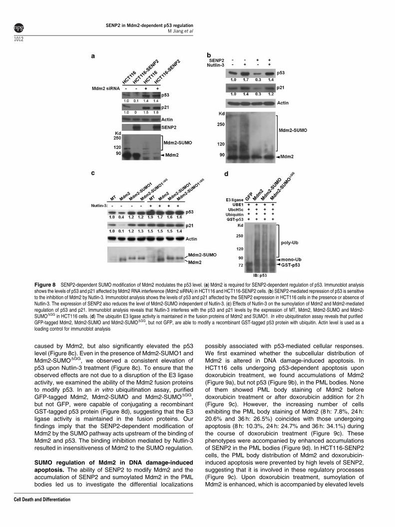
caused by Mdm2, but also significantly elevated the p53level (Figure 8c). Even in the presence of Mdm2-SUMO1 andMdm2-SUMODGG, we observed a consistent elevation ofp53 upon Nutlin-3 treatment (Figure 8c). To ensure that theobserved effects are not due to a disruption of the E3 ligaseactivity, we examined the ability of the Mdm2 fusion proteinsto modify p53. In an in vitro ubiquitination assay, purifiedGFP-tagged Mdm2, Mdm2-SUMO and Mdm2-SUMODGG,but not GFP, were capable of conjugating a recombinantGST-tagged p53 protein (Figure 8d), suggesting that the E3ligase activity is maintained in the fusion proteins. Ourfindings imply that the SENP2-dependent modification ofMdm2 by the SUMO pathway acts upstream of the binding ofMdm2 and p53. The binding inhibition mediated by Nutlin-3resulted in insensitiveness of Mdm2 to the SUMO regulation.
SUMO regulation of Mdm2 in DNA damage-inducedapoptosis. The ability of SENP2 to modify Mdm2 and theaccumulation of SENP2 and sumoylated Mdm2 in the PMLbodies led us to investigate the differential localizations
possibly associated with p53-mediated cellular responses.We first examined whether the subcellular distribution ofMdm2 is altered in DNA damage-induced apoptosis. InHCT116 cells undergoing p53-dependent apoptosis upondoxorubicin treatment, we found accumulations of Mdm2(Figure 9a), but not p53 (Figure 9b), in the PML bodies. Noneof them showed PML body staining of Mdm2 beforedoxorubicin treatment or after doxorubicin addition for 2 h(Figure 9c). However, the increasing number of cellsexhibiting the PML body staining of Mdm2 (8 h: 7.8%, 24 h:20.6% and 36 h: 26.5%) coincides with those undergoingapoptosis (8 h: 10.3%, 24 h: 24.7% and 36 h: 34.1%) duringthe course of doxorubicin treatment (Figure 9c). Thesephenotypes were accompanied by enhanced accumulationsof SENP2 in the PML bodies (Figure 9d). In HCT116-SENP2cells, the PML body distribution of Mdm2 and doxorubicin-induced apoptosis were prevented by high levels of SENP2,suggesting that it is involved in these regulatory processes(Figure 9c). Upon doxorubicin treatment, sumoylation ofMdm2 is enhanced, which is accompanied by elevated levels
Figure 8 SENP2-dependent SUMO modification of Mdm2 modulates the p53 level. (a) Mdm2 is required for SENP2-dependent regulation of p53. Immunoblot analysisshows the levels of p53 and p21 affected by Mdm2 RNA interference (Mdm2 siRNA) in HCT116 and HCT116-SENP2 cells. (b) SENP2-mediated repression of p53 is sensitiveto the inhibition of Mdm2 by Nutlin-3. Immunoblot analysis shows the levels of p53 and p21 affected by the SENP2 expression in HCT116 cells in the presence or absence ofNutlin-3. The expression of SENP2 also reduces the level of Mdm2-SUMO independent of Nutlin-3. (c) Effects of Nutlin-3 on the sumoylation of Mdm2 and Mdm2-mediatedregulation of p53 and p21. Immunoblot analysis reveals that Nutlin-3 interferes with the p53 and p21 levels by the expression of MT, Mdm2, Mdm2-SUMO and Mdm2-SUMODGG in HCT116 cells. (d) The ubiquitin E3 ligase activity is maintained in the fusion proteins of Mdm2 and SUMO1. In vitro ubiquitination assay reveals that purifiedGFP-tagged Mdm2, Mdm2-SUMO and Mdm2-SUMODGG, but not GFP, are able to modify a recombinant GST-tagged p53 protein with ubiquitin. Actin level is used as aloading control for immunoblot analysis
SENP2 in Mdm2-dependent p53 regulationM Jiang et al
1012
Cell Death and Differentiation

of p53 and p21 (Figure 9e). Before the addition of doxorubicin,free SUMO1 as well as Mdm2 proteins did not localize to
the PML bodies (Figure 9f). However, doxorubicin prom-oted the accumulations of Mdm2 and SUMO1 to the PMLbodies (Figure 9f). This differential co-localization implies thatsumoylated Mdm2 accumulates in the PML bodies in DNAdamage-induced responses. The data suggest that the SUMO-dependent regulation of Mdm2 distribution may have a crucialrole in genotoxic stresses mediated by p53.
Discussion
This study demonstrated an isoform-specific effect of SENP2on the p53/Mdm2 regulatory circuit. This effect is likelyattributed to the differential compartmentalization of SENP2isoforms within the cells. The longest form of SENP2 isnecessary and sufficient for modulating the stability of p53,and the p53-dependent transcription and stress responses.Biochemical studies further show that SENP2 promotes thedesumoylation of Mdm2, contributing to the p53-dependentregulations. The accumulation of SENP2 in the PML bodiesfurther indicates its importance in modifying the activity ofMdm2 and p53. Although both SENP2 and Mdm2 preferen-tially accumulate in the nucleus, their localizations do notseem to overlap significantly. However, SUMO conjugationof Mdm2 leads to protein translocation, resulting in strongaccumulations in the PML bodies where it co-localizedsignificantly with SENP2. The PML body distribution ofMdm2 and SENP2 is also modulated by doxorubicin-induced
apoptosis, suggesting a role in stress responses mediatedby p53. On the basis of this work, we have proposed amechanism for the SENP2–Mdm2–p53 regulatory pathway(Figure 10). Although p53 does not seem to be regulated bySENP2 at the PML bodies, it is possible that SENP2 may alsocatalyze the desumoylation of p53. If true, this would addcomplexities to this multilayered regulatory network. How-ever, the SENP2-mediated SUMO modification of p53 mighthave functional consequences distinct to that of Mdm2, whichremains to be explored.
We have previously reported that Nutlin-3 treatment is stillcapable of increasing the p53 level in SENP2-null cells.7
Nutlin-3 elevates p53 under normal (SENP2þ /þ ) and hyper-sumoylated (SENP2�/�) conditions of Mdm2, implying thatsumoylated Mdm2 is still able to interact with p53. Similar to
Figure 9 DNA damage-induced apoptosis promotes the interaction of Mdm2 and SENP2 at the PML bodies. (a) Triple labeling of Mdm2, PML and activated caspase-3reveals that Mdm2 accumulates in the PML bodies of cells undergoing p53-dependent apoptosis. Mdm2-GFP was expressed in HCT116 cells without or with the addition ofdoxorubicin for 24 h. Mdm2 co-localized with PML in cells undergoing apoptosis identified by immunostaining of activated caspase-3. (b) Triple labeling of p53, PML andactivated caspase-3 shows that the doxorubicin-induced apoptosis does not promote p53 accumulation in the PML bodies. (c) Graph indicating the percentage of Mdm2-GFPpositive cells displaying PML body distribution, and undergoing programmed cell death in the course of doxorubicin treatment. (d) Graph showing the percentage of HCT116-SENP2 cells displaying the PML body distribution of SENP2 in the course of doxorubicin treatment (P-value: *Po0.035; **Po0.021; ***Po0.017). (e) Immunoblot andimmunoprecipitation–immunoblot analyses reveal that the induction of p53 and p21 coincides with the accumulation of sumoylated Mdm2 upon doxorubicin treatment. Actinlevel is used as a loading control. (f) DNA damage response promotes accumulations of Mdm2 and SUMO1 in the PML bodies. HCT116 cells transfected with GFP-taggedMdm2 were cultured in the presence or absence of doxorubicin for 24 h, followed by triple labeling analysis
Figure 10 Model for SENP2-mediated modulation of Mdm2 in regulating p53.The diagram illustrates the mechanism underlying the regulation of Mdm2 by theSUMO pathway to control the cellular level of p53. SENP2 regulates p53 throughmodulation of Mdm2. SENP2 interacts with sumoylated Mdm2 and regulates itsSUMO conjugation at the PML body. The SENP2-dependent SUMO modificationacts upstream of the binding of Mdm2 and p53 in the regulatory pathway.Desumoylation of Mdm2 permits its binding and ubiquitination of p53, which is thendegraded through the proteolysis system
SENP2 in Mdm2-dependent p53 regulationM Jiang et al
1013
Cell Death and Differentiation

that observed in SENP2-null cells, Nutlin-3 treatment onMdm2-SUMO1 causes an elevation effect of p53. Onepossibility is that both Mdm2 and sumoylated Mdm2 formcomplexes with p53. Desumoylation of Mdm2 then activatesits E3 ligase activity. The fact that E3 ligase activity ismaintained in the Mdm2–SUMO fusion protein seems toargue against this. Alternatively, desumoylation of Mdm2promotes the cytoplasmic translocation of p53 for degrada-tion. Mdm2-SUMO and p53 could form a sequestrationcomplex accumulating in the nucleus without initiating thedegradation and nuclear–cytoplasmic translocation pro-cesses. Stabilization of p53 then is capable of promoting thesubsequent responses to cellular stresses. The findingssupport our hypothesis, further implying that Mdm2-SUMOis sequestered at the PML bodies unable to promote p53degradation under hyper-sumoylated conditions (Figure 10).Indeed, we have found that the cellular levels of sumoylatedMdm2 are increased upon doxorubicin treatment. Further-more, Mdm2 and SUMO1 co-localize at the PML bodies of thedoxorubicin-treated cells. Our data suggest that the SUMO-conjugated Mdm2 recruits to the PML bodies, causingdeficiency in targeting p53 degradation. However, due to thelack of information on identification of the sumoylation site ofMdm2, this hypothesis cannot be definitively assessed. Oncethe sumoylation site has been identified, it would be importantto determine whether mutation of the sumoylation siteinterferes with the PML body distribution of Mdm2.
An important function of p53 as a guardian of the genomeensures that the genetic information is properly propagatedduring these processes.21 In addition, p53 preserves genomicstability in stress responses of various insults, including DNAdamage, hypoxia, metabolic stress and oncogene activa-tion.22–24 The most documented mechanism by which p53exerts its protective effects is as a transcription factor.25
SENP2 is apparently a negative regulator for the p53 level andthe p53-dependent transcription. Although recent evidencehas challenged the classical model of p53 activation,26 itsstabilization remains to be a key regulatory step. SENP2,involved in the p53-induced regulations, may have a role ingenotoxic stresses. Further investigation focusing on theaction of SENP2 promises new insights into the mechanismunderlying cellular stress mediated by the SUMO pathway. Asour prior study indicated that the SENP2–Mdm2–p53 pathwayis critical for the G–S checkpoint of mitotic divisions andendoreduplication,7 it is possible that aberrant stimulation ofSENP2 causes cell cycle defects, leading to the prevention ofcell death and senescence. Recent reports suggest that theSUMO pathway is involved in cellular responses to DNAdouble-strand breaks.27,28 Whether SENP2 participates inthese genotoxic stresses essential for maintaining genomeintegrity remains an interesting question to be determined.
Mutations in the p53 gene have been linked to familial andsporadic forms of human cancer. Li–Fraumeni syndrome is arare familial disorder that greatly increases the risk ofdeveloping several types of cancers in children and youngadults.29 In sporadic forms, mutations have been identified inother key regulators, for example, p53, Mdm2 and p14ARF,which result in alteration of the p53 activity.22 Mice withdisruption of RanBP2, a SUMO E3 ligase, developedtumors,30 suggesting that aberrant regulation of the SUMO
pathway induces cells to undergo malignant transformation.Previous reports also suggest that SENP1 might be asso-ciated with development of prostate cancers.31 As SUMO canregulate p53 directly10 or indirectly,7 SENPs might contributeto the oncogenic processes at multiple levels. Therefore, it isconceivable that SENP2 has a crucial role in p53-inducedtumorigenesis, which remains to be explored. However,embryonic lethality associated with the SENP2 deletion inmice prevents investigation of its oncogenic role.7 Mousemodels permitting conditional inactivation of SENP2 arenecessary to further decipher this genetic regulatory network.
Materials and MethodsDNA. The generation of pCS2-SENP2, pGFP-Mdm2, pGFP-Mdm2-SUMO1,pGEX-4T-SAE1/2 and pGEX-2T-Ubc9 clones was described previously.7,32,33
The pGFP-Mdm2-SUMO1GG96�97D clone was generated by mutating two glycineresidues at the C terminus of SUMO1 contained in the pGFP-Mdm2-SUMO1plasmid using site-directed mutagenesis (Stratagene, La Jolla, CA, USA). Togenerate pGEX-2T-SUMO1, pGEX-2T-Mdm2 and pGEX-2T-p53 clones, thecorresponding DNA fragments were inserted into pGEX-2T vector (GEHealthCare, Waukesha, WI, USA). The myc-tagged SENP2 cDNAs, encodingthe middle and short isoforms, were inserted into the pCS2 vector to create thepCS2-SENP2-M and pCS2-SENP2-S clones, respectively. To generate the pHA-SUMO1 clone, SUMO1 fragment was inserted into the pCI-Neo-HA vector(Promega, Madison, WI, USA).
Cells. The establishment and culture of SENP2þ /þ and SENP2�/� TS celllines were described previously.7 To create the HCT116-SENP2, HCT116-SENP2M and HCT116-SENP2S clones, DNA fragment containing pGK-Neo wasfirst inserted into the pCS2-SENP2, pCS2-SENP2-M and pCS2-SENP2-Splasmids. The plasmids were then stably transfected into HCT116 cells, followedby G418 (800mg/ml) selection for 14 days. Colonies, which grow under drugresistance condition, were picked to establish cell clones. For DNA damageinduction, cells were cultured with 1 mM doxorubicin. For growth factor deprivation,cells were cultured in 0.1% fetal bovine serum. The number of cells was countedeach day to assess the growth curve. Cells undergoing apoptosis were detected byimmunostaining of activated caspase-3 or TUNEL staining (Millipore, Billerica, MA,USA) after the doxorubicin treatment for 24 h. The stained images were takenrandomly to determine the percentage of apoptotic cells by counting the activatedcaspase-3 or TUNEL-positive and -negative cells. Nutlin-3 (10 mM) was added tothe culture media to prevent the binding of p53 and Mdm2. To measure the p53-dependent transcriptional activity, cells transfected with p53-luciferase, containingthe luciferase reporter under control of p53 responsive elements, were subject toluciferase assay.
Immunostaining and immunoblot. Cells were fixed and subjected toimmunological staining and fluorescent imaging analysis as described.34–38 Imageswere taken using Zeiss Axio Observer microscope, followed by deconvolution, 3Dimaging and cut-view analyses.34,39 Immunoblot analysis was performed byisolating protein extracts from cells in the presence of protease inhibitor cocktail(Sigma–Aldrich, St. Louis, MO, USA), 1 mM sodium molybdate, 1 mM sodiumvanadate and 1 mM PMSF, or SDS lysis buffer (2% SDS, 2% 2-mercaptoethanol,10% glycerol, and 50 mM Tris, pH 6.8), followed by electrophoresis asdescribed.7,34,36,39,40 Bound primary antibodies were detected with horseradishperoxidase-conjugated secondary antibodies (Vector Laboratories, Burlingame,CA, USA), followed by ECL-mediated visualization (GE HealthCare) andautoradiography.7,34,36,39,40 Mouse monoclonal antibodies Actin (Thermo Fisher,Waltham, MA, USA), GS28 (BD Biosciences, San Jose, CA, USA), HA (CellSignaling, Danvers, MA, USA), PML (Santa Cruz, Santa Cruz, CA, USA), Mdm2(Santa Cruz), myc tag (Santa Cruz), p53 (Santa Cruz), SUMO1 (Invitrogen,Carlsbad, CA, USA), rabbit monoclonal antibody SUMO1 (Cell Signaling), rabbitpolyclonal antibody caspase-3 (BD Biosciences), goat polyclonal antibodies lamin B(Santa Cruz) and p53 (Santa Cruz) were used in the analyses.
Ubiquitin, sumoylation and desumoylation assays. For in vitroubiquitin assay, purified GFP-tagged Mdm2, Mdm2-SUMO1 or GFP-Mdm2-SUMO1DGG was added to the reaction buffer containing 50 mM Hepes (pH 8.0),
SENP2 in Mdm2-dependent p53 regulationM Jiang et al
1014
Cell Death and Differentiation
1 mM Mg-ATP, UBE1 (Boston Biochem, Cambridge, MA, USA), UbcH5c (BostonBiochem) and recombinant GST-p53 in a final volume of 10 ml, followed byincubation for 60 min at 371C. The reaction was then stopped by the addition of aloading buffer containing 150 mM Tris-HCl (pH 6.8), 6% SDS, 30% glycerol and 6%2-mercaptoethanol and analyzed by SDS-PAGE and immunoblot analysis of p53.
To perform the in vitro sumoylation and desumoylation assays, recombinantGST-SAE1/SAE2, GST-Ubc9, GST-SUMO1 and GST-Mdm2 proteins expressed inEscherichia coli were affinity purified. The 20-ml reaction buffer containing 50 mMTris-HCl (pH 7.6), 5 mM magnesium chloride, 10 mM ATP, 1mg of GST-SAE1/2,2mg of GST-Ubc9, 10mg of GST-SUMO1 and 200 ng GST-Mdm2 withthe presence of protease inhibitor cocktail was incubated for 3 h at 371C.The desumoylation reaction was then carried out in 10 ml of the above sumoylationmixture with the addition of purified MT-SENP2. After incubation overnight at371C, the samples were analyzed by SDS-PAGE and immunoblot analysis of Mdm2and SUMO1.
For desumoylation analysis using cell extracts, 293T cells were transfected withHA-SUMO1 plus or minus pCS2-SENP2, as well as HCT116, and HCT116-SENP2cells were transfected with HA-SUMO1 using Lipofectamine 2000 (Invitrogen).After 48 h, cell lysates were collected with buffer containing 100 mM sodium chloride,20 mM Tris-HCl (pH 7.5), 1% Triton X-100, protease inhibitor cocktail (Sigma-Aldrich),1 mM sodium molybdate, 1 mM sodium vanadate and 1 mM PMSF, followed byimmunoprecipitation with anti-Mdm2 antibody and protein G agarose beads. Theprecipitates were analyzed by SDS-PAGE and immunoblot. To perform desumoyla-tion analysis, cell lysates were collected from 293T cells transfected with pCS2-SENP2 or pCS2, followed by purification with anti-MT antibody-mediatedimmunoprecipitation. The purified MT or MT-SENP2 protein was then incubatedwith cell lysates isolated from the HA-SUMO1-transfected 293T cells with or withoutthe presence of 10 mM NEM for 4 h at 371C, followed by immunoblot analysis.
Conflict of interest
The authors declare no conflict of interest.
Acknowledgements. We thank Ronald Hay, Bert Vogelstein and MichaelO’Reilly for reagents, H-M Ivy Yu for technical assistance, and Anthony Mirando andPeter Keng for comments and suggestions. This work is supported by NationalInstitutes of Health Grant CA106308 to WH.
1. Melchior F. SUMO – nonclassical ubiquitin. Annu Rev Cell Dev Biol 2000; 16: 591–626.2. Schwartz DC, Hochstrasser M. A superfamily of protein tags: ubiquitin, SUMO and related
modifiers. Trends Biochem Sci 2003; 28: 321–328.3. Seeler JS, Dejean A. Nuclear and unclear functions of SUMO. Nat Rev 2003; 4: 690–699.4. Muller S, Ledl A, Schmidt D. SUMO: a regulator of gene expression and genome integrity.
Oncogene 2004; 23: 1998–2008.5. Melchior F, Schergaut M, Pichler A. SUMO: ligases, isopeptidases and nuclear pores.
Trends Biochem Sci 2003; 28: 612–618.6. Gill G. SUMO and ubiquitin in the nucleus: different functions, similar mechanisms? Genes
Dev 2004; 18: 2046–2059.7. Chiu SY, Asai N, Costantini F, Hsu W. SUMO-specific protease 2 is essential for
modulating p53-Mdm2 in development of trophoblast stem cell niches and lineages. PLoSBiol 2008; 6: e310.
8. Marine JC, Lozano G. Mdm2-mediated ubiquitylation: p53 and beyond. Cell Death Differ2010; 17: 93–102.
9. Meek DW, Knippschild U. Posttranslational modification of MDM2. Mol Cancer Res 2003;1: 1017–1026.
10. Melchior F, Hengst L. SUMO-1 and p53. Cell Cycle 2002; 1: 245–249.11. Marine JC. p53 stabilization: the importance of nuclear import. Cell Death Differ 2010; 17:
191–192.12. Storchova Z, Pellman D. From polyploidy to aneuploidy, genome instability and cancer.
Nat Rev 2004; 5: 45–54.
13. Lanni JS, Jacks T. Characterization of the p53-dependent postmitotic checkpoint followingspindle disruption. Mol Cell Biol 1998; 18: 1055–1064.
14. Andreassen PR, Lohez OD, Lacroix FB, Margolis RL. Tetraploid state inducesp53-dependent arrest of nontransformed mammalian cells in G1. Mol Biol Cell 2001; 12:1315–1328.
15. Yates KE, Korbel GA, Shtutman M, Roninson IB, DiMaio D. Repression of the SUMO-specific protease Senp1 induces p53-dependent premature senescence in normal humanfibroblasts. Aging Cell 2008; 7: 609–621.
16. Green DR, Kroemer G. Cytoplasmic functions of the tumour suppressor p53. Nature 2009;458: 1127–1130.
17. Vazquez A, Bond EE, Levine AJ, Bond GL. The genetics of the p53 pathway, apoptosisand cancer therapy. Nat Rev Drug Discov 2008; 7: 979–987.
18. Polyak K, Waldman T, He TC, Kinzler KW, Vogelstein B. Genetic determinants ofp53-induced apoptosis and growth arrest. Genes Dev 1996; 10: 1945–1952.
19. Fogal V, Gostissa M, Sandy P, Zacchi P, Sternsdorf T, Jensen K et al. Regulationof p53 activity in nuclear bodies by a specific PML isoform. EMBO J 2000; 19:6185–6195.
20. Pearson M, Carbone R, Sebastiani C, Cioce M, Fagioli M, Saito S et al. PML regulatesp53 acetylation and premature senescence induced by oncogenic Ras. Nature 2000; 406:207–210.
21. Levine AJ, Oren M. The first 30 years of p53: growing ever more complex. Nat Rev Cancer2009; 9: 749–758.
22. Vogelstein B, Lane D, Levine AJ. Surfing the p53 network. Nature 2000; 408: 307–310.23. Vousden KH, Lane DP. p53 in health and disease. Nat Rev 2007; 8: 275–283.24. Meek DW. Tumour suppression by p53: a role for the DNA damage response? Nat Rev
Cancer 2009; 9: 714–723.25. Riley T, Sontag E, Chen P, Levine A. Transcriptional control of human p53-regulated
genes. Nat Rev 2008; 9: 402–412.26. Kruse JP, Gu W. Modes of p53 regulation. Cell 2009; 137: 609–622.27. Morris JR, Boutell C, Keppler M, Densham R, Weekes D, Alamshah A et al. The SUMO
modification pathway is involved in the BRCA1 response to genotoxic stress. Nature 2009;462: 886–890.
28. Galanty Y, Belotserkovskaya R, Coates J, Polo S, Miller KM, Jackson SP. MammalianSUMO E3-ligases PIAS1 and PIAS4 promote responses to DNA double-strand breaks.Nature 2009; 462: 935–939.
29. Li FP, Fraumeni Jr JF. Soft-tissue sarcomas, breast cancer, and other neoplasms.A familial syndrome? Ann Intern Med 1969; 71: 747–752.
30. Dawlaty MM, Malureanu L, Jeganathan KB, Kao E, Sustmann C, Tahk S et al. Resolutionof sister centromeres requires RanBP2-mediated SUMOylation of topoisomerase IIalpha.Cell 2008; 133: 103–115.
31. Kaikkonen S, Jaaskelainen T, Karvonen U, Rytinki MM, Makkonen H, Gioeli D et al.SUMO-specific protease 1 (SENP1) reverses the hormone-augmented SUMOylation ofandrogen receptor and modulates gene responses in prostate cancer cells. Mol Endocrinol(Baltimore, Md) 2009; 23: 292–307.
32. Tatham MH, Jaffray E, Vaughan OA, Desterro JM, Botting CH, Naismith JH et al. Polymericchains of SUMO-2 and SUMO-3 are conjugated to protein substrates by SAE1/SAE2 andUbc9. J Biol Chem 2001; 276: 35368–35374.
33. Desterro JM, Rodriguez MS, Hay RT. SUMO-1 modification of IkappaBalpha inhibitsNF-kappaB activation. Mol Cell 1998; 2: 233–239.
34. Fu J, Jiang M, Mirando AJ, Yu HM, Hsu W. Reciprocal regulation of Wnt and Gpr177/mouse Wntless is required for embryonic axis formation. Proc Natl Acad Sci USA 2009;106: 18598–18603.
35. Liu B, Yu HM, Hsu W. Craniosynostosis caused by Axin2 deficiency is mediated throughdistinct functions of beta-catenin in proliferation and differentiation. Dev Biol 2007; 301:298–308.
36. Liu B, Yu HM, Huang J, Hsu W. Co-opted JNK/SAPK signaling in Wnt/beta-catenin-induced tumorigenesis. Neoplasia (New York, NY) 2008; 10: 1004–1013.
37. Maruyama T, Mirando AJ, Deng CX, Hsu W. The balance of WNT and FGF signalinginfluences mesenchymal stem cell fate during skeletal development. Sci Signal 2010;3: ra40.
38. Yu HM, Liu B, Costantini F, Hsu W. Impaired neural development caused by inducibleexpression of Axin in transgenic mice. Mech Dev 2007; 124: 146–156.
39. Yu HM, Jin Y, Fu J, Hsu W. Expression of Gpr177, a Wnt trafficking regulator, in mouseembryogenesis. Dev Dyn 2010; 239: 2102–2109.
40. Yu HM, Jerchow B, Sheu TJ, Liu B, Costantini F, Puzas JE et al. The role of Axin2 incalvarial morphogenesis and craniosynostosis. Development (Cambridge, England) 2005;132: 1995–2005.
SENP2 in Mdm2-dependent p53 regulationM Jiang et al
1015
Cell Death and Differentiation