journal club paper p53 mdm2
DESCRIPTION
jjTRANSCRIPT
A Potent Small-Molecule Inhibitor of the MDM2−p53 Interaction (MI-888) Achieved Complete and Durable Tumor Regression in MiceYujun Zhao,†,§ Shanghai Yu,†,§ Wei Sun,†,§ Liu Liu,† Jianfeng Lu,† Donna McEachern,† Sanjeev Shargary,†
Denzil Bernard,† Xiaoqin Li,‡ Ting Zhao,‡ Peng Zou,‡ Duxin Sun,‡ and Shaomeng Wang*,†
†Comprehensive Cancer Center and Departments of Internal Medicine, Pharmacology, and Medicinal Chemistry, University ofMichigan, Ann Arbor, Michigan 48109, United States‡Department of Pharmaceutical Sciences, College of Pharmacy, University of Michigan, Ann Arbor, Michigan 48109, United States
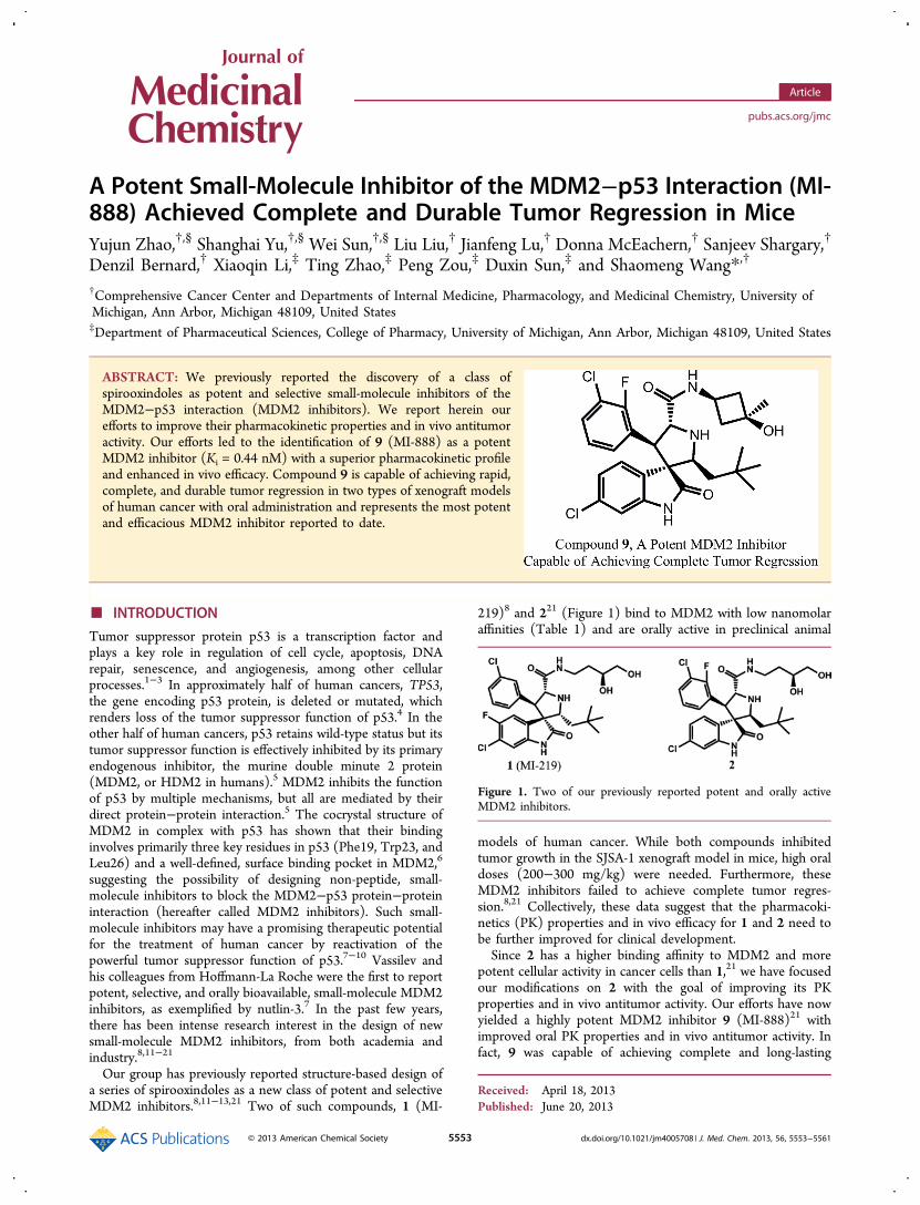
ABSTRACT: We previously reported the discovery of a class ofspirooxindoles as potent and selective small-molecule inhibitors of theMDM2−p53 interaction (MDM2 inhibitors). We report herein ourefforts to improve their pharmacokinetic properties and in vivo antitumoractivity. Our efforts led to the identification of 9 (MI-888) as a potentMDM2 inhibitor (Ki = 0.44 nM) with a superior pharmacokinetic profileand enhanced in vivo efficacy. Compound 9 is capable of achieving rapid,complete, and durable tumor regression in two types of xenograft modelsof human cancer with oral administration and represents the most potentand efficacious MDM2 inhibitor reported to date.
■ INTRODUCTION
Tumor suppressor protein p53 is a transcription factor andplays a key role in regulation of cell cycle, apoptosis, DNArepair, senescence, and angiogenesis, among other cellularprocesses.1−3 In approximately half of human cancers, TP53,the gene encoding p53 protein, is deleted or mutated, whichrenders loss of the tumor suppressor function of p53.4 In theother half of human cancers, p53 retains wild-type status but itstumor suppressor function is effectively inhibited by its primaryendogenous inhibitor, the murine double minute 2 protein(MDM2, or HDM2 in humans).5 MDM2 inhibits the functionof p53 by multiple mechanisms, but all are mediated by theirdirect protein−protein interaction.5 The cocrystal structure ofMDM2 in complex with p53 has shown that their bindinginvolves primarily three key residues in p53 (Phe19, Trp23, andLeu26) and a well-defined, surface binding pocket in MDM2,6
suggesting the possibility of designing non-peptide, small-molecule inhibitors to block the MDM2−p53 protein−proteininteraction (hereafter called MDM2 inhibitors). Such small-molecule inhibitors may have a promising therapeutic potentialfor the treatment of human cancer by reactivation of thepowerful tumor suppressor function of p53.7−10 Vassilev andhis colleagues from Hoffmann-La Roche were the first to reportpotent, selective, and orally bioavailable, small-molecule MDM2inhibitors, as exemplified by nutlin-3.7 In the past few years,there has been intense research interest in the design of newsmall-molecule MDM2 inhibitors, from both academia andindustry.8,11−21
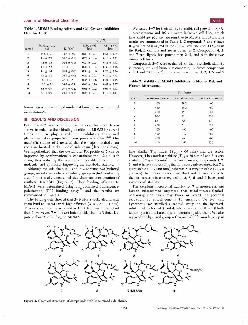
Our group has previously reported structure-based design ofa series of spirooxindoles as a new class of potent and selectiveMDM2 inhibitors.8,11−13,21 Two of such compounds, 1 (MI-
219)8 and 221 (Figure 1) bind to MDM2 with low nanomolaraffinities (Table 1) and are orally active in preclinical animal
models of human cancer. While both compounds inhibitedtumor growth in the SJSA-1 xenograft model in mice, high oraldoses (200−300 mg/kg) were needed. Furthermore, theseMDM2 inhibitors failed to achieve complete tumor regres-sion.8,21 Collectively, these data suggest that the pharmacoki-netics (PK) properties and in vivo efficacy for 1 and 2 need tobe further improved for clinical development.Since 2 has a higher binding affinity to MDM2 and more
potent cellular activity in cancer cells than 1,21 we have focusedour modifications on 2 with the goal of improving its PKproperties and in vivo antitumor activity. Our efforts have nowyielded a highly potent MDM2 inhibitor 9 (MI-888)21 withimproved oral PK properties and in vivo antitumor activity. Infact, 9 was capable of achieving complete and long-lasting
Received: April 18, 2013Published: June 20, 2013
Figure 1. Two of our previously reported potent and orally activeMDM2 inhibitors.
Article
pubs.acs.org/jmc
© 2013 American Chemical Society 5553 dx.doi.org/10.1021/jm4005708 | J. Med. Chem. 2013, 56, 5553−5561
tumor regression in animal models of human cancer upon oraladministration.
■ RESULTS AND DISCUSSIONBoth 1 and 2 have a flexible 1,2-diol side chain, which wasshown to enhance their binding affinities to MDM2 by severaltimes and to play a role in modulating their oralpharmacokinetics properties in our previous studies.11,21 Ourmetabolic studies of 2 revealed that the major metabolic softspots are located in the 1,2-diol side chain (data not shown).We hypothesized that the overall oral PK profile of 2 can beimproved by conformationally constraining the 1,2-diol sidechain, thus reducing the number of rotatable bonds in themolecule, and by further improving the metabolic stability.Although the side chain in 1 and in 2 contains two hydroxyl
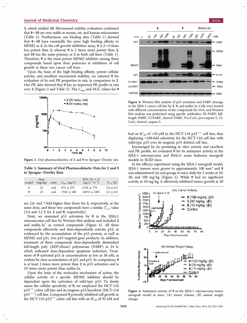
groups, we retained only one hydroxyl group in 3−7 containinga conformationally constrained side chain for consideration ofsynthetic feasibility (Figure 2). Their binding affinities toMDM2 were determined using our optimized fluorescence-polarization (FP) binding assay,21 and the results aresummarized in Table 1.The binding data showed that 3−6 with a cyclic alcohol side
chain bind to MDM2 with high affinities (Ki = 0.61−1.1 nM).These compounds are as potent as 2 but 10 times more potentthan 1. However, 7 with a tert-butanol side chain is 3 times lesspotent than 2 in binding to MDM2.
We tested 1−7 for their ability to inhibit cell growth in SJSA-1 osteosarcoma and RS4;11 acute leukemia cell lines, whichhave wild-type p53 and are sensitive to MDM2 inhibitors. Theresults are summarized in Table 1. Compounds 3 and 6 haveIC50 values of 0.24 μM in the SJSA-1 cell line and 0.12 μM inthe RS4;11 cell line and are as potent as 2. Compounds 4, 5,and 7 are slightly less potent than 2, 3, and 6 in these twocancer cell lines.Compounds 3−7 were evaluated for their metabolic stability
in mouse, rat, and human microsomes, in direct comparisonwith 1 and 2 (Table 2). In mouse microsomes, 1, 2, 3, 6, and 7
have similar T1/2 values (T1/2 > 60 min) and are stable.However, 4 has modest stability (T1/2 = 20.6 min) and 5 is veryunstable (T1/2 = 1.5 min). In rat microsomes, compounds 1, 2,3, and 6 have a shorter T1/2 than in mouse microsomes, but 7 isquite stable (T1/2 >60 min), whereas 5 is very unstable (T1/2 =3.8 min). In human microsomes, the trend is very similar tothat in mouse microsomes, and 1, 2, 3, 6, and 7 have goodmicrosomal stability.The excellent microsomal stability for 7 in mouse, rat, and
human microsomes suggested that trisubstituted-alcohol-containing side chain may block or retard the potentialoxidation by cytochrome P450 enzymes. To test thishypothesis, we installed a methyl group on the hydroxyl-substituted carbon of 3 and 4, which resulted in 8 and 9 bothtethering a trisubstituted-alcohol-containing side chain. We alsoreplaced the hydroxyl group with a methylsulfonamide group in
Table 1. MDM2 Binding Affinity and Cell Growth InhibitionData for 1−10
IC50 (μM)
compdbinding IC50
(nM) Ki (nM)SJSA-1 cell
lineRS4;11 cell
line
1 44.6 ± 2.7 10.5 ± 2.0 0.89 ± 0.21 0.54 ± 0.142 9.8 ± 2.7 0.86 ± 0.11 0.25 ± 0.04 0.10 ± 0.013 7.1 ± 1.5 0.61 ± 0.10 0.24 ± 0.05 0.12 ± 0.014 8.2 ± 2.3 1.1 ± 0.3 0.41 ± 0.04 0.30 ± 0.065 8.8 ± 1.4 0.94 ± 0.07 0.32 ± 0.06 0.23 ± 0.046 8.4 ± 1.1 0.62 ± 0.05 0.20 ± 0.02 0.10 ± 0.027 16.3 ± 2.1 2.4 ± 0.5 0.35 ± 0.06 0.21 ± 0.058 11.1 ± 2.1 0.97 ± 0.3 0.60 ± 0.16 0.43 ± 0.079 6.8 ± 0.9 0.44 ± 0.22 0.08 ± 0.02 0.06 ± 0.0110 7.6 ± 0.9 0.62 ± 0.19 0.25 ± 0.04 0.10 ± 0.01
Figure 2. Chemical structures of compounds with constrained side chains.
Table 2. Stability of MDM2 Inhibitors in Mouse, Rat, andHuman Microsomes
T1/2 (min)
compd mouse microsome rat microsome human microsome
1 >60 20.2 >602 >60 16.3 >603 >60 19.1 54.14 20.6 22.1 30.85 1.5 3.8 4.06 >60 21.1 55.17 >60 >60 >608 >60 >60 >609 >60 >60 >6010 >60 >60 53.3
Journal of Medicinal Chemistry Article
dx.doi.org/10.1021/jm4005708 | J. Med. Chem. 2013, 56, 5553−55615554
3, which yielded 10. Microsomal stability evaluation confirmedthat 8−10 are very stable in mouse, rat, and human microsomes(Table 2). Furthermore, our binding data (Table 1) showedthat 8−10 have essentially the same high binding affinity toMDM2 as 2. In the cell growth inhibition assay, 8 is 2−4 timesless potent than 2, whereas 9 is 2 times more potent than 2,and 10 has the same potency as 2 in both cell lines (Table 1).Therefore, 9 is the most potent MDM2 inhibitor among thesecompounds based upon their potencies in inhibition of cellgrowth in these two cancer cell lines.Upon the basis of the high binding affinity, potent cellular
activity, and excellent microsomal stability, we selected 9 forevaluation of its oral PK properties in rats, in comparison to 2.Our PK data showed that 9 has an improved PK profile in ratsover 2 (Figure 3 and Table 3). The Cmax and AUC values for 9
are 2.6- and 7-fold higher than those for 2, respectively, at thesame dose, and these two compounds have a similar T1/2 value(3.4 and 3.2 h for 2 and 9, respectively).Next, we examined p53 activation by 9 in the SJSA-1
osteosarcoma cell line by Western blot analysis and included 2and nutlin-3a7 as control compounds (Figure 4). All threecompounds effectively and dose-dependently activate p53, asevidenced by the accumulation of the p53 protein, as well asMDM2 and p21, two p53 targeted gene products. In addition,treatment of three compounds dose-dependently diminishedfull-length poly (ADP-ribose) polymerase (PARP) at 24 h,which indicated dose-dependent apoptosis induction. Treat-ment of 9 activated p53 at concentrations as low as 30 nM, asevident by clear accumulation of p21 and p53. In comparison, 9is at least 3 times more potent than 2 in p53 activation and is10 times more potent than nutlin-3a.Upon the basis of the molecular mechanism of action, the
cellular activity of a specific MDM2 inhibitor should bedependent upon the activation of wild-type p53. To firmlyassess the cellular specificity of 9, we employed the HCT-116p53+/+ colon cell line and its isogenic p53 knockout (HCT-116p53−/−) cell line. Compound 9 potently inhibited cell growth inthe HCT-116 p53+/+ colon cell line with an IC50 of 92 nM and
had an IC50 of >10 μM in the HCT-116 p53−/− cell line, thusdisplaying >100-fold selectivity for the HCT-116 cell line withwild-type p53 over its isogenic p53 deleted cell line.Encouraged by its promising in vitro activity and excellent
oral PK profile, we evaluated 9 for its antitumor activity in theSJSA-1 osteosarcoma and RS4;11 acute leukemia xenograftmodels in SCID mice.In the efficacy experiment using the SJSA-1 xenograft model,
SJSA-1 tumors were grown to approximately 100 mm3 and 9was administered via oral gavage to mice daily for 2 weeks at 10,30, and 100 mg/kg (Figure 5). While 9 had no significantactivity at 10 mg/kg, it effectively inhibited tumor growth at 30
Figure 3. Oral pharmacokinetics of 2 and 9 in Sprague−Dawley rats.
Table 3. Summary of Oral Pharmacokinetic Data for 2 and 9in Sprague−Dawley Rats
compddose
(mg/kg) route Cmax (μg/L)AUC (0 → t)(μg L−1 h−1) T1/2 (h)
2 25 oral 675 ± 274 2138 ± 775 3.4 ± 0.59 25 oral 1726 ± 100 14874 ± 2491 3.2 ± 0.5
Figure 4. Western blot analysis of p53 activation and PARP cleavagein the SJSA-1 cancer cell line by 2, 9, and nutlin-3a. Cells were treatedwith different concentrations of the compounds for 24 h, and Westernblot analysis was performed using specific antibodies: FL-PARP, full-length PARP; Cl-PARP, cleaved PARP; Pro-Cas3, pro-caspase-3; Cl-Cas3, cleaved caspase-3.
Figure 5. Antitumor activity of 9 in the SJSA-1 osteosarcoma tumorxenograft model in mice: (A) tumor volume; (B) animal weightchange.
Journal of Medicinal Chemistry Article
dx.doi.org/10.1021/jm4005708 | J. Med. Chem. 2013, 56, 5553−55615555
mg/kg. Impressively, 9 at 100 mg/kg achieved rapid andcomplete tumor regression. After 5 days of daily dosing, theaverage tumor volume was decreased by >70%, and after 10days of dosing, all the mice (8 out of 8 mice) treated with 9 hadundetectable tumor. The complete tumor regression wasdurable; all the mice remained tumor free for 60 days afterthe last dose. There was no significant difference in animalweight between the vehicle control group of mice and the threegroups of mice treated with 9. Furthermore, there was minimalweight loss and no sign of toxicity observed in mice treatedwith 9 at all doses during the entire experiment. Collectively,these data showed that 9 was well tolerated in mice at all thedoses tested.To gain an insight into the in vivo mechanism of action of 9,
we tested its ability and kinetics in activation of p53 andinduction of apoptosis in the SJSA-1 xenograft tissue (Figure6). Mice bearing SJSA-1 xenograft tumors were given a single
oral dose of 9 at 100 mg/kg. Mice were then sacrificed atdifferent time points, and tumors were harvested for Westernblot analysis. Our Western blot data showed that 9 inducedrobust up-regulation of p53, as well as p21 and MDM2 proteinsat 3 and 6 h time points, indicative of strong p53 activation intumor tissue. The levels of p53, p21, and MDM2 proteins weresignificantly diminished at the 24 h time point, suggesting thatp53 activation was transient in tumor tissue. Interestingly,cleavage of PARP and caspase-3 was minimal at the 1, 3, and 6h time points but became very clear at the 24 h time point,indicating that while p53 was activated by 9 in tumor tissuequickly, apoptosis induction occurred at later time points.In the efficacy experiment using the RS4;11 acute leukemia
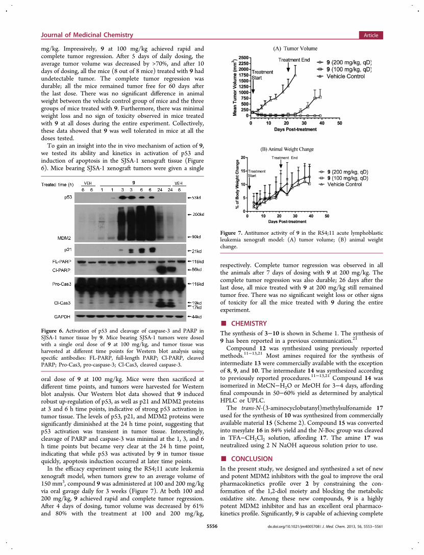
xenograft model, when tumors grew to an average volume of150 mm3, compound 9 was administered at 100 and 200 mg/kgvia oral gavage daily for 3 weeks (Figure 7). At both 100 and200 mg/kg, 9 achieved rapid and complete tumor regression.After 4 days of dosing, tumor volume was decreased by 61%and 80% with the treatment at 100 and 200 mg/kg,
respectively. Complete tumor regression was observed in allthe animals after 7 days of dosing with 9 at 200 mg/kg. Thecomplete tumor regression was also durable; 26 days after thelast dose, all mice treated with 9 at 200 mg/kg still remainedtumor free. There was no significant weight loss or other signsof toxicity for all the mice treated with 9 during the entireexperiment.
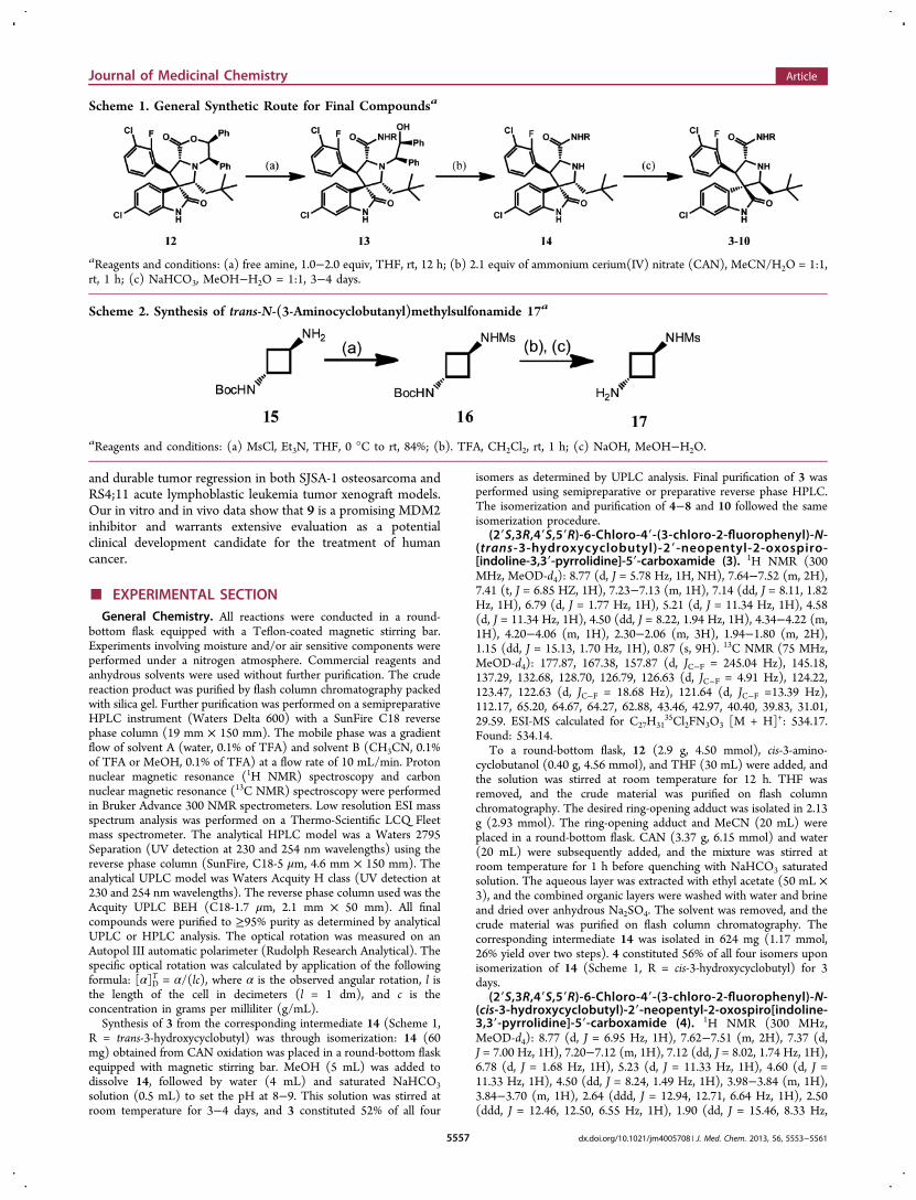
■ CHEMISTRYThe synthesis of 3−10 is shown in Scheme 1. The synthesis of9 has been reported in a previous communication.21
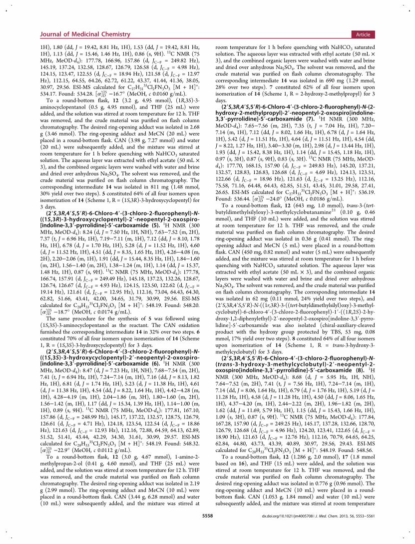
Compound 12 was synthesized using previously reportedmethods.11−13,21 Most amines required for the synthesis ofintermediate 13 were commercially available with the exceptionof 8, 9, and 10. The intermediate 14 was synthesized accordingto previously reported procedures.11−13,21 Compound 14 wasisomerized in MeCN−H2O or MeOH for 3−4 days, affordingfinal compounds in 50−60% yield as determined by analyticalHPLC or UPLC.The trans-N-(3-aminocyclobutanyl)methylsulfonamide 17
used for the synthesis of 10 was synthesized from commerciallyavailable material 15 (Scheme 2). Compound 15 was convertedinto mesylate 16 in 84% yield and the N-Boc group was cleavedin TFA−CH2Cl2 solution, affording 17. The amine 17 wasneutralized using 2 N NaOH aqueous solution prior to use.
■ CONCLUSIONIn the present study, we designed and synthesized a set of newand potent MDM2 inhibitors with the goal to improve the oralpharmacokinetics profile over 2 by constraining the con-formation of the 1,2-diol moiety and blocking the metabolicoxidative site. Among these new compounds, 9 is a highlypotent MDM2 inhibitor and has an excellent oral pharmaco-kinetics profile. Significantly, 9 is capable of achieving complete
Figure 6. Activation of p53 and cleavage of caspase-3 and PARP inSJSA-1 tumor tissue by 9. Mice bearing SJSA-1 tumors were dosedwith a single oral dose of 9 at 100 mg/kg, and tumor tissue washarvested at different time points for Western blot analysis usingspecific antibodies: FL-PARP, full-length PARP; Cl-PARP, cleavedPARP; Pro-Cas3, pro-caspase-3; Cl-Cas3, cleaved caspase-3.
Figure 7. Antitumor activity of 9 in the RS4;11 acute lymphoblasticleukemia xenograft model: (A) tumor volume; (B) animal weightchange.
Journal of Medicinal Chemistry Article
dx.doi.org/10.1021/jm4005708 | J. Med. Chem. 2013, 56, 5553−55615556
and durable tumor regression in both SJSA-1 osteosarcoma andRS4;11 acute lymphoblastic leukemia tumor xenograft models.Our in vitro and in vivo data show that 9 is a promising MDM2inhibitor and warrants extensive evaluation as a potentialclinical development candidate for the treatment of humancancer.
■ EXPERIMENTAL SECTIONGeneral Chemistry. All reactions were conducted in a round-
bottom flask equipped with a Teflon-coated magnetic stirring bar.Experiments involving moisture and/or air sensitive components wereperformed under a nitrogen atmosphere. Commercial reagents andanhydrous solvents were used without further purification. The crudereaction product was purified by flash column chromatography packedwith silica gel. Further purification was performed on a semipreparativeHPLC instrument (Waters Delta 600) with a SunFire C18 reversephase column (19 mm × 150 mm). The mobile phase was a gradientflow of solvent A (water, 0.1% of TFA) and solvent B (CH3CN, 0.1%of TFA or MeOH, 0.1% of TFA) at a flow rate of 10 mL/min. Protonnuclear magnetic resonance (1H NMR) spectroscopy and carbonnuclear magnetic resonance (13C NMR) spectroscopy were performedin Bruker Advance 300 NMR spectrometers. Low resolution ESI massspectrum analysis was performed on a Thermo-Scientific LCQ Fleetmass spectrometer. The analytical HPLC model was a Waters 2795Separation (UV detection at 230 and 254 nm wavelengths) using thereverse phase column (SunFire, C18-5 μm, 4.6 mm × 150 mm). Theanalytical UPLC model was Waters Acquity H class (UV detection at230 and 254 nm wavelengths). The reverse phase column used was theAcquity UPLC BEH (C18-1.7 μm, 2.1 mm × 50 mm). All finalcompounds were purified to ≥95% purity as determined by analyticalUPLC or HPLC analysis. The optical rotation was measured on anAutopol III automatic polarimeter (Rudolph Research Analytical). Thespecific optical rotation was calculated by application of the followingformula: [α]D
T = α/(lc), where α is the observed angular rotation, l isthe length of the cell in decimeters (l = 1 dm), and c is theconcentration in grams per milliliter (g/mL).Synthesis of 3 from the corresponding intermediate 14 (Scheme 1,
R = trans-3-hydroxycyclobutyl) was through isomerization: 14 (60mg) obtained from CAN oxidation was placed in a round-bottom flaskequipped with magnetic stirring bar. MeOH (5 mL) was added todissolve 14, followed by water (4 mL) and saturated NaHCO3solution (0.5 mL) to set the pH at 8−9. This solution was stirred atroom temperature for 3−4 days, and 3 constituted 52% of all four
isomers as determined by UPLC analysis. Final purification of 3 wasperformed using semipreparative or preparative reverse phase HPLC.The isomerization and purification of 4−8 and 10 followed the sameisomerization procedure.
(2′S,3R,4′S,5′R)-6-Chloro-4′-(3-chloro-2-fluorophenyl)-N-(trans-3-hydroxycyclobutyl)-2′-neopentyl-2-oxospiro-[indoline-3,3′-pyrrolidine]-5′-carboxamide (3). 1H NMR (300MHz, MeOD-d4): 8.77 (d, J = 5.78 Hz, 1H, NH), 7.64−7.52 (m, 2H),7.41 (t, J = 6.85 HZ, 1H), 7.23−7.13 (m, 1H), 7.14 (dd, J = 8.11, 1.82Hz, 1H), 6.79 (d, J = 1.77 Hz, 1H), 5.21 (d, J = 11.34 Hz, 1H), 4.58(d, J = 11.34 Hz, 1H), 4.50 (dd, J = 8.22, 1.94 Hz, 1H), 4.34−4.22 (m,1H), 4.20−4.06 (m, 1H), 2.30−2.06 (m, 3H), 1.94−1.80 (m, 2H),1.15 (dd, J = 15.13, 1.70 Hz, 1H), 0.87 (s, 9H). 13C NMR (75 MHz,MeOD-d4): 177.87, 167.38, 157.87 (d, JC−F = 245.04 Hz), 145.18,137.29, 132.68, 128.70, 126.79, 126.63 (d, JC−F = 4.91 Hz), 124.22,123.47, 122.63 (d, JC−F = 18.68 Hz), 121.64 (d, JC−F =13.39 Hz),112.17, 65.20, 64.67, 64.27, 62.88, 43.46, 42.97, 40.40, 39.83, 31.01,29.59. ESI-MS calculated for C27H31
35Cl2FN3O3 [M + H]+: 534.17.Found: 534.14.
To a round-bottom flask, 12 (2.9 g, 4.50 mmol), cis-3-amino-cyclobutanol (0.40 g, 4.56 mmol), and THF (30 mL) were added, andthe solution was stirred at room temperature for 12 h. THF wasremoved, and the crude material was purified on flash columnchromatography. The desired ring-opening adduct was isolated in 2.13g (2.93 mmol). The ring-opening adduct and MeCN (20 mL) wereplaced in a round-bottom flask. CAN (3.37 g, 6.15 mmol) and water(20 mL) were subsequently added, and the mixture was stirred atroom temperature for 1 h before quenching with NaHCO3 saturatedsolution. The aqueous layer was extracted with ethyl acetate (50 mL ×3), and the combined organic layers were washed with water and brineand dried over anhydrous Na2SO4. The solvent was removed, and thecrude material was purified on flash column chromatography. Thecorresponding intermediate 14 was isolated in 624 mg (1.17 mmol,26% yield over two steps). 4 constituted 56% of all four isomers uponisomerization of 14 (Scheme 1, R = cis-3-hydroxycyclobutyl) for 3days.
(2′S,3R,4′S,5′R)-6-Chloro-4′-(3-chloro-2-fluorophenyl)-N-(cis-3-hydroxycyclobutyl)-2′-neopentyl-2-oxospiro[indoline-3,3′-pyrrolidine]-5′-carboxamide (4). 1H NMR (300 MHz,MeOD-d4): 8.77 (d, J = 6.95 Hz, 1H), 7.62−7.51 (m, 2H), 7.37 (d,J = 7.00 Hz, 1H), 7.20−7.12 (m, 1H), 7.12 (dd, J = 8.02, 1.74 Hz, 1H),6.78 (d, J = 1.68 Hz, 1H), 5.23 (d, J = 11.33 Hz, 1H), 4.60 (d, J =11.33 Hz, 1H), 4.50 (dd, J = 8.24, 1.49 Hz, 1H), 3.98−3.84 (m, 1H),3.84−3.70 (m, 1H), 2.64 (ddd, J = 12.94, 12.71, 6.64 Hz, 1H), 2.50(ddd, J = 12.46, 12.50, 6.55 Hz, 1H), 1.90 (dd, J = 15.46, 8.33 Hz,
Scheme 1. General Synthetic Route for Final Compoundsa
aReagents and conditions: (a) free amine, 1.0−2.0 equiv, THF, rt, 12 h; (b) 2.1 equiv of ammonium cerium(IV) nitrate (CAN), MeCN/H2O = 1:1,rt, 1 h; (c) NaHCO3, MeOH−H2O = 1:1, 3−4 days.
Scheme 2. Synthesis of trans-N-(3-Aminocyclobutanyl)methylsulfonamide 17a
aReagents and conditions: (a) MsCl, Et3N, THF, 0 °C to rt, 84%; (b). TFA, CH2Cl2, rt, 1 h; (c) NaOH, MeOH−H2O.
Journal of Medicinal Chemistry Article
dx.doi.org/10.1021/jm4005708 | J. Med. Chem. 2013, 56, 5553−55615557
1H), 1.80 (dd, J = 19.42, 8.81 Hz, 1H), 1.53 (dd, J = 19.42, 8.81 Hz,1H), 1.13 (dd, J = 15.46, 1.46 Hz, 1H), 0.86 (s, 9H). 13C NMR (75MHz, MeOD-d4): 177.78, 166.96, 157.86 (d, JC−F = 249.82 Hz),145.19, 137.24, 132.58, 128.67, 126.79, 126.58 (d, JC−F = 4.98 Hz),124.15, 123.47, 122.55 (d, JC−F = 18.94 Hz), 121.58 (d, JC−F = 12.97Hz), 112.15, 64.55, 64.26, 62.72, 61.22, 43.37, 41.44, 41.36, 38.05,30.97, 29.56. ESI-MS calculated for C27H31
35Cl2FN3O3 [M + H]+:534.17. Found: 534.28. [α]D
25 −16.7° (MeOH, c 0.0160 g/mL).To a round-bottom flask, 12 (3.2 g, 4.95 mmol), (1R,3S)-3-
aminocyclopentanol (0.5 g, 4.95 mmol), and THF (25 mL) wereadded, and the solution was stirred at room temperature for 12 h. THFwas removed, and the crude material was purified on flash columnchromatography. The desired ring-opening adduct was isolated in 2.68g (3.46 mmol). The ring-opening adduct and MeCN (20 mL) wereplaced in a round-bottom flask. CAN (3.98 g, 7.27 mmol) and water(20 mL) were subsequently added, and the mixture was stirred atroom temperature for 1 h before quenching with NaHCO3 saturatedsolution. The aqueous layer was extracted with ethyl acetate (50 mL ×3), and the combined organic layers were washed with water and brineand dried over anhydrous Na2SO4. The solvent was removed, and thecrude material was purified on flash column chromatography. Thecorresponding intermediate 14 was isolated in 811 mg (1.48 mmol,30% yield over two steps). 5 constituted 64% of all four isomers uponisomerization of 14 (Scheme 1, R = (1S,3R)-3-hydroxycyclopentyl) for3 days.(2′S,3R,4′S,5′R)-6-Chloro-4′-(3-chloro-2-fluorophenyl)-N-
((1S,3R)-3-hydroxycyclopentyl)-2′-neopentyl-2-oxospiro-[indoline-3,3′-pyrrolidine]-5′-carboxamide (5). 1H NMR (300MHz, MeOD-d4): 8.24 (d, J = 7.50 Hz, 1H, NH), 7.63−7.52 (m, 2H),7.37 (t, J = 6.96 Hz, 1H), 7.19−7.11 (m, 1H), 7.12 (dd, J = 8.10, 1.78Hz, 1H), 6.78 (d, J = 1.70 Hz, 1H), 5.28 (d, J = 11.52 Hz, 1H), 4.60(d, J = 11.52 Hz, 1H), 4.51 (dd, J = 8.35, 1.65 Hz, 1H), 4.26−4.08 (m,2H), 2.20−2.06 (m, 1H), 1.91 (dd, J = 15.44, 8.35 Hz, 1H), 1.84−1.60(m, 2H), 1.56−1.40 (m, 2H), 1.38−1.24 (m, 1H), 1.14 (dd, J = 15.37,1.48 Hz, 1H), 0.87 (s, 9H). 13C NMR (75 MHz, MeOD-d4): 177.78,166.74, 157.91 (d, JC−F = 249.49 Hz), 145.18, 137.23, 132.26, 128.67,126.74, 126.67 (d, JC−F = 4.93 Hz), 124.15, 123.50, 122.62 (d, JC−F =19.14 Hz), 121.61 (d, JC−F = 12.95 Hz), 112.16, 73.04, 64.43, 64.30,62.82, 51.66, 43.41, 42.00, 34.65, 31.79, 30.99, 29.56. ESI-MScalculated for C28H33
35Cl2FN3O3 [M + H]+: 548.19. Found: 548.20.[α]D
25 −18.7° (MeOH, c 0.0174 g/mL).The same procedure for the synthesis of 5 was followed using
(1S,3S)-3-aminocyclopentanol as the reactant. The CAN oxidationfurnished the corresponding intermediate 14 in 32% over two steps. 6constituted 70% of all four isomers upon isomerization of 14 (Scheme1, R = (1S,3S)-3-hydroxycyclopentyl) for 3 days.(2′S,3R,4′S,5′R)-6-Chloro-4′-(3-chloro-2-fluorophenyl)-N-
((1S,3S)-3-hydroxycyclopentyl)-2′-neopentyl-2-oxospiro-[indoline-3,3′-pyrrolidine]-5′-carboxamide (6). 1H NMR (300MHz, MeOD-d4): 8.47 (d, J = 7.23 Hz, 1H, NH), 7.68−7.54 (m, 2H),7.41 (t, J = 6.94 Hz, 1H), 7.24−7.14 (m, 1H), 7.16 (dd, J = 8.13, 1.82Hz, 1H), 6.81 (d, J = 1.74 Hz, 1H), 5.23 (d, J = 11.38 Hz, 1H), 4.61(d, J = 11.38 Hz, 1H), 4.54 (dd, J = 8.22, 1.64 Hz, 1H), 4.42−4.28 (m,1H), 4.28−4.19 (m, 1H), 2.04−1.86 (m, 3H), 1.80−1.60 (m, 2H),1.56−1.42 (m, 1H), 1.17 (dd, J = 15.34, 1.39 Hz, 1H), 1.14−1.00 (m,1H), 0.89 (s, 9H). 13C NMR (75 MHz, MeOD-d4): 177.81, 167.10,157.86 (d, JC−F = 248.99 Hz), 145.17, 137.22, 132.57, 128.75, 126.79,126.61 (d, JC−F = 4.71 Hz), 124.18, 123.54, 122.54 (d, JC−F = 18.86Hz), 121.63 (d, JC−F = 12.93 Hz), 112.16, 72.88, 64.59, 64.13, 62.89,51.52, 51.41, 43.44, 42.29, 34.30, 31.61, 30.99, 29.57. ESI-MScalculated for C28H33
35Cl2FN3O3 [M + H]+: 548.19. Found: 548.32.[α]D
25 −22.9° (MeOH, c 0.0112 g/mL).To a round-bottom flask, 12 (3.0 g, 4.67 mmol), 1-amino-2-
methylpropan-2-ol (0.41 g, 4.60 mmol), and THF (25 mL) wereadded, and the solution was stirred at room temperature for 12 h. THFwas removed, and the crude material was purified on flash columnchromatography. The desired ring-opening adduct was isolated in 2.19g (2.99 mmol). The ring-opening adduct and MeCN (10 mL) wereplaced in a round-bottom flask. CAN (3.44 g, 6.28 mmol) and water(10 mL) were subsequently added, and the mixture was stirred at
room temperature for 1 h before quenching with NaHCO3 saturatedsolution. The aqueous layer was extracted with ethyl acetate (50 mL ×3), and the combined organic layers were washed with water and brineand dried over anhydrous Na2SO4. The solvent was removed, and thecrude material was purified on flash column chromatography. Thecorresponding intermediate 14 was isolated in 690 mg (1.29 mmol,28% over two steps). 7 constituted 62% of all four isomers uponisomerization of 14 (Scheme 1, R = 2-hydroxy-2-methylpropyl) for 3days.
(2′S,3R,4′S,5′R)-6-Chloro-4′-(3-chloro-2-fluorophenyl)-N-(2-hydroxy-2-methylpropyl)-2′-neopentyl-2-oxospiro[indoline-3,3′-pyrrolidine]-5′-carboxamide (7). 1H NMR (300 MHz,MeOD-d4): 7.65−7.56 (m, 2H), 7.35 (t, J = 7.04 Hz, 1H), 7.20−7.14 (m, 1H), 7.12 (dd, J = 8.02, 1.66 Hz, 1H), 6.78 (d, J = 1.64 Hz,1H), 5.42 (d, J = 11.51 Hz, 1H), 4.64 (d, J = 11.51 Hz, 1H), 4.54 (dd,J = 8.22, 1.27 Hz, 1H), 3.40−3.30 (m, 1H), 2.98 (d, J = 13.44 Hz, 1H),1.93 (dd, J = 15.42, 8.38 Hz, 1H), 1.14 (dd, J = 15.45, 1.18 Hz, 1H),0.97 (s, 3H), 0.87 (s, 9H), 0.83 (s, 3H). 13C NMR (75 MHz, MeOD-d4): 177.70, 168.15, 157.90 (d, JC−F = 249.83 Hz), 145.20, 137.21,132.57, 128.83, 126.83, 126.68 (d, JC−F = 4.69 Hz), 124.13, 123.51,122.66 (d, JC−F = 18.96 Hz), 121.63 (d, JC−F = 13.25 Hz), 112.16,75.58, 71.16, 64.48, 64.43, 62.85, 51.51, 43.45, 31.01, 29.58, 27.41,26.65. ESI-MS calculated for C27H33
35Cl2FN3O3 [M + H]+: 536.19.Found: 536.44. [α]D
25 −24.0° (MeOH, c 0.0186 g/mL).To a round-bottom flask, 12 (643 mg, 1.0 mmol), trans-3-(tert-
butyldimethylsilyloxy)-3-methylcyclobutanamine21 (0.10 g, 0.46mmol), and THF (10 mL) were added, and the solution was stirredat room temperature for 12 h. THF was removed, and the crudematerial was purified on flash column chromatography. The desiredring-opening adduct was isolated in 0.36 g (0.41 mmol). The ring-opening adduct and MeCN (5 mL) were placed in a round-bottomflask. CAN (450 mg, 0.82 mmol) and water (5 mL) were subsequentlyadded, and the mixture was stirred at room temperature for 1 h beforequenching with NaHCO3 saturated solution. The aqueous layer wasextracted with ethyl acetate (50 mL × 3), and the combined organiclayers were washed with water and brine and dried over anhydrousNa2SO4. The solvent was removed, and the crude material was purifiedon flash column chromatography. The corresponding intermediate 14was isolated in 62 mg (0.11 mmol, 24% yield over two steps), and(2′S,3R,4′S,5′R)-N-((1r,3R)-3-((tert-butyldimethylsilyl)oxy)-3-methyl-cyclobutyl)-6-chloro-4′-(3-chloro-2-fluorophenyl)-1′-((1R,2S)-2-hy-droxy-1,2-diphenylethyl)-2′-neopentyl-2-oxospiro[indoline-3,3′-pyrro-lidine]-5′-carboxamide was also isolated (chiral-auxiliary-cleavedproduct with the hydroxy group protected by TBS, 53 mg, 0.08mmol, 17% yield over two steps). 8 constituted 64% of all four isomersupon isomerization of 14 (Scheme 1, R = trans-3-hydroxy-3-methylcyclobutyl) for 3 days.
(2′S,3R,4′S,5′R)-6-Chloro-4′-(3-chloro-2-fluorophenyl)-N-(trans-3-hydroxy-3-methylcyclobutyl)-2′-neopentyl-2-oxospiro[indoline-3,3′-pyrrolidine]-5′-carboxamide (8). 1HNMR (300 MHz, MeOD-d4): 8.68 (d, J = 5.95 Hz, 1H, NH),7.64−7.52 (m, 2H), 7.41 (t, J = 7.56 Hz, 1H), 7.24−7.14 (m, 1H),7.14 (dd, J = 8.06, 1.64 Hz, 1H), 6.79 (d, J = 1.76 Hz, 1H), 5.19 (d, J =11.28 Hz, 1H), 4.58 (d, J = 11.28 Hz, 1H), 4.50 (dd, J = 8.06, 1.65 Hz,1H), 4.37−4.20 (m, 1H), 2.44−2.22 (m, 2H), 1.96−1.82 (m, 2H),1.62 (dd, J = 11.69, 5.79 Hz, 1H), 1.15 (dd, J = 15.43, 1.66 Hz, 1H),1.09 (s, 3H), 0.87 (s, 9H). 13C NMR (75 MHz, MeOD-d4): 177.84,167.28, 157.90 (d, JC−F = 249.25 Hz), 145.17, 137.28, 132.66, 128.70,126.79, 126.68 (d, JC−F = 4.96 Hz), 124.20, 123.41, 122.65 (d, JC−F =18.90 Hz), 121.63 (d, JC−F = 12.76 Hz), 112.16, 70.79, 64.65, 64.25,62.84, 44.80, 43.73, 43.39, 40.89, 30.97, 29.56, 29.43. ESI-MScalculated for C28H33
35Cl2FN3O3 [M + H]+: 548.19. Found: 548.56.To a round-bottom flask, 12 (1.286 g, 2.0 mmol), 17 (1.8 mmol
based on 16), and THF (15 mL) were added, and the solution wasstirred at room temperature for 12 h. THF was removed, and thecrude material was purified on flash column chromatography. Thedesired ring-opening adduct was isolated in 0.776 g (0.96 mmol). Thering-opening adduct and MeCN (10 mL) were placed in a round-bottom flask. CAN (1.053 g, 1.84 mmol) and water (10 mL) weresubsequently added, and the mixture was stirred at room temperature
Journal of Medicinal Chemistry Article
dx.doi.org/10.1021/jm4005708 | J. Med. Chem. 2013, 56, 5553−55615558
for 1 h before quenching with NaHCO3 saturated solution. Theaqueous layer was extracted with ethyl acetate (50 mL × 3), and thecombined organic layers were washed with water and brine and driedover anhydrous Na2SO4. The solvent was removed, and the crudematerial was purified on flash column chromatography. Thecorresponding intermediate 14 was isolated in 626 mg (1.023 mmol,56% yield over two steps). 10 constituted 62% of all four isomers uponisomerization of 14 (Scheme 1, R = trans-3-(methylsulfonamido)-cyclobutyl) for 3 days.(2′S,3R,4′S,5′R)-6-Chloro-4′-(3-chloro-2-fluorophenyl)-N-
(trans-3-(methylsulfonamido)cyclobutyl)-2′-neopentyl-2-oxospiro[indoline-3,3′-pyrrolidine]-5′-carboxamide (10). 1HNMR (300 MHz, MeOD-d4): 8.96 (d, J = 6.37 Hz, 1H, NH),7.64−7.54 (m, 2H), 7.36 (t, J = 6.96 Hz, 1H), 7.20−7.16 (m, 1H),7.11 (dd, J = 8.13, 1.79 Hz, 1H), 6.78 (d, J = 1.75 Hz, 1H), 5.32 (d, J =11.51 Hz, 1H), 4.63 (d, J = 11.51 Hz, 1H), 4.52 (d, J = 8.47, 1.49 Hz,1H), 4.28−4.14 (m, 1H), 3.83 (quintet, J = 3.83 Hz, 1H), 2.84 (s,3H), 2.40−2.30 (m, 2H), 2.30−2.20 (m, 1H), 2.06−1.96 (m, 1H),1.93 (dd, J = 15.47, 8.42 Hz, 1H), 1.13 (dd, J = 15.47, 1.23 Hz, 1H),0.86 (s, 9H). 13C NMR (75 MHz, MeOD-d4): 177.73, 167.29, 157.80(d, JC−F = 249.64 Hz), 145.13, 137.16, 132.63, 128.69, 126.75, 126.61(d, JC−F = 4.84 Hz), 124.13, 123.52, 122.51 (d, JC−F = 18.77 Hz),121.55 (d, JC−F = 13.01 Hz), 112.15, 64.51, 64.11, 62.70, 46.60, 43.43,40.93, 38.47, 38.11, 30.97, 29.54. ESI-MS calculated forC28H34
35Cl2FN4O4S [M + H]+: 611.17. Found: 611.42. [α]D25 −13.8°
(MeOH, c 0.0208 g/mL).N-(trans-3-Aminocyclobutyl)methanesulfonamide (17). To a
round-bottom flask, 15 (400 mg, 2.15 mmol, 1.0 equiv), Et3N (1 mL,7.5 mmol, 3.5 equiv), and THF (25 mL) were added. The reactionmixture was cooled in an ice−water bath, and methanesulfonylchloride (MsCl, 492 mg, 4.3 mmol, 2.0 equiv) was added dropwise viaa syringe. The mixture was stirred at room temperature for 12 h beforequenching with water. The aqueous layer was extracted with ethylacetate (70 mL × 2), and the combined organic layers were washedwith brine (50 mL) and dried over anhydrous Na2SO4. The solventswere removed, and the crude product was purified on flash columnchromatography. 16 was obtained as a white solid (0.475 g, 84%). 16:1H NMR (MeOD-d4, 300 MHz) 4.10−3.92 (m, 2H), 2.87 (s, 3H),2.30 (t, J = 6.71 Hz, 4H), 1.43 (s, 9H) 13C (MeOD-d4, 75 M Hz):158.07, 80.52, 46.61, 43.58, 40.90, 38.90, 28.91. ESI-MS: calculated forC10H20N2NaO4S [M + Na]+ = 287.10; obtained, 287.16.To a round-bottom flask, 16 (0.475 g, 1.8 mmol), Et3SiH (0.1 mL),
and CH2Cl2 (15 mL) were added. TFA (4 mL) was added via asyringe, and the mixture was stirred at room temperature for 1 h. Thereaction mixture was concentrated to remove solvent and TFA.MeOH (15 mL) was added, and the pH was adjusted to be 10−11 byNaOH (2 N aqueous solution). MeOH and water were then removedaffording crude free amine 17, which was directly used for the nextstep without further purification.FP-Based Protein Binding Assay. The binding affinity of MDM2
inhibitors was determined by an optimized, sensitive, and quantitativefluorescence-polarization-based (FP-based) binding assay using arecombinant human His-tagged MDM2 protein (residues 1−118)and a FAM tagged p53-based peptide as the fluorescent probe. Thedesign of the fluorescent probe was based upon a previously reportedhigh affinity p53-based peptidomimetic compound (5-FAM-βAla-βAla-Phe-Met-Aib-pTyr-(6-Cl-LTrp)-Glu-Ac3c-Leu-Asn-NH2).
22 Thistagged peptide was named PMDM6-F. The Kd value of PMDM6-F tothe MDM2 protein was determined to be 1.4 ± 0.3 nM by monitoringthe total fluorescence polarization of mixtures composed with thefluorescent probe at a fixed concentration and the MDM2 protein withincreasing concentrations up to full saturation. Fluorescence polar-ization values were measured using the Infinite M-1000 plate reader(Tecan U.S., Research Triangle Park, NC) in Microfluor 2 96-well,black, round-bottom plates (Thermo Scientific). In the saturationexperiments, 1 nM PMDM6-F and increasing concentrations ofproteins were added to each well to a final volume of 125 μL in theassay buffer (100 mM potassium phosphate, pH 7.5, 100 μg/mLbovine γ-globulin, 0.02% sodium azide [Invitrogen], with 0.01% TritonX-100 and 4% DMSO). Plates were mixed and incubated at room
temperature for 15−30 min with gentle shaking to ensure equilibrium.The polarization values in millipolarization units (mP) were measuredat an excitation wavelength of 485 nm and an emission wavelength of530 nm. Equilibrium dissociation constants (Kd) were then calculatedby fitting the sigmoidal dose-dependent FP increases as a function ofprotein concentrations using Graphpad Prism 5.0 software (GraphpadSoftware, San Diego, CA). Ki values of tested compounds weredetermined in a dose-dependent competitive binding experiment.Mixtures of 5 μL of the tested compound in different concentrations inDMSO and 120 μL of preincubated protein/fluorescent probecomplex with fixed concentrations in the assay buffer (100 mMpotassium phosphate, pH 7.5, 100 μg/mL bovine γ-globulin, 0.02%sodium azide, with 0.01% Triton X-100) were added into assay platesand incubated at room temperature with gentle shaking. Theincubation time was precisely controlled at 15 min to minimize theinfluence of the initial isomerization to the binding affinities. Finalconcentrations of the protein and fluorescent probe in the competitiveassays were 10 and 1 nM, respectively, and final DMSO concentrationis 4%. Negative controls containing protein/fluorescent probe complexonly (equivalent to 0% inhibition), and positive controls containingfree fluorescent probe only (equivalent to 100% inhibition), wereincluded in each assay plate. FP values were measured as describedabove. IC50 values were determined by nonlinear regression fitting ofthe sigmoidal dose-dependent FP decreases as a function of totalcompound concentrations using Graphpad Prism 5.0 software(Graphpad Software, San Diego, CA). Ki values of tested compoundsto the MDM2 protein were calculated using the measured IC50 values,the Kd value of the fluorescent probe to the protein, and theconcentrations of the protein and fluorescent probe in the competitiveassays.23
Cell Growth Inhibition Assay. The SJSA-1 osteosarcoma cell lineand RS4;11 leukemia cell line were purchased from the AmericanType Culture Collection (Manassas, VA). HCT-116 p53+/+ colon cellline (HCT-116 p53+/+) and its isogenic p53 knockout (HCT-116p53−/−) cell line are kind gifts from Dr. Bert Vogelstein, JohnsHopkins University at Baltimore, MD. All cell lines were grown inPRMI 1640 medium (Gibco, Life Technologies, Grand Island, NY)supplemented with 10% FBS (Invitrogen, Carlsbad, CA) in ahumidified tissue culture incubator at 37 °C, 5% CO2. For cell growthinhibition assay, cells were seeded in 96-well, flat-bottom cell cultureplates at a density of (3−4) × 103 cells/well and allowed to growovernight, then incubated with a compound at different concen-trations. The effect of cell growth inhibition of a compound wasdetermined with a lactate dehydrogenase based WST-8 assay (WST-8;Dojindo Molecular Technologies Inc., Gaithersburg, MD). For theRS4;11 cell line, WST-8 solution was directly added to culturemedium in each well to a final concentration of 10% upon the end oftreatment. For SJSA-1, HCT-116 p53+/+, and HCT-116 p53−/−,culture medium in each well was discharged and fresh RPMI 1640medium with no phenol red (Gibco, life Technologies, Grand Island,NY) containing 10% WST-8 solution was added to each well in thepresence of 5% FBS upon the end of treatment. Then the plates wereincubated at 37 °C for 2−3 h. The absorbance of the samples wasmeasured at 450 nm using a TECAN ULTRA reader. The effect ofeach treatment was calculated by the equation [(absorbance value oftreatment) − (absorbance value of medium with FBS + WST solutiononly)]/[(absorbance value of untreated control) − (absorbance valueof medium with FBS + WST solution only)]. The IC50 value of acompound is the concentration that inhibits cell growth by 50% overthe control.
Pharmacodynamics (PD) Study. For PD studies, the SJSA-1xenograft tumor model was used. To develop xenograft tumors, 5 ×10⁶ SJSA-1 cancer cells with 50% Matrigel were injected sc on thedorsal side of SCID mice from Charles River, one tumor per mouse.When xenograft tumors reached a mean of ∼100 mm3 (70−130 mm3),two mice were treated with vehicle control (10% PEG400/3%Cremophor/87% PBS) and eight mice were treated with a single doseof 9 at 100 mg/kg via oral gavage. Mice treated with vehicle controlwere sacrificed at 6 h, and tumor tissue was harvested for Western blot
Journal of Medicinal Chemistry Article
dx.doi.org/10.1021/jm4005708 | J. Med. Chem. 2013, 56, 5553−55615559
analysis. Two mice treated with 9 were sacrificed at 1, 3, 6, and 24 h(two mice at each time point), and tumor tissue was harvested.Western Blotting. For Western blot analysis, tumor cells or tumor
tissues were lysed in ice-cold RIPA buffer: 20 mM Tris-HCl (pH 7.5),150 mM NaCl, 1 mM EDTA, 1 mM EGTA, 1% sodium deoxycholate,2.5 mM sodium pyrophosphate, 1 mM β-glycerophosphate, 1 mMsodium orthovanadate, and 1 μg/mL leupeptin. The expressions ofindicated proteins in the whole cell lysates were detected by Westernblot analysis using the following antibodies: anti-p53 (OP43,Calbiochem), anti-MDM2 (sc-965, Santa Cruz), anti-p21 (556431,BD Biosciences), anti-PARP (9542, Cell Signaling Technology),anticaspase-3 (AAP-113, Stressgen Bioreagents), and HRP-conjugatedanti-GAPDH (sc-5778, Santa Cruz).In Vivo Efficacy Experiments. For in vivo efficacy experiments
using the SJSA-1 tumor xenograft model, 5 × 10⁶ SJSA-1 cancer cellswith 50% Matrigel were injected sc on the dorsal side of SCID micefrom Charles River, one tumor per mouse. When xenograft tumorsreached a mean of ∼100 mm3 (70−130 mm3), mice were randomizedinto groups of eight mice. Vehicle control (10% PEG400/3%Cremophor/87% PBS) was administered orally (po) once per dayfor 14 days. Compound 9 was administered orally once per day for 14days at 10, 30, and 100 mg/kg. Tumor sizes and animal weights weremeasured three times per week. Data are represented as mean tumorvolumes. Tumor volume (mm3) = (AB2)/2 where A and B are thetumor length and width (in mm), respectively. Statistical analyses weredone using two-way ANOVA and unpaired two-tailed t test, usingPrism (version 4.0, GraphPad). P < 0.05 was considered statisticallysignificant.For in vivo efficacy studies using the RS4;11 tumor xenograft
model, the same protocol was used with the following modifications:RS4;11 tumor cells were used; dose schedule of 100 and 200 mg/kg.
■ AUTHOR INFORMATION
Corresponding Author*Telephone: 734-615-0362. Fax: 734-647-9647. E-mail:[email protected].
Author Contributions§Y.Z., S.Y., and W.S. provided equal contributions.
NotesThe authors declare the following competing financialinterest(s): Dr. Shaomeng Wang is an inventor on thecompounds described in this study. The technology has beenlicensed by Ascenta and Sanofi for clinical development, andDr. Shaomeng Wang receives royalty from the licensing. Dr.Wang is also a consultant for Ascenta.
■ ACKNOWLEDGMENTS
We are grateful for financial support from the National CancerInstitute, National Institutes of Health (Grants R01CA121279,P50CA06956, and P50CA097248), the University of MichiganCancer Center (Core Grant P30CA046592), Ascenta Ther-apeutics, Inc., and Sanofi S.A.
■ ABBREVIATIONS USED
AUC, area under the curve; CAN, ceric ammonium nitrate;Cmax, the maximum compound concentration from oral dosing;Cl, clearance; CYP, cytochrome P450; CH2Cl2, dichloro-methane; EtOAc, ethyl acetate; H2O, water; LC/MS, liquidchromatography−mass spectrometry; MDM2, murine doubleminute 2; MeCN, acetonitrile; NaOH, sodium hydroxide; PD,pharmacodynamics; PK, pharmacokinetics; po, oral adminis-tration; qD, once a day dosing; SCID, severe combinedimmunodeficient; T1/2, half-life; TFA, trifluoroacetic acid; Tmax,the time at which Cmax is reached
■ REFERENCES(1) Levine, A. J. p53, the cellular gatekeeper for growth and division.Cell 1997, 88, 323−331.(2) El-Deiry, W. S. Regulation of p53 downstream genes. Semin.Cancer Biol. 1998, 8, 345−357.(3) Vogelstein, B.; Lane, D.; Levine, A. J. Surfing the p53 network.Nature 2000, 408, 307−310.(4) Hainaut, P.; Hollstein, M. p53 and human cancer: the first tenthousand mutations. Adv. Cancer Res. 2000, 77, 81−137.(5) Wu, X.; Bayle, J. H.; Olson, D.; Levine, A. J. The p53-mdm2autoregulatory feedback loop. Genes Dev. 1993, 7, 1126−1132.(6) Kussie, P. H.; Gorina, S.; Marechal, V.; Elenbaas, B.; Moreau, J.;Levine, A. J.; Pavletich, N. P. Structure of the MDM2 oncoproteinbound to the p53 tumor suppressor transactivation domain. Science1996, 274, 948−953.(7) Vassilev, L. T.; Vu, B. T.; Graves, B.; Carvajal, D.; Podlaski, F.;Filipovic, Z.; Kong, N.; Kammlott, U.; Lukacs, C.; Klein, C.; Fotouhi,N.; Liu, E. A. In vivo activation of the p53 pathway by small-moleculeantagonists of MDM2. Science 2004, 303, 844−848.(8) Shangary, S.; Qin, D.; McEachern, D.; Liu, M.; Miller, R. S.; Qiu,S.; Nikolovska-Coleska, Z.; Ding, K.; Wang, G.; Chen, J.; Bernard, D.;Zhang, J.; Lu, Y.; Gu, Q.; Shah, R. B.; Pienta, K. J.; Ling, X.; Kang, S.;Guo, M.; Sun, Y.; Yang, D.; Wang, S. Temporal activation of p53 by aspecific MDM2 inhibitor is selectively toxic to tumors and leads tocomplete tumor growth inhibition. Proc. Natl. Acad. Sci. U.S.A. 2008,105, 3933−3938.(9) Vassilev, L. T. p53 activation by small molecules: application inoncology. J. Med. Chem. 2005, 48, 4491−4499.(10) Shangary, S.; Wang, S. Targeting the MDM2-p53 interaction forcancer therapy. Clin. Cancer Res. 2008, 14, 5318−5324.(11) Ding, K.; Lu, Y.; Nikolovska-Coleska, Z.; Qiu, S.; Ding, Y.; Gao,W.; Stuckey, J.; Krajewski, K.; Roller, P. P.; Tomita, Y.; Parrish, D. A.;Deschamps, J. R.; Wang, S. Structure-based design of potent non-peptide MDM2 inhibitors. J. Am. Chem. Soc. 2005, 127, 10130−10131.(12) Ding, K.; Lu, Y.; Nikolovska-Coleska, Z.; Wang, G.; Qiu, S.;Shangary, S.; Gao, W.; Qin, D.; Stuckey, J.; Krajewski, K.; Roller, P. P.;Wang, S. Structure-based design of spiro-oxindoles as potent, specificsmall-molecule inhibitors of the MDM2-p53 interaction. J. Med. Chem.2006, 49, 3432−3435.(13) Yu, S.; Qin, D.; Shangary, S.; Chen, J.; Wang, G.; Ding, K.;McEachern, D.; Qiu, S.; Nikolovska-Coleska, Z.; Miller, R.; Kang, S.;Yang, D.; Wang, S. Potent and orally active small-molecule inhibitorsof the MDM2-p53 interaction. J. Med. Chem. 2009, 52, 7970−7973.(14) Rew, Y.; Sun, D. Q.; De Turiso, F. G. L.; Bartberger, M. D.;Beck, H. P.; Canon, J.; Chen, A.; Chow, D.; Deignan, J.; Fox, B. M.;Gustin, D.; Huang, X.; Jiang, M.; Jiao, X. Y.; Jin, L. X.; Kayser, F.;Kopecky, D. J.; Li, Y. H.; Lo, M. C.; Long, A. M.; Michelsen, K.;Oliner, J. D.; Osgood, T.; Ragains, M.; Saiki, A. Y.; Schneider, S.;Toteva, M.; Yakowec, P.; Yan, X. L.; Ye, Q. P.; Yu, D. Y.; Zhao, X. N.;Zhou, J.; Medina, J. C.; Olson, S. H. Structure-based design of novelinhibitors of the MDM2-p53 interaction. J. Med. Chem. 2012, 55,4936−4954.(15) Allen, J. G.; Bourbeau, M. P.; Wohlhieter, G. E.; Bartberger, M.D.; Michelsen, K.; Hungate, R.; Gadwood, R. C.; Gaston, R. D.; Evans,B.; Mann, L. W.; Matison, M. E.; Schneider, S.; Huang, X.; Yu, D. Y.;Andrews, P. S.; Reichelt, A.; Long, A. M.; Yakowec, P.; Yang, E. Y.;Lee, T. A.; Oliner, J. D. Discovery and optimization of chromeno-triazolopyrimidines as potent inhibitors of the mouse double minute 2-tumor protein 53 protein−protein interaction. J. Med. Chem. 2009, 52,7044−7053.(16) Czarna, A.; Beck, B.; Srivastava, S.; Popowicz, G. M.; Wolf, S.;Huang, Y. J.; Bista, M.; Holak, T. A.; Domling, A. Robust generation oflead compounds for protein−protein interactions by computationaland MCR chemistry: p53/Hdm2 antagonists. Angew. Chem., Int. Ed.2010, 49, 5352−5356.(17) Gomez-Monterrey, I.; Bertamino, A.; Porta, A.; Carotenuto, A.;Musella, S.; Aquino, C.; Granata, I.; Sala, M.; Brancaccio, D.; Picone,D.; Ercole, C.; Stiuso, P.; Campiglia, P.; Grieco, P.; Ianelli, P.; Maresca,B.; Novellino, E. Identification of the spiro(oxindole-3,3′-thiazolidine)-
Journal of Medicinal Chemistry Article
dx.doi.org/10.1021/jm4005708 | J. Med. Chem. 2013, 56, 5553−55615560

based derivatives as potential p53 activity modulators. J. Med. Chem.2010, 53, 8319−8329.(18) Hardcastle, I. R.; Liu, J. F.; Valeur, E.; Watson, A.; Ahmed, S. U.;Blackburn, T. J.; Bennaceur, K.; Clegg, W.; Drummond, C.; Endicott,J. A.; Golding, B. T.; Griffin, R. J.; Gruber, J.; Haggerty, K.; Harrington,R. W.; Hutton, C.; Kemp, S.; Lu, X. C.; McDonnell, J. M.; Newell, D.R.; Noble, M. E. M.; Payne, S. L.; Revill, C. H.; Riedinger, C.; Xu, Q.;Lunec, J. Isoindolinone inhibitors of the murine double minute 2(MDM2)-p53 protein−protein interaction: structure−activity studiesleading to improved potency. J. Med. Chem. 2011, 54, 1233−1243.(19) Bold, G.; Furet, P.; Gessier, F.; Kallen, J.; Hergovich Lisztwan,J.; Masuya, K.; Vaupel, A. Tetra-Substituted Heteroaryl Compoundsand Their Use as MDM2 and/or MDM4 Modulators. PCT PatentWO/2011/023677, 2011.(20) Uoto, K.; Kawato, H.; Sugimoto, Y.; Naito, H.; Miyazaki, M.;Taniguchi, T.; Aonuma, M. Imidazothiazole Derivative Having 4,7-Diazaspiro[2.5]octane Ring Structure. PCT Patent WO/2009/151069,2009.(21) Zhao, Y.; Liu, L.; Sun, W.; Lu, J.; McEachern, D.; Li, X.; Yu, S.;Bernard, D.; Ochsenbein, P.; Ferey, V.; Carry, J.-C.; Deschamps, J. R.;Sun, D.; Wang, S. Diastereomeric spirooxindoles as highly potent andefficacious MDM2 inhibitors. J. Am. Chem. Soc. 2013, 135, 7223−7734.(22) Garcia-Echeverria, C.; Chene, P.; Blommers, M. J. J.; Furet, P.Discovery of potent antagonists of the interaction between humandouble minute 2 and tumor suppressor p53. J. Med. Chem. 2000, 43,3205−3208.(23) Nikolovska-Coleska, Z.; Wang, R. X.; Fang, X. L.; Pan, H. G.;Tomita, Y.; Li, P.; Roller, P. P.; Krajewski, K.; Saito, N. G.; Stuckey, J.A.; Wang, S. M. Development and optimization of a binding assay forthe XIAP BIR3 domain using fluorescence polarization. Anal. Biochem.2004, 332, 261−273.
Journal of Medicinal Chemistry Article
dx.doi.org/10.1021/jm4005708 | J. Med. Chem. 2013, 56, 5553−55615561