study on the interaction of kojic acid with tyrosinase by
TRANSCRIPT

Wenbin Liu W et al
mdashmdashmdashmdashmdashmdashmdashmdashmdashmdashmdashmdashmdashmdashmdashmdashmdashmdashmdashmdashmdashmdashmdashmdashmdashmdashmdashmdashmdashmdashmdashmdashmdashmdashmdashmdashmdashmdashmdashmdashmdashmdashmdashmdashmdashmdashmdashmdashmdashmdashmdash
WWWSIFTDESKORG 365 Vol-4 Issue-2
SIFT DESK
Accepted Date 10th Mar 2020 Published Date20th Mar 2020
Xue You1 Cailian Wang1 Nihong Guo1 Wenbin Liu1
1 Department of Pharmaceutical and Biological Engineering School of Chemical Engineering Si-
chuan University Chengdu 610065 China
CORRESPONDENCE AUTHOR Wenbin Liu Tel 86-28-85408255 E-mail wbliuscueducn CITATION Xue You Cailian Wang Nihong Guo Wenbin Liu W Study on the interaction of kojic acid with tyrosinase by spectroscopic methods(2020) Journal of Computational Chemistry amp Molecular Mod-eling 4(2) pp365-375 ABSTRACT Tyrosinase is the key enzyme in melanin synthesis and kojic acid is a widely available inhibitor
against tyrosinase with extensive application values The inhibitory mechanism of kojic acid was
elaborated by studying the mutual interaction and the effects on tyrosinase conformation using
spectroscopy Ultraviolet-visible absorption spectra showed kojic acid caused both the secondary
and tertiary structure changed Fluorescence lifetime measurements implied that kojic acid
quenched the intrinsic fluorescence of tyrosinase via static process A complex formed through one
single binding site with a binding constant of 135times105 M-1 Thermodynamic parameters suggested
that the binding was a spontaneous process with hydrogen bonds and van der Waals forces playing
main role Synchronous fluorescence spectra and three-dimensional fluorescence spectra showed
that kojic acid induced obvious conformational changes in tyrosinase and increased the polarity of
microenvironment Circular dichroism revealed an increase of the content of α-helix and β-strand
This study will provide reliable basis concerning the inhibitory mechanism of kojic acid against ty-
rosinase and therefore contribute to development of tyrosinase inhibitor
Keywords Tyrosinase Kojic acid Inhibitory mechanism Spectroscopy Conformation
Copy rights copy 2020 The Author(s) Published by Sift Desk Journals This is an Open Access article distributed under the terms of the Creative Commons Attribution License (httpcreativecommonsorglicensesby40) which permits unrestricted use distribution and reproduction in any me-dium provided the original work is properly cited
Study on the interaction of kojic acid with
tyrosinase by spectroscopic methods
Journal of Computational Chemistry amp Molecular Modeling (ISSN 2473-6260)
DOI 1025177JCCMM42RA10607 Research
Wenbin Liu W et al
mdashmdashmdashmdashmdashmdashmdashmdashmdashmdashmdashmdashmdashmdashmdashmdashmdashmdashmdashmdashmdashmdashmdashmdashmdashmdashmdashmdashmdashmdashmdashmdashmdashmdashmdashmdashmdashmdashmdashmdashmdashmdashmdashmdashmdashmdashmdashmdashmdashmdashndash
WWWSIFTDESKORG 366 Vol-4 Issue-2
SIFT DESK
1 INTRODUCTION
Tyrosinase is widely distributed in organisms with an
important role in melanin biosynthesis Tyrosinase is
the key enzyme for pigmentation of skin wound
healing and molting process and immunity in insects
[1] However overproduction of melanin could cause
considerable problems such as melanoma Moreover
tyrosinase is also involved in the unfavorable food
browning reaction Therefore the tyrosinase inhibi-
tors therefore have been extensively applied in the
field of cosmetics food pharmaceuticals and agri-
culture[1] Based on the safety apprehension it is
decisive to elaborate the inhibitory mechanism for the
applications of tyrosinase inhibitors
Kojic acid a secondary metabolite produced by As-
pergillus and Penicillum has emerged as a cosmetic
agent with an excellent skin lightener effect and also
as a food preservative to prevent browning due to
inhibitory effect on tyrosinase[2] Kojic acid is usual-
ly served as the positive control to discover new tyro-
sinase inhibitors[1] Previous studies only focus on
the inhibitory effect and kinetic analysis[34] How-
ever the inhibitory mechanism and structure-activity
relationship have been seldom explored in-depth so
far There is little knowledge on the modulation of the
overall structures of tyrosinase involved in the inhibi-
tion by kojic acid The gap between plenty of docu-
ments on inhibitory activity and lack of knowledge on
inhibitory mechanism is striking for that well-known
tyrosinase inhibitor which has become an obstacle
for development of potent lead compounds and the
further application It is urgent to further study the
inhibitory mechanism of kojic acid towards to tyrosi-
nase with respect to mutual interaction and conforma-
tional changes
Given the structure of kojic acid it was hypothesized
that inhibition of tyrosinase activity by kojic acid re-
sulted from conformational changes due to the inter-
action In this study the mechanism of tyrosinase in-
hibition was systematically investigated via examin-
ing the effects of kojic acid on tyrosinase structure as
well as the potential binding through the multispec-
troscopic studies Binding mechanism was proposed
Three-dimensional fluorescence spectra and synchro-
nous fluorescence spectra technique were performed
to evaluate the conformational effect This research
provided qualitative understanding of the regulation
of tyrosinase activity at the molecular level The re-
sults could provide some important theoretic infor-
mation for various industrial applications of kojic
acid as tyrosinase inhibitor
2MATERIALS AND METHODS
21 Materials
Mushroom tyrosinase was purchased from Worthing-
ton (New Jersey USA) Tyrosinase was dissolved
with potassium phosphate buffer (005 M pH 65)
Kojic acid (purity gt 98) was purchased from Jinsui
Bio-Technology (Shanghai China)
22 Ultraviolet-visible spectra measurements
The MAPAD UV-3100PC spectrophotometer
(Shanghai China) was employed to measure the Ul-
traviolet-visible (UV-Vis) absorption using quartz
cuvette with 10 cm path length Scans of absorption
spectra were collected at 25oC Differential spectra
were obtained by subtracting the corresponding spec-
tra from the spectra of the mixture
23 Fluorescence spectra measurements
Steady state fluorescence spectra were carried out on a
Shimadzu spectrofluorophotometer RF-6000 (Kyoto
Japan) All measurements were performed in a stand-
ard quartz cell of 10 cm path length embedded in a
thermostatic cell holder Fluorescence emission spectra
were recorded upon excitation wavelength at 280 nm
The slit widths for excitation and emission were set to
3 nm and 5 nm respectively A 3 ml solution in cu-
vette containing of 065μM tyrosinase within the line-
ar concentration region for fluorescence intensity (data
not shown) was added successively with 1μl of 30
mM kojic acid aliquot using a trace syringe for 10
times The addition of small volume of buffer solution
during titration had little effect on the extent of fluo-
rescence quenching[56] Fluorescence quenching was
analyzed by Stern-Volmer equation (1)[78]
(1)
where F0 and F represent the fluorescence intensities
Wenbin Liu W et al
mdashmdashmdashmdashmdashmdashmdashmdashmdashmdashmdashmdashmdashmdashmdashmdashmdashmdashmdashmdashmdashmdashmdashmdashmdashmdashmdashmdashmdashmdashmdashmdashmdashmdashmdashmdashmdashmdashmdashmdashmdashmdashmdashmdashmdashmdashmdashmdashmdashmdashmdash
WWWSIFTDESKORG 367 Vol-4 Issue-2
SIFT DESK
of tyrosinase without and with kojic acid respective-
ly The inner filter effect was corrected[7] kq is the
quenching rate constant Ksv denotes the Stern-Volmer
quenching constant τ0 is the average lifetime of the
unquenched fluorophore (τ0=10-8 s) and [Q] is the
concentration of kojic acid
The equilibrium between free and bound molecules is
described by equation (2)[9]
(2)
where Kb is the binding constant and n is the number
of binding sites
24 Time-resolved fluorescence lifetime measure-
ments
Time-resolved fluorescence lifetime was examined
with a Horiba Jobin Yvon Fluorolog-3 spectrofluo-
rometer (Longjumeau France) using 10 mm quartz
cuvette The excitation wavelength and the emission
wavelength were fixed at 280 nm and 334 nm re-
spectively The concentration of tyrosinase was kept
fixed at 04μM whereas the concentration of kojic
acid varied from 0 to 60μM The fluorescence decay
curves were analyzed by triexponential iterative fit-
ting program The average fluorescence lifetime aacutetAVntilde
was calculated from the lifetimes (ti) and preexponen-
tial factors (αi) by using the following relationship
(3)
(3)
25 Electrochemical measurements
The electrochemical experiments were conducted
through a Chenhua CHI660C electrochemical work-
station (Shanghai China) Three-electrode electro-
chemical system was employed including a bare
nickel electrode as the working electrode an Hg
Hg2Cl2 electrode as the reference electrode and a
graphite rod as the counter electrode PBB buffer
serve as electrolyte Tyrosinase and kojic acid solu-
tion at different concentrations was directly added
into 20 mL electrolyte Each measurement of cyclic
voltammetry followed stirring for 3 min and resting
for 2 min The scan range was set from minus06 to 06 V
with a scan rate of 50 mV s-1
26 Thermodynamic parameters
If temperature varies in a limited range the thermo-
dynamic parameters including enthalpy change (ΔH)
entropy change (ΔS) and Gibbs free energy change
(ΔG) could be evaluated on the basis of the equation
(4) and equation (5)[10-12]
(4)
(5)
where R is the gas constant T is the experimental
temperature in Kelvin
27 Synchronous and three-dimensional fluores-
cence spectra
Synchronous fluorescence spectra were gained with a
fixed interval (Δλ) between excitation and emission
wavelength at 25oC Tyrosinase (065μM) was titrated
by increasing concentrations of kojic acid from 0μM
to 200μM with increment of 10μM each time The
emission wavelength were recorded from 245 nm to
385 nm (for Δλ= 15 nm) and from 200 nm to 340 nm
(for Δλ= 60 nm) at which the spectrum could only
show the characteristic information of Tyr and Trp
residues of tyrosinase respectively[13]
In order to measure the three-dimensional fluores-
cence different concentrations of kojic acid were in-
cubated with 065μM tyrosinase Excitation wave-
lengths from 225 to 350 nm and emission wave-
lengths from 280 to 450 nm were scanned at 25oC
28 Circular dichroism spectra measurements
Circular dichroism (CD) spectra were collected using
an Applied Photophysics Chirascan CD spectrometer
(Leatherhead UK) by a 1 mm path length quartz cu-
vette which was attached with a Peltier type tempera-
ture control system Far-UV CD spectrum was exam-
ined at wavelength in the range from 185 nm to 250
nm at a bandwidth of 2nm 300μL tyrosinase (1μM)
was incubated with 1μL various concentration of
kojic acid (0 1 4μM) at for 1 minutes and then was
measured at 25oC The contents for different second-
ary structures of tyrosinase were quantitatively ana-
lyzed by the online CDSSTR program with Set 4 as
the reference set[1415]
Wenbin Liu W et al
mdashmdashmdashmdashmdashmdashmdashmdashmdashmdashmdashmdashmdashmdashmdashmdashmdashmdashmdashmdashmdashmdashmdashmdashmdashmdashmdashmdashmdashmdashmdashmdashmdashmdashmdashmdashmdashmdashmdashmdashmdashmdashmdashmdashmdashmdashmdashmdashmdashmdashndash
WWWSIFTDESKORG 368 Vol-4 Issue-2
SIFT DESK
29 Data process
All assays were individually repeated in triplicate
For the sake of clarity error bars were not shown
Originreg 81was used for data process
3 RESULTS AND DISCUSSION
31 Ultraviolet-visible spectra for interaction be-
tween kojic acid and copper ion or tyrosinase
It was reported that the ability to chelate copper ion
exerts an enormous function on the tyrosinase inhibi-
tion[4] The direct interaction of kojic acid with the
copper ion was revealed by the UV-Vis spectra of
kojic acid treated with gradually increasing concen-
trations of copper ion (Figure 1A) The peak at 217
nm and 269 nm for kojic acid shifted to 223 nm and
309 nm respectively The characteristic red shift sug-
gested the formation of chelate between copper ion
and kojic acid[41617] Isosbestic points mirrored
the existence of free and bound forms of kojic acid in
equilibrium (Figure 1A) The chelation between kojic
acid and copper ion in the enzyme was also investi-
gated (Figure 1B) In contrast to a bathochromic shift
for copper ions in solution no significant wavelength
shift was observed after incubation with tyrosinase
(Figure 1B) Therefore the inhibitory mechanism of
kojic acid differed from those of simple copper chela-
tors[418]
UV-Vis absorption spectroscopy is also an effective
method to examine the formation of complex as well
as the structural alteration[1517] The UV-Vis ab-
sorption spectra of tyrosinase in the absence and
presence of kojic acid were shown in Fig 1B The
increased intensity of the differential spectrum for
tyrosinase suggested binding of kojic acid to tyrosi-
nase as well as changes in conformation of the pro-
tein skeleton[520] Transformation of secondary
structure made the polypeptide strand more extended
and induced global conformational change Further-
more the weak absorption bands around 280 nm
seemed to slightly shift towards shorter wavelength
upon addition of kojic acid demonstrating that the
interaction of kojic acid with tyrosinase decreased the
hydrophobicity of the microenvironment surrounding
Trp residues
Figure 1 Ultraviolet-visible spectra of kojic acid (A) Absorption spectrum for kojic acid (002 mM) upon addi-
tion of various concentration of copper ion Kojic acid was incubated with 002 004 006 008 and 010 mM
CuSO4 Dash-dot line a to e minus 002 mM kojic acid with 002 004 006 008 and 010 mM CuSO4 Gray line f
minus 002 mM kojic aicd Dashed line g minus 010 mM CuSO4 (B) Absorption spectrum for kojic acid (002 mM) up-
on addition of tyrosinase (90 nM) Differential absorption spectra were obtained by deducting the corresponding
spectra from the spectrum of the mixture Dash-dot-dot line h minus 90 nM tyrosinase Black line i minus 90 nM tyrosi-
nase with 002 mM kojic aicd Short dashed line j minus differential absorption spectra by deducting tyrosinase
Short dash-dot line k minus differential absorption spectra by deducting kojic acid
Wenbin Liu W et al
mdashmdashmdashmdashmdashmdashmdashmdashmdashmdashmdashmdashmdashmdashmdashmdashmdashmdashmdashmdashmdashmdashmdashmdashmdashmdashmdashmdashmdashmdashmdashmdashmdashmdashmdashmdashmdashmdashmdashmdashmdashmdashmdashmdashmdashmdashmdashmdashmdashmdashmdash
WWWSIFTDESKORG 369 Vol-4 Issue-2
SIFT DESK
32 Fluorescence quenching of interaction between
kojic acid and tyrosinase
Tyrosinase at a fixed concentration was titrated by gradual
addition of kojic acid (Figure 2) It was apparent that suc-
cessive addition of kojic acid led to a regular decrease in
the fluorescence intensity of tyrosinase without discernable
shift of the maximum peak wavelength Approximately
60 of the fluorescence was quenched when the concen-
tration of kojic acid reached 100 μM
Figure 2 Intrinsic fluorescence changes in the presence of
various kojic acid concentrations Tyrosinase at concentra-
tion of 065 μM was titrated by kojic acid and then incubat-
ed for 30 seconds before measurement Solid lines a to k minus
emission spectra of 065 μM tyrosinase titrated by 0 10
20 30 40 50 60 70 80 90 100 μM kojic acid at 298
Kelvin Dahsed line l minus potassium phosphate buffer solu-
tion Dash-dot line m minus emission spectrum of 200 μM kojic
acid alone
To explain the probable mechanism for quenching the flu-
orescence quenching at three temperatures was carried out
including 288 298 and 310 Kelvin (Figure 3A) The Stern
-Volmer plot displayed a good linear relationship under the
concentration range suggesting that only a single quench-
ing type occurred[71219] The kq values at different tem-
peratures appeared much larger than the maximum diffu-
sion collision quenching rate constant (2times1010 M-1s-1)
(Table 1)[7921] In addition the corresponding Ksv values
which decreased with the rising temperature were inverse-
ly correlated with temperature Therefore it was confirmed
that quenching of tyrosinase by kojic acid might be mainly
governed by a static quenching procedure[7921] It was
concluded that a specific interaction most likely took place
and a ground-state complex without fluorescence was pos-
sibly formed
Figure 3B showed the resulting double-logarithm curves
The Kb value was calculated to be 135times105 M-1 at 298 K
(Table 1) suggesting that kojic acid exhibited relatively
strong binding affinity toward tyrosinase The values of n
was approximately close to 1 and thus indicated the pres-
ence of just a single binding site on tyrosinase[1223]
Table 1 Quenching constants and thermodynamic parameters for the interaction between kojic acid and tyrosinase
T (K) Ksv (times104 Moline1) kq (times1010 Moline1soline1) Kb (times105 Moline1) n ΔH (kJ mololine1) ΔS (J mololine1 Koline1) ΔG (kJ mololine1)
288 454 4542 135 111
-4004 -6031
-2270
298 336 3361 070 108 -2210
310 256 2556 041 106 -2138
Figure 3 Fluorescence quenching effect of kojic acid on tyrosinase at different temperatures (A) Stern-Volmer plots for the
quenching at different temperatures Open square () minus 288 Kelvin Open circle () minus 298 Kelvin Open triangle () minus
310 Kelvin [Kojic acid] minus concentration of kojic acid F0 minus fluorescence intensities of tyrosinase without kojic acid F minus fluorescence intensities of tyrosinase with kojic acid lg minus logarithm (B) Double-logarithm plots for the quenching at differ-ent temperatures
Wenbin Liu W et al
mdashmdashmdashmdashmdashmdashmdashmdashmdashmdashmdashmdashmdashmdashmdashmdashmdashmdashmdashmdashmdashmdashmdashmdashmdashmdashmdashmdashmdashmdashmdashmdashmdashmdashmdashmdashmdashmdashmdashmdashmdashmdashmdashmdashmdashmdashmdashmdashmdashmdashndash
WWWSIFTDESKORG 370 Vol-4 Issue-2
SIFT DESK
33 Fluorescence lifetimes
The type of quenching mechanism could be directly
identified by fluorescence lifetime[712] The fluores-
cence decay of tyrosinase in the presence of different
concentrations of kojic acid exhibited triexponential
profiles with good fitting (Figure 4) Evidently the
fluorescence decay patterns scarcely changed after
the addition of kojic acid The average lifetime de-
creased slightly from 164 ns to 162 ns (Table 2)
which could be considered null in the range of exper-
imental error These results further indicated that the
quenching was really a static mode because a non-
fluorescent complex was formed
Figure 4 Representative time-resolved fluorescence
decay profiles of tyrosinase at various molar ratios of
kojic acid Tyrosinase at concentration of 04 μM was
incubated by kojic acid (0 15 60 μM) for 30 seconds
before measurement Open square () a minus 04 μM
tyrosinase Open circle () b minus 04 μM tyrosinase
with 15 μM kojic acid Open triangle () c minus 04 μM
tyrosinase with 60 μM kojic acid
34 Electrochemistry
Cyclic voltammetry could provide additional infor-
mation on interaction mode[24] The cyclic voltam-
mogram of kojic acid in absence and presence of ty-
rosinase were monitored by keeping the concentra-
tion of tyrosinase constant while varying the concen-
tration of kojic acid (Figure 5) The increment in in-
tensities of the currents for increasing concentrations
of kojic acid in presence of tyrosinase was lower than
that in absence of tyrosinase which was suggestive
of the reduction of equilibrium concentration of free
kojic acid in solution[25] Thus it could be conclud-
ed that a non-electroactive complex formed to block
the electron transfer between kojic acid and electrode
[24]
35 Thermodynamic parameters and interaction
forces
Attention was focused on the acting force to interpret
the binding mode (Figure 6 and Table 1) ΔG values
were all negative indicating that the spontaneity of
the binding process[1226] The negative sign for ΔH
revealed that the binding process was an exothermic
reaction[2627] Based on the thermodynamic criteri-
on[11] the negative ΔH and ΔS values meant that
both hydrogen bond and van der Waals force played a
main role in the binding process In case of kojic ac-
id hydroxyl groups were easy to form intermolecular
hydrogen bonds with the polar side chains of residues
in the binding pocket[28] The property of electronic
configuration of kojic acid was advantage for van der
Waals force to take part in the interaction[13]
Table 2 Fluorescence lifetime of tyrosinase as a function of concentration of kojic acid
t1 (ns) t2 (ns) t3 (ns) α1 α2 α3 tAV (ns)
χ2
Tyrosinase 218 053 537 038 052 010 164 1129
Tyrosinase + kojic acid ( 3 μΜ) 212 053 524 038 052 010 161 1107
Tyrosinase + kojic acid (15 μΜ) 222 055 544 038 053 009 163 1083
Tyrosinase + kojic acid (30 μΜ) 218 053 537 038 053 009 161 1072
Tyrosinase + kojic acid (60 μΜ) 213 052 530 039 051 010 161 1058
Tyrosinase + kojic acid (90 μΜ) 219 053 538 038 052 009 162 1080
Wenbin Liu W et al
mdashmdashmdashmdashmdashmdashmdashmdashmdashmdashmdashmdashmdashmdashmdashmdashmdashmdashmdashmdashmdashmdashmdashmdashmdashmdashmdashmdashmdashmdashmdashmdashmdashmdashmdashmdashmdashmdashmdashmdashmdashmdashmdashmdashmdashmdashmdashmdashmdashmdashmdash
WWWSIFTDESKORG 371 Vol-4 Issue-2
SIFT DESK
Figure 6 Vanrsquot Hoff plot T minus experimental tempera-
ture (Kelvin) lg minus logarithm Kb minus binding constant
36 Conformational changes of tyrosinase upon
binding of kojic acid
The synchronous fluorescence for Δλ at 60 nm
showed changes on the peak shape and a noticeable
red shift of spectral peak from 340 nm to 351 nm up-
on gradual addition of kojic acid (Figure 7A) The
red shift suggested that binding of kojic acid exposed
the Trp residues more to the solvent and thus in-
creased the hydrophilicity surrounding the Trp resi-
dues[1028] This finding corroborated the results
obtained from UV-Vis absorption experiment Com-
paratively the synchronous fluorescence for Δλ at 15
nm exhibited only very slightly red shift (Figure 7B)
reflecting that kojic acid had little effect on the mi-
croenvironment around Tyr residues Dramatic de-
crease in intensities was observed in both synchro-
nous fluorescence spectra The degree of quenching
for Δλ at 60 nm was higher than that at 15 nm imply-
ing that kojic acid was probably located closer to Trp
residue compared to Tyr residue[29] The microenvi-
ronmental alterations of the Trp residues indicated
that structure changes occurred after the interaction
with kojic acid
Figure 5 Cyclic voltammograms of kojic acid with tyrosinase (A) Various concentrations of kojic acid in the absence of tyrosinase The scan rate was 50 mVs-1 Solid lines a to f minus 0 10 20 30 40 50 μM kojic acid (B) Various concentrations of kojic acid in the presence of 1 μM tyrosinase
Figure 7 Synchronous fluorescence spectrum of tyrosinase titrated by kojic acid (A) Interval between excitation and emis-sion wavelength (Δλ) = 60 nm Solid lines a to u minus synchronous fluorescence spectrum of 065 μM tyrosinase titrated 0 10 20 30 40 50 60 70 80 90 100 110 120 130 140 150 160 170 180 190 200 μM kojic acid at 298 Kelvin (B) Δλ = 15 nm
Wenbin Liu W et al
mdashmdashmdashmdashmdashmdashmdashmdashmdashmdashmdashmdashmdashmdashmdashmdashmdashmdashmdashmdashmdashmdashmdashmdashmdashmdashmdashmdashmdashmdashmdashmdashmdashmdashmdashmdashmdashmdashmdashmdashmdashmdashmdashmdashmdashmdashmdashmdashmdashmdashndash
WWWSIFTDESKORG 372 Vol-4 Issue-2
SIFT DESK
The modulation in the three dimensional fluorescence
spectra upon kojic acid corresponded to the presence
of binding in terms of the decrease of fluorescence
intensity (Figure 8) The typical Rayleigh scattering
peak (peak C) and the second-order scattering peak
(peak D) seemed to be seldom affected[2326] Peak
A was characterized by the spectral feature of both
the intrinsic Trp and Tyr residues[2326] Peak B with
the excitation wavelength around 230 nm essentially
exhibited the fluorescence property of the polypep-
tide backbone structure of tyrosinase[2326] Kojic
acid resulted in a decrement of fluorescence intensity
for both peaks with a more serious degree for peak B
(Table 3) Moreover the fluorescence quenching for
peak A was accompanied with red shift of the maxi-
mum emission wavelength It was drawn a conclu-
sion that the environment around both residues be-
came more polar[3031] The significant decrease of
Strokes shift for peak A corroborated the abovemen-
tioned red shift Thus it could be conjectured that in-
teraction with kojic acid caused a major disturbance
of the peptide backbone and improved the polarity of
the microenvironment via exposing the hydrophobic
regions[2326]
The interaction of kojic acid with the secondary
structure of tyrosinase was revealed by circular di-
chroism (Figure 9) Upon addition of kojic acid at
increasing molar ratio an increment of ellipticity was
found for both two negative bands at 208 nm and 220
nm indicating a slight gain of a-helix[222331] The
proportions of different secondary structure composi-
tions of tyrosinase were quantitatively estimated
(Table 4) [12] The content of a-helix increased from
30 in free tyrosinase to 36 at a molar ratio for
kojic acid to tyrosinase of 41 while the content of b-
strand increased from 17 to 21 Some significant
decrease was also observed for the content of b-turn
and random coil This results suggested that increase
in the b-strand would lead to partial extending of pol-
ypeptide backbone and thus the conformation of tyro-
sinase became looser[32] On the basis of the spec-
troscopic results above it could be drawn a conclu-
sion that the occupancy of active site by kojic acid
could lead to destabilization of the native confor-
mation of tyrosinase followed by disruption of its
biological activities[3233]
Figure 8 Three-dimensional fluorescence spectra of tyrosinase at different kojic acid concentrations (A) Tyrosinase alone
peak A minus intrinsic Trp and Tyr residues peak peak B minus polypeptide backbone peak peak C minus Rayleigh scattering peak peak
D minus second-order scattering peak [tyrosinase] = 065 μM temperature = 298 Kelvin (B) Tyrosinase with 008 mM kojic
acid (C) Tyrosinase with 016 mM kojic acid (D) Tyrosinase with 032 mM kojic acid
Table 3 Characteristic parameters for three-dimensional fluorescence spectra of tyrosinase in presence of kojic acid
peak A peak B
kojic acid (mM)
peak position λexλem (nmnm)
fluorescence signal
(F0- F) F0 ()
Stokes Δλ a (nm)
peak posi-tion λexλem
(nmnm)
fluores-cence signal
(F0- F) F0 ()
Stokes Δλ a (nm)
0 280333 2507 0 53 228334 4256 0 106
008 286333 2078 1711 47 228339 2321 4547 111
016 291337 1383 4483 46 230337 1375 6769 107
032 294336 649 7411 42 233346 556 8694 113
a Stokes Δλ=λem - λex
Wenbin Liu W et al
mdashmdashmdashmdashmdashmdashmdashmdashmdashmdashmdashmdashmdashmdashmdashmdashmdashmdashmdashmdashmdashmdashmdashmdashmdashmdashmdashmdashmdashmdashmdashmdashmdashmdashmdashmdashmdashmdashmdashmdashmdashmdashmdashmdashmdashmdashmdashmdashmdashmdashmdash
WWWSIFTDESKORG 373 Vol-4 Issue-2
SIFT DESK
Figure 9 Circular dichroism spectra of tyrosinase at
different kojic aicd concentrations at 298 K The con-
centration of tyrosinase was kept fixed at 1 μM the
molar ratios of kojic acid to tyrosinase were 01 11
and 41 Dot line a minus 1 μM tyrosinase Dash-dot line
b minus 1 μM tyrosinase with 1 μM kojic acid Black line
c minus 1 μM tyrosinase with 4 μM kojic acid
4 CONCLUSIONS
The inhibitory mechanism of kojic acid on tyrosinase
was disclosed by an integrated study on spectroscopy
in this study for the first time Kojic acid had a single
class of binding sites on tyrosinase A complex spon-
taneously formed between kojic acid and tyrosinase
through static process The binding constant was
135times105 M-1 The interaction caused global confor-
mational changes of tyrosinase and thus increased the
microenvironmental polarity These experimental
observations interpreted that kojic acid might inhibit
the oxidation of L-DOPA via inducing conformation-
al changes after binding to tyrosinase The results
obtained in this study provided a fresh insight into
the relationship between activity and structure for
tyrosinase regarding molecular recognition It has a
great significance in further design of safe and effec-
tive tyrosinase inhibitors
COMPETING INTEREST
The authors declare that they have no competing in-
terests
AUTHORrsquoS CONTRIBUTIONS
WL designed the experiments XY and CW per-
formed the experiments XY and NG analyzed the
data All authors wrote the manuscript
ACKNOWLEDGEMENTS
This work was supported by the Strategic Partnership
Funds of Sichuan University
REFERENCES
[1] Chang T S (2009) An Updated Review of Tyrosi-
nase Inhibitors Int J Mol Sci 10 2440-2475
PMid19582213 View Article PubMed
NCBI
[2] Burnett C L Bergfeld W F Belsito D V Hill R
A Klaassen C D Liebler D C Marks J G Shank
R C Slaga T J Snyder P W (2010) Final Report
of the Safety Assessment of Kojic Acid as Used
in Cosmetics Int J Toxicol 29 244S-273S
PMid21164073 View Article PubMed
NCBI
[3] Lima C R Silva J R De T C Silva E O Lameira
J Do Nascimento J L Do Socorro Barros Brasil
D Alves C N (2014) Combined Kinetic Studies
and Computational Analysis on Kojic Acid Anal-
ogous as Tyrosinase Inhibitors Molecules 19
9591-9605 PMid25004069 View Arti-
cle PubMedNCBI
[4] Kubo I Kinsthori I (1999) Flavonols from Saf-
fron Flower Tyrosinase Inhibitory Activity and
Inhibition Mechanism J Agric Food Chem 47
4121-4125 PMid10552777 View Arti-
cle PubMedNCBI
[5] Copeland R A (1994) Methods for Protein Anal-
ysis a Practical Guide to Laboratory Protocols
Table 4 The contents of secondary structures of tyrosinase in the presence of kojic acida
α-Helix () β-Strand () β-Turns () Random Coil ()
Tyrosinase 30 17 23 30
Tyrosinase + kojic acid (1 μΜ) 32 19 20 29
Tyrosinase + kojic acid (4 μΜ) 36 21 18 25
a Tyrosinase at 1 μM
Wenbin Liu W et al
mdashmdashmdashmdashmdashmdashmdashmdashmdashmdashmdashmdashmdashmdashmdashmdashmdashmdashmdashmdashmdashmdashmdashmdashmdashmdashmdashmdashmdashmdashmdashmdashmdashmdashmdashmdashmdashmdashmdashmdashmdashmdashmdashmdashmdashmdashmdashmdashmdashmdashndash
WWWSIFTDESKORG 374 Vol-4 Issue-2
SIFT DESK
Chapman amp Hall
[6] Zhang Q J Liu B S Li G X Han R (2016) Using
Resonance Light Scattering and UVVis Absorp-
tion Spectroscopy to Study the Interaction be-
tween Gliclazide and Bovine Serum Albumin
Luminescence 31 1109-1114 PMid26663583
View Article PubMedNCBI
[7] Lakowicz (2006) Principles of Fluorescence
Spectroscopy 3rd ed Springer New York View
Article
[8] Pathak M Mishra R Agarwala P K Ojha H
Singh B Singh A Kukreti S (2016) Binding of
ethyl pyruvate to bovine serum albumin Calori-
metric spectroscopic and molecular docking
studies Thermochim Acta 633 140-148 View
Article
[9] Wei X L Xiao J B Wang Y Bai Y (2009)
Which Model Based on Fluorescence Quenching
is Suitable to Study the Interaction between
Trans-Resveratrol and BSA Spectrochim Acta
A Mol Biomol Spectrosc 75 299-304
PMid19926336 View Article PubMed
NCBI
[10]Huang Y Yan J Liu B Yu Z Gao X Tang Y Zi
Y (2010) Investigation on Interaction of Pruli-
floxacin with Pepsin A Spectroscopic Analysis
Spectrochim Acta A Mol Biomol Spectrosc 75
1024-1029 PMid20045662 View Arti-
cle PubMedNCBI
[11]Ross P D Subramanian S (1981) Thermodynam-
ics of Protein Association Reactions Forces
Contributing to Stability Biochemistry 20 3096-
3102 PMid7248271 View Article PubMed
NCBI
[12]Wang Y J Zhang G W Yan J K Gong D M
(2014) Inhibitory Effect of Morin on Tyrosinase
Insights from Spectroscopic and Molecular
Docking Studies Food Chem 163 226-233
PMid24912720 View Article PubMed
NCBI
[13]Tu B Chen Z F Liu Z J Li R R Ouyang Y Hu
Y J (2015) Study of the Structure-Activity Rela-
tionship of Flavonoids Based on Their Interac-
tion with Human Serum Albumin Rsc Adv 5
73290-73300 View Article
[14]Whitmore L Wallace B A (2008) Protein Sec-
ondary Structure Analyses from Circular Dichro-
ism Spectroscopy Methods and Reference Data-
bases Biopolymers 89 392-400
PMid17896349 View Article PubMed
NCBI
[15]Guo N H Wang C L Shang C You X Zhang L
Y Liu W B (2018) Integrated Study of the
Mechanism of Tyrosinase Inhibition by Baicalein
Using Kinetic Multispectroscopic and Computa-
tional Simulation Analyses Int J Biol Macromol
118 57-68 PMid29908273 View Arti-
cle PubMedNCBI
[16]Kim D Park J Kim J Han C Yoon J Kim N
Seo J Lee C (2006) Flavonoids as Mushroom
Tyrosinase Inhibitors A Fluorescence Quench-
ing Study J Agric Food Chem 54 935-941
PMid16448205 View Article PubMed
NCBI
[17]Shang C Zhang Y K You X Guo N H Wang Y
Fan Y Liu W B (2018) The Effect of 784-
Trihydroxyflavone on Tyrosinase Activity and
Conformation Spectroscopy and Docking Stud-
ies Luminescence 33 681-691 PMid29479807
View Article PubMedNCBI
[18]Gao H Nishida J Saito S Kawabata J (2007)
Inhibitory Effects of 567-Trihydroxyflavones
on Tyrosinase Molecules 12 86-97
PMid17693955 View Article PubMed
NCBI
[19]Pasricha S Sharma D Ojha H Gahlot P Pathak
M Basu M Kukreti S (2017) Luminescence
circular dichroism and in silico studies of binding
interaction of synthesized naphthylchalcone de-
rivatives with bovine serum albumin Lumines-
cence 32 1252-1262 PMid28512990 View Ar-
ticle PubMedNCBI
[20]Huang J Yuan Y Z Liang H (2002) Binding
Equilibrium Study of Phosphotungstic Acid and
HSA or BSA with UV Spectrum Fluorescence
Spectrum and Equilibrium Dialysis Sci Chi
Chem 45 200-207 View Article
[21]Weert M V Stella L (2011) Fluorescence
Quenching and Ligand Binding A Critical Dis-
cussion of A Popular Methodology J Mol Struct
998 144-150 View Article
[22]Sharma D Ojha H Pathak M Singh B Sharma
Wenbin Liu W et al
mdashmdashmdashmdashmdashmdashmdashmdashmdashmdashmdashmdashmdashmdashmdashmdashmdashmdashmdashmdashmdashmdashmdashmdashmdashmdashmdashmdashmdashmdashmdashmdashmdashmdashmdashmdashmdashmdashmdashmdashmdashmdashmdashmdashmdashmdashmdashmdashmdashmdashmdash
WWWSIFTDESKORG 375 Vol-4 Issue-2
SIFT DESK
N Singh A Sharma R K (2016) Spectroscopic
and molecular modelling studies of binding mech-
anism of metformin with bovine serum albumin J
Mol Struct 1118 267-274 View Article
[23]Peng W Ding F Jiang Y T Sun Y Peng Y K
(2014) Evaluation of the Biointeraction of Color-
ant Flavazin with Human Serum Albumin In-
sights from Multiple Spectroscopic Studies in
Silico Docking and Molecular Dynamics Simula-
tion Food Funct 5 1203-1217 PMid24705828
View Article PubMedNCBI
[24]Wang Y J Zhang G W Yan J K Gong D (2014)
Inhibitory Effect of Morin on Tyrosinase Insights
from Spectroscopic and Molecular Docking Stud-
ies Food Chem 163 226-233 PMid24912720
View Article PubMedNCBI
[25]Magdum P A Gokavi N M Nandibewoor S T
(2016) Study on the Interaction between Anti-
Tuberculosis Drug Ethambutol and Bovine Serum
Albumin Multispectroscopic and Cyclic Voltam-
metric Approaches Luminescence 32 206-216
PMid27377878 View Article PubMed
NCBI
[26]Wang J Xiang C Tian F F Xu Z Q Jiang F L
Liu Y (2014) Investigating the Interactions of A
Novel Anticancer Delocalized Lipophilic Cation
and Its Precursor Compound with Human Serum
Albumin RSC Adv 4 18205-18216 View Arti-
cle
[27]Ali M S Al-Lohedan H A (2016) Multi-
Technique Approach on the Interaction between
Sugar-Based Surfactant N-Dodecyl Β-D-
Maltoside and Bovine Serum Albumin J Lumin
169 35-42 View Article
[28]Wang Y Q Chen T T Zhang H M (2010) Investi-
gation of the Interactions of Lysozyme and Tryp-
sin with Biphenol A Using Spectroscopic Meth-
ods Spectrochim Acta A Mol Biomol Spectrosc
75 1130-1137 PMid20093070 View Arti-
cle PubMedNCBI
[29]Ray D Paul B K Guchhait N (2012) Effect of
Biological Confinement on the Photophysics and
Dynamics of A Proton-Transfer Phototautomer
An Exploration of Excitation and Emission Wave-
length-Dependent Photophysics of the Protein-
Bound Drug Phys Chem Chem Phys 14 12182-
12192 PMid22870509 View Article PubMed
NCBI
[30]Chen Z Y Xu H Y Zhu Y L Liu J Y Wang K Y
Wang P X Shang S J Yi X N Wang Z L Shao
W (2014) Understanding The Fate of An Anes-
thetic Nalorphine upon Interaction with Human
Serum Albumin A Photophysical and Mass-
Spectroscopy Approach Rsc Adv 4 25410-
25419 View Article
[31]Samanta A Paul B K Guchhait N (2011) Spectro-
scopic Probe Analysis for Exploring Probe-
Protein Interaction A Mapping of Native Unfold-
ing and Refolding of Protein Bovine Serum Albu-
min by Extrinsic Fluorescence Probe Biophys
Chem 156 128-139 PMid21514035 View Arti-
cle PubMedNCBI
[32]Mansouri M Pirouzi M Saberi M R Ghaderabad
M Chamani J (2013) Investigation on the Interac-
tion between Cyclophosphamide and Lysozyme in
the Presence of Three Different Kind of Cy-
clodextrins Determination of the Binding Mecha-
nism by Spectroscopic and Molecular Modeling
Techniques Molecules 18 789 PMid23344194
View Article PubMedNCBI
[33]Ojha H Mishra K Hassan M I Chaudhury N K
(2012) Spectroscopic and isothermal titration cal-
orimetry studies of binding interaction of ferulic
acid with bovine serum albumin Thermochim
acta 548 56-64 View Article
SIFT DESK JOURNALS Email infosiftdeskorg
- Study on the interaction of kojic acid with tyrosinase by spectroscopic methods
- ABSTRACT
- 1 INTRODUCTION
- 2 MATERIALS AND METHODS
-
- 21 Materials
- 22 Ultraviolet-visible spectra measurements
- 23 Fluorescence spectra measurements
- 24 Time-resolved fluorescence lifetime measurements
- 25 Electrochemical measurements
- 26 Thermodynamic parameters
- 27 Synchronous and three-dimensional fluorescence spectra
- 28 Circular dichroism spectra measurements
-
- 3 RESULTS AND DISCUSSION
-
- 31 Ultraviolet-visible spectra for interaction between kojic acid and copper ion or tyrosinase
- 32 Fluorescence quenching of interaction between kojic acid and tyrosinase
- 33 Fluorescence lifetimes
- 34 Electrochemistry
- 35 Thermodynamic parameters and interaction forces
- 36 Conformational changes of tyrosinase upon binding of kojic acid
-
- 4 CONCLUSIONS
- ACKNOWLEDGEMENTS
- REFERENCES
-

Wenbin Liu W et al
mdashmdashmdashmdashmdashmdashmdashmdashmdashmdashmdashmdashmdashmdashmdashmdashmdashmdashmdashmdashmdashmdashmdashmdashmdashmdashmdashmdashmdashmdashmdashmdashmdashmdashmdashmdashmdashmdashmdashmdashmdashmdashmdashmdashmdashmdashmdashmdashmdashmdashndash
WWWSIFTDESKORG 366 Vol-4 Issue-2
SIFT DESK
1 INTRODUCTION
Tyrosinase is widely distributed in organisms with an
important role in melanin biosynthesis Tyrosinase is
the key enzyme for pigmentation of skin wound
healing and molting process and immunity in insects
[1] However overproduction of melanin could cause
considerable problems such as melanoma Moreover
tyrosinase is also involved in the unfavorable food
browning reaction Therefore the tyrosinase inhibi-
tors therefore have been extensively applied in the
field of cosmetics food pharmaceuticals and agri-
culture[1] Based on the safety apprehension it is
decisive to elaborate the inhibitory mechanism for the
applications of tyrosinase inhibitors
Kojic acid a secondary metabolite produced by As-
pergillus and Penicillum has emerged as a cosmetic
agent with an excellent skin lightener effect and also
as a food preservative to prevent browning due to
inhibitory effect on tyrosinase[2] Kojic acid is usual-
ly served as the positive control to discover new tyro-
sinase inhibitors[1] Previous studies only focus on
the inhibitory effect and kinetic analysis[34] How-
ever the inhibitory mechanism and structure-activity
relationship have been seldom explored in-depth so
far There is little knowledge on the modulation of the
overall structures of tyrosinase involved in the inhibi-
tion by kojic acid The gap between plenty of docu-
ments on inhibitory activity and lack of knowledge on
inhibitory mechanism is striking for that well-known
tyrosinase inhibitor which has become an obstacle
for development of potent lead compounds and the
further application It is urgent to further study the
inhibitory mechanism of kojic acid towards to tyrosi-
nase with respect to mutual interaction and conforma-
tional changes
Given the structure of kojic acid it was hypothesized
that inhibition of tyrosinase activity by kojic acid re-
sulted from conformational changes due to the inter-
action In this study the mechanism of tyrosinase in-
hibition was systematically investigated via examin-
ing the effects of kojic acid on tyrosinase structure as
well as the potential binding through the multispec-
troscopic studies Binding mechanism was proposed
Three-dimensional fluorescence spectra and synchro-
nous fluorescence spectra technique were performed
to evaluate the conformational effect This research
provided qualitative understanding of the regulation
of tyrosinase activity at the molecular level The re-
sults could provide some important theoretic infor-
mation for various industrial applications of kojic
acid as tyrosinase inhibitor
2MATERIALS AND METHODS
21 Materials
Mushroom tyrosinase was purchased from Worthing-
ton (New Jersey USA) Tyrosinase was dissolved
with potassium phosphate buffer (005 M pH 65)
Kojic acid (purity gt 98) was purchased from Jinsui
Bio-Technology (Shanghai China)
22 Ultraviolet-visible spectra measurements
The MAPAD UV-3100PC spectrophotometer
(Shanghai China) was employed to measure the Ul-
traviolet-visible (UV-Vis) absorption using quartz
cuvette with 10 cm path length Scans of absorption
spectra were collected at 25oC Differential spectra
were obtained by subtracting the corresponding spec-
tra from the spectra of the mixture
23 Fluorescence spectra measurements
Steady state fluorescence spectra were carried out on a
Shimadzu spectrofluorophotometer RF-6000 (Kyoto
Japan) All measurements were performed in a stand-
ard quartz cell of 10 cm path length embedded in a
thermostatic cell holder Fluorescence emission spectra
were recorded upon excitation wavelength at 280 nm
The slit widths for excitation and emission were set to
3 nm and 5 nm respectively A 3 ml solution in cu-
vette containing of 065μM tyrosinase within the line-
ar concentration region for fluorescence intensity (data
not shown) was added successively with 1μl of 30
mM kojic acid aliquot using a trace syringe for 10
times The addition of small volume of buffer solution
during titration had little effect on the extent of fluo-
rescence quenching[56] Fluorescence quenching was
analyzed by Stern-Volmer equation (1)[78]
(1)
where F0 and F represent the fluorescence intensities
Wenbin Liu W et al
mdashmdashmdashmdashmdashmdashmdashmdashmdashmdashmdashmdashmdashmdashmdashmdashmdashmdashmdashmdashmdashmdashmdashmdashmdashmdashmdashmdashmdashmdashmdashmdashmdashmdashmdashmdashmdashmdashmdashmdashmdashmdashmdashmdashmdashmdashmdashmdashmdashmdashmdash
WWWSIFTDESKORG 367 Vol-4 Issue-2
SIFT DESK
of tyrosinase without and with kojic acid respective-
ly The inner filter effect was corrected[7] kq is the
quenching rate constant Ksv denotes the Stern-Volmer
quenching constant τ0 is the average lifetime of the
unquenched fluorophore (τ0=10-8 s) and [Q] is the
concentration of kojic acid
The equilibrium between free and bound molecules is
described by equation (2)[9]
(2)
where Kb is the binding constant and n is the number
of binding sites
24 Time-resolved fluorescence lifetime measure-
ments
Time-resolved fluorescence lifetime was examined
with a Horiba Jobin Yvon Fluorolog-3 spectrofluo-
rometer (Longjumeau France) using 10 mm quartz
cuvette The excitation wavelength and the emission
wavelength were fixed at 280 nm and 334 nm re-
spectively The concentration of tyrosinase was kept
fixed at 04μM whereas the concentration of kojic
acid varied from 0 to 60μM The fluorescence decay
curves were analyzed by triexponential iterative fit-
ting program The average fluorescence lifetime aacutetAVntilde
was calculated from the lifetimes (ti) and preexponen-
tial factors (αi) by using the following relationship
(3)
(3)
25 Electrochemical measurements
The electrochemical experiments were conducted
through a Chenhua CHI660C electrochemical work-
station (Shanghai China) Three-electrode electro-
chemical system was employed including a bare
nickel electrode as the working electrode an Hg
Hg2Cl2 electrode as the reference electrode and a
graphite rod as the counter electrode PBB buffer
serve as electrolyte Tyrosinase and kojic acid solu-
tion at different concentrations was directly added
into 20 mL electrolyte Each measurement of cyclic
voltammetry followed stirring for 3 min and resting
for 2 min The scan range was set from minus06 to 06 V
with a scan rate of 50 mV s-1
26 Thermodynamic parameters
If temperature varies in a limited range the thermo-
dynamic parameters including enthalpy change (ΔH)
entropy change (ΔS) and Gibbs free energy change
(ΔG) could be evaluated on the basis of the equation
(4) and equation (5)[10-12]
(4)
(5)
where R is the gas constant T is the experimental
temperature in Kelvin
27 Synchronous and three-dimensional fluores-
cence spectra
Synchronous fluorescence spectra were gained with a
fixed interval (Δλ) between excitation and emission
wavelength at 25oC Tyrosinase (065μM) was titrated
by increasing concentrations of kojic acid from 0μM
to 200μM with increment of 10μM each time The
emission wavelength were recorded from 245 nm to
385 nm (for Δλ= 15 nm) and from 200 nm to 340 nm
(for Δλ= 60 nm) at which the spectrum could only
show the characteristic information of Tyr and Trp
residues of tyrosinase respectively[13]
In order to measure the three-dimensional fluores-
cence different concentrations of kojic acid were in-
cubated with 065μM tyrosinase Excitation wave-
lengths from 225 to 350 nm and emission wave-
lengths from 280 to 450 nm were scanned at 25oC
28 Circular dichroism spectra measurements
Circular dichroism (CD) spectra were collected using
an Applied Photophysics Chirascan CD spectrometer
(Leatherhead UK) by a 1 mm path length quartz cu-
vette which was attached with a Peltier type tempera-
ture control system Far-UV CD spectrum was exam-
ined at wavelength in the range from 185 nm to 250
nm at a bandwidth of 2nm 300μL tyrosinase (1μM)
was incubated with 1μL various concentration of
kojic acid (0 1 4μM) at for 1 minutes and then was
measured at 25oC The contents for different second-
ary structures of tyrosinase were quantitatively ana-
lyzed by the online CDSSTR program with Set 4 as
the reference set[1415]
Wenbin Liu W et al
mdashmdashmdashmdashmdashmdashmdashmdashmdashmdashmdashmdashmdashmdashmdashmdashmdashmdashmdashmdashmdashmdashmdashmdashmdashmdashmdashmdashmdashmdashmdashmdashmdashmdashmdashmdashmdashmdashmdashmdashmdashmdashmdashmdashmdashmdashmdashmdashmdashmdashndash
WWWSIFTDESKORG 368 Vol-4 Issue-2
SIFT DESK
29 Data process
All assays were individually repeated in triplicate
For the sake of clarity error bars were not shown
Originreg 81was used for data process
3 RESULTS AND DISCUSSION
31 Ultraviolet-visible spectra for interaction be-
tween kojic acid and copper ion or tyrosinase
It was reported that the ability to chelate copper ion
exerts an enormous function on the tyrosinase inhibi-
tion[4] The direct interaction of kojic acid with the
copper ion was revealed by the UV-Vis spectra of
kojic acid treated with gradually increasing concen-
trations of copper ion (Figure 1A) The peak at 217
nm and 269 nm for kojic acid shifted to 223 nm and
309 nm respectively The characteristic red shift sug-
gested the formation of chelate between copper ion
and kojic acid[41617] Isosbestic points mirrored
the existence of free and bound forms of kojic acid in
equilibrium (Figure 1A) The chelation between kojic
acid and copper ion in the enzyme was also investi-
gated (Figure 1B) In contrast to a bathochromic shift
for copper ions in solution no significant wavelength
shift was observed after incubation with tyrosinase
(Figure 1B) Therefore the inhibitory mechanism of
kojic acid differed from those of simple copper chela-
tors[418]
UV-Vis absorption spectroscopy is also an effective
method to examine the formation of complex as well
as the structural alteration[1517] The UV-Vis ab-
sorption spectra of tyrosinase in the absence and
presence of kojic acid were shown in Fig 1B The
increased intensity of the differential spectrum for
tyrosinase suggested binding of kojic acid to tyrosi-
nase as well as changes in conformation of the pro-
tein skeleton[520] Transformation of secondary
structure made the polypeptide strand more extended
and induced global conformational change Further-
more the weak absorption bands around 280 nm
seemed to slightly shift towards shorter wavelength
upon addition of kojic acid demonstrating that the
interaction of kojic acid with tyrosinase decreased the
hydrophobicity of the microenvironment surrounding
Trp residues
Figure 1 Ultraviolet-visible spectra of kojic acid (A) Absorption spectrum for kojic acid (002 mM) upon addi-
tion of various concentration of copper ion Kojic acid was incubated with 002 004 006 008 and 010 mM
CuSO4 Dash-dot line a to e minus 002 mM kojic acid with 002 004 006 008 and 010 mM CuSO4 Gray line f
minus 002 mM kojic aicd Dashed line g minus 010 mM CuSO4 (B) Absorption spectrum for kojic acid (002 mM) up-
on addition of tyrosinase (90 nM) Differential absorption spectra were obtained by deducting the corresponding
spectra from the spectrum of the mixture Dash-dot-dot line h minus 90 nM tyrosinase Black line i minus 90 nM tyrosi-
nase with 002 mM kojic aicd Short dashed line j minus differential absorption spectra by deducting tyrosinase
Short dash-dot line k minus differential absorption spectra by deducting kojic acid
Wenbin Liu W et al
mdashmdashmdashmdashmdashmdashmdashmdashmdashmdashmdashmdashmdashmdashmdashmdashmdashmdashmdashmdashmdashmdashmdashmdashmdashmdashmdashmdashmdashmdashmdashmdashmdashmdashmdashmdashmdashmdashmdashmdashmdashmdashmdashmdashmdashmdashmdashmdashmdashmdashmdash
WWWSIFTDESKORG 369 Vol-4 Issue-2
SIFT DESK
32 Fluorescence quenching of interaction between
kojic acid and tyrosinase
Tyrosinase at a fixed concentration was titrated by gradual
addition of kojic acid (Figure 2) It was apparent that suc-
cessive addition of kojic acid led to a regular decrease in
the fluorescence intensity of tyrosinase without discernable
shift of the maximum peak wavelength Approximately
60 of the fluorescence was quenched when the concen-
tration of kojic acid reached 100 μM
Figure 2 Intrinsic fluorescence changes in the presence of
various kojic acid concentrations Tyrosinase at concentra-
tion of 065 μM was titrated by kojic acid and then incubat-
ed for 30 seconds before measurement Solid lines a to k minus
emission spectra of 065 μM tyrosinase titrated by 0 10
20 30 40 50 60 70 80 90 100 μM kojic acid at 298
Kelvin Dahsed line l minus potassium phosphate buffer solu-
tion Dash-dot line m minus emission spectrum of 200 μM kojic
acid alone
To explain the probable mechanism for quenching the flu-
orescence quenching at three temperatures was carried out
including 288 298 and 310 Kelvin (Figure 3A) The Stern
-Volmer plot displayed a good linear relationship under the
concentration range suggesting that only a single quench-
ing type occurred[71219] The kq values at different tem-
peratures appeared much larger than the maximum diffu-
sion collision quenching rate constant (2times1010 M-1s-1)
(Table 1)[7921] In addition the corresponding Ksv values
which decreased with the rising temperature were inverse-
ly correlated with temperature Therefore it was confirmed
that quenching of tyrosinase by kojic acid might be mainly
governed by a static quenching procedure[7921] It was
concluded that a specific interaction most likely took place
and a ground-state complex without fluorescence was pos-
sibly formed
Figure 3B showed the resulting double-logarithm curves
The Kb value was calculated to be 135times105 M-1 at 298 K
(Table 1) suggesting that kojic acid exhibited relatively
strong binding affinity toward tyrosinase The values of n
was approximately close to 1 and thus indicated the pres-
ence of just a single binding site on tyrosinase[1223]
Table 1 Quenching constants and thermodynamic parameters for the interaction between kojic acid and tyrosinase
T (K) Ksv (times104 Moline1) kq (times1010 Moline1soline1) Kb (times105 Moline1) n ΔH (kJ mololine1) ΔS (J mololine1 Koline1) ΔG (kJ mololine1)
288 454 4542 135 111
-4004 -6031
-2270
298 336 3361 070 108 -2210
310 256 2556 041 106 -2138
Figure 3 Fluorescence quenching effect of kojic acid on tyrosinase at different temperatures (A) Stern-Volmer plots for the
quenching at different temperatures Open square () minus 288 Kelvin Open circle () minus 298 Kelvin Open triangle () minus
310 Kelvin [Kojic acid] minus concentration of kojic acid F0 minus fluorescence intensities of tyrosinase without kojic acid F minus fluorescence intensities of tyrosinase with kojic acid lg minus logarithm (B) Double-logarithm plots for the quenching at differ-ent temperatures
Wenbin Liu W et al
mdashmdashmdashmdashmdashmdashmdashmdashmdashmdashmdashmdashmdashmdashmdashmdashmdashmdashmdashmdashmdashmdashmdashmdashmdashmdashmdashmdashmdashmdashmdashmdashmdashmdashmdashmdashmdashmdashmdashmdashmdashmdashmdashmdashmdashmdashmdashmdashmdashmdashndash
WWWSIFTDESKORG 370 Vol-4 Issue-2
SIFT DESK
33 Fluorescence lifetimes
The type of quenching mechanism could be directly
identified by fluorescence lifetime[712] The fluores-
cence decay of tyrosinase in the presence of different
concentrations of kojic acid exhibited triexponential
profiles with good fitting (Figure 4) Evidently the
fluorescence decay patterns scarcely changed after
the addition of kojic acid The average lifetime de-
creased slightly from 164 ns to 162 ns (Table 2)
which could be considered null in the range of exper-
imental error These results further indicated that the
quenching was really a static mode because a non-
fluorescent complex was formed
Figure 4 Representative time-resolved fluorescence
decay profiles of tyrosinase at various molar ratios of
kojic acid Tyrosinase at concentration of 04 μM was
incubated by kojic acid (0 15 60 μM) for 30 seconds
before measurement Open square () a minus 04 μM
tyrosinase Open circle () b minus 04 μM tyrosinase
with 15 μM kojic acid Open triangle () c minus 04 μM
tyrosinase with 60 μM kojic acid
34 Electrochemistry
Cyclic voltammetry could provide additional infor-
mation on interaction mode[24] The cyclic voltam-
mogram of kojic acid in absence and presence of ty-
rosinase were monitored by keeping the concentra-
tion of tyrosinase constant while varying the concen-
tration of kojic acid (Figure 5) The increment in in-
tensities of the currents for increasing concentrations
of kojic acid in presence of tyrosinase was lower than
that in absence of tyrosinase which was suggestive
of the reduction of equilibrium concentration of free
kojic acid in solution[25] Thus it could be conclud-
ed that a non-electroactive complex formed to block
the electron transfer between kojic acid and electrode
[24]
35 Thermodynamic parameters and interaction
forces
Attention was focused on the acting force to interpret
the binding mode (Figure 6 and Table 1) ΔG values
were all negative indicating that the spontaneity of
the binding process[1226] The negative sign for ΔH
revealed that the binding process was an exothermic
reaction[2627] Based on the thermodynamic criteri-
on[11] the negative ΔH and ΔS values meant that
both hydrogen bond and van der Waals force played a
main role in the binding process In case of kojic ac-
id hydroxyl groups were easy to form intermolecular
hydrogen bonds with the polar side chains of residues
in the binding pocket[28] The property of electronic
configuration of kojic acid was advantage for van der
Waals force to take part in the interaction[13]
Table 2 Fluorescence lifetime of tyrosinase as a function of concentration of kojic acid
t1 (ns) t2 (ns) t3 (ns) α1 α2 α3 tAV (ns)
χ2
Tyrosinase 218 053 537 038 052 010 164 1129
Tyrosinase + kojic acid ( 3 μΜ) 212 053 524 038 052 010 161 1107
Tyrosinase + kojic acid (15 μΜ) 222 055 544 038 053 009 163 1083
Tyrosinase + kojic acid (30 μΜ) 218 053 537 038 053 009 161 1072
Tyrosinase + kojic acid (60 μΜ) 213 052 530 039 051 010 161 1058
Tyrosinase + kojic acid (90 μΜ) 219 053 538 038 052 009 162 1080
Wenbin Liu W et al
mdashmdashmdashmdashmdashmdashmdashmdashmdashmdashmdashmdashmdashmdashmdashmdashmdashmdashmdashmdashmdashmdashmdashmdashmdashmdashmdashmdashmdashmdashmdashmdashmdashmdashmdashmdashmdashmdashmdashmdashmdashmdashmdashmdashmdashmdashmdashmdashmdashmdashmdash
WWWSIFTDESKORG 371 Vol-4 Issue-2
SIFT DESK
Figure 6 Vanrsquot Hoff plot T minus experimental tempera-
ture (Kelvin) lg minus logarithm Kb minus binding constant
36 Conformational changes of tyrosinase upon
binding of kojic acid
The synchronous fluorescence for Δλ at 60 nm
showed changes on the peak shape and a noticeable
red shift of spectral peak from 340 nm to 351 nm up-
on gradual addition of kojic acid (Figure 7A) The
red shift suggested that binding of kojic acid exposed
the Trp residues more to the solvent and thus in-
creased the hydrophilicity surrounding the Trp resi-
dues[1028] This finding corroborated the results
obtained from UV-Vis absorption experiment Com-
paratively the synchronous fluorescence for Δλ at 15
nm exhibited only very slightly red shift (Figure 7B)
reflecting that kojic acid had little effect on the mi-
croenvironment around Tyr residues Dramatic de-
crease in intensities was observed in both synchro-
nous fluorescence spectra The degree of quenching
for Δλ at 60 nm was higher than that at 15 nm imply-
ing that kojic acid was probably located closer to Trp
residue compared to Tyr residue[29] The microenvi-
ronmental alterations of the Trp residues indicated
that structure changes occurred after the interaction
with kojic acid
Figure 5 Cyclic voltammograms of kojic acid with tyrosinase (A) Various concentrations of kojic acid in the absence of tyrosinase The scan rate was 50 mVs-1 Solid lines a to f minus 0 10 20 30 40 50 μM kojic acid (B) Various concentrations of kojic acid in the presence of 1 μM tyrosinase
Figure 7 Synchronous fluorescence spectrum of tyrosinase titrated by kojic acid (A) Interval between excitation and emis-sion wavelength (Δλ) = 60 nm Solid lines a to u minus synchronous fluorescence spectrum of 065 μM tyrosinase titrated 0 10 20 30 40 50 60 70 80 90 100 110 120 130 140 150 160 170 180 190 200 μM kojic acid at 298 Kelvin (B) Δλ = 15 nm
Wenbin Liu W et al
mdashmdashmdashmdashmdashmdashmdashmdashmdashmdashmdashmdashmdashmdashmdashmdashmdashmdashmdashmdashmdashmdashmdashmdashmdashmdashmdashmdashmdashmdashmdashmdashmdashmdashmdashmdashmdashmdashmdashmdashmdashmdashmdashmdashmdashmdashmdashmdashmdashmdashndash
WWWSIFTDESKORG 372 Vol-4 Issue-2
SIFT DESK
The modulation in the three dimensional fluorescence
spectra upon kojic acid corresponded to the presence
of binding in terms of the decrease of fluorescence
intensity (Figure 8) The typical Rayleigh scattering
peak (peak C) and the second-order scattering peak
(peak D) seemed to be seldom affected[2326] Peak
A was characterized by the spectral feature of both
the intrinsic Trp and Tyr residues[2326] Peak B with
the excitation wavelength around 230 nm essentially
exhibited the fluorescence property of the polypep-
tide backbone structure of tyrosinase[2326] Kojic
acid resulted in a decrement of fluorescence intensity
for both peaks with a more serious degree for peak B
(Table 3) Moreover the fluorescence quenching for
peak A was accompanied with red shift of the maxi-
mum emission wavelength It was drawn a conclu-
sion that the environment around both residues be-
came more polar[3031] The significant decrease of
Strokes shift for peak A corroborated the abovemen-
tioned red shift Thus it could be conjectured that in-
teraction with kojic acid caused a major disturbance
of the peptide backbone and improved the polarity of
the microenvironment via exposing the hydrophobic
regions[2326]
The interaction of kojic acid with the secondary
structure of tyrosinase was revealed by circular di-
chroism (Figure 9) Upon addition of kojic acid at
increasing molar ratio an increment of ellipticity was
found for both two negative bands at 208 nm and 220
nm indicating a slight gain of a-helix[222331] The
proportions of different secondary structure composi-
tions of tyrosinase were quantitatively estimated
(Table 4) [12] The content of a-helix increased from
30 in free tyrosinase to 36 at a molar ratio for
kojic acid to tyrosinase of 41 while the content of b-
strand increased from 17 to 21 Some significant
decrease was also observed for the content of b-turn
and random coil This results suggested that increase
in the b-strand would lead to partial extending of pol-
ypeptide backbone and thus the conformation of tyro-
sinase became looser[32] On the basis of the spec-
troscopic results above it could be drawn a conclu-
sion that the occupancy of active site by kojic acid
could lead to destabilization of the native confor-
mation of tyrosinase followed by disruption of its
biological activities[3233]
Figure 8 Three-dimensional fluorescence spectra of tyrosinase at different kojic acid concentrations (A) Tyrosinase alone
peak A minus intrinsic Trp and Tyr residues peak peak B minus polypeptide backbone peak peak C minus Rayleigh scattering peak peak
D minus second-order scattering peak [tyrosinase] = 065 μM temperature = 298 Kelvin (B) Tyrosinase with 008 mM kojic
acid (C) Tyrosinase with 016 mM kojic acid (D) Tyrosinase with 032 mM kojic acid
Table 3 Characteristic parameters for three-dimensional fluorescence spectra of tyrosinase in presence of kojic acid
peak A peak B
kojic acid (mM)
peak position λexλem (nmnm)
fluorescence signal
(F0- F) F0 ()
Stokes Δλ a (nm)
peak posi-tion λexλem
(nmnm)
fluores-cence signal
(F0- F) F0 ()
Stokes Δλ a (nm)
0 280333 2507 0 53 228334 4256 0 106
008 286333 2078 1711 47 228339 2321 4547 111
016 291337 1383 4483 46 230337 1375 6769 107
032 294336 649 7411 42 233346 556 8694 113
a Stokes Δλ=λem - λex
Wenbin Liu W et al
mdashmdashmdashmdashmdashmdashmdashmdashmdashmdashmdashmdashmdashmdashmdashmdashmdashmdashmdashmdashmdashmdashmdashmdashmdashmdashmdashmdashmdashmdashmdashmdashmdashmdashmdashmdashmdashmdashmdashmdashmdashmdashmdashmdashmdashmdashmdashmdashmdashmdashmdash
WWWSIFTDESKORG 373 Vol-4 Issue-2
SIFT DESK
Figure 9 Circular dichroism spectra of tyrosinase at
different kojic aicd concentrations at 298 K The con-
centration of tyrosinase was kept fixed at 1 μM the
molar ratios of kojic acid to tyrosinase were 01 11
and 41 Dot line a minus 1 μM tyrosinase Dash-dot line
b minus 1 μM tyrosinase with 1 μM kojic acid Black line
c minus 1 μM tyrosinase with 4 μM kojic acid
4 CONCLUSIONS
The inhibitory mechanism of kojic acid on tyrosinase
was disclosed by an integrated study on spectroscopy
in this study for the first time Kojic acid had a single
class of binding sites on tyrosinase A complex spon-
taneously formed between kojic acid and tyrosinase
through static process The binding constant was
135times105 M-1 The interaction caused global confor-
mational changes of tyrosinase and thus increased the
microenvironmental polarity These experimental
observations interpreted that kojic acid might inhibit
the oxidation of L-DOPA via inducing conformation-
al changes after binding to tyrosinase The results
obtained in this study provided a fresh insight into
the relationship between activity and structure for
tyrosinase regarding molecular recognition It has a
great significance in further design of safe and effec-
tive tyrosinase inhibitors
COMPETING INTEREST
The authors declare that they have no competing in-
terests
AUTHORrsquoS CONTRIBUTIONS
WL designed the experiments XY and CW per-
formed the experiments XY and NG analyzed the
data All authors wrote the manuscript
ACKNOWLEDGEMENTS
This work was supported by the Strategic Partnership
Funds of Sichuan University
REFERENCES
[1] Chang T S (2009) An Updated Review of Tyrosi-
nase Inhibitors Int J Mol Sci 10 2440-2475
PMid19582213 View Article PubMed
NCBI
[2] Burnett C L Bergfeld W F Belsito D V Hill R
A Klaassen C D Liebler D C Marks J G Shank
R C Slaga T J Snyder P W (2010) Final Report
of the Safety Assessment of Kojic Acid as Used
in Cosmetics Int J Toxicol 29 244S-273S
PMid21164073 View Article PubMed
NCBI
[3] Lima C R Silva J R De T C Silva E O Lameira
J Do Nascimento J L Do Socorro Barros Brasil
D Alves C N (2014) Combined Kinetic Studies
and Computational Analysis on Kojic Acid Anal-
ogous as Tyrosinase Inhibitors Molecules 19
9591-9605 PMid25004069 View Arti-
cle PubMedNCBI
[4] Kubo I Kinsthori I (1999) Flavonols from Saf-
fron Flower Tyrosinase Inhibitory Activity and
Inhibition Mechanism J Agric Food Chem 47
4121-4125 PMid10552777 View Arti-
cle PubMedNCBI
[5] Copeland R A (1994) Methods for Protein Anal-
ysis a Practical Guide to Laboratory Protocols
Table 4 The contents of secondary structures of tyrosinase in the presence of kojic acida
α-Helix () β-Strand () β-Turns () Random Coil ()
Tyrosinase 30 17 23 30
Tyrosinase + kojic acid (1 μΜ) 32 19 20 29
Tyrosinase + kojic acid (4 μΜ) 36 21 18 25
a Tyrosinase at 1 μM
Wenbin Liu W et al
mdashmdashmdashmdashmdashmdashmdashmdashmdashmdashmdashmdashmdashmdashmdashmdashmdashmdashmdashmdashmdashmdashmdashmdashmdashmdashmdashmdashmdashmdashmdashmdashmdashmdashmdashmdashmdashmdashmdashmdashmdashmdashmdashmdashmdashmdashmdashmdashmdashmdashndash
WWWSIFTDESKORG 374 Vol-4 Issue-2
SIFT DESK
Chapman amp Hall
[6] Zhang Q J Liu B S Li G X Han R (2016) Using
Resonance Light Scattering and UVVis Absorp-
tion Spectroscopy to Study the Interaction be-
tween Gliclazide and Bovine Serum Albumin
Luminescence 31 1109-1114 PMid26663583
View Article PubMedNCBI
[7] Lakowicz (2006) Principles of Fluorescence
Spectroscopy 3rd ed Springer New York View
Article
[8] Pathak M Mishra R Agarwala P K Ojha H
Singh B Singh A Kukreti S (2016) Binding of
ethyl pyruvate to bovine serum albumin Calori-
metric spectroscopic and molecular docking
studies Thermochim Acta 633 140-148 View
Article
[9] Wei X L Xiao J B Wang Y Bai Y (2009)
Which Model Based on Fluorescence Quenching
is Suitable to Study the Interaction between
Trans-Resveratrol and BSA Spectrochim Acta
A Mol Biomol Spectrosc 75 299-304
PMid19926336 View Article PubMed
NCBI
[10]Huang Y Yan J Liu B Yu Z Gao X Tang Y Zi
Y (2010) Investigation on Interaction of Pruli-
floxacin with Pepsin A Spectroscopic Analysis
Spectrochim Acta A Mol Biomol Spectrosc 75
1024-1029 PMid20045662 View Arti-
cle PubMedNCBI
[11]Ross P D Subramanian S (1981) Thermodynam-
ics of Protein Association Reactions Forces
Contributing to Stability Biochemistry 20 3096-
3102 PMid7248271 View Article PubMed
NCBI
[12]Wang Y J Zhang G W Yan J K Gong D M
(2014) Inhibitory Effect of Morin on Tyrosinase
Insights from Spectroscopic and Molecular
Docking Studies Food Chem 163 226-233
PMid24912720 View Article PubMed
NCBI
[13]Tu B Chen Z F Liu Z J Li R R Ouyang Y Hu
Y J (2015) Study of the Structure-Activity Rela-
tionship of Flavonoids Based on Their Interac-
tion with Human Serum Albumin Rsc Adv 5
73290-73300 View Article
[14]Whitmore L Wallace B A (2008) Protein Sec-
ondary Structure Analyses from Circular Dichro-
ism Spectroscopy Methods and Reference Data-
bases Biopolymers 89 392-400
PMid17896349 View Article PubMed
NCBI
[15]Guo N H Wang C L Shang C You X Zhang L
Y Liu W B (2018) Integrated Study of the
Mechanism of Tyrosinase Inhibition by Baicalein
Using Kinetic Multispectroscopic and Computa-
tional Simulation Analyses Int J Biol Macromol
118 57-68 PMid29908273 View Arti-
cle PubMedNCBI
[16]Kim D Park J Kim J Han C Yoon J Kim N
Seo J Lee C (2006) Flavonoids as Mushroom
Tyrosinase Inhibitors A Fluorescence Quench-
ing Study J Agric Food Chem 54 935-941
PMid16448205 View Article PubMed
NCBI
[17]Shang C Zhang Y K You X Guo N H Wang Y
Fan Y Liu W B (2018) The Effect of 784-
Trihydroxyflavone on Tyrosinase Activity and
Conformation Spectroscopy and Docking Stud-
ies Luminescence 33 681-691 PMid29479807
View Article PubMedNCBI
[18]Gao H Nishida J Saito S Kawabata J (2007)
Inhibitory Effects of 567-Trihydroxyflavones
on Tyrosinase Molecules 12 86-97
PMid17693955 View Article PubMed
NCBI
[19]Pasricha S Sharma D Ojha H Gahlot P Pathak
M Basu M Kukreti S (2017) Luminescence
circular dichroism and in silico studies of binding
interaction of synthesized naphthylchalcone de-
rivatives with bovine serum albumin Lumines-
cence 32 1252-1262 PMid28512990 View Ar-
ticle PubMedNCBI
[20]Huang J Yuan Y Z Liang H (2002) Binding
Equilibrium Study of Phosphotungstic Acid and
HSA or BSA with UV Spectrum Fluorescence
Spectrum and Equilibrium Dialysis Sci Chi
Chem 45 200-207 View Article
[21]Weert M V Stella L (2011) Fluorescence
Quenching and Ligand Binding A Critical Dis-
cussion of A Popular Methodology J Mol Struct
998 144-150 View Article
[22]Sharma D Ojha H Pathak M Singh B Sharma
Wenbin Liu W et al
mdashmdashmdashmdashmdashmdashmdashmdashmdashmdashmdashmdashmdashmdashmdashmdashmdashmdashmdashmdashmdashmdashmdashmdashmdashmdashmdashmdashmdashmdashmdashmdashmdashmdashmdashmdashmdashmdashmdashmdashmdashmdashmdashmdashmdashmdashmdashmdashmdashmdashmdash
WWWSIFTDESKORG 375 Vol-4 Issue-2
SIFT DESK
N Singh A Sharma R K (2016) Spectroscopic
and molecular modelling studies of binding mech-
anism of metformin with bovine serum albumin J
Mol Struct 1118 267-274 View Article
[23]Peng W Ding F Jiang Y T Sun Y Peng Y K
(2014) Evaluation of the Biointeraction of Color-
ant Flavazin with Human Serum Albumin In-
sights from Multiple Spectroscopic Studies in
Silico Docking and Molecular Dynamics Simula-
tion Food Funct 5 1203-1217 PMid24705828
View Article PubMedNCBI
[24]Wang Y J Zhang G W Yan J K Gong D (2014)
Inhibitory Effect of Morin on Tyrosinase Insights
from Spectroscopic and Molecular Docking Stud-
ies Food Chem 163 226-233 PMid24912720
View Article PubMedNCBI
[25]Magdum P A Gokavi N M Nandibewoor S T
(2016) Study on the Interaction between Anti-
Tuberculosis Drug Ethambutol and Bovine Serum
Albumin Multispectroscopic and Cyclic Voltam-
metric Approaches Luminescence 32 206-216
PMid27377878 View Article PubMed
NCBI
[26]Wang J Xiang C Tian F F Xu Z Q Jiang F L
Liu Y (2014) Investigating the Interactions of A
Novel Anticancer Delocalized Lipophilic Cation
and Its Precursor Compound with Human Serum
Albumin RSC Adv 4 18205-18216 View Arti-
cle
[27]Ali M S Al-Lohedan H A (2016) Multi-
Technique Approach on the Interaction between
Sugar-Based Surfactant N-Dodecyl Β-D-
Maltoside and Bovine Serum Albumin J Lumin
169 35-42 View Article
[28]Wang Y Q Chen T T Zhang H M (2010) Investi-
gation of the Interactions of Lysozyme and Tryp-
sin with Biphenol A Using Spectroscopic Meth-
ods Spectrochim Acta A Mol Biomol Spectrosc
75 1130-1137 PMid20093070 View Arti-
cle PubMedNCBI
[29]Ray D Paul B K Guchhait N (2012) Effect of
Biological Confinement on the Photophysics and
Dynamics of A Proton-Transfer Phototautomer
An Exploration of Excitation and Emission Wave-
length-Dependent Photophysics of the Protein-
Bound Drug Phys Chem Chem Phys 14 12182-
12192 PMid22870509 View Article PubMed
NCBI
[30]Chen Z Y Xu H Y Zhu Y L Liu J Y Wang K Y
Wang P X Shang S J Yi X N Wang Z L Shao
W (2014) Understanding The Fate of An Anes-
thetic Nalorphine upon Interaction with Human
Serum Albumin A Photophysical and Mass-
Spectroscopy Approach Rsc Adv 4 25410-
25419 View Article
[31]Samanta A Paul B K Guchhait N (2011) Spectro-
scopic Probe Analysis for Exploring Probe-
Protein Interaction A Mapping of Native Unfold-
ing and Refolding of Protein Bovine Serum Albu-
min by Extrinsic Fluorescence Probe Biophys
Chem 156 128-139 PMid21514035 View Arti-
cle PubMedNCBI
[32]Mansouri M Pirouzi M Saberi M R Ghaderabad
M Chamani J (2013) Investigation on the Interac-
tion between Cyclophosphamide and Lysozyme in
the Presence of Three Different Kind of Cy-
clodextrins Determination of the Binding Mecha-
nism by Spectroscopic and Molecular Modeling
Techniques Molecules 18 789 PMid23344194
View Article PubMedNCBI
[33]Ojha H Mishra K Hassan M I Chaudhury N K
(2012) Spectroscopic and isothermal titration cal-
orimetry studies of binding interaction of ferulic
acid with bovine serum albumin Thermochim
acta 548 56-64 View Article
SIFT DESK JOURNALS Email infosiftdeskorg
- Study on the interaction of kojic acid with tyrosinase by spectroscopic methods
- ABSTRACT
- 1 INTRODUCTION
- 2 MATERIALS AND METHODS
-
- 21 Materials
- 22 Ultraviolet-visible spectra measurements
- 23 Fluorescence spectra measurements
- 24 Time-resolved fluorescence lifetime measurements
- 25 Electrochemical measurements
- 26 Thermodynamic parameters
- 27 Synchronous and three-dimensional fluorescence spectra
- 28 Circular dichroism spectra measurements
-
- 3 RESULTS AND DISCUSSION
-
- 31 Ultraviolet-visible spectra for interaction between kojic acid and copper ion or tyrosinase
- 32 Fluorescence quenching of interaction between kojic acid and tyrosinase
- 33 Fluorescence lifetimes
- 34 Electrochemistry
- 35 Thermodynamic parameters and interaction forces
- 36 Conformational changes of tyrosinase upon binding of kojic acid
-
- 4 CONCLUSIONS
- ACKNOWLEDGEMENTS
- REFERENCES
-

Wenbin Liu W et al
mdashmdashmdashmdashmdashmdashmdashmdashmdashmdashmdashmdashmdashmdashmdashmdashmdashmdashmdashmdashmdashmdashmdashmdashmdashmdashmdashmdashmdashmdashmdashmdashmdashmdashmdashmdashmdashmdashmdashmdashmdashmdashmdashmdashmdashmdashmdashmdashmdashmdashmdash
WWWSIFTDESKORG 367 Vol-4 Issue-2
SIFT DESK
of tyrosinase without and with kojic acid respective-
ly The inner filter effect was corrected[7] kq is the
quenching rate constant Ksv denotes the Stern-Volmer
quenching constant τ0 is the average lifetime of the
unquenched fluorophore (τ0=10-8 s) and [Q] is the
concentration of kojic acid
The equilibrium between free and bound molecules is
described by equation (2)[9]
(2)
where Kb is the binding constant and n is the number
of binding sites
24 Time-resolved fluorescence lifetime measure-
ments
Time-resolved fluorescence lifetime was examined
with a Horiba Jobin Yvon Fluorolog-3 spectrofluo-
rometer (Longjumeau France) using 10 mm quartz
cuvette The excitation wavelength and the emission
wavelength were fixed at 280 nm and 334 nm re-
spectively The concentration of tyrosinase was kept
fixed at 04μM whereas the concentration of kojic
acid varied from 0 to 60μM The fluorescence decay
curves were analyzed by triexponential iterative fit-
ting program The average fluorescence lifetime aacutetAVntilde
was calculated from the lifetimes (ti) and preexponen-
tial factors (αi) by using the following relationship
(3)
(3)
25 Electrochemical measurements
The electrochemical experiments were conducted
through a Chenhua CHI660C electrochemical work-
station (Shanghai China) Three-electrode electro-
chemical system was employed including a bare
nickel electrode as the working electrode an Hg
Hg2Cl2 electrode as the reference electrode and a
graphite rod as the counter electrode PBB buffer
serve as electrolyte Tyrosinase and kojic acid solu-
tion at different concentrations was directly added
into 20 mL electrolyte Each measurement of cyclic
voltammetry followed stirring for 3 min and resting
for 2 min The scan range was set from minus06 to 06 V
with a scan rate of 50 mV s-1
26 Thermodynamic parameters
If temperature varies in a limited range the thermo-
dynamic parameters including enthalpy change (ΔH)
entropy change (ΔS) and Gibbs free energy change
(ΔG) could be evaluated on the basis of the equation
(4) and equation (5)[10-12]
(4)
(5)
where R is the gas constant T is the experimental
temperature in Kelvin
27 Synchronous and three-dimensional fluores-
cence spectra
Synchronous fluorescence spectra were gained with a
fixed interval (Δλ) between excitation and emission
wavelength at 25oC Tyrosinase (065μM) was titrated
by increasing concentrations of kojic acid from 0μM
to 200μM with increment of 10μM each time The
emission wavelength were recorded from 245 nm to
385 nm (for Δλ= 15 nm) and from 200 nm to 340 nm
(for Δλ= 60 nm) at which the spectrum could only
show the characteristic information of Tyr and Trp
residues of tyrosinase respectively[13]
In order to measure the three-dimensional fluores-
cence different concentrations of kojic acid were in-
cubated with 065μM tyrosinase Excitation wave-
lengths from 225 to 350 nm and emission wave-
lengths from 280 to 450 nm were scanned at 25oC
28 Circular dichroism spectra measurements
Circular dichroism (CD) spectra were collected using
an Applied Photophysics Chirascan CD spectrometer
(Leatherhead UK) by a 1 mm path length quartz cu-
vette which was attached with a Peltier type tempera-
ture control system Far-UV CD spectrum was exam-
ined at wavelength in the range from 185 nm to 250
nm at a bandwidth of 2nm 300μL tyrosinase (1μM)
was incubated with 1μL various concentration of
kojic acid (0 1 4μM) at for 1 minutes and then was
measured at 25oC The contents for different second-
ary structures of tyrosinase were quantitatively ana-
lyzed by the online CDSSTR program with Set 4 as
the reference set[1415]
Wenbin Liu W et al
mdashmdashmdashmdashmdashmdashmdashmdashmdashmdashmdashmdashmdashmdashmdashmdashmdashmdashmdashmdashmdashmdashmdashmdashmdashmdashmdashmdashmdashmdashmdashmdashmdashmdashmdashmdashmdashmdashmdashmdashmdashmdashmdashmdashmdashmdashmdashmdashmdashmdashndash
WWWSIFTDESKORG 368 Vol-4 Issue-2
SIFT DESK
29 Data process
All assays were individually repeated in triplicate
For the sake of clarity error bars were not shown
Originreg 81was used for data process
3 RESULTS AND DISCUSSION
31 Ultraviolet-visible spectra for interaction be-
tween kojic acid and copper ion or tyrosinase
It was reported that the ability to chelate copper ion
exerts an enormous function on the tyrosinase inhibi-
tion[4] The direct interaction of kojic acid with the
copper ion was revealed by the UV-Vis spectra of
kojic acid treated with gradually increasing concen-
trations of copper ion (Figure 1A) The peak at 217
nm and 269 nm for kojic acid shifted to 223 nm and
309 nm respectively The characteristic red shift sug-
gested the formation of chelate between copper ion
and kojic acid[41617] Isosbestic points mirrored
the existence of free and bound forms of kojic acid in
equilibrium (Figure 1A) The chelation between kojic
acid and copper ion in the enzyme was also investi-
gated (Figure 1B) In contrast to a bathochromic shift
for copper ions in solution no significant wavelength
shift was observed after incubation with tyrosinase
(Figure 1B) Therefore the inhibitory mechanism of
kojic acid differed from those of simple copper chela-
tors[418]
UV-Vis absorption spectroscopy is also an effective
method to examine the formation of complex as well
as the structural alteration[1517] The UV-Vis ab-
sorption spectra of tyrosinase in the absence and
presence of kojic acid were shown in Fig 1B The
increased intensity of the differential spectrum for
tyrosinase suggested binding of kojic acid to tyrosi-
nase as well as changes in conformation of the pro-
tein skeleton[520] Transformation of secondary
structure made the polypeptide strand more extended
and induced global conformational change Further-
more the weak absorption bands around 280 nm
seemed to slightly shift towards shorter wavelength
upon addition of kojic acid demonstrating that the
interaction of kojic acid with tyrosinase decreased the
hydrophobicity of the microenvironment surrounding
Trp residues
Figure 1 Ultraviolet-visible spectra of kojic acid (A) Absorption spectrum for kojic acid (002 mM) upon addi-
tion of various concentration of copper ion Kojic acid was incubated with 002 004 006 008 and 010 mM
CuSO4 Dash-dot line a to e minus 002 mM kojic acid with 002 004 006 008 and 010 mM CuSO4 Gray line f
minus 002 mM kojic aicd Dashed line g minus 010 mM CuSO4 (B) Absorption spectrum for kojic acid (002 mM) up-
on addition of tyrosinase (90 nM) Differential absorption spectra were obtained by deducting the corresponding
spectra from the spectrum of the mixture Dash-dot-dot line h minus 90 nM tyrosinase Black line i minus 90 nM tyrosi-
nase with 002 mM kojic aicd Short dashed line j minus differential absorption spectra by deducting tyrosinase
Short dash-dot line k minus differential absorption spectra by deducting kojic acid
Wenbin Liu W et al
mdashmdashmdashmdashmdashmdashmdashmdashmdashmdashmdashmdashmdashmdashmdashmdashmdashmdashmdashmdashmdashmdashmdashmdashmdashmdashmdashmdashmdashmdashmdashmdashmdashmdashmdashmdashmdashmdashmdashmdashmdashmdashmdashmdashmdashmdashmdashmdashmdashmdashmdash
WWWSIFTDESKORG 369 Vol-4 Issue-2
SIFT DESK
32 Fluorescence quenching of interaction between
kojic acid and tyrosinase
Tyrosinase at a fixed concentration was titrated by gradual
addition of kojic acid (Figure 2) It was apparent that suc-
cessive addition of kojic acid led to a regular decrease in
the fluorescence intensity of tyrosinase without discernable
shift of the maximum peak wavelength Approximately
60 of the fluorescence was quenched when the concen-
tration of kojic acid reached 100 μM
Figure 2 Intrinsic fluorescence changes in the presence of
various kojic acid concentrations Tyrosinase at concentra-
tion of 065 μM was titrated by kojic acid and then incubat-
ed for 30 seconds before measurement Solid lines a to k minus
emission spectra of 065 μM tyrosinase titrated by 0 10
20 30 40 50 60 70 80 90 100 μM kojic acid at 298
Kelvin Dahsed line l minus potassium phosphate buffer solu-
tion Dash-dot line m minus emission spectrum of 200 μM kojic
acid alone
To explain the probable mechanism for quenching the flu-
orescence quenching at three temperatures was carried out
including 288 298 and 310 Kelvin (Figure 3A) The Stern
-Volmer plot displayed a good linear relationship under the
concentration range suggesting that only a single quench-
ing type occurred[71219] The kq values at different tem-
peratures appeared much larger than the maximum diffu-
sion collision quenching rate constant (2times1010 M-1s-1)
(Table 1)[7921] In addition the corresponding Ksv values
which decreased with the rising temperature were inverse-
ly correlated with temperature Therefore it was confirmed
that quenching of tyrosinase by kojic acid might be mainly
governed by a static quenching procedure[7921] It was
concluded that a specific interaction most likely took place
and a ground-state complex without fluorescence was pos-
sibly formed
Figure 3B showed the resulting double-logarithm curves
The Kb value was calculated to be 135times105 M-1 at 298 K
(Table 1) suggesting that kojic acid exhibited relatively
strong binding affinity toward tyrosinase The values of n
was approximately close to 1 and thus indicated the pres-
ence of just a single binding site on tyrosinase[1223]
Table 1 Quenching constants and thermodynamic parameters for the interaction between kojic acid and tyrosinase
T (K) Ksv (times104 Moline1) kq (times1010 Moline1soline1) Kb (times105 Moline1) n ΔH (kJ mololine1) ΔS (J mololine1 Koline1) ΔG (kJ mololine1)
288 454 4542 135 111
-4004 -6031
-2270
298 336 3361 070 108 -2210
310 256 2556 041 106 -2138
Figure 3 Fluorescence quenching effect of kojic acid on tyrosinase at different temperatures (A) Stern-Volmer plots for the
quenching at different temperatures Open square () minus 288 Kelvin Open circle () minus 298 Kelvin Open triangle () minus
310 Kelvin [Kojic acid] minus concentration of kojic acid F0 minus fluorescence intensities of tyrosinase without kojic acid F minus fluorescence intensities of tyrosinase with kojic acid lg minus logarithm (B) Double-logarithm plots for the quenching at differ-ent temperatures
Wenbin Liu W et al
mdashmdashmdashmdashmdashmdashmdashmdashmdashmdashmdashmdashmdashmdashmdashmdashmdashmdashmdashmdashmdashmdashmdashmdashmdashmdashmdashmdashmdashmdashmdashmdashmdashmdashmdashmdashmdashmdashmdashmdashmdashmdashmdashmdashmdashmdashmdashmdashmdashmdashndash
WWWSIFTDESKORG 370 Vol-4 Issue-2
SIFT DESK
33 Fluorescence lifetimes
The type of quenching mechanism could be directly
identified by fluorescence lifetime[712] The fluores-
cence decay of tyrosinase in the presence of different
concentrations of kojic acid exhibited triexponential
profiles with good fitting (Figure 4) Evidently the
fluorescence decay patterns scarcely changed after
the addition of kojic acid The average lifetime de-
creased slightly from 164 ns to 162 ns (Table 2)
which could be considered null in the range of exper-
imental error These results further indicated that the
quenching was really a static mode because a non-
fluorescent complex was formed
Figure 4 Representative time-resolved fluorescence
decay profiles of tyrosinase at various molar ratios of
kojic acid Tyrosinase at concentration of 04 μM was
incubated by kojic acid (0 15 60 μM) for 30 seconds
before measurement Open square () a minus 04 μM
tyrosinase Open circle () b minus 04 μM tyrosinase
with 15 μM kojic acid Open triangle () c minus 04 μM
tyrosinase with 60 μM kojic acid
34 Electrochemistry
Cyclic voltammetry could provide additional infor-
mation on interaction mode[24] The cyclic voltam-
mogram of kojic acid in absence and presence of ty-
rosinase were monitored by keeping the concentra-
tion of tyrosinase constant while varying the concen-
tration of kojic acid (Figure 5) The increment in in-
tensities of the currents for increasing concentrations
of kojic acid in presence of tyrosinase was lower than
that in absence of tyrosinase which was suggestive
of the reduction of equilibrium concentration of free
kojic acid in solution[25] Thus it could be conclud-
ed that a non-electroactive complex formed to block
the electron transfer between kojic acid and electrode
[24]
35 Thermodynamic parameters and interaction
forces
Attention was focused on the acting force to interpret
the binding mode (Figure 6 and Table 1) ΔG values
were all negative indicating that the spontaneity of
the binding process[1226] The negative sign for ΔH
revealed that the binding process was an exothermic
reaction[2627] Based on the thermodynamic criteri-
on[11] the negative ΔH and ΔS values meant that
both hydrogen bond and van der Waals force played a
main role in the binding process In case of kojic ac-
id hydroxyl groups were easy to form intermolecular
hydrogen bonds with the polar side chains of residues
in the binding pocket[28] The property of electronic
configuration of kojic acid was advantage for van der
Waals force to take part in the interaction[13]
Table 2 Fluorescence lifetime of tyrosinase as a function of concentration of kojic acid
t1 (ns) t2 (ns) t3 (ns) α1 α2 α3 tAV (ns)
χ2
Tyrosinase 218 053 537 038 052 010 164 1129
Tyrosinase + kojic acid ( 3 μΜ) 212 053 524 038 052 010 161 1107
Tyrosinase + kojic acid (15 μΜ) 222 055 544 038 053 009 163 1083
Tyrosinase + kojic acid (30 μΜ) 218 053 537 038 053 009 161 1072
Tyrosinase + kojic acid (60 μΜ) 213 052 530 039 051 010 161 1058
Tyrosinase + kojic acid (90 μΜ) 219 053 538 038 052 009 162 1080
Wenbin Liu W et al
mdashmdashmdashmdashmdashmdashmdashmdashmdashmdashmdashmdashmdashmdashmdashmdashmdashmdashmdashmdashmdashmdashmdashmdashmdashmdashmdashmdashmdashmdashmdashmdashmdashmdashmdashmdashmdashmdashmdashmdashmdashmdashmdashmdashmdashmdashmdashmdashmdashmdashmdash
WWWSIFTDESKORG 371 Vol-4 Issue-2
SIFT DESK
Figure 6 Vanrsquot Hoff plot T minus experimental tempera-
ture (Kelvin) lg minus logarithm Kb minus binding constant
36 Conformational changes of tyrosinase upon
binding of kojic acid
The synchronous fluorescence for Δλ at 60 nm
showed changes on the peak shape and a noticeable
red shift of spectral peak from 340 nm to 351 nm up-
on gradual addition of kojic acid (Figure 7A) The
red shift suggested that binding of kojic acid exposed
the Trp residues more to the solvent and thus in-
creased the hydrophilicity surrounding the Trp resi-
dues[1028] This finding corroborated the results
obtained from UV-Vis absorption experiment Com-
paratively the synchronous fluorescence for Δλ at 15
nm exhibited only very slightly red shift (Figure 7B)
reflecting that kojic acid had little effect on the mi-
croenvironment around Tyr residues Dramatic de-
crease in intensities was observed in both synchro-
nous fluorescence spectra The degree of quenching
for Δλ at 60 nm was higher than that at 15 nm imply-
ing that kojic acid was probably located closer to Trp
residue compared to Tyr residue[29] The microenvi-
ronmental alterations of the Trp residues indicated
that structure changes occurred after the interaction
with kojic acid
Figure 5 Cyclic voltammograms of kojic acid with tyrosinase (A) Various concentrations of kojic acid in the absence of tyrosinase The scan rate was 50 mVs-1 Solid lines a to f minus 0 10 20 30 40 50 μM kojic acid (B) Various concentrations of kojic acid in the presence of 1 μM tyrosinase
Figure 7 Synchronous fluorescence spectrum of tyrosinase titrated by kojic acid (A) Interval between excitation and emis-sion wavelength (Δλ) = 60 nm Solid lines a to u minus synchronous fluorescence spectrum of 065 μM tyrosinase titrated 0 10 20 30 40 50 60 70 80 90 100 110 120 130 140 150 160 170 180 190 200 μM kojic acid at 298 Kelvin (B) Δλ = 15 nm
Wenbin Liu W et al
mdashmdashmdashmdashmdashmdashmdashmdashmdashmdashmdashmdashmdashmdashmdashmdashmdashmdashmdashmdashmdashmdashmdashmdashmdashmdashmdashmdashmdashmdashmdashmdashmdashmdashmdashmdashmdashmdashmdashmdashmdashmdashmdashmdashmdashmdashmdashmdashmdashmdashndash
WWWSIFTDESKORG 372 Vol-4 Issue-2
SIFT DESK
The modulation in the three dimensional fluorescence
spectra upon kojic acid corresponded to the presence
of binding in terms of the decrease of fluorescence
intensity (Figure 8) The typical Rayleigh scattering
peak (peak C) and the second-order scattering peak
(peak D) seemed to be seldom affected[2326] Peak
A was characterized by the spectral feature of both
the intrinsic Trp and Tyr residues[2326] Peak B with
the excitation wavelength around 230 nm essentially
exhibited the fluorescence property of the polypep-
tide backbone structure of tyrosinase[2326] Kojic
acid resulted in a decrement of fluorescence intensity
for both peaks with a more serious degree for peak B
(Table 3) Moreover the fluorescence quenching for
peak A was accompanied with red shift of the maxi-
mum emission wavelength It was drawn a conclu-
sion that the environment around both residues be-
came more polar[3031] The significant decrease of
Strokes shift for peak A corroborated the abovemen-
tioned red shift Thus it could be conjectured that in-
teraction with kojic acid caused a major disturbance
of the peptide backbone and improved the polarity of
the microenvironment via exposing the hydrophobic
regions[2326]
The interaction of kojic acid with the secondary
structure of tyrosinase was revealed by circular di-
chroism (Figure 9) Upon addition of kojic acid at
increasing molar ratio an increment of ellipticity was
found for both two negative bands at 208 nm and 220
nm indicating a slight gain of a-helix[222331] The
proportions of different secondary structure composi-
tions of tyrosinase were quantitatively estimated
(Table 4) [12] The content of a-helix increased from
30 in free tyrosinase to 36 at a molar ratio for
kojic acid to tyrosinase of 41 while the content of b-
strand increased from 17 to 21 Some significant
decrease was also observed for the content of b-turn
and random coil This results suggested that increase
in the b-strand would lead to partial extending of pol-
ypeptide backbone and thus the conformation of tyro-
sinase became looser[32] On the basis of the spec-
troscopic results above it could be drawn a conclu-
sion that the occupancy of active site by kojic acid
could lead to destabilization of the native confor-
mation of tyrosinase followed by disruption of its
biological activities[3233]
Figure 8 Three-dimensional fluorescence spectra of tyrosinase at different kojic acid concentrations (A) Tyrosinase alone
peak A minus intrinsic Trp and Tyr residues peak peak B minus polypeptide backbone peak peak C minus Rayleigh scattering peak peak
D minus second-order scattering peak [tyrosinase] = 065 μM temperature = 298 Kelvin (B) Tyrosinase with 008 mM kojic
acid (C) Tyrosinase with 016 mM kojic acid (D) Tyrosinase with 032 mM kojic acid
Table 3 Characteristic parameters for three-dimensional fluorescence spectra of tyrosinase in presence of kojic acid
peak A peak B
kojic acid (mM)
peak position λexλem (nmnm)
fluorescence signal
(F0- F) F0 ()
Stokes Δλ a (nm)
peak posi-tion λexλem
(nmnm)
fluores-cence signal
(F0- F) F0 ()
Stokes Δλ a (nm)
0 280333 2507 0 53 228334 4256 0 106
008 286333 2078 1711 47 228339 2321 4547 111
016 291337 1383 4483 46 230337 1375 6769 107
032 294336 649 7411 42 233346 556 8694 113
a Stokes Δλ=λem - λex
Wenbin Liu W et al
mdashmdashmdashmdashmdashmdashmdashmdashmdashmdashmdashmdashmdashmdashmdashmdashmdashmdashmdashmdashmdashmdashmdashmdashmdashmdashmdashmdashmdashmdashmdashmdashmdashmdashmdashmdashmdashmdashmdashmdashmdashmdashmdashmdashmdashmdashmdashmdashmdashmdashmdash
WWWSIFTDESKORG 373 Vol-4 Issue-2
SIFT DESK
Figure 9 Circular dichroism spectra of tyrosinase at
different kojic aicd concentrations at 298 K The con-
centration of tyrosinase was kept fixed at 1 μM the
molar ratios of kojic acid to tyrosinase were 01 11
and 41 Dot line a minus 1 μM tyrosinase Dash-dot line
b minus 1 μM tyrosinase with 1 μM kojic acid Black line
c minus 1 μM tyrosinase with 4 μM kojic acid
4 CONCLUSIONS
The inhibitory mechanism of kojic acid on tyrosinase
was disclosed by an integrated study on spectroscopy
in this study for the first time Kojic acid had a single
class of binding sites on tyrosinase A complex spon-
taneously formed between kojic acid and tyrosinase
through static process The binding constant was
135times105 M-1 The interaction caused global confor-
mational changes of tyrosinase and thus increased the
microenvironmental polarity These experimental
observations interpreted that kojic acid might inhibit
the oxidation of L-DOPA via inducing conformation-
al changes after binding to tyrosinase The results
obtained in this study provided a fresh insight into
the relationship between activity and structure for
tyrosinase regarding molecular recognition It has a
great significance in further design of safe and effec-
tive tyrosinase inhibitors
COMPETING INTEREST
The authors declare that they have no competing in-
terests
AUTHORrsquoS CONTRIBUTIONS
WL designed the experiments XY and CW per-
formed the experiments XY and NG analyzed the
data All authors wrote the manuscript
ACKNOWLEDGEMENTS
This work was supported by the Strategic Partnership
Funds of Sichuan University
REFERENCES
[1] Chang T S (2009) An Updated Review of Tyrosi-
nase Inhibitors Int J Mol Sci 10 2440-2475
PMid19582213 View Article PubMed
NCBI
[2] Burnett C L Bergfeld W F Belsito D V Hill R
A Klaassen C D Liebler D C Marks J G Shank
R C Slaga T J Snyder P W (2010) Final Report
of the Safety Assessment of Kojic Acid as Used
in Cosmetics Int J Toxicol 29 244S-273S
PMid21164073 View Article PubMed
NCBI
[3] Lima C R Silva J R De T C Silva E O Lameira
J Do Nascimento J L Do Socorro Barros Brasil
D Alves C N (2014) Combined Kinetic Studies
and Computational Analysis on Kojic Acid Anal-
ogous as Tyrosinase Inhibitors Molecules 19
9591-9605 PMid25004069 View Arti-
cle PubMedNCBI
[4] Kubo I Kinsthori I (1999) Flavonols from Saf-
fron Flower Tyrosinase Inhibitory Activity and
Inhibition Mechanism J Agric Food Chem 47
4121-4125 PMid10552777 View Arti-
cle PubMedNCBI
[5] Copeland R A (1994) Methods for Protein Anal-
ysis a Practical Guide to Laboratory Protocols
Table 4 The contents of secondary structures of tyrosinase in the presence of kojic acida
α-Helix () β-Strand () β-Turns () Random Coil ()
Tyrosinase 30 17 23 30
Tyrosinase + kojic acid (1 μΜ) 32 19 20 29
Tyrosinase + kojic acid (4 μΜ) 36 21 18 25
a Tyrosinase at 1 μM
Wenbin Liu W et al
mdashmdashmdashmdashmdashmdashmdashmdashmdashmdashmdashmdashmdashmdashmdashmdashmdashmdashmdashmdashmdashmdashmdashmdashmdashmdashmdashmdashmdashmdashmdashmdashmdashmdashmdashmdashmdashmdashmdashmdashmdashmdashmdashmdashmdashmdashmdashmdashmdashmdashndash
WWWSIFTDESKORG 374 Vol-4 Issue-2
SIFT DESK
Chapman amp Hall
[6] Zhang Q J Liu B S Li G X Han R (2016) Using
Resonance Light Scattering and UVVis Absorp-
tion Spectroscopy to Study the Interaction be-
tween Gliclazide and Bovine Serum Albumin
Luminescence 31 1109-1114 PMid26663583
View Article PubMedNCBI
[7] Lakowicz (2006) Principles of Fluorescence
Spectroscopy 3rd ed Springer New York View
Article
[8] Pathak M Mishra R Agarwala P K Ojha H
Singh B Singh A Kukreti S (2016) Binding of
ethyl pyruvate to bovine serum albumin Calori-
metric spectroscopic and molecular docking
studies Thermochim Acta 633 140-148 View
Article
[9] Wei X L Xiao J B Wang Y Bai Y (2009)
Which Model Based on Fluorescence Quenching
is Suitable to Study the Interaction between
Trans-Resveratrol and BSA Spectrochim Acta
A Mol Biomol Spectrosc 75 299-304
PMid19926336 View Article PubMed
NCBI
[10]Huang Y Yan J Liu B Yu Z Gao X Tang Y Zi
Y (2010) Investigation on Interaction of Pruli-
floxacin with Pepsin A Spectroscopic Analysis
Spectrochim Acta A Mol Biomol Spectrosc 75
1024-1029 PMid20045662 View Arti-
cle PubMedNCBI
[11]Ross P D Subramanian S (1981) Thermodynam-
ics of Protein Association Reactions Forces
Contributing to Stability Biochemistry 20 3096-
3102 PMid7248271 View Article PubMed
NCBI
[12]Wang Y J Zhang G W Yan J K Gong D M
(2014) Inhibitory Effect of Morin on Tyrosinase
Insights from Spectroscopic and Molecular
Docking Studies Food Chem 163 226-233
PMid24912720 View Article PubMed
NCBI
[13]Tu B Chen Z F Liu Z J Li R R Ouyang Y Hu
Y J (2015) Study of the Structure-Activity Rela-
tionship of Flavonoids Based on Their Interac-
tion with Human Serum Albumin Rsc Adv 5
73290-73300 View Article
[14]Whitmore L Wallace B A (2008) Protein Sec-
ondary Structure Analyses from Circular Dichro-
ism Spectroscopy Methods and Reference Data-
bases Biopolymers 89 392-400
PMid17896349 View Article PubMed
NCBI
[15]Guo N H Wang C L Shang C You X Zhang L
Y Liu W B (2018) Integrated Study of the
Mechanism of Tyrosinase Inhibition by Baicalein
Using Kinetic Multispectroscopic and Computa-
tional Simulation Analyses Int J Biol Macromol
118 57-68 PMid29908273 View Arti-
cle PubMedNCBI
[16]Kim D Park J Kim J Han C Yoon J Kim N
Seo J Lee C (2006) Flavonoids as Mushroom
Tyrosinase Inhibitors A Fluorescence Quench-
ing Study J Agric Food Chem 54 935-941
PMid16448205 View Article PubMed
NCBI
[17]Shang C Zhang Y K You X Guo N H Wang Y
Fan Y Liu W B (2018) The Effect of 784-
Trihydroxyflavone on Tyrosinase Activity and
Conformation Spectroscopy and Docking Stud-
ies Luminescence 33 681-691 PMid29479807
View Article PubMedNCBI
[18]Gao H Nishida J Saito S Kawabata J (2007)
Inhibitory Effects of 567-Trihydroxyflavones
on Tyrosinase Molecules 12 86-97
PMid17693955 View Article PubMed
NCBI
[19]Pasricha S Sharma D Ojha H Gahlot P Pathak
M Basu M Kukreti S (2017) Luminescence
circular dichroism and in silico studies of binding
interaction of synthesized naphthylchalcone de-
rivatives with bovine serum albumin Lumines-
cence 32 1252-1262 PMid28512990 View Ar-
ticle PubMedNCBI
[20]Huang J Yuan Y Z Liang H (2002) Binding
Equilibrium Study of Phosphotungstic Acid and
HSA or BSA with UV Spectrum Fluorescence
Spectrum and Equilibrium Dialysis Sci Chi
Chem 45 200-207 View Article
[21]Weert M V Stella L (2011) Fluorescence
Quenching and Ligand Binding A Critical Dis-
cussion of A Popular Methodology J Mol Struct
998 144-150 View Article
[22]Sharma D Ojha H Pathak M Singh B Sharma
Wenbin Liu W et al
mdashmdashmdashmdashmdashmdashmdashmdashmdashmdashmdashmdashmdashmdashmdashmdashmdashmdashmdashmdashmdashmdashmdashmdashmdashmdashmdashmdashmdashmdashmdashmdashmdashmdashmdashmdashmdashmdashmdashmdashmdashmdashmdashmdashmdashmdashmdashmdashmdashmdashmdash
WWWSIFTDESKORG 375 Vol-4 Issue-2
SIFT DESK
N Singh A Sharma R K (2016) Spectroscopic
and molecular modelling studies of binding mech-
anism of metformin with bovine serum albumin J
Mol Struct 1118 267-274 View Article
[23]Peng W Ding F Jiang Y T Sun Y Peng Y K
(2014) Evaluation of the Biointeraction of Color-
ant Flavazin with Human Serum Albumin In-
sights from Multiple Spectroscopic Studies in
Silico Docking and Molecular Dynamics Simula-
tion Food Funct 5 1203-1217 PMid24705828
View Article PubMedNCBI
[24]Wang Y J Zhang G W Yan J K Gong D (2014)
Inhibitory Effect of Morin on Tyrosinase Insights
from Spectroscopic and Molecular Docking Stud-
ies Food Chem 163 226-233 PMid24912720
View Article PubMedNCBI
[25]Magdum P A Gokavi N M Nandibewoor S T
(2016) Study on the Interaction between Anti-
Tuberculosis Drug Ethambutol and Bovine Serum
Albumin Multispectroscopic and Cyclic Voltam-
metric Approaches Luminescence 32 206-216
PMid27377878 View Article PubMed
NCBI
[26]Wang J Xiang C Tian F F Xu Z Q Jiang F L
Liu Y (2014) Investigating the Interactions of A
Novel Anticancer Delocalized Lipophilic Cation
and Its Precursor Compound with Human Serum
Albumin RSC Adv 4 18205-18216 View Arti-
cle
[27]Ali M S Al-Lohedan H A (2016) Multi-
Technique Approach on the Interaction between
Sugar-Based Surfactant N-Dodecyl Β-D-
Maltoside and Bovine Serum Albumin J Lumin
169 35-42 View Article
[28]Wang Y Q Chen T T Zhang H M (2010) Investi-
gation of the Interactions of Lysozyme and Tryp-
sin with Biphenol A Using Spectroscopic Meth-
ods Spectrochim Acta A Mol Biomol Spectrosc
75 1130-1137 PMid20093070 View Arti-
cle PubMedNCBI
[29]Ray D Paul B K Guchhait N (2012) Effect of
Biological Confinement on the Photophysics and
Dynamics of A Proton-Transfer Phototautomer
An Exploration of Excitation and Emission Wave-
length-Dependent Photophysics of the Protein-
Bound Drug Phys Chem Chem Phys 14 12182-
12192 PMid22870509 View Article PubMed
NCBI
[30]Chen Z Y Xu H Y Zhu Y L Liu J Y Wang K Y
Wang P X Shang S J Yi X N Wang Z L Shao
W (2014) Understanding The Fate of An Anes-
thetic Nalorphine upon Interaction with Human
Serum Albumin A Photophysical and Mass-
Spectroscopy Approach Rsc Adv 4 25410-
25419 View Article
[31]Samanta A Paul B K Guchhait N (2011) Spectro-
scopic Probe Analysis for Exploring Probe-
Protein Interaction A Mapping of Native Unfold-
ing and Refolding of Protein Bovine Serum Albu-
min by Extrinsic Fluorescence Probe Biophys
Chem 156 128-139 PMid21514035 View Arti-
cle PubMedNCBI
[32]Mansouri M Pirouzi M Saberi M R Ghaderabad
M Chamani J (2013) Investigation on the Interac-
tion between Cyclophosphamide and Lysozyme in
the Presence of Three Different Kind of Cy-
clodextrins Determination of the Binding Mecha-
nism by Spectroscopic and Molecular Modeling
Techniques Molecules 18 789 PMid23344194
View Article PubMedNCBI
[33]Ojha H Mishra K Hassan M I Chaudhury N K
(2012) Spectroscopic and isothermal titration cal-
orimetry studies of binding interaction of ferulic
acid with bovine serum albumin Thermochim
acta 548 56-64 View Article
SIFT DESK JOURNALS Email infosiftdeskorg
- Study on the interaction of kojic acid with tyrosinase by spectroscopic methods
- ABSTRACT
- 1 INTRODUCTION
- 2 MATERIALS AND METHODS
-
- 21 Materials
- 22 Ultraviolet-visible spectra measurements
- 23 Fluorescence spectra measurements
- 24 Time-resolved fluorescence lifetime measurements
- 25 Electrochemical measurements
- 26 Thermodynamic parameters
- 27 Synchronous and three-dimensional fluorescence spectra
- 28 Circular dichroism spectra measurements
-
- 3 RESULTS AND DISCUSSION
-
- 31 Ultraviolet-visible spectra for interaction between kojic acid and copper ion or tyrosinase
- 32 Fluorescence quenching of interaction between kojic acid and tyrosinase
- 33 Fluorescence lifetimes
- 34 Electrochemistry
- 35 Thermodynamic parameters and interaction forces
- 36 Conformational changes of tyrosinase upon binding of kojic acid
-
- 4 CONCLUSIONS
- ACKNOWLEDGEMENTS
- REFERENCES
-

Wenbin Liu W et al
mdashmdashmdashmdashmdashmdashmdashmdashmdashmdashmdashmdashmdashmdashmdashmdashmdashmdashmdashmdashmdashmdashmdashmdashmdashmdashmdashmdashmdashmdashmdashmdashmdashmdashmdashmdashmdashmdashmdashmdashmdashmdashmdashmdashmdashmdashmdashmdashmdashmdashndash
WWWSIFTDESKORG 368 Vol-4 Issue-2
SIFT DESK
29 Data process
All assays were individually repeated in triplicate
For the sake of clarity error bars were not shown
Originreg 81was used for data process
3 RESULTS AND DISCUSSION
31 Ultraviolet-visible spectra for interaction be-
tween kojic acid and copper ion or tyrosinase
It was reported that the ability to chelate copper ion
exerts an enormous function on the tyrosinase inhibi-
tion[4] The direct interaction of kojic acid with the
copper ion was revealed by the UV-Vis spectra of
kojic acid treated with gradually increasing concen-
trations of copper ion (Figure 1A) The peak at 217
nm and 269 nm for kojic acid shifted to 223 nm and
309 nm respectively The characteristic red shift sug-
gested the formation of chelate between copper ion
and kojic acid[41617] Isosbestic points mirrored
the existence of free and bound forms of kojic acid in
equilibrium (Figure 1A) The chelation between kojic
acid and copper ion in the enzyme was also investi-
gated (Figure 1B) In contrast to a bathochromic shift
for copper ions in solution no significant wavelength
shift was observed after incubation with tyrosinase
(Figure 1B) Therefore the inhibitory mechanism of
kojic acid differed from those of simple copper chela-
tors[418]
UV-Vis absorption spectroscopy is also an effective
method to examine the formation of complex as well
as the structural alteration[1517] The UV-Vis ab-
sorption spectra of tyrosinase in the absence and
presence of kojic acid were shown in Fig 1B The
increased intensity of the differential spectrum for
tyrosinase suggested binding of kojic acid to tyrosi-
nase as well as changes in conformation of the pro-
tein skeleton[520] Transformation of secondary
structure made the polypeptide strand more extended
and induced global conformational change Further-
more the weak absorption bands around 280 nm
seemed to slightly shift towards shorter wavelength
upon addition of kojic acid demonstrating that the
interaction of kojic acid with tyrosinase decreased the
hydrophobicity of the microenvironment surrounding
Trp residues
Figure 1 Ultraviolet-visible spectra of kojic acid (A) Absorption spectrum for kojic acid (002 mM) upon addi-
tion of various concentration of copper ion Kojic acid was incubated with 002 004 006 008 and 010 mM
CuSO4 Dash-dot line a to e minus 002 mM kojic acid with 002 004 006 008 and 010 mM CuSO4 Gray line f
minus 002 mM kojic aicd Dashed line g minus 010 mM CuSO4 (B) Absorption spectrum for kojic acid (002 mM) up-
on addition of tyrosinase (90 nM) Differential absorption spectra were obtained by deducting the corresponding
spectra from the spectrum of the mixture Dash-dot-dot line h minus 90 nM tyrosinase Black line i minus 90 nM tyrosi-
nase with 002 mM kojic aicd Short dashed line j minus differential absorption spectra by deducting tyrosinase
Short dash-dot line k minus differential absorption spectra by deducting kojic acid
Wenbin Liu W et al
mdashmdashmdashmdashmdashmdashmdashmdashmdashmdashmdashmdashmdashmdashmdashmdashmdashmdashmdashmdashmdashmdashmdashmdashmdashmdashmdashmdashmdashmdashmdashmdashmdashmdashmdashmdashmdashmdashmdashmdashmdashmdashmdashmdashmdashmdashmdashmdashmdashmdashmdash
WWWSIFTDESKORG 369 Vol-4 Issue-2
SIFT DESK
32 Fluorescence quenching of interaction between
kojic acid and tyrosinase
Tyrosinase at a fixed concentration was titrated by gradual
addition of kojic acid (Figure 2) It was apparent that suc-
cessive addition of kojic acid led to a regular decrease in
the fluorescence intensity of tyrosinase without discernable
shift of the maximum peak wavelength Approximately
60 of the fluorescence was quenched when the concen-
tration of kojic acid reached 100 μM
Figure 2 Intrinsic fluorescence changes in the presence of
various kojic acid concentrations Tyrosinase at concentra-
tion of 065 μM was titrated by kojic acid and then incubat-
ed for 30 seconds before measurement Solid lines a to k minus
emission spectra of 065 μM tyrosinase titrated by 0 10
20 30 40 50 60 70 80 90 100 μM kojic acid at 298
Kelvin Dahsed line l minus potassium phosphate buffer solu-
tion Dash-dot line m minus emission spectrum of 200 μM kojic
acid alone
To explain the probable mechanism for quenching the flu-
orescence quenching at three temperatures was carried out
including 288 298 and 310 Kelvin (Figure 3A) The Stern
-Volmer plot displayed a good linear relationship under the
concentration range suggesting that only a single quench-
ing type occurred[71219] The kq values at different tem-
peratures appeared much larger than the maximum diffu-
sion collision quenching rate constant (2times1010 M-1s-1)
(Table 1)[7921] In addition the corresponding Ksv values
which decreased with the rising temperature were inverse-
ly correlated with temperature Therefore it was confirmed
that quenching of tyrosinase by kojic acid might be mainly
governed by a static quenching procedure[7921] It was
concluded that a specific interaction most likely took place
and a ground-state complex without fluorescence was pos-
sibly formed
Figure 3B showed the resulting double-logarithm curves
The Kb value was calculated to be 135times105 M-1 at 298 K
(Table 1) suggesting that kojic acid exhibited relatively
strong binding affinity toward tyrosinase The values of n
was approximately close to 1 and thus indicated the pres-
ence of just a single binding site on tyrosinase[1223]
Table 1 Quenching constants and thermodynamic parameters for the interaction between kojic acid and tyrosinase
T (K) Ksv (times104 Moline1) kq (times1010 Moline1soline1) Kb (times105 Moline1) n ΔH (kJ mololine1) ΔS (J mololine1 Koline1) ΔG (kJ mololine1)
288 454 4542 135 111
-4004 -6031
-2270
298 336 3361 070 108 -2210
310 256 2556 041 106 -2138
Figure 3 Fluorescence quenching effect of kojic acid on tyrosinase at different temperatures (A) Stern-Volmer plots for the
quenching at different temperatures Open square () minus 288 Kelvin Open circle () minus 298 Kelvin Open triangle () minus
310 Kelvin [Kojic acid] minus concentration of kojic acid F0 minus fluorescence intensities of tyrosinase without kojic acid F minus fluorescence intensities of tyrosinase with kojic acid lg minus logarithm (B) Double-logarithm plots for the quenching at differ-ent temperatures
Wenbin Liu W et al
mdashmdashmdashmdashmdashmdashmdashmdashmdashmdashmdashmdashmdashmdashmdashmdashmdashmdashmdashmdashmdashmdashmdashmdashmdashmdashmdashmdashmdashmdashmdashmdashmdashmdashmdashmdashmdashmdashmdashmdashmdashmdashmdashmdashmdashmdashmdashmdashmdashmdashndash
WWWSIFTDESKORG 370 Vol-4 Issue-2
SIFT DESK
33 Fluorescence lifetimes
The type of quenching mechanism could be directly
identified by fluorescence lifetime[712] The fluores-
cence decay of tyrosinase in the presence of different
concentrations of kojic acid exhibited triexponential
profiles with good fitting (Figure 4) Evidently the
fluorescence decay patterns scarcely changed after
the addition of kojic acid The average lifetime de-
creased slightly from 164 ns to 162 ns (Table 2)
which could be considered null in the range of exper-
imental error These results further indicated that the
quenching was really a static mode because a non-
fluorescent complex was formed
Figure 4 Representative time-resolved fluorescence
decay profiles of tyrosinase at various molar ratios of
kojic acid Tyrosinase at concentration of 04 μM was
incubated by kojic acid (0 15 60 μM) for 30 seconds
before measurement Open square () a minus 04 μM
tyrosinase Open circle () b minus 04 μM tyrosinase
with 15 μM kojic acid Open triangle () c minus 04 μM
tyrosinase with 60 μM kojic acid
34 Electrochemistry
Cyclic voltammetry could provide additional infor-
mation on interaction mode[24] The cyclic voltam-
mogram of kojic acid in absence and presence of ty-
rosinase were monitored by keeping the concentra-
tion of tyrosinase constant while varying the concen-
tration of kojic acid (Figure 5) The increment in in-
tensities of the currents for increasing concentrations
of kojic acid in presence of tyrosinase was lower than
that in absence of tyrosinase which was suggestive
of the reduction of equilibrium concentration of free
kojic acid in solution[25] Thus it could be conclud-
ed that a non-electroactive complex formed to block
the electron transfer between kojic acid and electrode
[24]
35 Thermodynamic parameters and interaction
forces
Attention was focused on the acting force to interpret
the binding mode (Figure 6 and Table 1) ΔG values
were all negative indicating that the spontaneity of
the binding process[1226] The negative sign for ΔH
revealed that the binding process was an exothermic
reaction[2627] Based on the thermodynamic criteri-
on[11] the negative ΔH and ΔS values meant that
both hydrogen bond and van der Waals force played a
main role in the binding process In case of kojic ac-
id hydroxyl groups were easy to form intermolecular
hydrogen bonds with the polar side chains of residues
in the binding pocket[28] The property of electronic
configuration of kojic acid was advantage for van der
Waals force to take part in the interaction[13]
Table 2 Fluorescence lifetime of tyrosinase as a function of concentration of kojic acid
t1 (ns) t2 (ns) t3 (ns) α1 α2 α3 tAV (ns)
χ2
Tyrosinase 218 053 537 038 052 010 164 1129
Tyrosinase + kojic acid ( 3 μΜ) 212 053 524 038 052 010 161 1107
Tyrosinase + kojic acid (15 μΜ) 222 055 544 038 053 009 163 1083
Tyrosinase + kojic acid (30 μΜ) 218 053 537 038 053 009 161 1072
Tyrosinase + kojic acid (60 μΜ) 213 052 530 039 051 010 161 1058
Tyrosinase + kojic acid (90 μΜ) 219 053 538 038 052 009 162 1080
Wenbin Liu W et al
mdashmdashmdashmdashmdashmdashmdashmdashmdashmdashmdashmdashmdashmdashmdashmdashmdashmdashmdashmdashmdashmdashmdashmdashmdashmdashmdashmdashmdashmdashmdashmdashmdashmdashmdashmdashmdashmdashmdashmdashmdashmdashmdashmdashmdashmdashmdashmdashmdashmdashmdash
WWWSIFTDESKORG 371 Vol-4 Issue-2
SIFT DESK
Figure 6 Vanrsquot Hoff plot T minus experimental tempera-
ture (Kelvin) lg minus logarithm Kb minus binding constant
36 Conformational changes of tyrosinase upon
binding of kojic acid
The synchronous fluorescence for Δλ at 60 nm
showed changes on the peak shape and a noticeable
red shift of spectral peak from 340 nm to 351 nm up-
on gradual addition of kojic acid (Figure 7A) The
red shift suggested that binding of kojic acid exposed
the Trp residues more to the solvent and thus in-
creased the hydrophilicity surrounding the Trp resi-
dues[1028] This finding corroborated the results
obtained from UV-Vis absorption experiment Com-
paratively the synchronous fluorescence for Δλ at 15
nm exhibited only very slightly red shift (Figure 7B)
reflecting that kojic acid had little effect on the mi-
croenvironment around Tyr residues Dramatic de-
crease in intensities was observed in both synchro-
nous fluorescence spectra The degree of quenching
for Δλ at 60 nm was higher than that at 15 nm imply-
ing that kojic acid was probably located closer to Trp
residue compared to Tyr residue[29] The microenvi-
ronmental alterations of the Trp residues indicated
that structure changes occurred after the interaction
with kojic acid
Figure 5 Cyclic voltammograms of kojic acid with tyrosinase (A) Various concentrations of kojic acid in the absence of tyrosinase The scan rate was 50 mVs-1 Solid lines a to f minus 0 10 20 30 40 50 μM kojic acid (B) Various concentrations of kojic acid in the presence of 1 μM tyrosinase
Figure 7 Synchronous fluorescence spectrum of tyrosinase titrated by kojic acid (A) Interval between excitation and emis-sion wavelength (Δλ) = 60 nm Solid lines a to u minus synchronous fluorescence spectrum of 065 μM tyrosinase titrated 0 10 20 30 40 50 60 70 80 90 100 110 120 130 140 150 160 170 180 190 200 μM kojic acid at 298 Kelvin (B) Δλ = 15 nm
Wenbin Liu W et al
mdashmdashmdashmdashmdashmdashmdashmdashmdashmdashmdashmdashmdashmdashmdashmdashmdashmdashmdashmdashmdashmdashmdashmdashmdashmdashmdashmdashmdashmdashmdashmdashmdashmdashmdashmdashmdashmdashmdashmdashmdashmdashmdashmdashmdashmdashmdashmdashmdashmdashndash
WWWSIFTDESKORG 372 Vol-4 Issue-2
SIFT DESK
The modulation in the three dimensional fluorescence
spectra upon kojic acid corresponded to the presence
of binding in terms of the decrease of fluorescence
intensity (Figure 8) The typical Rayleigh scattering
peak (peak C) and the second-order scattering peak
(peak D) seemed to be seldom affected[2326] Peak
A was characterized by the spectral feature of both
the intrinsic Trp and Tyr residues[2326] Peak B with
the excitation wavelength around 230 nm essentially
exhibited the fluorescence property of the polypep-
tide backbone structure of tyrosinase[2326] Kojic
acid resulted in a decrement of fluorescence intensity
for both peaks with a more serious degree for peak B
(Table 3) Moreover the fluorescence quenching for
peak A was accompanied with red shift of the maxi-
mum emission wavelength It was drawn a conclu-
sion that the environment around both residues be-
came more polar[3031] The significant decrease of
Strokes shift for peak A corroborated the abovemen-
tioned red shift Thus it could be conjectured that in-
teraction with kojic acid caused a major disturbance
of the peptide backbone and improved the polarity of
the microenvironment via exposing the hydrophobic
regions[2326]
The interaction of kojic acid with the secondary
structure of tyrosinase was revealed by circular di-
chroism (Figure 9) Upon addition of kojic acid at
increasing molar ratio an increment of ellipticity was
found for both two negative bands at 208 nm and 220
nm indicating a slight gain of a-helix[222331] The
proportions of different secondary structure composi-
tions of tyrosinase were quantitatively estimated
(Table 4) [12] The content of a-helix increased from
30 in free tyrosinase to 36 at a molar ratio for
kojic acid to tyrosinase of 41 while the content of b-
strand increased from 17 to 21 Some significant
decrease was also observed for the content of b-turn
and random coil This results suggested that increase
in the b-strand would lead to partial extending of pol-
ypeptide backbone and thus the conformation of tyro-
sinase became looser[32] On the basis of the spec-
troscopic results above it could be drawn a conclu-
sion that the occupancy of active site by kojic acid
could lead to destabilization of the native confor-
mation of tyrosinase followed by disruption of its
biological activities[3233]
Figure 8 Three-dimensional fluorescence spectra of tyrosinase at different kojic acid concentrations (A) Tyrosinase alone
peak A minus intrinsic Trp and Tyr residues peak peak B minus polypeptide backbone peak peak C minus Rayleigh scattering peak peak
D minus second-order scattering peak [tyrosinase] = 065 μM temperature = 298 Kelvin (B) Tyrosinase with 008 mM kojic
acid (C) Tyrosinase with 016 mM kojic acid (D) Tyrosinase with 032 mM kojic acid
Table 3 Characteristic parameters for three-dimensional fluorescence spectra of tyrosinase in presence of kojic acid
peak A peak B
kojic acid (mM)
peak position λexλem (nmnm)
fluorescence signal
(F0- F) F0 ()
Stokes Δλ a (nm)
peak posi-tion λexλem
(nmnm)
fluores-cence signal
(F0- F) F0 ()
Stokes Δλ a (nm)
0 280333 2507 0 53 228334 4256 0 106
008 286333 2078 1711 47 228339 2321 4547 111
016 291337 1383 4483 46 230337 1375 6769 107
032 294336 649 7411 42 233346 556 8694 113
a Stokes Δλ=λem - λex
Wenbin Liu W et al
mdashmdashmdashmdashmdashmdashmdashmdashmdashmdashmdashmdashmdashmdashmdashmdashmdashmdashmdashmdashmdashmdashmdashmdashmdashmdashmdashmdashmdashmdashmdashmdashmdashmdashmdashmdashmdashmdashmdashmdashmdashmdashmdashmdashmdashmdashmdashmdashmdashmdashmdash
WWWSIFTDESKORG 373 Vol-4 Issue-2
SIFT DESK
Figure 9 Circular dichroism spectra of tyrosinase at
different kojic aicd concentrations at 298 K The con-
centration of tyrosinase was kept fixed at 1 μM the
molar ratios of kojic acid to tyrosinase were 01 11
and 41 Dot line a minus 1 μM tyrosinase Dash-dot line
b minus 1 μM tyrosinase with 1 μM kojic acid Black line
c minus 1 μM tyrosinase with 4 μM kojic acid
4 CONCLUSIONS
The inhibitory mechanism of kojic acid on tyrosinase
was disclosed by an integrated study on spectroscopy
in this study for the first time Kojic acid had a single
class of binding sites on tyrosinase A complex spon-
taneously formed between kojic acid and tyrosinase
through static process The binding constant was
135times105 M-1 The interaction caused global confor-
mational changes of tyrosinase and thus increased the
microenvironmental polarity These experimental
observations interpreted that kojic acid might inhibit
the oxidation of L-DOPA via inducing conformation-
al changes after binding to tyrosinase The results
obtained in this study provided a fresh insight into
the relationship between activity and structure for
tyrosinase regarding molecular recognition It has a
great significance in further design of safe and effec-
tive tyrosinase inhibitors
COMPETING INTEREST
The authors declare that they have no competing in-
terests
AUTHORrsquoS CONTRIBUTIONS
WL designed the experiments XY and CW per-
formed the experiments XY and NG analyzed the
data All authors wrote the manuscript
ACKNOWLEDGEMENTS
This work was supported by the Strategic Partnership
Funds of Sichuan University
REFERENCES
[1] Chang T S (2009) An Updated Review of Tyrosi-
nase Inhibitors Int J Mol Sci 10 2440-2475
PMid19582213 View Article PubMed
NCBI
[2] Burnett C L Bergfeld W F Belsito D V Hill R
A Klaassen C D Liebler D C Marks J G Shank
R C Slaga T J Snyder P W (2010) Final Report
of the Safety Assessment of Kojic Acid as Used
in Cosmetics Int J Toxicol 29 244S-273S
PMid21164073 View Article PubMed
NCBI
[3] Lima C R Silva J R De T C Silva E O Lameira
J Do Nascimento J L Do Socorro Barros Brasil
D Alves C N (2014) Combined Kinetic Studies
and Computational Analysis on Kojic Acid Anal-
ogous as Tyrosinase Inhibitors Molecules 19
9591-9605 PMid25004069 View Arti-
cle PubMedNCBI
[4] Kubo I Kinsthori I (1999) Flavonols from Saf-
fron Flower Tyrosinase Inhibitory Activity and
Inhibition Mechanism J Agric Food Chem 47
4121-4125 PMid10552777 View Arti-
cle PubMedNCBI
[5] Copeland R A (1994) Methods for Protein Anal-
ysis a Practical Guide to Laboratory Protocols
Table 4 The contents of secondary structures of tyrosinase in the presence of kojic acida
α-Helix () β-Strand () β-Turns () Random Coil ()
Tyrosinase 30 17 23 30
Tyrosinase + kojic acid (1 μΜ) 32 19 20 29
Tyrosinase + kojic acid (4 μΜ) 36 21 18 25
a Tyrosinase at 1 μM
Wenbin Liu W et al
mdashmdashmdashmdashmdashmdashmdashmdashmdashmdashmdashmdashmdashmdashmdashmdashmdashmdashmdashmdashmdashmdashmdashmdashmdashmdashmdashmdashmdashmdashmdashmdashmdashmdashmdashmdashmdashmdashmdashmdashmdashmdashmdashmdashmdashmdashmdashmdashmdashmdashndash
WWWSIFTDESKORG 374 Vol-4 Issue-2
SIFT DESK
Chapman amp Hall
[6] Zhang Q J Liu B S Li G X Han R (2016) Using
Resonance Light Scattering and UVVis Absorp-
tion Spectroscopy to Study the Interaction be-
tween Gliclazide and Bovine Serum Albumin
Luminescence 31 1109-1114 PMid26663583
View Article PubMedNCBI
[7] Lakowicz (2006) Principles of Fluorescence
Spectroscopy 3rd ed Springer New York View
Article
[8] Pathak M Mishra R Agarwala P K Ojha H
Singh B Singh A Kukreti S (2016) Binding of
ethyl pyruvate to bovine serum albumin Calori-
metric spectroscopic and molecular docking
studies Thermochim Acta 633 140-148 View
Article
[9] Wei X L Xiao J B Wang Y Bai Y (2009)
Which Model Based on Fluorescence Quenching
is Suitable to Study the Interaction between
Trans-Resveratrol and BSA Spectrochim Acta
A Mol Biomol Spectrosc 75 299-304
PMid19926336 View Article PubMed
NCBI
[10]Huang Y Yan J Liu B Yu Z Gao X Tang Y Zi
Y (2010) Investigation on Interaction of Pruli-
floxacin with Pepsin A Spectroscopic Analysis
Spectrochim Acta A Mol Biomol Spectrosc 75
1024-1029 PMid20045662 View Arti-
cle PubMedNCBI
[11]Ross P D Subramanian S (1981) Thermodynam-
ics of Protein Association Reactions Forces
Contributing to Stability Biochemistry 20 3096-
3102 PMid7248271 View Article PubMed
NCBI
[12]Wang Y J Zhang G W Yan J K Gong D M
(2014) Inhibitory Effect of Morin on Tyrosinase
Insights from Spectroscopic and Molecular
Docking Studies Food Chem 163 226-233
PMid24912720 View Article PubMed
NCBI
[13]Tu B Chen Z F Liu Z J Li R R Ouyang Y Hu
Y J (2015) Study of the Structure-Activity Rela-
tionship of Flavonoids Based on Their Interac-
tion with Human Serum Albumin Rsc Adv 5
73290-73300 View Article
[14]Whitmore L Wallace B A (2008) Protein Sec-
ondary Structure Analyses from Circular Dichro-
ism Spectroscopy Methods and Reference Data-
bases Biopolymers 89 392-400
PMid17896349 View Article PubMed
NCBI
[15]Guo N H Wang C L Shang C You X Zhang L
Y Liu W B (2018) Integrated Study of the
Mechanism of Tyrosinase Inhibition by Baicalein
Using Kinetic Multispectroscopic and Computa-
tional Simulation Analyses Int J Biol Macromol
118 57-68 PMid29908273 View Arti-
cle PubMedNCBI
[16]Kim D Park J Kim J Han C Yoon J Kim N
Seo J Lee C (2006) Flavonoids as Mushroom
Tyrosinase Inhibitors A Fluorescence Quench-
ing Study J Agric Food Chem 54 935-941
PMid16448205 View Article PubMed
NCBI
[17]Shang C Zhang Y K You X Guo N H Wang Y
Fan Y Liu W B (2018) The Effect of 784-
Trihydroxyflavone on Tyrosinase Activity and
Conformation Spectroscopy and Docking Stud-
ies Luminescence 33 681-691 PMid29479807
View Article PubMedNCBI
[18]Gao H Nishida J Saito S Kawabata J (2007)
Inhibitory Effects of 567-Trihydroxyflavones
on Tyrosinase Molecules 12 86-97
PMid17693955 View Article PubMed
NCBI
[19]Pasricha S Sharma D Ojha H Gahlot P Pathak
M Basu M Kukreti S (2017) Luminescence
circular dichroism and in silico studies of binding
interaction of synthesized naphthylchalcone de-
rivatives with bovine serum albumin Lumines-
cence 32 1252-1262 PMid28512990 View Ar-
ticle PubMedNCBI
[20]Huang J Yuan Y Z Liang H (2002) Binding
Equilibrium Study of Phosphotungstic Acid and
HSA or BSA with UV Spectrum Fluorescence
Spectrum and Equilibrium Dialysis Sci Chi
Chem 45 200-207 View Article
[21]Weert M V Stella L (2011) Fluorescence
Quenching and Ligand Binding A Critical Dis-
cussion of A Popular Methodology J Mol Struct
998 144-150 View Article
[22]Sharma D Ojha H Pathak M Singh B Sharma
Wenbin Liu W et al
mdashmdashmdashmdashmdashmdashmdashmdashmdashmdashmdashmdashmdashmdashmdashmdashmdashmdashmdashmdashmdashmdashmdashmdashmdashmdashmdashmdashmdashmdashmdashmdashmdashmdashmdashmdashmdashmdashmdashmdashmdashmdashmdashmdashmdashmdashmdashmdashmdashmdashmdash
WWWSIFTDESKORG 375 Vol-4 Issue-2
SIFT DESK
N Singh A Sharma R K (2016) Spectroscopic
and molecular modelling studies of binding mech-
anism of metformin with bovine serum albumin J
Mol Struct 1118 267-274 View Article
[23]Peng W Ding F Jiang Y T Sun Y Peng Y K
(2014) Evaluation of the Biointeraction of Color-
ant Flavazin with Human Serum Albumin In-
sights from Multiple Spectroscopic Studies in
Silico Docking and Molecular Dynamics Simula-
tion Food Funct 5 1203-1217 PMid24705828
View Article PubMedNCBI
[24]Wang Y J Zhang G W Yan J K Gong D (2014)
Inhibitory Effect of Morin on Tyrosinase Insights
from Spectroscopic and Molecular Docking Stud-
ies Food Chem 163 226-233 PMid24912720
View Article PubMedNCBI
[25]Magdum P A Gokavi N M Nandibewoor S T
(2016) Study on the Interaction between Anti-
Tuberculosis Drug Ethambutol and Bovine Serum
Albumin Multispectroscopic and Cyclic Voltam-
metric Approaches Luminescence 32 206-216
PMid27377878 View Article PubMed
NCBI
[26]Wang J Xiang C Tian F F Xu Z Q Jiang F L
Liu Y (2014) Investigating the Interactions of A
Novel Anticancer Delocalized Lipophilic Cation
and Its Precursor Compound with Human Serum
Albumin RSC Adv 4 18205-18216 View Arti-
cle
[27]Ali M S Al-Lohedan H A (2016) Multi-
Technique Approach on the Interaction between
Sugar-Based Surfactant N-Dodecyl Β-D-
Maltoside and Bovine Serum Albumin J Lumin
169 35-42 View Article
[28]Wang Y Q Chen T T Zhang H M (2010) Investi-
gation of the Interactions of Lysozyme and Tryp-
sin with Biphenol A Using Spectroscopic Meth-
ods Spectrochim Acta A Mol Biomol Spectrosc
75 1130-1137 PMid20093070 View Arti-
cle PubMedNCBI
[29]Ray D Paul B K Guchhait N (2012) Effect of
Biological Confinement on the Photophysics and
Dynamics of A Proton-Transfer Phototautomer
An Exploration of Excitation and Emission Wave-
length-Dependent Photophysics of the Protein-
Bound Drug Phys Chem Chem Phys 14 12182-
12192 PMid22870509 View Article PubMed
NCBI
[30]Chen Z Y Xu H Y Zhu Y L Liu J Y Wang K Y
Wang P X Shang S J Yi X N Wang Z L Shao
W (2014) Understanding The Fate of An Anes-
thetic Nalorphine upon Interaction with Human
Serum Albumin A Photophysical and Mass-
Spectroscopy Approach Rsc Adv 4 25410-
25419 View Article
[31]Samanta A Paul B K Guchhait N (2011) Spectro-
scopic Probe Analysis for Exploring Probe-
Protein Interaction A Mapping of Native Unfold-
ing and Refolding of Protein Bovine Serum Albu-
min by Extrinsic Fluorescence Probe Biophys
Chem 156 128-139 PMid21514035 View Arti-
cle PubMedNCBI
[32]Mansouri M Pirouzi M Saberi M R Ghaderabad
M Chamani J (2013) Investigation on the Interac-
tion between Cyclophosphamide and Lysozyme in
the Presence of Three Different Kind of Cy-
clodextrins Determination of the Binding Mecha-
nism by Spectroscopic and Molecular Modeling
Techniques Molecules 18 789 PMid23344194
View Article PubMedNCBI
[33]Ojha H Mishra K Hassan M I Chaudhury N K
(2012) Spectroscopic and isothermal titration cal-
orimetry studies of binding interaction of ferulic
acid with bovine serum albumin Thermochim
acta 548 56-64 View Article
SIFT DESK JOURNALS Email infosiftdeskorg
- Study on the interaction of kojic acid with tyrosinase by spectroscopic methods
- ABSTRACT
- 1 INTRODUCTION
- 2 MATERIALS AND METHODS
-
- 21 Materials
- 22 Ultraviolet-visible spectra measurements
- 23 Fluorescence spectra measurements
- 24 Time-resolved fluorescence lifetime measurements
- 25 Electrochemical measurements
- 26 Thermodynamic parameters
- 27 Synchronous and three-dimensional fluorescence spectra
- 28 Circular dichroism spectra measurements
-
- 3 RESULTS AND DISCUSSION
-
- 31 Ultraviolet-visible spectra for interaction between kojic acid and copper ion or tyrosinase
- 32 Fluorescence quenching of interaction between kojic acid and tyrosinase
- 33 Fluorescence lifetimes
- 34 Electrochemistry
- 35 Thermodynamic parameters and interaction forces
- 36 Conformational changes of tyrosinase upon binding of kojic acid
-
- 4 CONCLUSIONS
- ACKNOWLEDGEMENTS
- REFERENCES
-
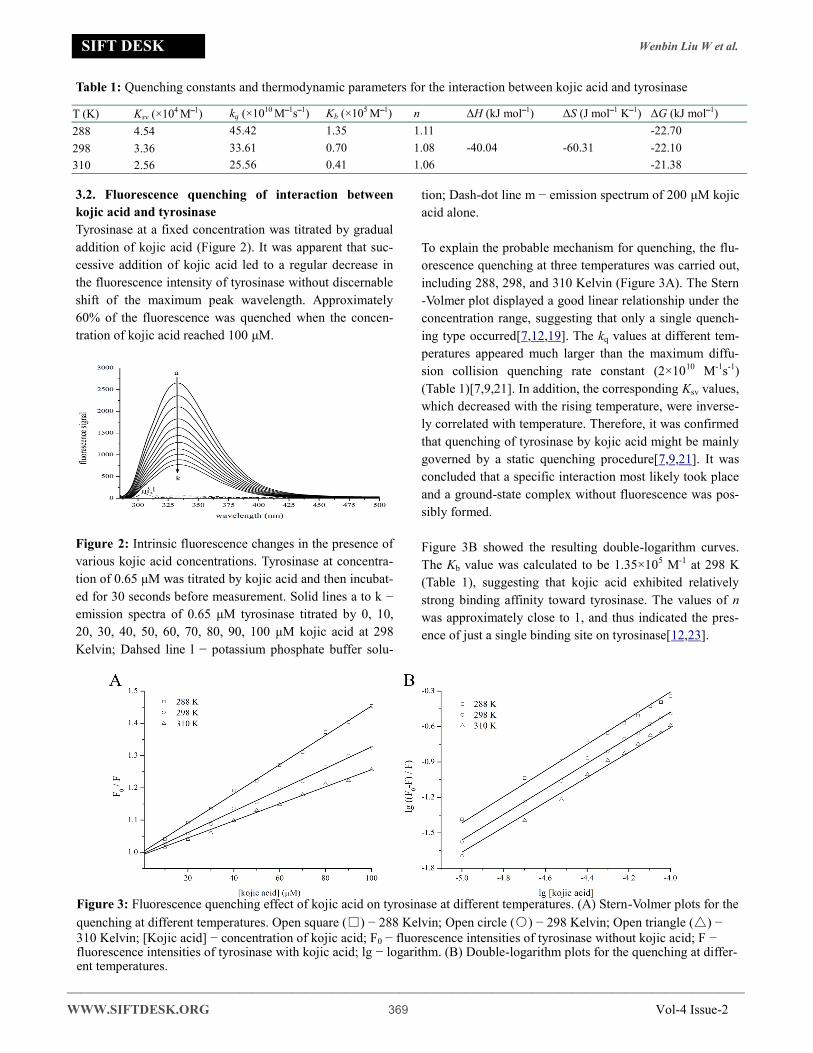
Wenbin Liu W et al
mdashmdashmdashmdashmdashmdashmdashmdashmdashmdashmdashmdashmdashmdashmdashmdashmdashmdashmdashmdashmdashmdashmdashmdashmdashmdashmdashmdashmdashmdashmdashmdashmdashmdashmdashmdashmdashmdashmdashmdashmdashmdashmdashmdashmdashmdashmdashmdashmdashmdashmdash
WWWSIFTDESKORG 369 Vol-4 Issue-2
SIFT DESK
32 Fluorescence quenching of interaction between
kojic acid and tyrosinase
Tyrosinase at a fixed concentration was titrated by gradual
addition of kojic acid (Figure 2) It was apparent that suc-
cessive addition of kojic acid led to a regular decrease in
the fluorescence intensity of tyrosinase without discernable
shift of the maximum peak wavelength Approximately
60 of the fluorescence was quenched when the concen-
tration of kojic acid reached 100 μM
Figure 2 Intrinsic fluorescence changes in the presence of
various kojic acid concentrations Tyrosinase at concentra-
tion of 065 μM was titrated by kojic acid and then incubat-
ed for 30 seconds before measurement Solid lines a to k minus
emission spectra of 065 μM tyrosinase titrated by 0 10
20 30 40 50 60 70 80 90 100 μM kojic acid at 298
Kelvin Dahsed line l minus potassium phosphate buffer solu-
tion Dash-dot line m minus emission spectrum of 200 μM kojic
acid alone
To explain the probable mechanism for quenching the flu-
orescence quenching at three temperatures was carried out
including 288 298 and 310 Kelvin (Figure 3A) The Stern
-Volmer plot displayed a good linear relationship under the
concentration range suggesting that only a single quench-
ing type occurred[71219] The kq values at different tem-
peratures appeared much larger than the maximum diffu-
sion collision quenching rate constant (2times1010 M-1s-1)
(Table 1)[7921] In addition the corresponding Ksv values
which decreased with the rising temperature were inverse-
ly correlated with temperature Therefore it was confirmed
that quenching of tyrosinase by kojic acid might be mainly
governed by a static quenching procedure[7921] It was
concluded that a specific interaction most likely took place
and a ground-state complex without fluorescence was pos-
sibly formed
Figure 3B showed the resulting double-logarithm curves
The Kb value was calculated to be 135times105 M-1 at 298 K
(Table 1) suggesting that kojic acid exhibited relatively
strong binding affinity toward tyrosinase The values of n
was approximately close to 1 and thus indicated the pres-
ence of just a single binding site on tyrosinase[1223]
Table 1 Quenching constants and thermodynamic parameters for the interaction between kojic acid and tyrosinase
T (K) Ksv (times104 Moline1) kq (times1010 Moline1soline1) Kb (times105 Moline1) n ΔH (kJ mololine1) ΔS (J mololine1 Koline1) ΔG (kJ mololine1)
288 454 4542 135 111
-4004 -6031
-2270
298 336 3361 070 108 -2210
310 256 2556 041 106 -2138
Figure 3 Fluorescence quenching effect of kojic acid on tyrosinase at different temperatures (A) Stern-Volmer plots for the
quenching at different temperatures Open square () minus 288 Kelvin Open circle () minus 298 Kelvin Open triangle () minus
310 Kelvin [Kojic acid] minus concentration of kojic acid F0 minus fluorescence intensities of tyrosinase without kojic acid F minus fluorescence intensities of tyrosinase with kojic acid lg minus logarithm (B) Double-logarithm plots for the quenching at differ-ent temperatures
Wenbin Liu W et al
mdashmdashmdashmdashmdashmdashmdashmdashmdashmdashmdashmdashmdashmdashmdashmdashmdashmdashmdashmdashmdashmdashmdashmdashmdashmdashmdashmdashmdashmdashmdashmdashmdashmdashmdashmdashmdashmdashmdashmdashmdashmdashmdashmdashmdashmdashmdashmdashmdashmdashndash
WWWSIFTDESKORG 370 Vol-4 Issue-2
SIFT DESK
33 Fluorescence lifetimes
The type of quenching mechanism could be directly
identified by fluorescence lifetime[712] The fluores-
cence decay of tyrosinase in the presence of different
concentrations of kojic acid exhibited triexponential
profiles with good fitting (Figure 4) Evidently the
fluorescence decay patterns scarcely changed after
the addition of kojic acid The average lifetime de-
creased slightly from 164 ns to 162 ns (Table 2)
which could be considered null in the range of exper-
imental error These results further indicated that the
quenching was really a static mode because a non-
fluorescent complex was formed
Figure 4 Representative time-resolved fluorescence
decay profiles of tyrosinase at various molar ratios of
kojic acid Tyrosinase at concentration of 04 μM was
incubated by kojic acid (0 15 60 μM) for 30 seconds
before measurement Open square () a minus 04 μM
tyrosinase Open circle () b minus 04 μM tyrosinase
with 15 μM kojic acid Open triangle () c minus 04 μM
tyrosinase with 60 μM kojic acid
34 Electrochemistry
Cyclic voltammetry could provide additional infor-
mation on interaction mode[24] The cyclic voltam-
mogram of kojic acid in absence and presence of ty-
rosinase were monitored by keeping the concentra-
tion of tyrosinase constant while varying the concen-
tration of kojic acid (Figure 5) The increment in in-
tensities of the currents for increasing concentrations
of kojic acid in presence of tyrosinase was lower than
that in absence of tyrosinase which was suggestive
of the reduction of equilibrium concentration of free
kojic acid in solution[25] Thus it could be conclud-
ed that a non-electroactive complex formed to block
the electron transfer between kojic acid and electrode
[24]
35 Thermodynamic parameters and interaction
forces
Attention was focused on the acting force to interpret
the binding mode (Figure 6 and Table 1) ΔG values
were all negative indicating that the spontaneity of
the binding process[1226] The negative sign for ΔH
revealed that the binding process was an exothermic
reaction[2627] Based on the thermodynamic criteri-
on[11] the negative ΔH and ΔS values meant that
both hydrogen bond and van der Waals force played a
main role in the binding process In case of kojic ac-
id hydroxyl groups were easy to form intermolecular
hydrogen bonds with the polar side chains of residues
in the binding pocket[28] The property of electronic
configuration of kojic acid was advantage for van der
Waals force to take part in the interaction[13]
Table 2 Fluorescence lifetime of tyrosinase as a function of concentration of kojic acid
t1 (ns) t2 (ns) t3 (ns) α1 α2 α3 tAV (ns)
χ2
Tyrosinase 218 053 537 038 052 010 164 1129
Tyrosinase + kojic acid ( 3 μΜ) 212 053 524 038 052 010 161 1107
Tyrosinase + kojic acid (15 μΜ) 222 055 544 038 053 009 163 1083
Tyrosinase + kojic acid (30 μΜ) 218 053 537 038 053 009 161 1072
Tyrosinase + kojic acid (60 μΜ) 213 052 530 039 051 010 161 1058
Tyrosinase + kojic acid (90 μΜ) 219 053 538 038 052 009 162 1080
Wenbin Liu W et al
mdashmdashmdashmdashmdashmdashmdashmdashmdashmdashmdashmdashmdashmdashmdashmdashmdashmdashmdashmdashmdashmdashmdashmdashmdashmdashmdashmdashmdashmdashmdashmdashmdashmdashmdashmdashmdashmdashmdashmdashmdashmdashmdashmdashmdashmdashmdashmdashmdashmdashmdash
WWWSIFTDESKORG 371 Vol-4 Issue-2
SIFT DESK
Figure 6 Vanrsquot Hoff plot T minus experimental tempera-
ture (Kelvin) lg minus logarithm Kb minus binding constant
36 Conformational changes of tyrosinase upon
binding of kojic acid
The synchronous fluorescence for Δλ at 60 nm
showed changes on the peak shape and a noticeable
red shift of spectral peak from 340 nm to 351 nm up-
on gradual addition of kojic acid (Figure 7A) The
red shift suggested that binding of kojic acid exposed
the Trp residues more to the solvent and thus in-
creased the hydrophilicity surrounding the Trp resi-
dues[1028] This finding corroborated the results
obtained from UV-Vis absorption experiment Com-
paratively the synchronous fluorescence for Δλ at 15
nm exhibited only very slightly red shift (Figure 7B)
reflecting that kojic acid had little effect on the mi-
croenvironment around Tyr residues Dramatic de-
crease in intensities was observed in both synchro-
nous fluorescence spectra The degree of quenching
for Δλ at 60 nm was higher than that at 15 nm imply-
ing that kojic acid was probably located closer to Trp
residue compared to Tyr residue[29] The microenvi-
ronmental alterations of the Trp residues indicated
that structure changes occurred after the interaction
with kojic acid
Figure 5 Cyclic voltammograms of kojic acid with tyrosinase (A) Various concentrations of kojic acid in the absence of tyrosinase The scan rate was 50 mVs-1 Solid lines a to f minus 0 10 20 30 40 50 μM kojic acid (B) Various concentrations of kojic acid in the presence of 1 μM tyrosinase
Figure 7 Synchronous fluorescence spectrum of tyrosinase titrated by kojic acid (A) Interval between excitation and emis-sion wavelength (Δλ) = 60 nm Solid lines a to u minus synchronous fluorescence spectrum of 065 μM tyrosinase titrated 0 10 20 30 40 50 60 70 80 90 100 110 120 130 140 150 160 170 180 190 200 μM kojic acid at 298 Kelvin (B) Δλ = 15 nm
Wenbin Liu W et al
mdashmdashmdashmdashmdashmdashmdashmdashmdashmdashmdashmdashmdashmdashmdashmdashmdashmdashmdashmdashmdashmdashmdashmdashmdashmdashmdashmdashmdashmdashmdashmdashmdashmdashmdashmdashmdashmdashmdashmdashmdashmdashmdashmdashmdashmdashmdashmdashmdashmdashndash
WWWSIFTDESKORG 372 Vol-4 Issue-2
SIFT DESK
The modulation in the three dimensional fluorescence
spectra upon kojic acid corresponded to the presence
of binding in terms of the decrease of fluorescence
intensity (Figure 8) The typical Rayleigh scattering
peak (peak C) and the second-order scattering peak
(peak D) seemed to be seldom affected[2326] Peak
A was characterized by the spectral feature of both
the intrinsic Trp and Tyr residues[2326] Peak B with
the excitation wavelength around 230 nm essentially
exhibited the fluorescence property of the polypep-
tide backbone structure of tyrosinase[2326] Kojic
acid resulted in a decrement of fluorescence intensity
for both peaks with a more serious degree for peak B
(Table 3) Moreover the fluorescence quenching for
peak A was accompanied with red shift of the maxi-
mum emission wavelength It was drawn a conclu-
sion that the environment around both residues be-
came more polar[3031] The significant decrease of
Strokes shift for peak A corroborated the abovemen-
tioned red shift Thus it could be conjectured that in-
teraction with kojic acid caused a major disturbance
of the peptide backbone and improved the polarity of
the microenvironment via exposing the hydrophobic
regions[2326]
The interaction of kojic acid with the secondary
structure of tyrosinase was revealed by circular di-
chroism (Figure 9) Upon addition of kojic acid at
increasing molar ratio an increment of ellipticity was
found for both two negative bands at 208 nm and 220
nm indicating a slight gain of a-helix[222331] The
proportions of different secondary structure composi-
tions of tyrosinase were quantitatively estimated
(Table 4) [12] The content of a-helix increased from
30 in free tyrosinase to 36 at a molar ratio for
kojic acid to tyrosinase of 41 while the content of b-
strand increased from 17 to 21 Some significant
decrease was also observed for the content of b-turn
and random coil This results suggested that increase
in the b-strand would lead to partial extending of pol-
ypeptide backbone and thus the conformation of tyro-
sinase became looser[32] On the basis of the spec-
troscopic results above it could be drawn a conclu-
sion that the occupancy of active site by kojic acid
could lead to destabilization of the native confor-
mation of tyrosinase followed by disruption of its
biological activities[3233]
Figure 8 Three-dimensional fluorescence spectra of tyrosinase at different kojic acid concentrations (A) Tyrosinase alone
peak A minus intrinsic Trp and Tyr residues peak peak B minus polypeptide backbone peak peak C minus Rayleigh scattering peak peak
D minus second-order scattering peak [tyrosinase] = 065 μM temperature = 298 Kelvin (B) Tyrosinase with 008 mM kojic
acid (C) Tyrosinase with 016 mM kojic acid (D) Tyrosinase with 032 mM kojic acid
Table 3 Characteristic parameters for three-dimensional fluorescence spectra of tyrosinase in presence of kojic acid
peak A peak B
kojic acid (mM)
peak position λexλem (nmnm)
fluorescence signal
(F0- F) F0 ()
Stokes Δλ a (nm)
peak posi-tion λexλem
(nmnm)
fluores-cence signal
(F0- F) F0 ()
Stokes Δλ a (nm)
0 280333 2507 0 53 228334 4256 0 106
008 286333 2078 1711 47 228339 2321 4547 111
016 291337 1383 4483 46 230337 1375 6769 107
032 294336 649 7411 42 233346 556 8694 113
a Stokes Δλ=λem - λex
Wenbin Liu W et al
mdashmdashmdashmdashmdashmdashmdashmdashmdashmdashmdashmdashmdashmdashmdashmdashmdashmdashmdashmdashmdashmdashmdashmdashmdashmdashmdashmdashmdashmdashmdashmdashmdashmdashmdashmdashmdashmdashmdashmdashmdashmdashmdashmdashmdashmdashmdashmdashmdashmdashmdash
WWWSIFTDESKORG 373 Vol-4 Issue-2
SIFT DESK
Figure 9 Circular dichroism spectra of tyrosinase at
different kojic aicd concentrations at 298 K The con-
centration of tyrosinase was kept fixed at 1 μM the
molar ratios of kojic acid to tyrosinase were 01 11
and 41 Dot line a minus 1 μM tyrosinase Dash-dot line
b minus 1 μM tyrosinase with 1 μM kojic acid Black line
c minus 1 μM tyrosinase with 4 μM kojic acid
4 CONCLUSIONS
The inhibitory mechanism of kojic acid on tyrosinase
was disclosed by an integrated study on spectroscopy
in this study for the first time Kojic acid had a single
class of binding sites on tyrosinase A complex spon-
taneously formed between kojic acid and tyrosinase
through static process The binding constant was
135times105 M-1 The interaction caused global confor-
mational changes of tyrosinase and thus increased the
microenvironmental polarity These experimental
observations interpreted that kojic acid might inhibit
the oxidation of L-DOPA via inducing conformation-
al changes after binding to tyrosinase The results
obtained in this study provided a fresh insight into
the relationship between activity and structure for
tyrosinase regarding molecular recognition It has a
great significance in further design of safe and effec-
tive tyrosinase inhibitors
COMPETING INTEREST
The authors declare that they have no competing in-
terests
AUTHORrsquoS CONTRIBUTIONS
WL designed the experiments XY and CW per-
formed the experiments XY and NG analyzed the
data All authors wrote the manuscript
ACKNOWLEDGEMENTS
This work was supported by the Strategic Partnership
Funds of Sichuan University
REFERENCES
[1] Chang T S (2009) An Updated Review of Tyrosi-
nase Inhibitors Int J Mol Sci 10 2440-2475
PMid19582213 View Article PubMed
NCBI
[2] Burnett C L Bergfeld W F Belsito D V Hill R
A Klaassen C D Liebler D C Marks J G Shank
R C Slaga T J Snyder P W (2010) Final Report
of the Safety Assessment of Kojic Acid as Used
in Cosmetics Int J Toxicol 29 244S-273S
PMid21164073 View Article PubMed
NCBI
[3] Lima C R Silva J R De T C Silva E O Lameira
J Do Nascimento J L Do Socorro Barros Brasil
D Alves C N (2014) Combined Kinetic Studies
and Computational Analysis on Kojic Acid Anal-
ogous as Tyrosinase Inhibitors Molecules 19
9591-9605 PMid25004069 View Arti-
cle PubMedNCBI
[4] Kubo I Kinsthori I (1999) Flavonols from Saf-
fron Flower Tyrosinase Inhibitory Activity and
Inhibition Mechanism J Agric Food Chem 47
4121-4125 PMid10552777 View Arti-
cle PubMedNCBI
[5] Copeland R A (1994) Methods for Protein Anal-
ysis a Practical Guide to Laboratory Protocols
Table 4 The contents of secondary structures of tyrosinase in the presence of kojic acida
α-Helix () β-Strand () β-Turns () Random Coil ()
Tyrosinase 30 17 23 30
Tyrosinase + kojic acid (1 μΜ) 32 19 20 29
Tyrosinase + kojic acid (4 μΜ) 36 21 18 25
a Tyrosinase at 1 μM
Wenbin Liu W et al
mdashmdashmdashmdashmdashmdashmdashmdashmdashmdashmdashmdashmdashmdashmdashmdashmdashmdashmdashmdashmdashmdashmdashmdashmdashmdashmdashmdashmdashmdashmdashmdashmdashmdashmdashmdashmdashmdashmdashmdashmdashmdashmdashmdashmdashmdashmdashmdashmdashmdashndash
WWWSIFTDESKORG 374 Vol-4 Issue-2
SIFT DESK
Chapman amp Hall
[6] Zhang Q J Liu B S Li G X Han R (2016) Using
Resonance Light Scattering and UVVis Absorp-
tion Spectroscopy to Study the Interaction be-
tween Gliclazide and Bovine Serum Albumin
Luminescence 31 1109-1114 PMid26663583
View Article PubMedNCBI
[7] Lakowicz (2006) Principles of Fluorescence
Spectroscopy 3rd ed Springer New York View
Article
[8] Pathak M Mishra R Agarwala P K Ojha H
Singh B Singh A Kukreti S (2016) Binding of
ethyl pyruvate to bovine serum albumin Calori-
metric spectroscopic and molecular docking
studies Thermochim Acta 633 140-148 View
Article
[9] Wei X L Xiao J B Wang Y Bai Y (2009)
Which Model Based on Fluorescence Quenching
is Suitable to Study the Interaction between
Trans-Resveratrol and BSA Spectrochim Acta
A Mol Biomol Spectrosc 75 299-304
PMid19926336 View Article PubMed
NCBI
[10]Huang Y Yan J Liu B Yu Z Gao X Tang Y Zi
Y (2010) Investigation on Interaction of Pruli-
floxacin with Pepsin A Spectroscopic Analysis
Spectrochim Acta A Mol Biomol Spectrosc 75
1024-1029 PMid20045662 View Arti-
cle PubMedNCBI
[11]Ross P D Subramanian S (1981) Thermodynam-
ics of Protein Association Reactions Forces
Contributing to Stability Biochemistry 20 3096-
3102 PMid7248271 View Article PubMed
NCBI
[12]Wang Y J Zhang G W Yan J K Gong D M
(2014) Inhibitory Effect of Morin on Tyrosinase
Insights from Spectroscopic and Molecular
Docking Studies Food Chem 163 226-233
PMid24912720 View Article PubMed
NCBI
[13]Tu B Chen Z F Liu Z J Li R R Ouyang Y Hu
Y J (2015) Study of the Structure-Activity Rela-
tionship of Flavonoids Based on Their Interac-
tion with Human Serum Albumin Rsc Adv 5
73290-73300 View Article
[14]Whitmore L Wallace B A (2008) Protein Sec-
ondary Structure Analyses from Circular Dichro-
ism Spectroscopy Methods and Reference Data-
bases Biopolymers 89 392-400
PMid17896349 View Article PubMed
NCBI
[15]Guo N H Wang C L Shang C You X Zhang L
Y Liu W B (2018) Integrated Study of the
Mechanism of Tyrosinase Inhibition by Baicalein
Using Kinetic Multispectroscopic and Computa-
tional Simulation Analyses Int J Biol Macromol
118 57-68 PMid29908273 View Arti-
cle PubMedNCBI
[16]Kim D Park J Kim J Han C Yoon J Kim N
Seo J Lee C (2006) Flavonoids as Mushroom
Tyrosinase Inhibitors A Fluorescence Quench-
ing Study J Agric Food Chem 54 935-941
PMid16448205 View Article PubMed
NCBI
[17]Shang C Zhang Y K You X Guo N H Wang Y
Fan Y Liu W B (2018) The Effect of 784-
Trihydroxyflavone on Tyrosinase Activity and
Conformation Spectroscopy and Docking Stud-
ies Luminescence 33 681-691 PMid29479807
View Article PubMedNCBI
[18]Gao H Nishida J Saito S Kawabata J (2007)
Inhibitory Effects of 567-Trihydroxyflavones
on Tyrosinase Molecules 12 86-97
PMid17693955 View Article PubMed
NCBI
[19]Pasricha S Sharma D Ojha H Gahlot P Pathak
M Basu M Kukreti S (2017) Luminescence
circular dichroism and in silico studies of binding
interaction of synthesized naphthylchalcone de-
rivatives with bovine serum albumin Lumines-
cence 32 1252-1262 PMid28512990 View Ar-
ticle PubMedNCBI
[20]Huang J Yuan Y Z Liang H (2002) Binding
Equilibrium Study of Phosphotungstic Acid and
HSA or BSA with UV Spectrum Fluorescence
Spectrum and Equilibrium Dialysis Sci Chi
Chem 45 200-207 View Article
[21]Weert M V Stella L (2011) Fluorescence
Quenching and Ligand Binding A Critical Dis-
cussion of A Popular Methodology J Mol Struct
998 144-150 View Article
[22]Sharma D Ojha H Pathak M Singh B Sharma
Wenbin Liu W et al
mdashmdashmdashmdashmdashmdashmdashmdashmdashmdashmdashmdashmdashmdashmdashmdashmdashmdashmdashmdashmdashmdashmdashmdashmdashmdashmdashmdashmdashmdashmdashmdashmdashmdashmdashmdashmdashmdashmdashmdashmdashmdashmdashmdashmdashmdashmdashmdashmdashmdashmdash
WWWSIFTDESKORG 375 Vol-4 Issue-2
SIFT DESK
N Singh A Sharma R K (2016) Spectroscopic
and molecular modelling studies of binding mech-
anism of metformin with bovine serum albumin J
Mol Struct 1118 267-274 View Article
[23]Peng W Ding F Jiang Y T Sun Y Peng Y K
(2014) Evaluation of the Biointeraction of Color-
ant Flavazin with Human Serum Albumin In-
sights from Multiple Spectroscopic Studies in
Silico Docking and Molecular Dynamics Simula-
tion Food Funct 5 1203-1217 PMid24705828
View Article PubMedNCBI
[24]Wang Y J Zhang G W Yan J K Gong D (2014)
Inhibitory Effect of Morin on Tyrosinase Insights
from Spectroscopic and Molecular Docking Stud-
ies Food Chem 163 226-233 PMid24912720
View Article PubMedNCBI
[25]Magdum P A Gokavi N M Nandibewoor S T
(2016) Study on the Interaction between Anti-
Tuberculosis Drug Ethambutol and Bovine Serum
Albumin Multispectroscopic and Cyclic Voltam-
metric Approaches Luminescence 32 206-216
PMid27377878 View Article PubMed
NCBI
[26]Wang J Xiang C Tian F F Xu Z Q Jiang F L
Liu Y (2014) Investigating the Interactions of A
Novel Anticancer Delocalized Lipophilic Cation
and Its Precursor Compound with Human Serum
Albumin RSC Adv 4 18205-18216 View Arti-
cle
[27]Ali M S Al-Lohedan H A (2016) Multi-
Technique Approach on the Interaction between
Sugar-Based Surfactant N-Dodecyl Β-D-
Maltoside and Bovine Serum Albumin J Lumin
169 35-42 View Article
[28]Wang Y Q Chen T T Zhang H M (2010) Investi-
gation of the Interactions of Lysozyme and Tryp-
sin with Biphenol A Using Spectroscopic Meth-
ods Spectrochim Acta A Mol Biomol Spectrosc
75 1130-1137 PMid20093070 View Arti-
cle PubMedNCBI
[29]Ray D Paul B K Guchhait N (2012) Effect of
Biological Confinement on the Photophysics and
Dynamics of A Proton-Transfer Phototautomer
An Exploration of Excitation and Emission Wave-
length-Dependent Photophysics of the Protein-
Bound Drug Phys Chem Chem Phys 14 12182-
12192 PMid22870509 View Article PubMed
NCBI
[30]Chen Z Y Xu H Y Zhu Y L Liu J Y Wang K Y
Wang P X Shang S J Yi X N Wang Z L Shao
W (2014) Understanding The Fate of An Anes-
thetic Nalorphine upon Interaction with Human
Serum Albumin A Photophysical and Mass-
Spectroscopy Approach Rsc Adv 4 25410-
25419 View Article
[31]Samanta A Paul B K Guchhait N (2011) Spectro-
scopic Probe Analysis for Exploring Probe-
Protein Interaction A Mapping of Native Unfold-
ing and Refolding of Protein Bovine Serum Albu-
min by Extrinsic Fluorescence Probe Biophys
Chem 156 128-139 PMid21514035 View Arti-
cle PubMedNCBI
[32]Mansouri M Pirouzi M Saberi M R Ghaderabad
M Chamani J (2013) Investigation on the Interac-
tion between Cyclophosphamide and Lysozyme in
the Presence of Three Different Kind of Cy-
clodextrins Determination of the Binding Mecha-
nism by Spectroscopic and Molecular Modeling
Techniques Molecules 18 789 PMid23344194
View Article PubMedNCBI
[33]Ojha H Mishra K Hassan M I Chaudhury N K
(2012) Spectroscopic and isothermal titration cal-
orimetry studies of binding interaction of ferulic
acid with bovine serum albumin Thermochim
acta 548 56-64 View Article
SIFT DESK JOURNALS Email infosiftdeskorg
- Study on the interaction of kojic acid with tyrosinase by spectroscopic methods
- ABSTRACT
- 1 INTRODUCTION
- 2 MATERIALS AND METHODS
-
- 21 Materials
- 22 Ultraviolet-visible spectra measurements
- 23 Fluorescence spectra measurements
- 24 Time-resolved fluorescence lifetime measurements
- 25 Electrochemical measurements
- 26 Thermodynamic parameters
- 27 Synchronous and three-dimensional fluorescence spectra
- 28 Circular dichroism spectra measurements
-
- 3 RESULTS AND DISCUSSION
-
- 31 Ultraviolet-visible spectra for interaction between kojic acid and copper ion or tyrosinase
- 32 Fluorescence quenching of interaction between kojic acid and tyrosinase
- 33 Fluorescence lifetimes
- 34 Electrochemistry
- 35 Thermodynamic parameters and interaction forces
- 36 Conformational changes of tyrosinase upon binding of kojic acid
-
- 4 CONCLUSIONS
- ACKNOWLEDGEMENTS
- REFERENCES
-

Wenbin Liu W et al
mdashmdashmdashmdashmdashmdashmdashmdashmdashmdashmdashmdashmdashmdashmdashmdashmdashmdashmdashmdashmdashmdashmdashmdashmdashmdashmdashmdashmdashmdashmdashmdashmdashmdashmdashmdashmdashmdashmdashmdashmdashmdashmdashmdashmdashmdashmdashmdashmdashmdashndash
WWWSIFTDESKORG 370 Vol-4 Issue-2
SIFT DESK
33 Fluorescence lifetimes
The type of quenching mechanism could be directly
identified by fluorescence lifetime[712] The fluores-
cence decay of tyrosinase in the presence of different
concentrations of kojic acid exhibited triexponential
profiles with good fitting (Figure 4) Evidently the
fluorescence decay patterns scarcely changed after
the addition of kojic acid The average lifetime de-
creased slightly from 164 ns to 162 ns (Table 2)
which could be considered null in the range of exper-
imental error These results further indicated that the
quenching was really a static mode because a non-
fluorescent complex was formed
Figure 4 Representative time-resolved fluorescence
decay profiles of tyrosinase at various molar ratios of
kojic acid Tyrosinase at concentration of 04 μM was
incubated by kojic acid (0 15 60 μM) for 30 seconds
before measurement Open square () a minus 04 μM
tyrosinase Open circle () b minus 04 μM tyrosinase
with 15 μM kojic acid Open triangle () c minus 04 μM
tyrosinase with 60 μM kojic acid
34 Electrochemistry
Cyclic voltammetry could provide additional infor-
mation on interaction mode[24] The cyclic voltam-
mogram of kojic acid in absence and presence of ty-
rosinase were monitored by keeping the concentra-
tion of tyrosinase constant while varying the concen-
tration of kojic acid (Figure 5) The increment in in-
tensities of the currents for increasing concentrations
of kojic acid in presence of tyrosinase was lower than
that in absence of tyrosinase which was suggestive
of the reduction of equilibrium concentration of free
kojic acid in solution[25] Thus it could be conclud-
ed that a non-electroactive complex formed to block
the electron transfer between kojic acid and electrode
[24]
35 Thermodynamic parameters and interaction
forces
Attention was focused on the acting force to interpret
the binding mode (Figure 6 and Table 1) ΔG values
were all negative indicating that the spontaneity of
the binding process[1226] The negative sign for ΔH
revealed that the binding process was an exothermic
reaction[2627] Based on the thermodynamic criteri-
on[11] the negative ΔH and ΔS values meant that
both hydrogen bond and van der Waals force played a
main role in the binding process In case of kojic ac-
id hydroxyl groups were easy to form intermolecular
hydrogen bonds with the polar side chains of residues
in the binding pocket[28] The property of electronic
configuration of kojic acid was advantage for van der
Waals force to take part in the interaction[13]
Table 2 Fluorescence lifetime of tyrosinase as a function of concentration of kojic acid
t1 (ns) t2 (ns) t3 (ns) α1 α2 α3 tAV (ns)
χ2
Tyrosinase 218 053 537 038 052 010 164 1129
Tyrosinase + kojic acid ( 3 μΜ) 212 053 524 038 052 010 161 1107
Tyrosinase + kojic acid (15 μΜ) 222 055 544 038 053 009 163 1083
Tyrosinase + kojic acid (30 μΜ) 218 053 537 038 053 009 161 1072
Tyrosinase + kojic acid (60 μΜ) 213 052 530 039 051 010 161 1058
Tyrosinase + kojic acid (90 μΜ) 219 053 538 038 052 009 162 1080
Wenbin Liu W et al
mdashmdashmdashmdashmdashmdashmdashmdashmdashmdashmdashmdashmdashmdashmdashmdashmdashmdashmdashmdashmdashmdashmdashmdashmdashmdashmdashmdashmdashmdashmdashmdashmdashmdashmdashmdashmdashmdashmdashmdashmdashmdashmdashmdashmdashmdashmdashmdashmdashmdashmdash
WWWSIFTDESKORG 371 Vol-4 Issue-2
SIFT DESK
Figure 6 Vanrsquot Hoff plot T minus experimental tempera-
ture (Kelvin) lg minus logarithm Kb minus binding constant
36 Conformational changes of tyrosinase upon
binding of kojic acid
The synchronous fluorescence for Δλ at 60 nm
showed changes on the peak shape and a noticeable
red shift of spectral peak from 340 nm to 351 nm up-
on gradual addition of kojic acid (Figure 7A) The
red shift suggested that binding of kojic acid exposed
the Trp residues more to the solvent and thus in-
creased the hydrophilicity surrounding the Trp resi-
dues[1028] This finding corroborated the results
obtained from UV-Vis absorption experiment Com-
paratively the synchronous fluorescence for Δλ at 15
nm exhibited only very slightly red shift (Figure 7B)
reflecting that kojic acid had little effect on the mi-
croenvironment around Tyr residues Dramatic de-
crease in intensities was observed in both synchro-
nous fluorescence spectra The degree of quenching
for Δλ at 60 nm was higher than that at 15 nm imply-
ing that kojic acid was probably located closer to Trp
residue compared to Tyr residue[29] The microenvi-
ronmental alterations of the Trp residues indicated
that structure changes occurred after the interaction
with kojic acid
Figure 5 Cyclic voltammograms of kojic acid with tyrosinase (A) Various concentrations of kojic acid in the absence of tyrosinase The scan rate was 50 mVs-1 Solid lines a to f minus 0 10 20 30 40 50 μM kojic acid (B) Various concentrations of kojic acid in the presence of 1 μM tyrosinase
Figure 7 Synchronous fluorescence spectrum of tyrosinase titrated by kojic acid (A) Interval between excitation and emis-sion wavelength (Δλ) = 60 nm Solid lines a to u minus synchronous fluorescence spectrum of 065 μM tyrosinase titrated 0 10 20 30 40 50 60 70 80 90 100 110 120 130 140 150 160 170 180 190 200 μM kojic acid at 298 Kelvin (B) Δλ = 15 nm
Wenbin Liu W et al
mdashmdashmdashmdashmdashmdashmdashmdashmdashmdashmdashmdashmdashmdashmdashmdashmdashmdashmdashmdashmdashmdashmdashmdashmdashmdashmdashmdashmdashmdashmdashmdashmdashmdashmdashmdashmdashmdashmdashmdashmdashmdashmdashmdashmdashmdashmdashmdashmdashmdashndash
WWWSIFTDESKORG 372 Vol-4 Issue-2
SIFT DESK
The modulation in the three dimensional fluorescence
spectra upon kojic acid corresponded to the presence
of binding in terms of the decrease of fluorescence
intensity (Figure 8) The typical Rayleigh scattering
peak (peak C) and the second-order scattering peak
(peak D) seemed to be seldom affected[2326] Peak
A was characterized by the spectral feature of both
the intrinsic Trp and Tyr residues[2326] Peak B with
the excitation wavelength around 230 nm essentially
exhibited the fluorescence property of the polypep-
tide backbone structure of tyrosinase[2326] Kojic
acid resulted in a decrement of fluorescence intensity
for both peaks with a more serious degree for peak B
(Table 3) Moreover the fluorescence quenching for
peak A was accompanied with red shift of the maxi-
mum emission wavelength It was drawn a conclu-
sion that the environment around both residues be-
came more polar[3031] The significant decrease of
Strokes shift for peak A corroborated the abovemen-
tioned red shift Thus it could be conjectured that in-
teraction with kojic acid caused a major disturbance
of the peptide backbone and improved the polarity of
the microenvironment via exposing the hydrophobic
regions[2326]
The interaction of kojic acid with the secondary
structure of tyrosinase was revealed by circular di-
chroism (Figure 9) Upon addition of kojic acid at
increasing molar ratio an increment of ellipticity was
found for both two negative bands at 208 nm and 220
nm indicating a slight gain of a-helix[222331] The
proportions of different secondary structure composi-
tions of tyrosinase were quantitatively estimated
(Table 4) [12] The content of a-helix increased from
30 in free tyrosinase to 36 at a molar ratio for
kojic acid to tyrosinase of 41 while the content of b-
strand increased from 17 to 21 Some significant
decrease was also observed for the content of b-turn
and random coil This results suggested that increase
in the b-strand would lead to partial extending of pol-
ypeptide backbone and thus the conformation of tyro-
sinase became looser[32] On the basis of the spec-
troscopic results above it could be drawn a conclu-
sion that the occupancy of active site by kojic acid
could lead to destabilization of the native confor-
mation of tyrosinase followed by disruption of its
biological activities[3233]
Figure 8 Three-dimensional fluorescence spectra of tyrosinase at different kojic acid concentrations (A) Tyrosinase alone
peak A minus intrinsic Trp and Tyr residues peak peak B minus polypeptide backbone peak peak C minus Rayleigh scattering peak peak
D minus second-order scattering peak [tyrosinase] = 065 μM temperature = 298 Kelvin (B) Tyrosinase with 008 mM kojic
acid (C) Tyrosinase with 016 mM kojic acid (D) Tyrosinase with 032 mM kojic acid
Table 3 Characteristic parameters for three-dimensional fluorescence spectra of tyrosinase in presence of kojic acid
peak A peak B
kojic acid (mM)
peak position λexλem (nmnm)
fluorescence signal
(F0- F) F0 ()
Stokes Δλ a (nm)
peak posi-tion λexλem
(nmnm)
fluores-cence signal
(F0- F) F0 ()
Stokes Δλ a (nm)
0 280333 2507 0 53 228334 4256 0 106
008 286333 2078 1711 47 228339 2321 4547 111
016 291337 1383 4483 46 230337 1375 6769 107
032 294336 649 7411 42 233346 556 8694 113
a Stokes Δλ=λem - λex
Wenbin Liu W et al
mdashmdashmdashmdashmdashmdashmdashmdashmdashmdashmdashmdashmdashmdashmdashmdashmdashmdashmdashmdashmdashmdashmdashmdashmdashmdashmdashmdashmdashmdashmdashmdashmdashmdashmdashmdashmdashmdashmdashmdashmdashmdashmdashmdashmdashmdashmdashmdashmdashmdashmdash
WWWSIFTDESKORG 373 Vol-4 Issue-2
SIFT DESK
Figure 9 Circular dichroism spectra of tyrosinase at
different kojic aicd concentrations at 298 K The con-
centration of tyrosinase was kept fixed at 1 μM the
molar ratios of kojic acid to tyrosinase were 01 11
and 41 Dot line a minus 1 μM tyrosinase Dash-dot line
b minus 1 μM tyrosinase with 1 μM kojic acid Black line
c minus 1 μM tyrosinase with 4 μM kojic acid
4 CONCLUSIONS
The inhibitory mechanism of kojic acid on tyrosinase
was disclosed by an integrated study on spectroscopy
in this study for the first time Kojic acid had a single
class of binding sites on tyrosinase A complex spon-
taneously formed between kojic acid and tyrosinase
through static process The binding constant was
135times105 M-1 The interaction caused global confor-
mational changes of tyrosinase and thus increased the
microenvironmental polarity These experimental
observations interpreted that kojic acid might inhibit
the oxidation of L-DOPA via inducing conformation-
al changes after binding to tyrosinase The results
obtained in this study provided a fresh insight into
the relationship between activity and structure for
tyrosinase regarding molecular recognition It has a
great significance in further design of safe and effec-
tive tyrosinase inhibitors
COMPETING INTEREST
The authors declare that they have no competing in-
terests
AUTHORrsquoS CONTRIBUTIONS
WL designed the experiments XY and CW per-
formed the experiments XY and NG analyzed the
data All authors wrote the manuscript
ACKNOWLEDGEMENTS
This work was supported by the Strategic Partnership
Funds of Sichuan University
REFERENCES
[1] Chang T S (2009) An Updated Review of Tyrosi-
nase Inhibitors Int J Mol Sci 10 2440-2475
PMid19582213 View Article PubMed
NCBI
[2] Burnett C L Bergfeld W F Belsito D V Hill R
A Klaassen C D Liebler D C Marks J G Shank
R C Slaga T J Snyder P W (2010) Final Report
of the Safety Assessment of Kojic Acid as Used
in Cosmetics Int J Toxicol 29 244S-273S
PMid21164073 View Article PubMed
NCBI
[3] Lima C R Silva J R De T C Silva E O Lameira
J Do Nascimento J L Do Socorro Barros Brasil
D Alves C N (2014) Combined Kinetic Studies
and Computational Analysis on Kojic Acid Anal-
ogous as Tyrosinase Inhibitors Molecules 19
9591-9605 PMid25004069 View Arti-
cle PubMedNCBI
[4] Kubo I Kinsthori I (1999) Flavonols from Saf-
fron Flower Tyrosinase Inhibitory Activity and
Inhibition Mechanism J Agric Food Chem 47
4121-4125 PMid10552777 View Arti-
cle PubMedNCBI
[5] Copeland R A (1994) Methods for Protein Anal-
ysis a Practical Guide to Laboratory Protocols
Table 4 The contents of secondary structures of tyrosinase in the presence of kojic acida
α-Helix () β-Strand () β-Turns () Random Coil ()
Tyrosinase 30 17 23 30
Tyrosinase + kojic acid (1 μΜ) 32 19 20 29
Tyrosinase + kojic acid (4 μΜ) 36 21 18 25
a Tyrosinase at 1 μM
Wenbin Liu W et al
mdashmdashmdashmdashmdashmdashmdashmdashmdashmdashmdashmdashmdashmdashmdashmdashmdashmdashmdashmdashmdashmdashmdashmdashmdashmdashmdashmdashmdashmdashmdashmdashmdashmdashmdashmdashmdashmdashmdashmdashmdashmdashmdashmdashmdashmdashmdashmdashmdashmdashndash
WWWSIFTDESKORG 374 Vol-4 Issue-2
SIFT DESK
Chapman amp Hall
[6] Zhang Q J Liu B S Li G X Han R (2016) Using
Resonance Light Scattering and UVVis Absorp-
tion Spectroscopy to Study the Interaction be-
tween Gliclazide and Bovine Serum Albumin
Luminescence 31 1109-1114 PMid26663583
View Article PubMedNCBI
[7] Lakowicz (2006) Principles of Fluorescence
Spectroscopy 3rd ed Springer New York View
Article
[8] Pathak M Mishra R Agarwala P K Ojha H
Singh B Singh A Kukreti S (2016) Binding of
ethyl pyruvate to bovine serum albumin Calori-
metric spectroscopic and molecular docking
studies Thermochim Acta 633 140-148 View
Article
[9] Wei X L Xiao J B Wang Y Bai Y (2009)
Which Model Based on Fluorescence Quenching
is Suitable to Study the Interaction between
Trans-Resveratrol and BSA Spectrochim Acta
A Mol Biomol Spectrosc 75 299-304
PMid19926336 View Article PubMed
NCBI
[10]Huang Y Yan J Liu B Yu Z Gao X Tang Y Zi
Y (2010) Investigation on Interaction of Pruli-
floxacin with Pepsin A Spectroscopic Analysis
Spectrochim Acta A Mol Biomol Spectrosc 75
1024-1029 PMid20045662 View Arti-
cle PubMedNCBI
[11]Ross P D Subramanian S (1981) Thermodynam-
ics of Protein Association Reactions Forces
Contributing to Stability Biochemistry 20 3096-
3102 PMid7248271 View Article PubMed
NCBI
[12]Wang Y J Zhang G W Yan J K Gong D M
(2014) Inhibitory Effect of Morin on Tyrosinase
Insights from Spectroscopic and Molecular
Docking Studies Food Chem 163 226-233
PMid24912720 View Article PubMed
NCBI
[13]Tu B Chen Z F Liu Z J Li R R Ouyang Y Hu
Y J (2015) Study of the Structure-Activity Rela-
tionship of Flavonoids Based on Their Interac-
tion with Human Serum Albumin Rsc Adv 5
73290-73300 View Article
[14]Whitmore L Wallace B A (2008) Protein Sec-
ondary Structure Analyses from Circular Dichro-
ism Spectroscopy Methods and Reference Data-
bases Biopolymers 89 392-400
PMid17896349 View Article PubMed
NCBI
[15]Guo N H Wang C L Shang C You X Zhang L
Y Liu W B (2018) Integrated Study of the
Mechanism of Tyrosinase Inhibition by Baicalein
Using Kinetic Multispectroscopic and Computa-
tional Simulation Analyses Int J Biol Macromol
118 57-68 PMid29908273 View Arti-
cle PubMedNCBI
[16]Kim D Park J Kim J Han C Yoon J Kim N
Seo J Lee C (2006) Flavonoids as Mushroom
Tyrosinase Inhibitors A Fluorescence Quench-
ing Study J Agric Food Chem 54 935-941
PMid16448205 View Article PubMed
NCBI
[17]Shang C Zhang Y K You X Guo N H Wang Y
Fan Y Liu W B (2018) The Effect of 784-
Trihydroxyflavone on Tyrosinase Activity and
Conformation Spectroscopy and Docking Stud-
ies Luminescence 33 681-691 PMid29479807
View Article PubMedNCBI
[18]Gao H Nishida J Saito S Kawabata J (2007)
Inhibitory Effects of 567-Trihydroxyflavones
on Tyrosinase Molecules 12 86-97
PMid17693955 View Article PubMed
NCBI
[19]Pasricha S Sharma D Ojha H Gahlot P Pathak
M Basu M Kukreti S (2017) Luminescence
circular dichroism and in silico studies of binding
interaction of synthesized naphthylchalcone de-
rivatives with bovine serum albumin Lumines-
cence 32 1252-1262 PMid28512990 View Ar-
ticle PubMedNCBI
[20]Huang J Yuan Y Z Liang H (2002) Binding
Equilibrium Study of Phosphotungstic Acid and
HSA or BSA with UV Spectrum Fluorescence
Spectrum and Equilibrium Dialysis Sci Chi
Chem 45 200-207 View Article
[21]Weert M V Stella L (2011) Fluorescence
Quenching and Ligand Binding A Critical Dis-
cussion of A Popular Methodology J Mol Struct
998 144-150 View Article
[22]Sharma D Ojha H Pathak M Singh B Sharma
Wenbin Liu W et al
mdashmdashmdashmdashmdashmdashmdashmdashmdashmdashmdashmdashmdashmdashmdashmdashmdashmdashmdashmdashmdashmdashmdashmdashmdashmdashmdashmdashmdashmdashmdashmdashmdashmdashmdashmdashmdashmdashmdashmdashmdashmdashmdashmdashmdashmdashmdashmdashmdashmdashmdash
WWWSIFTDESKORG 375 Vol-4 Issue-2
SIFT DESK
N Singh A Sharma R K (2016) Spectroscopic
and molecular modelling studies of binding mech-
anism of metformin with bovine serum albumin J
Mol Struct 1118 267-274 View Article
[23]Peng W Ding F Jiang Y T Sun Y Peng Y K
(2014) Evaluation of the Biointeraction of Color-
ant Flavazin with Human Serum Albumin In-
sights from Multiple Spectroscopic Studies in
Silico Docking and Molecular Dynamics Simula-
tion Food Funct 5 1203-1217 PMid24705828
View Article PubMedNCBI
[24]Wang Y J Zhang G W Yan J K Gong D (2014)
Inhibitory Effect of Morin on Tyrosinase Insights
from Spectroscopic and Molecular Docking Stud-
ies Food Chem 163 226-233 PMid24912720
View Article PubMedNCBI
[25]Magdum P A Gokavi N M Nandibewoor S T
(2016) Study on the Interaction between Anti-
Tuberculosis Drug Ethambutol and Bovine Serum
Albumin Multispectroscopic and Cyclic Voltam-
metric Approaches Luminescence 32 206-216
PMid27377878 View Article PubMed
NCBI
[26]Wang J Xiang C Tian F F Xu Z Q Jiang F L
Liu Y (2014) Investigating the Interactions of A
Novel Anticancer Delocalized Lipophilic Cation
and Its Precursor Compound with Human Serum
Albumin RSC Adv 4 18205-18216 View Arti-
cle
[27]Ali M S Al-Lohedan H A (2016) Multi-
Technique Approach on the Interaction between
Sugar-Based Surfactant N-Dodecyl Β-D-
Maltoside and Bovine Serum Albumin J Lumin
169 35-42 View Article
[28]Wang Y Q Chen T T Zhang H M (2010) Investi-
gation of the Interactions of Lysozyme and Tryp-
sin with Biphenol A Using Spectroscopic Meth-
ods Spectrochim Acta A Mol Biomol Spectrosc
75 1130-1137 PMid20093070 View Arti-
cle PubMedNCBI
[29]Ray D Paul B K Guchhait N (2012) Effect of
Biological Confinement on the Photophysics and
Dynamics of A Proton-Transfer Phototautomer
An Exploration of Excitation and Emission Wave-
length-Dependent Photophysics of the Protein-
Bound Drug Phys Chem Chem Phys 14 12182-
12192 PMid22870509 View Article PubMed
NCBI
[30]Chen Z Y Xu H Y Zhu Y L Liu J Y Wang K Y
Wang P X Shang S J Yi X N Wang Z L Shao
W (2014) Understanding The Fate of An Anes-
thetic Nalorphine upon Interaction with Human
Serum Albumin A Photophysical and Mass-
Spectroscopy Approach Rsc Adv 4 25410-
25419 View Article
[31]Samanta A Paul B K Guchhait N (2011) Spectro-
scopic Probe Analysis for Exploring Probe-
Protein Interaction A Mapping of Native Unfold-
ing and Refolding of Protein Bovine Serum Albu-
min by Extrinsic Fluorescence Probe Biophys
Chem 156 128-139 PMid21514035 View Arti-
cle PubMedNCBI
[32]Mansouri M Pirouzi M Saberi M R Ghaderabad
M Chamani J (2013) Investigation on the Interac-
tion between Cyclophosphamide and Lysozyme in
the Presence of Three Different Kind of Cy-
clodextrins Determination of the Binding Mecha-
nism by Spectroscopic and Molecular Modeling
Techniques Molecules 18 789 PMid23344194
View Article PubMedNCBI
[33]Ojha H Mishra K Hassan M I Chaudhury N K
(2012) Spectroscopic and isothermal titration cal-
orimetry studies of binding interaction of ferulic
acid with bovine serum albumin Thermochim
acta 548 56-64 View Article
SIFT DESK JOURNALS Email infosiftdeskorg
- Study on the interaction of kojic acid with tyrosinase by spectroscopic methods
- ABSTRACT
- 1 INTRODUCTION
- 2 MATERIALS AND METHODS
-
- 21 Materials
- 22 Ultraviolet-visible spectra measurements
- 23 Fluorescence spectra measurements
- 24 Time-resolved fluorescence lifetime measurements
- 25 Electrochemical measurements
- 26 Thermodynamic parameters
- 27 Synchronous and three-dimensional fluorescence spectra
- 28 Circular dichroism spectra measurements
-
- 3 RESULTS AND DISCUSSION
-
- 31 Ultraviolet-visible spectra for interaction between kojic acid and copper ion or tyrosinase
- 32 Fluorescence quenching of interaction between kojic acid and tyrosinase
- 33 Fluorescence lifetimes
- 34 Electrochemistry
- 35 Thermodynamic parameters and interaction forces
- 36 Conformational changes of tyrosinase upon binding of kojic acid
-
- 4 CONCLUSIONS
- ACKNOWLEDGEMENTS
- REFERENCES
-

Wenbin Liu W et al
mdashmdashmdashmdashmdashmdashmdashmdashmdashmdashmdashmdashmdashmdashmdashmdashmdashmdashmdashmdashmdashmdashmdashmdashmdashmdashmdashmdashmdashmdashmdashmdashmdashmdashmdashmdashmdashmdashmdashmdashmdashmdashmdashmdashmdashmdashmdashmdashmdashmdashmdash
WWWSIFTDESKORG 371 Vol-4 Issue-2
SIFT DESK
Figure 6 Vanrsquot Hoff plot T minus experimental tempera-
ture (Kelvin) lg minus logarithm Kb minus binding constant
36 Conformational changes of tyrosinase upon
binding of kojic acid
The synchronous fluorescence for Δλ at 60 nm
showed changes on the peak shape and a noticeable
red shift of spectral peak from 340 nm to 351 nm up-
on gradual addition of kojic acid (Figure 7A) The
red shift suggested that binding of kojic acid exposed
the Trp residues more to the solvent and thus in-
creased the hydrophilicity surrounding the Trp resi-
dues[1028] This finding corroborated the results
obtained from UV-Vis absorption experiment Com-
paratively the synchronous fluorescence for Δλ at 15
nm exhibited only very slightly red shift (Figure 7B)
reflecting that kojic acid had little effect on the mi-
croenvironment around Tyr residues Dramatic de-
crease in intensities was observed in both synchro-
nous fluorescence spectra The degree of quenching
for Δλ at 60 nm was higher than that at 15 nm imply-
ing that kojic acid was probably located closer to Trp
residue compared to Tyr residue[29] The microenvi-
ronmental alterations of the Trp residues indicated
that structure changes occurred after the interaction
with kojic acid
Figure 5 Cyclic voltammograms of kojic acid with tyrosinase (A) Various concentrations of kojic acid in the absence of tyrosinase The scan rate was 50 mVs-1 Solid lines a to f minus 0 10 20 30 40 50 μM kojic acid (B) Various concentrations of kojic acid in the presence of 1 μM tyrosinase
Figure 7 Synchronous fluorescence spectrum of tyrosinase titrated by kojic acid (A) Interval between excitation and emis-sion wavelength (Δλ) = 60 nm Solid lines a to u minus synchronous fluorescence spectrum of 065 μM tyrosinase titrated 0 10 20 30 40 50 60 70 80 90 100 110 120 130 140 150 160 170 180 190 200 μM kojic acid at 298 Kelvin (B) Δλ = 15 nm
Wenbin Liu W et al
mdashmdashmdashmdashmdashmdashmdashmdashmdashmdashmdashmdashmdashmdashmdashmdashmdashmdashmdashmdashmdashmdashmdashmdashmdashmdashmdashmdashmdashmdashmdashmdashmdashmdashmdashmdashmdashmdashmdashmdashmdashmdashmdashmdashmdashmdashmdashmdashmdashmdashndash
WWWSIFTDESKORG 372 Vol-4 Issue-2
SIFT DESK
The modulation in the three dimensional fluorescence
spectra upon kojic acid corresponded to the presence
of binding in terms of the decrease of fluorescence
intensity (Figure 8) The typical Rayleigh scattering
peak (peak C) and the second-order scattering peak
(peak D) seemed to be seldom affected[2326] Peak
A was characterized by the spectral feature of both
the intrinsic Trp and Tyr residues[2326] Peak B with
the excitation wavelength around 230 nm essentially
exhibited the fluorescence property of the polypep-
tide backbone structure of tyrosinase[2326] Kojic
acid resulted in a decrement of fluorescence intensity
for both peaks with a more serious degree for peak B
(Table 3) Moreover the fluorescence quenching for
peak A was accompanied with red shift of the maxi-
mum emission wavelength It was drawn a conclu-
sion that the environment around both residues be-
came more polar[3031] The significant decrease of
Strokes shift for peak A corroborated the abovemen-
tioned red shift Thus it could be conjectured that in-
teraction with kojic acid caused a major disturbance
of the peptide backbone and improved the polarity of
the microenvironment via exposing the hydrophobic
regions[2326]
The interaction of kojic acid with the secondary
structure of tyrosinase was revealed by circular di-
chroism (Figure 9) Upon addition of kojic acid at
increasing molar ratio an increment of ellipticity was
found for both two negative bands at 208 nm and 220
nm indicating a slight gain of a-helix[222331] The
proportions of different secondary structure composi-
tions of tyrosinase were quantitatively estimated
(Table 4) [12] The content of a-helix increased from
30 in free tyrosinase to 36 at a molar ratio for
kojic acid to tyrosinase of 41 while the content of b-
strand increased from 17 to 21 Some significant
decrease was also observed for the content of b-turn
and random coil This results suggested that increase
in the b-strand would lead to partial extending of pol-
ypeptide backbone and thus the conformation of tyro-
sinase became looser[32] On the basis of the spec-
troscopic results above it could be drawn a conclu-
sion that the occupancy of active site by kojic acid
could lead to destabilization of the native confor-
mation of tyrosinase followed by disruption of its
biological activities[3233]
Figure 8 Three-dimensional fluorescence spectra of tyrosinase at different kojic acid concentrations (A) Tyrosinase alone
peak A minus intrinsic Trp and Tyr residues peak peak B minus polypeptide backbone peak peak C minus Rayleigh scattering peak peak
D minus second-order scattering peak [tyrosinase] = 065 μM temperature = 298 Kelvin (B) Tyrosinase with 008 mM kojic
acid (C) Tyrosinase with 016 mM kojic acid (D) Tyrosinase with 032 mM kojic acid
Table 3 Characteristic parameters for three-dimensional fluorescence spectra of tyrosinase in presence of kojic acid
peak A peak B
kojic acid (mM)
peak position λexλem (nmnm)
fluorescence signal
(F0- F) F0 ()
Stokes Δλ a (nm)
peak posi-tion λexλem
(nmnm)
fluores-cence signal
(F0- F) F0 ()
Stokes Δλ a (nm)
0 280333 2507 0 53 228334 4256 0 106
008 286333 2078 1711 47 228339 2321 4547 111
016 291337 1383 4483 46 230337 1375 6769 107
032 294336 649 7411 42 233346 556 8694 113
a Stokes Δλ=λem - λex
Wenbin Liu W et al
mdashmdashmdashmdashmdashmdashmdashmdashmdashmdashmdashmdashmdashmdashmdashmdashmdashmdashmdashmdashmdashmdashmdashmdashmdashmdashmdashmdashmdashmdashmdashmdashmdashmdashmdashmdashmdashmdashmdashmdashmdashmdashmdashmdashmdashmdashmdashmdashmdashmdashmdash
WWWSIFTDESKORG 373 Vol-4 Issue-2
SIFT DESK
Figure 9 Circular dichroism spectra of tyrosinase at
different kojic aicd concentrations at 298 K The con-
centration of tyrosinase was kept fixed at 1 μM the
molar ratios of kojic acid to tyrosinase were 01 11
and 41 Dot line a minus 1 μM tyrosinase Dash-dot line
b minus 1 μM tyrosinase with 1 μM kojic acid Black line
c minus 1 μM tyrosinase with 4 μM kojic acid
4 CONCLUSIONS
The inhibitory mechanism of kojic acid on tyrosinase
was disclosed by an integrated study on spectroscopy
in this study for the first time Kojic acid had a single
class of binding sites on tyrosinase A complex spon-
taneously formed between kojic acid and tyrosinase
through static process The binding constant was
135times105 M-1 The interaction caused global confor-
mational changes of tyrosinase and thus increased the
microenvironmental polarity These experimental
observations interpreted that kojic acid might inhibit
the oxidation of L-DOPA via inducing conformation-
al changes after binding to tyrosinase The results
obtained in this study provided a fresh insight into
the relationship between activity and structure for
tyrosinase regarding molecular recognition It has a
great significance in further design of safe and effec-
tive tyrosinase inhibitors
COMPETING INTEREST
The authors declare that they have no competing in-
terests
AUTHORrsquoS CONTRIBUTIONS
WL designed the experiments XY and CW per-
formed the experiments XY and NG analyzed the
data All authors wrote the manuscript
ACKNOWLEDGEMENTS
This work was supported by the Strategic Partnership
Funds of Sichuan University
REFERENCES
[1] Chang T S (2009) An Updated Review of Tyrosi-
nase Inhibitors Int J Mol Sci 10 2440-2475
PMid19582213 View Article PubMed
NCBI
[2] Burnett C L Bergfeld W F Belsito D V Hill R
A Klaassen C D Liebler D C Marks J G Shank
R C Slaga T J Snyder P W (2010) Final Report
of the Safety Assessment of Kojic Acid as Used
in Cosmetics Int J Toxicol 29 244S-273S
PMid21164073 View Article PubMed
NCBI
[3] Lima C R Silva J R De T C Silva E O Lameira
J Do Nascimento J L Do Socorro Barros Brasil
D Alves C N (2014) Combined Kinetic Studies
and Computational Analysis on Kojic Acid Anal-
ogous as Tyrosinase Inhibitors Molecules 19
9591-9605 PMid25004069 View Arti-
cle PubMedNCBI
[4] Kubo I Kinsthori I (1999) Flavonols from Saf-
fron Flower Tyrosinase Inhibitory Activity and
Inhibition Mechanism J Agric Food Chem 47
4121-4125 PMid10552777 View Arti-
cle PubMedNCBI
[5] Copeland R A (1994) Methods for Protein Anal-
ysis a Practical Guide to Laboratory Protocols
Table 4 The contents of secondary structures of tyrosinase in the presence of kojic acida
α-Helix () β-Strand () β-Turns () Random Coil ()
Tyrosinase 30 17 23 30
Tyrosinase + kojic acid (1 μΜ) 32 19 20 29
Tyrosinase + kojic acid (4 μΜ) 36 21 18 25
a Tyrosinase at 1 μM
Wenbin Liu W et al
mdashmdashmdashmdashmdashmdashmdashmdashmdashmdashmdashmdashmdashmdashmdashmdashmdashmdashmdashmdashmdashmdashmdashmdashmdashmdashmdashmdashmdashmdashmdashmdashmdashmdashmdashmdashmdashmdashmdashmdashmdashmdashmdashmdashmdashmdashmdashmdashmdashmdashndash
WWWSIFTDESKORG 374 Vol-4 Issue-2
SIFT DESK
Chapman amp Hall
[6] Zhang Q J Liu B S Li G X Han R (2016) Using
Resonance Light Scattering and UVVis Absorp-
tion Spectroscopy to Study the Interaction be-
tween Gliclazide and Bovine Serum Albumin
Luminescence 31 1109-1114 PMid26663583
View Article PubMedNCBI
[7] Lakowicz (2006) Principles of Fluorescence
Spectroscopy 3rd ed Springer New York View
Article
[8] Pathak M Mishra R Agarwala P K Ojha H
Singh B Singh A Kukreti S (2016) Binding of
ethyl pyruvate to bovine serum albumin Calori-
metric spectroscopic and molecular docking
studies Thermochim Acta 633 140-148 View
Article
[9] Wei X L Xiao J B Wang Y Bai Y (2009)
Which Model Based on Fluorescence Quenching
is Suitable to Study the Interaction between
Trans-Resveratrol and BSA Spectrochim Acta
A Mol Biomol Spectrosc 75 299-304
PMid19926336 View Article PubMed
NCBI
[10]Huang Y Yan J Liu B Yu Z Gao X Tang Y Zi
Y (2010) Investigation on Interaction of Pruli-
floxacin with Pepsin A Spectroscopic Analysis
Spectrochim Acta A Mol Biomol Spectrosc 75
1024-1029 PMid20045662 View Arti-
cle PubMedNCBI
[11]Ross P D Subramanian S (1981) Thermodynam-
ics of Protein Association Reactions Forces
Contributing to Stability Biochemistry 20 3096-
3102 PMid7248271 View Article PubMed
NCBI
[12]Wang Y J Zhang G W Yan J K Gong D M
(2014) Inhibitory Effect of Morin on Tyrosinase
Insights from Spectroscopic and Molecular
Docking Studies Food Chem 163 226-233
PMid24912720 View Article PubMed
NCBI
[13]Tu B Chen Z F Liu Z J Li R R Ouyang Y Hu
Y J (2015) Study of the Structure-Activity Rela-
tionship of Flavonoids Based on Their Interac-
tion with Human Serum Albumin Rsc Adv 5
73290-73300 View Article
[14]Whitmore L Wallace B A (2008) Protein Sec-
ondary Structure Analyses from Circular Dichro-
ism Spectroscopy Methods and Reference Data-
bases Biopolymers 89 392-400
PMid17896349 View Article PubMed
NCBI
[15]Guo N H Wang C L Shang C You X Zhang L
Y Liu W B (2018) Integrated Study of the
Mechanism of Tyrosinase Inhibition by Baicalein
Using Kinetic Multispectroscopic and Computa-
tional Simulation Analyses Int J Biol Macromol
118 57-68 PMid29908273 View Arti-
cle PubMedNCBI
[16]Kim D Park J Kim J Han C Yoon J Kim N
Seo J Lee C (2006) Flavonoids as Mushroom
Tyrosinase Inhibitors A Fluorescence Quench-
ing Study J Agric Food Chem 54 935-941
PMid16448205 View Article PubMed
NCBI
[17]Shang C Zhang Y K You X Guo N H Wang Y
Fan Y Liu W B (2018) The Effect of 784-
Trihydroxyflavone on Tyrosinase Activity and
Conformation Spectroscopy and Docking Stud-
ies Luminescence 33 681-691 PMid29479807
View Article PubMedNCBI
[18]Gao H Nishida J Saito S Kawabata J (2007)
Inhibitory Effects of 567-Trihydroxyflavones
on Tyrosinase Molecules 12 86-97
PMid17693955 View Article PubMed
NCBI
[19]Pasricha S Sharma D Ojha H Gahlot P Pathak
M Basu M Kukreti S (2017) Luminescence
circular dichroism and in silico studies of binding
interaction of synthesized naphthylchalcone de-
rivatives with bovine serum albumin Lumines-
cence 32 1252-1262 PMid28512990 View Ar-
ticle PubMedNCBI
[20]Huang J Yuan Y Z Liang H (2002) Binding
Equilibrium Study of Phosphotungstic Acid and
HSA or BSA with UV Spectrum Fluorescence
Spectrum and Equilibrium Dialysis Sci Chi
Chem 45 200-207 View Article
[21]Weert M V Stella L (2011) Fluorescence
Quenching and Ligand Binding A Critical Dis-
cussion of A Popular Methodology J Mol Struct
998 144-150 View Article
[22]Sharma D Ojha H Pathak M Singh B Sharma
Wenbin Liu W et al
mdashmdashmdashmdashmdashmdashmdashmdashmdashmdashmdashmdashmdashmdashmdashmdashmdashmdashmdashmdashmdashmdashmdashmdashmdashmdashmdashmdashmdashmdashmdashmdashmdashmdashmdashmdashmdashmdashmdashmdashmdashmdashmdashmdashmdashmdashmdashmdashmdashmdashmdash
WWWSIFTDESKORG 375 Vol-4 Issue-2
SIFT DESK
N Singh A Sharma R K (2016) Spectroscopic
and molecular modelling studies of binding mech-
anism of metformin with bovine serum albumin J
Mol Struct 1118 267-274 View Article
[23]Peng W Ding F Jiang Y T Sun Y Peng Y K
(2014) Evaluation of the Biointeraction of Color-
ant Flavazin with Human Serum Albumin In-
sights from Multiple Spectroscopic Studies in
Silico Docking and Molecular Dynamics Simula-
tion Food Funct 5 1203-1217 PMid24705828
View Article PubMedNCBI
[24]Wang Y J Zhang G W Yan J K Gong D (2014)
Inhibitory Effect of Morin on Tyrosinase Insights
from Spectroscopic and Molecular Docking Stud-
ies Food Chem 163 226-233 PMid24912720
View Article PubMedNCBI
[25]Magdum P A Gokavi N M Nandibewoor S T
(2016) Study on the Interaction between Anti-
Tuberculosis Drug Ethambutol and Bovine Serum
Albumin Multispectroscopic and Cyclic Voltam-
metric Approaches Luminescence 32 206-216
PMid27377878 View Article PubMed
NCBI
[26]Wang J Xiang C Tian F F Xu Z Q Jiang F L
Liu Y (2014) Investigating the Interactions of A
Novel Anticancer Delocalized Lipophilic Cation
and Its Precursor Compound with Human Serum
Albumin RSC Adv 4 18205-18216 View Arti-
cle
[27]Ali M S Al-Lohedan H A (2016) Multi-
Technique Approach on the Interaction between
Sugar-Based Surfactant N-Dodecyl Β-D-
Maltoside and Bovine Serum Albumin J Lumin
169 35-42 View Article
[28]Wang Y Q Chen T T Zhang H M (2010) Investi-
gation of the Interactions of Lysozyme and Tryp-
sin with Biphenol A Using Spectroscopic Meth-
ods Spectrochim Acta A Mol Biomol Spectrosc
75 1130-1137 PMid20093070 View Arti-
cle PubMedNCBI
[29]Ray D Paul B K Guchhait N (2012) Effect of
Biological Confinement on the Photophysics and
Dynamics of A Proton-Transfer Phototautomer
An Exploration of Excitation and Emission Wave-
length-Dependent Photophysics of the Protein-
Bound Drug Phys Chem Chem Phys 14 12182-
12192 PMid22870509 View Article PubMed
NCBI
[30]Chen Z Y Xu H Y Zhu Y L Liu J Y Wang K Y
Wang P X Shang S J Yi X N Wang Z L Shao
W (2014) Understanding The Fate of An Anes-
thetic Nalorphine upon Interaction with Human
Serum Albumin A Photophysical and Mass-
Spectroscopy Approach Rsc Adv 4 25410-
25419 View Article
[31]Samanta A Paul B K Guchhait N (2011) Spectro-
scopic Probe Analysis for Exploring Probe-
Protein Interaction A Mapping of Native Unfold-
ing and Refolding of Protein Bovine Serum Albu-
min by Extrinsic Fluorescence Probe Biophys
Chem 156 128-139 PMid21514035 View Arti-
cle PubMedNCBI
[32]Mansouri M Pirouzi M Saberi M R Ghaderabad
M Chamani J (2013) Investigation on the Interac-
tion between Cyclophosphamide and Lysozyme in
the Presence of Three Different Kind of Cy-
clodextrins Determination of the Binding Mecha-
nism by Spectroscopic and Molecular Modeling
Techniques Molecules 18 789 PMid23344194
View Article PubMedNCBI
[33]Ojha H Mishra K Hassan M I Chaudhury N K
(2012) Spectroscopic and isothermal titration cal-
orimetry studies of binding interaction of ferulic
acid with bovine serum albumin Thermochim
acta 548 56-64 View Article
SIFT DESK JOURNALS Email infosiftdeskorg
- Study on the interaction of kojic acid with tyrosinase by spectroscopic methods
- ABSTRACT
- 1 INTRODUCTION
- 2 MATERIALS AND METHODS
-
- 21 Materials
- 22 Ultraviolet-visible spectra measurements
- 23 Fluorescence spectra measurements
- 24 Time-resolved fluorescence lifetime measurements
- 25 Electrochemical measurements
- 26 Thermodynamic parameters
- 27 Synchronous and three-dimensional fluorescence spectra
- 28 Circular dichroism spectra measurements
-
- 3 RESULTS AND DISCUSSION
-
- 31 Ultraviolet-visible spectra for interaction between kojic acid and copper ion or tyrosinase
- 32 Fluorescence quenching of interaction between kojic acid and tyrosinase
- 33 Fluorescence lifetimes
- 34 Electrochemistry
- 35 Thermodynamic parameters and interaction forces
- 36 Conformational changes of tyrosinase upon binding of kojic acid
-
- 4 CONCLUSIONS
- ACKNOWLEDGEMENTS
- REFERENCES
-

Wenbin Liu W et al
mdashmdashmdashmdashmdashmdashmdashmdashmdashmdashmdashmdashmdashmdashmdashmdashmdashmdashmdashmdashmdashmdashmdashmdashmdashmdashmdashmdashmdashmdashmdashmdashmdashmdashmdashmdashmdashmdashmdashmdashmdashmdashmdashmdashmdashmdashmdashmdashmdashmdashndash
WWWSIFTDESKORG 372 Vol-4 Issue-2
SIFT DESK
The modulation in the three dimensional fluorescence
spectra upon kojic acid corresponded to the presence
of binding in terms of the decrease of fluorescence
intensity (Figure 8) The typical Rayleigh scattering
peak (peak C) and the second-order scattering peak
(peak D) seemed to be seldom affected[2326] Peak
A was characterized by the spectral feature of both
the intrinsic Trp and Tyr residues[2326] Peak B with
the excitation wavelength around 230 nm essentially
exhibited the fluorescence property of the polypep-
tide backbone structure of tyrosinase[2326] Kojic
acid resulted in a decrement of fluorescence intensity
for both peaks with a more serious degree for peak B
(Table 3) Moreover the fluorescence quenching for
peak A was accompanied with red shift of the maxi-
mum emission wavelength It was drawn a conclu-
sion that the environment around both residues be-
came more polar[3031] The significant decrease of
Strokes shift for peak A corroborated the abovemen-
tioned red shift Thus it could be conjectured that in-
teraction with kojic acid caused a major disturbance
of the peptide backbone and improved the polarity of
the microenvironment via exposing the hydrophobic
regions[2326]
The interaction of kojic acid with the secondary
structure of tyrosinase was revealed by circular di-
chroism (Figure 9) Upon addition of kojic acid at
increasing molar ratio an increment of ellipticity was
found for both two negative bands at 208 nm and 220
nm indicating a slight gain of a-helix[222331] The
proportions of different secondary structure composi-
tions of tyrosinase were quantitatively estimated
(Table 4) [12] The content of a-helix increased from
30 in free tyrosinase to 36 at a molar ratio for
kojic acid to tyrosinase of 41 while the content of b-
strand increased from 17 to 21 Some significant
decrease was also observed for the content of b-turn
and random coil This results suggested that increase
in the b-strand would lead to partial extending of pol-
ypeptide backbone and thus the conformation of tyro-
sinase became looser[32] On the basis of the spec-
troscopic results above it could be drawn a conclu-
sion that the occupancy of active site by kojic acid
could lead to destabilization of the native confor-
mation of tyrosinase followed by disruption of its
biological activities[3233]
Figure 8 Three-dimensional fluorescence spectra of tyrosinase at different kojic acid concentrations (A) Tyrosinase alone
peak A minus intrinsic Trp and Tyr residues peak peak B minus polypeptide backbone peak peak C minus Rayleigh scattering peak peak
D minus second-order scattering peak [tyrosinase] = 065 μM temperature = 298 Kelvin (B) Tyrosinase with 008 mM kojic
acid (C) Tyrosinase with 016 mM kojic acid (D) Tyrosinase with 032 mM kojic acid
Table 3 Characteristic parameters for three-dimensional fluorescence spectra of tyrosinase in presence of kojic acid
peak A peak B
kojic acid (mM)
peak position λexλem (nmnm)
fluorescence signal
(F0- F) F0 ()
Stokes Δλ a (nm)
peak posi-tion λexλem
(nmnm)
fluores-cence signal
(F0- F) F0 ()
Stokes Δλ a (nm)
0 280333 2507 0 53 228334 4256 0 106
008 286333 2078 1711 47 228339 2321 4547 111
016 291337 1383 4483 46 230337 1375 6769 107
032 294336 649 7411 42 233346 556 8694 113
a Stokes Δλ=λem - λex
Wenbin Liu W et al
mdashmdashmdashmdashmdashmdashmdashmdashmdashmdashmdashmdashmdashmdashmdashmdashmdashmdashmdashmdashmdashmdashmdashmdashmdashmdashmdashmdashmdashmdashmdashmdashmdashmdashmdashmdashmdashmdashmdashmdashmdashmdashmdashmdashmdashmdashmdashmdashmdashmdashmdash
WWWSIFTDESKORG 373 Vol-4 Issue-2
SIFT DESK
Figure 9 Circular dichroism spectra of tyrosinase at
different kojic aicd concentrations at 298 K The con-
centration of tyrosinase was kept fixed at 1 μM the
molar ratios of kojic acid to tyrosinase were 01 11
and 41 Dot line a minus 1 μM tyrosinase Dash-dot line
b minus 1 μM tyrosinase with 1 μM kojic acid Black line
c minus 1 μM tyrosinase with 4 μM kojic acid
4 CONCLUSIONS
The inhibitory mechanism of kojic acid on tyrosinase
was disclosed by an integrated study on spectroscopy
in this study for the first time Kojic acid had a single
class of binding sites on tyrosinase A complex spon-
taneously formed between kojic acid and tyrosinase
through static process The binding constant was
135times105 M-1 The interaction caused global confor-
mational changes of tyrosinase and thus increased the
microenvironmental polarity These experimental
observations interpreted that kojic acid might inhibit
the oxidation of L-DOPA via inducing conformation-
al changes after binding to tyrosinase The results
obtained in this study provided a fresh insight into
the relationship between activity and structure for
tyrosinase regarding molecular recognition It has a
great significance in further design of safe and effec-
tive tyrosinase inhibitors
COMPETING INTEREST
The authors declare that they have no competing in-
terests
AUTHORrsquoS CONTRIBUTIONS
WL designed the experiments XY and CW per-
formed the experiments XY and NG analyzed the
data All authors wrote the manuscript
ACKNOWLEDGEMENTS
This work was supported by the Strategic Partnership
Funds of Sichuan University
REFERENCES
[1] Chang T S (2009) An Updated Review of Tyrosi-
nase Inhibitors Int J Mol Sci 10 2440-2475
PMid19582213 View Article PubMed
NCBI
[2] Burnett C L Bergfeld W F Belsito D V Hill R
A Klaassen C D Liebler D C Marks J G Shank
R C Slaga T J Snyder P W (2010) Final Report
of the Safety Assessment of Kojic Acid as Used
in Cosmetics Int J Toxicol 29 244S-273S
PMid21164073 View Article PubMed
NCBI
[3] Lima C R Silva J R De T C Silva E O Lameira
J Do Nascimento J L Do Socorro Barros Brasil
D Alves C N (2014) Combined Kinetic Studies
and Computational Analysis on Kojic Acid Anal-
ogous as Tyrosinase Inhibitors Molecules 19
9591-9605 PMid25004069 View Arti-
cle PubMedNCBI
[4] Kubo I Kinsthori I (1999) Flavonols from Saf-
fron Flower Tyrosinase Inhibitory Activity and
Inhibition Mechanism J Agric Food Chem 47
4121-4125 PMid10552777 View Arti-
cle PubMedNCBI
[5] Copeland R A (1994) Methods for Protein Anal-
ysis a Practical Guide to Laboratory Protocols
Table 4 The contents of secondary structures of tyrosinase in the presence of kojic acida
α-Helix () β-Strand () β-Turns () Random Coil ()
Tyrosinase 30 17 23 30
Tyrosinase + kojic acid (1 μΜ) 32 19 20 29
Tyrosinase + kojic acid (4 μΜ) 36 21 18 25
a Tyrosinase at 1 μM
Wenbin Liu W et al
mdashmdashmdashmdashmdashmdashmdashmdashmdashmdashmdashmdashmdashmdashmdashmdashmdashmdashmdashmdashmdashmdashmdashmdashmdashmdashmdashmdashmdashmdashmdashmdashmdashmdashmdashmdashmdashmdashmdashmdashmdashmdashmdashmdashmdashmdashmdashmdashmdashmdashndash
WWWSIFTDESKORG 374 Vol-4 Issue-2
SIFT DESK
Chapman amp Hall
[6] Zhang Q J Liu B S Li G X Han R (2016) Using
Resonance Light Scattering and UVVis Absorp-
tion Spectroscopy to Study the Interaction be-
tween Gliclazide and Bovine Serum Albumin
Luminescence 31 1109-1114 PMid26663583
View Article PubMedNCBI
[7] Lakowicz (2006) Principles of Fluorescence
Spectroscopy 3rd ed Springer New York View
Article
[8] Pathak M Mishra R Agarwala P K Ojha H
Singh B Singh A Kukreti S (2016) Binding of
ethyl pyruvate to bovine serum albumin Calori-
metric spectroscopic and molecular docking
studies Thermochim Acta 633 140-148 View
Article
[9] Wei X L Xiao J B Wang Y Bai Y (2009)
Which Model Based on Fluorescence Quenching
is Suitable to Study the Interaction between
Trans-Resveratrol and BSA Spectrochim Acta
A Mol Biomol Spectrosc 75 299-304
PMid19926336 View Article PubMed
NCBI
[10]Huang Y Yan J Liu B Yu Z Gao X Tang Y Zi
Y (2010) Investigation on Interaction of Pruli-
floxacin with Pepsin A Spectroscopic Analysis
Spectrochim Acta A Mol Biomol Spectrosc 75
1024-1029 PMid20045662 View Arti-
cle PubMedNCBI
[11]Ross P D Subramanian S (1981) Thermodynam-
ics of Protein Association Reactions Forces
Contributing to Stability Biochemistry 20 3096-
3102 PMid7248271 View Article PubMed
NCBI
[12]Wang Y J Zhang G W Yan J K Gong D M
(2014) Inhibitory Effect of Morin on Tyrosinase
Insights from Spectroscopic and Molecular
Docking Studies Food Chem 163 226-233
PMid24912720 View Article PubMed
NCBI
[13]Tu B Chen Z F Liu Z J Li R R Ouyang Y Hu
Y J (2015) Study of the Structure-Activity Rela-
tionship of Flavonoids Based on Their Interac-
tion with Human Serum Albumin Rsc Adv 5
73290-73300 View Article
[14]Whitmore L Wallace B A (2008) Protein Sec-
ondary Structure Analyses from Circular Dichro-
ism Spectroscopy Methods and Reference Data-
bases Biopolymers 89 392-400
PMid17896349 View Article PubMed
NCBI
[15]Guo N H Wang C L Shang C You X Zhang L
Y Liu W B (2018) Integrated Study of the
Mechanism of Tyrosinase Inhibition by Baicalein
Using Kinetic Multispectroscopic and Computa-
tional Simulation Analyses Int J Biol Macromol
118 57-68 PMid29908273 View Arti-
cle PubMedNCBI
[16]Kim D Park J Kim J Han C Yoon J Kim N
Seo J Lee C (2006) Flavonoids as Mushroom
Tyrosinase Inhibitors A Fluorescence Quench-
ing Study J Agric Food Chem 54 935-941
PMid16448205 View Article PubMed
NCBI
[17]Shang C Zhang Y K You X Guo N H Wang Y
Fan Y Liu W B (2018) The Effect of 784-
Trihydroxyflavone on Tyrosinase Activity and
Conformation Spectroscopy and Docking Stud-
ies Luminescence 33 681-691 PMid29479807
View Article PubMedNCBI
[18]Gao H Nishida J Saito S Kawabata J (2007)
Inhibitory Effects of 567-Trihydroxyflavones
on Tyrosinase Molecules 12 86-97
PMid17693955 View Article PubMed
NCBI
[19]Pasricha S Sharma D Ojha H Gahlot P Pathak
M Basu M Kukreti S (2017) Luminescence
circular dichroism and in silico studies of binding
interaction of synthesized naphthylchalcone de-
rivatives with bovine serum albumin Lumines-
cence 32 1252-1262 PMid28512990 View Ar-
ticle PubMedNCBI
[20]Huang J Yuan Y Z Liang H (2002) Binding
Equilibrium Study of Phosphotungstic Acid and
HSA or BSA with UV Spectrum Fluorescence
Spectrum and Equilibrium Dialysis Sci Chi
Chem 45 200-207 View Article
[21]Weert M V Stella L (2011) Fluorescence
Quenching and Ligand Binding A Critical Dis-
cussion of A Popular Methodology J Mol Struct
998 144-150 View Article
[22]Sharma D Ojha H Pathak M Singh B Sharma
Wenbin Liu W et al
mdashmdashmdashmdashmdashmdashmdashmdashmdashmdashmdashmdashmdashmdashmdashmdashmdashmdashmdashmdashmdashmdashmdashmdashmdashmdashmdashmdashmdashmdashmdashmdashmdashmdashmdashmdashmdashmdashmdashmdashmdashmdashmdashmdashmdashmdashmdashmdashmdashmdashmdash
WWWSIFTDESKORG 375 Vol-4 Issue-2
SIFT DESK
N Singh A Sharma R K (2016) Spectroscopic
and molecular modelling studies of binding mech-
anism of metformin with bovine serum albumin J
Mol Struct 1118 267-274 View Article
[23]Peng W Ding F Jiang Y T Sun Y Peng Y K
(2014) Evaluation of the Biointeraction of Color-
ant Flavazin with Human Serum Albumin In-
sights from Multiple Spectroscopic Studies in
Silico Docking and Molecular Dynamics Simula-
tion Food Funct 5 1203-1217 PMid24705828
View Article PubMedNCBI
[24]Wang Y J Zhang G W Yan J K Gong D (2014)
Inhibitory Effect of Morin on Tyrosinase Insights
from Spectroscopic and Molecular Docking Stud-
ies Food Chem 163 226-233 PMid24912720
View Article PubMedNCBI
[25]Magdum P A Gokavi N M Nandibewoor S T
(2016) Study on the Interaction between Anti-
Tuberculosis Drug Ethambutol and Bovine Serum
Albumin Multispectroscopic and Cyclic Voltam-
metric Approaches Luminescence 32 206-216
PMid27377878 View Article PubMed
NCBI
[26]Wang J Xiang C Tian F F Xu Z Q Jiang F L
Liu Y (2014) Investigating the Interactions of A
Novel Anticancer Delocalized Lipophilic Cation
and Its Precursor Compound with Human Serum
Albumin RSC Adv 4 18205-18216 View Arti-
cle
[27]Ali M S Al-Lohedan H A (2016) Multi-
Technique Approach on the Interaction between
Sugar-Based Surfactant N-Dodecyl Β-D-
Maltoside and Bovine Serum Albumin J Lumin
169 35-42 View Article
[28]Wang Y Q Chen T T Zhang H M (2010) Investi-
gation of the Interactions of Lysozyme and Tryp-
sin with Biphenol A Using Spectroscopic Meth-
ods Spectrochim Acta A Mol Biomol Spectrosc
75 1130-1137 PMid20093070 View Arti-
cle PubMedNCBI
[29]Ray D Paul B K Guchhait N (2012) Effect of
Biological Confinement on the Photophysics and
Dynamics of A Proton-Transfer Phototautomer
An Exploration of Excitation and Emission Wave-
length-Dependent Photophysics of the Protein-
Bound Drug Phys Chem Chem Phys 14 12182-
12192 PMid22870509 View Article PubMed
NCBI
[30]Chen Z Y Xu H Y Zhu Y L Liu J Y Wang K Y
Wang P X Shang S J Yi X N Wang Z L Shao
W (2014) Understanding The Fate of An Anes-
thetic Nalorphine upon Interaction with Human
Serum Albumin A Photophysical and Mass-
Spectroscopy Approach Rsc Adv 4 25410-
25419 View Article
[31]Samanta A Paul B K Guchhait N (2011) Spectro-
scopic Probe Analysis for Exploring Probe-
Protein Interaction A Mapping of Native Unfold-
ing and Refolding of Protein Bovine Serum Albu-
min by Extrinsic Fluorescence Probe Biophys
Chem 156 128-139 PMid21514035 View Arti-
cle PubMedNCBI
[32]Mansouri M Pirouzi M Saberi M R Ghaderabad
M Chamani J (2013) Investigation on the Interac-
tion between Cyclophosphamide and Lysozyme in
the Presence of Three Different Kind of Cy-
clodextrins Determination of the Binding Mecha-
nism by Spectroscopic and Molecular Modeling
Techniques Molecules 18 789 PMid23344194
View Article PubMedNCBI
[33]Ojha H Mishra K Hassan M I Chaudhury N K
(2012) Spectroscopic and isothermal titration cal-
orimetry studies of binding interaction of ferulic
acid with bovine serum albumin Thermochim
acta 548 56-64 View Article
SIFT DESK JOURNALS Email infosiftdeskorg
- Study on the interaction of kojic acid with tyrosinase by spectroscopic methods
- ABSTRACT
- 1 INTRODUCTION
- 2 MATERIALS AND METHODS
-
- 21 Materials
- 22 Ultraviolet-visible spectra measurements
- 23 Fluorescence spectra measurements
- 24 Time-resolved fluorescence lifetime measurements
- 25 Electrochemical measurements
- 26 Thermodynamic parameters
- 27 Synchronous and three-dimensional fluorescence spectra
- 28 Circular dichroism spectra measurements
-
- 3 RESULTS AND DISCUSSION
-
- 31 Ultraviolet-visible spectra for interaction between kojic acid and copper ion or tyrosinase
- 32 Fluorescence quenching of interaction between kojic acid and tyrosinase
- 33 Fluorescence lifetimes
- 34 Electrochemistry
- 35 Thermodynamic parameters and interaction forces
- 36 Conformational changes of tyrosinase upon binding of kojic acid
-
- 4 CONCLUSIONS
- ACKNOWLEDGEMENTS
- REFERENCES
-

Wenbin Liu W et al
mdashmdashmdashmdashmdashmdashmdashmdashmdashmdashmdashmdashmdashmdashmdashmdashmdashmdashmdashmdashmdashmdashmdashmdashmdashmdashmdashmdashmdashmdashmdashmdashmdashmdashmdashmdashmdashmdashmdashmdashmdashmdashmdashmdashmdashmdashmdashmdashmdashmdashmdash
WWWSIFTDESKORG 373 Vol-4 Issue-2
SIFT DESK
Figure 9 Circular dichroism spectra of tyrosinase at
different kojic aicd concentrations at 298 K The con-
centration of tyrosinase was kept fixed at 1 μM the
molar ratios of kojic acid to tyrosinase were 01 11
and 41 Dot line a minus 1 μM tyrosinase Dash-dot line
b minus 1 μM tyrosinase with 1 μM kojic acid Black line
c minus 1 μM tyrosinase with 4 μM kojic acid
4 CONCLUSIONS
The inhibitory mechanism of kojic acid on tyrosinase
was disclosed by an integrated study on spectroscopy
in this study for the first time Kojic acid had a single
class of binding sites on tyrosinase A complex spon-
taneously formed between kojic acid and tyrosinase
through static process The binding constant was
135times105 M-1 The interaction caused global confor-
mational changes of tyrosinase and thus increased the
microenvironmental polarity These experimental
observations interpreted that kojic acid might inhibit
the oxidation of L-DOPA via inducing conformation-
al changes after binding to tyrosinase The results
obtained in this study provided a fresh insight into
the relationship between activity and structure for
tyrosinase regarding molecular recognition It has a
great significance in further design of safe and effec-
tive tyrosinase inhibitors
COMPETING INTEREST
The authors declare that they have no competing in-
terests
AUTHORrsquoS CONTRIBUTIONS
WL designed the experiments XY and CW per-
formed the experiments XY and NG analyzed the
data All authors wrote the manuscript
ACKNOWLEDGEMENTS
This work was supported by the Strategic Partnership
Funds of Sichuan University
REFERENCES
[1] Chang T S (2009) An Updated Review of Tyrosi-
nase Inhibitors Int J Mol Sci 10 2440-2475
PMid19582213 View Article PubMed
NCBI
[2] Burnett C L Bergfeld W F Belsito D V Hill R
A Klaassen C D Liebler D C Marks J G Shank
R C Slaga T J Snyder P W (2010) Final Report
of the Safety Assessment of Kojic Acid as Used
in Cosmetics Int J Toxicol 29 244S-273S
PMid21164073 View Article PubMed
NCBI
[3] Lima C R Silva J R De T C Silva E O Lameira
J Do Nascimento J L Do Socorro Barros Brasil
D Alves C N (2014) Combined Kinetic Studies
and Computational Analysis on Kojic Acid Anal-
ogous as Tyrosinase Inhibitors Molecules 19
9591-9605 PMid25004069 View Arti-
cle PubMedNCBI
[4] Kubo I Kinsthori I (1999) Flavonols from Saf-
fron Flower Tyrosinase Inhibitory Activity and
Inhibition Mechanism J Agric Food Chem 47
4121-4125 PMid10552777 View Arti-
cle PubMedNCBI
[5] Copeland R A (1994) Methods for Protein Anal-
ysis a Practical Guide to Laboratory Protocols
Table 4 The contents of secondary structures of tyrosinase in the presence of kojic acida
α-Helix () β-Strand () β-Turns () Random Coil ()
Tyrosinase 30 17 23 30
Tyrosinase + kojic acid (1 μΜ) 32 19 20 29
Tyrosinase + kojic acid (4 μΜ) 36 21 18 25
a Tyrosinase at 1 μM
Wenbin Liu W et al
mdashmdashmdashmdashmdashmdashmdashmdashmdashmdashmdashmdashmdashmdashmdashmdashmdashmdashmdashmdashmdashmdashmdashmdashmdashmdashmdashmdashmdashmdashmdashmdashmdashmdashmdashmdashmdashmdashmdashmdashmdashmdashmdashmdashmdashmdashmdashmdashmdashmdashndash
WWWSIFTDESKORG 374 Vol-4 Issue-2
SIFT DESK
Chapman amp Hall
[6] Zhang Q J Liu B S Li G X Han R (2016) Using
Resonance Light Scattering and UVVis Absorp-
tion Spectroscopy to Study the Interaction be-
tween Gliclazide and Bovine Serum Albumin
Luminescence 31 1109-1114 PMid26663583
View Article PubMedNCBI
[7] Lakowicz (2006) Principles of Fluorescence
Spectroscopy 3rd ed Springer New York View
Article
[8] Pathak M Mishra R Agarwala P K Ojha H
Singh B Singh A Kukreti S (2016) Binding of
ethyl pyruvate to bovine serum albumin Calori-
metric spectroscopic and molecular docking
studies Thermochim Acta 633 140-148 View
Article
[9] Wei X L Xiao J B Wang Y Bai Y (2009)
Which Model Based on Fluorescence Quenching
is Suitable to Study the Interaction between
Trans-Resveratrol and BSA Spectrochim Acta
A Mol Biomol Spectrosc 75 299-304
PMid19926336 View Article PubMed
NCBI
[10]Huang Y Yan J Liu B Yu Z Gao X Tang Y Zi
Y (2010) Investigation on Interaction of Pruli-
floxacin with Pepsin A Spectroscopic Analysis
Spectrochim Acta A Mol Biomol Spectrosc 75
1024-1029 PMid20045662 View Arti-
cle PubMedNCBI
[11]Ross P D Subramanian S (1981) Thermodynam-
ics of Protein Association Reactions Forces
Contributing to Stability Biochemistry 20 3096-
3102 PMid7248271 View Article PubMed
NCBI
[12]Wang Y J Zhang G W Yan J K Gong D M
(2014) Inhibitory Effect of Morin on Tyrosinase
Insights from Spectroscopic and Molecular
Docking Studies Food Chem 163 226-233
PMid24912720 View Article PubMed
NCBI
[13]Tu B Chen Z F Liu Z J Li R R Ouyang Y Hu
Y J (2015) Study of the Structure-Activity Rela-
tionship of Flavonoids Based on Their Interac-
tion with Human Serum Albumin Rsc Adv 5
73290-73300 View Article
[14]Whitmore L Wallace B A (2008) Protein Sec-
ondary Structure Analyses from Circular Dichro-
ism Spectroscopy Methods and Reference Data-
bases Biopolymers 89 392-400
PMid17896349 View Article PubMed
NCBI
[15]Guo N H Wang C L Shang C You X Zhang L
Y Liu W B (2018) Integrated Study of the
Mechanism of Tyrosinase Inhibition by Baicalein
Using Kinetic Multispectroscopic and Computa-
tional Simulation Analyses Int J Biol Macromol
118 57-68 PMid29908273 View Arti-
cle PubMedNCBI
[16]Kim D Park J Kim J Han C Yoon J Kim N
Seo J Lee C (2006) Flavonoids as Mushroom
Tyrosinase Inhibitors A Fluorescence Quench-
ing Study J Agric Food Chem 54 935-941
PMid16448205 View Article PubMed
NCBI
[17]Shang C Zhang Y K You X Guo N H Wang Y
Fan Y Liu W B (2018) The Effect of 784-
Trihydroxyflavone on Tyrosinase Activity and
Conformation Spectroscopy and Docking Stud-
ies Luminescence 33 681-691 PMid29479807
View Article PubMedNCBI
[18]Gao H Nishida J Saito S Kawabata J (2007)
Inhibitory Effects of 567-Trihydroxyflavones
on Tyrosinase Molecules 12 86-97
PMid17693955 View Article PubMed
NCBI
[19]Pasricha S Sharma D Ojha H Gahlot P Pathak
M Basu M Kukreti S (2017) Luminescence
circular dichroism and in silico studies of binding
interaction of synthesized naphthylchalcone de-
rivatives with bovine serum albumin Lumines-
cence 32 1252-1262 PMid28512990 View Ar-
ticle PubMedNCBI
[20]Huang J Yuan Y Z Liang H (2002) Binding
Equilibrium Study of Phosphotungstic Acid and
HSA or BSA with UV Spectrum Fluorescence
Spectrum and Equilibrium Dialysis Sci Chi
Chem 45 200-207 View Article
[21]Weert M V Stella L (2011) Fluorescence
Quenching and Ligand Binding A Critical Dis-
cussion of A Popular Methodology J Mol Struct
998 144-150 View Article
[22]Sharma D Ojha H Pathak M Singh B Sharma
Wenbin Liu W et al
mdashmdashmdashmdashmdashmdashmdashmdashmdashmdashmdashmdashmdashmdashmdashmdashmdashmdashmdashmdashmdashmdashmdashmdashmdashmdashmdashmdashmdashmdashmdashmdashmdashmdashmdashmdashmdashmdashmdashmdashmdashmdashmdashmdashmdashmdashmdashmdashmdashmdashmdash
WWWSIFTDESKORG 375 Vol-4 Issue-2
SIFT DESK
N Singh A Sharma R K (2016) Spectroscopic
and molecular modelling studies of binding mech-
anism of metformin with bovine serum albumin J
Mol Struct 1118 267-274 View Article
[23]Peng W Ding F Jiang Y T Sun Y Peng Y K
(2014) Evaluation of the Biointeraction of Color-
ant Flavazin with Human Serum Albumin In-
sights from Multiple Spectroscopic Studies in
Silico Docking and Molecular Dynamics Simula-
tion Food Funct 5 1203-1217 PMid24705828
View Article PubMedNCBI
[24]Wang Y J Zhang G W Yan J K Gong D (2014)
Inhibitory Effect of Morin on Tyrosinase Insights
from Spectroscopic and Molecular Docking Stud-
ies Food Chem 163 226-233 PMid24912720
View Article PubMedNCBI
[25]Magdum P A Gokavi N M Nandibewoor S T
(2016) Study on the Interaction between Anti-
Tuberculosis Drug Ethambutol and Bovine Serum
Albumin Multispectroscopic and Cyclic Voltam-
metric Approaches Luminescence 32 206-216
PMid27377878 View Article PubMed
NCBI
[26]Wang J Xiang C Tian F F Xu Z Q Jiang F L
Liu Y (2014) Investigating the Interactions of A
Novel Anticancer Delocalized Lipophilic Cation
and Its Precursor Compound with Human Serum
Albumin RSC Adv 4 18205-18216 View Arti-
cle
[27]Ali M S Al-Lohedan H A (2016) Multi-
Technique Approach on the Interaction between
Sugar-Based Surfactant N-Dodecyl Β-D-
Maltoside and Bovine Serum Albumin J Lumin
169 35-42 View Article
[28]Wang Y Q Chen T T Zhang H M (2010) Investi-
gation of the Interactions of Lysozyme and Tryp-
sin with Biphenol A Using Spectroscopic Meth-
ods Spectrochim Acta A Mol Biomol Spectrosc
75 1130-1137 PMid20093070 View Arti-
cle PubMedNCBI
[29]Ray D Paul B K Guchhait N (2012) Effect of
Biological Confinement on the Photophysics and
Dynamics of A Proton-Transfer Phototautomer
An Exploration of Excitation and Emission Wave-
length-Dependent Photophysics of the Protein-
Bound Drug Phys Chem Chem Phys 14 12182-
12192 PMid22870509 View Article PubMed
NCBI
[30]Chen Z Y Xu H Y Zhu Y L Liu J Y Wang K Y
Wang P X Shang S J Yi X N Wang Z L Shao
W (2014) Understanding The Fate of An Anes-
thetic Nalorphine upon Interaction with Human
Serum Albumin A Photophysical and Mass-
Spectroscopy Approach Rsc Adv 4 25410-
25419 View Article
[31]Samanta A Paul B K Guchhait N (2011) Spectro-
scopic Probe Analysis for Exploring Probe-
Protein Interaction A Mapping of Native Unfold-
ing and Refolding of Protein Bovine Serum Albu-
min by Extrinsic Fluorescence Probe Biophys
Chem 156 128-139 PMid21514035 View Arti-
cle PubMedNCBI
[32]Mansouri M Pirouzi M Saberi M R Ghaderabad
M Chamani J (2013) Investigation on the Interac-
tion between Cyclophosphamide and Lysozyme in
the Presence of Three Different Kind of Cy-
clodextrins Determination of the Binding Mecha-
nism by Spectroscopic and Molecular Modeling
Techniques Molecules 18 789 PMid23344194
View Article PubMedNCBI
[33]Ojha H Mishra K Hassan M I Chaudhury N K
(2012) Spectroscopic and isothermal titration cal-
orimetry studies of binding interaction of ferulic
acid with bovine serum albumin Thermochim
acta 548 56-64 View Article
SIFT DESK JOURNALS Email infosiftdeskorg
- Study on the interaction of kojic acid with tyrosinase by spectroscopic methods
- ABSTRACT
- 1 INTRODUCTION
- 2 MATERIALS AND METHODS
-
- 21 Materials
- 22 Ultraviolet-visible spectra measurements
- 23 Fluorescence spectra measurements
- 24 Time-resolved fluorescence lifetime measurements
- 25 Electrochemical measurements
- 26 Thermodynamic parameters
- 27 Synchronous and three-dimensional fluorescence spectra
- 28 Circular dichroism spectra measurements
-
- 3 RESULTS AND DISCUSSION
-
- 31 Ultraviolet-visible spectra for interaction between kojic acid and copper ion or tyrosinase
- 32 Fluorescence quenching of interaction between kojic acid and tyrosinase
- 33 Fluorescence lifetimes
- 34 Electrochemistry
- 35 Thermodynamic parameters and interaction forces
- 36 Conformational changes of tyrosinase upon binding of kojic acid
-
- 4 CONCLUSIONS
- ACKNOWLEDGEMENTS
- REFERENCES
-

Wenbin Liu W et al
mdashmdashmdashmdashmdashmdashmdashmdashmdashmdashmdashmdashmdashmdashmdashmdashmdashmdashmdashmdashmdashmdashmdashmdashmdashmdashmdashmdashmdashmdashmdashmdashmdashmdashmdashmdashmdashmdashmdashmdashmdashmdashmdashmdashmdashmdashmdashmdashmdashmdashmdash
WWWSIFTDESKORG 375 Vol-4 Issue-2
SIFT DESK
N Singh A Sharma R K (2016) Spectroscopic
and molecular modelling studies of binding mech-
anism of metformin with bovine serum albumin J
Mol Struct 1118 267-274 View Article
[23]Peng W Ding F Jiang Y T Sun Y Peng Y K
(2014) Evaluation of the Biointeraction of Color-
ant Flavazin with Human Serum Albumin In-
sights from Multiple Spectroscopic Studies in
Silico Docking and Molecular Dynamics Simula-
tion Food Funct 5 1203-1217 PMid24705828
View Article PubMedNCBI
[24]Wang Y J Zhang G W Yan J K Gong D (2014)
Inhibitory Effect of Morin on Tyrosinase Insights
from Spectroscopic and Molecular Docking Stud-
ies Food Chem 163 226-233 PMid24912720
View Article PubMedNCBI
[25]Magdum P A Gokavi N M Nandibewoor S T
(2016) Study on the Interaction between Anti-
Tuberculosis Drug Ethambutol and Bovine Serum
Albumin Multispectroscopic and Cyclic Voltam-
metric Approaches Luminescence 32 206-216
PMid27377878 View Article PubMed
NCBI
[26]Wang J Xiang C Tian F F Xu Z Q Jiang F L
Liu Y (2014) Investigating the Interactions of A
Novel Anticancer Delocalized Lipophilic Cation
and Its Precursor Compound with Human Serum
Albumin RSC Adv 4 18205-18216 View Arti-
cle
[27]Ali M S Al-Lohedan H A (2016) Multi-
Technique Approach on the Interaction between
Sugar-Based Surfactant N-Dodecyl Β-D-
Maltoside and Bovine Serum Albumin J Lumin
169 35-42 View Article
[28]Wang Y Q Chen T T Zhang H M (2010) Investi-
gation of the Interactions of Lysozyme and Tryp-
sin with Biphenol A Using Spectroscopic Meth-
ods Spectrochim Acta A Mol Biomol Spectrosc
75 1130-1137 PMid20093070 View Arti-
cle PubMedNCBI
[29]Ray D Paul B K Guchhait N (2012) Effect of
Biological Confinement on the Photophysics and
Dynamics of A Proton-Transfer Phototautomer
An Exploration of Excitation and Emission Wave-
length-Dependent Photophysics of the Protein-
Bound Drug Phys Chem Chem Phys 14 12182-
12192 PMid22870509 View Article PubMed
NCBI
[30]Chen Z Y Xu H Y Zhu Y L Liu J Y Wang K Y
Wang P X Shang S J Yi X N Wang Z L Shao
W (2014) Understanding The Fate of An Anes-
thetic Nalorphine upon Interaction with Human
Serum Albumin A Photophysical and Mass-
Spectroscopy Approach Rsc Adv 4 25410-
25419 View Article
[31]Samanta A Paul B K Guchhait N (2011) Spectro-
scopic Probe Analysis for Exploring Probe-
Protein Interaction A Mapping of Native Unfold-
ing and Refolding of Protein Bovine Serum Albu-
min by Extrinsic Fluorescence Probe Biophys
Chem 156 128-139 PMid21514035 View Arti-
cle PubMedNCBI
[32]Mansouri M Pirouzi M Saberi M R Ghaderabad
M Chamani J (2013) Investigation on the Interac-
tion between Cyclophosphamide and Lysozyme in
the Presence of Three Different Kind of Cy-
clodextrins Determination of the Binding Mecha-
nism by Spectroscopic and Molecular Modeling
Techniques Molecules 18 789 PMid23344194
View Article PubMedNCBI
[33]Ojha H Mishra K Hassan M I Chaudhury N K
(2012) Spectroscopic and isothermal titration cal-
orimetry studies of binding interaction of ferulic
acid with bovine serum albumin Thermochim
acta 548 56-64 View Article
SIFT DESK JOURNALS Email infosiftdeskorg
- Study on the interaction of kojic acid with tyrosinase by spectroscopic methods
- ABSTRACT
- 1 INTRODUCTION
- 2 MATERIALS AND METHODS
-
- 21 Materials
- 22 Ultraviolet-visible spectra measurements
- 23 Fluorescence spectra measurements
- 24 Time-resolved fluorescence lifetime measurements
- 25 Electrochemical measurements
- 26 Thermodynamic parameters
- 27 Synchronous and three-dimensional fluorescence spectra
- 28 Circular dichroism spectra measurements
-
- 3 RESULTS AND DISCUSSION
-
- 31 Ultraviolet-visible spectra for interaction between kojic acid and copper ion or tyrosinase
- 32 Fluorescence quenching of interaction between kojic acid and tyrosinase
- 33 Fluorescence lifetimes
- 34 Electrochemistry
- 35 Thermodynamic parameters and interaction forces
- 36 Conformational changes of tyrosinase upon binding of kojic acid
-
- 4 CONCLUSIONS
- ACKNOWLEDGEMENTS
- REFERENCES
-