isolation, cloning and characterization of a tyrosinase
TRANSCRIPT

Fax +41 61 306 12 34E-Mail [email protected]
Research Article
J Mol Microbiol Biotechnol 2009;17:188–200 DOI: 10.1159/000233506
Isolation, Cloning and Characterization of a Tyrosinase with Improved Activity in Organic Solvents from Bacillus megaterium
Vered Shuster Ayelet Fishman
Department of Biotechnology and Food Engineering, Technion-Israel Institute of Technology, Haifa , Israel
Introduction
Tyrosinases (EC 1.14.18.1) are copper-containing en-zymes which are ubiquitously distributed in all domains of life [Halaouli et al., 2006]. Tyrosinases are found in prokaryotic as well as in eukaryotic micro-organisms, and in mammals, invertebrates and plants. Molecular ox-ygen is used by tyrosinases to catalyze two different en-zymatic reactions: (1) the orthohydroxylation of mono-phenols to o- diphenols (monophenolase activity) and (2) the oxidation of o- diphenols to o- quinones (diphenolase activity). The active quinones polymerize spontaneously to the macromolecular melanin [Decker and Tuczek, 2000; Seo et al., 2003]. Tyrosinases belong to a larger group of proteins named type-3 copper proteins, which include the catechol oxidases that exhibit only catecho-lase activity, and the oxygen-carrying hemocyanins from mollusks and arthropods [Decker and Tuczek, 2000; Halaouli et al., 2006]. Tyrosinase is perhaps the most thoroughly studied enzyme of this family due to its role in skin pigmentation and melanoma [Yu and Chang, 2004], as well as undesired browning in fruits and vege-tables [Martinez and Whitaker, 1995; Seo et al., 2003].
Although much of the work on tyrosinases and their applications has been done with mushroom tyrosinase, there are some examples of well characterized bacterial tyrosinases. They were first described in several species of Streptomyces [Katz et al., 1983; Lerch and Ettinger, 1972], but the enzyme has also been reported in other species such as Rhizobium, Symbiobacterium thermophi-
Key Words
Bacillus megaterium � L -Tyrosine � Tyrosinase � Monophenolase activity � Diphenolase activity � Organic solvents
Abstract
A tyrosinase-expressing bacterium was isolated from soil, and extracellular enzymatic activity was induced by the presence of tyrosine and CuSO 4 . Amplification of the 16S rDNA genes revealed a high similarity with Bacillus mega-terium . The enzyme was over-expressed in Escherichia coli BL21 and purified using an affinity column. The tyrosinase was composed of 297 amino acids and was determined to be a monomer with a relative molecular mass of 31 kDa ac-cording to gel filtration. The K m values for 3,4-dihydroxy- L -phenylalanine ( L -DOPA) and L -tyrosine were 0.35 and 0.075 m M, respectively, and the k cat / K m values were 28.9 � 10 3 and 32.9 � 10 3 (s –1 � M –1 ). The maximum activity for both monophe-nolase and diphenolase was observed at 50 ° C and pH 7.0. Enzymatic activity was enhanced in the presence of 10–50% water-miscible organic solvents, which included ethanol, methanol, 2-propanol and dimethyl sulfoxide (DMSO). The activity in 30% DMSO was 170% of the activity in water and the enantioselectivity towards L -DOPA decreased by 40%. The residual activity following an incubation period of 17 h in 0–70% methanol was constant. This newly isolated and characterized tyrosinase may have potential applications in organic synthesis due to its high activity and stability at typ-ically denaturing conditions. Copyright © 2009 S. Karger AG, Basel
Published online: August 6, 2009
Ayelet Fishman Department of Biotechnology and Food Engineering Technion-Israel Institute of Technology Haifa 32000 (Israel) Tel. +972 4 829 5898, Fax +972 4 829 3399, E-Mail [email protected]
© 2009 S. Karger AG, Basel1464–1801/09/0174–0188$26.00/0
Accessible online at:www.karger.com/mmb

Tyrosinase from Bacillus megaterium J Mol Microbiol Biotechnol 2009;17:188–200 189
lum, Pseudomonas maltophilia, Sinorhizobium meliloti , Marinomonas mediterranea, Thermomicrobium roseum, Bacillus thuringiensis , and Pseudomonas putida F6 [Claus and Decker, 2006; Dalfard et al., 2006; Liu et al., 2004; McMahon et al., 2007; Ruan et al., 2005]. Recently, a unique tyrosinase with a high tyrosine-hydroxylation/dopa-oxidase ratio was discovered in Ralstonia sola-nacearum [Hernàndez-Romero et al., 2006]. The first crystal structure of a tyrosinase was determined recently by Matoba et al. [2006] for an enzyme from Streptomyces castaneoglobisporus, contributing new insights on the structure-function relationship.
Tyrosinases demonstrated usefulness in numerous biotechnological applications. Their main application is in the detoxification of phenol-containing waste water and contaminant soils [Burton, 2003a; Duran and Es-posito, 2000; Duran et al., 2002]. The phenolic com-pounds are transformed by the enzyme to quinones which auto-oxidize to form insoluble polymeric com-pounds that precipitate from water [Girelli et al., 2006]. Another application is the synthesis of chemicals of com-mercial importance such as 3,4-dihydroxy- L -phenylala-nine ( L -DOPA), the preferred drug for treatment of Par-kinson’s disease [Ates et al., 2007; Burton, 2003b]. Hy-droxytyrosol, a potent antioxidant abundant in olives, was also synthesized with tyrosinase from tyrosol [Espin et al., 2001]. Chen et al. [2002] reported the novel use of tyrosinase for the in vitro conjugation of the protein gel-atin to the polysaccharide chitosan. Finally, biosensors based on tyrosinases were designed for measuring phe-nols, polyphenols and pesticides [Abhijith et al., 2007; Tanimoto de Albuquerque and Ferreira, 2007].
It is now well established that enzymes function in or-ganic solvents providing advantages, such as higher sub-strate solubility, reversal of hydrolytic reactions and modified enzyme specificity, which result in new enzyme activities. As a result, enzymatic catalysis in organic sol-vents has a variety of applications, which include chiral resolution of pharmaceuticals, synthesis of fine chemi-cals and enantio- and regioselective polymerization [Schmid et al., 2001; Yang and Russell, 1996]. Mushroom tyrosinase has been well studied in pure organic solvents [Kermasha and Tse, 2000; Kermasha et al., 2001] and aqueous media containing water miscible-solvents [Ito and Oda, 2000]. Tyrosinase from Streptomyces sp. REN-21, characterized as organic solvent resistant, had 44% of the activity of the control in the presence of 50% ethanol, while mushroom tyrosinase exhibited only 6% of the ac-tivity under the same conditions [Ito and Oda, 2000].
This work describes the isolation of a novel tyrosinase-producing bacterium from soil, the cloning of the tyrosi-nase gene into Escherichia coli and the subsequent purifi-cation and characterization of the enzyme. The increased activity of the purified tyrosinase in the presence of water-miscible organic solvents makes this enzyme unique com-pared with other tyrosinases reported to date.
Results and Discussion
Nowadays, there is an increasing interest in using ty-rosinases in industrial applications [Selinheimo et al., 2007] due to their ability to convert monophenols into diphenols which possess beneficial attributes as pharma-ceutical drugs and food additives [Halaouli et al., 2006]. Our aim was to discover and characterize a novel bacte-rial tyrosinase with biotechnologically interesting fea-tures using functional-based screening in soil samples.
Isolation and Identification of a Tyrosinase-Expressing Bacterial Strain A bacterium capable of forming melanin from L -tyro-
sine was isolated from soil samples collected from differ-ent areas in Israel. Diluted samples were streaked on se-lection plates (MMB) containing 0.1% L -tyrosine and glu-cose, and black colonies suspected of melanin production were isolated and grown again on L -tyrosine-containing plates and on similar plates without tyrosine (negative control). One strain, named VS1, consistently formed black colonies on L -tyrosine-containing plates, but was white on plates without tyrosine. Similar results were ob-tained in liquid culture; however, the addition of 0.2 m M CuSO 4 was required to obtain black pigment formation. The bacteria had the morphology of a rod shape. Mono-phenolase and diphenolase activities of the extracellular liquid broth were determined by monitoring the oxida-tion of L -tyrosine or L -DOPA to dopachrome at 475 nm (results not shown). It was observed that the addition of a catalytic amount of L -DOPA (0.025 m M ) to L -tyrosine suppressed the lag period characteristic of tyrosinase transforming tyrosine [Dalfard et al., 2006; Halaouli et al., 2005]. Activity measurements were also performed using reverse-phase high-performance liquid chroma-tography, and the conversion of L -tyrosine to L -DOPA was confirmed by comparing the product peak with au-thentic standards and the UV absorbance spectrum (data not shown).
rRNAs are essential elements in protein synthesis which are conserved in all living organisms and, there-

Shuster/Fishman
J Mol Microbiol Biotechnol 2009;17:188–200190
fore, used for species identification. The 16S rDNA se-quence obtained from the melanin-producing bacteria revealed high similarity ( 1 99%) with Bacillus megateri-um species using a BLAST program and the NCBI data-bases ( http://www.ncbi.nlm.nih.gov ) . The identity of the bacteria was also confirmed by biochemical assays. To date, there are several reports on tyrosinase from B. thuringiensis strains [Dalfard et al., 2006; Liu et al., 2004; Ruan et al., 2005], and there is a putative tyrosinase gene located on a 208-kb plasmid of B. cereus 10987 [Rasko et al., 2004]. There is no description of a tyrosinase from B. megaterium , making tyrosinase-VS1 a novel enzyme. Ex-tracellular tyrosinases have been reported previously in bacteria and fungi. For example, some strains of Strepto-myces have extracellular tyrosinases that are secreted with the assistance of a helper protein [Claus and Decker, 2006]. This protein is also needed for the incorporation of copper and activation of the apotyrosinase. Recently, it was discovered that the filamentous fungus Trichoder-ma reesei has a secreted tyrosinase that is processed by cleavage of a 20-kDa peptide from its C-terminus [Selin-heimo et al., 2006]. As the B. megaterium tyrosinase was successfully cloned in an active form in E. coli , it is as-sumed that there is no helper protein for this enzyme.
Cloning and Overexpression of the tyr Gene from B. megaterium The putative sequence of B. megaterium tyrosinase
was disclosed to us by Dr. Jibin Sun from the Helmholtz Centre for Infection Research in Braunschweig, Germa-ny, who is engaged in sequencing the entire B. megateri-um genome [Sun et al., 2006]. Primers which included the Nco I and Bgl II restriction sites and a His6-tag in the C-terminus were designed and used to amplify the gene from the genomic DNA. The PCR product was cloned into the pET9d vector and transformed into E. coli BL21 (DE3) cells. The cloned gene was overexpressed efficient-ly using the T7 RNA polymerase expression system. The new E. coli transformants harboring pET9d/tyr exhibited strong intracellular tyrosinase activity indicating the successful cloning of the gene. In addition, sequence analysis confirmed correct insertion of the gene into the plasmid. Alignment of the CuA and CuB regions of the B. megaterium tyrosinase with similar tyrosinases is pre-sented in figure 1 . B. megaterium tyrosinase contains the 6 conserved histidine residues involved in the binding of the copper pair [Hernàndez-Romero et al., 2006; Lopez-Serrano et al., 2002; Matoba et al., 2006]. Furthermore, the typical distance of H-x(8)-H in CuA and H-x(3)-H in CuB [Claus and Decker, 2006; Olivares et al., 2002] was
Organism Acc. number
Aa CuA binding site B.megaterium EU627691 297 -KRDFVRTVLILKEKGIYDRYIAWHGAAGKFHT--------------------------------PPGSDRNAAHMSSAFLPWHREYLLRFERDLQ---- 81 B.thuringiensis AAR88107 247 -KAAFVDAIQELKRNGEYQPYVDVHRKH-----------------------------------------FFHPIHQSAMFLPWHREFLHKFEIELQ---- 68 S.castaneoglobisporus AAP33665 275 -KRRFVAAVLELKRSGRYDEFVRTHNEFIMSDT------------------------------DSG----ERTGHRSPSFLPWHRRFLLDFEQALQ---- 75
M.mediterranea AAV49996 484 -LLWYSKAVESMKQKDITDPSSWWYQGAIHGYGLDKRPNLANNESWSESSVWEQAEGFPPSEGLVNSQFWQQCQHGTWFFLPWHRMYLQFFEAIVAKTVV 114
N.winogradskyi ABA04230 514 IVATYRDAVGIMKQKPANDKFNWVQLANFHGNISTG---------------------------------FRYCPHGDWYFLPWHRAYTAMYERIVR---- 113N.europaea NP_841294 500 -RAEFVAAIRVLKAEGIYDRFVLRHANA-----------------------------------------NMSAIHRCSAFLPWHRRFIYDLELELQ---- 68
S.antibioticus P07524 273 -KRRFVAALLELKRTGRYDAFVTTHNAFILGDT------------------------------DNG----ERTGHRSPSFLPWHRRFLLEFERALQ---- 75A.bisporus CAA59432 568 QFSLYVQALDRMYATPQNETASYFQVAGVHGYPLIPFDDAVG-----------------PTEFSPFDQWTGYCTHGSTLFPTWHRPYVLILEQILSGHAQ 110
C.efficiens NP_738366 415 -LERFQDAVNGIKADGTYDHFTEQHHHSMHEATVFPW--------------------------ESGGHLLRNSAHRGPAFLPWHRYYCREFELALQ---- 83
: * : ***. * * : : :: :
Organism Acc. number
Aa CuB binding site B.megaterium EU627691 297 EAPTLPTRDDVLNALK-ITQYDTPPWDMTSQNSFRNQLEGF-INGPQLHNRVHRWVG--GQMGVVPTAPNDPVFFLHHANVDRIWAVWQIIHRNQNYQPM 253 B.thuringiensis AAR88107 247 -NPFLPTRTQVKEAID-TTPYDTAPWRQVT-SGFRSALE-------ELHNGPHNWVG--GVM-AGAGSPEDPVFWLHHSNINRLWAIWQREHLNEPYLPT 196 S.castaneoglobisporus AAP33665 275 SVAELPTRAEVESVLA-ISAYDLPPYNSAS-EGFRNHLEGW-R-GVNLHNRVHVWVG--GQM-ATGVSPNDPVFWLHHAYVDKLWAEWQRRHPDSAYVPT 238 M.mediterranea AAV49996 484 LSSQADASCSVAMKLQNFTASSPATSFGGVQTGFSHDSGTFGAVENNPHNLVHVDIG--GAMGDPNTAALDPIFWLHHANIDRLWQCWIDQGRENTNDIT 271 N.winogradskyi ABA04230 514 STILNASPYEVFGTSRPAGQNSLDPSWITGGGGVQGTLEAT------PHNQVHNNIG--GWM-PTAASPRDPIFFMHHGNIDRIWALWNLKHQNSTDPLW 269 N.europaea NP_841294 500 GLPTLPTQAAINQVMA-VTPYDTSPWNMNSNPSFRNQLEGW-IG-PNLHNRGHVWVG--GSM-LPMTSPNDPVFFMHHCMVDKIWHEWQLRFPNQGYLPA 232 S.antibioticus P07524 273 GVSELPTRAEVDSVLA-MATYDMAPWNSGS-DGFRNHLEGW-R-GVNLHNRVHVWVG--GQM-ATGVSPNDPVFWLHHAYIDKLWAEWQRRHPSSPYLPG 238 A.bisporus CAA59432 568 KSVLKNAQASLTRATYDMFNRVTTWPHFSSHTPASGGSTSN--SIEAIHDNIHVLVGGNGHMSDPSVAPFDPIFFLHHANVDRLIALWSAIRYDVWTSPG 308 C.efficiens NP_738366 415 AVPTLPTPDEVTACITDLPVYDTDPWHPGSADSFRNQLEGW-PNGPAMHNRVHVWVG--GDM-GPGTSPNDPVFYLHHAFVDLIWARWQQTHAGG-YLPD 257
: : *: * :* * * :. **:*::** :: : *
Fig. 1. Amino acid sequence alignment at the CuA and CuB re-gions of tyrosinase from B. megaterium , with the proteins display-ing the highest sequence similarity. Tyrosinase from Agaricus bisporus has been included as an important model tyrosinase [Hernàndez-Romero et al., 2006]. Conserved histidine residues
directly involved in copper binding are marked in the shaded background. Asterisks ( * ) denote conserved residues in all se-quences. Alignment was generated using CLUSTAL-W software (http://www.ebi.ac.uk/clustalw).

Tyrosinase from Bacillus megaterium J Mol Microbiol Biotechnol 2009;17:188–200 191
kept. The closet homologues of the B. megaterium tyros-inase are the bacterial Nitrosomonas europaea (43% iden-tity) and B. thuringiensis (42% identity), while the iden-tity to Agaricus bisporus tyrosinase (mushroom tyrosi-nase) is only 15%.
Purification of Active His 6 -tag Tyrosinase The enzyme was purified in one step using a Ni(II)-
bound affinity column. 43 mg of purified enzyme were obtained from 0.5 liter of cell culture, with a 9-fold in-crease of the specific activity in the preparation. The pu-rified tyrosinase appeared as a single band on SDS-PAGE ( fig. 2 ). The molecular mass of the purified B. megateri-um tyrosinase was approximately 35 kDa as determined by SDS-PAGE (sodium dodecyl sulfate polyacrylamide gel electrophoresis; confirming the calculated value), and 31 kDa as determined by size exclusion chromatography (results not shown). The latter value suggests that the ty-rosinase is active in the cell in the form of a monomer. Tyrosinases originating from Streptomyces nigrifaciens , Streptomyces glaucescens , P. putida F6 and B. thuringien-sis are all monomers, while Vibrio tyrosinaticus, and
T. roseum tyrosinases are dimers [Kong et al., 2000; Liu et al., 2004; McMahon et al., 2007].
The addition of a gel filtration step in the present work did not increase the purity of the protein as evidenced by the SDS gel in figure 2 . A combination of the N i (II)-bound affinity column and gel filtration was used to pu-rify tyrosinase from T. reesei and remove dark brown im-purities [Westerholm-Parvinen et al., 2007]. The protein content obtained for tyrosinase from B. megaterium was high (43 mg from 0.5 liter of culture) compared to S. cas-taneoglobisporus and T. reesei tyrosinases (12 mg from 1.25 liters of culture and 24 mg from 1 liter of culture, respectively) suggesting good overexpression and high growth yield. The purification yield was slightly lower than the S. castaneoglobisporus procedure (88 vs. 102%) but higher than the T. reesei tyrosinase purification pro-cedure (88 vs. 54%, respectively).
pH and Temperature Dependency The catalytic properties of the purified B. megaterium
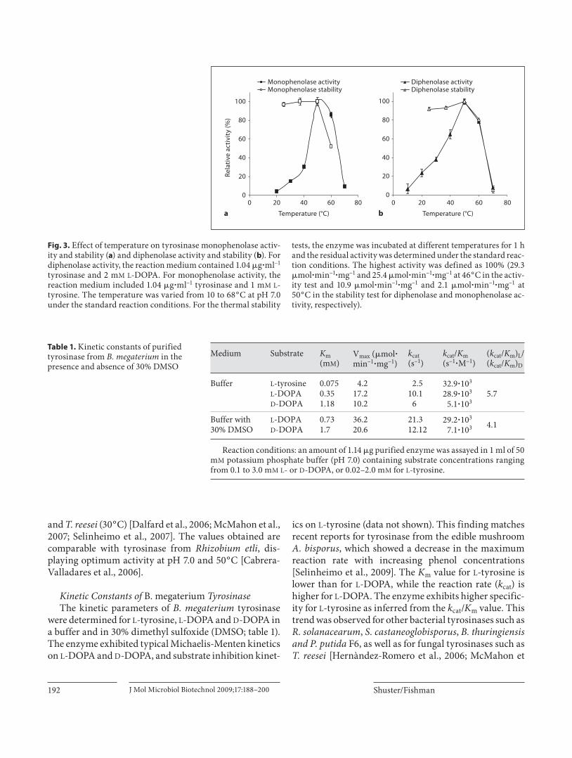
tyrosinase were investigated by following dopachrome appearance at 475 nm, using 1 m M L -DOPA and 1 m M L -tyrosine as substrates. For monophenolase activity, the pH optimum was determined over a range of pH 4.0–10.0; and the pH range for diphenolase activity was 4.0–7.0, as L -DOPA spontaneously converts to dopachrome at pH values above 7.5 (data not shown). The optimum pH of the enzyme was found to be 7.0 for the monophenolase and diphenolase activities, corresponding to that of other bacterial tyrosinases such as P. putida F6 (pH 7.0), Strep-tomyces sp. (pH 6.8) [McMahon et al., 2007] and fungal tyrosinase from Pycnoporus strains [Halaouli et al., 2005], while the optimum pH values for the diphenolase activi-ties of B. thuringiensis and T. roseum were 9.0 and 9.5, respectively [Kong et al., 2000; Liu et al., 2004]. In addi-tion, in both enzymatic reactions, the enzyme exhibited less than 50% of its maximum activity below pH 6.0 (data not shown). The effect of temperature on purified B. megaterium tyrosinase was determined at temperatures ranging from 10 to 70 ° C. The thermal stability of the en-zyme was determined at 25, 37, 50, 60 and 70 ° C by incu-bating the enzyme at the different temperatures for 1 h and determining the residual activity in 50 m M potas-sium phosphate buffer (pH 7.0) at room temperature (25 ° C). According to the results presented in figure 3 , the optimal temperature for this enzyme is approximately 50 ° C. The enzyme was fully stable below 50 ° C, but rap-idly lost its activity at 70 ° C. Thus, B. megaterium tyrosi-nase is quite stable at high temperatures in comparison with tyrosinases from other sources such as P. putida F6
250
150
100
75
50
37
25
20
1 2 3 4kDa
Fig. 2. Electrophoresis of B. megaterium tyrosinase under the de-naturing conditions of SDS-PAGE (12% acryl amide gel). Gel lane 1: Molecular mass marker; gel lane 2: crude cell extract from E. coli BL21(DE3)pET9d/tyr; gel lane 3: His6-tagged tyrosinase elut-ed from N i (II)-bound affinity column; gel lane 4: His6-tagged tyrosinase eluted from Superdex 200 26/10 gel filtration column. It is estimated that following the N i (II)-bound affinity column purification step tyrosinase accounts for approximately 95% of protein in the solution.
Shuster/Fishman
J Mol Microbiol Biotechnol 2009;17:188–200192
and T. reesei (30 ° C) [Dalfard et al., 2006; McMahon et al., 2007; Selinheimo et al., 2007]. The values obtained are comparable with tyrosinase from Rhizobium etli , dis-playing optimum activity at pH 7.0 and 50 ° C [Cabrera-Valladares et al., 2006].
Kinetic Constants of B. megaterium Tyrosinase The kinetic parameters of B. megaterium tyrosinase
were determined for L -tyrosine, L -DOPA and D -DOPA in a buffer and in 30% dimethyl sulfoxide (DMSO; table 1 ). The enzyme exhibited typical Michaelis-Menten kinetics on L -DOPA and D -DOPA, and substrate inhibition kinet-
ics on L -tyrosine (data not shown). This finding matches recent reports for tyrosinase from the edible mushroom A. bisporus , which showed a decrease in the maximum reaction rate with increasing phenol concentrations [Selinheimo et al., 2009]. The K m value for L -tyrosine is lower than for L -DOPA, while the reaction rate ( k cat ) is higher for L -DOPA. The enzyme exhibits higher specific-ity for L -tyrosine as inferred from the k cat / K m value. This trend was observed for other bacterial tyrosinases such as R. solanacearum , S. castaneoglobisporus , B. thuringiensis and P. putida F6, as well as for fungal tyrosinases such as T. reesei [Hernàndez-Romero et al., 2006; McMahon et
0
20
40
60
80
100
0 20 40 60 80
Temperature (°C)a b
0 20 40 60 80
Temperature (°C)
Rela
tive
act
ivit
y (%
)
Monophenolase activityMonophenolase stability
0
20
40
60
80
100
Diphenolase activityDiphenolase stability
Fig. 3. Effect of temperature on tyrosinase monophenolase activ-ity and stability ( a ) and diphenolase activity and stability ( b ). For diphenolase activity, the reaction medium contained 1.04 � g � ml –1 tyrosinase and 2 m M L -DOPA. For monophenolase activity, the reaction medium included 1.04 � g � ml –1 tyrosinase and 1 m M L -tyrosine. The temperature was varied from 10 to 68 ° C at pH 7.0 under the standard reaction conditions. For the thermal stability
tests, the enzyme was incubated at different temperatures for 1 h and the residual activity was determined under the standard reac-tion conditions. The highest activity was defined as 100% (29.3 � mol � min –1 � mg –1 and 25.4 � mol � min –1 � mg –1 at 46 ° C in the activ-ity test and 10.9 � mol � min –1 � mg –1 and 2.1 � mol � min –1 � mg –1 at 50 ° C in the stability test for diphenolase and monophenolase ac-tivity, respectively).
Medium Substrate Km(mM)
Vmax (�mol�min–1�mg–1)
kcat(s–1)
kcat/Km(s–1�M–1)
(kcat/Km)L/(kcat/Km)D
Buffer L-tyrosine 0.075 4.2 2.5 32.9�103
5.7L-DOPA 0.35 17.2 10.1 28.9�103
D-DOPA 1.18 10.2 6 5.1�103
Buffer with L-DOPA 0.73 36.2 21.3 29.2�1034.130% DMSO D-DOPA 1.7 20.6 12.12 7.1�103
Reaction conditions: an amount of 1.14 �g purified enzyme was assayed in 1 ml of 50 mM potassium phosphate buffer (pH 7.0) containing substrate concentrations ranging from 0.1 to 3.0 mM L- or D-DOPA, or 0.02–2.0 mM for L-tyrosine.
Table 1. Kinetic constants of purified tyrosinase from B. megaterium in the presence and absence of 30% DMSO

Tyrosinase from Bacillus megaterium J Mol Microbiol Biotechnol 2009;17:188–200 193
al., 2007; Selinheimo et al., 2006]. The K m value of B. megaterium tyrosinase for L -DOPA (0.35 m M ) is similar to the reported values for tyrosinase from T. roseum (0.18 m M ) [Cabrera-Valladares et al., 2006; Kong et al., 2000] or P. putida F6 (0.33 m M ) [McMahon et al., 2007], but lower than the value reported for the tyrosinase from B. thuringiensis (0.768 m M ) [Liu et al., 2004], S. castaneo-globisporus (8.1 m M ) [Kohashi et al., 2004] or T. reesei (3 m M ) [Selinheimo et al., 2006]. The K m value of B. mega-terium tyrosinase for L -tyrosine (0.075 m M ) is similar to tyrosinase from P. putida F6 (0.23 m M ) [McMahon et al., 2007] and mice (0.09–0.11 m M ) [Garcia-Molina et al., 2006; Olivares et al., 2002], but lower than the value re-ported for tyrosinase from Vibrio tyrosinaticus (3.1 m M ) [Cabrera-Valladares et al., 2006]. Thus, these results show that tyrosinase from B. megaterium displays a K m value
for L -DOPA and L -tyrosine similar to that of enzymes from other organisms. However, the k cat value of B. mega-terium tyrosinase for L -DOPA (10.1 s –1 ) is lower than the values reported for other tyrosinases [Garcia-Molina et al., 2007]. Tyrosinase from A. bisporus has a k cat value 1 order of magnitude higher (107 s –1 ), and tyrosinase from Streptomyces glaucescens is 2 orders of magnitude more active (1,440 s –1 ). This may be indicative of steric hin-drance in the catalytic site of B. megaterium tyrosinase [Olivares et al., 2002].
Substrate Specificity and Inhibitors Several monophenols, dihydroxyphenols and trihy-
droxyphenols were used to investigate the substrate spec-ificity of the enzyme ( table 2 ). B. megaterium tyrosinase had very low activity towards monohydroxyphenols but substantial activity towards dihydroxyphenols. In addi-tion, L -isomers of DOPA and tyrosine were much better substrates for B. megaterium tyrosinase than the corre-sponding D -isomers ( table 2 ). Very low oxidation of o -coumaric acid is in accordance with the literature regard-ing orthophenolic compounds, which are poor substrates for tyrosinases presumably because of steric hindrance [Selinheimo et al., 2006, 2007]. Low oxidation activity to-wards catechol is remarkably different from most tyrosi-nases from other sources such as S. glaucesens and T. rose-um [Kong et al., 2000; Lerch and Ettinger, 1972], but is comparable with reports for B. thuringiensis tyrosinase [Liu et al., 2004]. The results may denote differences in the substrate binding pockets of these groups of tyrosi-nases [Selinheimo et al., 2007].
Table 2. Substrate specificity of B. megaterium tyrosinase
Substrate (2.5 mM) Relative activity
MonohydroxyphenolsL-Tyrosine 35.6D-Tyrosine 20.3DL-Tyrosine 20.6p-Coumaric acid 6.1o-Coumaric acid <1p-Hydroxybenzoic acid <1Tyramine 2.5Phenol <13-Aminophenol 3.9Vanillic acid 0.01-Naphthol 0.02,6-Dimethoxyphenol 0.0
DihydroxyphenolsL-DOPA 100.0D-DOPA 34.0DL-DOPA 78.0Caffeic acid 60.9(=)-Catechin 60.8(8)-Catechin 58.7Catechol 10.3Chlorogenic acid 20.1Epicatechin 23.4Resorcinol 0.0
TrihydroxyphenolsPyrogallol 99.9Phloroglucin 1.6
Values represent percentages. Reaction conditions: B. megate-rium tyrosinase activity was measured using 1.88 �g enzyme and 2.5 mM of each substrate in 50 mM potassium phosphate buffer (pH 7.0). Relative activity is presented with the activity on L-DOPA set as 100%.
Table 3. Effect of inhibitors on B. megaterium tyrosinase
Inhibitor ConcentrationmM
Inhibition%
EDTA 1.0 270.1 20
Glutathione 1.0 1000.1 42
�-Mercaptoethanol 1.0 1000.1 50
Sodium diethyldithiocarbamate 1.0 1000.1 60
Reaction conditions: Purified enzyme was incubated with each inhibitor (0.1 or 1 mM) at room temperature (25° C) for 2 min and the activity assay was initiated by the addition of 2.5 mML-DOPA.

Shuster/Fishman
J Mol Microbiol Biotechnol 2009;17:188–200194
To further characterize the enzyme, various potential inhibitors of B. megaterium tyrosinase were examined. The inhibition was determined by measuring the enzy-matic activity on 2.5 m M L -DOPA in the presence of the inhibitor at two concentrations ( table 3 ). B. megaterium tyrosinase was inhibited strongly by glutathione, � -mer-captoethanol and sodium diethyldithiocarbamate, but EDTA did not inhibit the enzyme very efficiently. Thiol compounds, such as glutathione, inhibit the enzyme and may also affect the subsequent nonenzymatic reactions by reducing the quinones to diphenols (therefore retard-ing browning). These findings are comparable with other tyrosinases from bacterial, fungal and plant origins [Kong et al., 2000; Selinheimo et al., 2007]. Low inhibition of B. megaterium tyrosinase by EDTA is similar to other ty-rosinases from other organisms such as T. reesei [Selin-heimo et al., 2006; Selinheimo et al., 2007].
Effect of SDS on B. megaterium Tyrosinase Activity It has been reported that SDS, an anionic detergent, is
an activating agent of the enzyme. Recent experiments have confirmed that the activation is accompanied by an interaction between the detergent and the enzyme, re-sulting in a conformational change in the enzyme that improves the accessibility of the substrates to the active site without directly affecting its integrity [Gandia-Her-rero et al., 2005]. The effect of SDS on B. megaterium ty-rosinase was studied by incubating the enzyme solution with different concentrations of SDS (0.02–1%) at 50 ° C for 2 min and initiating the activity assay by adding 1 m M L -DOPA or L -tyrosine. The residual activity was deter-mined relative to L -DOPA or L -tyrosine activity without the SDS addition at 50 ° C (results not shown). The diphe-
nolase activity was increased by 20% in the presence of 0.02% SDS, while the presence of SDS concentrations above 0.5% inhibited the enzyme. The monophenolase activity was nearly constant in the presence of ! 0.1% SDS and deteriorated greatly at levels 1 0.5% similar to the di-phenolase activity. Tyrosinases from the South African clawed frog Xenopus laevis [Wittenberg and Triplett, 1985] and A. bisporus [Espin and Wichers, 1999] have been shown to be activated by detergents. Among bacte-rial tyrosinases, SDS activation was observed for Bacillus sp. tyrosinase [Dalfard et al., 2006] and tyrosinase from M. mediterranea [Lopez-Serrano et al., 2002], but was not useful for B. thuringiensis tyrosinase [Liu et al., 2004]. Further evaluation of this activation phenomenon on B. megaterium tryrosinase is currently being investigated in our lab.
Influence of Organic Solvents on B. megaterium Tyrosinase Activity, Stability and Enantioselectivity The effect of organic solvents on the activity of tyrosi-
nase from B. megaterium and mushroom tyrosinase was investigated using 1 m M L -DOPA as a substrate ( fig. 4 a). B. megaterium tyrosinase was activated in the presence of 10–30% of all miscible organic solvents examined. It re-tained full activity even in the presence of 70% ethanol and methanol. In contrast, mushroom tyrosinase lost its activity quite rapidly in the presence of the same solvents ( fig. 4 b). For instance, B. megaterium tyrosinase had 112 and 101% activity compared to that of the control in the presence of 50% ethanol and methanol, respectively ( fig. 4 a), while mushroom tyrosinase exhibited only 10 and 13% activity under the same conditions ( fig. 4 b). The kinetic constants were determined for the system con-
a
0
20
40
60
80
100
120
140
160
180
0
20
40
60
80
100
120
140
160
180
0 10 20 30 40 50 60 70 80Concentration of organic solvents (%) b
0 10 20 30 40 50 60Concentration of organic solvents (%)
Rela
tive
act
ivit
y (%
)
Ethanol Methanol 2-propanol Acetone DMSO
Fig. 4. Effect of organic solvents on the di-phenolase activity of B. megaterium tyros-inase ( a ) and commercial A. bisporus ty-rosinase ( b ). The reaction medium con-tained 1 m M L -DOPA and 1.1 � g � ml-1 B. megaterium tyrosinase or 0.5 � g � ml –1 A. bisporus tyrosinase. One hundred per-cent activity indicates the activity of each enzyme in 50 m M potassium phosphate buffer pH 7.0.

Tyrosinase from Bacillus megaterium J Mol Microbiol Biotechnol 2009;17:188–200 195
taining 30% DMSO in which the activation of tyrosinase was the most pronounced (170% activity; fig. 4 ). Both the K m and the k cat values increased 2-fold, thereby the selec-tivity factor, k cat / K m , remained unchanged ( table 1 ). In the literature, there is an example of a tyrosinase from Streptomyces sp. REN-21 which exhibited high stability in the presence of organic solvents compared with mush-room tyrosinase [Ito and Oda, 2000]. However, only 40–50% of the original activity was found in the presence of 50% ethanol or 30% DMSO, while B. megaterium tyrosi-nase showed 112 and 170% diphenolase activity, respec-tively.
The effect of organic solvents on the stability of B. megaterium and A. bisporus tyrosinase was investigated by incubating the enzyme solution at 30 ° C for 17 h with various concentrations of organic solvents followed by the measurement of the residual activity ( fig. 5 ). Under these conditions, B. megaterium tyrosinase was more sta-ble than A. bisporus tyrosinase in all of the tested condi-tions, although the activity in the buffers of both enzymes was reduced by 40% compared to the activity without in-cubation ( fig. 5 ). For example, B. megaterium tyrosinase had 44% activity to that of the nonincubated control in the presence of 40% DMSO ( fig. 5 a), while A. bisporus tyrosinase showed no activity under the same conditions ( fig. 5 b). Additionally, the enzyme retained its stability at methanol concentrations of 0–70%, which is very unusu-al for enzymes in general and was not seen with the sol-vent tolerant tyrosinase from Streptomyces sp. REN-21 [Ito and Oda, 2000]. High stability at 66% ethanol was reported for tyrosinase from grapes which lost only 4% diphenolase activity after 2 h of incubation [Valero et al., 1990]. The stability of B. megaterium tyrosinase at vari-
ous temperatures in the presence of 30% DMSO was also examined by incubating the enzyme at 50, 60 and 70 ° C for 1 h and subsequently determining the residual activ-ity. Under these conditions, B. megaterium tyrosinase was stable at 50 ° C (76% of the activity at buffer without DMSO) and rapidly lost its activity above this tempera-ture (data not shown). Thus, a combination of both 30% organic solvent and a high temperature are still tolerable for the enzyme.
In general, organic solvents can affect the enzymatic activity and specificity via interactions with water, en-zymes, substrates and products [Yang and Russell, 1996]. Kermasha et al. [2001] reported that among 100% metha-nol, acetone, heptanol and chloroform, the latter solvent was the only appropriate medium for commercial mush-room tyrosinase activity with catechin and vanillin as substrates since no activity was detected in the other sol-vents. In contrast, B. megaterium tyrosinase showed 97% and 48% diphenolase activity using L -DOPA as a sub-strate in the presence of 70% methanol and acetone, re-spectively. Mushroom tyrosinase exhibited higher activ-ity in pure chloroform compared to an aqueous medium using phenolic substrates such as chlorogenic acid, but it is essential to emphasize the immiscible nature of chlo-roform [Kermasha and Tse, 2000; Tse et al., 1997]. It is well documented that hydrophobic solvents are usually better than hydrophilic ones, as the latter have a greater tendency to strip tightly bound water from the enzyme molecule [Carrea et al., 1995; Gupta and Roy, 2004; Kil-banov, 2001]. However, our findings indicate that hydro-philic solvents (up to 70% in water) are suitable media for B. megaterium tyrosinase activity, supposedly due to its hydrophobic nature [Matoba et al., 2006]. Ogel et al.
a b
0102030405060708090
100
0102030405060708090
100
0 10 20 30 40 50 60 70Organic solvents concentrations (%)
0 10 20 30 40 50 60 70Organic solvents concentrations (%)
Resi
dua
l act
ivit
y (%
)
Ethanol Methanol DMSO
Fig. 5. Stability of B. megaterium ( a ) and commercial A. bisporus tyrosinase ( b ) in the presence of organic solvents. The reac-tion medium contained 1 m M L -DOPA and 1.75 � g � ml –1 B. megaterium tyrosi-nase or 0.9 � g � ml –1 A. bisporus tyrosinase, and was incubated at 30 ° C for 17 h. The residual activity was measured at 25 ° C. One hundred percent activity indicates the activity of each enzyme before incuba-tion.

Shuster/Fishman
J Mol Microbiol Biotechnol 2009;17:188–200196
[2006] reported that DMSO and ethanol were found to be beneficial for increasing the activity of a crude cell extract containing a phenol hydroxylase from Scytalidium ther-mophilum with activity on catechol, but not on L -tyro-sine. The identity of the enzyme was not elucidated; how-ever, these findings show that there are other oxidizing enzymes that exhibit a similar characteristic behavior as tyrosinase from B. megaterium .
B. megaterium tyrosinase was found to be stereospe-cific towards the L -enantiomer of tyrosine and DOPA ( ta-ble 2 ), similar to other tyrosinases from fungal and plant sources such as T. reesei, Pycnoporus sanguineus , A. bispo-rus , apples and potatos [Kawamura-Konishi et al., 2007; Selinheimo et al., 2006, 2007]. Tyrosinase from Strepto-myces sp. REN 21 was also highly selective towards L -DOPA and L -tyrosine [Ito and Oda, 2000]. The K m value of B. megaterium tyrosinase for the D -isomer was higher than for the L -isomer (1.18 vs . 0.35 m M , respectively), as was shown previously for tyrosinases from mushrooms, pears and strawberries. This observation is most likely due to the effect of a different spatial orientation of the D -isomer side chain [Espin et al., 1998a, b]. The V max val-ue of B. megaterium tyrosinase was higher for L -DOPA than for D -DOPA (17.2 � M � min –1 � mg –1 vs. 10.2 � M � min –1 � mg –1 , respectively), presumably due to better ori-entation and affinity of the more strongly bound isomer in the enzyme-substrate complex resulting in more rap-id electron-transfer steps during the catalytic cycle of the enzyme [Casella et al., 1991]. These findings are partly different from mushroom tyrosinase that exhibited lower K m values for the L -isomer than for D -isomer, but the V max values were comparable for both the L - and D -DOPA [Espin et al., 1998a]. It was suggested that the similar V max values resulted from the fact that the spatial orientation of the ring substituents did not affect the elec-tron donor capacity of the substrates which attack the copper atoms of the enzyme’s active site [Espin et al., 1998a, b]. Tyrosinase from Streptomyces sp. REN-21 showed a similar trend to B. megaterium tyrosinase, i.e. an increased K m value for D -DOPA accompanied by a de-crease in k cat value; however, the decline in selectivity ( k cat / K m ) was more dramatic (67-fold for Streptomyces tyrosinase vs. 6-fold for B. megaterium tyrosinase) [Ito and Oda, 2000].
Our findings also indicate that 30% DMSO influences the enantioselectivity of B. megaterium tyrosinase (5.7 in the absence of DMSO vs. 4.1 in the presence of DMSO as determined from the ratios of k cat / K m for the two enan-tiomers; table 1 ). To our knowledge, this is the first report to describe the influence of organic solvents on the enan-
tioselectivity of tyrosinases, although there are numer-ous reports describing the influence of organic solvents on the selectivity of other enzymes [Carrea et al., 1995; Intra et al., 2005; Kawashiro et al., 1997]. For example, enantioselectivity of � -chymotrypsin in the transesteri-fication of 3-hydroxy-2-phenylpropionate with propanol was changed from pro-S to pro-R simply by switching from 100% of one organic solvent to another [Wescott et al., 1996]. Another example is lipase from Pseudomonas cepacia which catalyzed hydrolysis of a prochiral diester and showed an opposite enantiomeric preference in iso-propyl ether than in cyclohexane [Berglund, 2001]. Wescott et al. [1996] and Berglund [2001] ascribe the changes in enantioselectivity to the influence of the or-ganic solvent on the substrate desolvation. Additionally, the effect of the organic solvent on enzyme selectivity was also shown to be highly dependent on the nature of both the enzyme and the solvent [Kawashiro et al., 1997]. The tyrosinase examined in the present work did not change its enantioselectivity in the presence of 30% ethanol. The effect of 30% methanol was subtle and was most pro-nounced in DMSO, indicating that the type of solvent is indeed highly influential as suggested previously [Ka-washiro et al., 1997].
The newly isolated and characterized tyrosinase, showing up to 70% activation in the presence of water-miscible organic solvents, is currently being further eval-uated for various biotechnological applications.
Experimental Procedures
Chemicals L -DOPA was purchased from Acros (Geel, Belgium). D -DOPA,
DL -DOPA, L -tyrosine, D -tyrosine, DL -tyrosine , p -coumaric acid, p -hydroxybenzoic acid, vanillic acid, 2,6-dimethoxyphenol, (+)-catechin, ( 8 )-catechin, catechol, chlorogenic acid, epicatechin, resorcinol, phloroglucin, EDTA, glutathione, sodium diethyldi-thiocarbamate, kanamycin, and SDS were purchased from Sigma-Aldrich (Rehovot, Israel). o -Coumaric acid, phenol, 1-naphthol, caffeic acid, DMSO and pyrogallol were purchased from Merck (Whitehouse Station, N.J., USA). � -Mercaptoethanol was pur-chased from Spectrum (Gardena, Calif., USA). Methanol and eth-anol were purchased from Bio Labs (Jerusalem, Israel). Acetone was purchased from Frutarom (Haifa, Israel). 2-Propanol was purchased from J.T. Baker (Deventer, The Netherlands). All ma-terials used were of the highest purity available and were used without further purification.
Strains B. megaterium was isolated from soil and stored in our lab, as
described below. E. coli BL21 (DE3; Novagen, Darmstadt, Ger-many) was used as the host for plasmid pET9d. E. coli transfor-mants were grown in a Luria-Bertani medium containing 0.025

Tyrosinase from Bacillus megaterium J Mol Microbiol Biotechnol 2009;17:188–200 197
mg/ml kanamycin. A. bisporus tyrosinase (mushroom tyrosi-nase) was purchased from Sigma-Aldrich (Rehovot, Israel, cat. No. T3824).
Isolation of a Tyrosinase-Expressing Bacterial Strain Soil samples were collected from 10 different areas in Israel.
0.2 g of soil was dissolved in a 10-ml saline solution and a 1-ml solution was added to 25 ml of rich medium known to favor bac-terial growth over fungal growth (tryptone 1%, glucose 0.2% and NaCl 0.5%). The flasks were incubated at 30 ° C overnight and se-rial diluted samples were streaked on MMB selection plates con-taining (% w/v): KH 2 PO 4 (0.0351%), MgSO 4 � 7H 2 O (0.015%), (NH 4 ) 2 SO 4 (0.2%), glucose (0.2%), agar (1.5%) and 50 m M Tris buffer (pH 7.0). After sterilization, L -tyrosine (0.1%) and a trace elements solution (0.3%) were added to the medium. The traceelements solution contained (% w/v): CuSO 4 � 5H 2 O (0.02%), FeSO 4 � 7H 2 O (0.2%), MnSO 4 � H 2 O (0.015%), ZnSO 4 � 7H 2 O (0.014%), CoCl 2 � 6H 2 O (0.026%), NaMoO 4 � 2H 2 O (0.023%) and CaCl 2 � 2H 2 O (0.061%). The plates were incubated at 30 ° C for 5–6 days. Melanin production was recorded by the appearance of black or black-brown color colonies. The selected strains were iso-lated and streaked again on L -tyrosine-containing MMB plates and on similar plates without tyrosine (negative control). The iso-late which formed black colonies on tyrosine-plates and white colonies on plates without tyrosine was grown in a MMB liquid medium supplemented with 0.2 m M CuSO 4 at 30 ° C for 4 days. The broth was assayed for extracellular tyrosinase activity.
PCR Amplification and 16S rDNA Sequencing and Identification of the Bacterial Strain Genomic DNA was extracted from the isolated bact eria using
standard methods [Sambrook and Russell, 2001]. Universal 16S rDNA PCR forward primer (5 � -GAGAGTTTGATCCTGGCT-CAG-3 � ) and reverse primer (5 � -CTACGGCTACCTTGTTGTT-ACGA-3 � ) were used in the amplification of 16S rDNA genes [Dalfard et al., 2006]. A DNA thermal cycler ( Apollo AC401; CLP, San Diego, Calif., USA) was used and programmed as follows: (1) the 1st cycle consisting of 94 ° C for 5 min, 50 ° C for 2 min and 72 ° C for 3 min; (2) a run of 28 cycles with each cycle consisting of 45 s at 94 ° C, 90 s at 50 ° C and 3 min at 72 ° C; and (3) the last cycle consisting of 45 s at 94 ° C and 90 s at 50 ° C, followed by in-cubation at 72 ° C for 10 min. The amplification products were purified with a PCR clean-up kit (RBC, Taipei, Taiwan) and DNA sequencing was performed using the dideoxy chain termination technique (Multidisciplinary Laboratories, Technion, Haifa, Is-rael).
The obtained 16S rDNA sequence was deposited in GenBank under accession No. EU627691 for isolate VS1.
Biochemical characterization of the new strain was performed by Milouda Biological Industries Services (Akko, Israel).
Analytical Methods Conversion of L -tyrosine to L -DOPA was determined by high
performance liquid chromatography with an Agilent 1100-series instrument (Agilent Technologies, Santa Clara, Calif., USA) us-ing an Eclipse XDB C18 column (5 � m, 4.6 ! 150 mm; Agilent Technologies, Santa Clara, Calif., USA). An isocratic method comprising acetonitrile/water (0.1% formic acid) at a ratio of 2/98, respectively, was applied at 1 ml � min –1 for 13 min. A diode array detector was used at a fixed wavelength of 275 nm to monitor the
reaction. Twenty microliters of filtered samples were injected into the column and under these conditions; L -DOPA eluted at 4.8 min and L -tyrosine at 7 min.
Cloning and Overexpression of the tyr Gene from Bacillus megaterium The genomic DNA was extracted from B. megaterium by stan-
dard methods [Sambrook and Russell, 2001] and used as a tem-plate to amplify the tyr gene by PCR amplification. The putative sequence of B. megaterium tyrosinase was disclosed to us by Dr. Jibin Sun from the Helmholtz Centre for Infection Research and a pair of primers were designed to amplify the tyr gene containing a C-terminal His6-tag. The forward primer ( Nco I front) contain-ing the Nco I restriction site (underlined) was 5 � -GAGGTTAAA C-CATGG GTAACAAGTATAGAG TTAGAAAAAACG-3 � . The re-verse primer containing the 6-His and BglII restriction site (un-derlined) was 5 � -CTGCTGTTTCT AGATCT GGTTAATGGTG-GTGATGGTGATGTGAGGAACGTTTTGATTTTC-3. Primer Nco l front generates an Nco I restriction site within the tyr gene by changing the 2nd codon AGT to GGT, creating a serine to glycine change at the 2nd position in the translated product of the modi-fied tyr gene. The PCR reaction was performed with BioTaq TM DNA polymerase (Bioline, London, UK) and carried out in a 200- � l reaction using a DNA thermal cycler (Apollo AC401; CLP, San Diego, Calif., USA). The PCR program had an initial denatur-ation step of 5 min at 94 ° C, followed by 30 cycles of 30 s at 94 ° C, 45 s at 52 ° C and 2 min at 72 ° C. This was followed by a final elon-gation step of 8 min at 72 ° C. The PCR product was purified using a PCR purification kit (Genomed GmbH, Löhne, Germany). The PCR product and the vector pET9d (Novagen, Darmstadt, Ger-many) were digested with Nco I and Bgl II, or with Nco I and Bam H I (Takara, Otsu, Japan), respectively, and then were ligated with T4 DNA ligase (Promega, Madison, Wisc., USA). The ligated plas-mid pET9d/tyr was transformed into competent E. coli BL21 (DE3) using GeneZapper (Bio-Rad, Hercules, Calif., USA).
Purification of Active His6-tag Tyrosinase The His 6 -tagged tyrosinase was purified in one step using an
Ni(II)-bound affinity column (HisTrap HP; Amersham Biosci-ences, Giles, UK). E. coli BL21 (DE3) cells harboring pET9d/tyr were grown overnight at 37 ° C in 0.5 liter of TB medium (tryptone 1.2%, yeast extract 2.4%, glycerol 0.4% and potassium phos-phate buffer 89 m M ). The cells were harvested by centrifugation (8,000 g for 10 min at room temperature) and yielded 15.6 g of wet pellets, suspended in a binding buffer (20 m M sodium phosphate buffer, pH 7.5, 500 m M NaCl; and 20 m M imidazole) and then treated twice in a pressure cell press (French Press, Spectronic Instruments Inc., Rochester, N.Y., USA). The cell debris was re-moved by centrifugation (16,000 g for 20 min at 15 ° C), whereas the supernatant was applied to the Ni(II)-bound affinity column (previously charged with a Ni ion and equilibrated with a binding buffer).
Elution was performed with an appropriate buffer (20 m M so-dium phosphate buffer, pH 7.5; 500 m M NaCl; and 500 m M imid-azole). The fractions containing tyrosinase were collected and dialyzed against a phosphate buffer at 4 ° C (50 m M sodium phos-phate buffer, pH 6.5, 0.02% sodium azide, 0.01 m M CuSO 4 ). En-zyme solutions were stored at –20 ° C without loss of activity.

Shuster/Fishman
J Mol Microbiol Biotechnol 2009;17:188–200198
Gel Filtration Analysis Gel filtration analysis was performed using a Superdex 200
26/10 column (Pharmacia, Giles, UK). � -Amylase (200 kDa), bo-vine serum albumin (66 kDa), carbonic anhydrase (29 kDa) and cytochrome c (12 kDa; Sigma-Aldrich, Rehovot, Israel) were used as marker proteins for determining molecular mass.
Tyrosinase Activity Assay Tyrosinase activity was assayed using L -DOPA and L-tyrosine
as substrates [Kohashi et al., 2004]. An aliquot of the purified en-zyme solution or liquid broth containing an extracellular enzyme was added to 50 m M potassium phosphate buffer pH 6.5 contain-ing 2 or 1 m M L -DOPA or L -tyrosine and the formation of L -dopa-chrome ( � = 3600) was monitored by measuring the absorbance at 475 nm (Ultrospec 2100pro; GE Healthcare, Giles, UK). The total volume of the reaction mixture was 1 ml. Control experi-ments without an enzyme were performed with all the substrates. The rate of dopachrome formation was defined as the slope of the linear zone of absorbance versus the time plot. Specific activity was calculated as the ratio of the conversion rate and the total protein content was determined by the Bradford analysis method, with bovine serum albumin as a calibration standard (Bio-Rad Protein Assay Kit). All measurements were carried out in tripli-cate.
Influence of pH and Temperature on Tyrosinase Activity, Substrate Specificity, Inhibitor Studies and SDS Effect Unless otherwise stated, activity was measured at room tem-
perature (25 ° C) by using the colorimetric assay described above. The reaction medium (1.0 ml) contained 50 m M potassium phos-phate buffer pH 7.0 and 1.88 � g � ml –1 of the enzyme with 1 m M L -tyrosine or 1 m M L -DOPA for monophenolase and diphenolase activities, respectively. Other conditions and reagents are detailed in the text and figure legends. All experiments were performed in triplicate.
The enzymatic activity at various pH values was determined by using the following buffers at 50 m M : acetic acid-sodium ace-tate (pH 4.0–5.0), potassium phosphate (pH 6.0–7.0) and glycine-NaOH (pH 8.0–10.0). For monophenolase activity, the reaction medium contained 2.08 � g � ml –1 tyrosinase and the pH optimum was determined over a range of pH 4.0–10.0, while for dipheno-lase activity, the reaction medium included 1.04 � g � ml –1 tyrosi-nase and the pH range was 4.0–7.0, due to spontaneous oxidation of the diphenol at alkaline pH.
Kinetic Analyses The values of K M and V max for the tyrosinase were determined
using the colorimetric assay described above (1 ml of final volume of 50 m M potassium phosphate buffer pH 7.0) employing 1.14 � g of a purified enzyme with increasing substrate concentrations ranging from 0.1–3.0 m M for L -DOPA and D-DOPA, or 0.02–2.00 m M for L -tyrosine. Measurements were performed in tetra-plicate. The Michaelis-Menten curves for determination of the kinetic constants were obtained with Sigmaplot 11.0 (Systat Soft-ware Inc., San Jose, Calif., USA).
Influence of Organic Solvents on Tyrosinase Activity, Stability and Enantioselectivity Determination of the organic solvents’ influence on B. mega-
terium tyrosinase activity was carried out using the colorimetric assay described above. Ethanol, methanol, 2-propanol, acetone and DMSO were added directly to the 1-ml reaction cuvette. Final organic solvent concentrations ranged from 0 to 80% for ethanol, methanol and 2-propanol; 0–70% for acetone, and 0–60% for DMSO. The activity on L -DOPA or D -DOPA in buffer without the presence of organic solvents was defined as 100%. To observe the possibility of spontaneous oxidation of L -DOPA, control experi-ments without an enzyme were performed with all the organic solvents. All assays were performed in tetraplicate.
For stability tests, the enzyme solution was kept at 30 ° C for17 h with various concentrations of organic solvents. Final or-ganic solvent concentrations ranged from 0 to 70% for ethanol and methanol, and 0–50% for DMSO. Reactions were measured at room temperature (25 ° C) using 96-well plates (final volume 200 � l) using a multiplate reader (OPTImax tunable microplate reader; Molecular Devices, Sunnyvale, Calif., USA). The activity on L -DOPA in buffer or organic solvents before incubation was defined as 100%. All assays were performed in triplicate.
Acknowledgements
This work was supported in part by the Israel Science Founda-tion (grant No. 535/07) and in part by the Mallat Family Fund. We would like to thank Dr. Jonathan Kuhn and Dr. SmadarShulami for their helpful advice.
References
Abhijith KS, Kumar PV, Kumar MA, Thakur MS: Immobilised tyrosinase-based biosen-sor for the detection of tea polyphenols. Anal Bioanal Chem 2007; 389: 2227–2234.
Ates S, Cortenlioglu E, Bayraktar E, Mehmeto-glu U: Production of L -DOPA using Cu-algi-nate gel immobilized tyrosinase in a batch and packed bed reactor. Enzyme Microb Technol 2007; 40: 683–687.
Berglund P: Controlling lipase enantioselectivi-ty for organic synthesis. Biomol Eng 2001; 18: 13–22.
Burton SG: Oxidizing enzymes as biocatalysts. Trends Biotechnol 2003a;21: 543–549.
Burton SG: Laccases and phenol oxidases in or-ganic synthesis – a review. Curr Org Chem 2003b;7: 1317–1331.
Cabrera-Valladares N, Martinez A, Pinero S,Lagunas-Munoz HV, Tinoco R, Anda R, Vazquez-Duhalt R, Bolivar F, Gosset G: Ex-pression of the melA gene from Rhizobium etli CFN42 in Escherichia coli and character-ization of the encoded tyrosinase. Enzyme Microb Technol 2006; 38: 772–779.
Carrea G, Ottolina G, Riva S: Role of solvents in the control of enzyme selectivity in organic media. Trends Biotechnol 1995; 13: 63–70.
Casella L, Gullotti M, Poli S, Bonfa M, Ferrari RP, Marchesini A: Spectroscopic and bind-ing studies on the stereoselective interaction of tyrosine with horseradish peroxidase and lactoperoxidase. Biochem J 1991; 279(part 1):245–250.

Tyrosinase from Bacillus megaterium J Mol Microbiol Biotechnol 2009;17:188–200 199
Chen T, Embree HD, Wu LQ, Payne GF: In vitro protein-polysaccharide conjugation: tyrosi-nase-catalyzed conjugation of gelatin and chitosan. Biopolymers 2002; 64: 292–302.
Claus H, Decker H: Bacterial tyrosinases. Syst Appl Microbiol 2006; 29: 3–14.
Dalfard AB, Kajeh K, Soudi MR, Naderi-Manesh H, Ranjbar B, Sajedi RH: Isolation and bio-chemical characterization of laccase and ty-rosinase activities in a novel melanogenic soil bacterium. Enzyme Microb Technol 2006; 39: 1409–1416.
Decker H, Tuczek F: Tyrosinase/catecholoxidase activity of hemocyanins: structural basis and molecular mechanism. Trends Biochem Sci 2000; 25: 392–397.
Duran N, Esposito E: Potential applications of oxidative enzymes and phenoloxidase-like compounds in wastewater and soil treat-ment: a review. Appl Catal B 2000; 28: 83–99.
Duran N, Rosa MA, D’Annibale A, Gianfreda L: Applications of laccases and tyrosinases (phenoloxidases) immobilized on different supports: a review. Enzyme Microb Technol 2002; 31: 907.
Espin JC, Wichers HJ: Activation of a latent mushroom ( Agaricus bisporus ) tyrosinase isoform by sodium dodecyl sulfate (SDS): ki-netic properties of the SDS-activated iso-form. J Agric Food Chem 1999; 47: 3518–3525.
Espin JC, Garcia-Ruiz PA, Tudela J, Garcia-Ca-novas F: Study of stereospecificity in mush-room tyrosinase. Biochem J 1998a;331: 547–551.
Espin JC, Garcia-Ruiz PA, Tudela J, Garcia-Ca-novas F: Study of stereospecificity in pear and strawberry polyphenol oxidases. J Agric Food Chem 1998b;46: 2469–2473.
Espin JC, Soler-Rivas C, Cantos E, Tomas-Bar-beran FA, Wichers HJ: Synthesis of the anti-oxidant hydroxytyrosol using tyrosinase as biocatalyst. J Agric Food Chem 2001; 49: 1187–1193.
Gandia-Herrero F, Jimenez-Atienzar M, Ca-banes J, Garcia-Carmona F, Escribano J: Dif-ferential activation of a latent polyphenol oxidase mediated by sodium dodecyl sulfate. J Agric Food Chem 2005; 53: 6825–6830.
Garcia-Molina F, Munoz JL, Garcia-Ruız PA, Rodriguez-Lopez JN, Garcia-Canovas F, Tudela J: A further step in the kinetic char-acterisation of the tyrosinase enzymatic sys-tem. J Math Chem 2006; 41: 393–406.
Garcia-Molina F, Munoz JL, Varon R, Rodri-guez-Lopez JN, Garcia-Canovas F, Tudela J: A review on spectrophotometric methods for measuring the monophenolase and di-phenolase activities of tyrosinase. J Agric Food Chem 2007; 55: 9739–9749.
Girelli AM, Mattei E, Messina A: Phenols re-moval by immobilized tyrosinase reactor in on-line high performance liquid chromatog-raphy. Anal Chim Acta 2006; 580: 271–277.
Gupta MN, Roy I: Enzymes in organic media: forms, functions and applications. Eur J Bio-chem 2004; 271: 2575–2583.
Halaouli S, Asther M, Kruus K, Guo L, Hamdi M, Sigoillot JC, Asther M, Lomascolo A: Characterization of a new tyrosinase from Pycnoporus species with high potential for food technological applications. J Appl Mi-crobiol 2005; 98: 332–343.
Halaouli S, Asther M, Sigoillot JC, Hamdi M, Lo-mascolo A: Fungal tyrosinases: new pros-pects in molecular characteristics, bioengi-neering and biotechnological applications. J Appl Microbiol 2006; 100: 219–232.
Hernàndez-Romero D, Sanchez-Amat A, Solano F: A tyrosinase with an abnormally high ty-rosine hydroxylase/dopa oxidase ratio. FEBS J 2006; 273: 257–270.
Intra A, Nicotra S, Riva S, Danieli B: Significant and unexpected solvent influence on the se-lectivity of laccase-catalyzed coupling of tet-rahydro-2-naphthol derivatives. Adv Synth Catal 2005; 347: 973–977.
Ito M, Oda K: An organic solvent resistant ty-rosinase from Streptomyces sp. REN-21: pu-rification and characterization. Biosci Bio-technol Biochem 2000; 64: 261–267.
Katz E, Thompson CJ, Hopwood DA: Cloning and expression of the tyrosinase gene from Streptomyces antibioticus in Streptomyces lividans . J Gen Microbiol 1983; 129: 2703–2714.
Kawamura-Konishi Y, Tsuji M, Hatana S, Asa-numa M, Kakuta D, Kawano T, Mukouyama EB, Goto H, Suzuki H: Purification, charac-terization, and molecular cloning of tyrosi-nase from Pholiota nameko . Biosci Biotech-nol Biochem 2007; 71: 1752–1760.
Kawashiro K, Sugahara H, Sugiyama S, Hayashi H: Effect of organic solvents on enantioselec-tivity of protease catalysis. Biotechnol Bio-eng 1997; 53: 26–31.
Kermasha S, Tse M: Biocatalysis of tyrosinase in chloroform medium, using selected phenolic substrates. J Chem Technol Biotechnol 2000; 75: 475–483.
Kermasha S, Bao H, Bisakowski B: Biocatalysis of tyrosinase using catechin as substrate in selected organic solvent media. J Mol Catal B Enzym 2001; 11: 929–938.
Kilbanov AM: Improving enzymes by using them in organic solvents. Nature 2001; 409: 241–246.
Kohashi PY, Kumagai T, Matoba Y, Yamamoto A, Maruyama M, Sugiyama M: An efficient method for the overexpression and purifica-tion of active tyrosinase from Streptomyces castaneoglobisporus . Protein Expr Purif 2004; 34: 202–210.
Kong KH, Hong MP, Choi SS, Kim YT, Cho SH: Purification and characterization of a highly stable tyrosinase from Thermomicrobium roseum . Biotechnol Appl Biochem 2000; 31: 113–118.
Lerch K, Ettinger L: Purification and character-ization of a tyrosinase from Streptomyces glaucescens . Eur J Biochem 1972; 31: 427–437.
Liu N, Zhang T, Wang YJ, Huang YP, Ou JH, Shen P: A heat inducible tyrosinase with dis-tinct properties from Bacillus thuringiensis . Lett Appl Microbiol 2004; 39: 407–412.
Lopez-Serrano D, Sanchez-Amat A, Solano F: Cloning and molecular characterization of a SDS-activated tyrosinase from Marinomo-nas mediterranea. Pigment Cell Res 2002; 15: 104–111.
Martinez MV, Whitaker JR: The biochemistry and control of enzymatic browning. Trends Food Sci Technol 1995; 6: 195–202.
Matoba Y, Kumagai T, Yamamoto A, Yoshitsu H, Sugiyama M: Crystallographic evidence that the dinuclear copper center of tyrosinase is f lexible during catalysis. J Biol Chem 2006; 281: 8981–8990.
McMahon AM, Doyle EM, Brooks S, O’Connor KE: Biochemical characterisation of the co-existing tyrosinase and laccase in the soil bacterium Pseudomonas putida F6. Enzyme Microb Technol 2007; 40: 1435–1441.
Ogel ZB, Yuzugullu Y, Mete S, Bakir U, Kaptan Y, Sutay D, Demir AS: Production, proper-ties and application to biocatalysis of a novel extracellular alkaline phenol oxidase from the thermophilic fungus Scytalidium ther-mophilum . Appl Microbiol Biotechnol 2006; 71: 853–862.
Olivares C, Garcia-Borron JC, Solano F: Identi-fication of active site residues involved in metal cofactor binding and stereospecific substrate recognition in mammalian tyrosi-nase: implications to the catalytic cycle. Bio-chemistry 2002; 41: 679–686.
Rasko DA, Ravel J, Okstad OA, Helgason E, Cer RZ, Jiang L, Shores KA, Fouts DE, Tourasse NJ, Angiuoli SV, Kolonay J, Nelson WC, Kolsto AB, Fraser CM, Read TD: The ge-nome sequence of Bacillus cereus ATCC 10987 reveals metabolic adaptations and a large plasmid related to Bacillus anthracis pXO1. Nucleic Acids Res 2004; 32: 977–988.
Ruan L, He W, He J, Sun M, Yu Z: Cloning and expression of mel gene from Bacillus thurin-giensis in Escherichia coli . Antonie Van Leeuwenhoek 2005; 87: 283–288.
Sambrook J, Russell DW: Molecular Cloning: A Laboratory Manual, ed 3. New York, Cold Spring Harbor Laboratory Press, 2001.
Schmid A, Dordick JS, Hauer B, Kiener A, Wub-bolts M, Witholt B: Industrial biocatalysis today and tomorrow. Nature 2001; 409: 258–268.
Selinheimo E, Gasparetti C, Mattinen ML, Stef-fensen CL, Buchert J, Kruus K: Comparison of substrate specificity of tyrosinases from Trichoderma reesei and Agaricus bisporus . Enzyme Microb Technol 2009; 44: 1–10.

Shuster/Fishman
J Mol Microbiol Biotechnol 2009;17:188–200200
Selinheimo E, Saloheimo M, Ahola E, Wester-holm-Parvinen A, Kalkkinen N, Buchert J, Kruus K: Production and characterization of a secreted, C-terminally processed tyrosi-nase from the filamentous fungus Trichoder-ma reesei . FEBS J 2006; 273: 4322–4335.
Selinheimo E, NiEidhin D, Steffensen C, Nielsen J, Lomascolo A, Halaouli S, Record E, O’Beirne D, Buchert J, Kruus K: Comparison of the characteristics of fungal and plant ty-rosinases. J Biotechnol 2007; 130: 471–480.
Seo SY, Sharma VK, Sharma N: Mushroom ty-rosinase: recent prospects. J Agric Food Chem 2003; 51: 2837–2853.
Sun J, Wang W, Hundertmark C, Zeng AP, Jahn D, Deckwer WD: A protein database con-structed from low-coverage genomic se-quence of Bacillus megaterium and its use for accelerated proteomic analysis. J Biotechnol 2006; 124: 486–495.
Tanimoto de Albuquerque YD, Ferreira LF: Am-perometric biosensing of carbamate and or-ganophosphate pesticides utilizing screen-printed tyrosinase-modified electrodes. Anal Chim Acta 2007; 596: 210–221.
Tse M, Kermasha S, Ismail A: Biocatalysis by ty-rosinase in organic solvent media: a model system using catechin and vanillin as sub-strates. J Mol Catal B Enzym 1997; 2: 199–213.
Valero E, Varon R, Garcia-Carmona F: Inhibi-tion of grape polyphenol oxidase by several natural aliphatic alcohols. J Agric Food Chem 1990; 38: 1097–1100.
Wescott CR, Noritomi H, Klibanov AM: Ratio-nal Control of Enzymatic Enantioselectivity through solvation thermodynamics. J Am Chem Soc 1996; 118: 10365–10370.
Westerholm-Parvinen A, Selinheimo E, Boer H, Kalkkinen N, Mattinen M, Saloheimo M: Expression of the Trichoderma reesei tyrosi-nase 2 in Pichia pastoris : isotopic labeling and physicochemical characterization. Pro-tein Expr Purif 2007; 55: 147–158.
Wittenberg C, Triplett EL: A detergent-activated tyrosinase from Xenopus laevis . II. Deter-gent activation and binding. J Biol Chem 1985; 260: 12542–12546.
Yang Z, Russell AJ: Fundamentals of non-aque-ous enzymology; in Koskinen A, Klibanov A (eds): Enzymatic Reactions in Organic Me-dia. London, Blackie Academic & Profes-sional, 1996, pp 43–65.
Yu B, Chang TMS: In vitro and in vivo enzyme studies of polyhemoglobin-tyrosinase. Bio-technol Bioeng 2004; 86: 835–841.