studies binding ofdifferent iron donors human serum ... · to removethe rivanol. then 1.5 litres...
TRANSCRIPT

Biochem. J. (1978) 173, 543-552Printed in Great Britain
Studies of the Binding of Different Iron Donors to Human SerumTransferrin and Isolation of Iron-Binding Fragments from the
N- and C-Terminal Regions of the Protein
By ROBERT W. EVANS and JOHN WILLIAMSMolecular Enzymology Laboratory, Department ofBiochemistry, University ofBristol,
Bristol BS8 1 TD, U.K.
(Received 25 November 1977)
1. Trypsin digestion of human serum transferrin partially saturated with iron(III)-nitrilotriacetate at pH5.5 or pH 8.5 produces a carbohydrate-containing iron-bindingfragment of mol.wt. 43000. 2. When iron(III) citrate, FeCI3, iron(II) ascorbate and(NH4)2SO4,FeSO4 are used as iron donors to saturate the protein partially, at pH 8.5,proteolytic digestion yields a fragment of mol.wt. 36000 that lacks carbohydrate.3. The two fragments differ in their antigenic structures, amino acid compositions andpeptide 'maps'. 4. The fragment with mol.wt. 36000 was assigned to the N-terminal regionof the protein and the other to the C-terminal region. 5. The distribution of iron in humanserum transferrin partially saturated with various iron donors was examined byelectrophoresis in urea/polyacrylamide gels and the two possible monoferric forms wereunequivocally identified. 6. The site designated A on human serum transferrin [Harris(1977) Biochemistry 16, 560-564] was assigned to the C-terminal region of the proteinand the B site to the N-terminal region. 7. The distribution of iron on transferrin inhuman plasma was determined.
Studies on the relative affinities of the twometal-binding sites of transferrins for iron haveproduced many conflicting results that are oftenfurther confused, unnecessarily, by the lack ofclarification of the source and species.For human serum transferrin, equilibrium dialysis
indicated that there was sequential occupancy of thesites (K1 = 400K2, where K1 is the association constantfor iron at the first site and K2 that for the second site)(Davis et al., 1962), although with the same techniqueAasa et al. (1963) concluded that the sites wereequivalent and independent. It was thought that inthe first determination true equilibrium was neverattained.A moving-boundary-electrophoretic study (Aisen
et al., 1966) in which (NH4)2SO4,FeSO4 was addedto human serum transferrin at pH 6.7 showed that theprotein can be separated into three species, with 0, 1
and 2 atoms of iron. The distribution of iron wasthought to support the theory of random ironbinding, but on careful re-examination the resultssuggest a sequential binding process. Gel isoelectricfocusing also resolves three forms of transferrin(Hovanessian & Awdeh, 1976); however, the distri-bution of iron, when added as iron(Ill)-nitrilotri-acetate to the protein in 1 mM-NaHCO3, agrees wellwith that predicted for equivalent sites. Both theseelectrophoretic methods fail to resolve the two
Vol. 173
possible monoferric forms of the protein, presumablybecause they have the same isoelectric point, aswith the two monoferric species of hen ovotransferrin(Williams, 1975). However, polyacrylamide-gel elec-trophoresis in the presence of 6M-urea (Makey &Seal, 1976) will resolve iron-free human serumtransferrin, the two monoferric transferrins and fullysaturated transferrin. Until now this technique hasnot been used to study in detail the distribution of ironwhen added to the iron-free protein.Lane (1975) has shown that it is possible to separate
two monoferric forms of human serum transferrinby chromatography on DEAE-cellulose at pH 7.9.From the distribution of iron in samples of proteinthat had been partially saturated with FeCl3 at pH 5.0and subsequently raised to pH 7.9 he concluded thatbinding had taken place randomly.
In spite of the results supporting the equivalenceof the sites, other workers have now shown that thetwo sites exhibit different pH-dependencies. One siteretains its iron down to about pH 5, whereas the otherloses its iron around pH6 (Princiotto & Zapolski,1975; Lestas, 1976). The site that retains its iron atlow pH, designated the A site, has also been shown tobind iron preferentially at pH 7.5 when iron(III)-nitrilotriacetate is used as the iron donor (Harris,1977a).
In view of the conflicting reports on the binding of
543
R. W. EVANS AND J. WILLIAMS
iron to human serum transferrin we set out toexamine the way in which various iron donors occupythe iron-binding sites on the protein. We have useda method, first reported by Williams (1974), for theisolation of the N-terminal fragment of hen ovo-transferrin, which involves partial iron saturation ofthe protein, proteolytic digestion of unoccupiedbinding sites and identification of the iron-bindingfragments remaining. We have then exploited theobserved differences in the order of binding ofdifferent iron donors to isolate and characterize thetwo iron-binding domains. With the electrophoreticmethod of Makey & Seal (1976) we have been able tocompare the distribution of iron when added totransferrin in different forms and under differentconditions and then been able to identify the mono-ferric form of transferrin present in normal blood.
Materials and Methods
Preparation ofhuman serum transferrin
Human serum transferrin was prepared by amodification ofthe method ofRoop & Putnam (1967)starting with Cohn IV fraction (Cohn et al., 1946)from outdated pooled human plasma. First 3 litresof Cohn IV fraction was dialysed twice for 12h at4°C against 10 litres of phosphate-buffered saline(Dulbecco & Vogt, 1954) and the precipitate removedby centrifugation at 6000g for 1 h at 4°C. Iron(III)-nitriloacetate and NaHCO3 were added to thesupernatant to final concentrations of 0.2 and 0.5 mmrespectively to saturate the transferrin and thesolution was then dialysed twice for 6h at 4°Cagainst water adjusted to pH9.4 with aq. NH3.An equal volume of 0.6% (w/v) rivanol (2-ethoxy-acridine-6,9-diamine lactate monohydrate) in 5mM-Tris/HCl, pH9.4, was added dropwise overnight at4°C to the stirred solution. The precipitate that formedwas removed by centrifugation at 6000g for I hat 4°C and the supernatant filtered through a pad ofpotato starch (BDH Chemicals, Poole, Dorset, U.K.),previously washed with 5 mM-Tris/HCI, pH 9.4,to remove the rivanol. Then 1.5 litres of a slurry ofDEAE-Sephadex A-50 equilibrated in 5 mM-Tris/HCI,pH8.8, was added to the filtrate, and after standingfor 2h at 20°C the suspension was filtered through aBuchner funnel. The ion-exchange resin was washedwith buffer until the eluate was protein-free and thenthe bound transferrin was eluted with 0.1 M-NaCl.After dialysis of the eluate overnight against 10 litresof 50mM-Tris/HCI, pH 8.8, the protein was adsorbedon a column (12cmx 30cm) of DEAE-SephadexA-50, in the same buffer, which was washed withbuffer until the eluate was free of protein as indicatedby A280. The transferrin was eluted with 0.1 M-NaCl,dialysed against water and freeze-dried. The yieldof protein was 20g.
Iron-free transferrinIron was removed from transferrin by the method
of Warner & Weber (1951) by using the anion-exchange resin Bio-Rad AGI (X2, 200-400 mesh,Cl- form) (Bio-Rad Laboratories, Richmond, CA,U.S.A.). The protein was shown to be iron-free by thelack of the characteristic absorption band of iron-transferrin at 470nm [A470 = 5 x 10³M-1 - cm-l (Aasaet al., 1963)].
Polyacrylamide-gel electrophoresisSamples for sodium dodecyl sulphate/polyacryl-
amide-gel electrophoresis were dissolved in a buffercontaining 5% (w/v) sodium dodecyl sulphate, 10%(v/v) glycerol, 1 % 2-mercaptoethanol, 0.01 % Bromo-phenol Blue and 5mM-sodium phosphate, pH 7.0,and heated for 3 min in a boiling-water bath. Whenunreduced samples were required the 2-mercapto-ethanol was omitted.
Electrophoresis was carried out on gels contain-ing 7.5% (w/v) acrylamide and 0.2% (w/v) NN'-methylenebisacrylamide in 0.1 % sodium dodecylsulphate and 0.05M-sodium phosphate, pH 7.0.
Gels were stained for protein with CoomassieBrilliant Blue R-250 by the method of Berg (1969)and for carbohydrate with the periodic acid/Schiff's-base stain (Zacharias et al., 1969). Molecularweights were determined by the use of a markermixture that contained ovotransferrin (mol.wt.80000), bovine serum albumin (mol.wt. 67000),lactate dehydrogenase (subunit mol.wt. 36000) andhorse heart cytochrome c (mol.wt. 12400).
Polyacrylamide-gel electrophoresis in 6M-ureawas carried out by a modification of the method ofMakey & Seal (1976) as described by Williams et al.(1978).
Before examination of transferrin in human plasmasamples the plasma was treated by the method ofMatson et al. (1966). To 0.1 ml of plasma was added0.3 ml of reservoir buffer and 0.5 ml of 0.6% (w/v)rivanol in reservoir buffer, and the precipitateremoved by centrifugation (lOOOg for 5min). Thisprocedure removes all plasma proteins except thefi- and y-globulins and simplifies the gel pattern.
Starch-gel electrophoresisFlat-bed starch-gel electrophoresis was carried
out in 13% (w/v) gels in the discontinuous buffersystem of Poulik (1957). Protein was stained with I %(w/v) Naphthalene Black (12B) in methanol/aceticacid/water (5:1:5, by vol.) and destained by washingin the same solvent.
Preparation of iron chelatesIron(III)-nitrilotriacetate and iron(II) ascorbate
were prepared by the method of Woodworth (1966).
1978
544

IRON BINDING TO HUMAN SERUM TRANSFERRIN
Iron(III) citrate was prepared with an iron/citratemolar ratio of 1:20. Under these conditions thepredominant species in solution is thought to beiron(III) dicitrate (Spiro et al., 1967).
Immunological methodsAn antiserum tohuman serum transferrin was raised
in a sheep. Antigen-antibody reactions were observedby the Ouchterlony (1958) method with undiluted anti-sera. Antigen solutions were either prepared bydissolving protein (1 mg/ml) in 0.1 M-sodium phos-phate, pH 7.0, or taken directly from column fractions.
Peptide 'maps'Protein was taken up in 5% (v/v) formic acid and
digested with pepsin (from pig stomach mucosa;Sigma Chemical Co., St. Louis, MO, U.S.A.) at anenzyme/substrate ratio of 1: 30 (w/w) for 15 h at 37°C.One-dimensional electrophoretic patterns were runat pH6.5 and 3.5. A sample (25nmol) of each digestwas applied directly to 2.54cm (1 in) of starting lineon Whatman 3MM paper and electrophoresis wascarried out for I h at 60V/cm. Peptides were stainedwith the cadmium acetate/ninhydrin reagent ofHeilmann et al. (1957) and in addition tyrosine- andtryptophan-containing peptides were detected by thespecific stains of Jepson & Smith (1953).
Amino acid analysisProtein samples were hydrolysed at 105°C with
5.7M-HCl in sealed evacuated tubes, after flushingseveral times with N2, for 24h and analysed on aRank Hilger Chromaspek J180 instrument.
Preparation ofhuman serum transferrin partially ironsaturated at pH8.5
Samples of human serum transferrin in 0.1 M-NaHCO3, pH 8.5, partially saturated with iron(III)-nitrilotriacetate, iron(II) ascorbate or (NH4)2SO4,-FeSO4 were prepared by addition of the calculatedamount of iron donor after titration of the iron.,freeprotein by monitoring the change in A470. As theuptake of iron by transferrin is a slow process wheniron(III) citrate and FeCI3 are used as iron donors(Bates et al., 1967; Bates & Schlabach, 1973) samplesof protein partially saturated by them were obtainedby addition of an amount based on the values for theother iron chelates. In all cases the partially iron-saturated samples were left for 15 h at 4°C to ensure aquantitative binding of the iron, which was confirmedby measurements of A470-
Preparation ofhuman serum transferrin partially iron-saturated with iron(III)-nitrilotriacetate atpH5.5
Iron-free protein (300mg) in 0ml of 0.1 M-Hepes[4 - (2 - hydroxyethyl) - 1 - piperazine - ethanesulphonicVol. 173
acid]/0.01M-NaHCO3, pH5.5, was 40%-iron-satur-ated with iron(lII)-nitrilotriacetate, as judged byA470. After standing at 20°C for 45min the solutionwas passed through a column (1cmx 10cm) ofBio-Rad AG 1-X2, which had been previouslyequilibrated with the same buffer, to remove anyunbound iron. The pH was then adjusted to 7.45 byaddition of 0.1 M-NaOH.
Digestion of partially iron-saturated human serumtransferrin
Partially iron-saturated samples of protein (50mg/ml) were digested with trypsin (bovine trypsin type XItreated with diphenylcarbamoyl chloride; Sigma)for 6h at 37°C at an enzyme/protein ratio of 1:30(w/w), followed by a second addition of the sameamount of enzyme and incubation for a further 15 h.To check that this procedure digested away all iron-free sites a sample of iron-free protein was treatedin the same way and shown to be unable to bind iron.
ResultsSusceptibility of iron-free and iron-saturated humanserum transferrin to proteolytic digestion
Treatment of iron-free transferrin with trypsin atpH 8.5 results in complete loss of its iron-bindingability, and sodium dodecyl sulphate/polyacrylamide-gel electrophoresis reveals that the protein has beendigested to low-molecular-weight peptides. Underthe same conditions the fully-saturated proteinretains its bound iron, as judged by its A470, andmoves as a single band of mol.wt. 80000 on sodiumdodecyl sulphate/polyacrylamide gels. However,when the fully saturated/protein was examined bysodium dodecyl sulphate-polyacrylamide-gel electro-phoresis in the presence of 2-mercaptoethanol,several bands were observed, suggesting that theprotein had undergone some internal cleavage butwas still held together by disulphide bridges. Thissuggestion is confirmed by starch-gel electrophoresis,which can detect charge heterogeneity in a givenprotein. Untreated iron-saturated transferrin movesas a single band on starch gel, but after treatment withtrypsin it gives multiple bands that move faster thanthe native protein. The effect of trypsin on fullysaturated human serum transferrin is different fromthat reported by Brock et al. (1976) on fullysaturated bovine serum transferrin. The bovineprotein is cleaved into separate iron-binding domains.
Proteolytic digestion of human serum transferrinpartially iron-saturated atpH 8.5
Samples of transferrin that had been 30%-iron-saturated, at pH8.5, with four different iron donors,iron(III)-nitrilotriacetate, FeCl3, iron(II) ascorbateand (NH4)2SO4,FeSO4, were treated with trypsin
S
545

R. W. EVANS AND J. WILLIAMS
| 1 ..
£r ::::1@M_
...: .:: .:.. :.: 0_
_ ._
_
::..R,- ......{-.%Eg..t .MN.{'3.
'.-.i szi:..:G a:...... : < :
:::_
OriginI
.SlX-" .,.rl
_i
*:
\:X#.t......... ..j....
E._
.E ._'...
_
..
S,-I94-a
(1) (2) (3) (4) (5) (6) (7) (8) (9) (10) (1 1) (12)
Fig. 1. Sodium dodecyl sulphate/polyacrylamide-gel electrophoresis of trypsin digests of 30%-iron-saturated humanserum transferrin
Samples of iron-free human serum transferrin in 0.1 M-NaHCO3, pH8.5, were 30%'-iron-saturated with iron(III)-nitrilotriacetate, FeCl3, iron(ll) ascorbate and (NH4)2SO4,/FeSO4 and digested with trypsin, as described in theMaterials and Methods section. Gels 1-6 were run in the absence of 2-mercaptoethanol and gels 7-12 were run inthe presence of 2-mercaptoethanol. Gels 1 and 7 are a marker mixture containing hen ovotransferrin (mol.wt. 80000),bovine serum albumin (mol.wt. 67000), lactate dehydrogenase (mol.wt. 36000) and cytochrome c (mol.wt. 12400).Gels 2 and 8 are samples of transferrin labelled with iron(III)-nitrilotriacetate. Gels 3 and 9 are samples labelledwith FeCl3. Gels 4 and 10 are samples labelled with iron(lI) ascorbate. Gels 5 and 11 are samples labelled with(NH4)2SO4,FeSO4. Gels 6 and 12 are samples of undigested transferrin.
to digest away unoccupied iron-binding sites. Thesodium dodecyl sulphate/polyacrylamide-gel-electro-phoretic pattern, in both the presence and theabsence of reducing agent, is shown in Fig. 1. As can
be seen from the unreduced samples (gels 2-5), thegel pattern of the sample partially saturated byusing iron(III)-nitrilotriacetate shows two mainspecies with mol.wts. 80000 and 43000, whereasthe three other samples have only a trace of thesecond species but two components with mol.wts.
33 500 and 36000. In the presence of reducing agent(Fig. 1, gels 8-11) the gel-electrophoretic pattern ofthe iron(III)-nitrilotriacetate sample is again differentfrom the other three; however, the interpretation iscomplicated by the fact that none of the main speciesobserved in the absence of reducing agent are singlechains. As the fully saturated protein has been shownto undergo limited proteolytic cleavage, it is notsurprising that the fragments do not have a single-chain structure.
1978
546
/,- 1_If.,:, 0.
Iir, I"'
I
I
4
MI-:X I ."i"W"",
.
.. s....

IRON BINDING TO HUMAN SERUM TRANSFERRIN
1.2
0.8 F
0.4
0X00 n
1 .
0.I
O.d
(a)A
B
Z A(b)
8 kB4 B
0 20 40Fraction no.
Fig. 2. Gel-filtration patterns on Sephade.135cm) in 0.02M-NH4HCO3 of trypsin
serum transferrin partially iron sature(a) Protein partially iron-saturatediron(II) ascorbate or (NH4)2S04,FeS(partially iron-saturated with ironacetate. Fractions of volume 2.35 ml i
C Immunological properties of the fragments from thedigestsWhenexamined by the Ouchterlonymethod against
an antiserum to whole human serum transferrin,each of the fragments from the samples partially
c saturated with FeCl3, iron(III) citrate, iron(II)C ascorbate and (NH4)2SO4,FeSO4 gave precipitin
lines that fused with each other but showed onlypartial identity with that of the whole protein.The fragment of mol.wt. 43000 from the sample
60 80 partially saturated with iron(III)-nitrilotriacetatealso showed partial identity with transferrin, but its
x G-100 (1.2cmx precipitin line crossed those from the other fourdigests of human samples, suggesting a complete lack of commonated at pH8.5 antigenic determinants. This is illustrated in Fig. 3(a).with FeCl3, The precipitin line produced by a mixture of the two
04; (b) protein types of fragment fuses with that produced by the(Il)-nitrilotri- whole protein (Fig. 3b), so together the fragmentswere collected. appear to account for all the determinants on
transferrin.
In a separate experiment two samples of transferrinwere partially saturated with iron(III) citrate andiron(III)-nitrilotriacetate and digested with trypsin,as in the above experiment. Gel electrophoresisshowed that the sample labelled with iron(III)citrate gives rise to the same components as FeCl3,iron(II) ascorbate and (NH4)2SO4,FeSO4.When unreduced gels were stained for carbohydrate
only the species with mol.wts. 80000 and 43 000 werefound to be carbohydrate-positive.
Gel filtration of each digest was carried out onSephadex G-100. The samples that had beenpartially saturated with FeCI3, iron(II) ascorbate,(NH4)2SO4,FeSO4 and iron(III) citrate were resolvedinto two protein peaks, A and B (Fig. 2a), both pink incolour, followed by low-molecular-weight peptides(peak C). Sodium dodecyl sulphate/polyacrylamide-gel electrophoresis showed that peak A had amol.wt. of 80000 and peak B had two componentswith mol.wts. 33500 and 36000 corresponding tothose observed on gel electrophoresis of the wholedigests (Fig. 1, gels 3-5). Gel filtration of the samplethat had been partially saturated with iron(III)-nitrilotriacetate failed to resolve the protein com-ponents completely and the fragment with mol.wt.43000 appeared as a shoulder on the main peak(Fig. 2b, peak A/B), the position of which was shiftedto a slightly higher elution volume relative to peak Ain the other samples. In spite of this poor resolution,a sample of the 43000-mol.wt. fragment with only aslight contamination by the other component wasobtained by taking a narrow cut from the fractions onthe shoulder. We will show below that the 43000-mol.wt. fragment can be resolved on Sephadex G-200.
Vol. 173
Proteolytic digestion of human serum transferrinpartially saturated with iron(III)-nitrilotriacetate atpH5.5
A sample of transferrin that had been 30%-iron-saturated with iron(III)-nitrilotriacetate at pH5.5,passed down a cation-exchange resin to remove anyunbound iron and digested with trypsin after the pHof the solution had been raised to 7.45 produced thesame gel-filtration pattern on Sephadex G-100 as thesample of protein that had been partially saturatedwith the same iron chelate at pH8.5.
Gel filtration of digests of partially iron-saturatedhuman serum transferrin on Sephadex G-200
The fragment derived from digestion of transferrinpartially iron-saturated with iron(III)-nitrilotriacet-ate at either pH 5.5 or 8.5 is not fully resolved from thewhole protein by gel filtration on Sephadex G-100(Fig. 2b); however, gel filtration on Sephadex G-200separated the two proteins (Fig. 4a). Peaks A and Bboth contain carbohydrate, as revealed by theorcinol/H2SO4 test (Winzler, 1955), and both bindiron, asjudged by the A470. Sodium dodecyl sulphate/polyacrylamide-gel electrophoresis confirmed thatpeak A was whole protein that had undergone limitedproteolytic cleavage and peak B contained thefragment of mol.wt. 43 000. Peaks C and Drepresent low-molecular-weight glycopeptides andpeptides. Whereas peaks A and B have similarA280/A470 ratios, peak B contains relatively morecarbohydrate than peak A.For comparison, gel filtration on Sephadex G-200
of a tryptic digest of transferrin partially iron-saturated with (NH4)2SO4,FeSO4 at pH 8.5 isshown in Fig. 4(b). Again two protein peaks, A and B,
547

R. W. EVANS AND J. WILLIAMS
2.0
1.6
1.2
0.8
0.4
cooo
L
0
0.
(b)
(a)
AX
D
0.06 -0.300 0
0.03 t-0.155'.- j-0 >ll Jo- 'IT30 40 50 60 70 80 90
Fraction no.D
u(b)
.6 A
.2-
.8 0.09
,.- 0.06., 0.30
_.34 5.J \\0 0.036
30 40 50 60 70 80 9
Fraction no.
Fig. 4. Gel-filtration patterns on Sephadex G-200 (2.4cm x120cm) in 0.1 M-NH4HCO3 oftrypsin digests ofpartially
iron-saturated human transferrin(a) Human serum transferrin partially iron-saturatedwith iron(III)-nitrilotriacetate at pH8.5; (b) humanserum transferrin partially iron-saturated with(NH4)2S04,FeSO4 at pH 8.5. , A280; -,A470; - -, A540 in the orcinol assay. For details ofpeaks A-D see the text. Fractions of volume 6.8mlwere collected.
Fig. 3. Agar-gel diffusion plate(a) Wells 1, 2 and 3 contained human serumtransferrin, the fragment of mol.wt. 36000 and thefragment of mol.wt. 43000 respectively. The centralwell contained antiserum to human serum transferrin.(b) Well I contained human serum transferrin. Well 2contained a mixture of equal amounts of the twofragments. Wells 3 and 4 contained the fragment ofmol.wt. 36000 and the fragment of mol.wt. 43000respectively. The central well contained antiserum tohuman serum transferrin.
phoresis showed that peak B represented a fragmentof mol.wt. 36000.During the isolation procedure for human serum
transferrin the protein loses some of its bound ironand the final preparation is about 40% iron-saturated. On trypsin digestion the protein yields thefragment of mol.wt. 43000.
Amino acid composition of the fragments
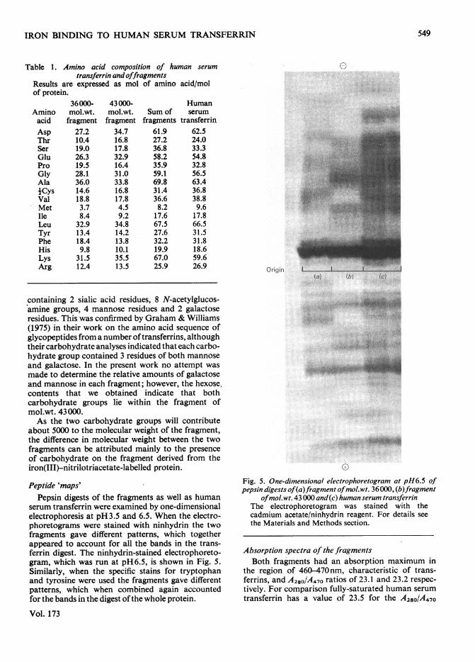
The amino acid compositions of human serumtransferrin, the fragment of mol.wt. 36000 and thefragment of mol.wt. 43000, as isolated by gelfiltration on Sephadex G-200 are given in Table 1.The fragments have similar compositions, althoughdifferences are apparent in their contents of asparticacid, glutamic acid, threonine and phenylalanine.The sum of the compositions of the fragments agreesreasonably well with that of the whole protein.
Carbohydrate composition of the fragments
The hexose content of the 36000- and 43000-mol.wt. fragments, as determined by the orcinol/H2SO4 test, were found to be 1.9 and 10.1 mol ofsugar/mol of protein respectively. Jamieson (1965)showed that human serum transferrin has twocarbohydrate groups of identical composition, each
1978
are found; however, although they both containiron, peak B is essentially free of carbohydrate.Sodium dodecyl sulphate/polyacrylamide-gel electro-
548
(a)
IRON BINDING TO HUMAN SERUM TRANSFERRIN
mino acid composition of human serumtransferrin and offragments
e expressed as mol of amino acid/mol
43000-mol.wt.fragment
34.716.817.832.916.431.033.816.817.84.59.2
34.814.213.810.135.513.5
Sum offragments
61.927.236.858.235.959.169.831.436.68.2
17.667.527.632.219.967.025.9
Humanserum
transferrin62.524.033.354.832.856.563.436.838.89.6
17.866.531.531.818.659.626.9
containing 2 sialic acid residues, 8 N-acetylglucos-amine groups, 4 mannose residues and 2 galactoseresidues. This was confirmed by Graham & Williams(1975) in their work on the amino acid sequence ofglycopeptides from a number oftransferrins, althoughtheir carbohydrate analyses indicated that each carbo-hydrate group contained 3 residues of both mannose
and galactose. In the present work no attempt was
made to determine the relative amounts of galactoseand mannose in each fragment; however, the hexose,contents that we obtained indicate that bothcarbohydrate groups lie within the fragment ofmol.wt. 43000.As the two carbohydrate groups will contribute
about 5000 to the molecular weight of the fragment,the difference in molecular weight between the twofragments can be attributed mainly to the presenceof carbohydrate on the fragment derived from theiron(III)-nitrilotriacetate-labelled protein.
Peptide 'maps'Pepsin digests of the fragments as well as human
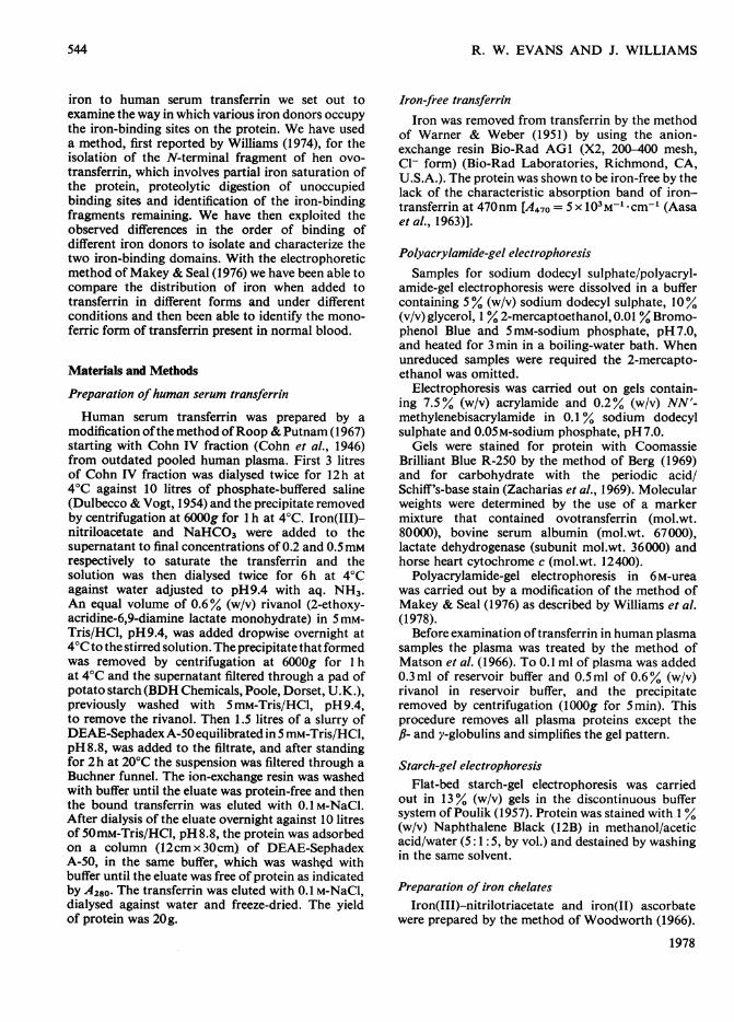
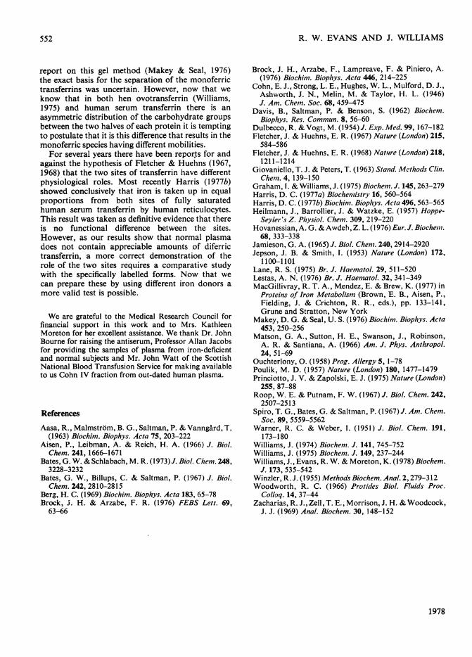
serum transferrin were examined by one-dimensionalelectrophoresis at pH3.5 and 6.5. When the electro-phoretograms were stained with ninhydrin the twofragments gave different patterns, which togetherappeared to account for all the bands in the trans-ferrin digest. The ninhydrin-stained electrophoreto-gram, which was run at pH6.5, is shown in Fig. 5.Similarly, when the specific stains for tryptophanand tyrosine were used the fragments gave differentpatterns, which when combined again accountedfor the bands in the digest of the whole protein.
Vol. 173
~~~~~~~~~~~~~. .. .....
Fig. 5. One-dimensional electrophoretogram at pH6.5 ofpepsin digests of(a)fragment ofmol.:wt. 36000, (b)fragment
ofmol.wt. 43000 and(c) human serum transferrinThe electrophoretogram was stained with thecadmium acetate/ninhydrin reagent. For details seethe Materials and Methods section.
Absorption spectra of the fragmentsBoth fragments had an absorption maximum in
the region of 460-470nm, characteristic of trans-ferrins, and A280/A470 ratios of 23.1 and 23.2 respec-tively. For comparison fully-saturated human serumtransferrin has a value of 23.5 for the A280/A470
0Table 1. A)
Results arcof protein.
Amino r
acid fiAspThrSerGluProGlyAlajCysValMetIleLeuTyrPheHisLysArg
36000-mol.wt.Iragment27.210.419.026.319.528.136.014.618.83.78.4
32.913.418.49.8
31.512.4 Origin
(a) (b) (c)
549
'lx. %"':;s :..- .X
1 ti

R. W. EVANS AND J. WILLIAMS
Origin
H ST Ak -:
H ST Fel---E..l_w01 _
Fe -H ST F! . @ w.
Fe2 H ST-*
(a) (b) (c) (d) (e) (f) Ig) (h) (i)
-HST
4-H ST-Fe
4- Fe-HST
4-Fe2 HST
Fig. 6. Urealpolyacrylamide-gel electrophoresis ofhuman serum transferrin partially iron-saturated with various iron donorsatpH6.0 and 8.5
Samples (a) (25,ug) and (n) (25,ug) are partially iron-saturated preparation of human serum transferrin. Samples (b)(25 pg) and (c) (50 pg) are proteins partially iron-saturated with iron(III)-nitrilotriacetate at pH 8.5. Samples (d) (25pg)and (e) (50g) are proteins partially iron-saturated with (NH4)2SO4,FeSO4 at pH8.5. Samples (f) (25/,g) and (g)(50ug) are proteins partially iron-saturated with FeCJ3 at pH8.5. Samples (h) (25pg) and (i) (50,ug) are proteinspartially iron-saturated with iron(III)-nitrilotriacetate at pH 6.0. Samples (j) (25pg) and (k) (50g) are proteins partiallyiron-saturated with (NH4)2SO4,/FeSO4 at pH6.0. Samples (I) (25,ug) and (m) (50,ug) are proteins partiallyiron-saturated with FeCl3 at pH6.0. HST indicates the position on the gel of iron-free human serum transferrin,HST-Fe the position of monoferric transferrin with iron in the C-terminal site, Fe-HST the position of monoferrictransferrin with iron in the N-terminal site and Fe2HST the position of fully saturated transferrin.
ratio. A slight difference in the absorption maximawas observed for the fragments, but as yet it is notknown whether this is of any significance.
Reversibility of iron binding to fragmentsBoth the iron-free fragments were titrated with
iron(III)-nitrilotriacetate and (NH4)2SO4,FeSO4 atpH 8.5; however, only the fragment of mol.wt. 43 000took up its original complement of iron, as indicatedby the A470. The lower-molecular-weight fragmentwould only take up about 30% of the theoreticalamount of iron, irrespective of the nature of theiron donor.
Urealpolyacrylamide-gel electrophoresis of partiallyiron-saturated human serum transferrin and humanplasmaHuman serum transferrin can be separated into
four species, namely iron-free protein, two monoferric
species and fully iron-saturated protein, by electro-phoresis at pH 8.4 in polyacrylamide gels containing6M-urea (Makey & Seal, 1976). As shown in Fig. 6addition of iron as iron(III)-nitrilotriacetate (samplesb and c) to iron-free transferrin, at pH 8.5, enhancesthe slower-migrating of the two intermediate forms,whereas when (NH4)2SO4,FeSO4 (samples d and e)and FeCl3 (samples f and g) are used as iron donorsthe faster-moving intermediate band is preferentiallyformed.When samples of transferrin in 0.1 M-Hepes/
0.01 M-NaHCO3, pH 6.0, were partially saturatedwith the same three iron donors electrophoresis inurea/polyacrylamide gels (Fig. 6, samples h-m)revealed that in all three cases only the apoproteinand the slower of the two intermediates were present.The isolation procedure for human serum trans-
ferrin yields protein that is about 40% iron-saturated and that on digestion with trypsin it givesrise to the fragment with mol.wt. 43000. Urea/
1978
550
(i) (k) U) (m) (n)
:-i:..ijii.:.::.-'xM-.s:
.i:",

IRON BINDING TO HUMAN SERUM TRANSFERRIN
polyacrylamide-gel electrophoresis of this partiallysaturated protein (Fig. 6, samples a and n) showedthat it contained mainly the slower-running mono-ferric species, together with small amounts ofiron-free and iron-saturated transferrin.Plasma from normal subjects was examined by this
gel-electrophoresis method and found to containonly iron-free protein and the first intermediate.To check that the second intermediate can exist inplasma, iron(III)-nitrilotriacetate and (NH4)2SO4,-FeSO4 were used to saturate the transferrin partiallyin plasma from iron-deficient patients. It wasobserved that, as with the pure protein, iron(III)-nitrilotriacetate enhances the slower-migrating inter-mediate band, whereas the other iron donor enhancesthe faster-migrating intermediate.
Discussion
In the present paper we have shown that proteo-lytic digestion of partially iron-saturated humanserum transferrin gives rise to iron-binding frag-ments. Under the same conditions the iron-freeprotein is completely degraded to low-molecular-weight peptides, whereas the iron-saturated proteinundergoes some internal cleavage but retains itscomplement of iron, and under non-reducing con-ditions has the same mobility on sodium dodecylsulphate/polyacrylamide-gel electrophoresis as thenative protein. Similar observations have been madewith hen ovotransferrin (Williams, 1974, 1975),except that the fully saturated protein was completelyresistant to attack by trypsin. For bovine serumtransferrin Brock et al. (1976) found that trypsindigestion of the iron-saturated protein producestwo fragments that were subsequently thought torepresent the two domains of the protein (Brock &Arzabe, 1976).
Proteolytic digestion of human serum transferrinsaturated to 30% with iron(III)-nitrilotriacetate ateither pH5.5 or pH8.5 produces a carbohydrate-containing fragment of mol.wt. 43000. However,when four other iron donors, iron(III) citrate,FeCI3, iron(II) ascorbate and (NH4)2SO4,FeSO4,are used partially to saturate the protein at pH8.5a different fragment is obtained after digestion withtrypsin. This second fragment lacks carbohydrateand has mol.wt. 36000. In some experiments thisfragment was accompanied by a minor fragment ofslightly lower molecular weight, possibly owing tofurther digestion. Neither the 36000-mol.wt. frag-ment nor the 43GO0-mol.wt. fragment possesses asingle chain; however, this is not surprising in viewof the fact that the fully saturated protein itselfundergoes limited cleavage with trypsin.Although the smaller fragment is readily separated
from undigested protein by gel filtration onSephadex G-100, the other fragment could only be
Vol. 173
completely resolved from transferrin by gel filtrationon Sephadex G-2C0. As well as differing in molecularweight and carbohydrate composition the fragmentshave different peptide 'maps' and amino acidcompositions and are immunologically distinct.Their only similarity is in their absorption spectra,which are almost identical and characteristic of alltransferrins. Studies on human serum transferrin(MacGillivray et al., 1977) have shown that the twocarbohydrate groups on the protein lie within theC-terminal half of the protein, so we conclude thatthe fragment with mol.wt. 43000 must represent theC-terminal region and the other fragment theN-terminal region. The availability of the isolatediron-binding domains will simplify further investi-gations into the structural properties of the iron-binding sites.The observation that iron(III)-nitrilotriacetate,
a non-physiological iron donor, has a preference forthe C-terminal site at both pH 5.5 and pH 8.5 agreeswith the work of Harris (1977a), who also found thatone particular site, designated A, exhibits a tendencyto bind iron first at both acidic and neutral pH. Wenow know that the A site lies in the C-terminalregion of the protein. In hen ovotransferrin theN-terminal site preferentially binds iron, whenadded as iron(III)-nitrilotriacetate, at pH8.5, butat acidic pH the C-terminal site takes up ironpreferentially (Williams et al., 1978).Although the other iron donors that we examined,
which showed a preference for the N-terminal site,might be expected to resemble more closely the formin which iron is present in the body, the distri-bution of iron on transferrin in normal plasmasuggests that this is not so. By using a urea/polyacrylamide gel, which resolves iron-free humanserum transferrin, the two monoferric transferrinsand fully saturated transferrin (Makey & Seal, 1976),we have compared the pattern obtained when iron indifferent forms is added to the pure protein with thepattern in normal blood. Human plasma, where thetransferrin is about 30% iron-saturated (Giovaniello& Peters, 1963), contains two species, iron-freetransferrin and a monoferric form that correspondsto that obtained when iron(III)-nitrilotriacetate isadded to the isolated protein atpH 6.0 or 8.5 and there-fore has iron in the C-terminal site. Transferrin inthe plasma of iron-deficient patients is essentiallyiron-free, so it was possible to confirm that, as withthe pure protein, addition of iron(III)-nitrilotri-acetate to plasma enhances the monoferric form withiron in the C-terminal site, whereas the other donorsenhance the other monoferric species.
It is noteworthy that, as for hen ovotransferrin(Williams et al., 1978), the monoferric human serumtransferrin species with iron in the C-terminal site hasa lower mobility in urea/polyacrylamide gels than theform with iron in the N-terminal site. In the original
551
552 R. W. EVANS AND J. WILLIAMS
report on this gel method (Makey & Seal, 1976)the exact basis for the separation of the monoferrictransferrins was uncertain. However, now that weknow that in both hen ovotransferrin (Williams,1975) and human serum transferrin there is anasymmetric distribution of the carbohydrate groupsbetween the two halves of each protein it is temptingto postulate that it is this difference that results in themonoferric species having different mobilities.For several years there have been repor.ts for and
against the hypothesis of Fletcher & Huehns (1967,1968) that the two sites of transferrin have differentphysiological roles. Most recently Harris (1 977b)showed conclusively that iron is taken up in equalproportions from both sites of fully saturatedhuman serum transferrin by human reticulocytes.This result was taken as definitive evidence that thereis no functional difference between the sites.However, as our results show that normal plasmadoes not contain appreciable amounts of diferrictransferrin, a more correct demonstration of therole of the two sites requires a comparative studywith the specifically labelled forms. Now that wecan prepare these by using different iron donors amore valid test is possible.
We are grateful to the Medical Research Council forfinancial support in this work and to Mrs. KathleenMoreton for her excellent assistance. We thank Dr. JohnBourne for raising the antiserum, Professor Allan Jacobsfor providing the samples of plasma from iron-deficientand normal subjects and Mr. John Watt of the ScottishNational Blood Transfusion Service for making availableto us Cohn IV fraction from out-dated human plasma.
References
Aasa, R., Malmstrom, B. G., Saltman, P. & Vanngard, T.(1963) Biochim. Biophys. Acta 75, 203-222
Aisen, P., Leibman, A. & Reich, H. A. (1966) J. Biol.Chem. 241, 1666-1671
Bates, G. W. & Schlabach, M. R. (1973)J. Biol. Chem. 248,3228-3232
Bates, G. W., Billups, C. & Saltman, P. (1967) J. Biol.Chem. 242, 2810-2815
Berg, H. C. (1969) Biochim. Biophys. Acta 183, 65-78Brock, J. H. & Arzabe, F. R. (1976) FEBS Lett. 69,
63-66
Brock, J. H., Arzabe, F., Lampreave, F. & Piniero, A.(1976) Biochim. Biophys. Acta 446, 214-225
Cohn, E. J., Strong, L. E., Hughes, W. L., Mulford, D. J.,Ashworth, J. N., Melin, M. & Taylor, H. L. (1946)J. Am. Chem. Soc. 68, 459-475
Davis, B., Saltman, P. & Benson, S. (1962) Biochem.Biophys. Res. Commun. 8, 56-60
Dulbecco, R. &Vogt, M. (1954) J. Exp.Med. 99, 167-182Fletcher, J. & Huehns, E. R. (1967) Nature (London) 215,
584-586Fletcher, J. & Huehns, E. R. (1968) Nature (London) 218,
1211-1214Giovaniello, T. J. & Peters, T. (1963) Stand. Methods Clin.
Che,ni. 4, 139-150Graham, I. & Williams, J. (1975) Biochem. J. 145, 263-279Harris, D. C. (1977a) Biochemistry 16, 560-564Harris, D. C. (1977b) Biochim. Biophys. Acta 496, 563-565Heilmann, J., Barrollier, J. & Watzke, E. (1957) Hoppe-
Seyler's Z. Physiol. Chem. 309, 219-220Hovanessian, A. G. & Awdeh, Z. L. (I 976) Eur. J. Biochemii.
68, 333-338Jamieson, G. A. (1965) J. Biol. Chem. 240, 2914-2920Jepson, J. B. & Smith, 1. (1953) Nature (London) 172,
1100-1101Lane, R. S. (1975) Br. J. Haematol. 29, 511-520Lestas, A. N. (1976) Br. J. Haematol. 32, 341-349MacGillivray, R. T. A., Mendez, E. & Brew, K. (1977) in
Proteins of Iron Metabolism (Brown, E. B., Aisen, P.,Fielding, J. & Crichton, R. R., eds.), pp. 133-141,Grune and Stratton, New York
Makey, D. G. & Seal, U. S. (1976) Biochim. Biophys. Acta453, 250-256
Matson, G. A., Sutton, H. E., Swanson, J., Robinson,A. R. & Santiana, A. (1966) Am. J. Phys. Anthropol.24, 51-69
Ouchterlony, 0. (1958) Prog. Allergy 5, 1-78Poulik, M. D. (1957) Nature (London) 180, 1477-1479Princiotto, J. V. & Zapolski, E. J. (1975) Nature (London)
255, 87-88Roop, W. E. & Putnam, F. W. (1967) J. Biol. Chem. 242,
2507-2513Spiro, T. G., Bates, G. & Saltnian, P. (1967) J. Am. Chem.
Soc. 89, 5559-5562Warner, R. C. & Weber, I. (1951) J. Biol. Chem. 191,
173-180Williams, J. (1974) Biochem. J. 141, 745-752Williams, J. (1975) Biochem. J. 149, 237-244Williams, J., Evans, R. W. & Moreton, K. (1978) Biochem.
J. 173, 535-542Winzler, R. J. (1955) Methods Biochem. Anal. 2,279-312Woodworth, R. C. (1966) Protides Biol. Fluids Proc.
Colloq. 14, 37-44Zacharias, R. J.,Zell, T. E., Morrison, J. H. & Woodcock,
J. J. (1969) Anal. Biochem. 30, 148-152
1978