structured reporting of focal masses in the …€¦ · 1 structured reporting of focal masses in...
TRANSCRIPT

1
STRUCTURED REPORTING OF FOCAL MASSES IN THE ABDOMEN: WITH A
FOCUS ON CONSISTENT COMMUNICATION & MONITORING OF
FOLLOW-UP RECOMMENDATIONS
Seetharam C. Chadalavada, MD, MS1; Hanna M. Zafar MD, MHS1; Darco Lalevic1; Caroline
Sloan2;Mitchell D. Schnall, MD, PhD1; Curtis P. Langlotz, MD, PhD3; Tessa S. Cook, MD, PhD1
Department of Radiology, Hospital of the University of Pennsylvania1
Pearlman School of Medicine, University of Pennsylvania2
Department of Radiology, Stanford University3
RSNA 2014: 14015451, QSE104
DISCLOSURES
• Seetharam C. Chadalavada:
• Shareholder, Docphin, Inc
• Curtis P. Langlotz:
• Shareholder, Montage Healthcare Solutions, Inc Advisory Board, General Electric Company
Advisory Board, Reed Elsevier Advisory Board, Activate Networks, Inc Spouse, Consultant,
Amgen Inc Spouse, Consultant, Novartis AG Spouse, Consultant, Johnson & Johnson
• All remaining authors:
• Nothing to disclose

2
PROBLEM
• Failure to complete follow-up
• Associated with preventable patient harm as result of
missed or delayed cancer diagnoses
• Costs to health system
• Patient safety and quality of care
BACKGROUND
• Poor communication
• Free text reports: variable & inconsistent
• Identification & monitoring of patients?
• Structured Reporting
• None outside of mammography
• Breast Imaging–Reporting and Data System (BI-RADS®)
• Monitor, follow-up, and pathology outcomes

3
Benign
Indeterminate
Suspicious
STRUCTURED REPORTING: ABDOMEN
• Implemented standardized language/categories for
reporting likelihood of cancer in focal lesions (modeling
after BI-RADS®)
• Since July 1, 2013 – Hospital of the University of Pennsylvania
• Focus on 4 organs:
• Liver
• Pancreas
• Kidneys
• Adrenal glands
CODE ABDOMEN: CATEGORIES
• MAC 0: Incompletely evaluated. If indicated within the patient’s
clinical context, follow up [INSERT MODALITY] is advised.
• MAC 1: No mass.
• MAC 2: Benign. No further evaluation needed.*
• MAC 3: Indeterminate. If indicated within the patient’s clinical
context, follow up [INSERT MODALITY] is advised within [INSERT
TIME FRAME] [weeks/months].
• MAC 4: Suspicious. May represent malignancy.
• MAC 5: Highly suspicious. Clear imaging evidence of malignancy.
• MAC 6: Known cancer.
• MAC 7: Completely treated cancer.
• MAC 99: Technically inadequate for evaluation of masses.
BENIGN
INDETERMINATE
SUSPICIOUS
INDETERMINATE
BENIGN (SURVEILLANCE)
TECHNICAL LIMITATIONS
KNOWN CANCER

4
SELECT EXAMPLE CASES
CT: CATEGORY 2 - BENIGN
63 year old male with history of diabetes mellitus presents to the emergency room for abdominal pain. Index CT
examination demonstrates single 2 cm fluid attenuation lesion (solid green arrow) within the upper pole of the left
kidney as demonstrated on axial (left image) and coronal (right image) enhanced CT images representing benign
renal cyst. Incidentally noted is a non-obstructing 2 mm calculus within the mid-pole of left kidney (solid yellow
arrow).

5
CT: CATEGORY 0 – INDETERMINATE
54 year old male with remote history of Whipple procedure for cystic pancreatic neoplasm presents with
abdominal pain. Axial (left image) and coronal (right image) enhanced CT images at the level of the adrenals
revealed a heterogeneously enhancing 2 cm mass within the left adrenal gland (solid green arrow) measuring
greater than 10 Hounsfield units. This was assigned a Category 0 and further characterization with MRI was
recommended.
F/U MRI: CATEGORY 2 - BENIGN
54 year old male with remote history of Whipple procedure for cystic pancreatic neoplasm. Subsequent MRI
exam, reveals that this mass contains microscopic lipid manifest as drop out of signal between the in phase (left
image, curved arrow) and out of phase (right image, arrow) gradient echo images. This lesion was diagnosed as
a benign adrenal adenoma and assigned a Category 2 on the MRI report.)

6
US: CATEGORY 3 – INDETERMINATE, INTERVAL TIME NECESSARY
(68 year old female presents for follow-up of known right renal cyst. Ultrasound exam from May 2014 (left
image), reveals an indeterminate complex cystic lesion with an avascular thickened septation that
demonstrates soft tissue echogenicity but no vascular flow. This is similar in size and appearance to an exam
from March 2012 (right image). In light of the interval stability conservative management with a subsequent
ultrasound exam in one year was recommended.)
May 2014 US Exam March 2012 US Exam
CT: CATEGORY 99 – TECHNICALLY INADEQUATE
54 year old male with history of cirrhosis presents with abdominal pain. Axial (left image) & coronal (right
image) unenhanced CT images at level of liver demonstrates nodularity of the liver contour, mild hypertrophy
of the caudate and left hepatic lobes and ascites; all consistent with known cirrhosis. In the absence of a triple
phase protocol, evaluation of hepatic masses, specifically hepatocellular carcinoma, is not possible and this
patient was assigned a Category 99.

7
US: CATEGORY 99 – TECHNICALLY INADEQUATE
34 year old male with neurogenic bladder presents with recurrent urinary tract infections. An ultrasound
examination of the retroperitoneum was performed to evaluate for hydronephrosis. Although the examination
is adequate to answer the clinical question, it is technically inadequate to evaluate for masses due to poor
acoustic penetration and shadowing from the ribs (solid green arrows) secondary to inability of the patient to
hold deep inspiration.)Accordingly this patient was assigned a Category 99.
DATA/RESULTS

8
COMPLIANCE
• Over 24,585 US, CT, MRI eligible exams captured in our database
• 93% compliance over 1st academic year of implementation
Graphical representation of compliance among the 3 (CT, MRI, US) modalities & cumulative (ALL)
EARLY COMPLIANCE
Expected low compliance initially. Dedicated educational orientation sessions for both radiology
trainees & faculty during first phase (~4 week “warm-up phase”), helped gradually improve compliance.

9
CATEGORIES - LIVER
CT MRI US ALL Modalities
30%
21%
14%
41%
23%
11%
36%19%
23%
12%
19%
32%
18%
Distribution of Code Abdomen Categories within the Liver among the 3 (CT, MRI, US) modalities &
collectively.
CATEGORIES - PANCREAS
CT MRI US ALL Modalities
13%
26%
26%
48%
14%
20%
77%
14% 23%
23%
13%
Distribution of Code Abdomen Categories within the Pancreas among the 3 (CT, MRI, US) modalities &
collectively.

10
CATEGORIES - KIDNEYS
CT MRI US ALL Modalities
17%
30%
22%
41%
16%
21% 46%
15%
27%
26%
20%
21%
Distribution of Code Abdomen Categories within the Kidneys among the 3 (CT, MRI, US) modalities &
collectively.
CATEGORIES - ADRENALS
CT MRI US ALL Modalities
79%
28%
10%10%
26%
14%
36%13%
13%
7%
15%
33%
16%
12%
Distribution of Code Abdomen Categories within the Adrenals among the 3 (CT, MRI, US) modalities &
collectively.

11
4 ORGANS – ALL MODALITIES
• Distribution of Code Abdomen Categories for the 4 organs (liver, pancreas, kidneys,
adrenals) among all 3 modalities (CT, MRI, US) collectively.
• Ability to monitor our patients (follow-ups) prospectively (1st year of implementation):
• 2,238 patients with indeterminate lesions (0 & 3’s)
• 1,302 patients with suspicious lesions (4 & 5’s)
LIVER PANCREAS KIDNEYS ADRENALS
15%
33%
16%
12%26%
20%
21%
23%
23%
13%
12%
19%
32%
18% 20%
FOLLOW-UP STATUS SUMMARY
• Added benefit of structured reporting is the ability to mine reports for meaningful data.
• Follow-up status summary allows us to actively track the status of follow-up
recommendations: completed, scheduled, missed/not-scheduled?

12
INDIVIDUAL PATIENT MONITORING
• Follow-up status allows us to track and determine outcomes at a patient level.
OUTCOMES OF RADIOLOGY FOLLOW-UP RECOMMENDATIONS IN ABDOMINAL IMAGING STUDIES
• 1,337 out of 8,801 patients (15%) with lesions indeterminate for cancer
between 8/11/13 & 12/31/13.
• Of these, 424 patients (32%) underwent follow-up abdominal imaging
within our hospital system over a 4 month span
• Included 787 CT & 260 MRI exams cumulatively.
• On subsequent imaging, lesions were:
• Downgraded to benign on 76% of patients (316/424)
• Upgraded to suspicious for cancer in 6% (31/424)
• Remained indeterminate in 18% (77/424).
13
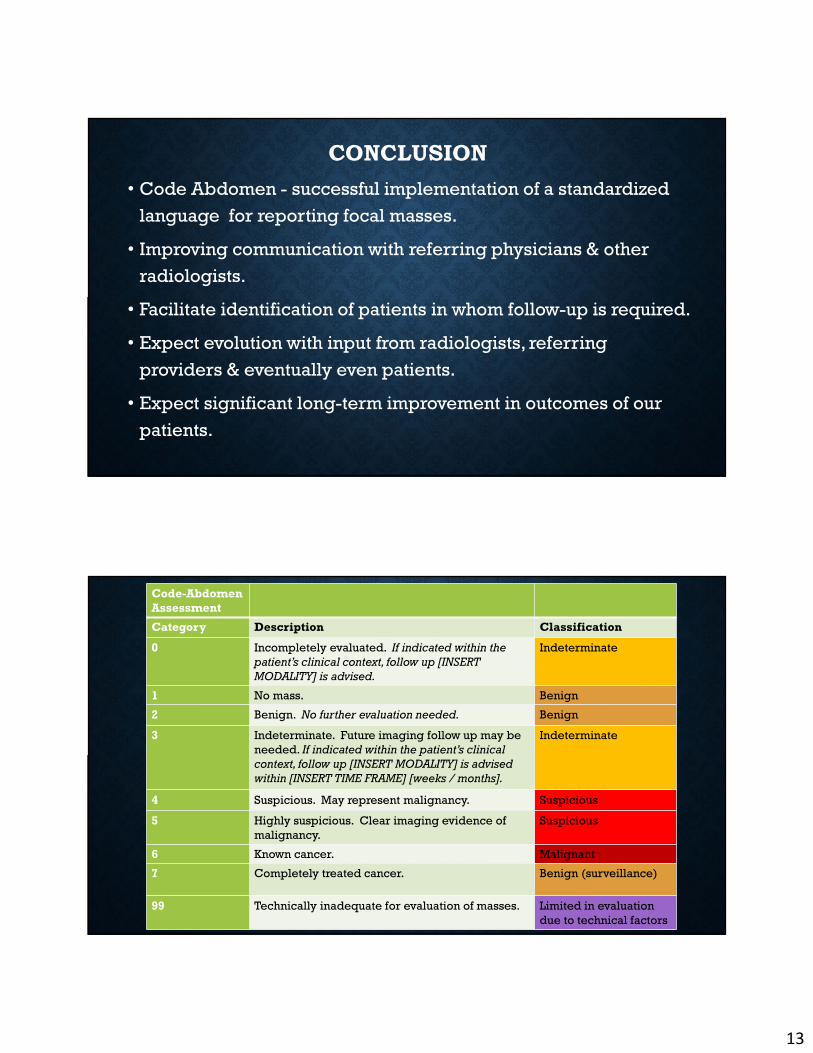
CONCLUSION
• Code Abdomen - successful implementation of a standardized
language for reporting focal masses.
• Improving communication with referring physicians & other
radiologists.
• Facilitate identification of patients in whom follow-up is required.
• Expect evolution with input from radiologists, referring
providers & eventually even patients.
• Expect significant long-term improvement in outcomes of our
patients.
Code-Abdomen
Assessment
Category Description Classification
0 Incompletely evaluated. If indicated within the
patient’s clinical context, follow up [INSERT
MODALITY] is advised.
Indeterminate
1 No mass. Benign
2 Benign. No further evaluation needed. Benign
3 Indeterminate. Future imaging follow up may be
needed. If indicated within the patient’s clinical
context, follow up [INSERT MODALITY] is advised
within [INSERT TIME FRAME] [weeks / months].
Indeterminate
4 Suspicious. May represent malignancy. Suspicious
5 Highly suspicious. Clear imaging evidence of
malignancy.
Suspicious
6 Known cancer. Malignant
7 Completely treated cancer. Benign (surveillance)
99 Technically inadequate for evaluation of masses. Limited in evaluation
due to technical factors

14
SUPPORT
• Abdominal Imaging Division – Penn Radiology
• Radiology Trainees – Penn Radiology
• T32 NIH-NIBIB Training grant
• Penn Medicine Center for Health Care Innovation