structure of corneal layers, collagen fibrils, and ... · structure of corneal layers, collagen...
TRANSCRIPT
Structure of corneal layers, collagen fibrils, and proteoglycans oftree shrew cornea
Turki Almubrad, Saeed Akhtar
Cornea Research Chair, Department of Optometry and Vision Sciences, College of Applied Medical Sciences, King Saud University,Riyadh, Saudi Arabia
Purpose: The stroma is the major part of the cornea, in which collagen fibrils and proteoglycans are distributed uniformly.We describe the ultrastructure of corneal layers, collagen fibrils (CF), and proteoglycans (PGs) in the tree shrew cornea.Methods: Tree shrew corneas (5, 6, and 10 week old animals) and normal human corneas (24, 25, and 54 years old) werefixed in 2.5% glutaraldehyde containing cuprolinic blue in a sodium acetate buffer. The tissue was processed for electronmicroscopy. The ‘iTEM Olympus Soft Imaging Solutions GmbH’ program was used to measure the corneal layers,collagen fibril diameters and proteoglycan areas.Results: The tree shrew cornea consists of 5 layers: the epithelium, Bowman’s layer, stroma, Descemet’s membrane, andendothelium. The epithelium was composed of squamous cells, wing cells and basal cells. The Bowman’s layer was5.5±1.0 µm thick and very similar to a normal human Bowman’s layer. The stroma was 258±7.00 µm thick and consistedof collagen fibril lamellae. The lamellae were interlaced with one another in the anterior stroma, but ran parallel to oneanother in the middle and posterior stroma. Collagen fibrils were decorated with proteoglycan filaments with an area sizeof 390 ±438 nm2. The collagen fibril had a minimum diameter of 39±4.25 nm. The interfibrillar spacing was 52.91±6.07nm. Within the collagen fibrils, very small electron-dense particles were present.Conclusions: The structure of the tree shrew cornea is very similar to that of the normal human cornea. As is the casewith the human cornea, the tree shrew cornea had a Bowman's layer, lamellar interlacing in the anterior stroma and electron-dense particles within the collagen fibrils. The similarities of the tree shrew cornea with the human cornea suggest that itcould be a good structural model to use when studying changes in collagen fibrils and proteoglycans in non-genetic cornealdiseases, such as ectasia caused after LASIK (laser-assisted in situ keratomileusis).
In recent years, the tree shrew has been used as a modelfor studying myopia [1,2]. Norton et al. [1] examined theeffect of a period of continuous darkness on the refractive stateand vitreous chamber depth of normal light-reared juveniletree shrew eyes. They observed that four out of the five myopiceyes in the dark-recovery group became more myopic indarkness, and all the control eyes shifted toward myopia. Theprevention of collagen cross linking and the expression oftransforming growth factor-beta and collagen synthesis on thecornea and sclera of the tree shrew were also studied [3,4].
Ishiko et al. [5] observed the early ocular changes in adiabetic tree shrew model. They developed a tree shrew modelof diabetes using streptozotocin (STZ) and studied the earlyocular changes of diabetes (after one week of diabetes).Corneal autofluorescence was significantly increased oneweek after the STZ injection. These changes may be relatedto the impairment of the ocular homeostatic mechanisms dueto the onset of diabetes. They suggested using the tree shrewas a model may make it possible to obtain diabetic ocular
Correspondence to: Dr. Saeed Akhtar, Cornea Research Chair,Department of Optometry and Vision Sciences, College of AppliedMedical Sciences, King Saud University, Riyadh, Saudi Arabia;Phone: 00966 4693532; Mobile: 00966 541032226; FAX: 009664350810; email: [email protected] or [email protected]
impairment data closer to human data than could be obtainedin previous models of diabetes. Another advantage of usingthe tree shrew as a model is that tree shrews are not primates[6]. Given the ethical concerns surrounding the use ofprimates in medical research, the tree shrew could be used asan alternative to primates.
Few ultra-structural analysis studies have beenperformed on the tree shrew ocular system. McBrian et al.[7] used the tree shrew as a model to assess changes in thediameter of collagen fibrils in the sclera of myopic tree shrews.They reported a significant reduction in the median collagenfibril diameter at the posterior pole. Albon et al. [8] describedthe ultrastructure and immuno-histochemistry of the laminacribrosa of the tree shrew. The authors showed the presenceof multilayered connective tissue plates in the tree shrewlamina cribrosa LC stretched across the optic nerve canal atthe level of the sclera and consisting of collagen types I, III,IV, V, and VI; and elastin and fibronectin. The ultrastructuraldistribution of collagen fibrils, elastic fibers, and axons hasbeen shown by scanning and transmission electronmicroscopy. To our knowledge, there have been no studiesperformed on the ultrastructure of the tree shrew cornea.
A substantial proportion of the cornea consists of thestroma, which accounts for 95% of the corneal thickness inhumans. The stroma is composed of parallel running lamellae
Molecular Vision 2011; 17:2283-2291 <http://www.molvis.org/molvis/v17/a248>Received 31 July 2011 | Accepted 19 August 2011 | Published 25 August 2011
© 2011 Molecular Vision
2283

which form the extracellular matrix. These lamellae arepacked with uniformly distributed collagen fibrils of uniformdiameters. This uniform distribution of collagen fibrils isresponsible for the transparency of the cornea [9]. Theuniform distribution of collagen fibrils is regulated bymacromolecular glycoconjugates known as proteoglycans.The extracellular matrix of the cornea contains a class of smallinterstitial PGs known as collagen-binding small leucine-richrepeat proteoglycans (SLRRPs). These proteoglycans carryeither kearatan sulfate chains or chondroitin sulfate⁄dermatansulfate chains. Lumican, keratocan and mimican carry keratansulfate chains, whereas decorin, biglycan and versican carrychondroitin sulfate⁄dermatan sulfate chains. Lumican isbelieved to be essential to the transparency of the cornea inthat it regulates the uniform diameter of the collagen fibrils[10].
Ultrastructural features of the collagen fibrils andproteoglycans of the cornea have been discussed in variousanimal models, such as bovine, mouse, rabbit, chick, andzebrafish [10-13]. Detailed ultrastructural studies ofBowman’s layers and stroma in tadpoles, cattle, mice, rabbits,guinea pigs and humans were performed by Hayashi et al.[14]. This is the first paper to describe the ultrastructuralorganization of corneal layers, collagen fibrils andproteoglycans in tree shrew cornea.
The minimum collagen fibril diameter and center-to-center spacing were measured using the ‘iTEM Olypmus SoftImaging Solutions GmbH’ program. A decision was made tomeasure the minimum diameter of each fibril, rather than theaverage value to avoid errors due to any obliqueness in thefibril cross-sections. Akhtar et al. [15] measured the‘minimum diameter’ of collagen fibrils of human cornea fixedin paraformaldehyde and embedded in LR White resin (TheLondon Resin Co Ltd, Reading , Berkshire, UK). In thepresent studies, the ‘minimum diameter’ of collagen fibrils ofthe cornea that were fixed in glutaradehyde plus cuprolinicblue and embedded in spurr resin was measured. The area sizeof the proteoglycans was measured in the anterior, middle andposterior stroma of the cornea. Data were analyzed using thestatistic program SPSS (IBM Corporation, Armonk, NY).Kolmogorov–Smirnov and Mann–Whitney tests wereperformed. The mean and (pooled) standard deviation valuesfor the 3 corneas were calculated taking into account thewithin-sample variances.
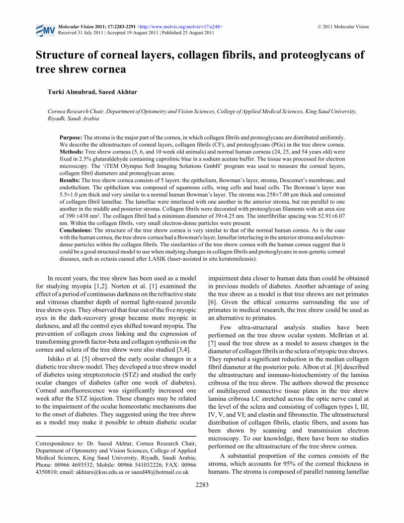
RESULTSLight microscopic observations of semithin sections showedthat the structure of the tree shrew cornea was very similar tothat of the human cornea. The cornea was 320.96±2.95 µmthick and contained 5 layers: the epithelium, Bowman’s layer,stroma, Descemet’s membrane, and endothelium (Figure 1A-D). It was composed of squamous cells, wing cells and basalcells (Figure 1B). The stroma consisted of collagen fibrillamellae containing keratocytes (Figure 1C). The posteriorpart of the stroma was covered by Descemet’s membrane anda single layer of the endothelium (Figure 1D).
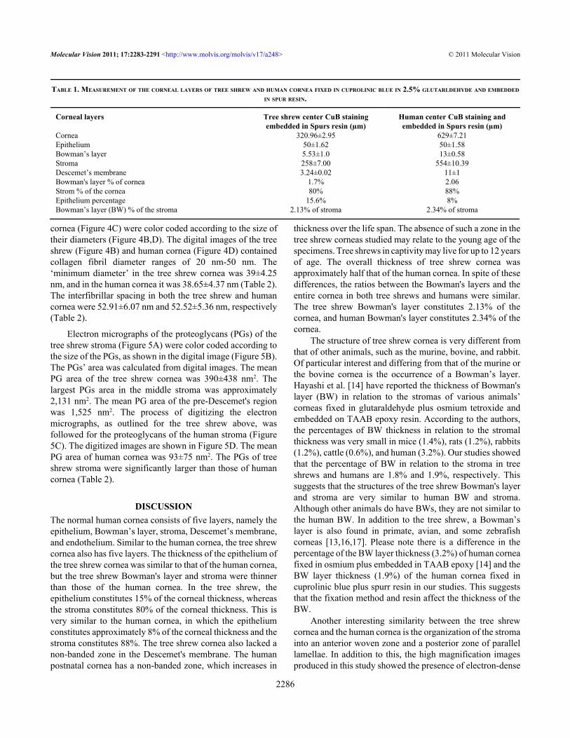
Light microscopic measurements of corneal layers fromsemi-thin sections of tree shrew and human cornea fixed inglutaraldehyde are shown in Table 1. The epithelium of thetree shrew cornea was 50.29±1.62 µm thick and constituted15% of the corneal thickness (Table 1). The thickness of theBowman’s layer and stroma of the tree shrew were5.53±1.0 µm and 258±7.0 µm, respectively. The tree shrewBowman's layer constituted 2.13% and the stroma constituted80% of the entire cornea (Table 1). In the human cornea, theepithelium was 50±2.00 µm thick, the Bowman's layer was13±0.58 µm thick, and the stroma was 554±10.39 µm thick.In the human cornea, the epithelium, Bowman's layer andstroma constituted 8%, 2.34%, and 88% of the thickness ofthe cornea, respectively (Table 1). All the data presented inthis paper in relation to the human cornea were producedduring the course of the present study.
Ultrastructural observations showed that the basalepithelial cells were columnar and contained large nuclei andprominent cytoplasmic filaments (Figure 2A,B). They were
Molecular Vision 2011; 17:2283-2291 <http://www.molvis.org/molvis/v17/a248> © 2011 Molecular Vision
2284
METHODS
Tissue procurement and use was conducted in accordancewith the Declaration of Helsinki and local regulations. It wasethically approved by the Local Ethical Committee of KingSaud University, Saudi Arabia.
The corneal buttons of 5, 6, and 10 week old tree shrewsand normal human corneas of 24, 25, and 54 year old maleswere fixed in 2.5% glutaraldehyde containing 0.05%cuprolinic blue (BDH Ltd, Dorset, UK) using a criticalelectrolyte concentration mode within 30 min of their removalfrom the eye of the dead animals [15]. The tissue wasembedded in Spurr resin (TAAB Laboratories Equipment Ltd,Aldermaston, Berkshire, UK) and polymerised for 12 h at70 °C. Three corneas were sampled from each age group, andfive blocks from each cornea were used to cut cross-sections.Semi-thin and ultra-thin normal perpendicular cross-sectionswere cut with a Reichert-Jung Ultracut® microtome(Reichert-Jung, Vienna, Austria). The semi-thin sections werecollected on glass slides and stained with toluedine blue. Thesections were observed with an Olympus BX1 lightmicroscope (Olympus, Tokyo Japan). Corneal layers weremeasured from the light micrographs taken by the lightmicroscope. Ultra-thin sections were collected on 200 meshcopper grids. The sections were stained with uranyl acetate +lead citrate and phosphotungstic acid + uranyl acetate andobserved by transmission electron microscopy (JEOL 1400;JEOL Ltd. Akishima, Japan). Digital images were capturedwith an Olympus 12 megapixel Qamisa camera using the
‘iTEM Olypmus Soft Imaging Solutions GmbH’ program(Münster, Germany). Five sections per cornea were observedusing light and electron microscopy.
attached to a basal lamina by hemidesmosomes (Figure 2C).The Bowman’s layer consisted of dense, randomly arrangedcollagen fibrils similar to those of the human cornea (Figure2C).
The major component of the cornea was the stroma,which consisted of lamellae. In the anterior part of the stroma,the lamellae interlaced antero-posteriorly (Figure 2D,E).There was a nonlinear, random distribution of collagen fibrils(curved arrowhead) present in the anterior stroma, just belowthe Bowman's layer (Figure 2D). Some of the collagen fibrilsran across the longitudinally-running collagen fibrils(arrowhead; Figure 2D). The lamellar interlacing (arrowhead)in the anterior stroma of tree shrews (Figure 2E) were verysimilar to lamellar interlacing (arrowhead) in anterior stromaof human cornea (Figure 2F). In the middle and posteriorstroma, the lamellae ran in the plane of the cornea, withneighboring lamellae crossing at varying angles (Figure 3A).
Keratocytes were present in between these lamellae (Figure3A). Within each stromal lamella, the collagen fibrils werepacked in an orderly, parallel array (Figure 3B,C). Thesecollagen fibrils were decorated with proteoglycans (Figure3B,C). In cross-section, the collagen fibrils exhibited tinyparticles, some of which were of high electron density (Figure3D). The Descemet’s membrane showed only a banded region(Figure 3E,F). In the pre-Descemet's stroma, proteoglycanswere very large in size (1,525 nm2; Figure 3E). There werenumerous very thin microfibrils present in the stroma (Figure3B) and in the pre-Descemet's area (Figure 3E). Theendothelial cells contained a large nucleus and all the normalcell organelles (Figure 3F).
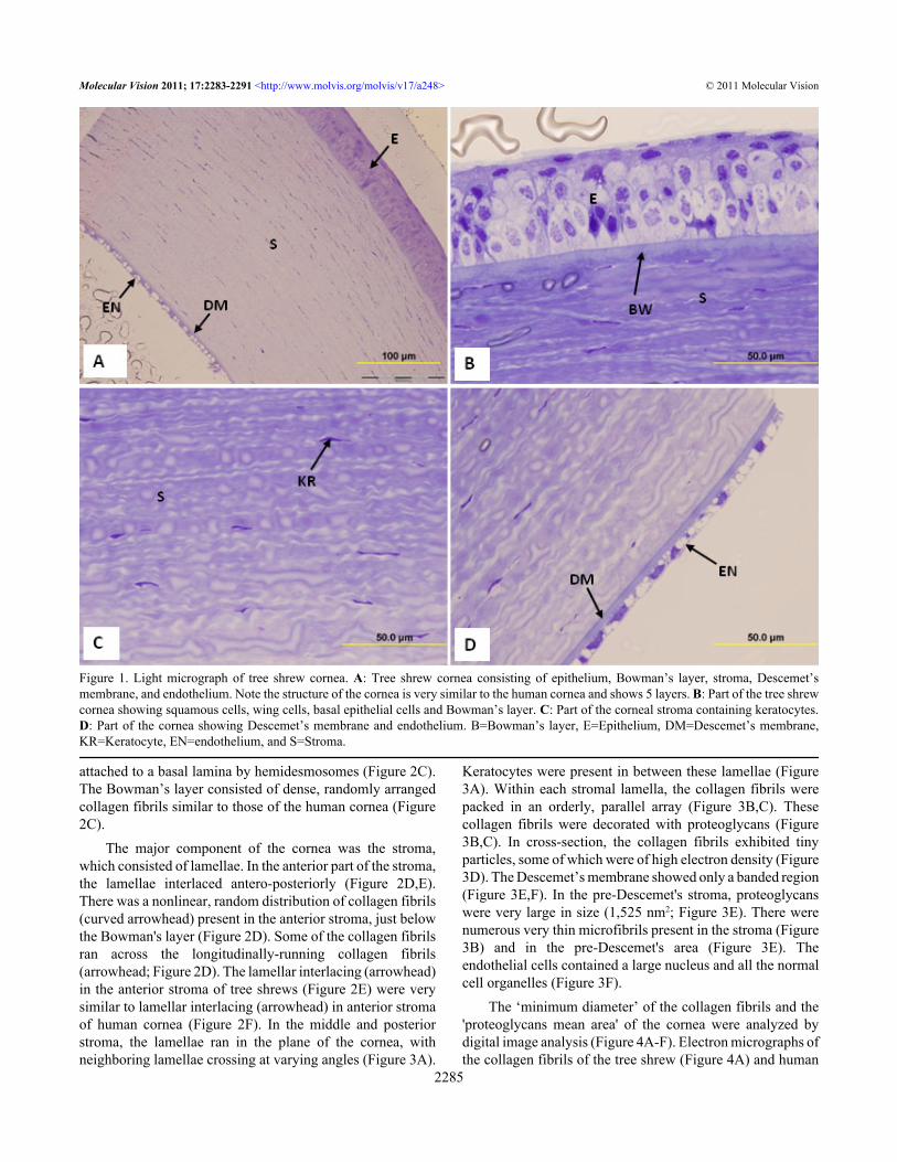
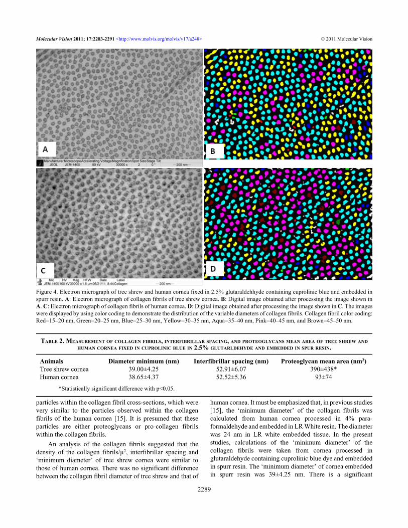
The ‘minimum diameter’ of the collagen fibrils and the'proteoglycans mean area' of the cornea were analyzed bydigital image analysis (Figure 4A-F). Electron micrographs ofthe collagen fibrils of the tree shrew (Figure 4A) and human
Figure 1. Light micrograph of tree shrew cornea. A: Tree shrew cornea consisting of epithelium, Bowman’s layer, stroma, Descemet’smembrane, and endothelium. Note the structure of the cornea is very similar to the human cornea and shows 5 layers. B: Part of the tree shrewcornea showing squamous cells, wing cells, basal epithelial cells and Bowman’s layer. C: Part of the corneal stroma containing keratocytes.D: Part of the cornea showing Descemet’s membrane and endothelium. B=Bowman’s layer, E=Epithelium, DM=Descemet’s membrane,KR=Keratocyte, EN=endothelium, and S=Stroma.
Molecular Vision 2011; 17:2283-2291 <http://www.molvis.org/molvis/v17/a248> © 2011 Molecular Vision
2285
cornea (Figure 4C) were color coded according to the size oftheir diameters (Figure 4B,D). The digital images of the treeshrew (Figure 4B) and human cornea (Figure 4D) containedcollagen fibril diameter ranges of 20 nm-50 nm. The‘minimum diameter’ in the tree shrew cornea was 39±4.25nm, and in the human cornea it was 38.65±4.37 nm (Table 2).The interfibrillar spacing in both the tree shrew and humancornea were 52.91±6.07 nm and 52.52±5.36 nm, respectively(Table 2).
Electron micrographs of the proteoglycans (PGs) of thetree shrew stroma (Figure 5A) were color coded according tothe size of the PGs, as shown in the digital image (Figure 5B).The PGs’ area was calculated from digital images. The meanPG area of the tree shrew cornea was 390±438 nm2. Thelargest PGs area in the middle stroma was approximately2,131 nm2. The mean PG area of the pre-Descemet's regionwas 1,525 nm2. The process of digitizing the electronmicrographs, as outlined for the tree shrew above, wasfollowed for the proteoglycans of the human stroma (Figure5C). The digitized images are shown in Figure 5D. The meanPG area of human cornea was 93±75 nm2. The PGs of treeshrew stroma were significantly larger than those of humancornea (Table 2).
DISCUSSIONThe normal human cornea consists of five layers, namely theepithelium, Bowman’s layer, stroma, Descemet’s membrane,and endothelium. Similar to the human cornea, the tree shrewcornea also has five layers. The thickness of the epithelium ofthe tree shrew cornea was similar to that of the human cornea,but the tree shrew Bowman's layer and stroma were thinnerthan those of the human cornea. In the tree shrew, theepithelium constitutes 15% of the corneal thickness, whereasthe stroma constitutes 80% of the corneal thickness. This isvery similar to the human cornea, in which the epitheliumconstitutes approximately 8% of the corneal thickness and thestroma constitutes 88%. The tree shrew cornea also lacked anon-banded zone in the Descemet's membrane. The humanpostnatal cornea has a non-banded zone, which increases in
thickness over the life span. The absence of such a zone in thetree shrew corneas studied may relate to the young age of thespecimens. Tree shrews in captivity may live for up to 12 yearsof age. The overall thickness of tree shrew cornea wasapproximately half that of the human cornea. In spite of thesedifferences, the ratios between the Bowman's layers and theentire cornea in both tree shrews and humans were similar.The tree shrew Bowman's layer constitutes 2.13% of thecornea, and human Bowman's layer constitutes 2.34% of thecornea.
The structure of tree shrew cornea is very different fromthat of other animals, such as the murine, bovine, and rabbit.Of particular interest and differing from that of the murine orthe bovine cornea is the occurrence of a Bowman’s layer.Hayashi et al. [14] have reported the thickness of Bowman'slayer (BW) in relation to the stromas of various animals’corneas fixed in glutaraldehyde plus osmium tetroxide andembedded on TAAB epoxy resin. According to the authors,the percentages of BW thickness in relation to the stromalthickness was very small in mice (1.4%), rats (1.2%), rabbits(1.2%), cattle (0.6%), and human (3.2%). Our studies showedthat the percentage of BW in relation to the stroma in treeshrews and humans are 1.8% and 1.9%, respectively. Thissuggests that the structures of the tree shrew Bowman's layerand stroma are very similar to human BW and stroma.Although other animals do have BWs, they are not similar tothe human BW. In addition to the tree shrew, a Bowman’slayer is also found in primate, avian, and some zebrafishcorneas [13,16,17]. Please note there is a difference in thepercentage of the BW layer thickness (3.2%) of human corneafixed in osmium plus embedded in TAAB epoxy [14] and theBW layer thickness (1.9%) of the human cornea fixed incuprolinic blue plus spurr resin in our studies. This suggeststhat the fixation method and resin affect the thickness of theBW.
Another interesting similarity between the tree shrewcornea and the human cornea is the organization of the stromainto an anterior woven zone and a posterior zone of parallellamellae. In addition to this, the high magnification imagesproduced in this study showed the presence of electron-dense
TABLE 1. MEASUREMENT OF THE CORNEAL LAYERS OF TREE SHREW AND HUMAN CORNEA FIXED IN CUPROLINIC BLUE IN 2.5% GLUTARLDEHYDE AND EMBEDDED
IN SPUR RESIN.
Corneal layers Tree shrew center CuB stainingembedded in Spurs resin (µm)
Human center CuB staining andembedded in Spurs resin (µm)
Cornea 320.96±2.95 629±7.21Epithelium 50±1.62 50±1.58Bowman’s layer 5.53±1.0 13±0.58Stroma 258±7.00 554±10.39Descemet’s membrane 3.24±0.02 11±1Bowman's layer % of cornea 1.7% 2.06Strom % of the cornea 80% 88%Epithelium percentage 15.6% 8%Bowman’s layer (BW) % of the stroma 2.13% of stroma 2.34% of stroma
Molecular Vision 2011; 17:2283-2291 <http://www.molvis.org/molvis/v17/a248> © 2011 Molecular Vision
2286

Figure 2. Electron micrograph of tree shrew and human cornea fixed in 2.5% glutaraldehyde containing cuprolinic blue and embedded in spurrresin. A: Basal epithelial cells are columnar and contain large nuclei. B: Prominent cytoplasmic filaments in basal epithelial cells. C: Basalepithelial cells attached by hemidesmosomes to a basement membrane followed by a Bowman’s layer consisting of dense, randomly arrangedcollagen fibrils. D: Non linear, random distribution of collagen fibrils (curved arrowhead) present in the anterior stroma just below theBowman's layer. Some of the collagen run across the longitudinally-running collagen fibrils (arrowhead). E: Lamellae are interlacing(arrowhead) in the anterior stroma of the tree shrew. F: Lamellae are interlacing (arrowhead) in the anterior stroma of the normal humancornea. B=Bowman’s layer, BM=Basement membrane, CF=Collagen fibrils, E=Epithelium, H=Hemidesmosomes, and MF=Cytoplasmicfilaments.
Molecular Vision 2011; 17:2283-2291 <http://www.molvis.org/molvis/v17/a248> © 2011 Molecular Vision
2287
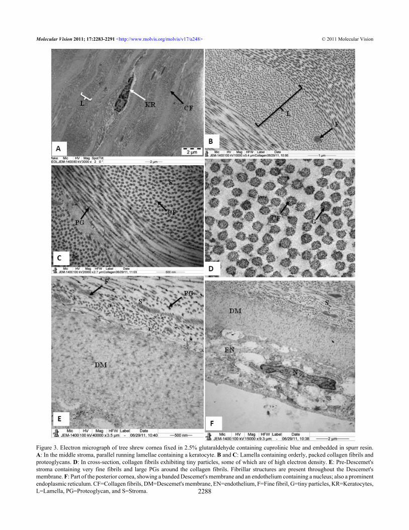
Figure 3. Electron micrograph of tree shrew cornea fixed in 2.5% glutaraldehyde containing cuprolinic blue and embedded in spurr resin.A: In the middle stroma, parallel running lamellae containing a keratocyte. B and C: Lamella containing orderly, packed collagen fibrils andproteoglycans. D: In cross-section, collagen fibrils exhibiting tiny particles, some of which are of high electron density. E: Pre-Descemet'sstroma containing very fine fibrils and large PGs around the collagen fibrils. Fibrillar structures are present throughout the Descemet'smembrane. F: Part of the posterior cornea, showing a banded Descemet's membrane and an endothelium containing a nucleus; also a prominentendoplasmic reticulum. CF=Collagen fibrils, DM=Descemet's membrane, EN=endothelium, F=Fine fibril, G=tiny particles, KR=Keratocytes,L=Lamella, PG=Proteoglycan, and S=Stroma.
Molecular Vision 2011; 17:2283-2291 <http://www.molvis.org/molvis/v17/a248> © 2011 Molecular Vision
2288
particles within the collagen fibril cross-sections, which werevery similar to the particles observed within the collagenfibrils of the human cornea [15]. It is presumed that theseparticles are either proteoglycans or pro-collagen fibrilswithin the collagen fibrils.
An analysis of the collagen fibrils suggested that thedensity of the collagen fibrils/µ2, interfibrillar spacing and‘minimum diameter’ of tree shrew cornea were similar tothose of human cornea. There was no significant differencebetween the collagen fibril diameter of tree shrew and that of
human cornea. It must be emphasized that, in previous studies[15], the ‘minimum diameter’ of the collagen fibrils wascalculated from human cornea processed in 4% para-formaldehyde and embedded in LR White resin. The diameterwas 24 nm in LR white embedded tissue. In the presentstudies, calculations of the ‘minimum diameter’ of thecollagen fibrils were taken from cornea processed inglutaraldehyde containing cuprolinic blue dye and embeddedin spurr resin. The ‘minimum diameter’ of cornea embeddedin spurr resin was 39±4.25 nm. There is a significant
Figure 4. Electron micrograph of tree shrew and human cornea fixed in 2.5% glutaraldehhyde containing cuprolinic blue and embedded inspurr resin. A: Electron micrograph of collagen fibrils of tree shrew cornea. B: Digital image obtained after processing the image shown inA. C: Electron micrograph of collagen fibrils of human cornea. D: Digital image obtained after processing the image shown in C. The imageswere displayed by using color coding to demonstrate the distribution of the variable diameters of collagen fibrils. Collagen fibril color coding:Red=15–20 nm, Green=20–25 nm, Blue=25–30 nm, Yellow=30–35 nm, Aqua=35–40 nm, Pink=40–45 nm, and Brown=45–50 nm.
TABLE 2. MEASUREMENT OF COLLAGEN FIBRILS, INTERFIBRILLAR SPACING, AND PROTEOGLYCANS MEAN AREA OF TREE SHREW ANDHUMAN CORNEA FIXED IN CUPROLINIC BLUE IN 2.5% GLUTARLDEHYDE AND EMBEDDED IN SPUR RESIN.
Animals Diameter minimum (nm) Interfibrillar spacing (nm) Proteoglycan mean area (nm2)Tree shrew cornea 39.00±4.25 52.91±6.07 390±438*Human cornea 38.65±4.37 52.52±5.36 93±74
*Statistically significant difference with p<0.05.
Molecular Vision 2011; 17:2283-2291 <http://www.molvis.org/molvis/v17/a248> © 2011 Molecular Vision
2289
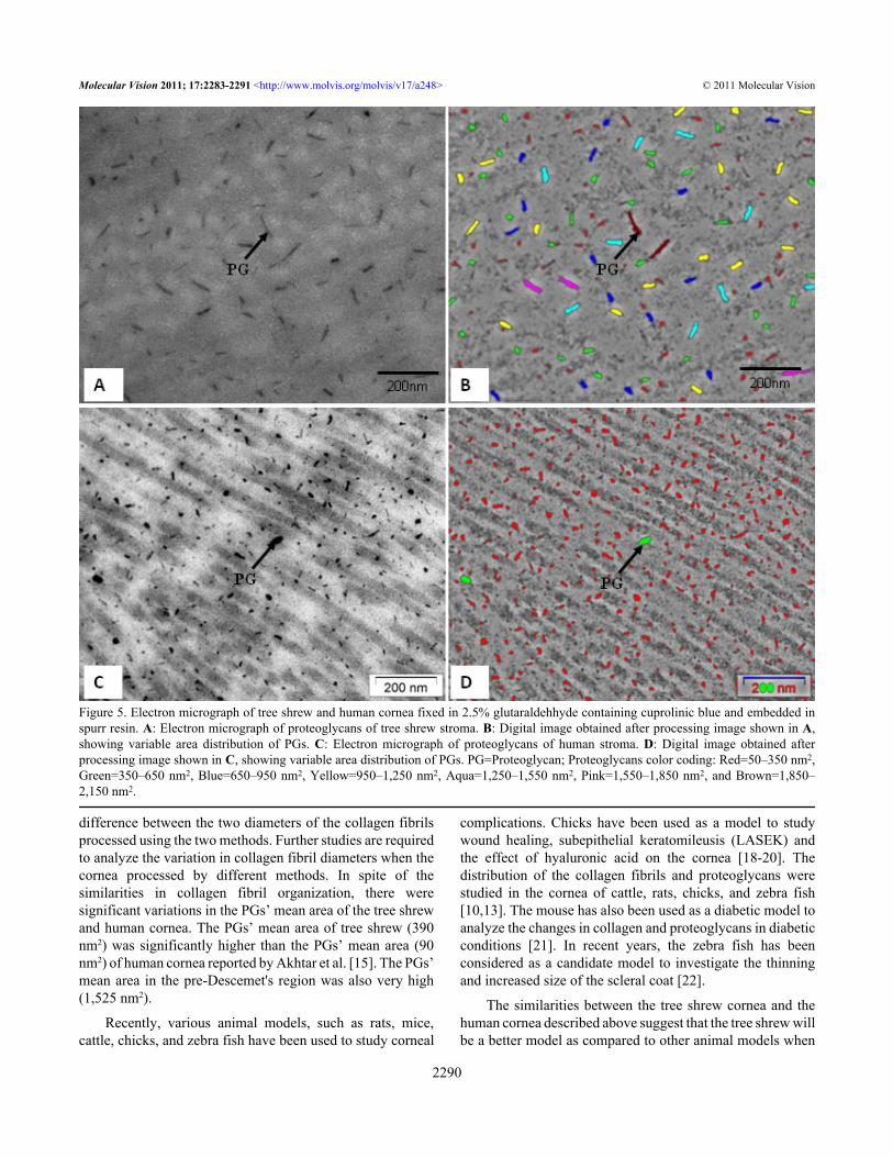
difference between the two diameters of the collagen fibrilsprocessed using the two methods. Further studies are requiredto analyze the variation in collagen fibril diameters when thecornea processed by different methods. In spite of thesimilarities in collagen fibril organization, there weresignificant variations in the PGs’ mean area of the tree shrewand human cornea. The PGs’ mean area of tree shrew (390nm2) was significantly higher than the PGs’ mean area (90nm2) of human cornea reported by Akhtar et al. [15]. The PGs’mean area in the pre-Descemet's region was also very high(1,525 nm2).
Recently, various animal models, such as rats, mice,cattle, chicks, and zebra fish have been used to study corneal
complications. Chicks have been used as a model to studywound healing, subepithelial keratomileusis (LASEK) andthe effect of hyaluronic acid on the cornea [18-20]. Thedistribution of the collagen fibrils and proteoglycans werestudied in the cornea of cattle, rats, chicks, and zebra fish[10,13]. The mouse has also been used as a diabetic model toanalyze the changes in collagen and proteoglycans in diabeticconditions [21]. In recent years, the zebra fish has beenconsidered as a candidate model to investigate the thinningand increased size of the scleral coat [22].
The similarities between the tree shrew cornea and thehuman cornea described above suggest that the tree shrew willbe a better model as compared to other animal models when
Figure 5. Electron micrograph of tree shrew and human cornea fixed in 2.5% glutaraldehhyde containing cuprolinic blue and embedded inspurr resin. A: Electron micrograph of proteoglycans of tree shrew stroma. B: Digital image obtained after processing image shown in A,showing variable area distribution of PGs. C: Electron micrograph of proteoglycans of human stroma. D: Digital image obtained afterprocessing image shown in C, showing variable area distribution of PGs. PG=Proteoglycan; Proteoglycans color coding: Red=50–350 nm2,Green=350–650 nm2, Blue=650–950 nm2, Yellow=950–1,250 nm2, Aqua=1,250–1,550 nm2, Pink=1,550–1,850 nm2, and Brown=1,850–2,150 nm2.
Molecular Vision 2011; 17:2283-2291 <http://www.molvis.org/molvis/v17/a248> © 2011 Molecular Vision
2290

used to study the causes of the thinning of the cornea, as wellas changes in collagen fibrils and proteoglycans inpathological conditions, such as diabetes. The occurrence ofa Bowman’s layer similar to that in humans also suggests thatthe tree shrew will be a good model to assess swelling and itseffect on the collagen fibrils and proteoglycans. The treeshrew cornea could also be a good model to study the collagenfibrils and proteoglycans present with non-genetic diseases,such as ectasia caused after LASIK.
ACKNOWLEDGMENTS
The research project was funded by the National Plan for Science and Technology, King Saud University, Riyadh,Saudi Arabia.
REFERENCES1. Siegwart JT Jr, Norton TT. The susceptible period for
deprivation-induced myopia in tree shrews. Vision Res 1998;38:3505-15. [PMID: 9893785]
2. Norton TT, Amedo AO, John T. Siegwart Jr. Darkness causesmyopia in visually experienced tree shrews. InvestOphthalmol Vis Sci 2006; 47:4700-7. [PMID: 17065476]
3. McBrien NA, Norton TT. Prevention of collagen crosslinkingincreases form-deprivation myopia in tree shrews. Exp EyeRes 1994; 59:475-86. [PMID: 7859823]
4. Jobling AI, Nguyen M, Gentle A, McBrien NA. Isoform-specific changes in scleral transforming growth factor-betaexpression and the regulation of collagen synthesis duringmyopia progression. J Biol Chem 2004; 279:18121-6.[PMID: 14752095]
5. Ishiko S, Yoshida A, Mori F, Abiko T, Kitaya N, Kojima M,Saito K. Early ocular changes in a tree shrew model ofdiabetes. Nippon Ganka Gakkai Zasshi 1997; 101:19-23.[PMID: 9028102]
6. Cao J, Yang EB, Su JJ, Li Y, Chow P. The tree shrews: adjunctsand alternatives to primates as models for biomedicalresearch. J Med Primatol 2003; 32:123-30. [PMID:12823622]
7. McBrien NA, Cornell LM, Gentle A. Structural andultrastructural changes to the sclera in a mammalian model ofhigh myopia. Invest Ophthalmol Vis Sci 2001; 42:2179-87.[PMID: 11527928]
8. Albon J, Farrant S, Akhtar S, Young R, Boulton ME, Smith G,Taylor M, Guggenheim J, Morgan JE. Connective tissuestructure of the tree shrew optic nerve and associated ageingchanges. Invest Ophthalmol Vis Sci 2007; 48:2134-44.[PMID: 17460272]
9. Maurice DM. The structure and transparency of the cornea. JPhysiol 1957; 136:263-86. [PMID: 13429485]
10. Chakravarti S, Petroll MW, Hassell JR, Jester JV, Lass JH, PaulJ, Birk DE. Corneal Opacity in Lumican-Null Mice: Defectsin Collagen Fibril Structure and Packing in the PosteriorStroma. Invest Ophthalmol Vis Sci 2000; 41:3365-73.[PMID: 11006226]
11. Scott JE, Haigh M. Identification of specific binding sites forkeratan sulphate proteoglycans and chondroitin-dermatansulphate proteoglycans on collagen fibrils in cornea by the useof cupromeronic blue in 'critical-electrolyte-concentration'techniques. Biochem J 1988; 253:607-10. [PMID: 2972275]
12. Birk DE, Fitch JM, Babiarz JP, Linsenmayer TF. Collagen typeI and type V are present in the same fibril in the avian cornealstroma. J Cell Biol 1988; 106:999-1008. [PMID: 3346334]
13. Akhtar S, Schonthaler HB, Bron AJ, Dahm R. Theultrastructural organization of stromal collagen andproteoglycan in the zebrafish cornea during development.Acta Ophthalmol 2008; 86:655-65. [PMID: 18221494]
14. Hayashi S, Osawa T, Tohyama K. Comparative observations ofcornea, with special reference to Bowman’s layer andDescemet’s membrane in Mammals and Amphibians. JMorphol 2002; 254:247-58. [PMID: 12386895]
15. Merindano MD, Canals M, Potau JM, Costa J, Ruano D.Morphological and morphometric aspects of primate cornea:a comparative study with human cornea. Eur J Morphol 1997;35:95-104. [PMID: 9253587]
16. Linsenmayer TF, Firch JM. May net R. Extracellular matricesin the developing avian eye: type V collagen in corneal andnon-corneal tissues. Invest Ophthalmol Vis Sci 1984;25:41-7. [PMID: 6365824]
17. Akhtar S, Bron AJ, Salvi SM, Hawksworth NR, Tuft SJ, MeekKM. Collagen fibrils and proteoglycans in keratoconus - aquantitative ultra-structural analysis. Acta Ophthalmol 2008;86:764-72. [PMID: 18422999]
18. Martínez-García MC, Merayo-Llovés J, Blanco-Mezquita T,Mar-Sardaña S. Wound healing following refractive surgeryin hens. Exp Eye Res 2006; 83:728-35. [PMID: 16701650]
19. Lee JB, Javier JA, Chang JH, Chen CC, Kato T, Azar DT.Confocal and electron microscopic studies of lasersubepithelial keratomileusis (LASEK) in the white leghornchick eye. Arch Ophthalmol 2002; 120:1700-6. [PMID:12470145]
20. Gómez S, Herreras JM, Merayo J, García M, Argüeso P, CuevasJ. Effect of hyaluronic acid on corneal haze in aphotorefractive keratectomy experimental model. J RefractSurg 2001; 17:549-54. [PMID: 11583225]
21. Akhtar S, Almubrad TM, Bron AJ, Yusif MHM, Benter IF,Akhtar S. Effect of EGFR in corneal remodelling in diabetes.Acta Ophthalmol 2009; 87:881-9. [PMID: 19416119]
22. Yeh LK, Liu CY, Kao WW, Huang CJ, Hu FR, Chien CL, WangIJ. Knockdown of zebrafish lumican gene (zlum) causesscleral thinning and increased size of scleral coats. J BiolChem 2010; 285:28141-55. [PMID: 20551313]
Molecular Vision 2011; 17:2283-2291 <http://www.molvis.org/molvis/v17/a248> © 2011 Molecular Vision
Articles are provided courtesy of Emory University and the Zhongshan Ophthalmic Center, Sun Yat-sen University, P.R. China.The print version of this article was created on 22 August 2011. This reflects all typographical corrections and errata to thearticle through that date. Details of any changes may be found in the online version of the article.
2291