structural and functional studies of sav0551 from ... · title structural and functional studies of...
TRANSCRIPT
Bioscience Reports (2017) 37 BSR20171106https://doi.org/10.1042/BSR20171106
Received: 08 June 2017Revised: 10 November 2017Accepted: 10 December 2017
Accepted Manuscript Online:17 October 2017Version of Record published:17 November 2017
Research Article
Structural and functional studies of SAV0551 fromStaphylococcus aureus as a chaperone andglyoxalase IIIHyo Jung Kim1, Ki-Young Lee1, Ae-Ran Kwon2 and Bong-Jin Lee1
1Research Institute of Pharmaceutical Sciences, College of Pharmacy, Seoul National University, Gwanak-gu, Seoul 151-742, Korea; 2Department of Herbal Skin Care, College ofHerbal Bio-industry, Deagu Haany University, Gyeongsan 712-715, Korea
Correspondence: Ae-Ran Kwon ([email protected]) or Bong-Jin Lee ([email protected])
The DJ-1/ThiJ/PfpI superfamily of proteins is highly conserved across all biological king-doms showing divergent multifunctions, such as chaperone, catalase, protease, and ki-nase. The common theme of these functions is responding to and managing various cellu-lar stresses. DJ-1/ThiJ/PfpI superfamily members are classified into three subfamilies ac-cording to their quaternary structure (DJ-1-, YhbO-, and Hsp-types). The Hsp-type sub-family includes Hsp31, a chaperone and glyoxalase III. SAV0551, an Hsp-type subfamilymember from Staphylococcus aureus, is a hypothetical protein that is predicted as Hsp31.Thus, to reveal the function and reaction mechanism of SAV0551, the crystal structure ofSAV0551 was determined. The overall folds in SAV0551 are similar to other members ofthe Hsp-type subfamily. We have shown that SAV0551 functions as a chaperone and thatthe surface structure is crucial for holding unfolded substrates. As many DJ-1/ThiJ/PfpI su-perfamily proteins have been characterized as glyoxalase III, our study also demonstratesSAV0551 as a glyoxalase III that is independent of any cofactors. The reaction mechanismwas evaluated via a glyoxylate-bound structure that mimics the hemithioacetal reaction in-termediate. We have confirmed that the components required for reaction are present inthe structure, including a catalytic triad for a catalytic action, His78 as a base, and a watermolecule for hydrolysis. Our functional studies based on the crystal structures of native andglyoxylate-bound SAV0551 will provide a better understanding of the reaction mechanismof a chaperone and glyoxalase III.
IntroductionThe DJ-1/ThiJ/Pf pI superfamily (DJ-1 superfamily hereinafter) is a group of proteins that are universal toall the kingdoms of life [1]. The members of this superfamily share structural and functional similarity tohuman DJ-1, which is known to be associated with Parkinson disease [2,3]. The crystal structures of sev-eral members of the DJ-1 superfamily have been solved. Most of them are oligomers, with each monomerconsisting of center-aligned β-strands surrounded by α-helices, which is called an α/β sandwich fold[4]. A completely conserved cysteine is located at a sharp turn between a β-strand and an α-helix. Thesharp turn motif is referred to as the ‘nucleophilic elbow’ and is known to allow easy access to the cysteineby substrates [5]. DJ-1 superfamily proteins are divided into the following distinct subfamilies based ontheir quaternary structure: (i) DJ-1-type, (ii) YhbO-type, and (iii) Hsp-type. Each of the DJ-1 subfamiliesalso differs in the architecture of the conserved cysteine. DJ-1-type subfamily proteins (DJ-1 and YajL)form dimeric interfaces consisting of α-helices, β-strands, and loops. The conserved cysteine does notform a catalytic triad in human DJ-1 and bacterial YajL [6,7]. Many functions of DJ-1 have been revealedincluding chaperone, neurone protection, and glyoxalase III [8-10]. The YhbO-type subfamily includes
c© 2017 The Author(s). This is an open access article published by Portland Press Limited on behalf of the Biochemical Society and distributed under the Creative Commons AttributionLicense 4.0 (CC BY).
1
Dow
nloaded from https://portlandpress.com
/HTTPH
andlers/ArticlePdfHandler.ashx?partialdoi=BSR
20171106&journal=bioscirep by guest on 28 Decem
ber 2019
Bioscience Reports (2017) 37 BSR20171106https://doi.org/10.1042/BSR20171106
the representative YhbO from Escherichia coli, SAV1875 from Staphylococcus aureus, Pf pI from Pyrococcushorikoshii, and Ton1285 from Thermococcus onnurineus. YhbO-type subfamily members form dimers with threeα-helices. The catalytic triad in this subfamily consists of a cysteine, histidine, and an acidic residue from the adja-cent subunit. Ton1285 and Pf pI from the YhbO-type subfamily are known to cleave peptide bonds [4,11]. Hsp-typesubfamily members have a unique dimerization surface consisting of N-terminal β-strands and loops [12]. As heatshock proteins (HSPs) are considered molecular chaperones, most members of the Hsp-type subfamily function aschaperones, specifically holding chaperones [13]. Holding chaperones capture unfolded proteins with their surfacestructures for proper folding. The discriminating surface structures, canyons, and bowls of Hsp31 from E. coli andVibrio cholerae distinguish them from other subfamily members [12,14]. Hsp-type DJ-1 subfamily proteins have acatalytic triad consisting of a cysteine, histidine, and an acidic residue from the cap domain of the same subunit. Withthe catalytic cysteine in the catalytic triad, most proteins in the Hsp-type subfamily function as glyoxalase III [15-18].
The distinct subclass classification correlates with structural characteristics, but known functions of DJ-1 super-family proteins cannot be classified into subclasses. From our previous study, we determined the surface structureof the YhbO-type protein SAV1875 differs from the YhbO-type subfamily proteins but is similar to Hsp-type sub-family proteins. SAV1875 shows chaperone activity similar to other Hsp-type subfamily proteins [19]. However, theHsp-type protein Glx3 from Candida albicans and YDR533C from Saccharomyces cerevisiae do not show chap-erone activity. The differences in the chaperone activity within the Hsp-type subfamily are shown to be based onthe importance of their surface structure, as these two proteins lack the 45 amino acid N-terminal region [16]. Theglyoxalase III function was first revealed in Hsp-type subfamily proteins, though YajL and DJ-1 from the DJ-1-typeand YhbO from the YhbO-type subfamily also show the same function [9,20]. Despite the structural and functionaldifferences in the classes, many DJ-1 superfamily proteins perform chaperone and glyoxalase III functions. Understress conditions, chaperone proteins protect cells by stabilizing unfolded proteins, giving the cell time to repair orresynthesize damaged proteins, which is important for protecting cells from severe stress conditions [21]. GlyoxalaseIII directly converts the toxic methylglyoxal substrate to non-toxic d-lactate without glutathione. Methlyglyoxal is atoxic endogenous electrophile component that damages proteins, nucleic acids, and lipids [22]. Therefore, studyingthe DJ-1 superfamily of proteins is important for understanding cellular protection mechanisms from various stressconditions.
In the present study, the structural and functional identification of hypothetical SAV0551 from S. aureus Mu50 wasconducted. S. aureus Mu50 (ATCC 700699) is a vancomycin-intermediate S. aureus that displays robust virulenceproperties. It causes severe infectious diseases ranging from mild infections, such as skin infections and food poi-soning, to life-threatening infections, such as sepsis, endocarditis, and toxic shock syndrome [23]. However, becauseof the bacterial resistance, the prognosis for S. aureus infection is still poor despite early diagnosis and appropriatetreatment [24]. Although there are various theories regarding how S. aureus acquires antibiotic resistance, very littleis known. In this regard, understanding the bacterial defense mechanisms in S. aureus against antibiotics is crucial.Furthermore, the DJ-1 superfamily of proteins is predicted to have stress-response functions under multiple stressconditions [25]. Therefore, we designed structural and functional experiments with SAV0551, a DJ-1 superfamilyprotein from S. aureus Mu50, as an extension of our previous work on SAV1875. We elucidated the structure andfunction of SAV0551 as a chaperone and glyoxalase III. The catalytic triad mutants (C190A, H191A, and D221A) andabsolutely conserved glutamate mutant (E81A) were designed to identify structure-related functional differences. Themutants lost glyoxalase III activity but maintained chaperone activity. We show a co-crystal structure of SAV0551 withthe known glyoxalase III inhibitor glyoxylate, and it shows the intermediate structure in the glyoxalase III reaction.Our structural and functional research on previously unknown SAV0551 presents new residues that are involved inglyoxalase III reaction.
ExperimentalCloning, protein expression, and purificationThe predicted ORF of SAV0551 was amplified from S. aureus Mu50 genomic DNA using standard PCR meth-ods. The forward and reverse oligonucleotide primers were designed using the published genome sequence, 5′-GACTGCATATGTCACAAGATGTAAATGAATTAAG and 5′- GTCACTCGAGT TTATTTTGTATTGCATTTAA-CAT, respectively, where the bases underlined represent the NdeI and XhoI restriction enzyme cleavage sites. Theamplified DNA was inserted into the NdeI/XhoI-digested expression vector pET-21a(+) (Novagen). The resultingconstruct contains eight non-native residues at the C-terminus (LEHHHHHH) that facilitate protein purification.The accuracy of the cloning was confirmed by DNA sequencing. The resulting expression plasmid was then trans-formed into E. coli BL21(DE3) cells (Novagen).
2 c© 2017 The Author(s). This is an open access article published by Portland Press Limited on behalf of the Biochemical Society and distributed under the Creative Commons AttributionLicense 4.0 (CC BY).
Dow
nloaded from https://portlandpress.com
/HTTPH
andlers/ArticlePdfHandler.ashx?partialdoi=BSR
20171106&journal=bioscirep by guest on 28 Decem
ber 2019
Bioscience Reports (2017) 37 BSR20171106https://doi.org/10.1042/BSR20171106
To prepare mutants, the EZchange Site-directed Mutagenesis kit (Enzynomics) was used to generate point muta-tions in the SAV0551 recombinant pET-21a(+) plasmid. The point mutations resulted in separate multiple recom-binant plasmids, specifically E81A, C190A, H191A, and D221A. The sequences of the reconstructed mutants wereconfirmed by DNA sequencing (results not shown).
The wild-type and SAV0551 mutants’ (E81A, C190A, H191A, and D221A) cells were grown at 37◦C until theOD600 reached 0.6 and expression was induced by the addition of IPTG to a final concentration of 0.5 mM. Afteran additional 4 h of growth at 37◦C, cells were harvested by centrifugation and resuspended in 50 mM Tris, pH 7.5,0.5 M NaCl, and 20 mM imidazole buffer. Cells were lysed by sonication at 4◦C and the supernatant was loadedon to Ni2+-NTA (Ni2+-nitrilotriacetate) affinity column (Qiagen; 3 ml of resin per liter of cell culture) previouslyequilibrated with the same buffer. The column was washed extensively with wash buffer (50 mM Tris, pH 7.5, 0.5M NaCl, and 50 mM imidazole); then the bound protein was eluted with elution buffer (50 mM Tris, pH 7.5, 0.5 MNaCl, and 500 mM imidazole) until there was no detectable absorbance at 280 nm in the elutant. Fractions containingprotein were concentrated to ∼2 ml and applied to a Superdex 75 (10/300 GL) column (GE Healthcare Life Sciences)that had been equilibrated with the final buffer (50 mM Tris, pH 7.5 and 0.15 M NaCl). The purities of SAV0551was judged to be over 95% by SDS/PAGE. The protein solution was concentrated using 10000 Da molecular-masscut-off spin columns (Millipore). The protein concentration was estimated by measuring the absorbance at 280 nm,employing the calculated extinction coefficient of 35535 M−1.cm−1 (Swiss-Prot; http://www.expasy.org).
Crystallization, data collection, structure determination, and refinementCrystallization was performed at 293 K by the hanging-drop vapor diffusion method using 24-well VDX plates(Hampton Research). Initial crystallization conditions were established using screening kits from Hampton Research(Crystal Screens I and II, Index, PEG/Ion, and MembFac) and from Emerald BioSystems (Wizard I, II, III, and IV). Forthe optimal growth of the SAV0551 crystals, each hanging drop was prepared on a siliconized cover slip by mixing 1μl of protein solution (15 mg/ml) and 1 μl of precipitant solution (23% (w/v) PEG3350, 100 mM BisTris, pH 6.0), andthis drop was equilibrated against a 1-ml reservoir of precipitant solution. These conditions yielded needle-shapedcrystals for each protein that grew to dimensions of 1.0 × 0.3 × 0.2 mm in two days. The co-crystal with glyoxylatewere prepared by addition of 50 mM glyoxylate, 0.2–1 μl of protein solution and 1 μl of precipitant solution (30%(w/v) PEG3350, 100 mM BisTris, pH 6.5). All crystals belonged to space group P212121 and contained eight moleculesper asymmetric unit.
For crystal freezing, the crystals were transferred to a cryoprotectant solution with 30% (v/v) ethylene glycol in thecrystallization condition for several minutes before being flash-frozen in a stream of nitrogen gas at 100 K. Diffractiondata were collected on beamline 5C at the Pohang Light Source, South Korea. The raw data were processed andscaled using the HKL2000 program suite [26]. Further data analysis was carried out using the CCP4 suite [27]. Datacollection statistics are summarized in Table 1.
To determine the structure of the wild-type SAV0551, molecular replacement was used with the program Molrep[28] within the CCP4 suite [27] using the homologous structure of Hsp31 from V. cholerae (PDB code: 4I4N) as asearch model. The SAV0551 and Hsp31 from V. cholerae shows 49% sequence identity. To determine the structure ofglyoxylate-bound SAV0551, molecular replacement was performed using the wild-type SAV0551 as the search model.Refinement of each crystal structure was done through iterative cycles of model building using COOT [29], followedby refinement of the models with Refmac5 and phenix.refine [30,31]. A 5% portion of the data were set aside prior torefinement for the Rfree calculations for each dataset [32]. Solvent and glyoxylate molecules became apparent in thelater stages of refinement and were added into the model. Further refinement was pursued until no further decreasein Rfree was observed. Structural alignments were carried out using the program PyMOL (http://www.pymol.org) andUCSF Chimera (http://www.cgl.ucsf.edu/chimera) [33], which were then used for the construction and generationof all the figures. Protein interfaces, surfaces, and assemblies were calculated using the PISA server at the EuropeanBioinformatics Institute (http://www.ebi.ac.uk/pdbe/prot int/pistart.html) [34].
Determination of chaperone activityTo monitor the chaperone activity of the wild-type and SAV0551 mutants, citrate synthase was employed as a substrate[35,36]. Initially, to identify chaperone activity, 75 μg of citrate synthase (Sigma–Aldrich) was mixed with a solutionof 100 mM Tris, pH 8.0, 20 mM DTT, 6 M guanidine chloride (GnCl). The citrate synthase mixture (75 μg of citratesynthase, 100 mM Tris, pH 8.0, 6 M GnCl, 20 mM DTT) was incubated for 1 h at 25◦C; consequently, the citratesynthase in this solution was denatured. After incubation, refolding of citrate synthase was achieved by 100-folddilution with a solution of 100 mM Tris (pH 8.0) containing 5 μM wild-type and SAV0551 mutants. The diluted
c© 2017 The Author(s). This is an open access article published by Portland Press Limited on behalf of the Biochemical Society and distributed under the Creative Commons AttributionLicense 4.0 (CC BY).
3
Dow
nloaded from https://portlandpress.com
/HTTPH
andlers/ArticlePdfHandler.ashx?partialdoi=BSR
20171106&journal=bioscirep by guest on 28 Decem
ber 2019
Bioscience Reports (2017) 37 BSR20171106https://doi.org/10.1042/BSR20171106
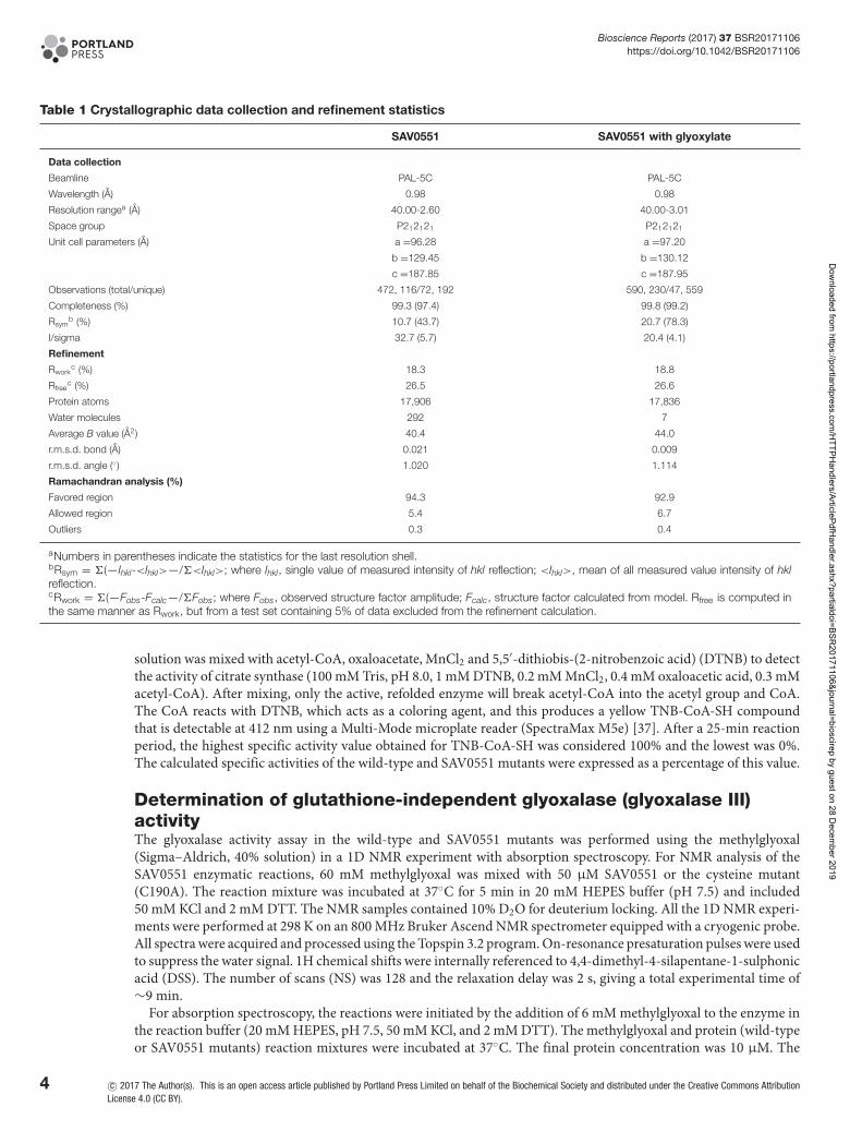
Table 1 Crystallographic data collection and refinement statistics
SAV0551 SAV0551 with glyoxylate
Data collection
Beamline PAL-5C PAL-5C
Wavelength (A) 0.98 0.98
Resolution rangea (A) 40.00-2.60 40.00-3.01
Space group P212121 P212121
Unit cell parameters (A) a =96.28 a =97.20
b =129.45 b =130.12
c =187.85 c =187.95
Observations (total/unique) 472, 116/72, 192 590, 230/47, 559
Completeness (%) 99.3 (97.4) 99.8 (99.2)
Rsymb (%) 10.7 (43.7) 20.7 (78.3)
I/sigma 32.7 (5.7) 20.4 (4.1)
Refinement
Rworkc (%) 18.3 18.8
Rfreec (%) 26.5 26.6
Protein atoms 17,906 17,836
Water molecules 292 7
Average B value (A2) 40.4 44.0
r.m.s.d. bond (A) 0.021 0.009
r.m.s.d. angle (◦) 1.020 1.114
Ramachandran analysis (%)
Favored region 94.3 92.9
Allowed region 5.4 6.7
Outliers 0.3 0.4
aNumbers in parentheses indicate the statistics for the last resolution shell.bRsym = �(—Ihkl-<Ihkl>—/�<Ihkl>; where Ihkl, single value of measured intensity of hkl reflection; <Ihkl>, mean of all measured value intensity of hklreflection.cRwork = �(—Fobs-Fcalc—/�Fobs; where Fobs, observed structure factor amplitude; Fcalc, structure factor calculated from model. Rfree is computed inthe same manner as Rwork, but from a test set containing 5% of data excluded from the refinement calculation.
solution was mixed with acetyl-CoA, oxaloacetate, MnCl2 and 5,5′-dithiobis-(2-nitrobenzoic acid) (DTNB) to detectthe activity of citrate synthase (100 mM Tris, pH 8.0, 1 mM DTNB, 0.2 mM MnCl2, 0.4 mM oxaloacetic acid, 0.3 mMacetyl-CoA). After mixing, only the active, refolded enzyme will break acetyl-CoA into the acetyl group and CoA.The CoA reacts with DTNB, which acts as a coloring agent, and this produces a yellow TNB-CoA-SH compoundthat is detectable at 412 nm using a Multi-Mode microplate reader (SpectraMax M5e) [37]. After a 25-min reactionperiod, the highest specific activity value obtained for TNB-CoA-SH was considered 100% and the lowest was 0%.The calculated specific activities of the wild-type and SAV0551 mutants were expressed as a percentage of this value.
Determination of glutathione-independent glyoxalase (glyoxalase III)activityThe glyoxalase activity assay in the wild-type and SAV0551 mutants was performed using the methylglyoxal(Sigma–Aldrich, 40% solution) in a 1D NMR experiment with absorption spectroscopy. For NMR analysis of theSAV0551 enzymatic reactions, 60 mM methylglyoxal was mixed with 50 μM SAV0551 or the cysteine mutant(C190A). The reaction mixture was incubated at 37◦C for 5 min in 20 mM HEPES buffer (pH 7.5) and included50 mM KCl and 2 mM DTT. The NMR samples contained 10% D2O for deuterium locking. All the 1D NMR experi-ments were performed at 298 K on an 800 MHz Bruker Ascend NMR spectrometer equipped with a cryogenic probe.All spectra were acquired and processed using the Topspin 3.2 program. On-resonance presaturation pulses were usedto suppress the water signal. 1H chemical shifts were internally referenced to 4,4-dimethyl-4-silapentane-1-sulphonicacid (DSS). The number of scans (NS) was 128 and the relaxation delay was 2 s, giving a total experimental time of∼9 min.
For absorption spectroscopy, the reactions were initiated by the addition of 6 mM methylglyoxal to the enzyme inthe reaction buffer (20 mM HEPES, pH 7.5, 50 mM KCl, and 2 mM DTT). The methylglyoxal and protein (wild-typeor SAV0551 mutants) reaction mixtures were incubated at 37◦C. The final protein concentration was 10 μM. The
4 c© 2017 The Author(s). This is an open access article published by Portland Press Limited on behalf of the Biochemical Society and distributed under the Creative Commons AttributionLicense 4.0 (CC BY).
Dow
nloaded from https://portlandpress.com
/HTTPH
andlers/ArticlePdfHandler.ashx?partialdoi=BSR
20171106&journal=bioscirep by guest on 28 Decem
ber 2019
Bioscience Reports (2017) 37 BSR20171106https://doi.org/10.1042/BSR20171106
remaining methylglyoxal at each time point was determined by reaction with 2,4-dinitrophenylhydrazine (DNPH)to generate the purple chromophore mehtylglyoxal-bis-2,4,-dinitrophenylhydrazone after alkali treatment [38]. Theassay was performed by removing a 50 μl sample from the reaction at fixed time points after enzyme addition (0,15, 30, 45, 60, 75, 90, 105, and 120 s) and rapidly mixing the sample with 0.9 ml of distilled water. To this solution,0.33 ml of a freshly prepared stock of DNPH reagent (0.2% DNPH dissolved in 2 N HCl) was immediately added andincubated at 37◦C for 15 min. This highly acidic solution stops the enzymatic reaction, whose rate was already greatlydiminished by the initial 19-fold dilution into water. The purple color of the hydrazine is developed by the addition of1.67 ml of 3.8 M NaOH and incubation for 10 min at room temperature followed by measurement of the absorbanceat 550 nm in a SpectraMax M5e. All the measurements were made in triplicate.
PDB codesProtein co-ordinates and structure factors have been deposited in the RCSB PDB under codes 5XR2 for the nativeSAV0551 and 5XR3 for SAV0551 with glyoxylate.
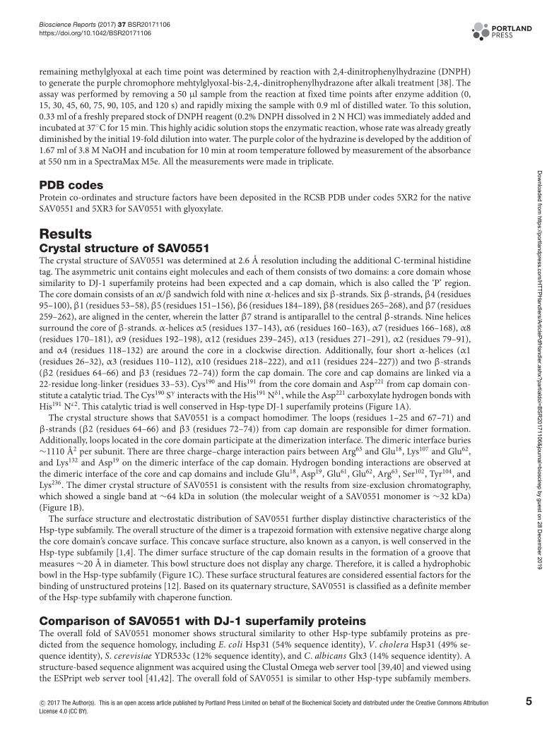
ResultsCrystal structure of SAV0551The crystal structure of SAV0551 was determined at 2.6 A resolution including the additional C-terminal histidinetag. The asymmetric unit contains eight molecules and each of them consists of two domains: a core domain whosesimilarity to DJ-1 superfamily proteins had been expected and a cap domain, which is also called the ‘P’ region.The core domain consists of an α/β sandwich fold with nine α-helices and six β-strands. Six β-strands, β4 (residues95–100),β1 (residues 53–58),β5 (residues 151–156),β6 (residues 184–189),β8 (residues 265–268), andβ7 (residues259–262), are aligned in the center, wherein the latter β7 strand is antiparallel to the central β-strands. Nine helicessurround the core of β-strands. α-helices α5 (residues 137–143), α6 (residues 160–163), α7 (residues 166–168), α8(residues 170–181), α9 (residues 192–198), α12 (residues 239–245), α13 (residues 271–291), α2 (residues 79–91),and α4 (residues 118–132) are around the core in a clockwise direction. Additionally, four short α-helices (α1(residues 26–32), α3 (residues 110–112), α10 (residues 218–222), and α11 (residues 224–227)) and two β-strands(β2 (residues 64–66) and β3 (residues 72–74)) form the cap domain. The core and cap domains are linked via a22-residue long-linker (residues 33–53). Cys190 and His191 from the core domain and Asp221 from cap domain con-stitute a catalytic triad. The Cys190 Sγ interacts with the His191 Nδ1, while the Asp221 carboxylate hydrogen bonds withHis191 Nε2. This catalytic triad is well conserved in Hsp-type DJ-1 superfamily proteins (Figure 1A).
The crystal structure shows that SAV0551 is a compact homodimer. The loops (residues 1–25 and 67–71) andβ-strands (β2 (residues 64–66) and β3 (residues 72–74)) from cap domain are responsible for dimer formation.Additionally, loops located in the core domain participate at the dimerization interface. The dimeric interface buries∼1110 A2 per subunit. There are three charge–charge interaction pairs between Arg63 and Glu18, Lys107 and Glu62,and Lys132 and Asp19 on the dimeric interface of the cap domain. Hydrogen bonding interactions are observed atthe dimeric interface of the core and cap domains and include Glu18, Asp19, Glu61, Glu62, Arg63, Ser102, Tyr104, andLys236. The dimer crystal structure of SAV0551 is consistent with the results from size-exclusion chromatography,which showed a single band at ∼64 kDa in solution (the molecular weight of a SAV0551 monomer is ∼32 kDa)(Figure 1B).
The surface structure and electrostatic distribution of SAV0551 further display distinctive characteristics of theHsp-type subfamily. The overall structure of the dimer is a trapezoid formation with extensive negative charge alongthe core domain’s concave surface. This concave surface structure, also known as a canyon, is well conserved in theHsp-type subfamily [1,4]. The dimer surface structure of the cap domain results in the formation of a groove thatmeasures ∼20 A in diameter. This bowl structure does not display any charge. Therefore, it is called a hydrophobicbowl in the Hsp-type subfamily (Figure 1C). These surface structural features are considered essential factors for thebinding of unstructured proteins [12]. Based on its quaternary structure, SAV0551 is classified as a definite memberof the Hsp-type subfamily with chaperone function.
Comparison of SAV0551 with DJ-1 superfamily proteinsThe overall fold of SAV0551 monomer shows structural similarity to other Hsp-type subfamily proteins as pre-dicted from the sequence homology, including E. coli Hsp31 (54% sequence identity), V. cholera Hsp31 (49% se-quence identity), S. cerevisiae YDR533c (12% sequence identity), and C. albicans Glx3 (14% sequence identity). Astructure-based sequence alignment was acquired using the Clustal Omega web server tool [39,40] and viewed usingthe ESPript web server tool [41,42]. The overall fold of SAV0551 is similar to other Hsp-type subfamily members.
c© 2017 The Author(s). This is an open access article published by Portland Press Limited on behalf of the Biochemical Society and distributed under the Creative Commons AttributionLicense 4.0 (CC BY).
5
Dow
nloaded from https://portlandpress.com
/HTTPH
andlers/ArticlePdfHandler.ashx?partialdoi=BSR
20171106&journal=bioscirep by guest on 28 Decem
ber 2019

Bioscience Reports (2017) 37 BSR20171106https://doi.org/10.1042/BSR20171106
Figure 1. Crystal structure of SAV0551
(A) Ribbon representation of the SAV0551 monomer with two domains. SAV0551 shows a sandwich–
folded main domain and a cap domain. Each domain is connected by a linker (green). SAV0551 has an
α1-linker-β1-β2-β3-α2-β4-α3-α4-α5-β5-α6-α7-α8-β6-α9-α10-α11-α12-β7-β8-α13 topology. The Cys190 at a sharp turn
between β6 and α9 forms a catalytic triad with His191 from the main domain and Asp221 from the cap domain. (B) The SAV0551
dimer is shown in ribbon representation from a side view. Chain A is colored in pink and chain B is colored in blue. The cap
domain of each subunit is shown in a darker color. The β-strands and loop from the cap domain are mainly involved at the dimeric
interface. (C) The potential surface charge of the SAV0551 crystal structure calculated by UCSF Chimera, where the surfaces are
colored between –10 kcal/mol · e (red) and +10 kcal/mol · e (blue) (1 kcal =4.184 kJ). Most of the surface is negatively charged.
The canyon and bowl are indicated.
The SAV0551 structure was submitted to the DALI server (http://ekhidna.biocenter.helsinki.fi/dali server/) to iden-tify structural homologs. The DALI algorithm reveals that SAV0551 is structurally similar to Hsp31 proteins withhigh Z scores. Most of the structural matches were DJ-1 family members, including Hsp31 from V. cholerae (Z score=46.5, 0.88 r.m.s.d., 277 equivalent Cα, 49% sequence identity), Hsp31 from E. coli (Z score =45.2, 0.73 r.m.s.d., 226
6 c© 2017 The Author(s). This is an open access article published by Portland Press Limited on behalf of the Biochemical Society and distributed under the Creative Commons AttributionLicense 4.0 (CC BY).
Dow
nloaded from https://portlandpress.com
/HTTPH
andlers/ArticlePdfHandler.ashx?partialdoi=BSR
20171106&journal=bioscirep by guest on 28 Decem
ber 2019

Bioscience Reports (2017) 37 BSR20171106https://doi.org/10.1042/BSR20171106
equivalent Cα, 54% sequence identity), YDR533c from S. cerevisiae (Z score =24.2, 1.85 r.m.s.d., 196 equivalent Cα,12% sequence identity), and Glx3 from C. albicans (Z score =23.8, 2.12 r.m.s.d., 200 equivalent Cα, 14% sequenceidentity). The other matches, such as Hsp33 from S. cerevisiae (Z score =23.5, 1.97 r.m.s.d., 189 equivalent Cα, 27%sequence identity), DJ-1 from human (Z score =19.6, 2.32 r.m.s.d., 139 equivalent Cα, 27% sequenced identity) andprotease Pf pI from P. horikoshii (Z score =18.9, 1.70 r.m.s.d., 158 equivalent Cα, 23% sequence identity), were alsoidentified. Sequence alignment and superposition of the SAV0551 structure with the Hsp31 proteins from E. coli andV. cholerae, YDR533c from S. cerevisiae, and Glx3 from C. albicans are shown in Figures 2A,B, respectively.
Although the tertiary structures of DJ-1 superfamily members are similar, quaternary structures vary accordingto the DJ-1 subfamilies (DJ-1-, YhbO-, and Hsp-types). DJ-1-type subfamily proteins form dimer using α-helices,β-strands, and loops. YhbO-type subfamily proteins use three α-helices to form a dimer, and Hsp-type subfamilymembers interact with other subunit by N-terminal β-strands and loops [4]. In addition to the difference in the qua-ternary structure, another obvious difference is the architecture of the catalytic triad. Although all DJ-1 superfamilyproteins have a reactive cysteine at a sharp turn between aβ-strand and anα-helix, this cysteine forms different activesite constellations depending on the subfamilies. The catalytic triad is not detected in DJ-1-type subfamily proteins.The DJ-1 type DJ-1 forms a catalytic dyad with a nearby histidine [43]. The DJ-1-type protein YajL does not form acatalytic dyad/triad [7]. YhbO-type proteins constitute a catalytic triad with cysteine, the histidine next to the cys-teine, and an acidic residue from the other subunit. The Hsp-type proteins form a catalytic triad using the cysteine,the histidine next to the cysteine, and an acidic residue from an intrasubunit from the cap domain [4]. YhbO-type andHsp-type subfamily proteins have an analogous catalytic triad, though the triads differ in the orientation of the acidicresidue (Figure 3). Because the constellation of the three residues in the catalytic triad is identical between YhbO-typeand Hsp-type subfamily proteins, the bond angles and distances between the atoms are similar. Although it is knownthat the catalytic triad may contribute to the protease and glyoxalase III mechanisms in the YhbO-type and Hsp-typesubfamily, respectively, the exact function of the catalytic triad in the DJ-1 superfamily has not yet been revealed. Inthat sense, this structural difference may implicate the function of the catalytic triad and type of potential substrates.
Chaperone activity of SAV0551 and its mutantsHsp-type DJ-1 subfamily proteins are highly conserved in many bacterial species and function as molecular chaper-ones that facilitate protein folding. Under stress conditions, such as heat shock, pH shift, or oxidation, the increasedexpression of chaperone proteins protects cells by stabilizing unfolded proteins. Hsp31 proteins are known as holdases,which holding molecular chaperones. The surface structure of Hsp31 is crucial for holding unfolded substrates. Hsp31from E. coli work as a dimer by forming a deep acidic canyon and bowl at its dimeric interfaces [44]. When the sur-faces of Hsp31 from E. coli were viewed, hydrophobic patches were detected around the canyon for the binding ofunstructured proteins. SAV0551 expresses similar surface patterns to Hsp31 from E. coli, including a canyon anda bowl. SAV0551 has a deep acidic canyon that winds from the dimeric interface to each side of the subunit, andbowl structure is located on the bottom side (Figure 1C). This characteristic surface structure is well conserved inHsp-type DJ-1 subfamily members. Other types of DJ-1 subfamily proteins have also shown chaperone activity in-cluding the DJ-1-type DJ-1 and the YhbO-type SAV1875 [8,19]. Even though the whole cap domain is absent fromthe DJ-1-type or YhbO-type subfamily members, the overall surface structure are conserved. Considering that themechanism of chaperone activity is dependent upon the surface structure, the catalytic triad is not shown to be rele-vant to its action. To determine the chaperone activity of SAV0551 and any correlation between the catalytic triad andchaperone activity, catalytic triad mutants C190A, H191A, and D221A were designed to identify the structural andfunctional differences of SAV0551. The observed data showed a chaperone-facilitated renaturation of citrate synthasewith the wild-type and SAV0551 mutants (Figure 4). There were no significant differences in the chaperone activityof the wild-type and SAV0551 mutants. From the present study, we identified that SAV0551 is an S. aureus chaperoneprotein and that the presence of cysteine is not a key element for the chaperone function.
Glyoxalase III activity of SAV0551 and its mutantsEndogenous methylglyoxal is generated as an unavoidable consequence of glycolysis. It is also formed by lipid perox-idation systems, acetone metabolism, and DNA degradation [22,45,46]. Methylglyoxal is a known endogenous andenvironmental mutagen that can modify both DNA and proteins. To detoxify methylglyoxal, there is a glyoxalasesystem that converts methylglyoxal to non-toxic d-lactate. The traditional glyoxalase system is accomplished by thesequential action of the two thiol-dependent enzymes glyoxalase I and II in the presence of glutathione (GSH) [47].Recently, a GSH-independent glyoxalase system that utilizes Hsp31 was identified in E. coli. In this system, the E.coli Hsp31 directly converts methylglyoxal to d-lactate in a single step, independent of GSH [15].
c© 2017 The Author(s). This is an open access article published by Portland Press Limited on behalf of the Biochemical Society and distributed under the Creative Commons AttributionLicense 4.0 (CC BY).
7
Dow
nloaded from https://portlandpress.com
/HTTPH
andlers/ArticlePdfHandler.ashx?partialdoi=BSR
20171106&journal=bioscirep by guest on 28 Decem
ber 2019
Bioscience Reports (2017) 37 BSR20171106https://doi.org/10.1042/BSR20171106
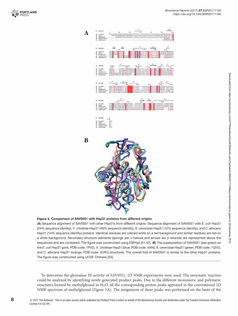
Figure 2. Comparison of SAV0551 with Hsp31 proteins from different origins
(A) Sequence alignment of SAV0551 with other Hsp31s from different origins. Sequence alignment of SAV0551 with E. coli Hsp31
(54% sequence identity), V. cholerae Hsp31 (49% sequence identity), S. cerevisiae Hsp31 (12% sequence identity), and C. albicans
Hsp31 (14% sequence identity) proteins. Identical residues are colored white on a red background and similar residues are red on
a white background. Secondary-structure elements (springs are α-helices and arrows are β-strands) are represented above the
sequences and are numbered. The figure was constructed using ESPript [41,42]. (B) The superposition of SAV0551 (sea green) on
the E. coli Hsp31 (pink, PDB code: 1PV2), V. cholerae Hsp31 (blue, PDB code: 4I4N), S. cerevisiae Hsp31 (green, PDB code: 1QVV),
and C. albicans Hsp31 (orange, PDB code: 4LRU) structures. The overall fold of SAV0551 is similar to the other Hsp31 proteins.
The figure was constructed using UCSF Chimera [33].
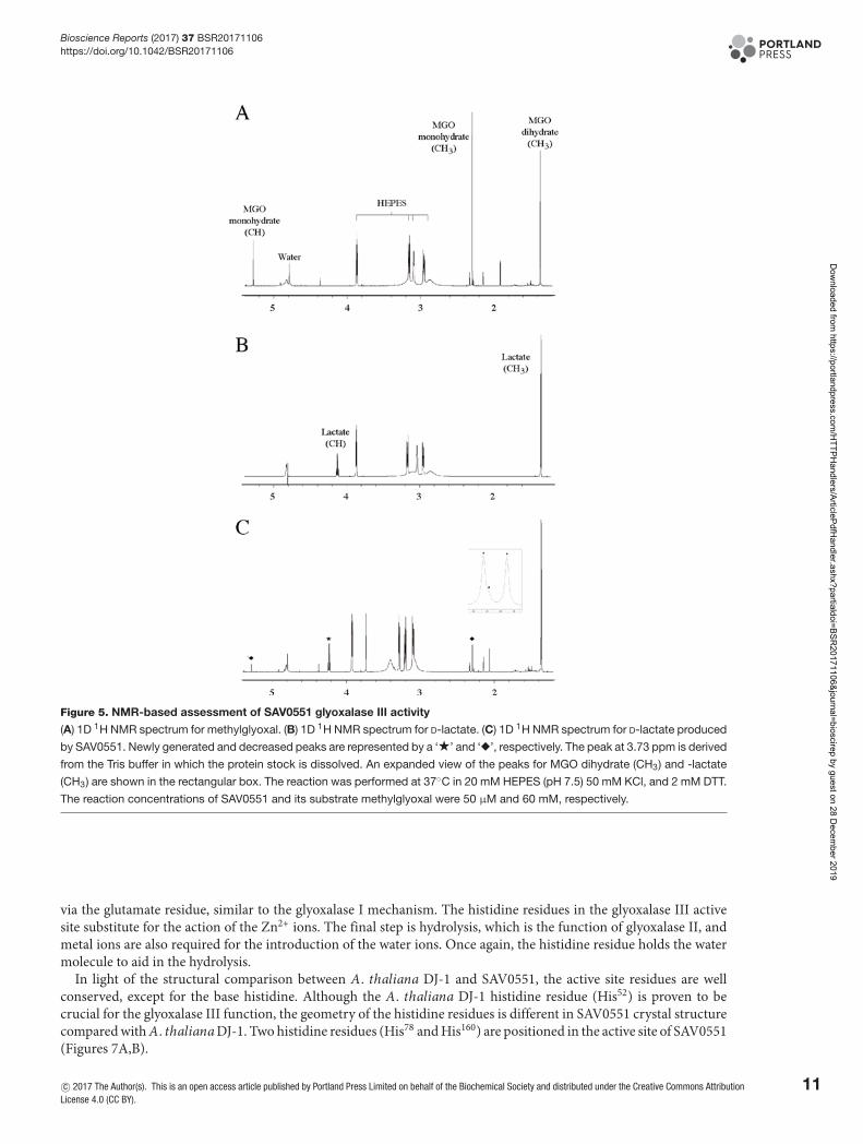
To determine the glyoxalase III activity of SAV0551, 1D NMR experiments were used. The enzymatic reactioncould be analyzed by identifying newly generated product peaks. Due to the different monomeric and polymericstructures formed by methylglyoxal in H2O, all the corresponding proton peaks appeared in the conventional 1DNMR spectrum of methylglyoxal (Figure 5A). The assignment of these peaks was performed on the basis of the
8 c© 2017 The Author(s). This is an open access article published by Portland Press Limited on behalf of the Biochemical Society and distributed under the Creative Commons AttributionLicense 4.0 (CC BY).
Dow
nloaded from https://portlandpress.com
/HTTPH
andlers/ArticlePdfHandler.ashx?partialdoi=BSR
20171106&journal=bioscirep by guest on 28 Decem
ber 2019
Bioscience Reports (2017) 37 BSR20171106https://doi.org/10.1042/BSR20171106
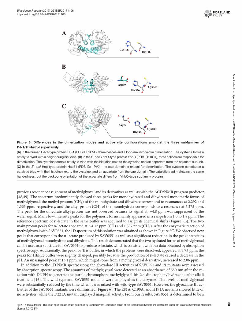
Figure 3. Differences in the dimerization modes and active site configurations amongst the three subfamilies of
DJ-1/ThiJ/PfpI superfamily
(A) In the human DJ-1-type protein DJ-1 (PDB ID: 1P5F), three helices and a loop are involved in dimerization. The cysteine forms a
catalytic dyad with a neighboring histidine. (B) In the E. coli YhbO-type protein YhbO (PDB ID: 1IO4), three helices are responsible for
dimerization. The cysteine forms a catalytic triad with the histidine next to the cysteine and an aspartate from the adjacent subunit.
(C) In the E. coli Hsp-type protein Hsp31 (PDB ID: 1PV2), the cap domain is critical for dimerization. The cysteine constitutes a
catalytic triad with the histidine next to the cysteine, and an aspartate from the cap domain. The catalytic triad maintains the same
handedness, but the backbone orientation of the aspartate differs from YhbO-type subfamily proteins.
previous resonance assignment of methylglyoxal and its derivatives as well as with the ACD/NMR program predictor[48,49]. The spectrum predominantly showed three peaks for monohydrated and dihydrated monomeric forms ofmethylglyoxal; the methyl protons (CH3) of the monohydrate and dihydrate correspond to resonances at 2.292 and1.363 ppm, respectively, and the alkyl proton (CH) of the monohydrate corresponds to a resonance at 5.275 ppm.The peak for the dihydrate alkyl proton was not observed because its signal at ∼4.8 ppm was suppressed by thewater signal. Many low-intensity peaks for the polymeric forms mainly appeared in a range from 1.0 to 1.8 ppm. Thereference spectrum of d-lactate in the same buffer was acquired to assign its chemical shifts (Figure 5B). The twomain proton peaks for d-lactate appeared at ∼4.12 ppm (CH) and 1.337 ppm (CH3). After the enzymatic reaction ofmethylglyoxal with SAV0551, the 1D spectrum of this solution was obtained as shown in Figure 5C. We observed newpeaks that correspond to the d-lactate produced by SAV0551 as well as a significant reduction in the peak intensitiesof methylglyoxal monohydrate and dihydrate. This result demonstrated that the two hydrated forms of methylglyoxalcan be used as a substrate for SAV0551 to produce d-lactate, which is consistent with our data obtained by absorptionspectroscopy. Additionally, the peak for Tris buffer, in which the proteins were dissolved, appeared at 3.73 ppm; thepeaks for HEPES buffer were slightly changed, possibly because the production of d-lactate caused a decrease in thepH. An unassigned peak at 1.91 ppm, which might come from a methylglyoxal derivative, increased to 2.06 ppm.
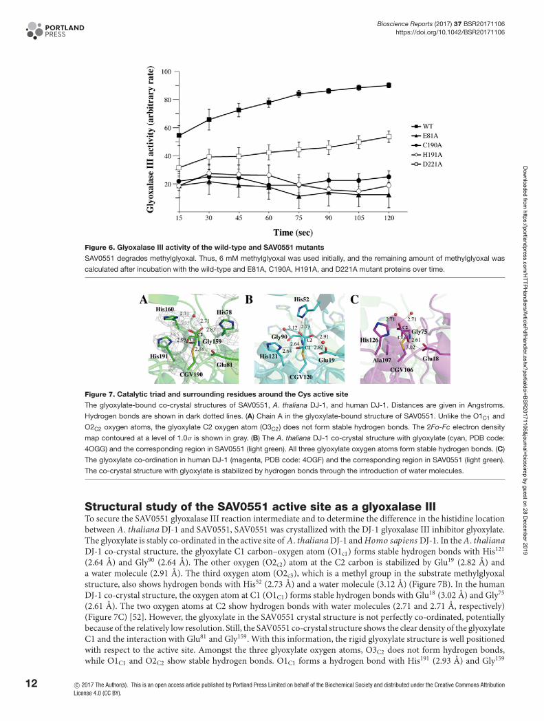
In addition to the 1D NMR spectroscopy, the glyoxalase III activities of SAV0551 and its mutants were assessedby absorption spectroscopy. The amounts of methylglyoxal were detected at an absorbance of 550 nm after the re-action with DNPH to generate the purple chromophore methylglyoxal-bis-2,4-dinitrophenylhydrazone after alkalitreatment [16]. The wild-type and SAV0551 mutants were employed as the enzymes. The levels of methylglyoxalwere substantially reduced by the time when it was mixed with wild-type SAV0551. However, the glyoxalase III ac-tivities of the SAV0551 mutants were diminished (Figure 6). The E81A, C190A, and H191A mutants showed little orno activities, while the D221A mutant displayed marginal activity. From our results, SAV0551 is determined to be a
c© 2017 The Author(s). This is an open access article published by Portland Press Limited on behalf of the Biochemical Society and distributed under the Creative Commons AttributionLicense 4.0 (CC BY).
9
Dow
nloaded from https://portlandpress.com
/HTTPH
andlers/ArticlePdfHandler.ashx?partialdoi=BSR
20171106&journal=bioscirep by guest on 28 Decem
ber 2019
Bioscience Reports (2017) 37 BSR20171106https://doi.org/10.1042/BSR20171106
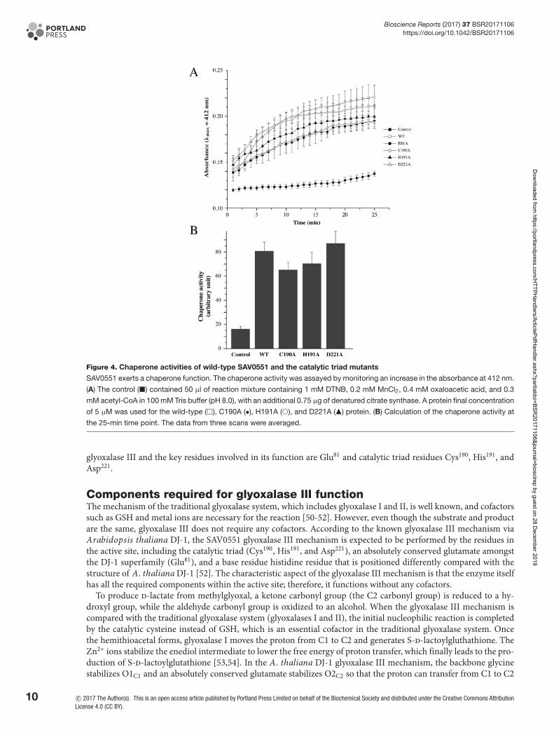
Figure 4. Chaperone activities of wild-type SAV0551 and the catalytic triad mutants
SAV0551 exerts a chaperone function. The chaperone activity was assayed by monitoring an increase in the absorbance at 412 nm.
(A) The control (�) contained 50 μl of reaction mixture containing 1 mM DTNB, 0.2 mM MnCl2, 0.4 mM oxaloacetic acid, and 0.3
mM acetyl-CoA in 100 mM Tris buffer (pH 8.0), with an additional 0.75 μg of denatured citrate synthase. A protein final concentration
of 5 μM was used for the wild-type (�), C190A (•), H191A (�), and D221A (�) protein. (B) Calculation of the chaperone activity at
the 25-min time point. The data from three scans were averaged.
glyoxalase III and the key residues involved in its function are Glu81 and catalytic triad residues Cys190, His191, andAsp221.
Components required for glyoxalase III functionThe mechanism of the traditional glyoxalase system, which includes glyoxalase I and II, is well known, and cofactorssuch as GSH and metal ions are necessary for the reaction [50-52]. However, even though the substrate and productare the same, glyoxalase III does not require any cofactors. According to the known glyoxalase III mechanism viaArabidopsis thaliana DJ-1, the SAV0551 glyoxalase III mechanism is expected to be performed by the residues inthe active site, including the catalytic triad (Cys190, His191, and Asp221), an absolutely conserved glutamate amongstthe DJ-1 superfamily (Glu81), and a base residue histidine residue that is positioned differently compared with thestructure of A. thaliana DJ-1 [52]. The characteristic aspect of the glyoxalase III mechanism is that the enzyme itselfhas all the required components within the active site; therefore, it functions without any cofactors.
To produce d-lactate from methylglyoxal, a ketone carbonyl group (the C2 carbonyl group) is reduced to a hy-droxyl group, while the aldehyde carbonyl group is oxidized to an alcohol. When the glyoxalase III mechanism iscompared with the traditional glyoxalase system (glyoxalases I and II), the initial nucleophilic reaction is completedby the catalytic cysteine instead of GSH, which is an essential cofactor in the traditional glyoxalase system. Oncethe hemithioacetal forms, glyoxalase I moves the proton from C1 to C2 and generates S-d-lactoylgluthathione. TheZn2+ ions stabilize the enediol intermediate to lower the free energy of proton transfer, which finally leads to the pro-duction of S-d-lactoylglutathione [53,54]. In the A. thaliana DJ-1 glyoxalase III mechanism, the backbone glycinestabilizes O1C1 and an absolutely conserved glutamate stabilizes O2C2 so that the proton can transfer from C1 to C2
10 c© 2017 The Author(s). This is an open access article published by Portland Press Limited on behalf of the Biochemical Society and distributed under the Creative Commons AttributionLicense 4.0 (CC BY).
Dow
nloaded from https://portlandpress.com
/HTTPH
andlers/ArticlePdfHandler.ashx?partialdoi=BSR
20171106&journal=bioscirep by guest on 28 Decem
ber 2019
Bioscience Reports (2017) 37 BSR20171106https://doi.org/10.1042/BSR20171106
Figure 5. NMR-based assessment of SAV0551 glyoxalase III activity
(A) 1D 1H NMR spectrum for methylglyoxal. (B) 1D 1H NMR spectrum for D-lactate. (C) 1D 1H NMR spectrum for D-lactate produced
by SAV0551. Newly generated and decreased peaks are represented by a ‘�’ and ‘�’, respectively. The peak at 3.73 ppm is derived
from the Tris buffer in which the protein stock is dissolved. An expanded view of the peaks for MGO dihydrate (CH3) and -lactate
(CH3) are shown in the rectangular box. The reaction was performed at 37◦C in 20 mM HEPES (pH 7.5) 50 mM KCl, and 2 mM DTT.
The reaction concentrations of SAV0551 and its substrate methylglyoxal were 50 μM and 60 mM, respectively.
via the glutamate residue, similar to the glyoxalase I mechanism. The histidine residues in the glyoxalase III activesite substitute for the action of the Zn2+ ions. The final step is hydrolysis, which is the function of glyoxalase II, andmetal ions are also required for the introduction of the water ions. Once again, the histidine residue holds the watermolecule to aid in the hydrolysis.
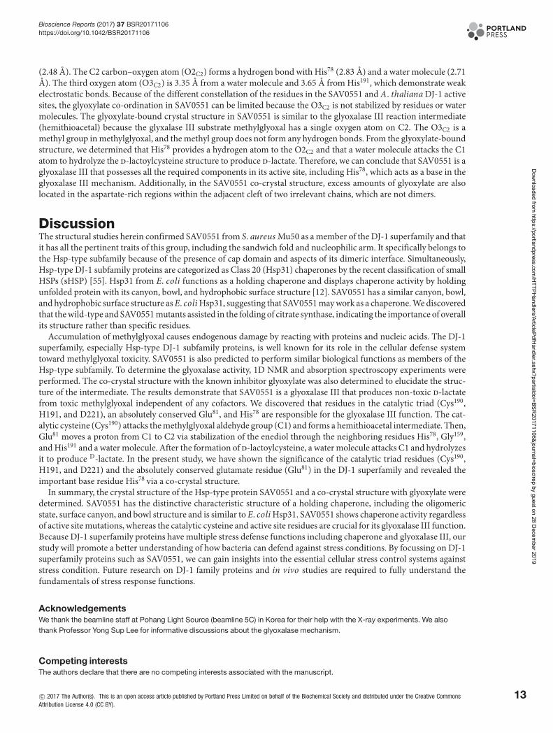
In light of the structural comparison between A. thaliana DJ-1 and SAV0551, the active site residues are wellconserved, except for the base histidine. Although the A. thaliana DJ-1 histidine residue (His52) is proven to becrucial for the glyoxalase III function, the geometry of the histidine residues is different in SAV0551 crystal structurecompared with A. thaliana DJ-1. Two histidine residues (His78 and His160) are positioned in the active site of SAV0551(Figures 7A,B).
c© 2017 The Author(s). This is an open access article published by Portland Press Limited on behalf of the Biochemical Society and distributed under the Creative Commons AttributionLicense 4.0 (CC BY).
11
Dow
nloaded from https://portlandpress.com
/HTTPH
andlers/ArticlePdfHandler.ashx?partialdoi=BSR
20171106&journal=bioscirep by guest on 28 Decem
ber 2019
Bioscience Reports (2017) 37 BSR20171106https://doi.org/10.1042/BSR20171106
Figure 6. Glyoxalase III activity of the wild-type and SAV0551 mutants
SAV0551 degrades methylglyoxal. Thus, 6 mM methylglyoxal was used initially, and the remaining amount of methylglyoxal was
calculated after incubation with the wild-type and E81A, C190A, H191A, and D221A mutant proteins over time.
Figure 7. Catalytic triad and surrounding residues around the Cys active site
The glyoxylate-bound co-crystal structures of SAV0551, A. thaliana DJ-1, and human DJ-1. Distances are given in Angstroms.
Hydrogen bonds are shown in dark dotted lines. (A) Chain A in the glyoxylate-bound structure of SAV0551. Unlike the O1C1 and
O2C2 oxygen atoms, the glyoxylate C2 oxygen atom (O3C2) does not form stable hydrogen bonds. The 2Fo-Fc electron density
map contoured at a level of 1.0σ is shown in gray. (B) The A. thaliana DJ-1 co-crystal structure with glyoxylate (cyan, PDB code:
4OGG) and the corresponding region in SAV0551 (light green). All three glyoxylate oxygen atoms form stable hydrogen bonds. (C)
The glyoxylate co-ordination in human DJ-1 (magenta, PDB code: 4OGF) and the corresponding region in SAV0551 (light green).
The co-crystal structure with glyoxylate is stabilized by hydrogen bonds through the introduction of water molecules.
Structural study of the SAV0551 active site as a glyoxalase IIITo secure the SAV0551 glyoxalase III reaction intermediate and to determine the difference in the histidine locationbetween A. thaliana DJ-1 and SAV0551, SAV0551 was crystallized with the DJ-1 glyoxalase III inhibitor glyoxylate.The glyoxylate is stably co-ordinated in the active site of A. thaliana DJ-1 and Homo sapiens DJ-1. In the A. thalianaDJ-1 co-crystal structure, the glyoxylate C1 carbon–oxygen atom (O1c1) forms stable hydrogen bonds with His121
(2.64 A) and Gly90 (2.64 A). The other oxygen (O2c2) atom at the C2 carbon is stabilized by Glu19 (2.82 A) anda water molecule (2.91 A). The third oxygen atom (O2c3), which is a methyl group in the substrate methylglyoxalstructure, also shows hydrogen bonds with His52 (2.73 A) and a water molecule (3.12 A) (Figure 7B). In the humanDJ-1 co-crystal structure, the oxygen atom at C1 (O1C1) forms stable hydrogen bonds with Glu18 (3.02 A) and Gly75
(2.61 A). The two oxygen atoms at C2 show hydrogen bonds with water molecules (2.71 and 2.71 A, respectively)(Figure 7C) [52]. However, the glyoxylate in the SAV0551 crystal structure is not perfectly co-ordinated, potentiallybecause of the relatively low resolution. Still, the SAV0551 co-crystal structure shows the clear density of the glyoxylateC1 and the interaction with Glu81 and Gly159. With this information, the rigid glyoxylate structure is well positionedwith respect to the active site. Amongst the three glyoxylate oxygen atoms, O3C2 does not form hydrogen bonds,while O1C1 and O2C2 show stable hydrogen bonds. O1C1 forms a hydrogen bond with His191 (2.93 A) and Gly159
12 c© 2017 The Author(s). This is an open access article published by Portland Press Limited on behalf of the Biochemical Society and distributed under the Creative Commons AttributionLicense 4.0 (CC BY).
Dow
nloaded from https://portlandpress.com
/HTTPH
andlers/ArticlePdfHandler.ashx?partialdoi=BSR
20171106&journal=bioscirep by guest on 28 Decem
ber 2019
Bioscience Reports (2017) 37 BSR20171106https://doi.org/10.1042/BSR20171106
(2.48 A). The C2 carbon–oxygen atom (O2C2) forms a hydrogen bond with His78 (2.83 A) and a water molecule (2.71A). The third oxygen atom (O3C2) is 3.35 A from a water molecule and 3.65 A from His191, which demonstrate weakelectrostatic bonds. Because of the different constellation of the residues in the SAV0551 and A. thaliana DJ-1 activesites, the glyoxylate co-ordination in SAV0551 can be limited because the O3C2 is not stabilized by residues or watermolecules. The glyoxylate-bound crystal structure in SAV0551 is similar to the glyoxalase III reaction intermediate(hemithioacetal) because the glyxalase III substrate methylglyoxal has a single oxygen atom on C2. The O3C2 is amethyl group in methylglyoxal, and the methyl group does not form any hydrogen bonds. From the glyoxylate-boundstructure, we determined that His78 provides a hydrogen atom to the O2C2 and that a water molecule attacks the C1atom to hydrolyze the d-lactoylcysteine structure to produce d-lactate. Therefore, we can conclude that SAV0551 is aglyoxalase III that possesses all the required components in its active site, including His78, which acts as a base in theglyoxalase III mechanism. Additionally, in the SAV0551 co-crystal structure, excess amounts of glyoxylate are alsolocated in the aspartate-rich regions within the adjacent cleft of two irrelevant chains, which are not dimers.
DiscussionThe structural studies herein confirmed SAV0551 from S. aureus Mu50 as a member of the DJ-1 superfamily and thatit has all the pertinent traits of this group, including the sandwich fold and nucleophilic arm. It specifically belongs tothe Hsp-type subfamily because of the presence of cap domain and aspects of its dimeric interface. Simultaneously,Hsp-type DJ-1 subfamily proteins are categorized as Class 20 (Hsp31) chaperones by the recent classification of smallHSPs (sHSP) [55]. Hsp31 from E. coli functions as a holding chaperone and displays chaperone activity by holdingunfolded protein with its canyon, bowl, and hydrophobic surface structure [12]. SAV0551 has a similar canyon, bowl,and hydrophobic surface structure as E. coli Hsp31, suggesting that SAV0551 may work as a chaperone. We discoveredthat the wild-type and SAV0551 mutants assisted in the folding of citrate synthase, indicating the importance of overallits structure rather than specific residues.
Accumulation of methylglyoxal causes endogenous damage by reacting with proteins and nucleic acids. The DJ-1superfamily, especially Hsp-type DJ-1 subfamily proteins, is well known for its role in the cellular defense systemtoward methylglyoxal toxicity. SAV0551 is also predicted to perform similar biological functions as members of theHsp-type subfamily. To determine the glyoxalase activity, 1D NMR and absorption spectroscopy experiments wereperformed. The co-crystal structure with the known inhibitor glyoxylate was also determined to elucidate the struc-ture of the intermediate. The results demonstrate that SAV0551 is a glyoxalase III that produces non-toxic d-lactatefrom toxic methylglyoxal independent of any cofactors. We discovered that residues in the catalytic triad (Cys190,H191, and D221), an absolutely conserved Glu81, and His78 are responsible for the glyoxalase III function. The cat-alytic cysteine (Cys190) attacks the methylglyoxal aldehyde group (C1) and forms a hemithioacetal intermediate. Then,Glu81 moves a proton from C1 to C2 via stabilization of the enediol through the neighboring residues His78, Gly159,and His191 and a water molecule. After the formation of d-lactoylcysteine, a water molecule attacks C1 and hydrolyzesit to produce D-lactate. In the present study, we have shown the significance of the catalytic triad residues (Cys190,H191, and D221) and the absolutely conserved glutamate residue (Glu81) in the DJ-1 superfamily and revealed theimportant base residue His78 via a co-crystal structure.
In summary, the crystal structure of the Hsp-type protein SAV0551 and a co-crystal structure with glyoxylate weredetermined. SAV0551 has the distinctive characteristic structure of a holding chaperone, including the oligomericstate, surface canyon, and bowl structure and is similar to E. coli Hsp31. SAV0551 shows chaperone activity regardlessof active site mutations, whereas the catalytic cysteine and active site residues are crucial for its glyoxalase III function.Because DJ-1 superfamily proteins have multiple stress defense functions including chaperone and glyoxalase III, ourstudy will promote a better understanding of how bacteria can defend against stress conditions. By focussing on DJ-1superfamily proteins such as SAV0551, we can gain insights into the essential cellular stress control systems againststress condition. Future research on DJ-1 family proteins and in vivo studies are required to fully understand thefundamentals of stress response functions.
AcknowledgementsWe thank the beamline staff at Pohang Light Source (beamline 5C) in Korea for their help with the X-ray experiments. We alsothank Professor Yong Sup Lee for informative discussions about the glyoxalase mechanism.
Competing interestsThe authors declare that there are no competing interests associated with the manuscript.
c© 2017 The Author(s). This is an open access article published by Portland Press Limited on behalf of the Biochemical Society and distributed under the Creative CommonsAttribution License 4.0 (CC BY).
13
Dow
nloaded from https://portlandpress.com
/HTTPH
andlers/ArticlePdfHandler.ashx?partialdoi=BSR
20171106&journal=bioscirep by guest on 28 Decem
ber 2019
Bioscience Reports (2017) 37 BSR20171106https://doi.org/10.1042/BSR20171106
FundingThis work was supported by the National Research Foundation of Korea (NRF) funded by the Ministry of Education, Sci-ence and Technology (MEST) of the Korean government [grant numbers 2015 R1A2A1A05001894, 2014 K1A3A1A19067618,2010-0025488]; the 2016 BK21 Plus Project for Medicine, Dentistry, and Pharmacy; and the Pohang Accelerator Laboratory (PAL)through the abroad beamtime programme of the Synchrotron Radiation Facility Project under MEST.
Author contributionH.J.K. designed and performed the experiments, analyzed the data, and wrote the paper under the supervision of A.-R.K. andB.-J.L. H.J.K. and K.-Y.L. performed the NMR measurements and analyzed the NMR data.
AbbreviationsDTT, dithiothreitol; DNPH, 2,4-dinitrophenylhydrazine; DTNB, 5,5′-dithiobis-(2-nitrobenzoic acid); GSH, glutathione; HSP, heatshock protein.
References1 Wei, Y. et al. (2007) Identification of functional subclasses in the DJ-1 superfamily proteins. PLoS Comput. Biol. 3, e102 Tang, B. et al. (2006) Association of PINK1 and DJ-1 confers digenic inheritance of early-onset Parkinson’s disease. Hum. Mol. Genet. 15, 1816–18253 Wang, Z.Q., Zhou, H.Y. and Chen, S.D. (2006) The role of DJ-1 in the pathogenesis of Parkinson’s disease. Neurosci. Bull. 22, 232–2344 Jung, H.J. et al. (2012) Dissection of the dimerization modes in the DJ-1 superfamily. Mol. Cells 33, 163–1715 Ollis, D.L. et al. (1992) The alpha/beta hydrolase fold. Protein Eng. 5, 197–2116 Honbou, K. et al. (2003) The crystal structure of DJ-1, a protein related to male fertility and Parkinson’s disease. J. Biol. Chem. 278, 31380–313847 Wilson, M.A., Ringe, D. and Petsko, G.A. (2005) The atomic resolution crystal structure of the YajL (ThiJ) protein from Escherichia coli : a close
prokaryotic homologue of the Parkinsonism-associated protein DJ-1. J. Mol. Biol. 353, 678–6918 Shendelman, S. et al. (2004) DJ-1 is a redox-dependent molecular chaperone that inhibits alpha-synuclein aggregate formation. PLoS Biol. 2, e3629 Lee, J.Y. et al. (2012) Human DJ-1 and its homologs are novel glyoxalases. Hum. Mol. Genet. 21, 3215–322510 Zhang, Q. et al. (2012) DJ-1 promotes the proteasomal degradation of Fis1: implications of DJ-1 in neuronal protection. Biochem. J. 447, 261–26911 Zhan, D. et al. (2014) Characterization of the PH1704 protease from Pyrococcus horikoshii OT3 and the critical functions of Tyr120. PLoS ONE 9,
e10390212 Quigley, P.M. et al. (2003) The 1.6-A crystal structure of the class of chaperones represented by Escherichia coli Hsp31 reveals a putative catalytic
triad. Proc. Natl. Acad. Sci. U.S.A. 100, 3137–314213 Mujacic, M., Bader, M.W. and Baneyx, F. (2004) Escherichia coli Hsp31 functions as a holding chaperone that cooperates with the DnaK-DnaJ-GrpE
system in the management of protein misfolding under severe stress conditions. Mol. Microbiol. 51, 849–85914 Das, S. et al. (2017) Structural and biochemical studies on Vibrio cholerae Hsp31 reveals a novel dimeric form and Glutathione-independent Glyoxalase
activity. PLoS ONE 12, e017262915 Subedi, K.P. et al. (2011) Hsp31 of Escherichia coli K-12 is glyoxalase III. Mol. Microbiol. 81, 926–93616 Hasim, S. et al. (2014) A glutathione-independent glyoxalase of the DJ-1 superfamily plays an important role in managing metabolically generated
methylglyoxal in Candida albicans. J. Biol. Chem. 289, 1662–167417 Zhao, Q. et al. (2014) Identification of glutathione (GSH)-independent glyoxalase III from Schizosaccharomyces pombe. BMC Evol. Biol. 14, 8618 Bankapalli, K. et al. (2015) Robust glyoxalase activity of Hsp31, a ThiJ/DJ-1/Pf pI family member protein, is critical for oxidative stress resistance in
Saccharomyces cerevisiae. J. Biol. Chem. 290, 26491–2650719 Kim, H.J., Kwon, A.R. and Lee, B.J. (2016) Structural and functional insight into the different oxidation states of SAV1875 from Staphylococcus aureus.
Biochem. J. 473, 55–6620 Abdallah, J. et al. (2016) The DJ-1 superfamily members YhbO and YajL from Escherichia coli repair proteins from glycation by methylglyoxal and
glyoxal. Biochem. Biophys. Res. Commun. 470, 282–28621 Papp, E. et al. (2003) Molecular chaperones, stress proteins and redox homeostasis. Biofactors 17, 249–25722 Allaman, I., Belanger, M. and Magistretti, P.J. (2015) Methylglyoxal, the dark side of glycolysis. Front. Neurosci. 9, 2323 Archer, G.L. (1998) Staphylococcus aureus: a well-armed pathogen. Clin. Infect. Dis. 26, 1179–118124 Woods, C. and Colice, G. (2014) Methicillin-resistant Staphylococcus aureus pneumonia in adults. Expert Rev. Respir. Med. 8, 641–65125 Di Nottia, M. et al. (2016) DJ-1 modulates mitochondrial response to oxidative stress: clues from a novel diagnosis of PARK7. Clin. Genet. 1284126 Otwinowski, Z. and Minor, W. (1997) Processing of X-ray diffraction data collected in oscillation mode. Methods Enzymol. 276, 307–32627 Winn, M.D. et al. (2011) Overview of the CCP4 suite and current developments. Acta Crystallogr. D Biol. Crystallogr. 67, 235–24228 Vagin, A. and Teplyakov, A. (2010) Molecular replacement with MOLREP. Acta Crystallogr. D Biol. Crystallogr. 66, 22–2529 Emsley, P. and Cowtan, K. (2004) Coot: model-building tools for molecular graphics. Acta Crystallogr. D Biol. Crystallogr. 60, 2126–213230 Adams, P.D. et al. (2010) PHENIX: a comprehensive Python-based system for macromolecular structure solution. Acta Crystallogr. D Biol. Crystallogr.
66, 213–22131 Murshudov, G.N. et al. (2011) REFMAC5 for the refinement of macromolecular crystal structures. Acta Crystallogr. D Biol. Crystallogr. 67, 355–36732 Brunger, A.T. (1992) Free R value: a novel statistical quantity for assessing the accuracy of crystal structures. Nature 355, 472–47533 Pettersen, E.F. et al. (2004) UCSF Chimera-a visualization system for exploratory research and analysis. J. Comput. Chem. 25, 1605–1612
14 c© 2017 The Author(s). This is an open access article published by Portland Press Limited on behalf of the Biochemical Society and distributed under the Creative CommonsAttribution License 4.0 (CC BY).
Dow
nloaded from https://portlandpress.com
/HTTPH
andlers/ArticlePdfHandler.ashx?partialdoi=BSR
20171106&journal=bioscirep by guest on 28 Decem
ber 2019

Bioscience Reports (2017) 37 BSR20171106https://doi.org/10.1042/BSR20171106
34 Krissinel, E. and Henrick, K. (2007) Inference of macromolecular assemblies from crystalline state. J. Mol. Biol. 372, 774–79735 Zhi, W. et al. (1992) Renaturation of citrate synthase: influence of denaturant and folding assstants. Protein Sci. 1, 522–52936 Lee, G.J. (1995) Assaying proteins for molecular chaperone activity. Methods Cell. Biol. 50, 325–33437 Morgunov, I. and Srere, P.A. (1998) Interaction between citrate synthase and malate dehydrogenase. Substrate channeling of oxaloacetate. J. Biol.
Chem. 273, 29540–2954438 Gilbert, R.P. and Brandt, R.B. (1975) Spectrophotometric determination of methyl glyoxal with 2,4-dinitrophenylhydrazine. Anal. Chem. 47, 2418–242239 Sievers, F. et al. (2011) Fast, scalable generation of high-quality protein multiple sequence alignments using Clustal Omega. Mol. Syst. Biol. 7, 53940 McWilliam, H. et al. (2013) Analysis Tool Web Services from the EMBL-EBI. Nucleic Acids Res. 41, W597–W60041 Gouet, P. et al. (1999) ESPript: analysis of multiple sequence alignments in PostScript. Bioinformatics 15, 305–30842 Robert, X. and Gouet, P. (2014) Deciphering key features in protein structures with the new ENDscript server. Nucleic Acids Res. 42, W320–W32443 Chen, J., Li, L. and Chin, L.S. (2010) Parkinson disease protein DJ-1 converts from a zymogen to a protease by carboxyl-terminal cleavage. Hum. Mol.
Genet. 19, 2395–240844 Sastry, M.S. et al. (2004) The linker-loop region of Escherichia coli chaperone Hsp31 functions as a gate that modulates high-affinity substrate binding
at elevated temperatures. Proc. Natl. Acad. Sci. U.S.A. 101, 8587–859245 Oya, T. et al. (1999) Methylglyoxal modification of protein. Chemical and immunochemical characterization of methylglyoxal-arginine adducts. J. Biol.
Chem. 274, 18492–1850246 Kang, J.H. (2003) Oxidative damage of DNA induced by methylglyoxal in vitro. Toxicol. Lett. 145, 181–18747 Martins, A.M. et al. (2001) In situ kinetic analysis of glyoxalase I and glyoxalase II in Saccharomyces cerevisiae. Eur. J. Biochem. 268, 3930–393648 Nemet, I., Vikic-Topic, D. and Varga-Defterdarovic, L. (2004) Spectroscopic studies of methylglyoxal in water and dimethylsulfoxide. Bioorg. Chem. 32,
560–57049 Zhang, J. et al. (2010) A novel capillary electrophoretic method for determining methylglyoxal and glyoxal in urine and water samples. J. Chromatogr. A
1217, 5124–512950 Vander Jagt, D.L. (1993) Glyoxalase II: molecular characteristics, kinetics and mechanism. Biochem. Soc. Trans. 21, 522–52751 Creighton, D.J. and Hamilton, D.S. (2001) Brief history of glyoxalase I and what we have learned about metal ion-dependent, enzyme-catalyzed
isomerizations. Arch. Biochem. Biophys. 387, 1–1052 Choi, D. et al. (2014) Stereospecific mechanism of DJ-1 glyoxalases inferred from their hemithioacetal-containing crystal structures. FEBS J. 281,
5447–546253 O’Young, J., Sukdeo, N. and Honek, J.F. (2007) Escherichia coli glyoxalase II is a binuclear zinc-dependent metalloenzyme. Arch. Biochem. Biophys.
459, 20–2654 Suttisansanee, U. et al. (2015) Modulating glyoxalase I metal selectivity by deletional mutagenesis: underlying structural factors contributing to nickel
activation profiles. Metallomics 7, 605–61255 Jaspard, E. and Hunault, G. (2016) sHSPdb: a database for the analysis of small Heat Shock Proteins. BMC Plant Biol. 16, 135
c© 2017 The Author(s). This is an open access article published by Portland Press Limited on behalf of the Biochemical Society and distributed under the Creative Commons AttributionLicense 4.0 (CC BY).
15
Dow
nloaded from https://portlandpress.com
/HTTPH
andlers/ArticlePdfHandler.ashx?partialdoi=BSR
20171106&journal=bioscirep by guest on 28 Decem
ber 2019