structural and functional studies of aspergillus oryzae...
TRANSCRIPT
Structural and Functional Studies of Aspergillus oryzaeCutinase: Enhanced Thermostability and Hydrolytic Activity of
Synthetic Ester and Polyester Degradation
Zhiqiang Liu,† Yuying Gosser,‡ Peter James Baker,† Yaniv Ravee,† Ziying Lu,‡
Girum Alemu,‡ Huiguang Li,§ Glenn L. Butterfoss,| Xiang-Peng Kong,§
Richard Gross,† and Jin Kim Montclare*,†,⊥
Department of Chemical and Biological Sciences, Polytechnic Institute of New York UniVersity,Brooklyn, New York 11201, Pathways Bioinformatics & Biomolecular Center, The City College,CUNY, New York, New York 10031, Department of Biochemistry, NYU School of Medicine, 550First AVenue, New York, New York 10016, Department of Biology and Computer Science, NewYork UniVersity, New York, New York 10003, Department of Biochemistry, SUNY-Downstate
Medical Center, Brooklyn, New York 11203
Received June 8, 2009; E-mail: [email protected]
Abstract: Cutinases are responsible for hydrolysis of the protective cutin lipid polyester matrix in plantsand thus have been exploited for hydrolysis of small molecule esters and polyesters. Here we explore thereactivity, stability, and structure of Aspergillus oryzae cutinase and compare it to the well-studied enzymefrom Fusarium solani. Two critical differences are highlighted in the crystallographic analysis of the A.oryzae structure: (i) an additional disulfide bond and (ii) a topologically favored catalytic triad with a continuousand deep groove. These structural features of A. oryzae cutinase are proposed to result in an improvedhydrolytic activity and altered substrate specificity profile, enhanced thermostability, and remarkable reactivitytoward the degradation of the synthetic polyester polycaprolactone. The results presented here provideinsight into engineering new cutinase-inspired biocatalysts with tailor-made properties.
Introduction
With environmental concerns over waste build-up andlimited petroleum resources, the demand has never beengreater for enzymatic or “green” approaches for efficientchemical synthesis and degradation reactions.1-3 Severalenzymes have been exploited not only to perform stereo- andregioselective chemical transformations on small moleculesbut also to break down or modify synthetic polymers usefulfor applications in chemical, pharmaceutical, and textileindustries,2,4,5 Serine hydrolases have been employed mostextensively for these biotransformations.1,6-14
Cutinases are R/� hydrolases commonly secreted by fungalphytopathogens that enable them to penetrate the protectivesurface cutin layer of plants.15-19 Cutin is an insoluble, cross-linked, lipid-polyester matrix comprised of n-C17 and n-C18
hydroxy and epoxy fatty acids that serve as a barrier againstdehydration and invasion. Cutinases hydrolyze and break downthese complex polyesters into smaller hydroxyacids.15,19
Because of the ability of cutinases to hydrolyze cutin, theyhave been exploited for reactions with small molecule estersand synthetic polyesters.1 The utility of cutinase or any enzyme
† Polytechnic Institute of New York University.‡ The City College, CUNY.§ NYU School of Medicine.| New York University.⊥ SUNY-Downstate Medical Center.
(1) Carvalho, C. M. L.; Aires-Barros, M. R.; Cabral, J. M. S. Biotechnol.Bioeng. 1999, 66, 17–34.
(2) Guebitz, G. M.; Cavaco-Paulo, A. Trends Biotechnol. 2008, 26, 32–38.
(3) Maeda, H.; Yamagata, Y.; Abe, K.; Hasegawa, F.; Machida, M.;Ishioka, R.; Gomi, K.; Nakajima, T. Appl. Microbiol. Biotechnol. 2005,67, 778–788.
(4) Costa, L.; Brissos, V.; Lemos, F.; Ribeiro, F. R.; Cabral, J. M. S.Bioprocess Biosyst. Eng. 2008, 31, 323–327.
(5) Liu, Y. B.; Wu, G. F.; Gu, L. H. AATCC ReV. 2008, 8, 44–48.(6) Masaki, K.; Kamini, N. R.; Ikeda, H.; Iefuji, H. Appl. EnViron.
Microbiol. 2005, 71, 7548–7550.(7) Vidinha, P.; Harper, N.; Micaelo, N. M.; Lourengo, N. M. T.; da Silva,
M.; Cabral, J. M. S.; Afonso, C. A. M.; Soares, C. M.; Barreiros, S.Biotechnol. Bioeng. 2004, 85, 442–449.
(8) Borreguero, I.; Carvalho, C. M. L.; Cabral, J. M. S.; Sinisterra, J. V.;Alcantara, A. R. J. Mol. Catal. B: Enzym. 2001, 11, 613–622.
(9) Bogel-Lukasik, R.; Lourenco, N. M. T.; Vidinha, P.; da Silva, M.;Afonso, C. A. M.; da Ponte, M. N.; Barreiros, S. Green Chem. 2008,10, 243–248.
(10) Chen, B.; Hu, J.; Miller, E. M.; Xie, W. C.; Cai, M. M.; Gross, R. A.Biomacromolecules 2008, 9, 463–471.
(11) Kulshrestha, A. S.; Gao, W.; Fu, H. Y.; Gross, R. A. Biomacromol-ecules 2007, 8, 1794–1801.
(12) Chen, B.; Miller, E. M.; Miller, L.; Maikner, J. J.; Gross, R. A.Langmuir 2007, 23, 1381–1387.
(13) Azim, H.; Dekhterman, A.; Jiang, Z. Z.; Gross, R. A. Biomacromol-ecules 2006, 7, 3093–3097.
(14) Hu, J.; Gao, W.; Kulshrestha, A.; Gross, R. A. Macromolecules 2006,39, 6789–6792.
(15) Purdy, R. E.; Kolattukudy, P. E. Biochemistry 1975, 14, 2824–2831.(16) Longhi, S.; Czjzek, M.; Lamzin, V.; Nicolas, A.; Cambillau, C. J.
Mol. Biol. 1997, 268, 779–799.(17) Sweigard, J. A.; Chumley, F. G.; Valent, B. Mol. Gen. Genet. 1992,
232, 174–182.(18) Martinez, C.; Degeus, P.; Lauwereys, M.; Matthyssens, G.; Cambillau,
C. Nature 1992, 356, 615–618.(19) Purdy, R. E.; Kolattukudy, P. E. Biochemistry 1975, 14, 2832–2840.
10.1021/ja9046697 CCC: $40.75 XXXX American Chemical Society J. AM. CHEM. SOC. XXXX, xxx, 000 9 A
Dow
nloa
ded
by N
EW
YO
RK
UN
IV o
n O
ctob
er 1
4, 2
009
| http
://pu
bs.a
cs.o
rg
Pub
licat
ion
Dat
e (W
eb):
Oct
ober
7, 2
009
| doi
: 10.
1021
/ja90
4669
7

for chemical reactions is commonly limited by intolerance tohigh temperatures and the constraints of the substrate recognitionpocket.1 Thus, the identification of enzymes exhibiting enhancedthermostability and altered reactivity for various substrateswould greatly expand the potential of cutinase for industrialand environmental applications.
Aspergillus oryzea is a filamentous fungus that has beenemployed in fermentation to produce traditional consumableproducts such as rice wine, soybean paste, and soy sauce in thefood industry for approximately 1000 years.9 It has been usedrecently as an expression host for recombinant proteins20 andto degrade poly(butylene succinate) (PBS) as well as emulsifiedpoly(butylenes succinate-co-adipate) (PBSA).3 Because of theremarkable ability of A. oryzae cutinase to readily break downsuch synthetic plastics,3 it represents an excellent target toperform detailed structure-activity analysis. Here we report thereactivity, stability, and crystal structure of A. oryzea cutinaseand perform a comparison to the well-characterized Fusariumsolani cutinase.
Methods
Enzyme Expression. The cutinase gene was expressed in Pichiapastoris, and recombinant cutinase was produced by using the strongmethanol-induced AOX1 promoter. Single colonies were pickedand cultured in BMGY medium (g/L) composed of 5 g of yeastextract, 10 g of peptone, supplemented with 50 mL of 1 M KH2PO4
buffer pH 6.0, 1.7 g of yeast nitrogen base, 5 g of ammoniumsulfate, 5 mL of glycerol, 500 × l mL biotin, and 96 × 5.2 mLhistidine. Precultures of P. pastoris harboring cutinase genes wereincubated at 30 °C, 200 rpm, overnight. After centrifugation at 6,000rpm for 10 min, cells were transferred into a parallel fermentor(DASGIP, Germany) containing 1 L of basal salt medium composedof glycerol 40 mL/L, CaSO4 0.9 g/L, K2SO4 14.67 g/L,MgSO4 ·7H2O 11.67 g/L, (NH4)2SO4 9 g/L, 12 mL/L hexameta-phosphate, trace salts (cupric sulfate ·5H2O 6.0 g/L, sodium iodide0.08 g /L, manganese sulfate H2O 3.0 g/L, sodium molybdate ·2H2O0.2 g/L, boric acid 0.02 g/L, cobalt chloride 0.5 g/L, zinc chloride20.0 g/L, ferrous sulfate ·7H2O 65 g/L, biotin 0.2 g/L). The constantdissolved oxygen was set to 40%, glycerol (50%, v/v) feeding timewas 6 h, and the rate of methanol feeding was 5 mL/h. When theactivity reached its maximum, the fermentation was stopped. Aftercentrifugation, the supernatant was collected for further use.
Enzyme Purification. Fermentation broth was centrifuged at8,000 rpm for 10 min at 4 °C, and then supernatant was concentratedabout 10 times using an ultrafiltration unit (Millipore TFE system)with a 10 kDa membrane. Cutinase was purified by FPLC usingVISION Workstation (Applied Biosystems Co.) with a 16 mmD/100 mmL POROS MC 20 µm column (Applied Biosystems Co.).The metal site in the column was saturated with NiCl2 solutionaccording to operating instructions for the column. Approximately50 mM NaH2PO4 buffer with 0.5 mM imidazole (pH 8.0) was usedas a starting buffer, and 50 mM NaH2PO4 buffer with 100 mMimidazole (pH 8.0) was used as an elution buffer at a flow rate of30 mL/min. Samples filtered by a 0.45 µm filter were loaded ontothe column. Approximately 2 column volumes (CVs) of startingbuffer were run to wash out any proteins that were boundnonspecifically to the column, and then 3 CVs of elution bufferwith imidazole were run with a concentration linear gradient from0 to 100 mM. The peak fraction in the gradient step was collected.The collected samples were desalted by an ultrafiltration unit witha 10 kDa membrane and then freeze-dried. SDS-PAGE analysisof the purified proteins was performed (Supporting Information).
Kinetic Analysis for pNP Ester Substrates. The pNP esters(pNPA, pNPB, pNPV, and pNPH) with concentration ranging from
60 to 0.9375 µM were used for the kinetic study.21,22 Assays wereperformed using a final concentration of 1.1 µM of A. oryzae andF. solani cutinases in 14.5 mM Tris-HCl buffer pH 7.5, 0.75%glycerol. Since pNPA and pNPH were dissolved in DMSO, pNPBin methanol, and pNPV in TritonX, there was approximately 25%DMSO, methanol or 0.5% Triton X in the final mixtures.23
Reactions were initiated by the addition of enzyme, and reactionprogress was monitored spectrophotometrically (Molecular DevicesSpectramax M2) at 405 nm. Softmax Pro v5 software was used toanalyze data. Enzyme-catalyzed initial rates were corrected bysubtracting background hydrolysis rate. All reactions were per-formed in triplicate. The Km and kcat values were determined by adouble-reciprocal Lineweaver-Burk plot (1/V vs 1/[pNPA/pNPB/pNPV/ pNPH]) (Supporting Information).
Thermoactivity. The thermoactivity or residual activity ofcutinases was investigated by incubating enzymes at temperaturesranging from 25 to 60 °C at a final concentration of 1.1 µM in14.5 mM Tris-HCl buffer pH 7.5, 0.75% glycerol. The incubationtook place in an Eppendorf Thermomixer at the specified temper-ature with a constant mixing of 350 rpm for 1 h. The samples wereincubated on ice for 5 min followed by incubation at roomtemperature for 15 min. Reactions were initiated by addition of 30µM pNPB, monitored at 405 nm for 1 min, and performed intriplicate. The data obtained was presented as activity withoutnormalization.
Circular Dichroism (CD) Measurements. CD spectra wererecorded on a JASCO J-815 Spectropolarimeter using SpectraManager 228 software. Temperature was controlled using a FisherIsotemp Model 3016S water bath. A protein concentration of 29µM in 10 mM sodium phosphate buffer, pH 8.0 was used for bothcutinases. Data were collected at 1-nm intervals from 190 to 250nm for wavelength scans and 0.3 °C/min from 4 to 85 °C fortemperature scans in duplicate (Supporting Information). Smallsignaling arising from buffer was subtracted.
Thermodynamic Parameters by Differential ScanningCalorimetry (DSC). Calorimetric measurements of melting tem-peratures (Tm) were carried out on a NanoDSC differential scanningcalorimeter (TA Instruments) with a sample cell volume of 0.3 mL.Unfolding data of both cutinases were obtained by heating thesamples, at a concentration 5 mg/mL, from 4 to 80 °C at a rate of1 °C/min in duplicate (Supporting Information). The protein sampleswere present in water. Water was used in the reference cell to obtainthe molar heat capacity by comparison. The observed thermogramswere baseline corrected, and normalized data were analyzed usingNanoAnalyze software.
Degradation of Polymer Thin Films. Thin films of poly(ε-caprolactone) (PCL) were cut to 1.0 cm × 1.0 cm with anapproximate thickness of 250 µm (30-35 mg) and placed in 20mL scintillation screw cap vials containing 2.5 mL of 100 mMTris-HCl buffer pH 8.0 with a final concentration of 8.8 µMenzyme. The control vials contained a film with buffer solution.All measurements were performed in triplicate, incubated for 6 hat 40 °C in an incubator shaker at 200 rpm, and weighed afterdrying.
Crystallization. The protein was crystallized by mixing equalvolumes of protein solution (15 mg/mL, in 100 mM Tris bufferpH 8.5) with mother liquor (30% PEG2KMME, 100 mM potassiumthiocyanate) at 296 K. The screening was conducted with 96crystallization conditions at 296 K using the hanging drop vapordiffusion technique. Crystals appeared within 10-15 days.
Structure Determination. X-ray diffraction data of the crystalwere collected at beamline X4A (λ ) 0.96785 Å) of the synchrotronlight source in the Brookhaven National Laboratory. Prior to data
(20) Christensen, T.; Woeldike, H.; Boel, E.; Mortensen, S. B.; Hjortshoejk,K.; Thim, L.; Hansen, M. T. Biotechnology 1988, 6, 1419–1422.
(21) Pedersen, S.; Nesgaard, L.; Baptista, R. P.; Melo, E. P.; Kristensen,S. R.; Otzen, D. E. Biopolymers 2006, 83, 619–629.
(22) Goncalves, A. M.; Serro, A. P.; Aires-Barros, M. R.; Cabral, J. M. S.Biochim. Biophys. Acta 2000, 1480, 92–106.
(23) Camacho, R. M.; Mateo, J. C.; Gonzalez-Reynoso, O.; Prado, L. A.;Cordova, J. J. Ind. Microbiol. Biotechnol. 2009, 36, 901–909.
B J. AM. CHEM. SOC. 9 VOL. xxx, NO. xx, XXXX
A R T I C L E S Liu et al.
Dow
nloa
ded
by N
EW
YO
RK
UN
IV o
n O
ctob
er 1
4, 2
009
| http
://pu
bs.a
cs.o
rg
Pub
licat
ion
Dat
e (W
eb):
Oct
ober
7, 2
009
| doi
: 10.
1021
/ja90
4669
7
collection, the crystal was soaked for 5 min in a mixture of themother liquid (30% PEG2000MME and 100 mM potassiumthiocyanate) and 15% glycerol for cryoprotection. A data set of1.75 Å resolution was collected from a single crystal at 100 K with98.5% completeness. This data set was processed using HKL2000software.24 The crystal belongs to the trigonal space group P3221with one subunit in the asymmetric unit. The unit cell parameterswere a ) 45.299 Å, b ) 45.299 Å, c ) 157.111 Å and R ) 90.00°,� ) 90.00°, � ) 120.00°. The structure was solved by molecularreplacement using the Molrep program in the CCP4 suite.25 Thestructure of the native F. solani cutinase (PDB accession number1CEX) was used as the search model. The initial model was builtusing O program. The structure was refined with CNS.26 Theiterative model building and refinement was carried out to a finalR-work factor of 19.4% and a final free-R factor of 19.9%. Thestereochemistry of the final structure was verified using theProcheck_NT and Sfcheck programs in the CCP4 suite.25 Procheckvalidation of local geometry revealed that 91.5% of residues arelocated in the most favored regions of the Ramachandran plot, 7.2%are in the additional allowed regions, and 1.3% are in the generouslyallowed region. The electron densities of the first 10 N-terminalresidues (17-25) were not observed. Structural analysis wasperformed using UCSF Chimera software (http://www.cgl.ucsf.edu/chimera).27 Electrostatic surface rendering was performed usingICN and the groove by the active site was generated by thePocketFinder function.28 For all above analysis 1CEX pdb file wasused for F. solani cutinase.
Modeling. 1CEX and the A. oryzae cutinase crystal structurepresented here were modeled with the butyrate, valerate, andhexanoate esters of the active site serine. Hydrogens were added,and starting structures of the serine esters were generated bysampling all combinations of -60°, 60°, and 180° for each � angle(with the exception of the ester C-O torsion, which was sampledat -180° and 180°). This generated 162, 486, and 1458 conforma-tions for the butyrate, valerate, and hexanoate esters, respectively.The modified serine residues were then subject to 11 × 20 steps ofconjugate gradient energy minimization using the CHARMM22potential29 while keeping all other protein atoms fixed. Modelingwas done using the SIGMA package.30
Results
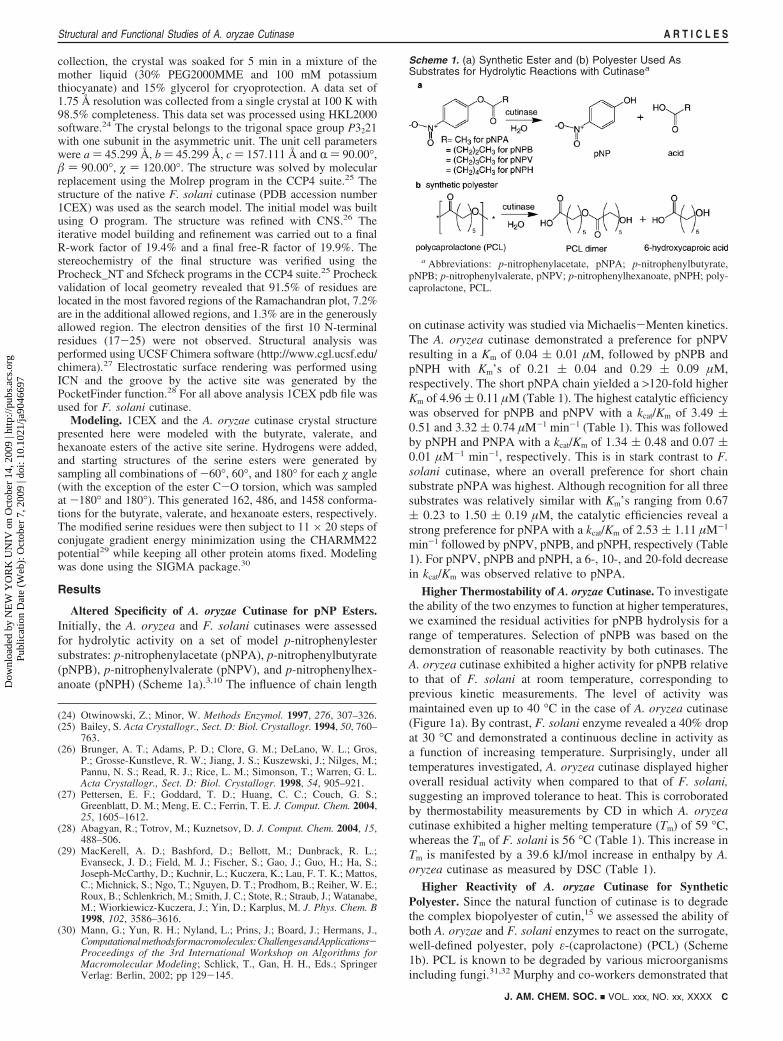
Altered Specificity of A. oryzae Cutinase for pNP Esters.Initially, the A. oryzea and F. solani cutinases were assessedfor hydrolytic activity on a set of model p-nitrophenylestersubstrates: p-nitrophenylacetate (pNPA), p-nitrophenylbutyrate(pNPB), p-nitrophenylvalerate (pNPV), and p-nitrophenylhex-anoate (pNPH) (Scheme 1a).3,10 The influence of chain length
on cutinase activity was studied via Michaelis-Menten kinetics.The A. oryzea cutinase demonstrated a preference for pNPVresulting in a Km of 0.04 ( 0.01 µM, followed by pNPB andpNPH with Km’s of 0.21 ( 0.04 and 0.29 ( 0.09 µM,respectively. The short pNPA chain yielded a >120-fold higherKm of 4.96 ( 0.11 µM (Table 1). The highest catalytic efficiencywas observed for pNPB and pNPV with a kcat/Km of 3.49 (0.51 and 3.32 ( 0.74 µM-1 min-1 (Table 1). This was followedby pNPH and PNPA with a kcat/Km of 1.34 ( 0.48 and 0.07 (0.01 µM-1 min-1, respectively. This is in stark contrast to F.solani cutinase, where an overall preference for short chainsubstrate pNPA was highest. Although recognition for all threesubstrates was relatively similar with Km’s ranging from 0.67( 0.23 to 1.50 ( 0.19 µM, the catalytic efficiencies reveal astrong preference for pNPA with a kcat/Km of 2.53 ( 1.11 µM-1
min-1 followed by pNPV, pNPB, and pNPH, respectively (Table1). For pNPV, pNPB and pNPH, a 6-, 10-, and 20-fold decreasein kcat/Km was observed relative to pNPA.
Higher Thermostability of A. oryzae Cutinase. To investigatethe ability of the two enzymes to function at higher temperatures,we examined the residual activities for pNPB hydrolysis for arange of temperatures. Selection of pNPB was based on thedemonstration of reasonable reactivity by both cutinases. TheA. oryzea cutinase exhibited a higher activity for pNPB relativeto that of F. solani at room temperature, corresponding toprevious kinetic measurements. The level of activity wasmaintained even up to 40 °C in the case of A. oryzea cutinase(Figure 1a). By contrast, F. solani enzyme revealed a 40% dropat 30 °C and demonstrated a continuous decline in activity asa function of increasing temperature. Surprisingly, under alltemperatures investigated, A. oryzea cutinase displayed higheroverall residual activity when compared to that of F. solani,suggesting an improved tolerance to heat. This is corroboratedby thermostability measurements by CD in which A. oryzeacutinase exhibited a higher melting temperature (Tm) of 59 °C,whereas the Tm of F. solani is 56 °C (Table 1). This increase inTm is manifested by a 39.6 kJ/mol increase in enthalpy by A.oryzea cutinase as measured by DSC (Table 1).
Higher Reactivity of A. oryzae Cutinase for SyntheticPolyester. Since the natural function of cutinase is to degradethe complex biopolyester of cutin,15 we assessed the ability ofboth A. oryzae and F. solani enzymes to react on the surrogate,well-defined polyester, poly ε-(caprolactone) (PCL) (Scheme1b). PCL is known to be degraded by various microorganismsincluding fungi.31,32 Murphy and co-workers demonstrated that
(24) Otwinowski, Z.; Minor, W. Methods Enzymol. 1997, 276, 307–326.(25) Bailey, S. Acta Crystallogr., Sect. D: Biol. Crystallogr. 1994, 50, 760–
763.(26) Brunger, A. T.; Adams, P. D.; Clore, G. M.; DeLano, W. L.; Gros,
P.; Grosse-Kunstleve, R. W.; Jiang, J. S.; Kuszewski, J.; Nilges, M.;Pannu, N. S.; Read, R. J.; Rice, L. M.; Simonson, T.; Warren, G. L.Acta Crystallogr., Sect. D: Biol. Crystallogr. 1998, 54, 905–921.
(27) Pettersen, E. F.; Goddard, T. D.; Huang, C. C.; Couch, G. S.;Greenblatt, D. M.; Meng, E. C.; Ferrin, T. E. J. Comput. Chem. 2004,25, 1605–1612.
(28) Abagyan, R.; Totrov, M.; Kuznetsov, D. J. Comput. Chem. 2004, 15,488–506.
(29) MacKerell, A. D.; Bashford, D.; Bellott, M.; Dunbrack, R. L.;Evanseck, J. D.; Field, M. J.; Fischer, S.; Gao, J.; Guo, H.; Ha, S.;Joseph-McCarthy, D.; Kuchnir, L.; Kuczera, K.; Lau, F. T. K.; Mattos,C.; Michnick, S.; Ngo, T.; Nguyen, D. T.; Prodhom, B.; Reiher, W. E.;Roux, B.; Schlenkrich, M.; Smith, J. C.; Stote, R.; Straub, J.; Watanabe,M.; Wiorkiewicz-Kuczera, J.; Yin, D.; Karplus, M. J. Phys. Chem. B1998, 102, 3586–3616.
(30) Mann, G.; Yun, R. H.; Nyland, L.; Prins, J.; Board, J.; Hermans, J.,Computationalmethodsformacromolecules:ChallengesandApplications-Proceedings of the 3rd International Workshop on Algorithms forMacromolecular Modeling; Schlick, T., Gan, H. H., Eds.; SpringerVerlag: Berlin, 2002; pp 129-145.
Scheme 1. (a) Synthetic Ester and (b) Polyester Used AsSubstrates for Hydrolytic Reactions with Cutinasea
a Abbreviations: p-nitrophenylacetate, pNPA; p-nitrophenylbutyrate,pNPB; p-nitrophenylvalerate, pNPV; p-nitrophenylhexanoate, pNPH; poly-caprolactone, PCL.
J. AM. CHEM. SOC. 9 VOL. xxx, NO. xx, XXXX C
Structural and Functional Studies of A. oryzae Cutinase A R T I C L E S
Dow
nloa
ded
by N
EW
YO
RK
UN
IV o
n O
ctob
er 1
4, 2
009
| http
://pu
bs.a
cs.o
rg
Pub
licat
ion
Dat
e (W
eb):
Oct
ober
7, 2
009
| doi
: 10.
1021
/ja90
4669
7

F. solani strains bearing cutinase were able to degrade PCLand use it as a carbon source as well as to induce the productionof cutinase,33 suggesting that the cutinase should be active forPCL hydrolysis in vitro. PCL films with dimensions of 1 cm2
and 250 µm thickness were incubated at 40 °C in the presenceof either A. oryzea or F. solani cutinase in 100 mM Tris-HClat pH 8.0. Remarkably, nearly complete degradation of 87%PCL was achieved within 6 h in the presence of A. oryzeaenzyme. Degradation of PCL films by F. solani cutinaseproceeded more slowly, reaching 30% degradation by 6 h(Figure 1b). These data support the results on the aforementionedsubstrate specificity in which the A. oryzea cutinase preferredlonger chain esters such as pNPB and pNPH, since PCLrepresents a longer chain polyester. Furthermore, this supportsthe thermoactivity data in which a dramatic loss in activity wasobserved for the F. solani enzyme at 40 °C while that of A.oryzae maintained a high level activity.
Crystal Structure of A. oryzae Cutinase and Comparisonto That of F. solani. To understand the observed reactivity andstability differences, the A. oryzea cutinase structure wasdetermined by molecular replacement method and refined to1.75 Å resolution (PDB ID:3GBS, Supporting Information). Thecrystal structure revealed a monomeric protein with an R/� foldhallmarked by a central �-sheet of 5 parallel strands surroundedby 10 R-helices. As a member of the R/� hydrolases, thisenzyme possesses an active site composed of the catalytic triadresidues Ser126, Asp181, and His194 (Figure 2a). The catalyticsite is surrounded by the two hydrophobic surfaces composed
of residues 87-93 within helix 3, and residues 186-194 thatrepresents the loop between helices 9 and 10 as well as the first3 residues of the latter. A. oryzea cutinase bears an oxyanionhole composed of Ser48 and Gln127 backbone amides that arecritical in polarizing the ester bond of the substrate andstabilizing the transition state of the formed substrate oxyanion(Figure 2a).12
Upon overlay, the A. oryzea and F. solani structures have asimilar fold with an overall rms deviation of 1.02 Å, main chaindeviation of 0.87 Å, and CR deviation of 0.84 Å (Figure 3a).However, the A. oryzae enzyme bears several structural featuresthat differ significantly from that of F. solani. Comparison ofthe two sequences reveal that the A. oryzea cutinase is shorterthan that of F. solani as exhibited by smaller loops in the N-and C-terminal regions as well as a missing �-strand in the A.oryzae structure (Figures 2a and 3a). Although the F. solanicutinase possesses 6 �-strands and 10 R-helices, the A. oryzaestructure bears the 5 �-strands and the same number of R-helices.The N-terminal region (residues Thr26 to Asp30), the loops inbetween helix 1 and strand 1 (residues Gly35 to Pro49), as wellas helix 2 and strand 2 (residues Ser71 to Asp73), helix 10(residues Ser199 to Asp203), and C-terminal residues beyondhelix 10 based on the A. oryzae structure deviate significantlyin the overlay as highlighted in Figure 3a.
Distinct Disulfide Bond. The A. oryzae structure contains aunique disulfide bond between Cys63 and Cys76 that ties helix2 to strand 2 of the central �-sheet (Figure 2a). This disulfidebond has not been previously reported for any cutinase orhydrolase structures.7,12-17 The other two disulfide bonds,Cys37-Cys115 connecting the loop between helix 1 and strand1 with the loop between helix 4 and strand 3 andCys177-Cys184 linking the loop following strand 5 to helix9, are well-conserved in previously published cutinase structures,including that from F. solani.14 Sequence analysis suggests thatthis disulfide bond is unique for the cutinases from theAspergillus family (sharing sequence identity of 50-77% withA. oryzae) and a few other filamentous fungi, such as Neosa-rtorya fischeri (53% sequence identity) and Emericella nidulans52% sequence identity) (Figure 2a). The cutinases from F. solaniand Glomerella cingulata represent another group of filamentousfungi that do not have this special disulfide bond, although theyshare about 50% sequence identity with the A. oryzae enzyme.
Continuous Groove by the Active Site. Although the catalytictriad is sequentially conserved in both cutinases, they aretopologically positioned differently. In the A. oryzea structure,His194 Nδ1 to Asp181 Oδ2 is 2.71 Å, and the Ser126 Oγ toHis194 Nε2 is 2.63 Å (Figure 3b), which are within hydrogenbond distance. The corresponding distances in the F. solanicutinase are 2.84 Å for Asp175 Oδ2 to His188 Nδ1 and 2.98Å from Ser120 Oγ to His188 Nε2, which is slightly larger thanthe hydrogen bond distance presented in the A. oryzae struc-ture.16 This average distance of 2.98 Å was calculated on thebasis of the presence of two possible positions of the Ser120Oγ in the native F. solani crystal structure: where the A formpossesses a 73% occupancy and the B form a 27% occupancy(PDB ID:1CEX, Figure 3b). Moreover, the A. oryzae cutinase
(31) Benedict, C. V.; Cook, W. J.; Jarrett, P.; Cameron, J. A.; Huang, S. J.;Bell, J. P. J. Appl. Polym. Sci. 1983, 28, 327–334.
(32) Nishida, H.; Tokiwa, Y. J. EnViron. Polym. Degrad. 1993, 1, 227–233.
(33) Murphy, C. A.; Cameron, J. A.; Huang, S. J.; Vinopal, R. T. Appl.EnViron. Microbiol. 1996, 62, 456–460.
Table 1. Kinetic and Thermodynamic Parameters
pNPA pNPB pNPV pNPH
cutinase Km (µM-1) kcat/Km (µM-1 min-1) Km (µM-1>) kcat/Km (µM-1 min-1) Km (µM-1) kcat/Km (µM-1 min-1) Km (µM-1) kcat/Km (µM-1 min-1) Tm (°C) ∆H (kJ/mol)
F. solani 0.67 ( 0.23 2.53 ( 1.11 1.26 ( 0.28 0.26 ( 0.06 1.48 ( 0.56 0.61 ( 0.40 1.50 ( 0.19 0.14 ( 0.02 56 562.21A. oryzae 4.96 ( 0.1 0.07 ( 0.01 0.21 ( 0.04 3.49 ( 0.51 0.04 ( 0.01 3.32 ( 0.74 0.29 ( 0.09 1.34 ( 0.48 59 622.61
Figure 1. (a) Thermoactivity profile as measured in terms of residualactivity for pNPB upon incubation at increasing temperatures. (b) Weightloss measurements of defined PCL films.
D J. AM. CHEM. SOC. 9 VOL. xxx, NO. xx, XXXX
A R T I C L E S Liu et al.
Dow
nloa
ded
by N
EW
YO
RK
UN
IV o
n O
ctob
er 1
4, 2
009
| http
://pu
bs.a
cs.o
rg
Pub
licat
ion
Dat
e (W
eb):
Oct
ober
7, 2
009
| doi
: 10.
1021
/ja90
4669
7

bears a longer and deeper groove by the active site relative tothat of F. solani as demonstrated in Figure 3c. While the A.oryzae structure reveals a continuous groove that spans acrossthe active site, the F. solani structure shows a narrow, shallowergroove that abruptly stops. Models of both cutinases with theesters of the active site demonstrate the presence of twogatekeeper residues (Leu87 and Val190) in A. oryzae and (Leu81and Val184) in F. solani that play a role in substrate recognition(Figure 4), The two residues in A. oryzae residues are 9.87 Å(C� to C�) apart, whereas the corresponding residues of the F.solani cutinase are separated by 8.74 Å. This results in the alkylchain populations in A. oryzae laying across the wider grooveregion surrounding the active site, whereas for the F. solanimodel, there is a preponderance of alkyl chain structures thatare oriented down the narrow tail of the groove.
Discussion
The molecular detail provided by the crystal structureelucidates the observed discrepancy in the activity and specificityof the two cutinases. The preference for the A. oryzea enzymeto hydrolyze longer chain substrates can be explained by thedeep continuous groove extending across the active site, whilethat of F. solani favors short chain substrates due to the shallowand interrupted groove. This is further confirmed by our modelsdemonstrating that the gatekeeper residues that line the groovewithin the A. oryzae are farther apart than the equivalent residueswithin the F. solani structure (Figure 4). In the A. oryzaestructure, modeled with the hexanoate ester (Figure 4a), thisadditional space allows the carbon chains greater conformationalfreedom and easier access to a large region of the groove
Figure 2. (a) Sequence alignment of A. oryzae cutinase with cutinases originating from representative filamentous fungi with sequence identity g50%. TheR-helical regions are highlighted in red, �-strands in yellow, catalytic residues in magenta, oxyanion hole residues in cyan, and cysteines bearing disulfidebonds in yellow. Residues identical in all sequences in the alignment are labeled with “*”, residues with conserved substations are labeled with “:”, andsemiconserved substitutions with “.”. The alignment was performed using NCBI BLAST (http://blast.ncbi.nlm.nih.gov/) and EBI ClustalW (http://www.ebi.ac.uk/Tools/clustalw2/). (b) Ribbon structure rendering of A. oryzae cutinase with the helices, strands, and catalytic residues labeled as in the alignment. Thedisulfide bonds between the cysteines are displayed in yellow. (c) Stereoview of A. oryzae cutinase with catalytic triad in magenta and oxyanion hole incyan. Abbreviations: Aspergillus oryzae, A.ory; Aspergillus fumigatus, A.fum; Aspergillus claVatus, A.cla; Emericella nidulans, E.nid; Neosartorya fischeri,N.fis; Glomerella cingulata, G.cin; Fusarium solani, F.sol.
Figure 3. (a) Superposition of A. oryzae (red) and F. solani (gray)cutinases revealing nearly identical structural similarity. The nonover-lapping regions are highlighted in yellow. (b) Overlay of the A. oryzaecutinase (colored) residues of the catalytic triad and oxyanion hole withthat of F. solani residues (gray). Residues are labeled accordingly.Electrostatic surface rendering of A. oryzae (c) and F. solani (d) cutinasesas rendered by ICM software package.28 The green solid densitygenerated by the PocketFinder function of ICM illustrates the grooveon the surface proximal to the active site.28 Note that the green densitycannot fit in the narrow groove on the F. solani cutinase.
J. AM. CHEM. SOC. 9 VOL. xxx, NO. xx, XXXX E
Structural and Functional Studies of A. oryzae Cutinase A R T I C L E S
Dow
nloa
ded
by N
EW
YO
RK
UN
IV o
n O
ctob
er 1
4, 2
009
| http
://pu
bs.a
cs.o
rg
Pub
licat
ion
Dat
e (W
eb):
Oct
ober
7, 2
009
| doi
: 10.
1021
/ja90
4669
7

surrounding the active site. However, in the F. solani structure(Figure 4b), the close distance between the two gatekeeperresidues creates a barrier, constraining the alkyl chains to anarrower region of the groove pointing away from the activesite. The presence of a wider, continuous groove by the openingof the active site in A. oryzae cutinase can also explain its abilityto rapidly hydrolyze PCL relative to that of F. solani (Figures3c and 4a).
Enhanced thermotolerance in comparison to that of F. solaniis observed for A. oryzae cutinase by thermoactivity andthermodynamic stability experiments. This improved stabilitycan be attributed to the presence of an additional disulfide bondas observed in the A. oryzea structure (Figure 2b). Specifically,the disulfide bond between Cys63 and Cys76 links a peripheralhelix to the central �-sheet, providing extra stability. In general,covalent bonds stabilize proteins; specifically disulfide bondsconnecting disparate and adjacent regions of a protein improvesits thermostability.34-36
Understanding how the cutinase structure plays an importantrole in function and stability not only provides insight into thereactivity of A. oryzae enzyme but also offers useful guidelinesfor the design of proteins in general. Much of the research oncutinases has thus far focused on that of F. solani. This is largelyattributable to the existence of a crystal structure and extensivebiochemical studies.16,18,37-40 Recently, the structure of Glom-erella cingulata cutinase has been determined, which suggeststhat the catalytic triad undergoes a significant conformationalrearrangement during the catalytic cycle,41 providing insight intoits reactivity. Although a large number of other cutinases have
been functionally investigated,18,37-40,42-45 none have beenstructurally characterized and linked to their reactivities.
Cutinases have demonstrated unique properties that can beexploited for degradation of various synthetic polymers and mayin the near future find use in plastic recycling.46 Critical featureshighlighted in this work include (i) engineering a continuousgroove across the active site and (ii) including appropriatedisulfide bonds as structural features that can be used in theredesign of other cutinases and related enzymes. From ourstudies, we have obtained useful guidelines for the redesign ofproteins for environmentally compatible biocatalytic reactionson small molecules and polymer substrates useful for industryand academia.1-3 Further detailed characterization to elucidatestructural and functional information are underway.
Acknowledgment. This work was supported in part by NYU-Poly Seed Fund (XPK, YG, RG and JKM), AFOSR DURIP (FA-9550-08-1-0266) (JKM), IUCRC (RG), NSF GK-12 Fellows grant0741714 (PJB), NIH grant GM70841 (HL and XPK) and the HHMIScience education project at CCNY (YG, ZL and GA). We thankJeremy Minshull and Jonathan Ness from DNA 2.0 for theirassistance with DNA and protein expression and purificationoptimization.
Supporting Information Available: The coordinates of theA. oryzae cutinase structure are available in the protein databank with the ID 3GBS. SDS PAGE, CD, DSC and modelingcharacterization as well as kinetics is available free of chargevia the Internet at http://pubs.acs.org.
JA9046697(34) Pace, C. N. J. Mol. Biol. 1992, 226, 29–35.(35) Green, S. M.; Meeker, A. K.; Shortle, D. Biochemistry 1992, 31, 5717–
5728.(36) Doig, A. J.; Williams, D. H. J. Mol. Biol. 1991, 217, 389–398.(37) Abergel, C.; Martinez, C.; Fontecillacamps, J.; Cambillau, C.; Degeus,
P.; Lauwereys, M. J. Mol. Biol. 1990, 215, 215–216.(38) Longhi, S.; Mannesse, M.; Verheij, H. M.; DeHaas, G. H.; Egmond,
M.; KnoopsMouthuy, E.; Cambillau, C. Protein Sci. 1997, 6, 275–286.
(39) Longhi, S.; Nicolas, A.; Creveld, L.; Egmond, M.; Verrips, C. T.;deVlieg, J.; Martinez, C.; Cambillau, C. Proteins: Struct. Funct. Genet.1996, 26, 442–458.
(40) Ettinger, W. F.; Thukral, S. K.; Kolattukudy, P. E. Biochemistry 1987,26, 7883–7892.
(41) Nyon, M. P.; Rice, D. W.; Berrisford, J. M.; Hounslow, A. M.; Moir,A. J. G.; Huang, H. Z.; Nathan, S.; Mahadi, N. M.; Abu Bakar, F. D.;Craven, C. J. J. Mol. Biol. 2009, 385, 226–235.
(42) Nielsen, A. D.; Arleth, L.; Westh, P. Biochim. Biophys. Acta 2005,1752, 124–132.
(43) Figueroa, Y.; Hinks, D.; Montero, G. Biotechnol. Prog. 2006, 22,1209–1214.
(44) Genencor, Cutinase for use in detergent composition produced byculturing Pseudomonas putida, U.S. Patent 89-02922, 1988.
(45) Yoon, M. Y.; Kellis, J.; Poulose, A. J. AATCC ReV. 2002, 2, 33–36.(46) Seo, H. S.; Um, H. J.; Min, J.; Rhee, S. K.; Cho, T. J.; Kim, Y. H.;
Lee, J. FEMS Yeast Res. 2007, 7, 1035–1045.
Figure 4. Predicted low energy conformations of serine hexanoate esters for (a) A. oryzae and (b) F. solani cutinases. Ester heavy atoms are shown as sticksfor all conformations within 5 kcal mol-1 of the lowest energy conformation after systematic screening of torsions and minimization. The gatekeeper residues,Leu87 and Val190 in A. oryzae and Leu81 and Val184 in F. solani, are shown as spheres.
F J. AM. CHEM. SOC. 9 VOL. xxx, NO. xx, XXXX
A R T I C L E S Liu et al.
Dow
nloa
ded
by N
EW
YO
RK
UN
IV o
n O
ctob
er 1
4, 2
009
| http
://pu
bs.a
cs.o
rg
Pub
licat
ion
Dat
e (W
eb):
Oct
ober
7, 2
009
| doi
: 10.
1021
/ja90
4669
7