stem cell review - komabiotech.co.kr · high-throughput screening of compound libraries and the...
TRANSCRIPT

DR
IV
IN
G
RE
SE
AR
CH
F
UR
TH
ER
Tocris Bioscience Scientific Review Series
Victoria B. Christie1,2 and Stefan A. Przyborski1,2
1Biological Sciences, Durham University, South Road, Durham, DH1 3LE, UK.2Reinnervate Limited, NETPark Incubator, Thomas Wright Way, Sedgefield, County Durham, TS21 3FD, UK.Dr Victoria Christie is currently a postdoctoral research scientist working in the laboratory of Professor Stefan Przyborski at Durham University (UK). Research in Professor Przyborski’s group focuses on the development of technology to improve the growth, differentiation and function of cultured cells, including developing small molecules that control stem cell differentiation. Aspects of this work have been commercialized via the Durham University spin-out company, Reinnervate Limited. Correspondence e-mail: [email protected]
Stem Cell Growth and Differentiation
The Use of Small Molecules
ContentsIntroduction ........................................................................ 1
Identification of Small Molecules ..................................... 2
Pathways Which Modulate Stem Cell Activity ................ 3
Canonical Wnt Pathway .................................................. 3
Hedgehog Pathway ......................................................... 4
Transforming Growth Factor-β Superfamily .................... 6
Retinoic Acid Receptor, FGF and Notch Signaling Pathways ........................................................................ 7
Promotion of ES Cell Proliferation ................................... 8
Somatic Cell Reprogramming .......................................... 9
Conclusion ....................................................................... 10
References ....................................................................... 11
Stem Cell Compounds .................................................... 12
contain specific cell subtypes which are responsible for cell proliferation within a tumor. If such cells are cultured singly, they are able to form all the cell types found within the tumor mass. Finally the most recent category, and potentially most exciting therapeutically, are induced pluripotent stem (iPS) cells. iPS cells are created from somatic cells by a reprogramming event – either by genetic manipulation or by exposing cells to a multitude of factors – which appears to revert them back to a stem cell-like state able to differentiate into all three of the germ layers in a similar fashion to ES cells.
The use of both naturally occurring and synthetic small molecules has played an integral part in the advancement of stem cell research. Small molecules have been administered to cultures either to enhance and maintain the proliferation of stem cells, or to induce controlled differentiation into more defined cell lineages. This article provides a brief review concerning the use of small molecules which interact with the primary signaling pathways that mediate stem cell behavior. In addition, the use of small molecules to dedifferentiate somatic cells, creating pluripotent stem cells which are viable for research, and their potential therapeutic benefits, are also discussed.
Numerous molecular mechanisms control stem cell proliferation and differentiation, including signaling by Wnt, hedgehog (Hh), and retinoic acid. Compounds can be designed to act at a particular stage of a pathway, leading to specific responses which can be minutely controlled by the concentration of the compound. This ability to act on only the desired pathway in a controlled manner enables investigators to direct stem cell proliferation and differentiation, which is important in the formation of new cells and tissues for use in basic research, disease modeling and drug screening, and which may also have therapeutic benefits. As we extend our knowledge of the molecular mechanisms that modulate cell growth
IntroductionThe term ‘stem cell’ is given to a cell which has the ability to self-renew as well as to differentiate into defined cellular subtypes.1 Generally, stem cells can be split into four main categories. Embryonic stem (ES) cells are historically the most potent and are able to differentiate into cells representing all three of the developing germ layers, as well as all the extra-embryonic cell types. Adult stem (AS) cells have a much more restricted lineage potential and they are typically able to repopulate cells residing in their particular cell niche within tissues. The third category is cancer stem (CS) cells. It has recently been demonstrated that certain types of cancer

� |
Tocris Bioscience Scientific Review Series
and function, the opportunity to interact with such pathways increases. High-throughput screening of compound libraries and the custom design of molecular structures provide two alternative approaches to discover new small molecules that control stem cell behavior.
Identification of Small MoleculesThere are a number of different chemical approaches routinely used to identify small molecules which affect cell fate (see review by Lyssiotis et al 2). The most common of these approaches is a high-throughput cell-based phenotypic screen of chemical libraries using immortalized cell lines. Depending on the complexity of the test, i.e. whether it is a simple reporter based assay or a more complex morphological identification/verification method, the assay can be run in up to a 1536-well format with single cells in each well. Large libraries of small molecules containing >1 million compounds are usually held by pharmaceutical companies and tend to be used for general unbiased cell-based screening. Smaller, more focused libraries of <10,000 compounds often contain specific groups of compounds, such as those which act on a specific pathway, or a group of defined
drugs with related molecular structures. While the screening approach is high-throughput, it does have a few drawbacks. Molecules which record positive hits may not necessarily be ideal small molecule cell activators due to low potency, poor solubility or cytotoxic activity. Compounds may also activate numerous alternative pathways which may not be desired.
A direct approach involving a more detailed study of the target and assessment of compound structure-activity relationships is sometimes a more profitable alternative. In general, a small group of compounds with known biological activity are analyzed to try and elucidate the mechanisms of their actions. Common assays include the affinity-based target assay, microarray gene expression analysis or protein expression identification techniques. Data resulting from such experiments in relation to compound structure-activity can be used to design more effective compounds for more focused trials. For example, the development of synthetic retinoid EC 23, was the outcome of structural design guided by biological activity, resulting in the identification of a potent compound that has the ability to induce stem cell differentiation.3
Figure 1 | The Wnt signaling pathwayWnt
CK1
Axin
APC
TCF/LEF β-catenin
GSK-3
TCF/LEFβ-catenin
Target gene expression
In the presence of ligand, the breakdown of β-catenin by the destruction complex is inhibited allowing the accumulation of β-catenin within the cell. This then interacts with the transcription factors TCF/LEF and translocates to the nucleus where it activates the expression of target genes.

www.tocris.com | �
Stem Cell Growth and Differentiation: The Use of Small Molecules
Pathways Which Modulate Stem Cell ActivityNumerous synthetic small molecules have been developed to target the primary pathways known to mediate cell behavior with great effect. Like their natural counterparts, many of these molecules are able to minutely control embryonic patterning, determination of cell fate, and differentiation processes.
Canonical Wnt PathwayThe canonical Wnt pathway is one of the most studied biological pathways that influences the proliferation, self-renewal and differentiation of both stem cells and progenitors during development.4 The pathway has been shown to be centrally involved in numerous areas of development including bone formation, hematopoiesis and neural differentiation. Dysregulation of this pathway has also been associated with various cancers where cancer stem cells are proposed to play a part in their malignancy, examples include breast and brain tumors and colon cancers. Mutations to molecules central to the Wnt pathway are the most prevalent genetic alteration in colorectal carcinomas (fully reviewed by Reya and Clevers4). The central involvement of Wnt signaling in both stem cell renewal and differentiation, and
cancer stem cell biology makes it an important pathway to target in stem cell research.
In brief, in the absence of Wnt-receptor binding, β-catenin is bound to a destruction complex comprised of Axin 1, adenomatosis polyposis coli (APC), casein kinase 1 (CK1), and glycogen synthase kinase 3 (GSK-3), which results in its phosphorylation leading to degradation. This in turn results in a decrease in stem cell renewal and proliferation. In the presence of Wnt, β-catenin is released from the destruction complex, resulting in its increased accumulation within the cell. This facilitates the translocation of the β-catenin to the nucleus, where it interacts with other specific transcription factors such as T-cell factor and lymphoid enhancer factor (TCF/LEF), and activates target genes (Figure 1).
While it stands to reason that small molecules which interact with this pathway may affect the growth of stem cell cultures and may have potentially exciting therapeutic applications, few pharmacological targets exist and historically only a small number of groups have pursued this avenue of research. More recent screening of chemical libraries has, however, identified some small molecules which inhibit or activate Wnt signaling (Table 1). For example, a
Table 1 | Selected small molecules that act on the Wnt signaling pathwayCat. No. Compound Name Activity Structure
3533 IWP 2 Inhibitor of Wnt production
N
N
O
S
SNH
O
N
S
3532and 3947
(negative control)
IWR 1 Inhibitor of Wnt signalingN
H
H
O
O
O
HN
N
3748 XAV 939 Inhibitor of Wnt signaling
N
N
SCF3
HO
CHIR 99021 GSK-3β inhibitor
N
NHN
NH
N
N
HN
Cl ClNC
2296 Prostaglandin E2 β-catenin stabilizer O
HOOH
CO2H
1626 PKF 115584 (Calphostin C)
Inhibitor of Wnt signaling OOH
O
O
OOH
O
O O
O
O O
O
OH

� |
Tocris Bioscience Scientific Review Series
study in 2009 reported the synthesis of a family of benzothiazole derived compounds which antagonize the Wnt pathway.5 These compounds are split into two separate groups relating to their mode of action: IWP compounds inhibit the production of Wnt by inhibiting Wnt secretion and therefore initial pathway activation; whereas IWR compounds inhibit the Wnt response by enhancing the activity of the destruction complex, thereby increasing the breakdown of free β-catenin. IWR stabilizes one of the components of the complex, Axin 1, which has been shown to be the concentration-limiting factor within the complex. The small molecule XAV 939 has also been shown to antagonize the pathway in a similar fashion to IWR.6 In this study, XAV 939 inhibited the growth of APC-deficient colorectal cancer cells, highlighting the therapeutic potential of this compound.
There are also reagents which activate the Wnt pathway. The most common and widely used group of these compounds are the GSK-3β inhibitors. These molecules deactivate the destruction complex, thereby allowing β-catenin to accumulate within the cell. The small molecule CHIR 99021 is another example of such a compound.7 β-catenin can also
be stabilized by the small molecule prostaglandin E2, acting via cAMP/PKC activity.8 Another part of the pathway which can be targeted is the interaction of β-catenin with the transcription factors TCF/LEF. Following a comprehensive screen, the small molecule PKF 115-584 (Calphostin C) was discovered to elicit desired effects, in both cultured cells and embryos.9
Hedgehog PathwayThe hedgehog (Hh) signaling pathway is involved in the regulation of many different developmental processes including neural cell fate and digit formation. The pathway works in a tissue-specific, dose-dependent manner (for details, see review by Ingham and McMahon10). In the absence of Hh ligand, the transmembrane protein Smoothened (Smo) is inhibited by its neighboring transmembrane protein Patched-1 (Ptc1). This inhibition of Smo results in the sequential phosphorylation of Gli transcription factors by protein kinase A (PKA), CK1 and GSK-3. Proteolytic processing of the Gli transcription factors results in modifying Gli into a transcriptional repressor which in turn inhibits gene expression. In the presence of Hh ligand, Ptc1 is
Figure 2 | The Hedgehog signaling pathwayHedgehog
Ptc1 Smo
PKA
GSK-3 CK1
Gli Gli repressor
Target gene expression
In the presence of ligand the inhibitory effect of Patched-1 is blocked, allowing Smo activation. Activated Smo inhibits the phosphorylation of Gli into a Gli repressor by the protein complex PKA, GSK-3 and CK1. Activated Gli translocates to the nucleus of the cell where it activates the expression of target genes.

www.tocris.com | �
Stem Cell Growth and Differentiation: The Use of Small Molecules
inhibited and Smo becomes active, in turn activating the Gli transcription factors, which results in the translocation of active Gli to the nucleus promoting the expression of target genes (Figure 2). Different developmental pathways are activated by different modifications of the Hh ligand, for example, in mammals Sonic Hh regulates neural cell fate and Indian Hh regulates digit formation.
The Hh pathway has the most small molecule modulators to date (Table 2) the majority of which act on the Smo transmembrane protein. Indeed, the first small molecule to be discovered, cyclopamine, is a direct inhibitor of Smo.11,12 High-throughput screens of chemical libraries have identified several other antagonists of Smo, including GANT 61,13 and SANT1-4.12 Smo antagonists have shown to be potential chemotherapeutic agents against many different types of cancer. For example, the
effectiveness of the small molecule GDC 0449 against basal cell carcinomas in phase 1 clinical trials has recently been demonstrated.14 High-throughput screens have identified Smo agonists, such as purmorphamine, which was originally found during a screen for osteogenic compounds and has also been shown to modulate various neural patterning events.15,16 A family of compounds termed SAG (Smo agonist) have also been identified.12 These molecules are able to induce motor neurons from mouse ES cells and induce hair growth on mouse skin.
Small molecules which target the pathway at alternative sites to Smo have also been described. Robotnikinin is the only molecule to date to act upstream of Smo.17 Robotnikinin is a 12-membered macrocycle compound which was discovered during assays looking for recombinant Sonic Hh ligand binding molecules. Its precise mechanism of action
Table 2 | Selected small molecules that act on the Hedgehog signaling pathwayCat. No. Compound Name Activity Structure
1623 Cyclopamine Smo inhibitor
Me
HO
H
H
Me
O
HN MeH
H
H
Me
3191 GANT 61 Gli antagonist
N N
N
NN
GDC-0449 Smo inhibitor
N
Cl
NH
O Cl
SO
O
Purmorphamine Smo agonist
O N
N
N
N
NH
N
O
SAG Smo agonistN
ON
S
Cl
NH
Robotnikinin Exact mechanism unknown
OO
NHHN
O
Cl
O
3341 JK 184 Inhibitor of downstream Hh signaling; exact mechanism unknown
N
N
S
N
NH
OEt

� |
Tocris Bioscience Scientific Review Series
is unknown. Another Hh antagonist with no activity at Smo, JK 184, was discovered using a Gli-driven reporter based assay which looked at inhibitors of hedgehog signaling.18
Transforming Growth Factor-β SuperfamilyThe transforming growth factor-β (TGF-β) superfamily covers over 30 different ligands for the TGF-β (including activin/nodal ligands) and bone morphogenetic protein (BMP) pathways. These ligands activate numerous developmental pathways and are important for a wide variety of embryonic and adult homeostatic processes. For example, Nodal ligands are important in embryogenesis and the formation of mesoderm and endoderm, whilst BMP proteins are involved in the differentiation of skin, neural cells and bone.19,20 Both pathways signal through heterotetrameric membrane-bound receptor complexes, made up of two types of serine/threonine kinases. The activated kinase complex recruits and phosphorylates intracellular SMAD proteins, causing them to bind to the SMAD4-CoSMAD complex. This SMAD-CoSMAD complex translocates to the nucleus and initiates the expression of target genes (Figure 3).
While signaling of the TGF-β superfamily covers a wide variety of developmental processes, relatively few small molecule modulators of the pathway have been developed (Table 3). All of the inhibitors of the pathways act on type 1 kinases of the heterotetrameric complex. The small molecule SB 431542 is an activin receptor-like kinase (ALK) 4/5 and 7 antagonist and is observed to affect many different biological processes. For example, SB 431542 stimulates proliferation, differentiation, and sheet formation of endothelial cells derived from embryonic stem cells.21 It has also been shown to promote differentiation of glioblastoma CS cells,22 and has recently been shown to be able to replace one of the factors used to generate murine iPS cells.23 The antagonist dorsomorphin was identified through a whole organism zebrafish developmental screen.24 This molecule blocks activin receptor-like kinases 2/3 and 6 which are associated with the BMP pathway, enhancing myocardial differentiation from mouse ES cells. Two recent agonists of TGF-β signaling have been reported, namely the alkyl hydrazone derivatives IDE-1 and IDE-2.25 These compounds have been shown to induce the differentiation of definitive endoderm from both mouse and human ES
Figure 3 | The TGF-β superfamily pathwayTGF β
superfamily ligands
Co-SmadSmad4-Smad
Co-Smad
Smad
Smad4
Type IIreceptor
Type Ireceptor
Target gene expression
Homodimers of ligands interact with their target transmembrane protein. The activated protein then phosphorylates intracellular Smad proteins, allowing them to bind to the Co-Smad/Smad4 complex. This complex then translocates to the nucleus where it activates the expression of target genes.

www.tocris.com | �
Stem Cell Growth and Differentiation: The Use of Small Molecules
Similar to the TGF-β superfamily, FGF (fibroblast growth factor) signaling incorporates a wide range of ligand and signaling pathways, which is reflected in its involvement in numerous developmental processes within an organism, and its association with many cancer phenotypes when dysregulation occurs.30 FGF ligands are commonly used in routine ES cell cultures. Indeed, one of the main uses of basic FGF in vitro is to maintain the undifferentiated state of ES cells and promote proliferation of neural stem cells. FGF signaling is activated by ligands binding to their specific receptor causing it to dimerize and the tyrosine kinase component to phosphorolyze, resulting in the recruitment of signaling complexes which in turn activate subsequent responsive signaling pathways. There are three main pathways involved in FGF signaling; phospholipase C γ (PLC γ), phosphatidylinositol-3-kinase (PI-3K), and mitogen-activated protein kinase (MAPK) pathways. Due to the diverse nature of these pathways and their interaction with other signaling pathways, small molecules that specifically act on FGF signaling usually target the initial stages of the signaling process. One such molecule is PD 173074 which inhibits the FGF receptor.31 Small molecules have also been developed which act on specific pathways within FGF signaling, for example, PD 0325901 targets the mitogen-activated protein kinase (MAPK) pathway and has undergone phase 2 clinical trials for the use as a therapeutic agent in specific types of cancer.32 MAPK inhibitors are also being targeted to aid the formation of mammalian iPS cells.33
The Notch pathway acts in a more unusual way than the other pathways discussed, in that its ligands only activate signaling in adjacent cells and not the larger surrounding area. This type of process is
cells, and were able to integrate into the developing gut tube when transplanted into mouse embryos. Both derivatives appeared to function at least in part via activation of TGF-β signaling.
Retinoic Acid Receptor, FGF and Notch Signaling PathwaysThese three signaling pathways are each very complex and are involved in numerous cell fate determination processes throughout embryonic development and on into adult cell turnover and homeostasis. As such, each pathway has been targeted by assays looking to identify specific small molecule modulators, either to probe the physiology of the pathways further or for the identification of potential therapeutic agents (Table 4).
The retinoic acid receptor pathway modulates several developmental processes including formation of the mesoderm and neural cell differentiation.26 Retinoic acid is a powerful morphogen, acting in a gradient concentration-dependent manner via its receptors located in the nucleus. Naturally occurring all-trans retinoic acid (ATRA) is unstable and is sensitive to light and heat exposure, causing the compound to break down and isomerize to other biologically active compounds when used in the laboratory.27 This is a potential problem since ATRA is an important component of many stem cell and progenitor cell differentiation protocols. To avoid such variability, the synthetic retinoid EC 23 was developed as a replacement. This compound remains completely stable and mimics the biological activity of ATRA (Table 3).28 EC 23 is a potent inducer of pluripotent stem cell differentiation,28 regulates neural development,29 and is known to activate key proteins involved in the retinoic acid signaling pathway.3
Table 3 | Selected small molecules that act on the TGF-β signaling pathwayCat. No. Compound Name Activity Structure
1614 SB 431542 ALK 4, 5, 7 inhibitor
NH
N
N
OO
O
NH2
3093 Dorsomorphin ALK 2, 3, 6 inhibitor
N
N N
N
ON
4015 IDE 1 Exact mechanism unknown HN
O
HO
O
N
O
OH
4016 IDE 2 Exact mechanism unknownHN
O
HO
O
N

� |
Tocris Bioscience Scientific Review Series
important for the creation of specific cellular niches, for example the neural cell niche where a balance between differentiated neural cell types and neural progenitors is established.34 Small molecules which specifically target this pathway have been difficult to identify due to the fact that many of the target areas are not specific to the Notch pathway. Two antagonists of Notch are DAPT35 and MRK 00336 which have been shown to reduce neural cell differentiation and induce programmed cell death respectively.
Promotion of ES Cell ProliferationThe long-term culture of ES cells in vitro can be a challenge due to spontaneous cell differentiation. To overcome this, cocktails of proteins and other exogenous factors are included in the culture media to block differentiation-inducing pathways such as the MAPK pathway. Conventionally, human ES cells require a feeder layer of mouse embryonic fibroblasts, or a conditioned media, for their continued self-renewal and the incorporation of basic FGF into the media. The exact identity of the active factors within the feeder layer are unknown, however recent papers describe specialist protocols for the proliferation of ES cells without the requirement for the feeder layers.37 Mouse ES cells require leukemia inhibitory factor (LIF) and BMP4 proteins in their culture medium
for continued self-renewal. Both methods of culture present particular problems when trying to maintain large-scale, consistent and robust ES cell cultures. Such problems include the variability between serum and feeder layer batches, the unknown active elements of such serum and feeder layers, and also that these may bias stem cells towards certain lineage types if they activate particular signaling pathways (for details see review by Xu et al38). Such culture conditions are also not suitable if the aim is to develop therapeutic applications. Many groups have therefore turned to high-throughput screens of small molecules to try and identify compounds which overcome these issues (Table 5). Compounds which have been identified that promote mouse ES cell self-renewal include the GSK-3 inhibitor CHIR 99021, acting via GSK-3-mediated Wnt suppression1; PD 0325901, which inhibits the MEK pathway as mentioned previously1; and pluripotin, a dihydropyrimidine.39 This small molecule was shown to allow the long-term maintenance of mouse ES cells without the need for feeder layers, serum, LIF, BMPs or Wnt proteins, and acted independently of the main stem cell signaling pathways. Through further structure-activity assays pluripotin was found to bind to Ras GTPase-activating protein and extracellular signal-related kinase (ERK1), and its
Table 4 | Selected small molecules that act on the retinoic acid, FGF and Notch signaling pathways
Cat. No. Compound Name Activity Structure0695 ATRA
(retinoic acid)Retinoic acid receptor agonist
CO2H
4011 EC 23 Retinoic acid receptor agonist CO2H
3044 PD 173074 FGF receptor inhibitor
N
N N
O
O
NH
NH
N
NHO
4192 PD 0325901 MEK-ERK 1 inhibitorHN
F
IF
F
HN
OHOO
OH
MRK-003 Notch pathway inhibitorN
CF3
N
SNH
F3C
O
O
2634 DAPT Notch pathway inhibitor
HN
NH
OF
F
O
O
O

www.tocris.com | �
Stem Cell Growth and Differentiation: The Use of Small Molecules
activity is proposed to be mediated via the PI-3K arm of Ras signaling. Finally, the compound ID 8 has been described which promotes mouse ES cell proliferation in serum-free conditions via an unknown mechanism.40
To date, very few small molecules which promote human ES cell self-renewal have been identified. One such compound is 6-bromoindirubin-3´-oxime (BIO).41 BIO is a potent GSK-3 inhibitor working via the Wnt signaling pathway. Another GSK-3 inhibitor, TWS 119, is shown to induce neural differentiation of ES cells,42 outlining the complexity of these pathways and also how the ground-state of the initial ES cell culture is an important consideration when studying small molecule activity.
Somatic Cell ReprogrammingOver the last few years an exciting area of stem cell biology has emerged. In 2006 Yamanaka’s group were able to group reprogrammed mouse somatic cells so that they formed pluripotent populations which were able to differentiate into cells representative of all three of the embryonic germ layers.43 These cells were termed iPS cells. The process involved the exogenous genetic manipulation of the transcription factors Oct4, Sox2, Klf4 and c-Myc using retroviruses. This process has subsequently been demonstrated in human cells,44 which holds great promise for the use of this technology to study human disease processes. Potentially cells could be taken from an individual suffering from a disease, reprogrammed in vitro and the resulting cells studied in depth. This would overcome some of the ethical issues concerning nuclear transfer and the use of ES cells. Ultimately iPS cells could aid the development of
cell based therapies for many different diseases and disorders. However, before this can be realized the creation of iPS cells needs to be optimized. Firstly, the yield of iPS cells is very small and the protocol long and inefficient, it takes between two and three weeks to generate small iPS cell populations. Secondly, if the cells are to be considered for therapy the use of retroviruses will have to be eliminated along with the use of transcription factors that are associated with tumorigenesis.
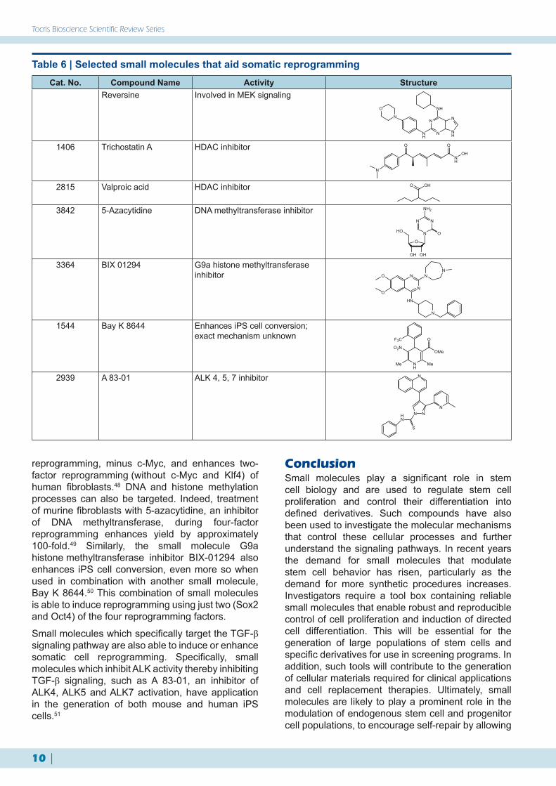
One way that scientists are trying to overcome these limitations is by investigating chemically defined approaches to reprogram cells rather than by genetic manipulation. Screens of small molecules which could potentially replace one or all of the transcription factors have been studied (Table 6). One such assay took lineage committed myoblasts and screened small molecules, looking for cell dedifferentiation. Positive hits were then taken to the next stage, where the cells were induced to differentiate into other lineage-specific cell types. A 2,6-disubstituted purine small molecule termed reversine was identified,39 and shown to work via MEK signaling and non-muscle myosin II heavy chain.45 Reversine has since been reported to reprogram other cell types including human dermal fibroblasts with high efficiency both in vitro and in vivo.46
Small molecules which target histone modification have been proven to increase the efficiency of iPS generation. Both valproic acid and trichostatin A inhibit histone deacetylase (HDAC) activity and greatly enhance the yield of iPS cells created via the traditional four-factor method.47 Valproic acid also increases the yield of three-factor
Table 5 | Selected small molecules that act on ES self-renewalCat. No. Compound Name Activity Structure
Pluripotin Proposed Ras signaling agonistN
N N
N NH
O
CF3
ONH
NN
3194 BIO GSK-3β inhibitor
NH
NHO
NH
O
Br
3853 ID 8 Sustains ES self-renewal; exact mechanism unknown
N
NO2
HO
O
3835 TWS 119 GSK-3β inhibitor
N
N NH
O
OH
NH2
10 |
Tocris Bioscience Scientific Review Series
reprogramming, minus c-Myc, and enhances two-factor reprogramming (without c-Myc and Klf4) of human fibroblasts.48 DNA and histone methylation processes can also be targeted. Indeed, treatment of murine fibroblasts with 5-azacytidine, an inhibitor of DNA methyltransferase, during four-factor reprogramming enhances yield by approximately 100-fold.49 Similarly, the small molecule G9a histone methyltransferase inhibitor BIX-01294 also enhances iPS cell conversion, even more so when used in combination with another small molecule, Bay K 8644.50 This combination of small molecules is able to induce reprogramming using just two (Sox2 and Oct4) of the four reprogramming factors.
Small molecules which specifically target the TGF-β signaling pathway are also able to induce or enhance somatic cell reprogramming. Specifically, small molecules which inhibit ALK activity thereby inhibiting TGF-β signaling, such as A 83-01, an inhibitor of ALK4, ALK5 and ALK7 activation, have application in the generation of both mouse and human iPS cells.51
ConclusionSmall molecules play a significant role in stem cell biology and are used to regulate stem cell proliferation and control their differentiation into defined derivatives. Such compounds have also been used to investigate the molecular mechanisms that control these cellular processes and further understand the signaling pathways. In recent years the demand for small molecules that modulate stem cell behavior has risen, particularly as the demand for more synthetic procedures increases. Investigators require a tool box containing reliable small molecules that enable robust and reproducible control of cell proliferation and induction of directed cell differentiation. This will be essential for the generation of large populations of stem cells and specific derivatives for use in screening programs. In addition, such tools will contribute to the generation of cellular materials required for clinical applications and cell replacement therapies. Ultimately, small molecules are likely to play a prominent role in the modulation of endogenous stem cell and progenitor cell populations, to encourage self-repair by allowing
Table 6 | Selected small molecules that aid somatic reprogrammingCat. No. Compound Name Activity Structure
Reversine Involved in MEK signaling
N
NH
N
N
O
NH
N
NH
1406 Trichostatin A HDAC inhibitor
N
O
NH
O
OH
2815 Valproic acid HDAC inhibitor OHO
3842 5-Azacytidine DNA methyltransferase inhibitor
O
OHOH
HO N
NN
O
NH2
3364 BIX 01294 G9a histone methyltransferase inhibitor
N
NO
O
NN
HN
N
1544 Bay K 8644 Enhances iPS cell conversion; exact mechanism unknown
NH
OMe
O
MeMe
O2N
F3C
2939 A 83-01 ALK 4, 5, 7 inhibitor N
N N
S
HN
N

www.tocris.com | 11
Stem Cell Growth and Differentiation: The Use of Small Molecules
References1. Ying et al (2008) The ground state of embryonic stem cell self-
renewal. Nature 453 519.2. Lyssiotis et al (2010) Chemical control of stem cell fate and
developmental potential. Angew.Chem.Int.Ed.Engl. 50 200.3. Maltman et al (2009) Proteomic profiling of the stem cell
response to retinoic acid and synthetic retinoid analogues: identification of major retinoid-inducible proteins. Mol.Biosyst. 5 458.
4. Reya and Clevers (2005) Wnt signaling in stem cells and cancer. Nature 434 843.
5. Chen et al (2009) Small molecule-mediated disruption of Wnt-dependent signaling in tissue regeneration and cancer. Nat.Chem.Biol. 5 100.
6. Huang et al (2009) Tankyrase inhibition stabilizes axin and antagonizes Wnt signaling. Nature 461 614.
7. Doble and Woodgett (2003) GSK-3: tricks of the trade for a multi-tasking kinase. J.Cell.Sci. 116 1175.
8. North et al (2007) Prostaglandin E2 regulates vertebrate haematopoietic stem cell homeostasis. Nature 447 1007.
9. Lepourcelet et al (2004) Small-molecule antagonists of the oncogenic Tcf/β-catenin protein complex. Cancer Cell 5 91.
10. Ingham and McMahon (2001) Hedgehog signaling in animal development: paradigms and principles. Genes Dev.15 3059.
11. Binns et al (1959) A congenital cyclopiantype malformation in lambs. J.Am.Vet.Med.Assoc.134 180.
12. Chen et al (2002) Small molecule modulation of Smoothened activity. Proc.Natl.Acad.Sci.USA 99 14071.
13. Lauth et al (2007) Inhibition of GLI-mediated transcription and tumor cell growth by small-molecule antagonists. Proc.Natl.Acad.Sci.USA 104 8455.
14. Epstein (2008) Basal cell carcinomas: attack of the hedgehog. Nat.Rev.Cancer 8 743.
15. Sinha and Chen (2006) Purmorphamine activates the Hedgehog pathway by targeting Smoothened. Nat.Chem.Biol. 2 29.
16. Wu et al (2002) A small molecule with osteogenesis-inducing activity in multipotent mesenchymal progenitor cells. J.Am.Chem.Soc. 124 14520.
17. Stanton et al (2009) A small molecule that binds Hedgehog and blocks its signaling in human cells. Nat.Chem.Biol. 5 154.
18. Lee et al (2007) A small-molecule antagonist of the hedgehog signaling pathway. Chem.Biochem. 8 1916.
19. Wu and Hill (2009) TGF-β superfamily signaling in embryonic development and homeostasis. Dev.Cell 16 329.
20. Hong and Yu (2009) Applications of small molecule BMP inhibitors in physiology and disease. Cytokine Growth Factor Rev. 20 409.
21. Watabe et al (2003) TGF-β receptor kinase inhibitor enhances growth and integrity of embryonic stem cell-derived endothelial cells. J.Cell.Biol. 163 1303.
22. Golestaneh and Mishra (2005) TGF-β neuronal stem cells and glioblastoma. Oncogene 24 5722.
23. Ichida et al (2009) A small-molecule inhibitor of Tgf-β signaling replaces Sox2 in reprogramming by inducing Nanog. Cell Stem Cell 5 491.
24. Hao et al (2008) Dorsomorphin, a selective small molecule inhibitor of BMP signaling, promotes cardiomyogenesis in embryonic stem cells. PLoS One 3 e2904.
25. Borowiak et al (2009) Small molecules efficiently direct endodermal differentiation of mouse and human embryonic stem cells. Cell Stem Cell 4 348.
26. Mark et al (2009) Function of retinoic acid receptors during embryonic development. Nucl.Recept.Signal. 7 e002.
27. Han et al (2003) Efficacy validation of synthesized retinol derivatives In vitro: stability, toxicity, and activity. Bioorg.Med.Chem.11 3839.
28. Christie et al (2008) Synthesis and evaluation of synthetic retinoid derivatives as inducers of stem cell differentiation. Org.Biomol.Chem. 6 3497.
29. Christie et al (2010) Retinoid supplementation of differentiating human neural progenitors and embryonic stem cells leads to enhanced neurogenesis in vitro. J.Neurosci.Methods. 193 239.
30. Gotoh (2009) Control of stemness by fibroblast growth factor signaling in stem cells and cancer stem cells. Curr.Stem Cell Res.Ther. 4 9.
31. Bansal et al (2003) Specific inhibitor of FGF receptor signaling: FGF-2-mediated effects on proliferation, differentiation, and MAPK activation are inhibited by PD173074 in oligodendrocyte-lineage cells. J.Neurosci.Res. 74 486.
32. Rinehart et al (2004) Multicenter phase II study of the oral MEK inhibitor, CI-1040, in patients with advanced non-small-cell lung, breast, colon, and pancreatic cancer. J.Clin.Oncol. 22 4456.
33. Silva et al (2008) Promotion of reprogramming to ground state pluripotency by signal inhibition. PLoS Biol. 6 e253.
34. Weinmaster and Kopan (2006) A garden of Notch-ly delights. Development 133 3277.
35. Kanungo et al (2008) The Notch signaling inhibitor DAPT down-regulates cdk5 activity and modulates the distribution of neuronal cytoskeletal proteins. J.Neurochem.106 2236.
36. Cullion et al (2009) Targeting the Notch1 and mTOR pathways in a mouse T-ALL model. Blood 113 6172.
37. Mallon et al (2006) Toward xeno-free culture of human embryonic stem cells. Int.J.Biochem.Cell.Biol. 38 1063.
38. Xu et al (2008) A chemical approach to stem-cell biology and regenerative medicine. Nature 453 338.
39. Chen et al (2006) Self-renewal of embryonic stem cells by a small molecule. Proc.Natl.Acad.Sci.USA 103 17266.
40. Miyabayashi et al (2008) Indole derivatives sustain embryonic stem cell self-renewal in long-term culture. Biosci.Biotechnol.Biochem. 72 1242.
41. Sato et al (2004) Maintenance of pluripotency in human and mouse embryonic stem cells through activation of Wnt signaling by a pharmacological GSK-3-specific inhibitor. Nat.Med. 10 55.
42. Ding et al (2003) Synthetic small molecules that control stem cell fate. Proc.Natl.Acad.Sci.USA 100 7632.
43. Takahashi and Yamanaka (2006) Induction of pluripotent stem cells from mouse embryonic and adult fibroblast cultures by defined factors. Cell 126 663.
44. Yu et al (2007) Induced pluripotent stem cell lines derived from human somatic cells. Science 318 1917.
45. Chen et al (2004) Dedifferentiation of lineage-committed cells by a small molecule. J.Am.Chem.Soc. 126 410.
46. Anastasia et al (2006) Reversine-treated fibroblasts acquire myogenic competence in vitro and in regenerating skeletal muscle. Cell Death Differ.13 2042.
47. Huangfu et al (2008) Induction of pluripotent stem cells by defined factors is greatly improved by small-molecule compounds. Nat.Biotechnol. 26 795.
48. Huangfu et al (2008) Induction of pluripotent stem cells from primary human fibroblasts with only Oct4 and Sox2. Nat.Biotechnol. 26 1269.
49. Tsuji-Takayama et al (2004) Demethylating agent, 5-azacytidine, reverses differentiation of embryonic stem cells. Biochem.Biophys.Res.Commun. 323 86.
50. Shi et al (2008) Induction of pluripotent stem cells from mouse embryonic fibroblasts by Oct4 and Klf4 with small-molecule compounds. Cell Stem Cell 3 568.
51. Li et al (2009) Generation of rat and human induced pluripotent stem cells by combining genetic reprogramming and chemical inhibitors. Cell Stem Cell 4 16.
the proliferation and differentiation of host cells using pharmacological cues.
The development and characterization of small molecules that interact with the molecular pathways
that control cell renewal and differentiation is therefore integral to the advance and optimization of stem cell research and its applications to drug discovery and regenerative medicine.

www.tocris.com
UK:Phone: + 44 (0)117 916 3333
Fax: + 44 (0)117 916 3344 [email protected]
Tocris House, IO Centre, Moorend Farm Avenue,Avonmouth, Bristol, BS11 0QL, UK
US:Phone: 800-421-3701
Fax: [email protected]
16144 Westwoods Business Park,Ellisville, Missouri 63021 USA
Tocris Reviews No. 37©2011 Tocris Cookson
For a complete and up-to-date product listing please visit www.tocris.com.
Stem Cell Dedifferentiation3842 5-azacytidine
DNA methyltransferase inhibitor. Improves reprogramming efficiency1544 Bay K8644
Ca2+-channel activator (L-type); enhances iPSC conversion3364 BIX 01294
G9a-like protein and G9a histone lysine methyltransferase (HMTase) inhibitor
1398 KenpaulloneInhibitor of GSK-3; generates iPSCs from somatic cells
3845 ThiozovivinImproves the efficiency of fibroblast reprogramming and induction of iPSCs
1406 Trichostatin AHistone deacetylase inhibitor
2815 Valproic acidHistone deacetylase inhibitor
Stem Cell Differentiation3851 Cardiogenol C
Induces cardiomyogenesis in ESCs2634 DAPT
γ-secretase inhibitor1126 Dexamethasone
Anti-inflammatory glucocorticoid; induces differentiation of hMSCs3093 Dorsomorphin
Potent and selective AMPK inhibitor4011 EC 23
Synthetic retinoid; induces differentiation of stem cells1099 Forskolin
Adenylyl cyclase activator; induces neuronal differentiation3651 (-)-Indolactam V
Protein kinase C activator; directs hESC differentiation3656 Neurodazine
Induces neurogenesis in mature skeletal muscle cells3854 1-Oleoyl lysophosphatidic acid
LPA1 and LPA2 agonist. Inhibits differentiation of neural stem cells3044 PD 173074
Selective FGFR1 and -3 inhibitor1213 PD 98059
Specific inhibitor of MEK0695 Retinoic acid
Endogenous retinoic acid receptor agonist. Promotes ESC differentiation
3850 Sodium butyrateHistone deacetylase inhibitor
3741 StauprimideInhibits NME2 nuclear translocation; primes ESCs for differentiation
3835 TWS 119GSK-3β inhibitor; induces neuronal differentiation in ESCs
Stem Cell Proliferation2939 A 83-01
Selective inhibitor of TGF-βRI, ALK4 and ALK73194 BIO
Potent, selective GSK-3 inhibitor3849 Gatifloxacin
Antibiotic; activates short-term renewal of ESCs
Stem Cell Receptor Compounds Available from Tocris
3853 ID 8Sustains self-renewal and pluripotency of ESCs
3044 PD 173074Selective FGFR1 and -3 inhibitor
0238 O-Phospho-L-serineGroup III mGlu agonist; enhances neural differentiation in progenitor cells
1202 SB 203580Selective inhibitor of p38 MAPK; stimulates neural stem cell proliferation
1614 SB 431542Potent, selective inhibitor of TGF-βRI, ALK4 and ALK7
3848 SinomenineNatural anti-inflammatory morphinan analog; stimulates short term self-renewal of hESCs
3847 TheaninePromotes hESC self-renewal
Stem Cell Signaling3874 BIO-acetoxime
Selective GSK-3α/β inhibitor3364 BIX 01294
G9a-like protein and G9a histone lysine methyltransferase (HMTase) inhibitor
1626 Calphostin CPotent, selective and photo-dependent PKC inhibitor; inhibits Wnt signaling
1623 CyclopamineInhibitor of (Hh) signaling
4027 16,16-dimethyl prostaglandin E2Synthetic PGE2 (Cat.No. 2296) derivative; regulates HSC development
3889 GANT 58GLI1 antagonist; inhibits Hh signaling
3191 GANT 61GLI antagonist; inhibits Hh signaling
3533 IWP 2Wnt production inhibitor; inactivates porcn
3532 IWR 1Axin stabilizer; promotes β-catenin phosphorylation
3341 JK 184Downstream Hh signaling pathway inhibitor; inhibits alcohol dehydrogenase 7
1398 KenpaulloneInhibitor of GSK-3; generates iPSCs from somatic cells
2296 Prostaglandin E2Major endogenous prostanoid
3324 QS 11ARFGAP1 inhibitor; modulates Wnt/β-catenin signaling
3295 RG 108Non-nucleoside DNA methyltransferase inhibitor; enhances efficiency of iPSC generation
3742 SJN 2511Selective inhibitor of TGF-βRI
3748 XAV 939Tankyrase inhibitor; inhibits Wnt signaling