standard treatment guidelines gastrointestinal …
TRANSCRIPT
1
STANDARD TREATMENT GUIDELINES
GASTROINTESTINAL SURGERY
Ministry of Health & Family Welfare
Govt. of India
2
Contents
S.No. Topic
Page No.
1 ACUTE PANCREATITIS 4 2 PORTAL HYPERTENSION 12 3 CHOLEDOCHOLITHIASIS 24 4 CARCINOMA STOMACH 31 5 GALLBLADDER CARCINOMA (GBC) REQUIRING HOSPITALIZATION 38 6 CARCINOMA RECTUM 46
3
Group Head Coordinator of Development Team
Dr. V.P. Bhalla Department of Surgical Gastroenterology,
Bariatric & Minimal Access Surgery BLK Superspeciality Hospital
4
Acute Pancreatitis
Dr. V.P. Bhalla Dr. Deep Goel
Department of Surgical Gastroenterology, Bariatric & Minimal Access Surgery
BLK Superspeciality Hospital New Delhi
Peer Reviewer- Dr Samiran Nundy, Chairman, Surgical Gastro and Liver Transplant, Sir Ganga Ram Hospital
1. When to suspect/recognize
a) Introduction
Acute pancreatitis is an important cause of acute upper abdominal pain associated with vomiting. The common causes include gall bladder stone disease, alcoholism and idiopathic- where no obvious cause is discernible. Fortunately the majority of cases of acute pancreatitis are mild and respond to conservative treatment. In less than 10% the disease is more severe and follows a vicious course with immense clinical and socio economic implications. These guidelines will help in the initial management of these patients at the secondary district level hospital and also at the more advanced tertiary metro super specialty centre.
b) Case definition
A typical patient presents with severe agonizing upper abdominal pain which may radiate to the back and is associated with retching and vomiting. The patient may be a known case of gall bladder stone disease or give a history of chronic alcohol consumption or a recent alcoholic binge. Clinical examination early in the disease process may reveal upper abdominal tenderness guarding and later the patient will show all the features of hypovolaemia including shock as third spacing of fluids sets in.
I. A 3-4 fold increase in serum amylase level within 48 hours of onset of pain is highly
suggestive of the diagnosis of acute pancreatitis.
Initial management is aimed at relieving pain and administration of IV fluids to maintain core perfusion as evidenced by good urine output. Subsequently management of containing pancreatitis is best done in tertiary multi super specialty hospitals with expertise in dealing with such patients.
II. Incidence of the condition
The exact incidence of acute pancreatitis in India is unknown as no hard data is available. The incidence is rising world wide with the United Kingdom reporting an incidence range of 150-420 cases per million population. The experience of senior clinicians from personal experience seems to suggest that even in India incidence of Acute Pancreatitis appears to be rising and patients are being seen frequently.
5
III. Differential diagnosis
The differential diagnosis of acute pancreatitis include all the differentials of the syndrome of sudden onset severe epigastric pain associated with vomiting.
These are:
1. Severe acute gastritis 2. Peptic ulcer perforation 3. Liver abscess 4. Enteric perforation 5. Biliary colic 6. Acute cholecystitis 7. Acute gastroentertitis 8. Acute episode of chronic pancreatitis 9. Acute mesenteric ischaemia 10. Myocardial infarction
IV. Prevention and counselling
In a known case of alcohol induced pancreatitis the patient must be counselled about the role of alcohol and that abstaining from it will prevent a further episode of pancreatitis. Similarly avoidance of fatty food and early cholecystectomy in a known case of biliary or gall stone induced pancreatitis will prevent further attacks.
V. Optimal diagnostic criteria, investigations, treatment and referral criteria
Diagnosis of acute pancreatitis is based on the presentation with severe acute upper and abdominal pain and a three to four fold increase in the level of serum amylase within 48 hours of onset of pain.
Investigation to confirm the diagnosis and exclude other possibilities is a contrast enhanced CT examination of the abdomen. An early CT (within the first few hours or day 1-2) will be helpful if no diagnosis has been made in 48 hours. The best time for CECT abdomen to establish the diagnosis of acute pancreatitis and extent of necrosis is 5-7 days.
Further investigations need to be done to document severity of acute pancreatitis. These include CBC, BUN & serum creatinine, blood gas analysis, C-reactive protein. Chest X-ray PA and ultrasound to demonstrate pleural effusion.
Other investigations to help establish the cause of acute pancreatitis include, MRCP- Magnetic Retrograde Cholangio Pancreaticography, ultrasound and increasingly endo- ultrasound.
V. Referral criteria
Criteria have been developed to predict mortality in Acute Pancreatitis. These can be used to identify patients who will do well to be referred to tertiary centres for further management.
6
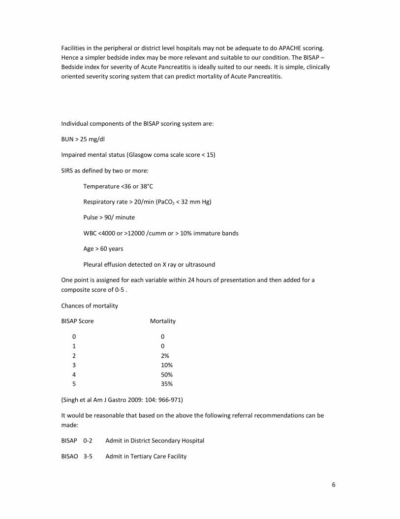
Facilities in the peripheral or district level hospitals may not be adequate to do APACHE scoring. Hence a simpler bedside index may be more relevant and suitable to our condition. The BISAP – Bedside index for severity of Acute Pancreatitis is ideally suited to our needs. It is simple, clinically oriented severity scoring system that can predict mortality of Acute Pancreatitis.
Individual components of the BISAP scoring system are:
BUN > 25 mg/dl
Impaired mental status (Glasgow coma scale score < 15)
SIRS as defined by two or more:
Temperature <36 or 38°C
Respiratory rate > 20/min (PaCO2 < 32 mm Hg)
Pulse > 90/ minute
WBC <4000 or >12000 /cumm or > 10% immature bands
Age > 60 years
Pleural effusion detected on X ray or ultrasound
One point is assigned for each variable within 24 hours of presentation and then added for a composite score of 0-5 .
Chances of mortality
BISAP Score
Mortality
0 0 1 0 2 2% 3 10% 4 50% 5 35%
(Singh et al Am J Gastro 2009: 104: 966-971)
It would be reasonable that based on the above the following referral recommendations can be made:
BISAP 0-2 Admit in District Secondary Hospital
BISAO 3-5 Admit in Tertiary Care Facility
7
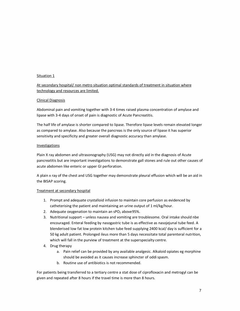
Situation 1
At secondary hospital/ non metro situation optimal standards of treatment in situation where technology and resources are limited.
Clinical Diagnosis
Abdominal pain and vomiting together with 3-4 times raised plasma concentration of amylase and lipase with 3-4 days of onset of pain is diagnostic of Acute Pancreatitis.
The half life of amylase is shorter compared to lipase. Therefore lipase levels remain elevated longer as compared to amylase. Also because the pancreas is the only source of lipase it has superior sensitivity and specificity and greater overall diagnostic accuracy than amylase.
Investigations
Plain X ray abdomen and ultrasonography (USG) may not directly aid in the diagnosis of Acute pancreatitis but are important investigations to demonstrate gall stones and rule out other causes of acute abdomen like enteric or upper GI perforation.
A plain x ray of the chest and USG together may demonstrate pleural effusion which will be an aid in the BISAP scoring.
Treatment at secondary hospital
1. Prompt and adequate crystalloid infusion to maintain core perfusion as evidenced by catheterising the patient and maintaining an urine output of 1 ml/kg/hour.
2. Adequate oxygenation to maintain an sPO2 above 95%. 3. Nutritional support – unless nausea and vomiting are troublesome. Oral intake should nbe
encouraged. Enteral feeding by nasogastric tube is as effective as nasojejunal tube feed. A blenderised low fat low protein kitchen tube feed supplying 2400 kcal/ day is sufficient for a 50 kg adult patient. Prolonged ileus more than 5 days necessitate total parenteral nutrition, which will fall in the purview of treatment at the superspecialty centre.
4. Drug therapy a. Pain relief can be provided by any available analgesic. Alkaloid opiates eg morphine
should be avoided as it causes increase sphincter of oddi spasm. b. Routine use of antibiotics is not recommended.
For patients being transferred to a tertiary centre a stat dose of ciprofloxacin and metrogyl can be given and repeated after 8 hours if the travel time is more than 8 hours.
8
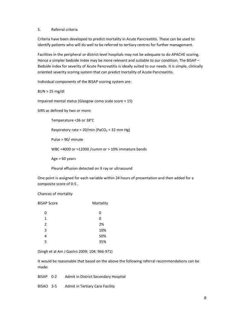
5. Referral criteria
Criteria have been developed to predict mortality in Acute Pancreatitis. These can be used to identify patients who will do well to be referred to tertiary centres for further management.
Facilities in the peripheral or district level hospitals may not be adequate to do APACHE scoring. Hence a simpler bedside index may be more relevant and suitable to our condition. The BISAP – Bedside index for severity of Acute Pancreatitis is ideally suited to our needs. It is simple, clinically oriented severity scoring system that can predict mortality of Acute Pancreatitis.
Individual components of the BISAP scoring system are:
BUN > 25 mg/dl
Impaired mental status (Glasgow coma scale score < 15)
SIRS as defined by two or more:
Temperature <36 or 38°C
Respiratory rate > 20/min (PaCO2 < 32 mm Hg)
Pulse > 90/ minute
WBC <4000 or >12000 /cumm or > 10% immature bands
Age > 60 years
Pleural effusion detected on X ray or ultrasound
One point is assigned for each variable within 24 hours of presentation and then added for a composite score of 0-5 .
Chances of mortality
BISAP Score
Mortality
0 0 1 0 2 2% 3 10% 4 50% 5 35%
(Singh et al Am J Gastro 2009: 104: 966-971)
It would be reasonable that based on the above the following referral recommendations can be made:
BISAP 0-2 Admit in District Secondary Hospital
BISAO 3-5 Admit in Tertiary Care Facility
9
Situation 2
At Super Specialty facility in Metro location where higher end technology is available
Clinical Diagnosis
As in situation 1. Record history of known gall stone disease, alcohol intake, drug intake, exposure to known viral causes
Investigations
Aims of investigations at super speciality facility are
1. Confirm diagnosis 2. Confirm aetiology 3. Confirm presence of pancreatic necrosis and infected pancreatic necrosis 4. Confirm developing complications of Acute Pancreatitis
a. Peri pancreatic fluid collection b. Peri pancreatic abscess c. Bowel ischemia and gangrene d. Bleeding
Investigations to be done at the super speciality centre
Blood tests
1. CBC – serial complete blood counts to look for the trends of neutrophilic leucocytosis which will indicate both severe pancreatitis and infected pancreatic necrosis
2. Serum pancreatic enzymes- amylase and lipase are not helpful after 4-5 days 3. LFT 4. Blood urea and serum creatinine 5. Serial monitoring of blood sugar and serum calcium 6. Fasting serum lipid profile 7. Viral antibody titres
Radiological tests
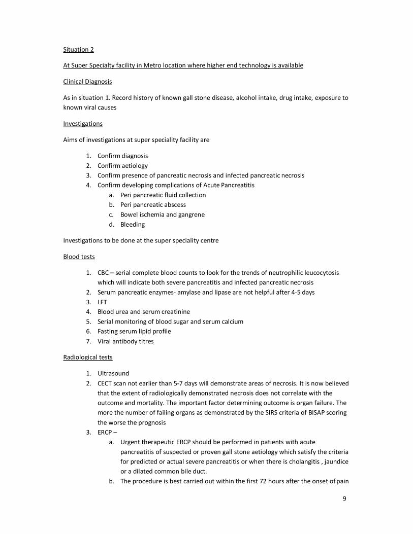
1. Ultrasound 2. CECT scan not earlier than 5-7 days will demonstrate areas of necrosis. It is now believed
that the extent of radiologically demonstrated necrosis does not correlate with the outcome and mortality. The important factor determining outcome is organ failure. The more the number of failing organs as demonstrated by the SIRS criteria of BISAP scoring the worse the prognosis
3. ERCP – a. Urgent therapeutic ERCP should be performed in patients with acute
pancreatitis of suspected or proven gall stone aetiology which satisfy the criteria for predicted or actual severe pancreatitis or when there is cholangitis , jaundice or a dilated common bile duct.
b. The procedure is best carried out within the first 72 hours after the onset of pain
10
c. All patients will with severe gall stone pancreatitis will require endoscopic sphincterotomy whether or not stones are found in the bile duct
d. Patients with signs of cholangitis may require duct drainage by stenting to ensure relief of biliary obstruction
4. EUS ( endoscopic ultrasound) – EUS has proven superiority over conventional abdominal ultrasound for the detection of CBD stones. It is of particular benefit in the evaluation of patients with recurrent acute pancreatitis.
5. MRCP – is an effective non invasive means of delineating biliary and pancreatic ductal anatomy. In patients with recurrent pancreatitis it can show CBD stones, ampullary strictures and presence of pancreatic divisium.
6. Image guided FNAC of pancreatic necrosis to confirm infected pancreatic necrosis.
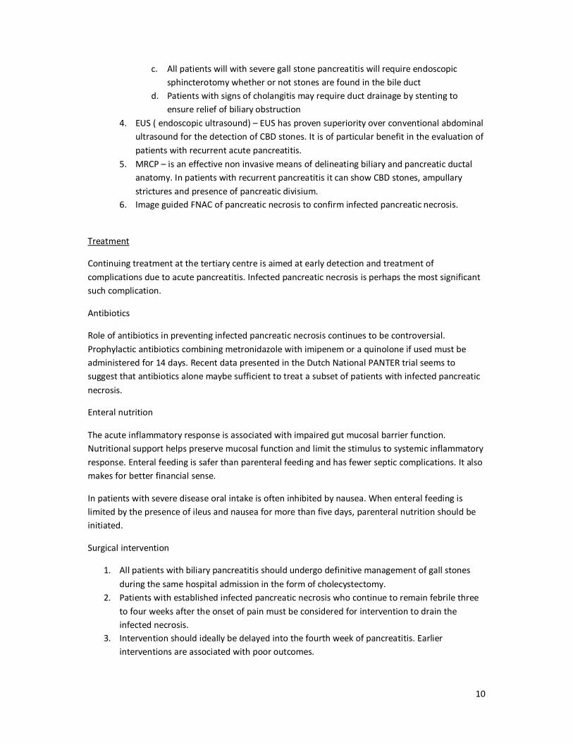
Treatment
Continuing treatment at the tertiary centre is aimed at early detection and treatment of complications due to acute pancreatitis. Infected pancreatic necrosis is perhaps the most significant such complication.
Antibiotics
Role of antibiotics in preventing infected pancreatic necrosis continues to be controversial. Prophylactic antibiotics combining metronidazole with imipenem or a quinolone if used must be administered for 14 days. Recent data presented in the Dutch National PANTER trial seems to suggest that antibiotics alone maybe sufficient to treat a subset of patients with infected pancreatic necrosis.
Enteral nutrition
The acute inflammatory response is associated with impaired gut mucosal barrier function. Nutritional support helps preserve mucosal function and limit the stimulus to systemic inflammatory response. Enteral feeding is safer than parenteral feeding and has fewer septic complications. It also makes for better financial sense.
In patients with severe disease oral intake is often inhibited by nausea. When enteral feeding is limited by the presence of ileus and nausea for more than five days, parenteral nutrition should be initiated.
Surgical intervention
1. All patients with biliary pancreatitis should undergo definitive management of gall stones
during the same hospital admission in the form of cholecystectomy. 2. Patients with established infected pancreatic necrosis who continue to remain febrile three
to four weeks after the onset of pain must be considered for intervention to drain the infected necrosis.
3. Intervention should ideally be delayed into the fourth week of pancreatitis. Earlier interventions are associated with poor outcomes.
11
4. A stepped up approach starting with radiological guided needle aspirations, endoscopic guided lesser sac aspiration and going on to video assisted retro peritoneal endoscopic drainage maybe preferred to open conventional necrosectomy if all facilities are available at the tertiary hospital.
5. Conventional necrosectomy is acceptable treatment if the above facilities are not available. It is recommended that a cholecystectomy be added during the necrosectomy particularly in patients with biliary pancreatitis.
6. A feeding jejunostomy must always be added in our Indian patients.
Tertiary treatment centre for patients with acute pancreatitis
1. Every tertiary hospital receiving patients with pancreatitis should have a nominated clinical team to manage these patients.
2. Components of team a. Clinicians : a multidisciplinary team of specialists including surgical and medical
gastroenterologists, intensivists, nutritionists and other support staff of the intensive care unit.
b. Facilities for dynamic multislice C.T., percutaneous needle aspiration and drainage procedure and MR imaging.
c. Facilities for ERCP and EUS.
Further reading
1. UK guidelines for the management of acute pancreatitis UK working party on acute pancreatitis Gut 2005;54;1-g
2. Singh VK et al A prospective evaluation of the bedside index for severity in acute pancreatitis score in assessing mortality and intermediate markers of severity in acute pancreatitis Am J Gastro, 2009;104;966-971
3. Hirota M et al Fundamental and intensive care of acute pancreatitis J Hepatobiliary Pancreat Sci (2010) 17:45-42
4. Acute pancreatitis: Problems in adherence to guidelines 2009:76.12;697-703
5. Wu Bu et al, The early prediction of mortality in acute pancreatitis: A large population- based study, Gut 2008: 57:1698-1703
12
Portal hypertension
S Thiagrajan Sorabh Kapoor Samiran Nundy
Department of Surgical Gastroenterology and Liver Transplantation Sir Ganga Ram Hospital
New Delhi
I. When to suspect / recognise
a)Introduction
Portal hypertension may manifest as variceal bleeding, ascites, splenomegaly, hepato renal syndrome and hepatopulmonary syndrome.The management of Portal Hypertension (PHTincludes treating acute bleeding from varices in the oesophagus and stomach, preventing recurrent bleeding (secondary prophylaxis), preventing the first bleeding episode (primary prophylaxis) and controlling ascites and liver failure. In these guidelines we will only deal with the management of bleeding varices in both cirhhotic and non cirrhotic portal hypertension [EHPVO(extra hepatic portal venous obstruction), NCPF (non cirrhotic portal fibrosis) etc.,]. The management of ascites and liver failure is mainly by drugs and liver transplants. In addition management of left sided portal hypertension usually accompanying chronic pancreatitis is also dealt separately
b) Case definition
Portal hypertension is defined as elevated pressure in portal venous system due to resistance to portal blood flow. The site of resistance may be prehepatic (EHPVO), hepatic (cirrhosis and NCPF which is presinusoidal) and post hepatic (Hepatic venous outflow tract obstruction – HVOTO). The normal portal pressure is 5-10 mmHg. Pressure more than 10 mmHg is defined as portal hypertension. Since portal pressure or HVPG is normally measured in specialized Gastroenterology /Hepatology units, presence of varices along with evidence of liver cirrhosis or portal vein thrombosis / HVOTO in the presence /absence of ascites and splenomegaly is considered sufficient for diagnosis of portal hypertension.
II. Incidence of portal hypertension in our country
The true incidence of the condition is unknown. However variceal bleeding is one of the common causes of Upper gastrointestinal haemorrhage , accounting for almost half of cases (depending on referral pattern). Variceal haemorrhage is also the commonest complication of liver cirrhosis. Almost 90% of patients with cirrhosis will have variceal formation and 30% of all patients with varices are likely to bleed. Most patients with EHPVO and NCPF are also diagnosed after evaluation for splenomegaly and variceal bleeding. In children, variceal bleeding is most common cause of UGI bleeding in India, accounting for almost 90% cases with EHPVO responsible for the majority and NCPF and cirrhosis responsible for the rest.
III. Differential diagnosis
13
a) Variceal hemorrhage has to be differentiated from GI bleed due to
Peptic ulcer disease,
esophagitis,
Dieulafoy’s lesion,
Mallory Weiss tear
NSAID induced ulcers
Arteriovenous malformations/ telengectasias
b) Ascites due to portal hypertension has to differentiated from ascites due to
Renal disease
Malignant ascites
Tuberculosis
Pancreatic ascites, etc
c) Similarly splenomegaly and hypersplenism have to be differentiated from hematological and other infiltrative causes of splenic enlargement
IV. Prevention and counseling
Since the etiology of EHPVO and NCPF are not fully known the prevention is mainly prophylaxis of bleeding or rebleeding. Simiarly, if HVOTO is diagnosed then etiology of probable hypercoagulable state needs to be evaluated for prevention of further thrombosis of other hepatic veins or re occlusion after interventional radiological management / shunt / transplant.
The prevention of cirrhosis involves essentially involves counseling in alcoholics, vaccination for Hepatitis B, and precautions for Hepatitis C and treatment of patients with NAFL (to prevent progression to NAFLD). In addition, all patients diagnosed with viral hepatitis( B, C and D) or chronic liver disease due to any etiology require management by specialized gastroenterogy / hepatology units with the aim of halting the progression of disease and early management of complications.
Prevention of Variceal haemorrhage
The prevention of index bleeding in cirrhotics or other causes of PHT in patients who are diagnosed prior to bleeding is important.
14
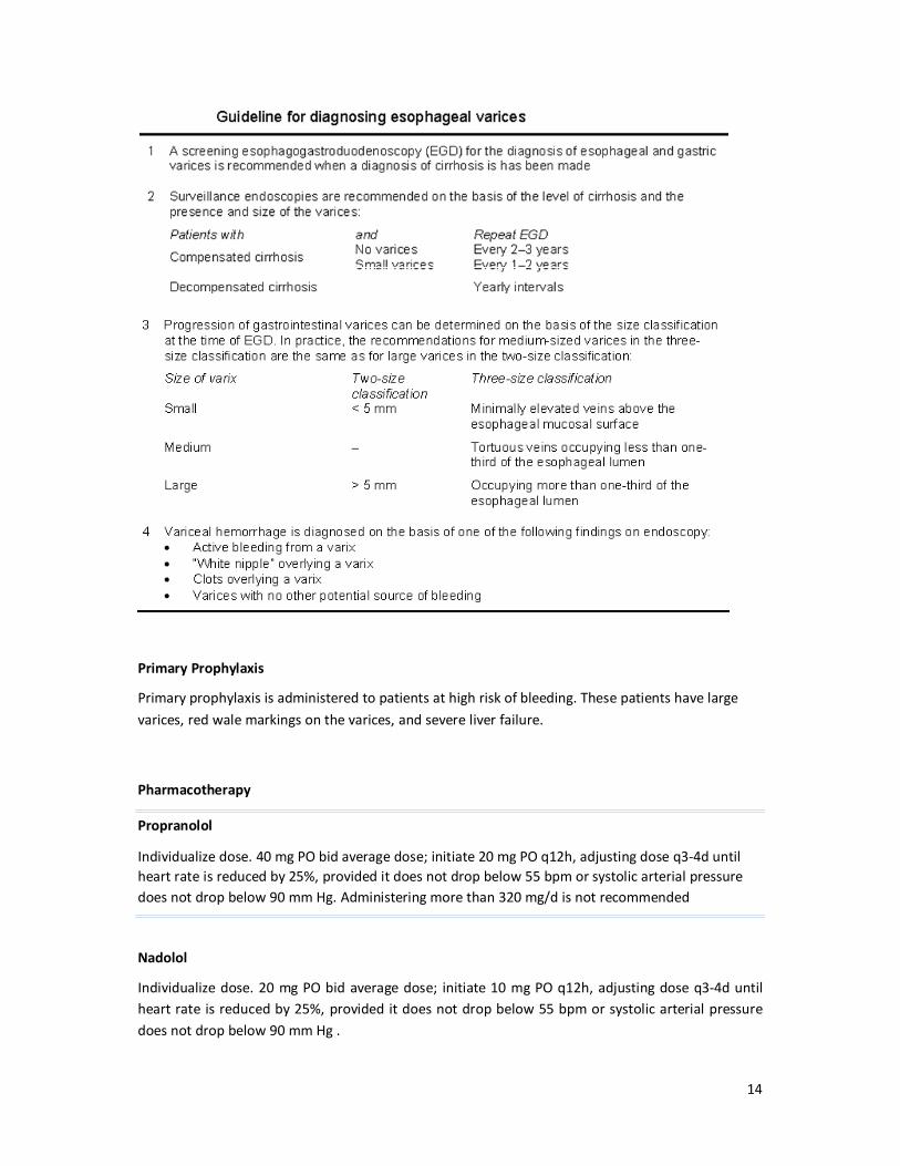
Primary Prophylaxis
Primary prophylaxis is administered to patients at high risk of bleeding. These patients have large varices, red wale markings on the varices, and severe liver failure.
Pharmacotherapy
Propranolol
Individualize dose. 40 mg PO bid average dose; initiate 20 mg PO q12h, adjusting dose q3-4d until heart rate is reduced by 25%, provided it does not drop below 55 bpm or systolic arterial pressure does not drop below 90 mm Hg. Administering more than 320 mg/d is not recommended
Nadolol
Individualize dose. 20 mg PO bid average dose; initiate 10 mg PO q12h, adjusting dose q3-4d until heart rate is reduced by 25%, provided it does not drop below 55 bpm or systolic arterial pressure does not drop below 90 mm Hg .
15
Response to treatment is monitored by a reduction of the portal pressure gradient by more than 20% of the baseline value or less than 12 mm Hg. Checking the HVPG response in primary prophylaxis is not mandatory because 60% of patients who do not achieve these targets do not bleed at 2-year follow-up evaluations.
Propranolol is contraindicated in patients with asthma, chronic obstructive pulmonary disease (COPD), atrioventricular (AV) block, intermittent claudication, and psychosis. Beta-blockers are best continued for the patient's lifetime because the risk of variceal hemorrhage returns to that of the untreated population once beta-blockers are withdrawn.
Vasodilators Available evidence does not support the use of Isosorbide mononitrate ISMN as monotherapy for primary prophylaxis, even in patients with contraindications or intolerance to beta-blockers.
Combination therapy
This involves both beta-blockers and ISMN. Combination therapy cannot be recommended presently until further studies prove efficacy.
Prophylactic sclerotherapy and Surgery No role in primary prophylaxis except perhaps in patients with EHPVO who have ‘dangerous’ varices and live far from tertiary medical centres.
Prophylactic endoscopic variceal ligation
Prophylactic EVL currently cannot be recommended as a routine measure for primary prevention but may be an option for patients with grade 3-4 varices who have contraindications to or cannot tolerate beta-blockers.
V. Optimal diagnostic criteria, investigations, treatment and referral criteria
Situation 1. At secondary hospital /Non metro situation : Optimal standards of treatment in situations where technology and resources are limited
Clinical diagnosis
Variceal hemorrhage is diagnosed when patients present with upper gastrointestinal hemorrhage in the background of preexisting chronic liver disease or cirrhosis or in patients in 1st decades of life for EHO and 2nd and 3rd decade who present with UGI bleeding associated with splenomegaly usually in the absence of any associated features of chronic liver disease.
Investigations
The initial investigations are aimed at guiding and assessing the adequacy of resuscitation by checking Hemogram , liver and renal function
16
Treatment
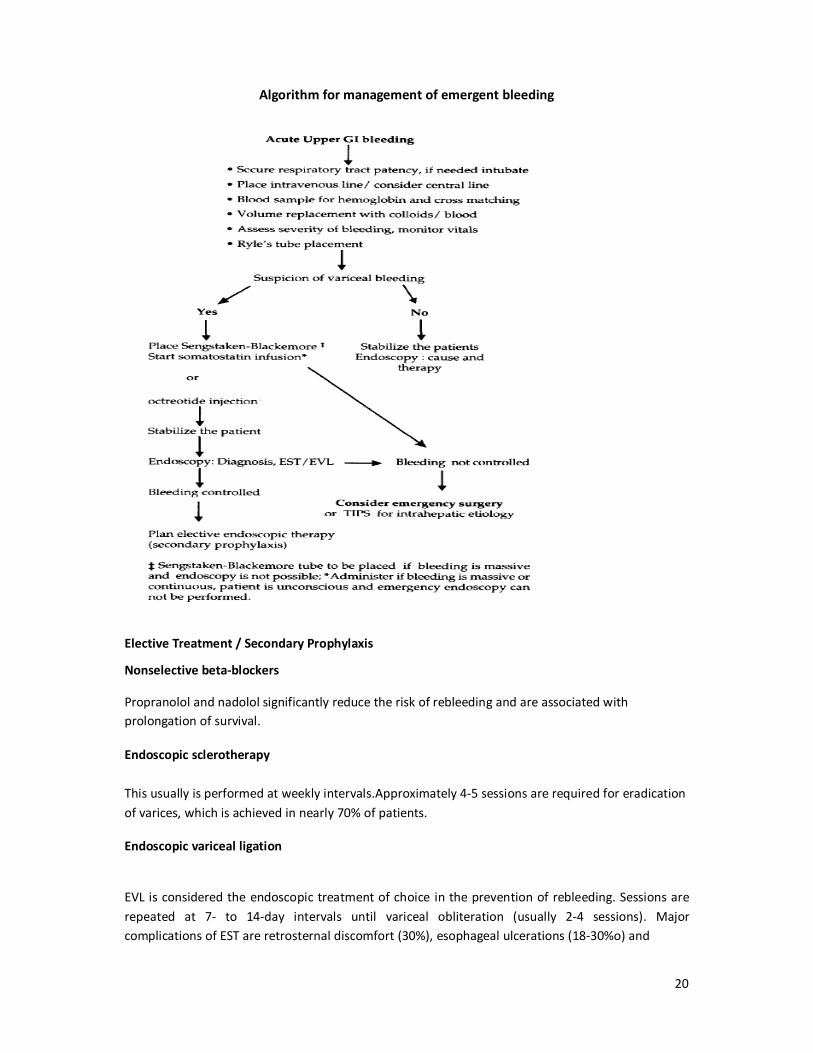
Initial resuscitation with replacement of blood volume loss Secure respiratory tract patency, if needed endotracheal intubation may be done.
Place two wide bore 16G intravenous lines preferably in the antecubital fossae and consider central venous line insertion.
Assess severity of bleeding, monitor vitals.
Blood sample for hemoglobin and cross matching.
Volume replacement with colloids/blood, guided initially, by blood pressure and urine output and central venous pressure(CVP) if possible. It is important to avoid under- than over-transfuse to avoid excessive intravascular volume and variceal expansion and consequent rebleeding.
Blood should be replaced at a modest target of HCT (hematocrit) of 25-30%.
Place a nasogastric tube
Prevention of complications (eg, hepatic encephalopathy, bronchial aspiration, renal failure, systemic infections, Spontaneous Bacterial Peritonitis) All patients with cirrhosis and upper GI bleeding are at a high risk of developing severe bacterial infections, which are associated with early rebleeding. The use of prophylactic antibiotics has been demonstrated to decrease the rate of bacterial infections and increase survival rates, thus prophylactic antibiotic use (norfloxacin 400 mg PO bid for 7 d; ciprofloxacin and other broad-spectrum antibiotics) in the setting of acute bleeding is recommended.
Pharmacological therapy
This acts by decreasing splanchnic blood flow Octreotide is a synthetic analogue of somatostatin that is usually administered at a constant infusion of 50 mcg/h.
Terlipressin,a synthetic analogue of vasopressin which is also useful during an acute bleeding episode. Dosage 0.5 mg to 2mg QID by slow IV infusion
The use of vasopressin is limited by adverse effects related to splanchnic vasoconstriction (eg, bowel ischaemia) and systemic vasoconstriction (eg, hypertension, myocardial ischemia). Continuous infusion of 0.2-0.4 IU/min (not exceeding 0.8 IU/min) is recommended. Vasopressin always should be accompanied by intravenous nitroglycerin at a dose of 40 mcg/min (not to exceed 400 mcg/min) to maintain systolic blood pressure greater than 90 mm Hg. Adding nitrates to vasopressin therapy improves its efficacy, although the adverse effects of combination therapy are higher than those associated with terlipressin or somatostatin.
Subsequent Management
Based on availability of expertise and equipment.
17
If the bleeding continues the Endoscopic therapy with sclerotherapy or band ligation should be attempted. In the absence of expertise or rebleeding after initial control Balloon tamponade should be instituted and the patient referred to higher center.
Situation 2. At superspeciaity facility in metro location where higher end technology is available
Clinical diagnosis
Variceal hemorrhage is diagnosed when patients present with upper gastrointestinal hemorrhage in the background of preexisting chronic liver disease or cirrhosis or in patients in 1st decades of life for EHO and 2nd and 3rd decade who present with UGI bleeding associated with splenomegaly usually in the absence of any associated features of chronic liver disease.
Investigations
The definitive diagnosis of variceal haemorrhage is done by demonstrating varices on esophagogastroscopy which should be done after adequate resuscitation and stabilization. Imaging of liver by ultrasound or CT scan is also done after initial resuscitation.
Following emergent treatment, the etiology of portal hypertension or cirrhosis needs to be identified. EHPVO is diagnosed by clinical presentation with preserved liver functions and splenomegaly with varices along with demonstration on USG Doppler of portal vein thrombosis or portal cavernoma formation. Similarly aforementioned presentation with normal liver function and normal portal vein with normal liver on ultrasound is considered sufficient for diagnosis of NCPF.
Doppler is also used for diagnosis of HVOTO which may be supplemented by venography.
Etiology of cirrhosis is identified by history of alcoholism , liver functions and viral serology, PCR, autoantibodies and tests for Wilsons disease and hemochromatosis. Liver biopsy may be needed in various situations.
Emergency Treatment
Bleeding from esophageal varices
Following resuscitation, treatment of acute variceal bleeding includes control of bleeding (24 h without bleeding within the first 48 h after starting therapy) and prevention of early recurrence.
Initial resuscitation with replacement of blood volume loss Secure respiratory tract patency, if needed endotracheal intubation may be done.
Place two wide bore 16G intravenous lines preferably in the antecubital fossae and consider central venous line insertion.
Assess severity of bleeding, monitor vitals.
17
Blood sample for hemoglobin and cross matching.
Volume replacement with colloids/blood, guided initially, by blood pressure and urine output and central venous pressure(CVP) if possible. It is important to avoid under- than over-transfuse to avoid excessive intravascular volume and variceal expansion and consequent rebleeding.
Blood should be replaced at a modest target of HCT (hematocrit) of 25-30%.
Place a nasogastric tube
Prevention of complications (eg, hepatic encephalopathy, bronchial aspiration, renal failure, systemic infections, Spontaneous Bacterial Peritonitis)
All patients with cirrhosis and upper GI bleeding are at a high risk of developing severe bacterial infections, which are associated with early rebleeding. The use of prophylactic antibiotics has been demonstrated to decrease the rate of bacterial infections and increase survival rates, thus prophylactic antibiotic use (norfloxacin 400 mg PO bid for 7 d; ciprofloxacin and other broad-spectrum antibiotics) in the setting of acute bleeding is recommended.
Pharmacological therapy
This acts by decreasing splanchnic blood flow Octreotide is a synthetic analogue of somatostatin that is usually administered at a constant infusion of 50 mcg/h.
Terlipressin,a synthetic analogue of vasopressin which is also useful during an acute bleeding episode. Dosage 0.5 mg to 2mg QID by slow IV infusion Vasopressin The use of vasopressin is limited by adverse effects related to splanchnic vasoconstriction (eg, bowel ischaemia) and systemic vasoconstriction (eg, hypertension, myocardial ischemia). Continuous infusion of 0.2-0.4 IU/min (not exceeding 0.8 IU/min) is recommended. Vasopressin always should be accompanied by intravenous nitroglycerin at a dose of 40 mcg/min (not to exceed 400 mcg/min) to maintain systolic blood pressure greater than 90 mm Hg. Adding nitrates to vasopressin therapy improves its efficacy, although the adverse effects of combination therapy are higher than those associated with terlipressin or somatostatin. .
Endoscopic therapy
Endoscopic therapy is a very effective emergency treatment for acute oesophageal variceal bleeding (though not optimal for patients bleeding from gastric fundal varices).
Failures of endoscopic treatments may be managed by a second session of endoscopic treatment, but no more than two sessions should be undertaken before deciding to insert a transjugular intrahepatic portosystemic shunt(TIPS) or perform surgery.
Endoscopic variceal ligation (EVL) is achieved by a banding device attached to the tip of the endoscope. The varix is aspirated into the banding chamber, and a trip wire dislodges the carried rubber band, ligating the entrapped varix. One to three bands are applied to each varix, resulting in thrombosis. EVL is less prone to complications than
18
injection sclerotherapy. However it has the same limitations of availability, cost, and difficulty in treating gastric varices as sclerotherapy.
Endoscopic injection sclerotherapy - Injecting a sclerosant solution into the bleeding varix, obliterating the lumen by thrombosis, or into the overlying submucosa. Sclerosants include 5% sodium morrhuate, 1% to 3% sodium tetradecyl sulphate, and 5% ethanolamine oleate. The typical volume used per injection is 1-2 mL of sclerosant, with the total volume ranging from 10-15 mL.
Although ligation is being considered the treatment of choice for esophageal varices, the choice of technique should be left up to the experience of the operator, as well as the particular circumstances found during endoscopic therapy.
Other interventions Balloon-tube tamponade should be used only in massive bleeding as a temporizing measure (less than 48 hours) until definitive treatment can be instituted. Continued bleeding during balloon tamponade indicates an incorrectly positioned tube or bleeding from another source.
The Sengstaken-Blakemore (S-B) tube has three lumens - one for gastric aspiration and two to inflate the gastric and esophageal balloons.. The tube is inserted through the mouth, and its position within the stomach is checked by auscultation while injecting air through the gastric lumen. The gastric balloon is inflated with 200 mL of air. Once fully inflated, the gastric balloon is pulled up against the oesophagogastric junction, using approximately 0.5 kg of traction, compressing the submucosal varices. Oesophageal balloon inflation however is rarely required. A plain X ray of the abdomen is performed to confirm the position of the inflated gastric balloon. The tube is usually removed before 48 h to permit definite evaluation by UGIE. The Minnesota tube is an adaptation of the SB tube, the difference is that it has and additional oesophageal suction port to prevent aspiration.
Endoscopic administration of cyanoacrylate monomer (superglue) is indicated in gastric varices.
TIPS
TIPS is a useful procedure for bleeding which continues despite medical and endoscopic treatment in Child’s C patients and selected patients with Child class B disease. Under local anaesthesia with sedation, the hepatic vein is cannulated with a needle via the internal jugular vein and a tract is created through the liver parenchyma from the hepatic to the portal vein. This is performed under ultrasonographic and fluoroscopic guidance. The tract is dilated, and an expandable metal stent is introduced, connecting the hepatic and portal systems. Blood from the hypertensive portal vein and sinusoidal bed is shunted to the hepatic vein.
Accepted indications include
(1) active variceal bleeding despite emergency endoscopic and/or pharmacological treatment, and
19
(2) recurrent variceal bleeding despite adequate endoscopic treatment.
Potential indications include (a) isolated bleeding from gastric fundal varices and (b) refractory ascites.
TIPS is a viable option and is less invasive for those whose bleeding is not controlled. However, if TIPS is not available, then staple transection of the esophagus is an option when endoscopic treatment and pharmacological therapy have failed.
Emergency Surgery
Patients with PHT may require emergency surgical intervention when endoscopic and/or pharmacotherapy and SBT fail to arrest acute variceal bleeding. The objective of emergency surgery is to control bleeding from the varices. The most important factor in choosing the surgical option in patients with uncontrolled variceal bleeding is the experience of the surgeon and the underlying etiology of PHT.
Shunt procedures have high control rates of bleeding and low rebleeding rates, therefore should be an option of choice in experienced hands in patients a with suitable venous anatomy. Patients with unshuntable anatomy and poor liver function (Child's B or C) should only be subjected to nonshunt procedures such as gastroesophageal devascularization with or without gastroesophageal transection, partial esophagogastrectomy and transthoracic ligation of varices. These procedures however are associated with a higher rebleeding rates.2
20
Algorithm for management of emergent bleeding
Elective Treatment / Secondary Prophylaxis
Nonselective beta-blockers
Propranolol and nadolol significantly reduce the risk of rebleeding and are associated with prolongation of survival.
Endoscopic sclerotherapy
This usually is performed at weekly intervals.Approximately 4-5 sessions are required for eradication of varices, which is achieved in nearly 70% of patients.
Endoscopic variceal ligation
EVL is considered the endoscopic treatment of choice in the prevention of rebleeding. Sessions are repeated at 7- to 14-day intervals until variceal obliteration (usually 2-4 sessions). Major complications of EST are retrosternal discomfort (30%), esophageal ulcerations (18-30%o) and
21
strictures (6-16%); and transient pyrexia (39%). Serious complications like esophageal perforation and mediastinitis can rarely occur.
Combination of EVL and pharmacologic therapy
EVL plus nadolol plus sucralfate is more effective in preventing variceal rebleeding than EVL alone. Combination of EVL with beta-blockers seems to be reasonable for patients in whom pharmacological therapy has failed.
Surgical Care
For prevention of rebleeding, when pharmacological therapy and/or endoscopic therapy have failed, consider surgery. Failure is defined as a single episode of clinically significant rebleeding (transfusion requirement of 2 U of blood or more within 24 h, a systolic blood pressure <100 mm Hg or a postural change of >20 mm Hg, and/or pulse rate greater than 100 bpm).
When the patient lives far from tertiary medical care cannot come for regular follow up with endoscopy there is a role for early shunt procedures in those with non cirrhotic portal hypertension with documented massive hematemesis and especially when there is growth retardation . These patients have normal liver function therefore, no risk of post- shunt hepatic decompensation and encephalopathy; and tolerate surgery well.
Indications of surgery in this group of children with EHPVO would be failure to control acute variceal bleeding by non- surgical methods, gastric varices (bleeding or large size), significant hypersplenism, bleeding ectopic varices and isolated splenic vein thrombosis. Each patient with EHPVO needs to be individualized for appropriate therapy. Children with PHT due to other non- cirrhotic conditions like congenital hepatic fibrosis and non- cirrhotic portal fibrosis may be managed on similar guidelines of EHPVO as these cases are expected to have well preserved liver function. Operations have the added advantages of being one time procedures, they reverse the problems associated with splenomegaly and improve post-operative growth parameters.
Surgical procedures performed are shunt and nonshunt operations.
Decompressive Shunts
The shunt procedures are designed to divert blood from the high-pressure portal venous to the low pressure systemic system. They have been divided into non-selective shunts; selective shunts, partial shunts and the more recently introduced "Rex shunt"(mesenterico-left portal bypass).
Total portal systemic shunts
These include any direct anastomosis between shunt between the portal vein (or one of its main tributaries) and the IVC (or one of its tributaries).The non-selective shunts completely decompress the entire portal venous system and divert all portal blood flow away from the liver. These are end- to-side and side-to-side portacaval shunts; central lienorenal shunts, mesocaval shunts and the large
22
diameter interposition portacaval or mesocaval shunts. These shunts achieve effective control of bleeding. However a major concern with them is that they may precipitate encephalopathy (rate of 40-50% in cirrhotics) and progressive liver failure. The procedure has relatively limited indications, which include massive variceal bleeding with ascites or acute Budd-Chiari syndrome without evidence of liver failure. Splenectomy with a central lienorenal shunt has not been found to be associated with an increased risk of post- splenectomy sepsis. The "Rex shunt" restores the physiological hepatopetal flow by interposing a jugular venous allograft between the superior mesenteric vein and the intrahepatic left portal vein. This shunt has been initially used for treating portal vein thrombosis after liver transplantation and its application has been extended to primary portal vein thrombosis.
Partial portal systemic shunts
These reduce the size of the anastomosis of a side-to-side shunt to 8 mm in diameter. Portal pressure is reduced to 12 mm Hg, and portal flow is maintained in 80% of patients. The operative approach is similar to side-to-side portacaval shunts, except the interposition graft must be placed between the portal vein and the IVC.
Selective shunts
The selective shunts compartmentalize the portal venous system into a decompressed gastrosplenic and hypertensive superior mesenteric circuit, thus maintaining portal perfusion. For instance a distal splenorenal shunt (Warren shunt) is a selective shunt used primarily in patients who present with refractory bleeding and continue to have good liver function. This shunt provides the best long- term maintenance of some portal flow and liver function with a lower incidence of encephalopathy (10-15%) compared to total shunts. The operation produces ascites because the retroperitoneal lymphatics are diverted.
Non shunt operations
A subgroup(approximately 5-10%) of patients with EHPVO have no suitable veins for shunting due to extensive thrombosis of the splenoportal axis, prior splenectomy or a previously performed but failed shunt procedure. This group poses special management problems. They merit non-surgical management and in case of its failure would necessitate non-shunt surgical procedures.
Devascularization Procedures
These include splenectomy, gastroesophageal devascularization, and, occasionally, esophageal transection. The incidence of liver failure and encephalopathy is low following devascularization procedures, presumably because of better maintenance of the portal flow.
23
Splenectomy
This should not be performed except in patients with gastric varices and isolated left sided portal hypertension following splenic vein thrombosis(usually following chronic pancreatitis. In them it is a curative procedure. The spleen is one of the major inflow paths to gastroesophageal varices.
Gastroesophageal devascularization (Sugiura procedure)
In this the whole greater curve of the stomach from the pylorus to the esophagus and the upper two thirds of the lesser curve of the stomach; the esophagus is devascularized for a minimum of 7 cm via a thoracic approach upto the level of the inferior pulmonary vein.
Follow-up
Further Outpatient Care
To prevent recurrent variceal hemorrhage, these patients should have EVL sessions scheduled until complete obliteration of varices is achieved. EVL sessions are repeated at 7- to 14-day intervals. These usually require 2-4 sessions for complete obliteration of varices. Patients should be included in an on-demand endoscopic program of varices eradication for postoperative follow-up as opposed a prophylactic program.
1. Chang YW. Indication of treatment for esophageal varices: who and when?. Dig Endosc. Jan
2006;18(1):10-5.
2. Uchiyama M, Iwafuchi M, Ohsawa Yet al. Long term results after non-shunt operations for esophageal varices in children. J Pediatr Surg 1994; 29 : 1429-1433
3. Dite P, Labrecque D,Michael F et al. World Gastroenterology Organisation practice guideline: Esophageal varices June 2008
4. Alvarez F, Bernard O, Brunelle F et al. Portal obstruction in children. II. Results of surgical portosystemic shunts. J Pediatr 1983; 103 : 703-707
5. Prasad AS, Gupta S, Kohli V, Pande GK, Sahni P, Nundy S. Proximal Splenorenal shunts for
extrahepatic portal venous obstruction in children. Ann Surg 1994; 219 : 193-196
6. Bambin/DA, Superina R, Almond PS, Wh/tington PF, Alonso E. Experience with the Rex (Mesenterico-Left portal Bypass) in children with Extrahepatic portal hypertension~ J Pediatr Surg 2000; 35 : 13-19
Garcia-Tsao G. Portal hypertension. Curr Opin stroenterol. May 2000;16:282-9
24
CHOLEDOCHOLITHIASIS
Anand Bharathan V Sitaram
Department of Hepatic Pancreatic & Biliary (HPB) Surgery Christian Medical College
Vellore.
I. WHEN TO SUSPECT/ RECOGNIZE?
a) Introduction: Choledocholithiasis is suspected in patients presenting with colicky upper abdominal pain that may or may not radiate to back (biliary colic). This may be associated with jaundice. About 8-20% of patients who have gallbladder stones were found to have choledocholithasis in published literature1. About 5% of common bile duct stones found during an operation may be unsuspected preoperatively2.
Case definition: Choledocholithiasis is occurrence of stones within the common bile duct or common hepatic duct.
II. INCIDENCE OF THE CONDITION IN OUR COUNTRY
Published data on the incidence of choledocholithiasis is limited.
III. DIFFERENTIAL DIAGNOSIS a. Gallbladder stone disease b. Mirrizzi syndrome c. Choledochal cyst d. Benign biliary stricture e. Malignant obstruction of extra-hepatic biliary tree
IV. PREVENTION AND COUNSELING No specific primary or secondary preventive measures are known.
V. OPTIMAL DIAGNOSTIC CRITERIA, INVESTIGATIONS, TREATMENT AND REFERRAL CRITERIA
History: Abdominal pain is the most common symptom. Jaundice with or without cholestatic features like pruritus and clay colored stools, fever with chills due to cholangitis, acute pancreatitis may also form part of history.
(Physical examination is usually non-contributory).
25
Diagnosis: Elevated levels of serum bilirubin and alkaline phosphatase indicate biliary obstruction but are not sensitive or specific for choledocholithiasis. Normal levels of bilirubin, alkaline phosphatase or liver enzymes like aspartate transaminase and alanine transaminase will not rule out choledocholithiasis.
Ultrasound scan abdomen is usually the first imaging modality to raise the suspicion of choledocholithiasis. Sensitivity and specificity of ultrasound scan for diagnosis are 30% and close to 100% respectively3. Magnetic resonance cholangio pancreatography (MRCP) has demonstrated sensitivity and specificity of 91% and 100% respectively. Sensitivity of MRCP decreases to about 71% for stones less than 5 mm4. Endoscopic ultrasound (EUS) scan of bile duct has been shown to have sensitivity and specificity of 84-100% and 96-100% respectively. Positive predictive values of MRCP and EUS for diagnosis were 0.87 and 0.93 respectively. Corresponding negative predictive values were 0.92 and 0.96. All these are in comparison to endoscopic retrograde cholangio pancreatography (ERCP) which has been given up as a diagnostic modality. It is currently recommended only for therapeutic use to remove bile duct stone after a reasonable diagnosis of choledocholithiasis has been arrived at3.
Treatment
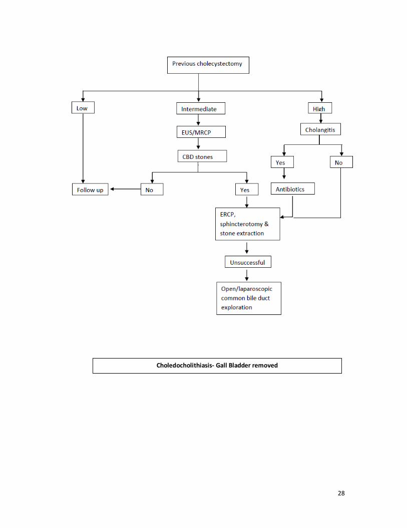
In patients who have undergone cholecystectomy earlier and diagnosis of choledocholithiasis, endoscopic retrograde cholangio pancreatography (ERCP) and extraction of bile duct stones using endoscopic techniques is the preferred approach. If this fails, open or laparoscopic common bile duct exploration should be performed. A possibility that dilated common bile duct with calculi is a choledochal cyst must be kept in mind as treatment & long term follow up of the latter is different.
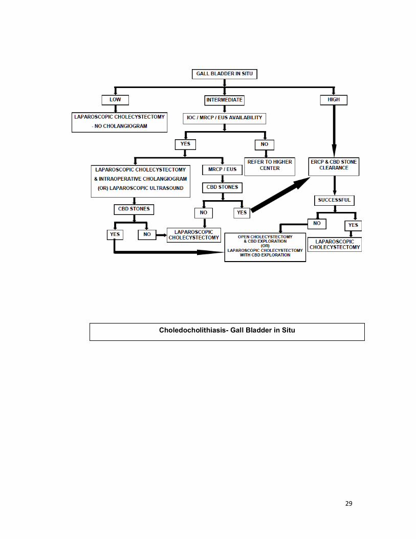
In patients with gall bladder stones and high risk of choledocholithiasis, ERCP and stone retrieval followed by laparoscopic cholecystectomy is the preferred treatment. If endoscopic therapy fails, they may undergo laparoscopic or open common bile duct exploration along with cholecystectomy.
A cautious decision to withhold cholecystectomy after endoscopic treatment of choledocholithiasis may be made in patients with unacceptable surgical risk.
If there is intermediate risk of choledocholithiasis in those with gallstones this should be confirmed with MRCP or EUS. Thereafter treatment is as outlined above.
Patients with low risk of choledocholithiasis and gallstones may undergo intraoperative cholangiogram (IOC). If choledocholithiasis if diagnosed, and the bile duct is of normal caliber, exploration is not advised. Post operative endoscopic therapy is an option. If the common bile duct is dilated options are: laparoscopic common bile duct exploration / conversion to open operation and common bile duct exploration.
Patient must be referred to higher centers if either of MRCP, EUS or intraoperative cholangiogram facilities are unavailable.
26
Situation 1
At secondary hospital / non-metro situation: optimal standards of treatment in situations where technology and resources are limited
Clinical diagnosis:
The most common presentation is with colicky upper abdominal pain (biliary colic) with or without jaundice. Fever with chills would indicate cholangitis. Pruritus and clay colored stools may be present if biliary obstruction is high grade. Fever and icterus may be present on general examination. There are no specific abdominal signs that would indicate choledocholithiasis. Presence of distended gallbladder will be a pointer against the diagnosis of choledocholithiasis in most cases.
Investigations
Haemogram, liver function test, ultrasound scan abdomen. MRCP should preferably be available even in this situation.
Treatment
We recommend two treatment protocols depending on whether cholecystectomy has been performed earlier. These protocols have been given in the form of two algorithms along with this document. It would be acceptable not to have high end technology like endoscopic ultrasound scan (EUS), laparoscopic ultrasound scan or instrumentation for laparoscopic common bile duct exploration in a secondary referral hospital setting.
Standard operating procedure (please see the two algorithms)
Most of the investigations may be performed as an outpatient. Cholangitis would make admission mandatory during the initial evaluation itself.
Referral criteria
Patients with cholangitis unresponsive to antibiotic therapy should be referred to a tertiary (super specialty) hospital. Patients with failed papillary access / biliary cannulation (at ERCP and attempt at
27
stone extraction) should also be considered for referral to tertiary care centers before decision to perform open common bile duct exploration.
Situation 2
At super specialty facility in metro location where high end technology is available
Clinical diagnosis:
Careful review of symptoms and signs must be done.
Investigations:
If cholangitis was the indication for referral, complete blood count, coagulation profile and blood culture and sensitivity must be done at admission. Liver function tests and ultrasound scan abdomen to confirm the diagnosis and to look for cholangitic abscess should be performed. Parenteral vitamin K supplementation must be initiated during the period of evaluation. After initiation of appropriate antibiotic therapy, it may be reasonable to proceed to ERCP and attempt at endoscopic retrieval of bile duct stones.
Treatment:
Our suggested treatment protocol is in the form of algorithms attached with this document.
Standard Operating procedure
All investigations can be done as outpatient/day care procedures. However, if cholangitis is present, patient should be hospitalized.
All surgical procedures require hospitalization.
Referral criteria
None.
28
Choledocholithiasis- Gall Bladder removed
29
Choledocholithiasis- Gall Bladder in Situ
30
References
1. Blumgart LH. Stones in the common bile duct-Clinical features and open surgical approaches
and techniques. In: Blumgart LH, Fong Y eds. Surgery of the liver and biliary tract. Saunders
Elsevier; 2000:528-547.
2. McFadden DW, Nigam A. Choledocholithiasis and cholangitis. In: Zinner MJ, Ashley SW eds.
Maingot's Abdominal operations. McGraw Hill Medical; 2007:865-888.
3. Williams EJ, Green J, Beckingham I, Parks R, Martin D, Lombard M. Guidelines on the
management of common bile duct stones (CBDS). Gut 57, 1004-1021. 2008.
Ref Type: Journal (Full)
4. Sugiyama M, Atomi Y, Hachiya J. Magentic resonance cholangiography using half-Fourier
acquisition for diagnosing choledocholithiasis. American Journal of Gastroenterology 93, 1886-
1890. 1998.
Ref Type: Journal (Full)
31
CARCINOMA STOMACH
Gen RP Chaubey (retd) Formerly from the Armed Forces Medical Services
Sri Balaji Action Medical Institute New Delhi
I. WHEN TO SUSPECT /RECOGNIZE?
a) Introduction
Carcinoma Stomach remains a common disease worldwide with dismal prognosis. It
represents the fourth most common malignancy and the second leading cause of cancer
related death. In Japan gastric cancer remains the most common type of cancer among
men. Its incidence, however, has been declining globally since World War II. Gastric
cancer is one of the least common cancers in North America. The incidence of proximal
gastric cancer is on the increase while the distal gastric cancer is declining in North
America. The five year survival rate of gastric carcinoma is low (10-20%).
b) Case Definition
Gastric Cancer refers to the malignant growth arising from the epithelial lining of the
stomach. It is an aggressive tumor with vague early symptoms and spreads to the
adjoining structures early in its course.
II. INCIDENCE IN INDIA
India falls in low incidence zone of gastric cancer. It is the fifth commonest cancer in
males and seventh commonest in females in India. Age adjusted rate (AAR) of gastric
cancer in six urban registries from India have reported the incidence 3.0-
13.2/1,00,000 population which is lower to the world incidence of 4.1-15.5/1,00,000
population.
There is a regional variation in its incidence. It occurs four times more commonly in
south India as compared to north India and also a decade earlier. Gastric cancer
follows the global trend of declining incidence in India as well.
III. DIFFERENTIAL DIAGNOSIS
32
• Lower Esophageal Cancer
• Lower Esophageal Stricture
• Lower Esophagitis
• Gastric Ulcers
• Acute Gastritis
• Atrophic Gastritis
• Chronic Gastritis
• Bacterial Gastroenteritis
• Viral Gastroenteritis
• Non-Hodgkin Lymphoma
• Malignant Neoplasms of the Small Intestine
IV. PREVENTION AND COUNSELING
Vast majority of Gastric Cancers are attributed to environmental factors, the most
common being infection with Helicobacter Pylori. This organism has been found in
almost 70% of the patients with Antral gastric cancer and is associated with nine fold
increased risk of developing gastric cancer. Inoculation most likely occurs in
childhood through the oro-fecal pathway and is transmitted from person to person.
Intake of certain food contents is also thought to be contributory; preserved diets
with high salt contents, smoked foods and diets with low fresh fruits and vegetable
contents have also been attributed to the increased incidence of gastric cancer.
Smoking and prolonged consumption of alcohol have also been attributed to the
increased occurrence of gastric cancer. Better living standard, better dietary habits,
eradication of Helicobacter Pylori infection, giving up of smoking and alcohol
consumption may decrease the occurrence of gastric cancer.
1-3% of gastric cancers are associated with inherited gastric cancer
predisposition syndromes. E-cadherin mutations occur in approximately 25% of
families with an autosomal dominant predisposition to diffuse gastric cancers also
called hereditary diffuse gastric cancer. This subset of persons may benefit from
genetic counseling and prophylactic gastrectomy.
33
V. OPTIMAL DIAGNOSTIC CRITERIA, INVESTIGATIONS, TREATMENT & REFERRAL CRITERIA
a) Clinical Diagnosis
Clinical diagnosis of Gastric Cancer, like all other diseases is based on astute history
taking and thorough physical examination.
There are no pathognomic symptoms of early gastric cancer; rather they are vague
and non-specific often mimicking peptic ulcer disease. Commonest complaint is
epigastric discomfort. Patient often present with Aneamia, weight loss (Aesthenia)
and loss of appetite (Anorexia), early satiety and rarely upper GI bleed.
Physical examination of early gastric cancer is usually uninformative. In late stage
they may present with palpable epigastric mass, cachexia, bowel obstruction, ascites
and pedal oedema. In advance cancers peritoneal seedling may involve ovaries
leading to Krukenberg tumor, pelvic cul-de-sac (Blumer’s shelf) palpable on digital
rectal examination, left supra clavicular lymphadenopathy (Virchow node), left
anterior axillary lymphadenopathy (Irish’s node) or a periumbilical lymph node
(Sister Mary Joseph node).
b) Investigations
Upper GI Endoscopy is the mainstay of diagnosis, accounting for > 90% of Gastric
Cancer diagnosis. Typically gastric cancer appears as irregular ulcer with raised
margins or a polypoidal or fungating mass lesion. Multiple, at least 6 or more
biopsies are to be taken for the best yield.
Barium UGI series is hardly required these days, though it may prove diagnostic in
patient with Linitis Plastica, who have undistensible stomach.
Contrast Enhanced Computed Tomography (CECT), is required to stage the disease
and evaluate the metastatic status.
34
Endoscopic Ultrasound (EUS) is used to asses the tumor depth and the adjacent
lymphadenopathy. EUS guided FNAC of adjacent lymph nodes can also be
performed.
Staging laparoscopy is the latest addition to the investigation armamentarium for
carcinoma stomach.
PET scan is not routinely recommended to evaluate Gastric Cancer.
Tumor Markers: There are no specific tumor markers for Gastric cancer hence their
assessment is not routinely advocated.
c) Treatment
Multi-disciplinary treatment planning is mandatory for a better outcome of this
rather dismal disease. Patients with Gastric cancer should be managed by an
experienced team of Surgeons, Onco-physicians, Gastroenterologist, Radiation-
Oncologist. Nutrion Specialist and Onco –Nurses.
Surgery remains the mainstay of treatment of gastric cancer. It is the only single
modality treatment capable of curing the disease. The goal of surgical cure requires
complete resection (R0). The standard recommendations for respectable gastric
cancer are free margin surgery
(at least 5 cm clearance) with at least D1 lymph node dissection removing minimum
of 15 lymph nodes.
Type of Gastectomy depends upon tumor location and its extent and consists of
partial ( ProximaL/ Distal) or Total Gastrectomy addition of Splenectomy and distal
Pancreatectomy significantly increases post operative mortality without significant
survival advantage, hence should not be performed routinely.
35
Lymph Node Excision: Extent of lymph node dissection though an important issue,
remains controversial. Results of D1 lymphadenectomy ( Perigastric nodes along the
lesser and greater curvature) are comparable with D2 lymhadenectomy ( nodes
along the coeliac trunk and its 3 branches), however more centres in even western
world are now resorting to D2 gastrectomy for better post operative outcome.
Laparoscopic Surgery For Gastric Cancer:
Laparoscopy –assisted distal gastectomy(LADG) first developed by Kitano et al in
Japan in 1991, has now become the standard of care in Japan for respectable
Gastric Cancer.
Neo-Adjuvant/Adjuvant Therapy: Large number of randomized phase 3 studies
have shown the efficacy of perioperative (pre & post operative) chemotherapy and
post operative chemoradiotherapy in combination with R0 tumor resection and
D1/D2 LN dissection.
Early Gastric Cancer: Endoscopic Mucosal Resection (EMR), and Endoscopic Sub-
Mucosal dissection are the latest surgical option in the management of early gastric
cancer (T1NoMo)., however such cancers are rarity in India and the western world.
Advanced Gastric Cancer:
In the treatment of advanced gastric Cancer (Unresectable, metastatic), surgery has
no role except as palliative gastrojejunostomy for gastric Outlet Obstruction, control
of bleeding or placement of feeding jejunostomy tube.
Multi disciplinary team, so necessary for the successful management of patients
with Gastric Carcinoma, may not be available even in most of Indian Metro
36
Hospitals; their management at secondary Hospital/ Non-Metro situation is not
advisable.
Referral Criteria:
All patients of gastric cancer, who are deemed respectable at secondary hospitals,
must be referred to super-specialty facility for a better post therapy outcome;
however patients with advanced disease requiring palliation or emergency surgery
can be tackled at secondary hospitals only.
VI. FURTHER READING/ REFERNCES
1) James McLoughlin, MD .Adenocarcinoma of the stomach: a review –
Baylor University Medical Centre Proceedings, Vol -17 (4), October 2004, 391-
399
2) Bryan J. Dicken,MD, David L. Bigam,MD, FRCS©, Carol Cass, I Mackey, MD,
FRCS©, Anil A. Joy, MD, FRCS©, and Stewart M,FRCS(C) Gastric
Adencarcinoma : Review and considerations for Future Directions- Ann Surg.
2005, January; 241(1):27-39
3) Farhat Aziz Khan, Aditya Nath Shukla .Pathogenesis and treatment of gastric
carcinoma: “An update with brief review” –. J Cancer Res Ther; December 2006-
Vol 2 (4), 196-199
4) Eric Van Cutsem, Cornelius Van de Velde, Arnaud Roth, Florian Lordick, Claus-
Henning Kohne, Stefano Cascinu, Matti Aapro . Expert opinion on management
of gastric and gastro-oesophegeal junction adenocarcinoma on behalf of the
37
European Organisation for Research and Treatment of Cancer (EORTC) –
gastrointestinal cancer group -, European Journal of Cancer 44 (2008) 182-194
5) NCCN Clinical Practice Guidelines in Oncology : Gastric Cancer- V.2.2009
6) Keechalat Pavithran, Dinesh C.Doval and Kamal K. Pandey
Gastric Cancer in India- Gastric Cancer. 2002;5(4):240-3.
7) Atul Sharma, Venkataraman Radhakrishnan. Gastric Cancer in India- Indian J of
Med & Paediatr Oncol 2011; Vol 32: 12-16
8) Naro Shiraishi, Tsuyashi Etoh, Seigo Kitano. Laparoscopic Surgery in Gastric
Cancer, Laparoscopic Gastrointestinal Surgery, ECAB Clinical Update: Surgical
Gastroenterology and Liver Transplantation,113-129
9) Elwyn C Cabebe, MD, Vivek K Mehta, MD, George Fisher Jr, MD, Michael Perry,
MD, MS, MACP, Francisco Talavera, PharmD, PhD, Benjamin Movsas, MD,
Rajalaxmi McKenna, MD, FACP, Jules E Harris, MD:
emedicine.medscape.com/article/278744-overview
38
Gallbladder carcinoma (GBC) requiring hospitalization
Anil K Aggarwal Department of Surgical Gastroenterology and Liver Transplantation
GB Pant Hospital New Delhi
Introduction
The gallbladder is a distensible pear-shaped structure located in a fossa on the undersurface of the right lobe of the liver. It is a storage reservoir that allows bile acids to be delivered in a high concentration and a controlled manner to the duodenum for the solubilization of dietary lipid. Gallbladder has a storage capacity of approximately 30 to 50 mL in a normal adult. The portions of the gallbladder are the fundus, body, infundibulum, and neck.
Case definition (for situation 1 and 2)
Ø The term Gallbladder carcinoma (GBC) refers to malignant tumor arising from epithelial
lining of gallbladder. It is an aggressive tumor which can spread to adjacent organs, lymph nodes and metastasize to distant sites resulting in death if left untreated.
Ø Incidental GBC - GBC that is not suspected before or at operation and even on gross
examination of the opened gallbladder specimen by the surgeon, but is detected for the first time on histopathological examination (HPE) of a gallbladder removed for presumed (clinical, ultrasound, operative) diagnosis of gallstone disease (GSD).
Incidence in our country
Ø GBC is more common in Northern and Eastern India compared to other regions.
Ø Age standardized incidence rate in males ranged from 0.3 /1,00,000 men in low incidence areas to 5.3/1,00,000 men in high incidence areas.
Ø Age standardized incidence rate in females ranged from 0.4/1,00,000 in low incidence
areas to 14.3/1,00,000 in high incidence areas.
Ø GBC is becoming one of the most common cancers among women in north and northeast India.
Diagnosis
Situation 1
Clinical : Clinical diagnosis is based on evaluation of symptoms and examination.
Symptoms due to tumor in gallbladder
39
Ø Right upper abdominal pain – colicky or continuous with or without radiation to shoulder or back
Ø Abdominal lump
Symptoms due to adjacent organ involvement
Ø Jaundice (bile duct involvement)
Ø Vomiting (gastroduodenal involvement)
Ø Intestinal obstruction (colonic involvement)
Constitutional symptoms
Ø Anorexia
Ø Weight loss
Symptoms due to metastasis
Ø Bone pain (bone metastasis)
Ø Abdominal distension (peritoneal dissemination with ascites)
Ø Dyspnoea (lung metastasis)
Situation 2 Clinical : Same as in situation 1
Differential diagnosis
Presentation with upper abdominal pain
Ø Cholelithiasis and cholecystitis
Ø Pancreatitis
Ø Peptic ulcer disease
Presentation with jaundice
Ø Choledocholithiasis (CBD stones)
Ø Periampullary carcinoma
Ø Carcinoma head of pancreas
Presentation with vomiting
40
Ø Benign gastric outlet obstruction (peptic ulcer disease related)
Ø Carcinoma stomach
Ø Duodenal tuberculosis
Presentation with abdominal lump
Ø Hepatocellular carcinoma
Ø Periampullary/carcinoma head of pancreas with palpable gallbladder
Ø Hydatid cyst
Ø Carcinoma hepatic flexure
Management (situation 1)
Investigations :
Ultrasound abdomen: Features suggestive of GBC are
Ø Irregular /focal GB wall thickening
Ø Large intraluminal polypoidal mass
Ø GB mass with liver infiltration.
Treatment
Situation 1
Out patient
Ø Patients with clinical findings suggestive of GBC should be evaluated with Ultrasound abdomen.
Ø If ultrasound findings are suggestive of GBC patient should be referred to tertiary centre
with expertise in management of GBC.
In patient
Ø Patients with clinical findings suggestive of GBC should be evaluated with Ultrasound abdomen.
Ø If ultrasound findings are suggestive of GBC patient should be referred to tertiary centre
with expertise in management of GBC
Intra-op
41
Patient taken up for cholecystectomy for suspected gall stone disease à Intraoperative findings suggestive of mass in gallbladder à If no expertise in management à it is preferable to refer the patient to tertiary centre with expertise in management of GBC instead of performing simple cholecystectomy
Post-op
Ø All cholecystectomy specimens performed for gallstone disease should be sent for
histopathological examination (HPE)
Ø If HPE suggestive of GBC patient should be referred to tertiary centre with expertise in management of GBC
Management (situation 2)
Investigations
For diagnosis and staging
Ultrasound with Doppler abdomen : Doppler to assess vascular involvement
Contrast enhanced computed tomography (CECT) abdomen or Magnetic resonance imaging (MRI) abdomen with Magnetic Resonance Cholangio Pancreatography (MRCP) Ø Both CECT and MRI abdomen are more sensitive for diagnosis and staging compared to
ultrasound abdomen
Ø MRI preferred in patients with jaundice
Whole body Positron emission tomography (PET)
Ø Not required in all patients
Ø In selected cases (locally advanced disease) with no evidence of metastasis on CECT/MRI abdomen to detect metastatic disease
Upper GI endoscopy : In patients with suspected gastroduodenal involvement
Tumor markers (CEA,CA 19-9, CA 125, CA 242)
Ø Not required for diagnosis
Ø Prognostic value
Ø Useful in follow up
Pathological diagnosis (image guided FNAC or biopsy)
Not required in all patients
42
Required in selected cases
Ø Planned for neoadjuvant therapy in view of locally advanced disease
Ø Planned for palliative therapy in view of metastatic disease
To assess fitness for surgery
Ø Hemogram
Ø Serum electrolytes
Ø Kidney function test
Ø Liver function test
Ø ECG
Ø Chest x-ray
Treatment
Outpatient
Ø Patients with clinical findings suggestive of GBC and fit for surgery should be evaluated with Ultrasound abdomen.
Ø If ultrasound findings are suggestive of GBC further evaluation with CECT/MRI abdomen for
diagnosis and staging.
Ø Early admission and surgical intervention should be advised
In patient
Staging laparoscopy should be preferably done in all patients prior to laparotomy
T1b –T2 GBC
Ø Radical cholecystectomy is the standard treatment.
Ø Radcical cholecystectomy includes – liver resection with lymphadenectomy
Ø Liver resection - cholecystectomy with 2cm wedge or anatomical segment IVb-V resection
Ø Lymphadenectomy – Extent of lymphadenectomy varies from clearance of only nodes along the hepatoduodenal ligament skeletonizing the vascular structures and the bile ducts to
43
additional clearance of nodes anterior and posterior to the head of the pancreas and the hepatic artery till its origin from the celiac axis.
T3 GBC
Ø Radical cholecystectomy is the standard treatment.
Ø Extended right hepatectomy in patients with extensive liver infiltration
T4 GBC
Ø Radical cholecystectomy with resection of adjacent involved organs if deemed resectable
IGBC
Completion radical cholecystectomy for all cases with stage T1b and above.
Contraindications for curative surgery (absolute and relative)
Ø Distant metastasis - liver metastasis and peritoneal deposits
Ø Vascular involvement (main portal vein, common hepatic artery)
Ø Extensive nodal disease or multiple adjacent organ involvement
Ø Extensive biliary involvement.
Adjuvant chemoradiotherapy
It can be considered in patients with
Ø Advanced stage disease (stage III and IV)
Ø Nodal positive disease
Ø Non curative resection (R1 and R2 resection)
Post-operative care
Ø Analgesics
Ø Antibiotics – duration depends upon postoperative course
Ø Intravenous fluid supplementation till oral feeds are started
Ø Wound care
Ø DVT prophylaxis in high risk patients
44
Complications
Ø Wound infection
Ø Chest infection
Ø Bleeding
Ø Bile leak
Ø Anastomotic leak in patients with resection of adjacent organs
Ø Liver failure following major hepatectomy
Prevention
Risk factors for GBC
Ø Female gender
Ø Increasing age
Ø Dietary factors (higher consumption of mustard oil contaminated with argemone oil, high cholesterol intake, intake of red meat, drinking water contaminated with pesticides)
Ø Exposure to potential carcinogens (methylcholanthrene, aflatoxin B)
Ø Cholelithiasis and chronic cholecystitis
Ø Gallbladder polyps
Ø Choledochal cysts
Ø Anomalous pancreaticobiliary duct junction
Ø Genetic factors (p53 and K-ras mutations)
Further reading/references
1. D’Angelica M, Dalal KM, DeMatteo RP, et al. Analysis of the extent of resection for adenocarcinoma of the gallbladder. Ann Surg Oncol. 2009;16(4): 806–816.
2. NCCN practice guide lines in Oncology 2011
3. Sikora SS, Singh RK. Surgical strategies in patients with gallbladder cancer: nihilism to optimism. J Surg Oncol. 2006 Jun 15;93(8):670-81. Review.
45
4. Nishio H, Ebata T, Yokoyama Y, Igami T, Sugawara G, Nagino M. Gallbladderm cancer involving the extrahepatic bile duct is worthy of resection. Ann Surg. 2011 May;253(5):953- 60.
5. Agarwal AK, Mandal S, Singh S, et al. Biliary obstruction in gall bladder cancer is not sine qua
non of inoperability. Ann Surg Oncol. 2007;14(10):2831–2837.
6. Regimbeau JM, Fuks D, Bachellier P, Le Treut YP, Pruvot FR, Navarro F, Chiche L, Farges O. Prognostic value of jaundice in patients with gallbladder cancer by the AFC-GBC-2009 study group. Eur J Surg Oncol. 2011 Jun;37(6):505-12.
7. Agarwal AK, Mandal S, Singh S, Sakhuja P, Puri S. Gallbladder cancer with duodenal infiltration: is it still resectable? J Gastrointest Surg. 2007 Dec;11(12):1722-7.
8. Hirano S, Tanaka E, Shichinohe T, Saitoh K, Takeuchi M, Senmaru N, Suzuki O, Kondo S. Feasibility of en-bloc wedge resection of the pancreas and/or the duodenum as an alternative to pancreatoduodenectomy for advanced gallbladder cancer. J Hepatobiliary Pancreat Surg. 2007;14(2):149-54.
9. Mekeel KL, Hemming AW. Surgical management of gallbladder carcinoma: a review.J
Gastrointest Surg. 2007 Sep;11(9):1188-93. Review.
10. Pilgrim C, Usatoff V, Evans PM. A review of the surgical strategies for the management of gallbladder carcinoma based on T stage and growth type of the tumour. Eur J Surg Oncol. 2009 Sep;35(9):903-7. Epub 2009 Mar 4. Review.
46
CARCINOMA RECTUM
Ameet Kumar Peush Sahni
Department of GI Surgery and Liver Transplantation All India Institute of Medical Sciences
New Delhi
I. WHEN TO SUSPECT/ RECOGNIZE?
b) Introduction: Colorectal cancer is common in developed countries such as the USA and Japan, and lower in frequency in developing countries like Africa and Asia. The incidence is slightly higher in men than women, and is highest in African American men. Colon and rectal cancer is the third most common cancer in both women and men in the US. Incidence rates range from 25.3 per 100,000 in Eastern Europe to 45.8 per 100,000 in Australia. The crude incidence of rectal cancer in the European Union is ∼35% of the total colorectal cancer incidence, i.e. 15–25/100 000 per year. The mortality is 4–10/100 000 per year with lower figures in women and the higher ones for men.
Case definition: A patient with bleeding per rectum and/or tenesmus with or without change in bowel habit who on rectal examination/proctoscopy or sigmoidoscopy is found to have a mass which on biopsy is a cancer.
II. INCIDENCE OF THE CONDITION IN OUR COUNTRY
The incidence rates of colorectal cancers in India are low––about 2 to 8 per 100,000. The incidence of rectal cancer in India has been constant over the past few years. Hospital- and population-based data also show that the incidence rates for rectal cancer are higher than colon cancer in all parts of India. However, a high incidence of these cancers is seen in the urban population. Data is limited.
III. DIFFERENTIAL DIAGNOSIS a. Haemorrhoids b. Ulcerative colitis c. Solitary rectal ulcer d. Rectal prolapse e. Radiation proctitis
IV. PREVENTION AND COUNSELING No specific intervention for primary prevention is known. However, the following dietary and lifestyle changes may play a role in prevention: physical activity, folate, fruits and vegetables,
47
calcium, vitamin D, high fiber diet, weight reduction, avoidance of red and processed meat, stopping smoking.
For secondary prevention, 2 broad groups have been identified a. High risk individuals (those with a history of adenomas or cancers, family history or genetic
syndrome, or inflammatory bowel disease) b. Average risk individuals (all others)
Among the high risk groups: a colonoscopy 3 years after removal of an adenoma/polyp and if
this is normal then after 5 years.
Previous Colorectal Cancer and Family History of Colorectal Cancer - The first surveillance colonoscopy at 1 year following cancer resection - If normal, the interval can be increased to 3 years. However, if additional disease is noted on postoperative colonoscopy, more frequent examinations are warranted.
Patients with a family history of colorectal cancer or adenoma, including affected first-degree
relatives - should undergo screening with colonoscopy beginning at 40 years of age or earlier, when they are 10 years younger than their affected family member(s) were at age of initial diagnosis.
Patients with long-standing IBD - In patients with pancolitis surveillance colonoscopy should
begin after 8 years of symptoms. Surveillance can start later in those patients with left-sided colitis, generally after 12 to 15 years of disease. Colonoscopy should be performed every 1 to 2 years.
Patients from FAP families who have not been tested for an APC mutation should begin routine
screening at puberty with annual flexible sigmoidoscopy. If polyps are not identified by age 40 years, then the frequency of examinations can be decreased to every 3 years. On the other hand, individuals who express the phenotype require upper endoscopy to examine the periampullary region. Patients with a known genetic mutation or members of an FAP kindred should undergo colectomy when they develop polyps, because stage-specific survival of colorectal cancer appears to be the same for polyposis patients as for those who have sporadic bowel cancers.
Colorectal screening for patients with HNPCC - endoscopy should thus be performed every 1 to 2
years. For individuals with known mutations or family history consistent with the Amsterdam Criteria, screening should begin at 21 years of age. Screening for extracolonic disease should be performed as well, including urine cytology, pelvic ultrasound, and periodic endometrial biopsy.
a. Average risk individuals
Combination of fecal occult blood test (FOBT) with flexible sigmoidoscopy at 5-year intervals after the age of 50 years
V. OPTIMAL DIAGNOSTIC CRITERIA, INVESTIGATIONS, TREATMENT AND REFERRAL CRITERIA
48
Diagnosis
History: Rectal bleeding is the commonest symptom. Other symptoms include tenesmus, altered bowel habits and mucus discharge, weight loss and loss of appetite.
Diagnosis: A digital rectal examination, proctoscopy and/or sigmoidoscopy with biopsy for histopathological examination. Tumours with distal extension to ≤15 cm (as measured by rigid sigmoidoscopy) from the anal margin are termed rectal tumours, while more proximal ones are called colonic.
Staging: Complete blood count, liver and renal function tests and a full colonoscopy to evaluate for synchronous lesions (present in up to 5% of colorectal cancers), rigid proctoscopy (to define the level of the tumour), abdominal CT and chest X-ray to evaluate for metastases, and baseline serum carcinoembryonic antigen (CEA) level. A PET-CT may be done to evaluate suspected extrahepatic metastasis. The depth of penetration can be estimated by digital rectal exam (superficial tumours are mobile, whereas fixed lesions have deeper infiltration), and endorectal ultrasound (ERUS) or MRI with endorectal coil can provide a good assessment of the extent of invasion of the bowel wall. ERUS for early tumours (T1–T2) or rectal MRI for all tumours, including the earliest ones, is usually suggested prior to planning treatment and extent of surgery.
Histopathological examination of the surgical specimen should assess the proximal, distal and circumferential margins and regional lymph nodes (at least 12 lymph nodes should be examined). Also, vascular and neural invasion should be assessed.
Treatment
Localized disease
Low anterior resection or abdominlperineal resection as required
Advanced disease
Locally advanced disease may require neoadjuvant therapy in an attempt to downstage the tumour and attempt sphincter preservation. Preoperative radiotherapy (short course or long course) may be used.
Situation 1
At Secondary Hospital/Non-Metro situation: Optimal Standards of Treatment in Situations where technology and resources are limited
49
Clinical diagnosis:
In a patient who presents with bleeding per rectum, a thorough history and clinical examination should be undertaken especially in the elderly. A history of tenesmus, change in bowel habits, anorexia and weight loss should be asked as also a family history of colorectal cancer. Next, a rectal examination/proctoscopy and if necessary a sigmoidoscopic examination should be done and if found to have a mass, a punch biopsy should be done. If on pathology this shows a malignancy then it confirms the diagnosis.
Investigations
Haemogram, liver function test, CEA levels, sigmoidoscopy, chest X-ray, CT abdomen and pelvis.
Treatment
All patients who have confirmed rectal cancer should have a surgical resection (anterior resection or abdomino-perineal resection). Neoadjuvant therapy if required for sphincter preservation may be used.
Standard Operating procedure
All investigations can be done as outpatient/day care procedures. However, if the general condition of patient is not good, hospitalization may be needed.
All surgical procedures require hospitalization.
Referral criteria
All patients with borderline resectability or where a low/ultralow anterior resection is required, or those with metastatic liver disease may benefit by referral to GI Surgery centres for complete evaluation and definitive management.
Situation 2
At Super Specialty Facility in Metro location where higher-end technology is available
Clinical Diagnosis:
50
Patients with rectal bleeding along with a suggestive history should be evaluated for colorectal cancer. All patients referred as cases of rectal cancer should have their diagnosis confirmed.
Investigations:
Review of all previous investigation including blocks and slides followed by colonoscopy to rule out synchronous lesions. Haemogram, liver function test, CEA levels, Sigmoidoscopy, chest X-ray, CT abdomen, MRI pelvis/ERUS, PET-CT.
Treatment:
Operable/potentially operable
Anterior resection or abdominoperineal resection
Advanced disease
If both primary and metastatic tumours are considered resectable, multidisciplinary teams should consider initial systemic treatment followed by surgery. If not resectable, consider palliative chemotherapy along with a palliative resection/diverting colostomy.
Standard Operating procedure
All investigations can be done as outpatient/day care procedures. However, if the general condition of patient is not good, hospitalization may be needed.
All surgical procedures require hospitalization.
Referral criteria
May require referral if neoadjuvant therapy is planned and if facilities for radiotherapy/chemotherapy are not available.