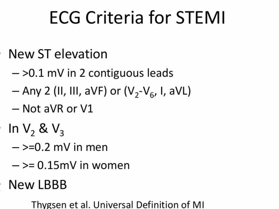
st elevation myocardial infarction (stemi) · 2013 stemi guideline •primary pci is the preferred...
TRANSCRIPT

ST ELEVATION MYOCARDIAL INFARCTION (STEMI)
Gordon Kritzer, MD, FACC
Virginia Mason Medical Center, Seattle

STEMI
• ACS and STEMI
• History and physical, differential diagnosis
• ECGs
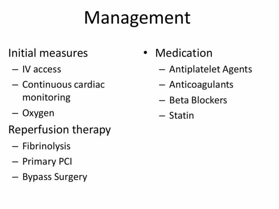
• Initial Therapy and management
• Reperfusion therapy
• Complications of acute STEMI


Hospitalizations in the U.S. Due to Acute
Coronary Syndromes (ACS)
Acute Coronary
Syndromes*
1.57 Million Hospital Admissions - ACS
UA/NSTEMI† STEMI
1.24 million Admissions per year
.33 million Admissions per year
Heart Disease and Stroke Statistics – 2007 Update. Circulation 2007; 115:69-171.
*Primary and secondary diagnoses. †About 0.57 million NSTEMI and 0.67 million UA.


STEMI PRE HOSPITAL RX GOALS
• Delivery patients to an appropriate health facility as quickly as possible
• Preventing sudden death and controlling arrhythmia using ACLS when necessary
• Initiating and continuing management of patients during inter facility transfer

Important direct signout from EMS team
• Person who initiated EMS involvement and why
• Complaints at the scene
• Vital signs, physical exam, and notable changes
• Therapies prior to arrival and patient response
• ECGs from outside and en route
• Code status
• Family contact for supplemental information

General Therapy
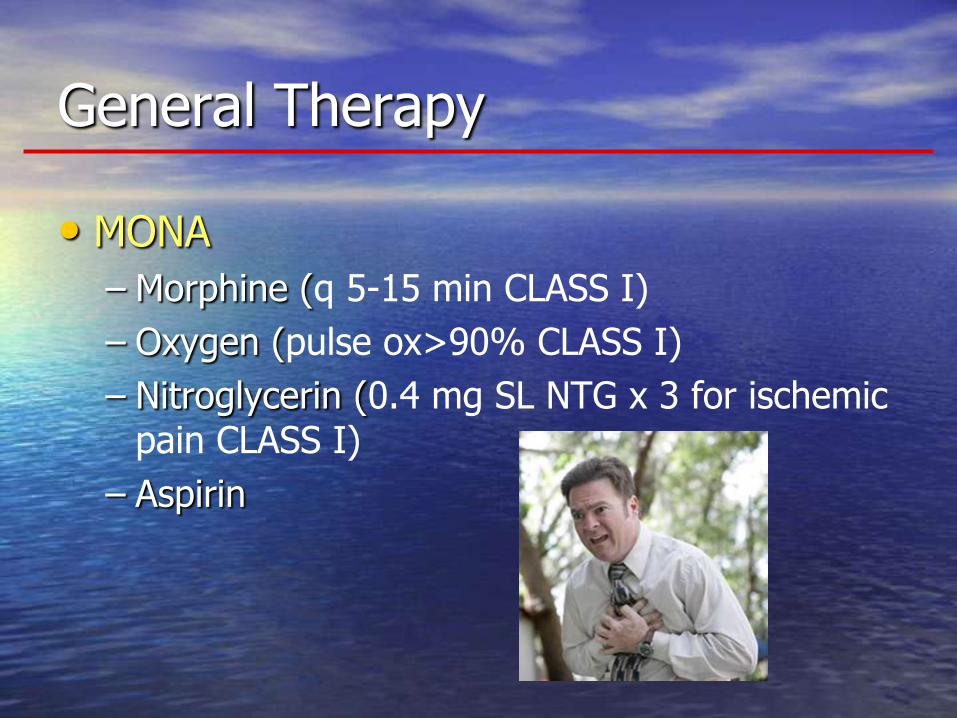
General Therapy
• MONA
– Morphine (q 5-15 min CLASS I)
– Oxygen (pulse ox>90% CLASS I)
– Nitroglycerin (0.4 mg SL NTG x 3 for ischemic pain CLASS I)
– Aspirin
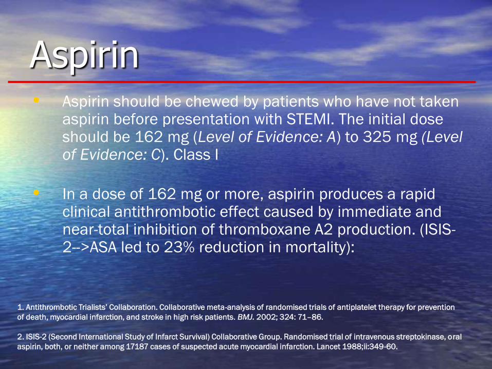
Aspirin • Aspirin should be chewed by patients who have not taken
aspirin before presentation with STEMI. The initial dose should be 162 mg (Level of Evidence: A) to 325 mg (Level of Evidence: C). Class I
• In a dose of 162 mg or more, aspirin produces a rapid clinical antithrombotic effect caused by immediate and near-total inhibition of thromboxane A2 production. (ISIS-2-->ASA led to 23% reduction in mortality):
1. Antithrombotic Trialists’ Collaboration. Collaborative meta-analysis of randomised trials of antiplatelet therapy for prevention
of death, myocardial infarction, and stroke in high risk patients. BMJ. 2002; 324: 71–86.
2. ISIS-2 (Second International Study of Infarct Survival) Collaborative Group. Randomised trial of intravenous streptokinase, oral
aspirin, both, or neither among 17187 cases of suspected acute myocardial infarction. Lancet 1988;ii:349-60.

Beta-Blockers

Recommendations - Class Ia (B) • ORAL beta-blocker therapy SHOULD BE initiated in the first
24 hours for patients who DO NOT have any of the
following:
1) signs of heart failure,
2) evidence of a low output state,
3) increased risk for cardiogenic shock, or
4) relative contraindications to beta blockade
1AVB > 0.24 sec,
2nd- or 3rd-degree heart block
reactive airway disease
** There is no study evaluating oral beta blockers alone
Beta-Blockers
*Risk factors for cardiogenic shock :heart failure, age > 70 , systolic blood pressure < 120,
sinus tachycardia > 110 or heart rate < 60, increased time since onset of STEMI symptoms

Recommendations - Class IIa (B) • It is reasonable to administer an IV BETA BLOCKER at the
time of presentation to STEMI patients who are
HYPERTENSIVE and who do not have any of the following:
1) signs of heart failure,
2) evidence of a low output state,
3) increased risk for cardiogenic shock, or
4) relative contraindications to beta blockade
1AVB > 0.24 sec,
2nd- or 3rd-degree heart block
reactive airway disease
Beta-Blockers
*Risk factors for cardiogenic shock :heart failure, age > 70 , systolic blood pressure < 120, sinus
tachycardia > 110 or heart rate < 60, increased time since onset of STEMI symptoms

Recommendations - Class III (A) • IV beta blockers SHOULD NOT be administered to STEMI
patients who have any of the following:
1) signs of heart failure
2) evidence of a low output state
3) increased risk* for cardiogenic shock
4) relative contraindications to beta blockade
1AVB > 0.24 sec,
2nd- or 3rd-degree heart block
reactive airway disease
Beta-Blockers
*Risk factors for cardiogenic shock :heart failure, age > 70 , systolic blood
pressure < 120, sinus tachycardia > 110 or heart rate < 60, increased time since
onset of STEMI symptoms

Reperfusion
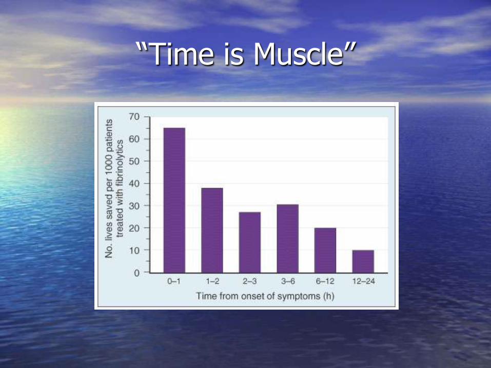
“Time is Muscle”
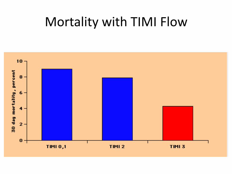
Mortality with TIMI Flow

Reperfusion
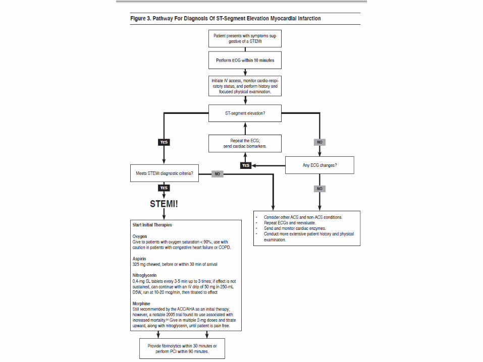
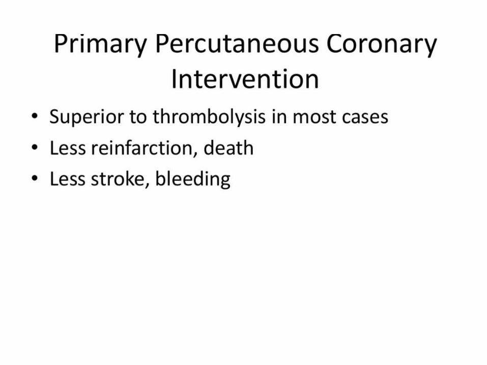
• STEMI patients presenting to a hospital with PCI capability
should be treated with primary PCI within 90 min of first
medical contact as a systems goal. Class Ia
• STEMI patients presenting to a hospital without PCI
capability, and who cannot be transferred to a PCI center and
undergo PCI within 90 min of first medical contact, should be
treated with fibrinolytic therapy within 30 min of hospital
presentation as a systems goal, unless fibrinolytic therapy is
contraindicated. Class Ib

PCI vs Fibrinolysis for STEMI: Short-Term Clinical Outcomes
7
52
6
1
78
97 7
21
21
5
13
5
10
15
20
25
30
35PCI
Fre
qu
en
cy (
%)
P=.0002
P=.0003 P<.0001
P<.0001
P<.0001 P=.0004
P=.032
P<.0001
Death Death, no shock
data
ReMI Rec.
Ischemia
Total
Stroke
Hem.
Stroke
Major
Bleed
Death
MI
CVA
Fibrinolysis
N=7739
Keeley E, et al. Lancet . 2003;361:13-20.

Brief Review of Thrombolytic Trials
GISSI-1: Streptokinase 18% reduction in mortality at 21 d
GUSTO-1: tPA. 15% reduction in 30-day mortality compared
to Streptokinase
GUSTO-3: Reteplase had no benefit over tPA but is easier to
use (double bolus)
ASSENT: TNKase is similar to tPA but with less non-cerebral
bleeding and better mortality with symptoms>4 hrs: Single
bolus, fibrin selective, resistance to PAI-1
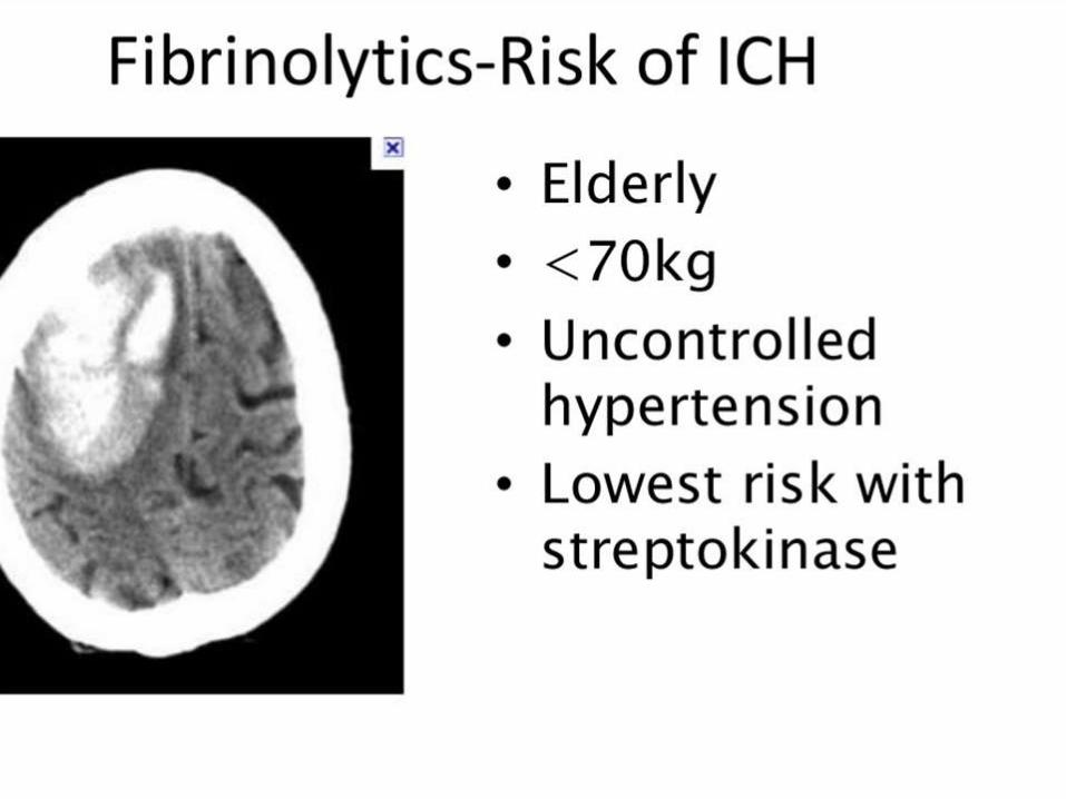
*Overall risk of ICH is 0.7%; Strokes occurred in 1.4%


Anticoagulants
•Patients undergoing reperfusion with fibrinolytics
should receive anticoagulant therapy for a minimum of
48 hours (unfractionated heparin) or up to 8 days
•Anticoagulant regimens with established efficacy
include:
♥ UFH (LOE: C)
♥ Enoxaparin (LOE:A)
♥ Fondaparinux (LOE:B)

2013 STEMI guideline
• Primary PCI is the preferred treatment for STEMI
– At a hospital with PCI, the goal is PCI within 90 minutes
– At a hospital without PCI, the goal is transfer and PCI within 120 minutes
– If transfer for PCI within 120 minutes is not feasible, use fibrinolytic therapy unless contraindicated, with a door-to-needle time less than 30 minutes
2013 ACCF/AHA Guideline for STEMI. Circulation. 2013; 127: 4529-555.

2013 STEMI guideline
• Patients with cardiogenic shock or severe HF should be transferred immediately for PCI
• Patients who have failed to reperfuse with fibrinolytic therapy should be transferred urgently for PCI
• Therapeutic hypothermia is recommended for comatose patients with STEMI and cardiac arrest

Andersen HR, et al. DANAMI-2 trial. NEJM 2003; 349: 733-742.


Complications of Myocardial Infarction
• Arrhythmias
• Ventricular Septal Perforation
• Ischemic Mitral Regurgitation, Papillary Muscle Rupture
• Ventricular Free Wall Rupture
• Systemic Embolism
• Ventricular Aneurysm
• Pericarditis
• Cardiogenic Shock (another lecture)

Ventricular Arrhythmias
•60-110 BPM; Up to 20% STEMI patients have this
•Usually a result of reperfusion; no specific therapy needed if HD
stable. Otherwise, atropine or even atrial pacing may increase
sinus rate to overdrive pace the AIVR
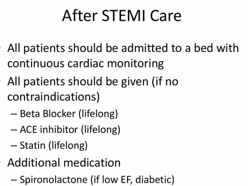
•Routine post-MI management with B-blockers, ACE, etc.

PVC’s
•Extremely common, along with short runs of
NSVT
•Amiodarone won’t increase mortality, other
antiarrhythmics (other than B-blockers) do.
•B-blockers, electrolytes
•Best if no antiarrhythmics are used

Not So Benign Rhythm
•Ischemic VT is often polymorphic; HR>100-110 BPM
•Higher risk with more LV damage and in first 2 days after MI
•Treat: DCCV, cath lab (if needed), electrolyte correction,
amiodarone, lidocaine, B-Blockers

If That Didn’t Make You Nervous…
Primary VF: Sudden event with no warning--10% STEMI patients
before lytics. MUCH MUCH less now
Secondary VF: Occurring in setting HF or shock
Late VF: >48 hrs after MI-->Increased risk with IVCD, anterior wall
MI, persistent SVT early in course, and RV infarction requiring
pacing
***Have to worry about structural complication (free wall
rupture)/ischemia
Treat: Non-synced DCCV, electrolyte correction

Why get worked up about electrolytes?
Nordrehaug JE, van der Lippe G: Hypokalemia and ventricular fibrillation in acute myocardial infarction.
Br Heart J 50:525, 1983.
NOTE: Pre-lytic
study

Sinus Bradycardia/Junctional Escape Rhythm
• 4-5% of STEMI patients have a bradyarrhythmia
• Sinus node ischemia--Blood supply to SA node is: 65% RCA, 25% LCX, 10% dual supply.
• Most commonly seen in Inferior/posterior MI’s.
• Often induced by vagal reaction that may be protective

Atrioventricular Block
• First-Degree: Usually the RCA and does not require treatment. Hold the B-blocker for PR>240 ms
• Second-Degree: Usually RCA disease and does not require treatment unless HR less than 50 and arrhythmia or symptoms. Otherwise, atropine or pace
• Third-Degree: Can be from any location of infarct. Can be preceded by Mobitz II Block – Pace for symptoms and for hemodynamic support. Usually not
needed in inferior MI’s as block is transient (pace for HR<40-50)

Post-MI VSD
• ~2% of acute MI’s prior to reperfusion era
• ~0.2% in GUSTO-I streptokinase trial
• Without reperfusion, usually occurs within first week
– Day 1--Large intramural hematomas that dissect
– Day 3-5--Coagulation necrosis
• 24 hr or less if receive lysis--Lytics reduce infarct size but may promote hemorrhagic dissection of myocardium

Symptoms, Exam, and Diagnosis
• Chest pain, dyspnea
• PE: Harsh, holosystolic murmur along sternal border radiating to base/apex/R parasternum; thrill in 1/2 patients; S3; Loud P2; TR.
• Compared to acute MR, murmur is loud. Up to 20% of patients may have MR as well though

CCU Management
• IABP
• Ventilation
• Diuresis/HF Management
• Inotropes (can increase shunt)
• Nitroprusside if tolerated (can cause hypotension)
• Mortality with conservative management is HIGH (24%, 46%, 67-82% at 24 hrs, 1 wk, and 2 months, respectively)
• Ultimately, mechanical closure needed (surgery vs. percutaneous)-TIMING is questionable but clinical status should not preclude this

Acute Mitral Regurgitation
• Caused by papillary muscle ischemia or rupture (less likely). Rupture is usually partial since total is essentially incompatible with life
• Usually in setting of inferior MI involving the posteromedial papillary muscle (single PDA blood supply as opposed to anterolateral)
• Rupture usually occurs 3-5 days post-MI and in 1% of MI’s and requires emergent operative repair (50% mortality in 24 hrs)
• Accounts for 7% of cardiogenic shock and 5% of mortality associated with acute MI
• Area of infarction does NOT have to be large

Symptoms, Exam, Diagnosis
•Symptoms: Those of heart failure
•PE: May or may not hear loud systolic murmur (need a gradient)
CCU Management
• Mechanical ventilation if needed
• IABP--especially for hypotension
• PCI if papillary m. ischemia (not rupture)
• Afterload reduction (nitroprusside if possible) to MAP of 70-80 mm Hg
• Since mortality is 90% with medical therapy alone, surgery is the major therapy of choice
– Perioperative mortality 20-25%
– Overall surgical mortality is even higher

Free Wall Rupture
• ~10% of patients who die in hospital from STEMI
• Most commonly between 1 and 4 days (up to 3 weeks)
• Caused by tear or dissecting hematoma
• More common with fibrinolysis compared to PCI
• More common in patients without previous infarction

Symptoms, Exam, Diagnosis
• Acute symptoms include sudden chest pain (esp with
cough, strain) and sudden death
• Subacute symptoms: Pericarditis-like symptoms (chest
pain, nausea, vomiting)
• Exam (think HF and tamponade): JVD, pulsus,
diminished heart sounds, rub, possibly a new murmur
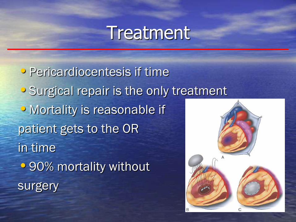
Treatment
•Pericardiocentesis if time
•Surgical repair is the only treatment
•Mortality is reasonable if
patient gets to the OR
in time
•90% mortality without
surgery

Summary of Acute STEMI Complications
• Much more rare in the reperfusion era
– Look for them especially in delayed presentation
• Arrhythmias are most common complication and
may require emergent treatment
• VSD’s, papillary muscle rupture, and free wall
ruptures carry a VERY high mortality and require
emergent surgical consultation
– Support mechanically until patient receives operation
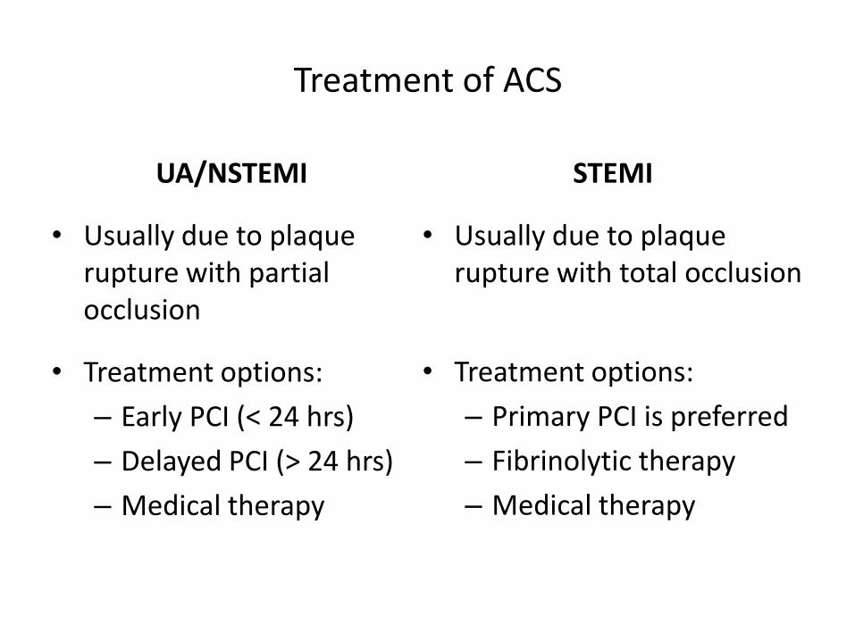
Treatment of ACS
UA/NSTEMI
• Usually due to plaque rupture with partial occlusion
• Treatment options:
– Early PCI (< 24 hrs)
– Delayed PCI (> 24 hrs)
– Medical therapy
STEMI
• Usually due to plaque rupture with total occlusion
• Treatment options:
– Primary PCI is preferred
– Fibrinolytic therapy
– Medical therapy

Initial treatment of ACS
• Aspirin at initial presentation
• P2Y12 inhibitor (clopidogrel, ticagrelor, . . .)
• Statin (atorvastatin 80 mg daily)
• Anticoagulation (heparin, enoxaparin, . . .)
• Nitrates / morphine for chest pain
• Revascularization
• Scenarios:
– STEMI with PCI available
– STEMI with PCI unavailable
– High-risk UA/NSTEMI
– Low-risk UA/NSTEMI

Initial treatment for STEMI with PCI available
• PCI with 90 minutes, or transfer for PCI within 120 min
• Aspirin 325 mg
• P2Y12 inhibitor
– Ticagrelor 180 mg
– Prasugrel 60 mg
– Clopidogrel 600 mg
• Anticoagulant
– Heparin 70 units/kg
– Bivalirudin (usually started in the cath lab)
• High dose statin (atorvastatin 80 mg)

Initial treatment for STEMI with PCI unavailable
• Fibrinolytic therapy within 30 minutes
• Aspirin 325 mg
• Clopidogrel
– initial dose 300 mg for patients < 75 years old
– initial dose 75 mg for patients ≥ 75 years old
• Enoxaparin, fondaparinux, or heparin
• High dose statin
2013 ACCF/AHA Guideline for STEMI. Circulation. 2013; 127: 4529-555.
