spect and pet in neurodegenerative diseases · spect and pet in neurodegenerative diseases isabel...
TRANSCRIPT
SPECT and PET in SPECT and PET in Neurodegenerative DiseasesNeurodegenerative Diseases
Isabel Roca, MDHU VALL HEBRONBarcelona, Spain
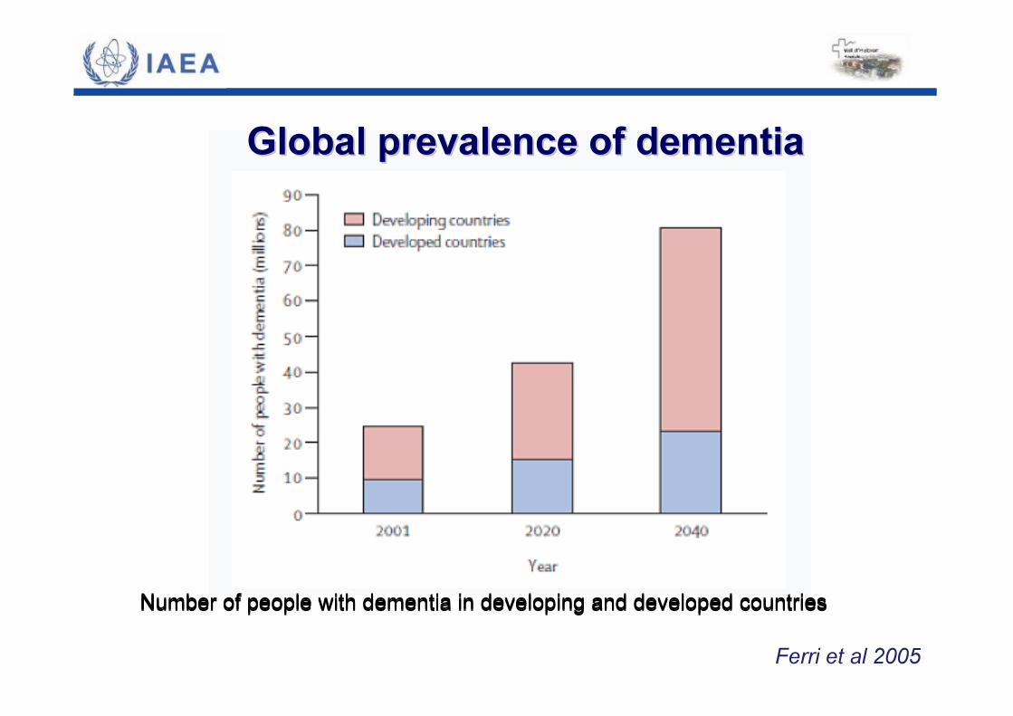
Global Global prevalenceprevalence ofof dementiadementia
Ferri et al 2005
Number of people with dementia in developing and developed countriesNumber of people with dementia in developing and developed countries
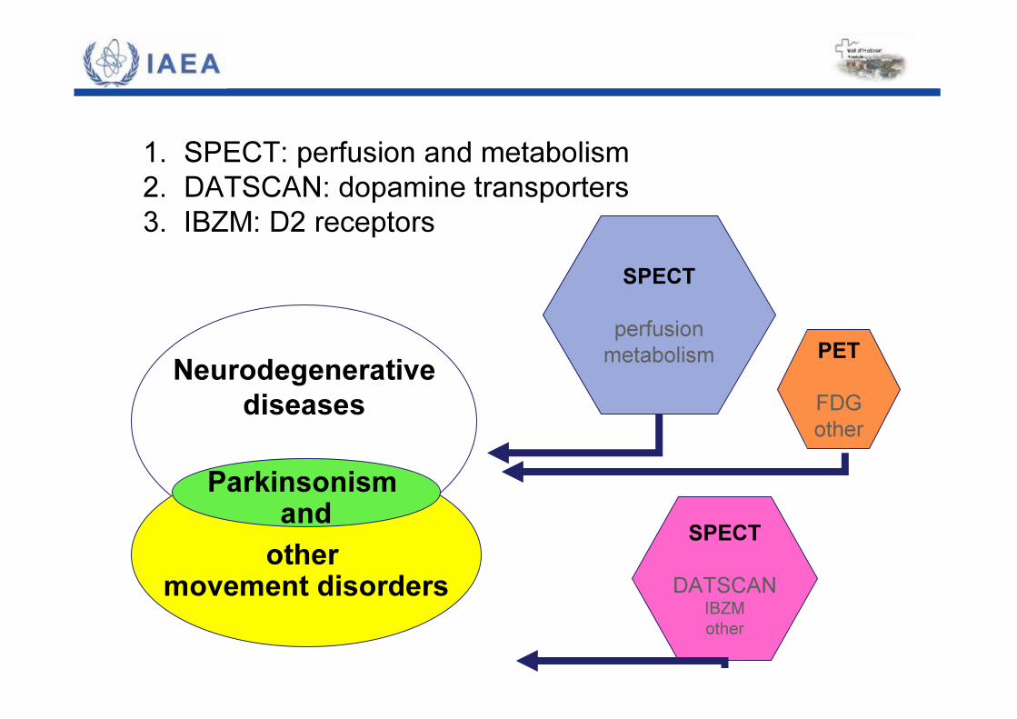
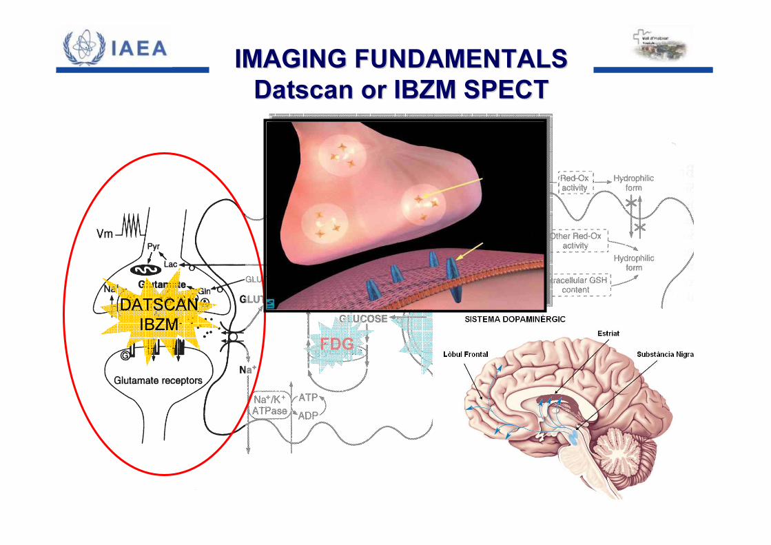
1. SPECT: perfusion and metabolism2. DATSCAN: dopamine transporters3. IBZM: D2 receptors
Neurodegenerativediseases
othermovement disorders
Parkinsonismand
SPECTperfusionmetabolism PET
FDGother
SPECTDATSCAN
IBZMother
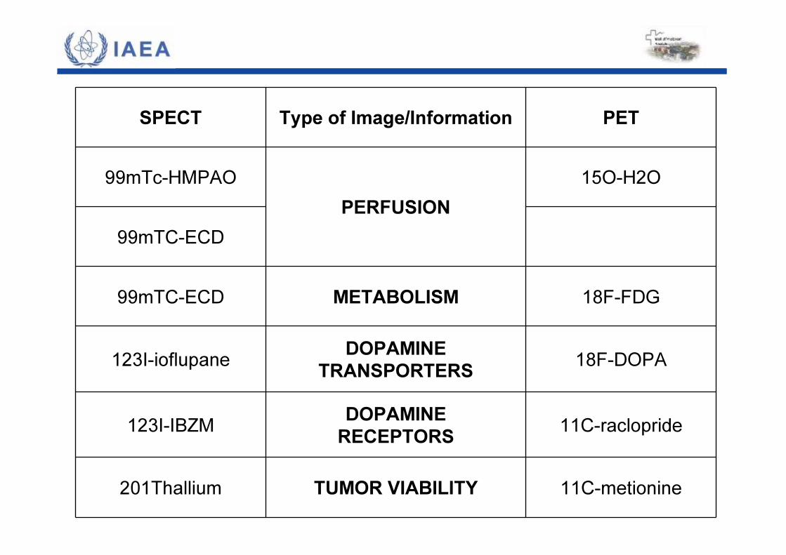
SPECT Type of Image/Information PET
99mTc-HMPAOPERFUSION
15O-H2O
99mTC-ECD
99mTC-ECD METABOLISM 18F-FDG
123I-ioflupane DOPAMINETRANSPORTERS 18F-DOPA
123I-IBZM DOPAMINERECEPTORS 11C-raclopride
201Thallium TUMOR VIABILITY 11C-metionine
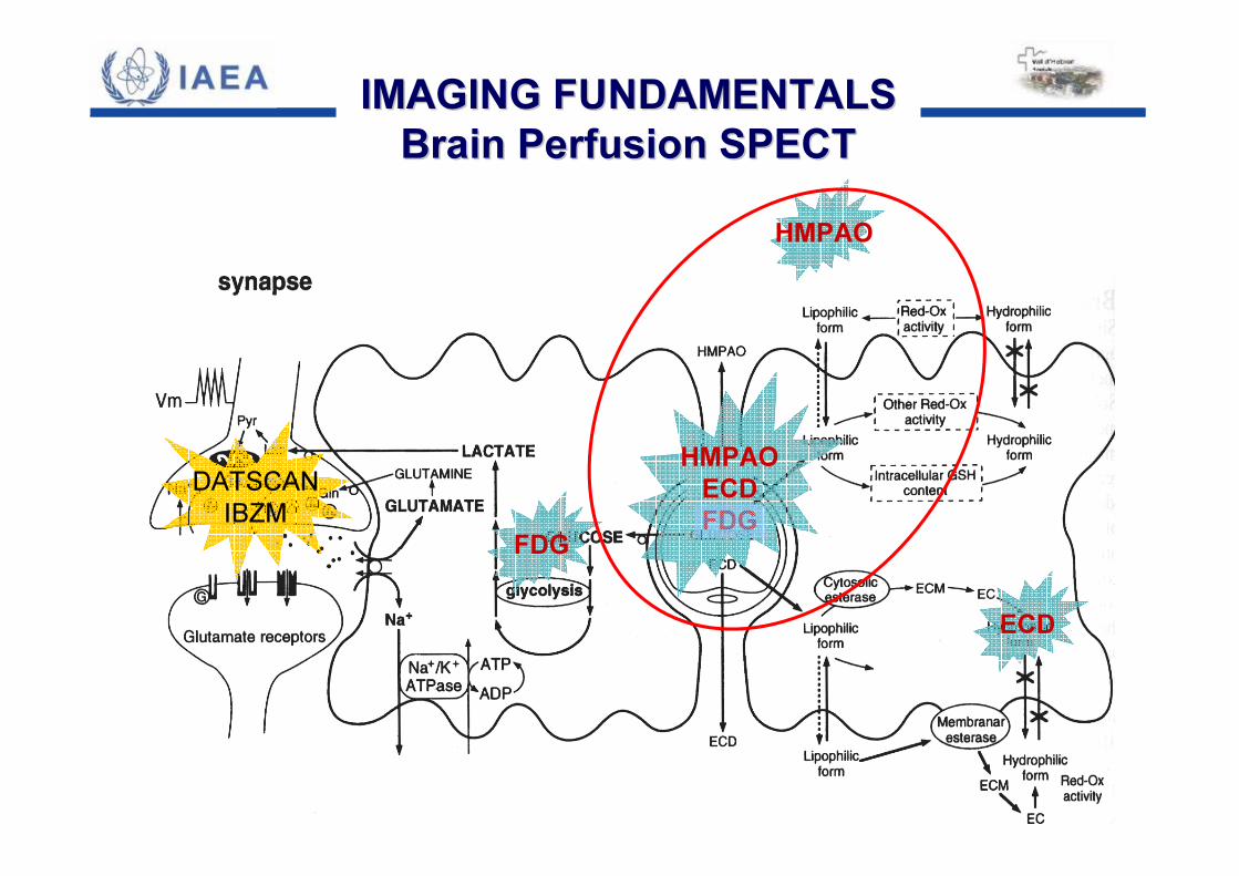
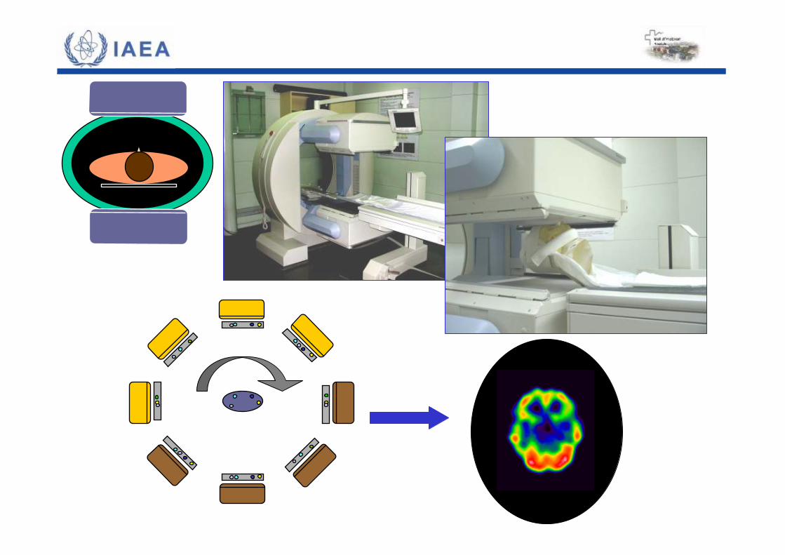
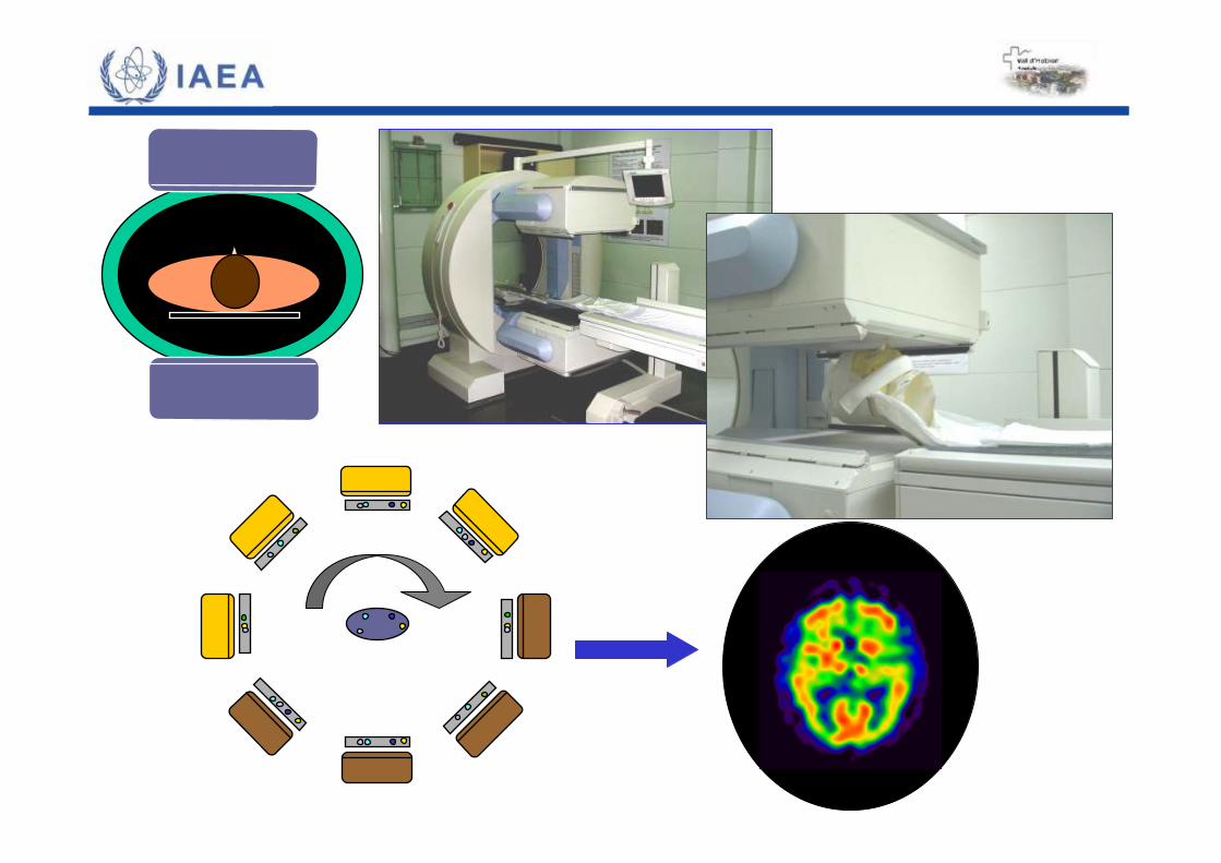
IMAGING FUNDAMENTALSIMAGING FUNDAMENTALSBrainBrain PerfusionPerfusion SPECTSPECT
HMPAOECDFDG
DATSCANIBZM
FDG
HMPAO
ECD
HMPAOECDFDG
DATSCANIBZM
FDG
HMPAO
ECD
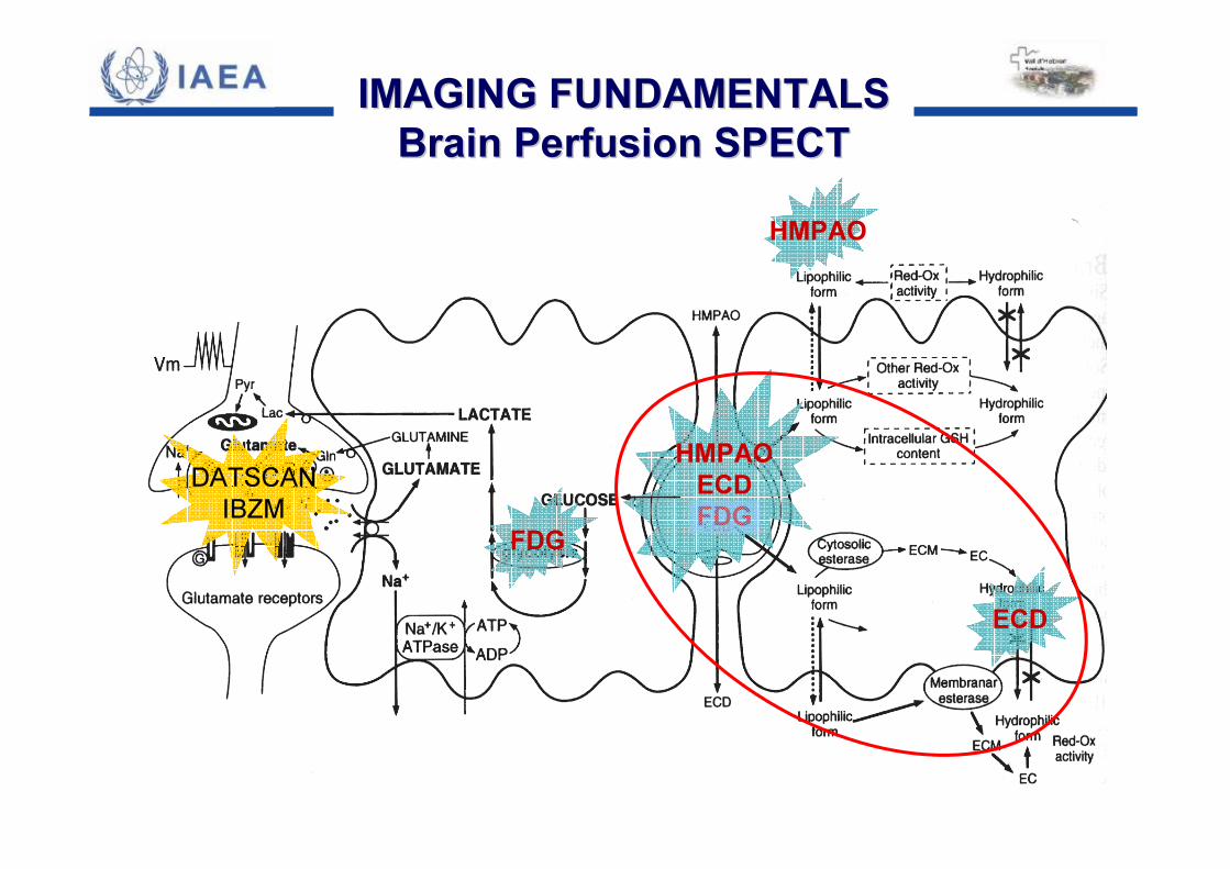
IMAGING FUNDAMENTALSIMAGING FUNDAMENTALSBrainBrain PerfusionPerfusion SPECTSPECT

DATSCANIBZM
FDG
HMPAO
ECD
HMPAOECDFDG
IMAGING FUNDAMENTALSIMAGING FUNDAMENTALSBrainBrain PerfusionPerfusion SPECTSPECT
HMPAOECDFDG
DATSCANIBZM
FDG
HMPAO
ECD
IMAGING FUNDAMENTALSIMAGING FUNDAMENTALSDatscanDatscan oror IBZM SPECTIBZM SPECT
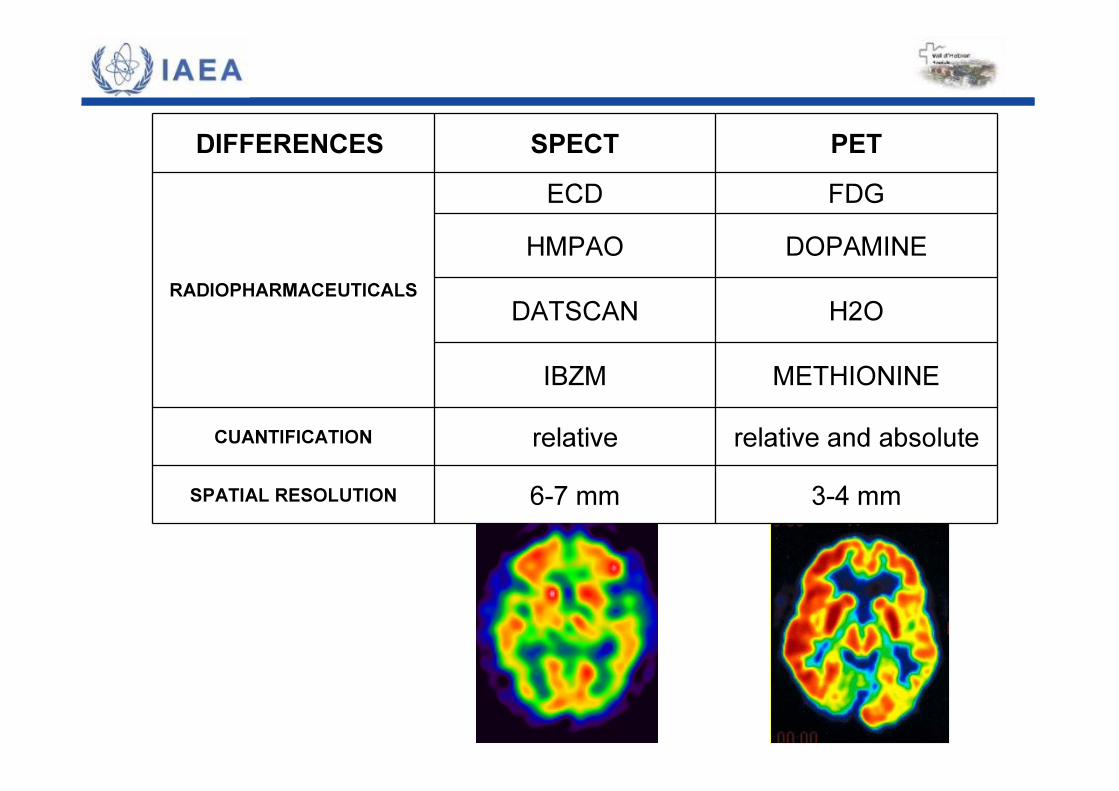
DIFFERENCES SPECT PET
RADIOPHARMACEUTICALS
ECD FDGHMPAO DOPAMINE
DATSCAN H2O
IBZM METHIONINECUANTIFICATION relative relative and absolute
SPATIAL RESOLUTION 6-7 mm 3-4 mm
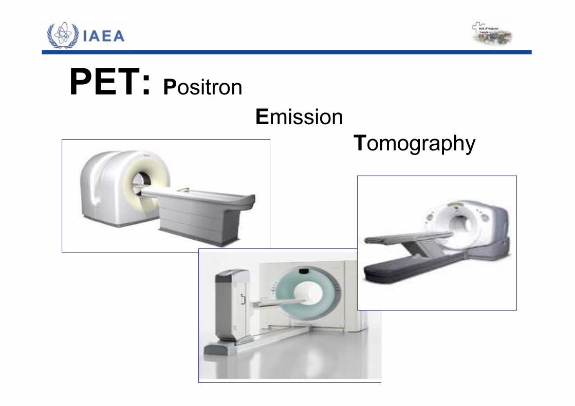
PET: PositronEmission
Tomography
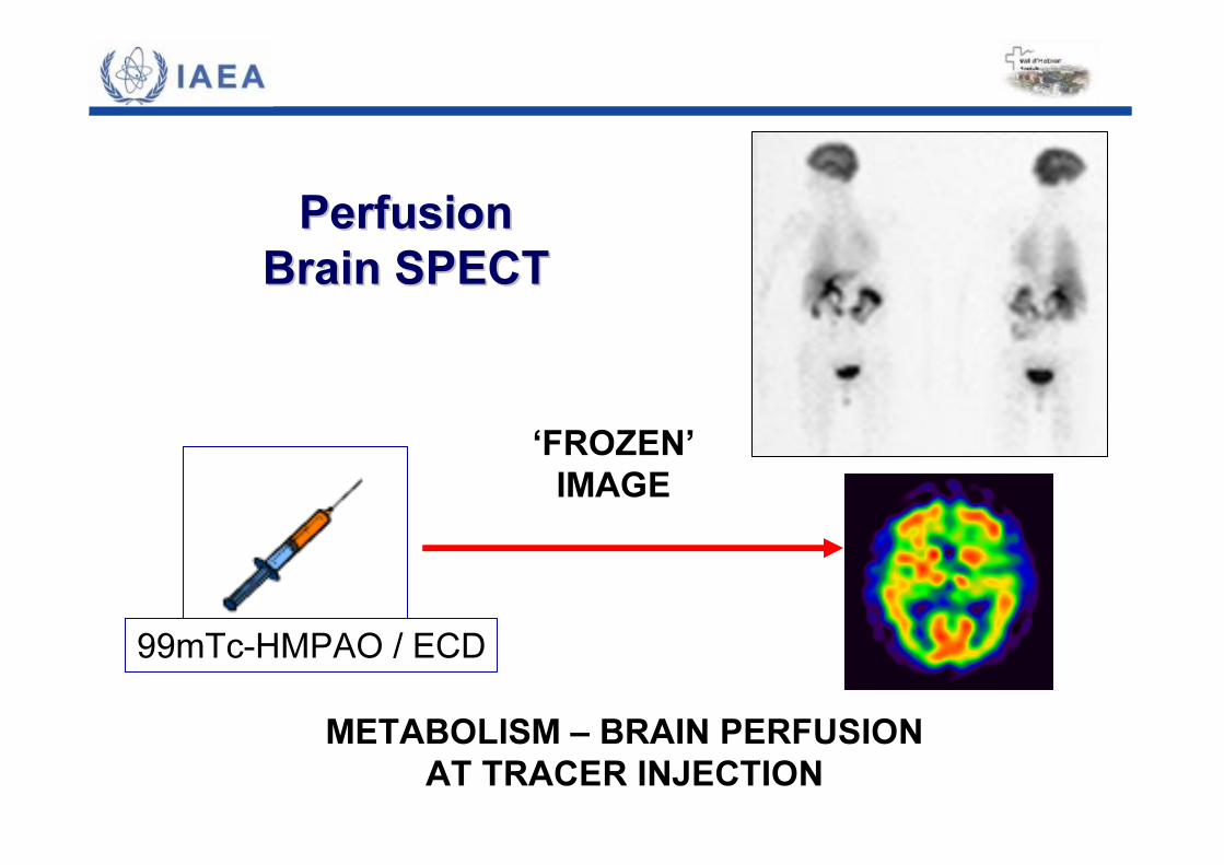
PerfusionPerfusionBrain SPEBrain SPECCTT
‘FROZEN’IMAGE
METABOLISM – BRAIN PERFUSIONAT TRACER INJECTION
99mTc-HMPAO / ECD
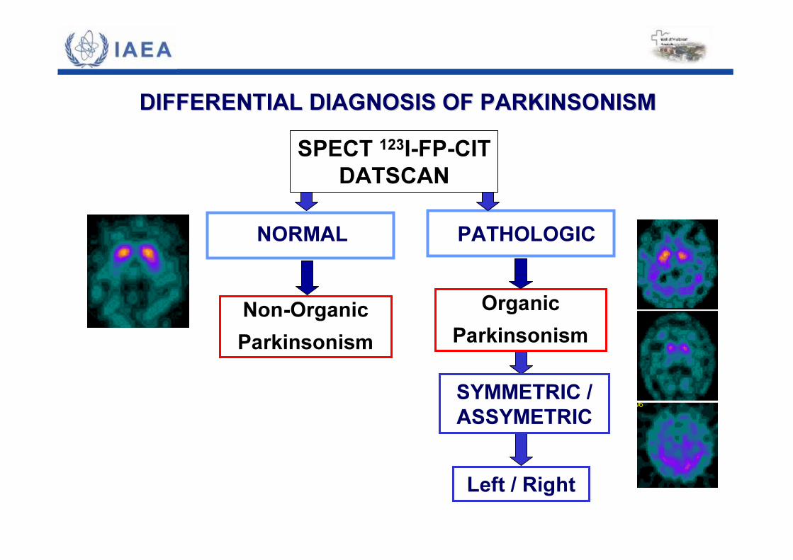
SYMMETRIC / ASSYMETRIC
Left / Right
DIFFERENTIAL DIAGNOSIS OF PARKINSONISM DIFFERENTIAL DIAGNOSIS OF PARKINSONISM
OrganicParkinsonism
Non-OrganicParkinsonism
NORMAL PATHOLOGIC
SPECT 123I-FP-CITDATSCAN
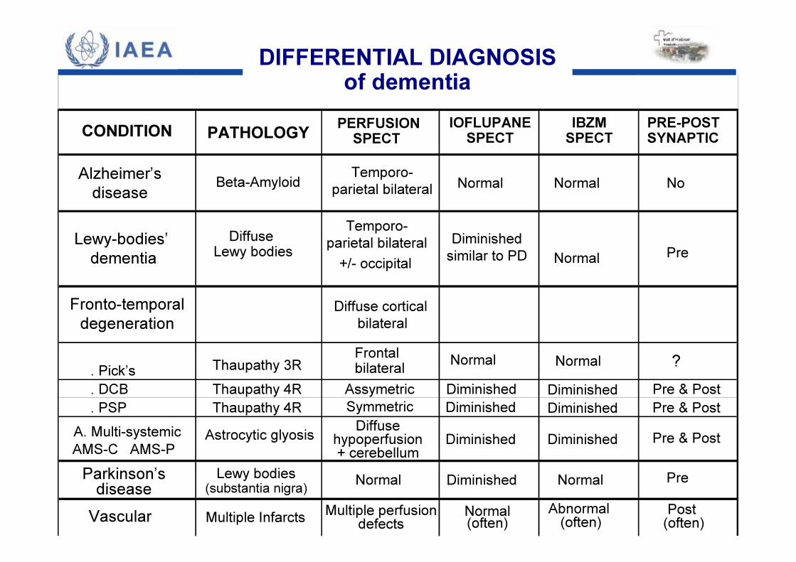
PATHOLOGY PERFUSIONSPECT
IOFLUPANESPECT
IBZMSPECT
PRE-POSTSYNAPTIC
Alzheimer’sdisease Beta-Amyloid Temporo-
parietal bilateral Normal Normal No
Lewy-bodies’dementia
Diffuse Lewy bodies +/- occipital
Diminishedsimilar to PD Normal Pre
Fronto-temporaldegeneration
Diffuse corticalbilateral
. Pick’s Thaupathy 3R Frontal bilateral Normal Normal ?
. DCB Thaupathy 4R Assymetric Diminished Pre & Post
. PSP Thaupathy 4R Symmetric Pre & PostA. Multi-systemicAMS-C AMS-P
Astrocytic glyosis Diffuse hypoperfusion+ cerebellum
Pre & Post
Parkinson’sdiseaseLewy bodies
(substantia nigra) Normal Normal Pre
Vascular Multiple Infarcts Multiple perfusiondefects Normal(often)
Abnormal (often) Post
(often)
DiminishedDiminishedDiminished
DiminishedDiminished
Diminished
CONDITION
Temporo-parietal bilateral
DIFFERENTIAL DIAGNOSISof dementia
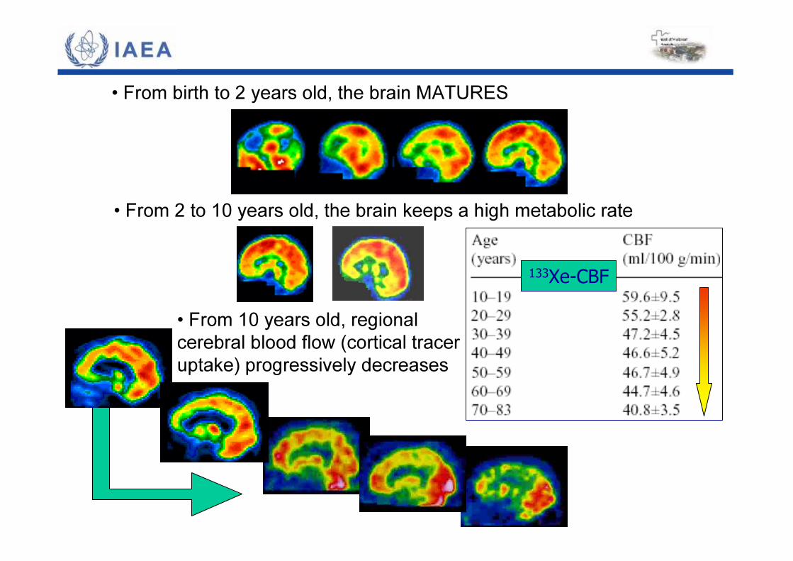
133Xe-CBF• From 10 years old, regional cerebral blood flow (cortical traceruptake) progressively decreases
• From birth to 2 years old, the brain MATURES
• From 2 to 10 years old, the brain keeps a high metabolic rate
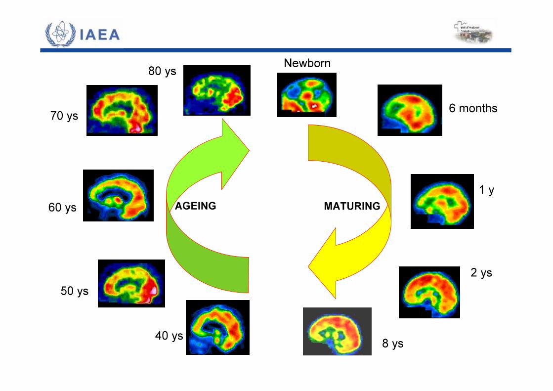
Newborn
6 months
1 y
2 ys
8 ys40 ys
50 ys
60 ys
70 ys
80 ys
MATURINGAGEING
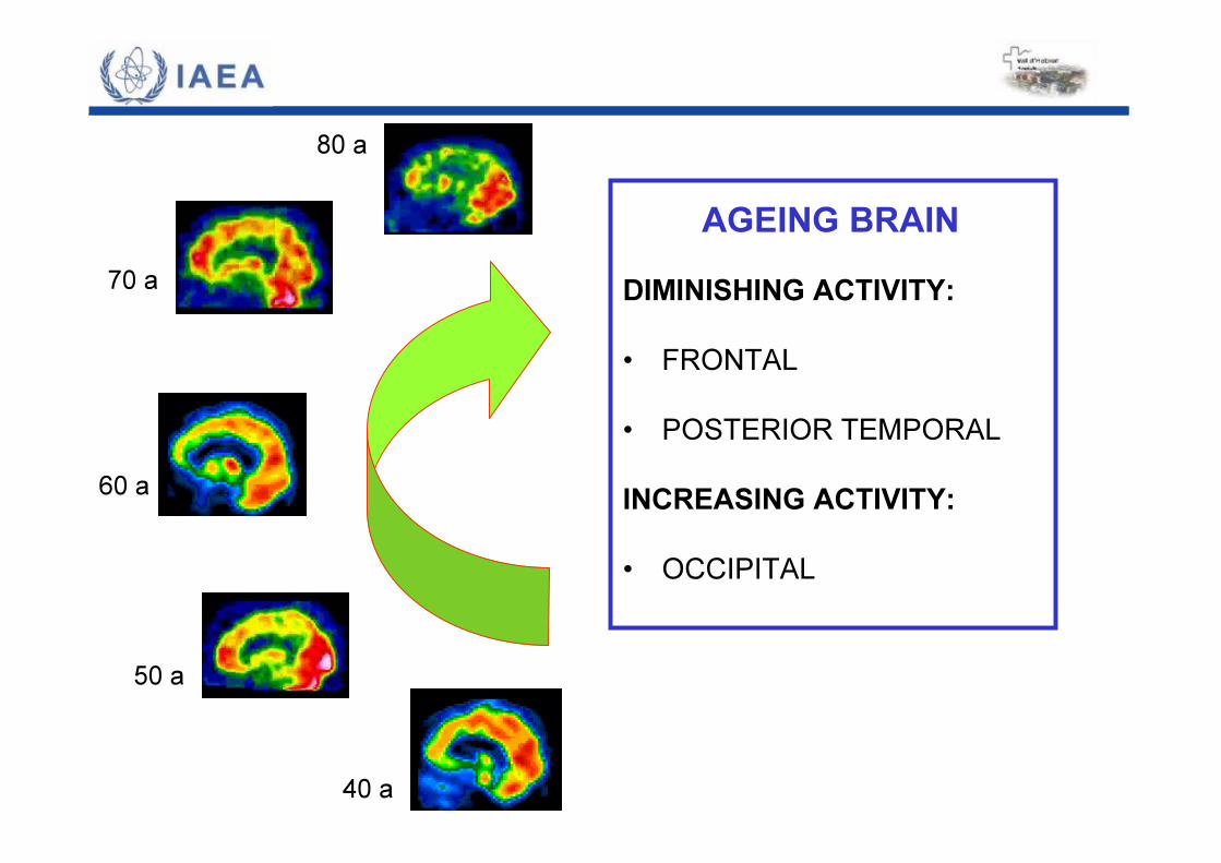
DIMINISHING ACTIVITY:• FRONTAL• POSTERIOR TEMPORALINCREASING ACTIVITY:• OCCIPITAL
40 a
50 a
60 a
70 a
80 a
AGEING BRAIN
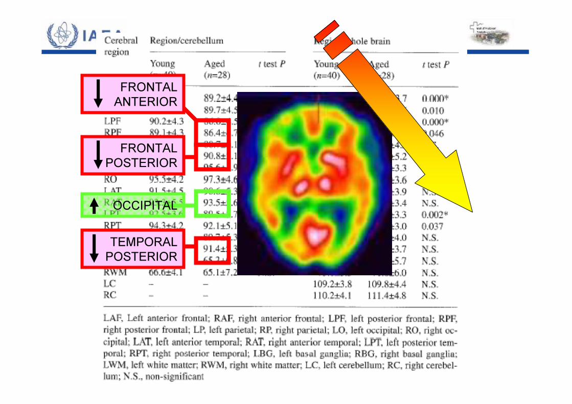
FRONTALANTERIOR
FRONTALPOSTERIOR
TEMPORALPOSTERIOR
OCCIPITAL
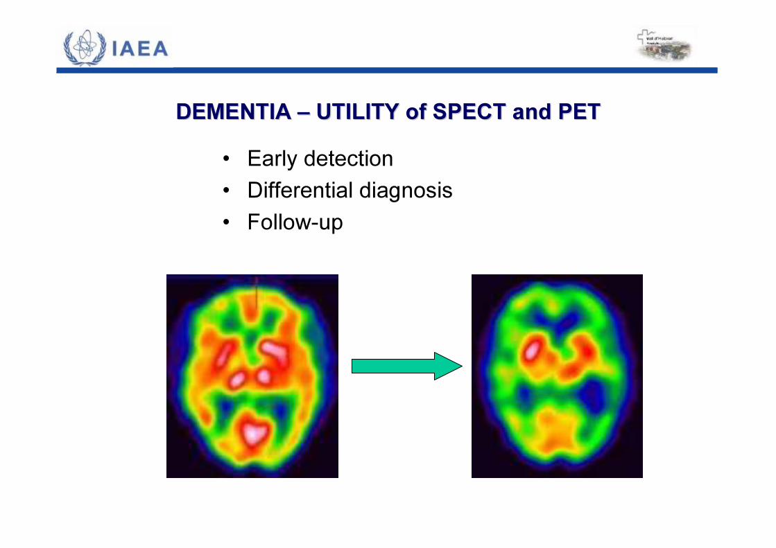
DEMENTIA DEMENTIA –– UTILITY of SPECT and PETUTILITY of SPECT and PET• Early detection• Differential diagnosis• Follow-up
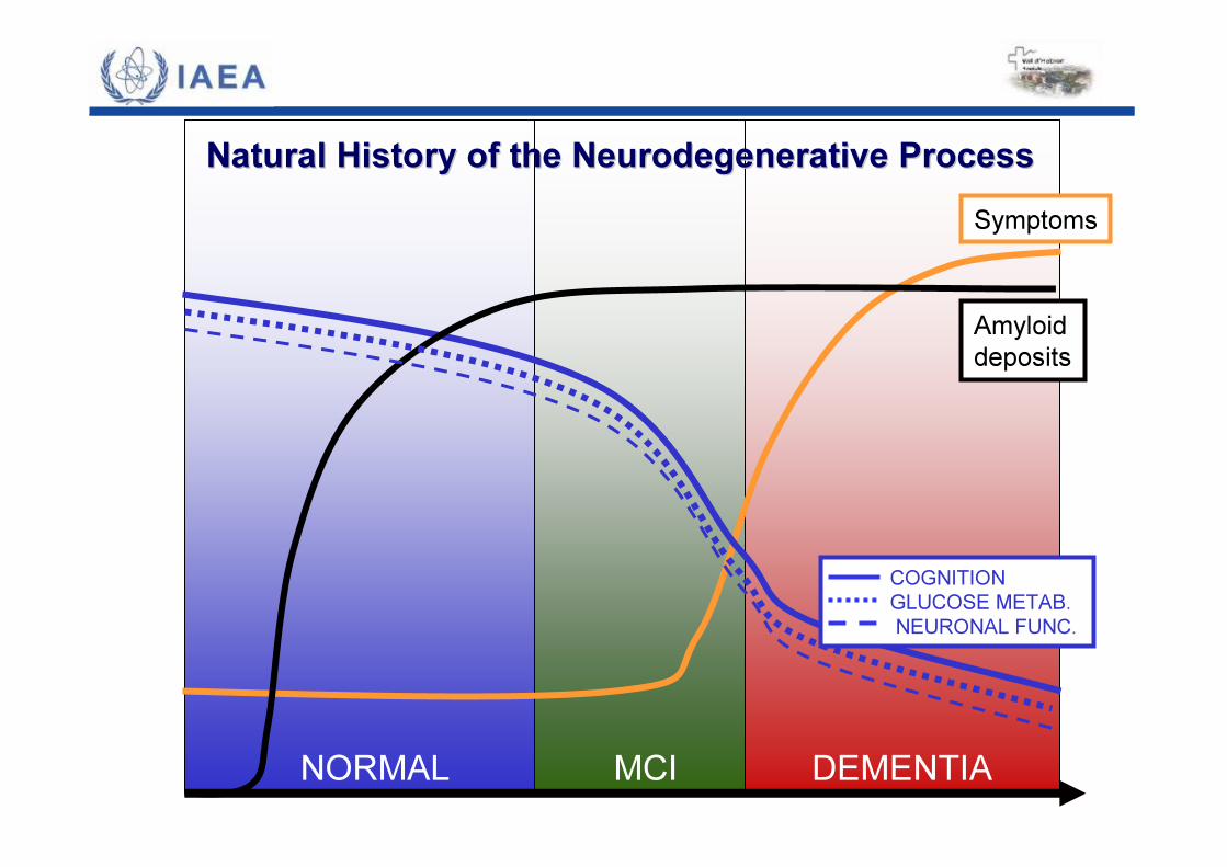
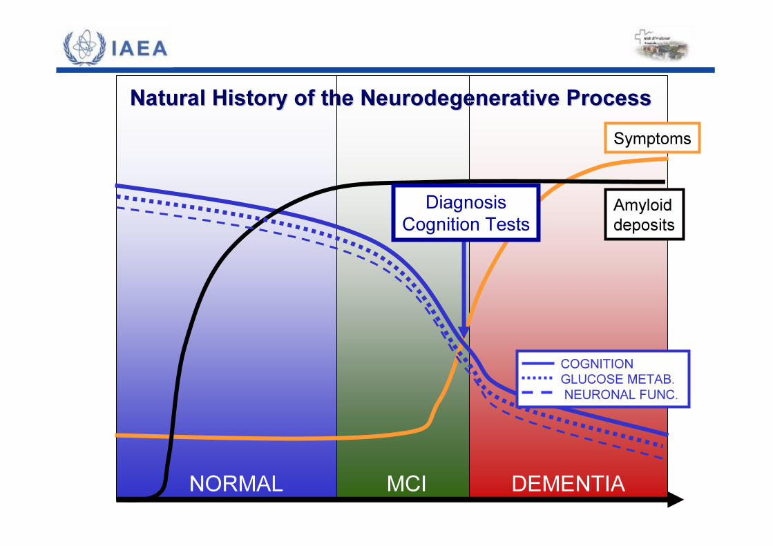
Natural Natural HistoryHistory ofof thethe NeurodegenerativeNeurodegenerative ProcessProcessSymptoms
NORMAL MCI DEMENTIA
Amyloiddeposits
COGNITIONGLUCOSE METAB.NEURONAL FUNC.
Natural Natural HistoryHistory ofof thethe NeurodegenerativeNeurodegenerative ProcessProcessSymptoms
NORMAL MCI DEMENTIA
Amyloiddeposits
COGNITIONGLUCOSE METAB.NEURONAL FUNC.
DiagnosisCognition Tests
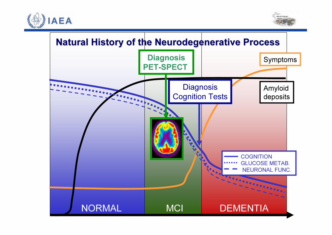
Natural Natural HistoryHistory ofof thethe NeurodegenerativeNeurodegenerative ProcessProcessSymptoms
NORMAL MCI DEMENTIA
Amyloiddeposits
COGNITIONGLUCOSE METAB.NEURONAL FUNC.
DiagnosisCognition Tests
DiagnosisPET-SPECT

Natural Natural HistoryHistory ofof thethe NeurodegenerativeNeurodegenerative ProcessProcessSymptoms
NORMAL MCI DEMENTIA
Amyloiddeposits
COGNITIONGLUCOSE METAB.NEURONAL FUNC.
DiagnosisCognition Tests
DiagnosisPET-SPECT
DiagnosisPIB
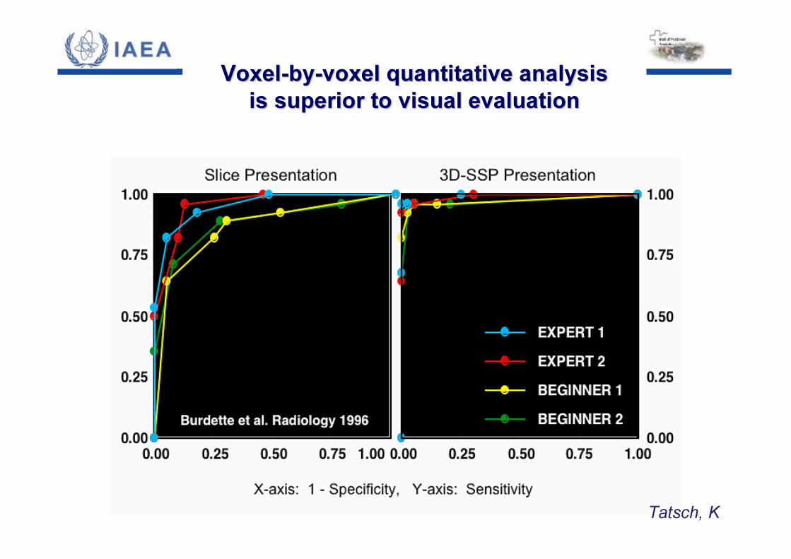
VoxelVoxel--byby--voxelvoxel quantitativequantitative analysisanalysisisis superior superior toto visual visual evaluationevaluation
Tatsch, K
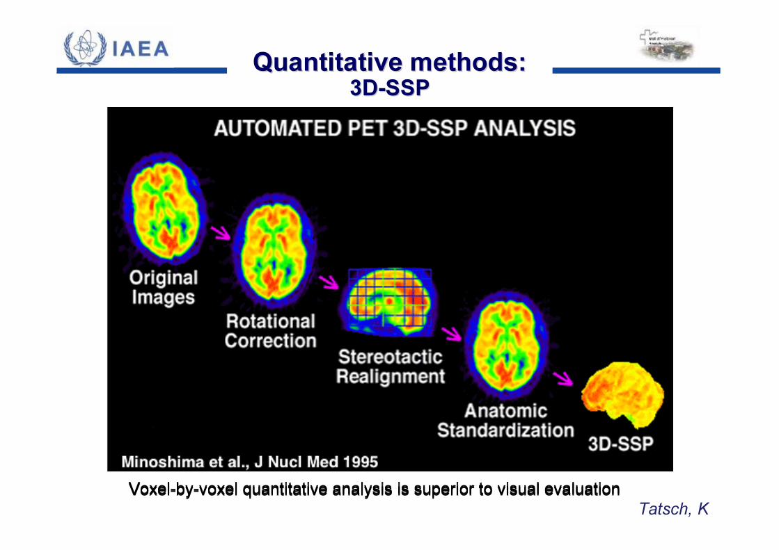
QuantitativeQuantitative methodsmethods::3D3D--SSPSSP
Voxel-by-voxel quantitative analysis is superior to visual evaluationVoxel-by-voxel quantitative analysis is superior to visual evaluationTatsch, K
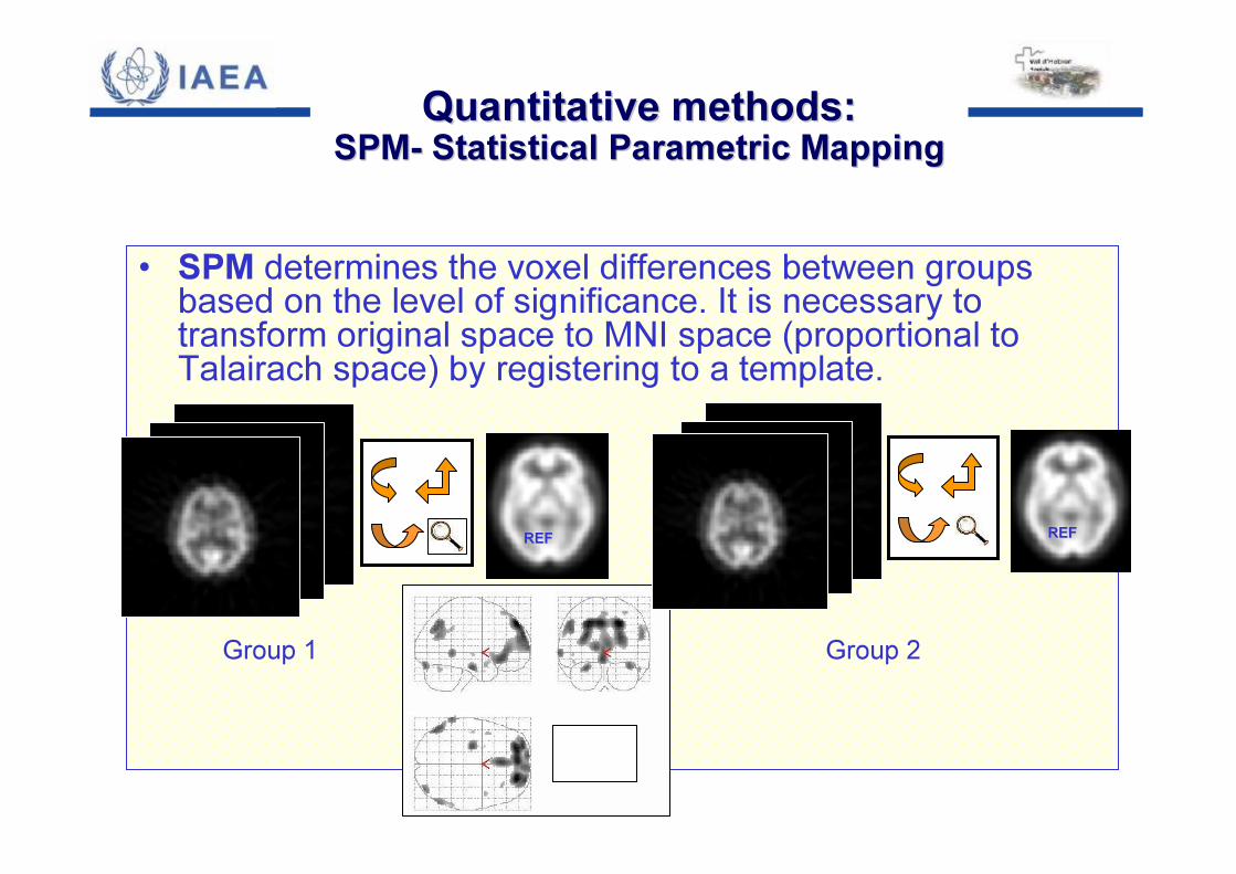
• SPM determines the voxel differences between groups based on the level of significance. It is necessary to transform original space to MNI space (proportional to Talairach space) by registering to a template.
REFREF
Group 1
REFREF
Group 2
QuantitativeQuantitative methodsmethods::SPMSPM-- StatisticalStatistical ParametricParametric MappingMapping
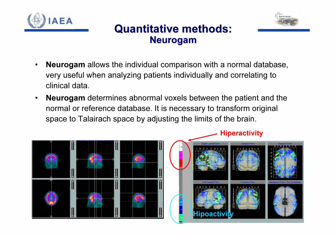
• Neurogam allows the individual comparison with a normal database, very useful when analyzing patients individually and correlating to clinical data.
• Neurogam determines abnormal voxels between the patient and the normal or reference database. It is necessary to transform original space to Talairach space by adjusting the limits of the brain.
Hiperactivity
HipoactivityHipoactivity
QuantitativeQuantitative methodsmethods::NeurogamNeurogam
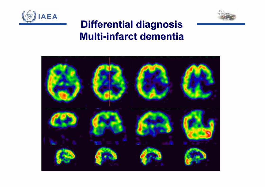
Differential diagnosisDifferential diagnosisMultiMulti--infarct dementiainfarct dementia
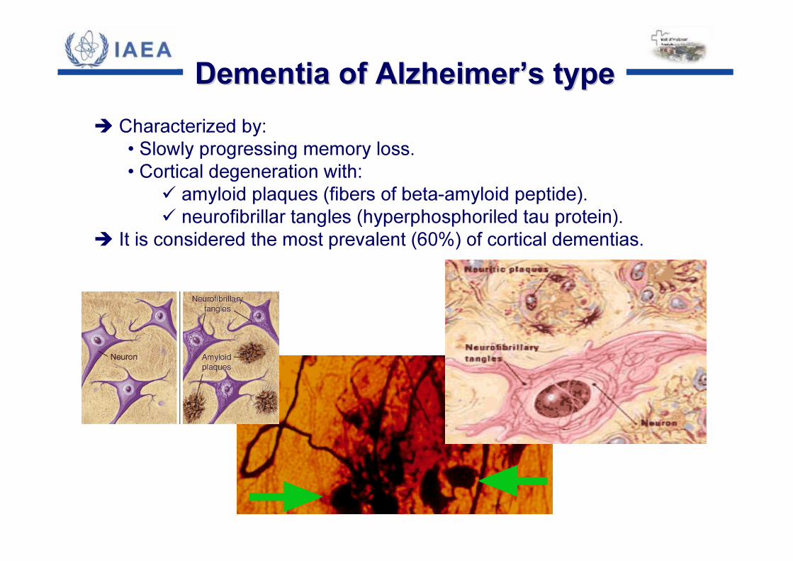
� Characterized by: • Slowly progressing memory loss.• Cortical degeneration with:
� amyloid plaques (fibers of beta-amyloid peptide).� neurofibrillar tangles (hyperphosphoriled tau protein).
� It is considered the most prevalent (60%) of cortical dementias.
Dementia of AlzheimerDementia of Alzheimer’’s types type
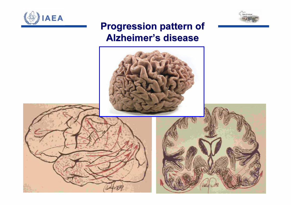
Progression pattern of Progression pattern of AlzheimerAlzheimer’’s diseases disease
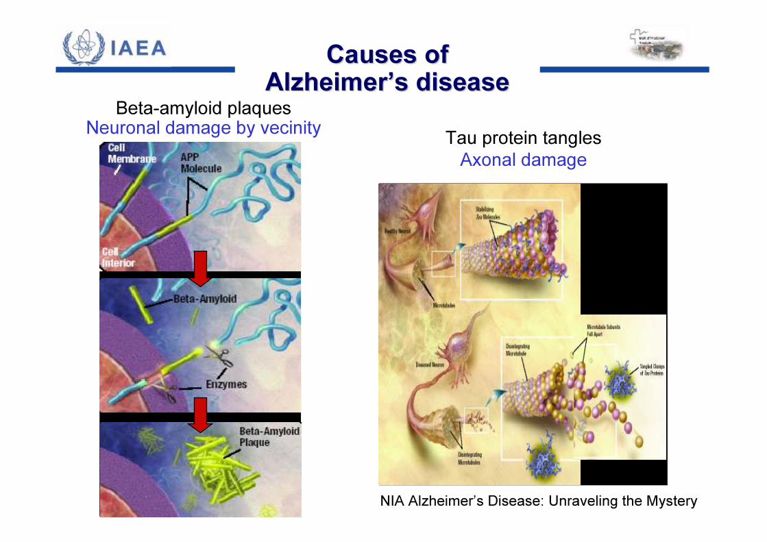
NIA Alzheimer’s Disease: Unraveling the Mystery
Beta-amyloid plaquesNeuronal damage by vecinity Tau protein tangles
Axonal damage
Causes ofCauses ofAlzheimerAlzheimer’’s diseases disease
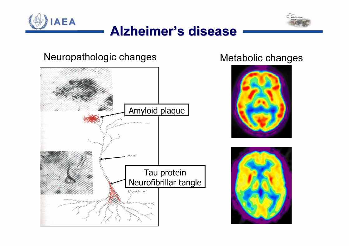
Amyloid plaque
Tau proteinNeurofibrillar tangle
AlzheimerAlzheimer’’s diseases diseaseNeuropathologic changes Metabolic changes
Tatsch, K
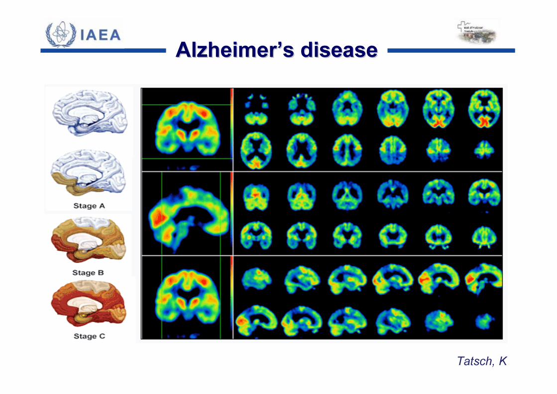
AlzheimerAlzheimer’’s diseases disease

Progression pattern of Progression pattern of AlzheimerAlzheimer’’s diseases disease
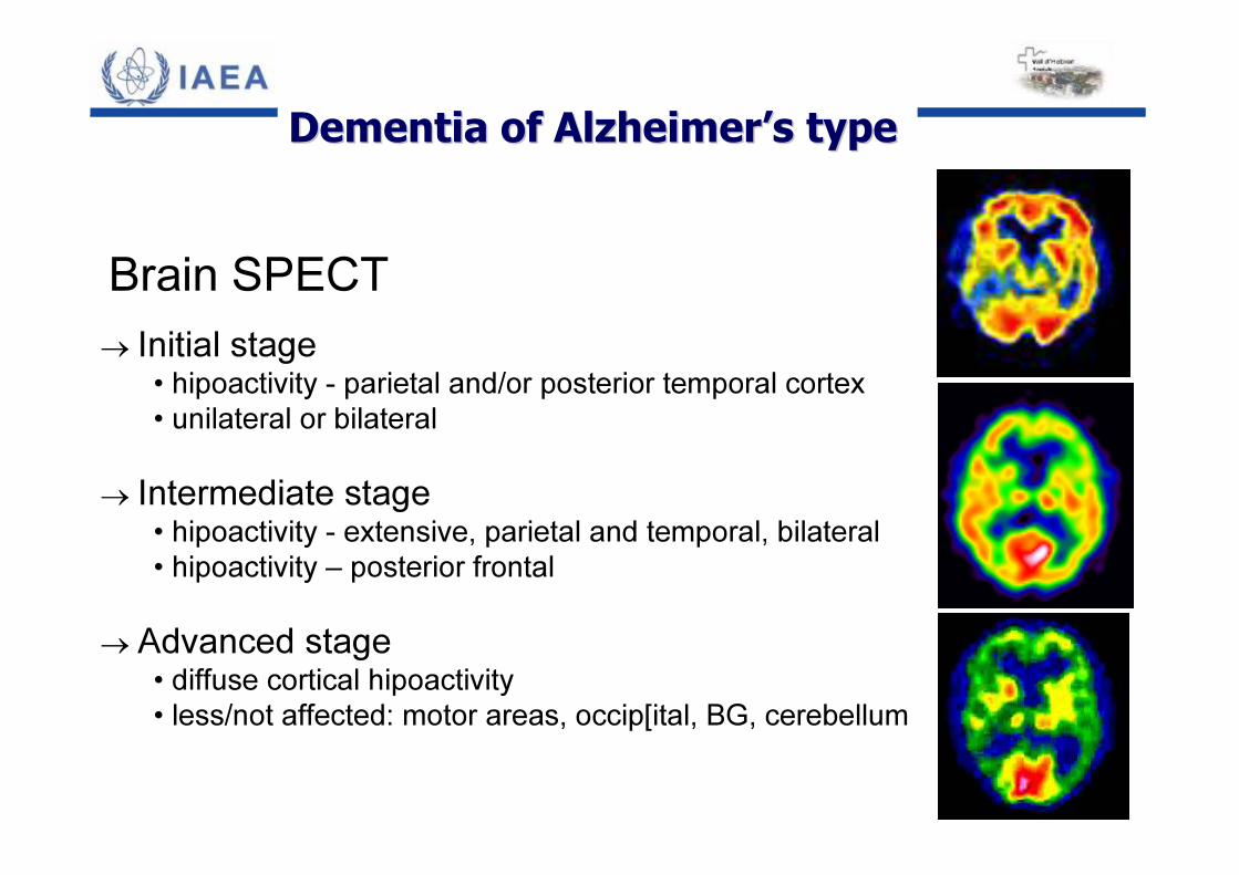
Brain SPECT→ Initial stage
• hipoactivity - parietal and/or posterior temporal cortex• unilateral or bilateral
→ Intermediate stage• hipoactivity - extensive, parietal and temporal, bilateral• hipoactivity – posterior frontal
→ Advanced stage• diffuse cortical hipoactivity• less/not affected: motor areas, occip[ital, BG, cerebellum
DementiaDementia of of AlzheimerAlzheimer’’ss typetype
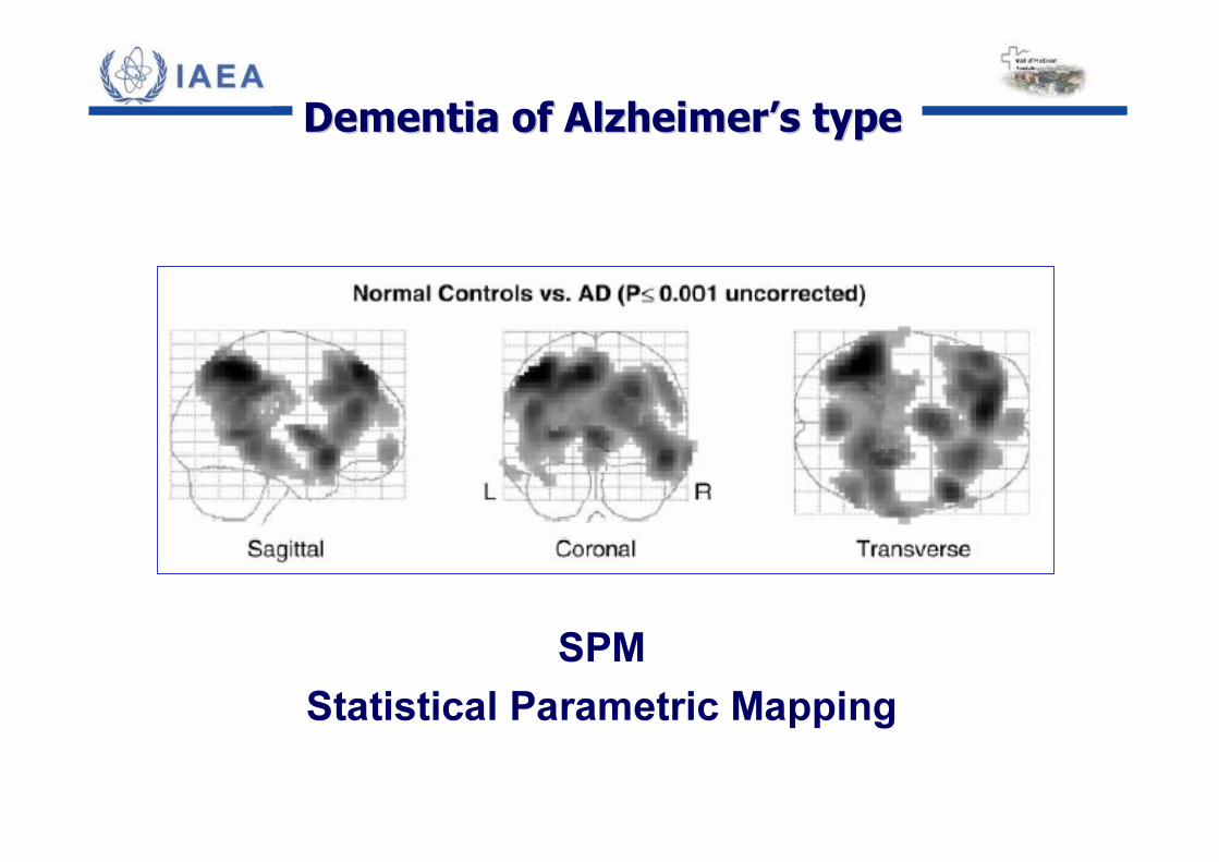
SPMStatistical Parametric Mapping
DementiaDementia of of AlzheimerAlzheimer’’ss typetype
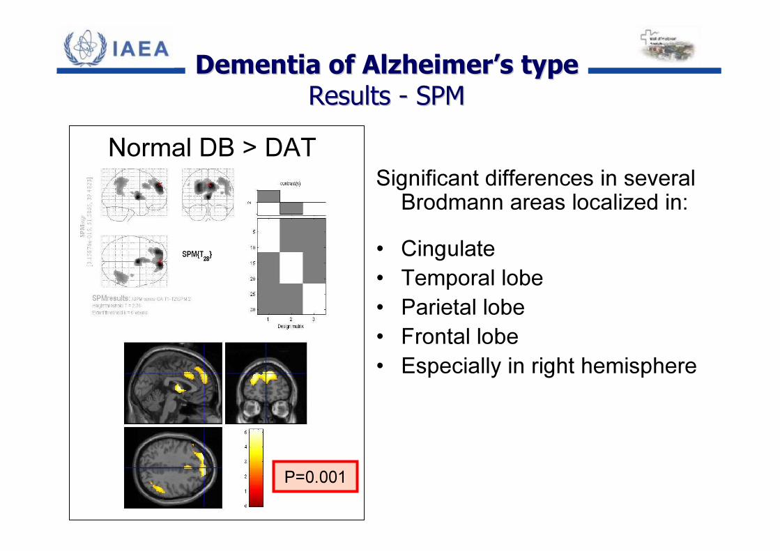
Normal DB > DAT
P=0.001
Significant differences in severalBrodmann areas localized in:
• Cingulate• Temporal lobe• Parietal lobe• Frontal lobe• Especially in right hemisphere
DementiaDementia of of AlzheimerAlzheimer’’ss typetypeResultsResults -- SPMSPM
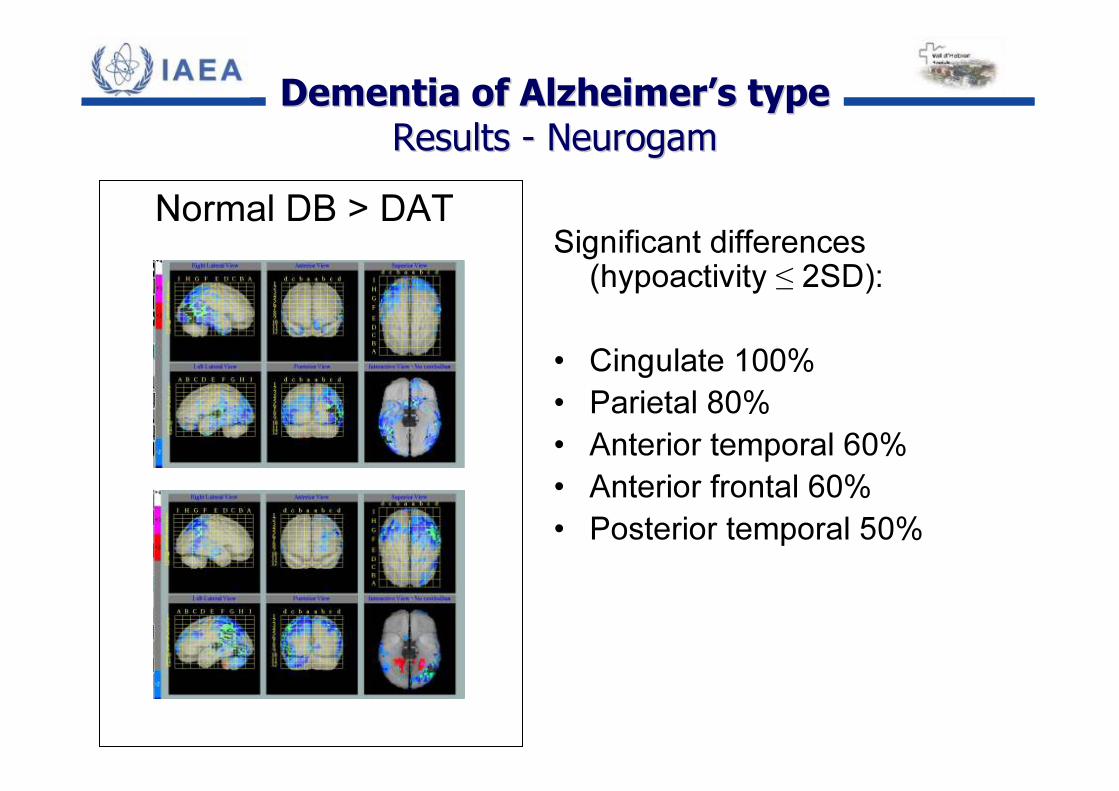
Normal DB > DATSignificant differences(hypoactivity ≤ 2SD):
• Cingulate 100%• Parietal 80%• Anterior temporal 60%• Anterior frontal 60%• Posterior temporal 50%
DementiaDementia of of AlzheimerAlzheimer’’ss typetypeResultsResults -- NeurogamNeurogam
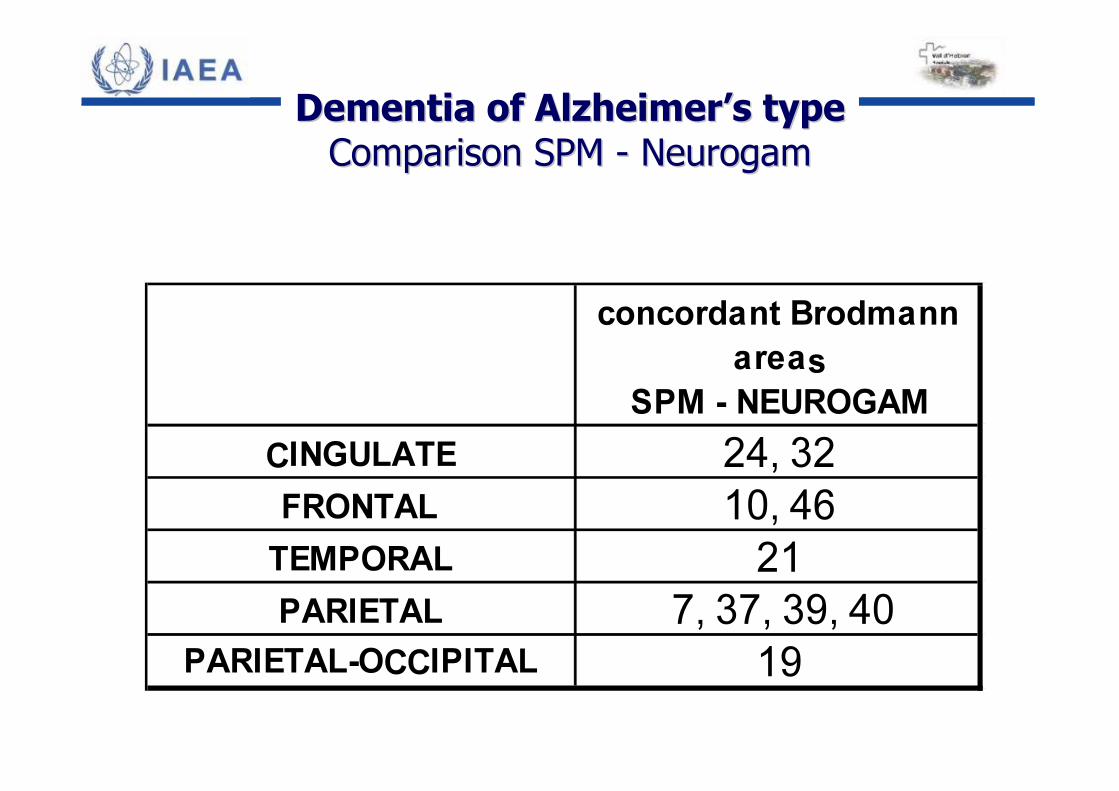
concordant Brodmann areas
SPM - NEUROGAMCINGULATE 24, 32FRONTAL 10, 46
TEMPORAL 21PARIETAL 7, 37, 39, 40
PARIETAL-OCCIPITAL 19
DementiaDementia of of AlzheimerAlzheimer’’ss typetypeComparisonComparison SPM SPM -- NeurogamNeurogam
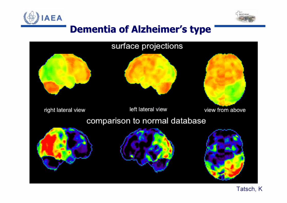
Tatsch, K
DementiaDementia of of AlzheimerAlzheimer’’ss typetype
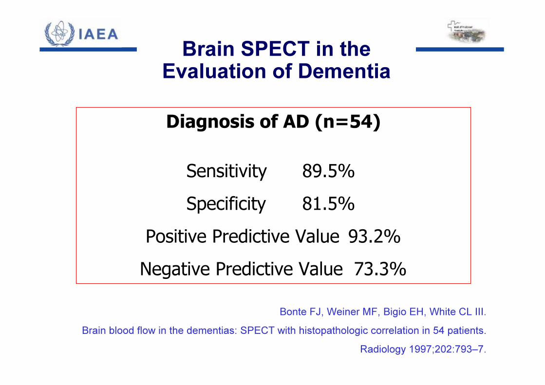
Diagnosis of AD (n=54)
Sensitivity 89.5%Specificity 81.5%
Positive Predictive Value 93.2%Negative Predictive Value 73.3%
Bonte FJ, Weiner MF, Bigio EH, White CL III. Brain blood flow in the dementias: SPECT with histopathologic correlation in 54 patients.
Radiology 1997;202:793–7.
Brain SPECT in the Evaluation of Dementia
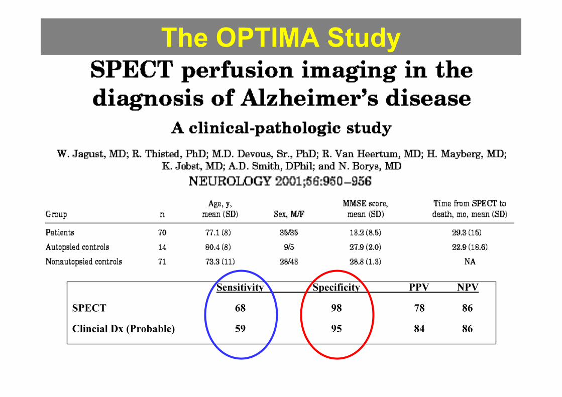
The OPTIMA Study
Sensitivity Specificity PPV NPVSPECT 68 98 78 86Clincial Dx (Probable) 59 95 84 86
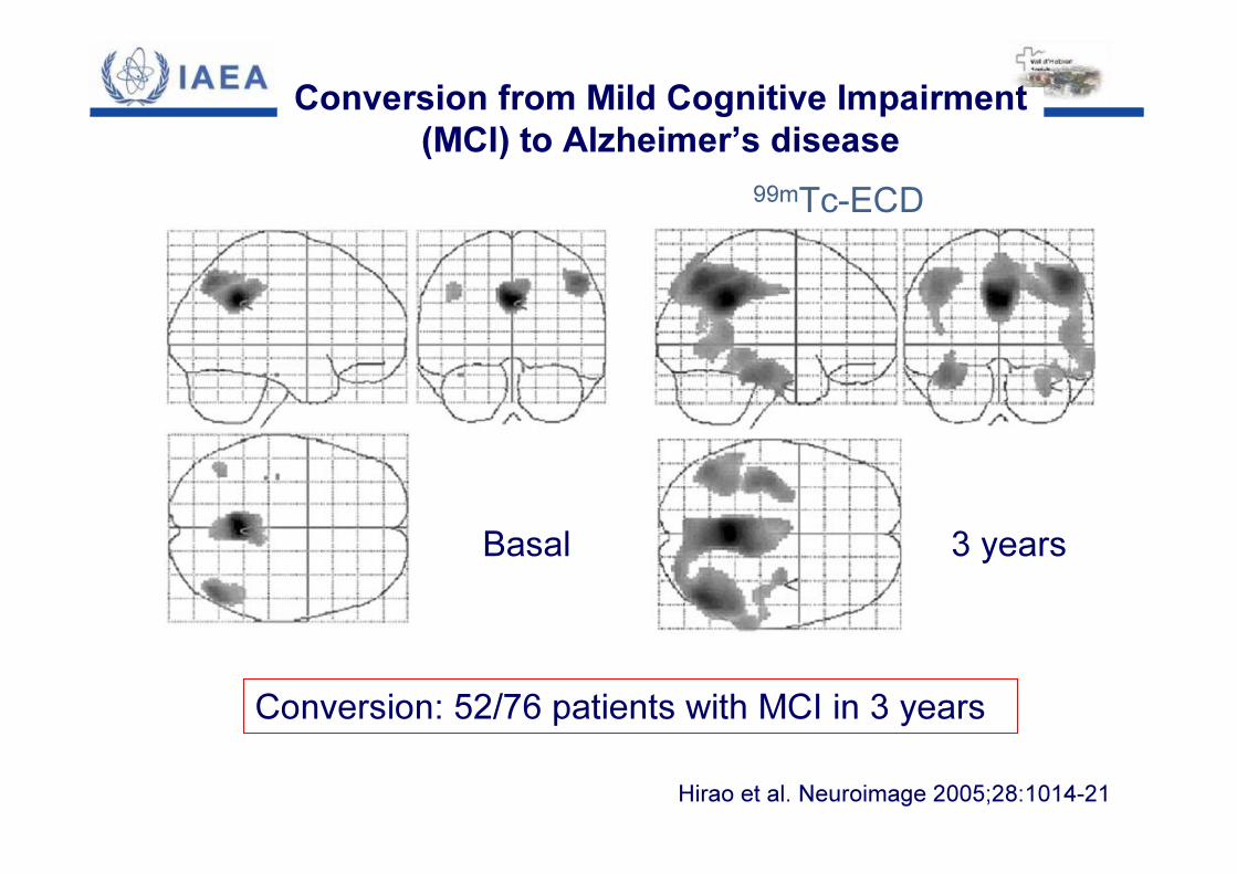
Conversion from Mild Cognitive Impairment (MCI) to Alzheimer’s disease
99mTc-ECD
Basal 3 years
Conversion: 52/76 patients with MCI in 3 yearsHirao et al. Neuroimage 2005;28:1014-21
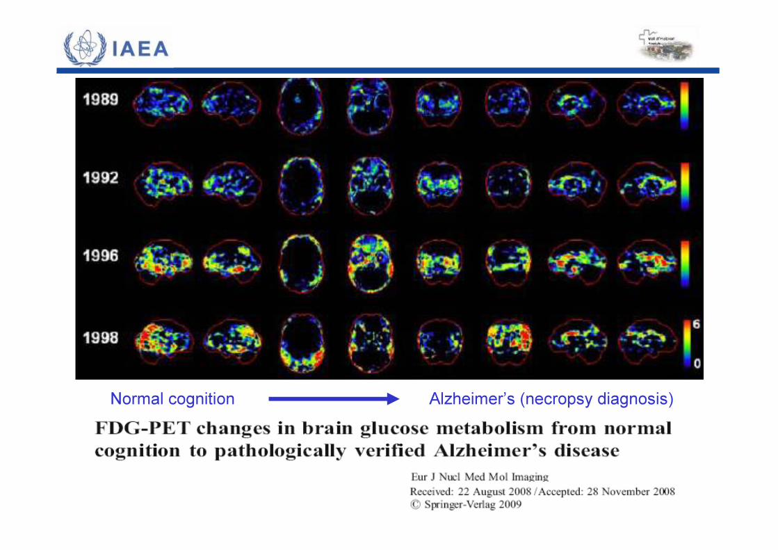
Normal cognition Alzheimer’s (necropsy diagnosis)
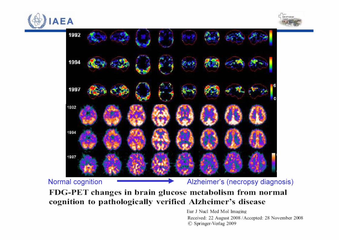
Normal cognition Alzheimer’s (necropsy diagnosis)
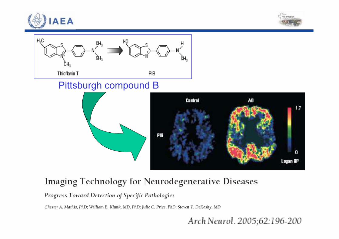
Pittsburgh compound B
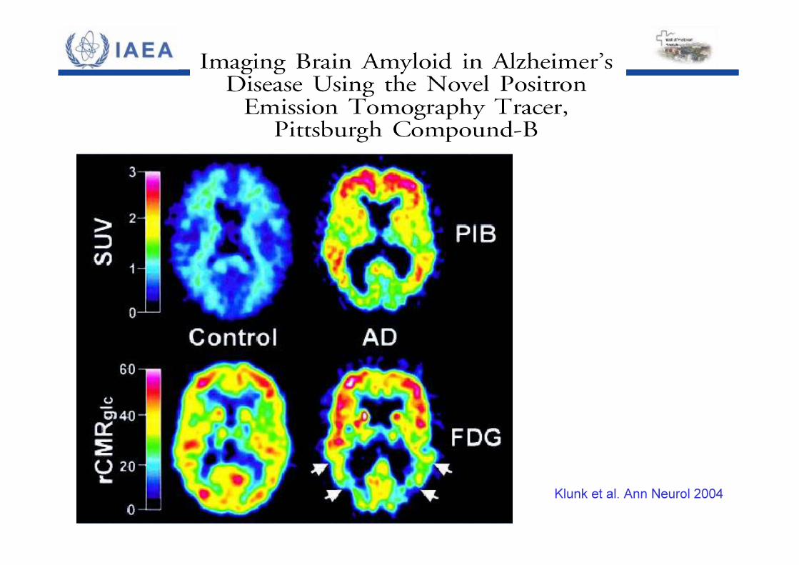
Klunk et al. Ann Neurol 2004
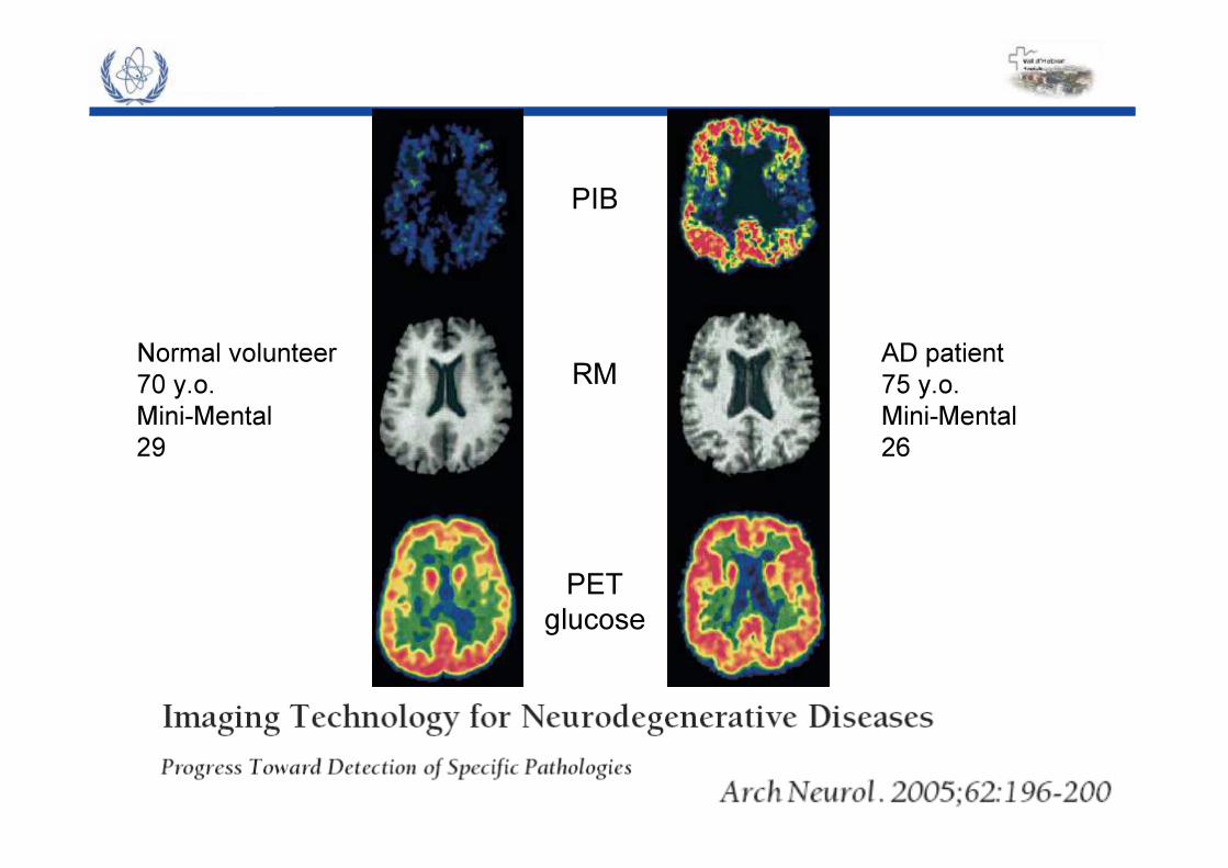
PIB
RM
PETglucose
Normal volunteer70 y.o.Mini-Mental 29
AD patient75 y.o.Mini-Mental 26
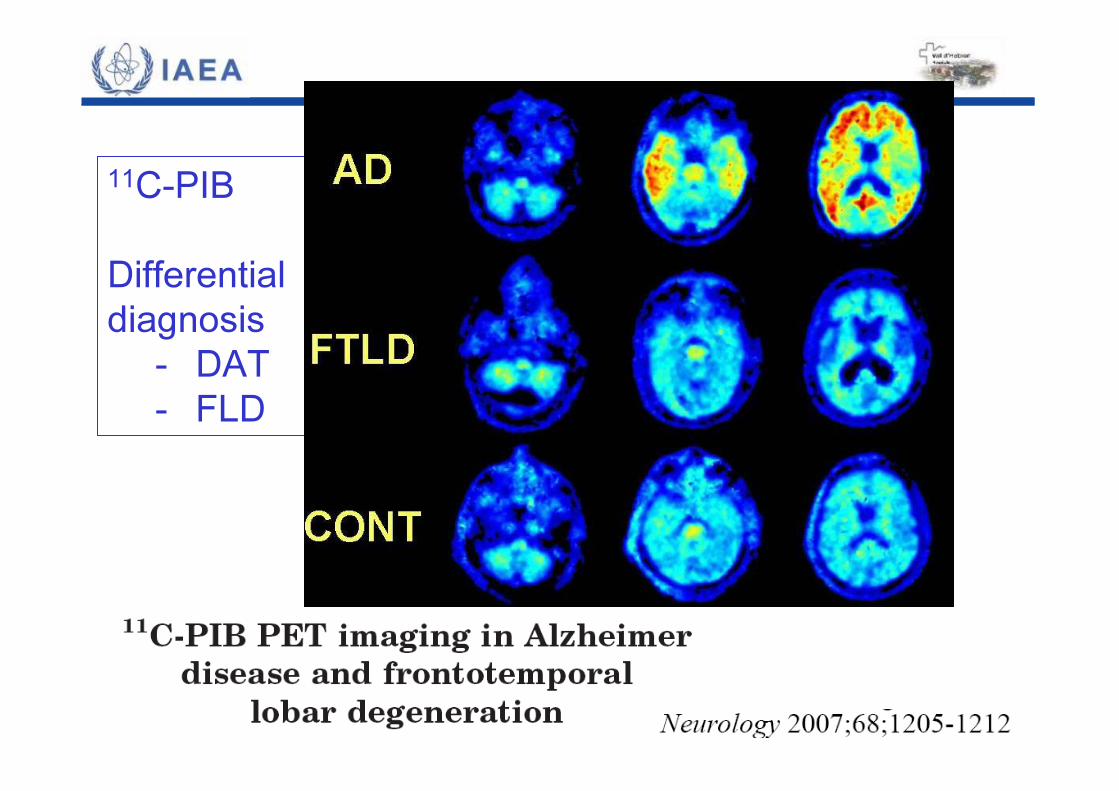
11C-PIBDifferential diagnosis
- DAT - FLD
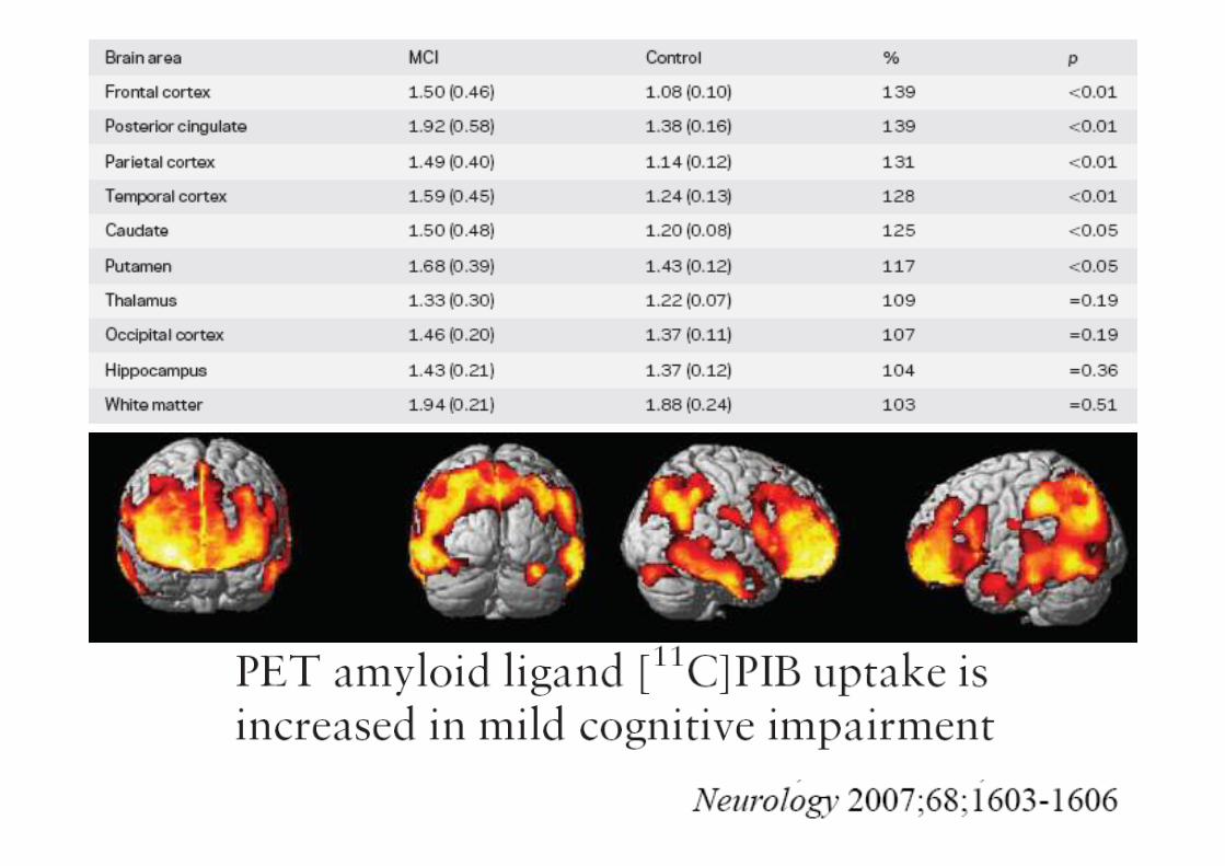

Alzheimer’s DiseaseAmyloid and hypometabolism
Increased PIB
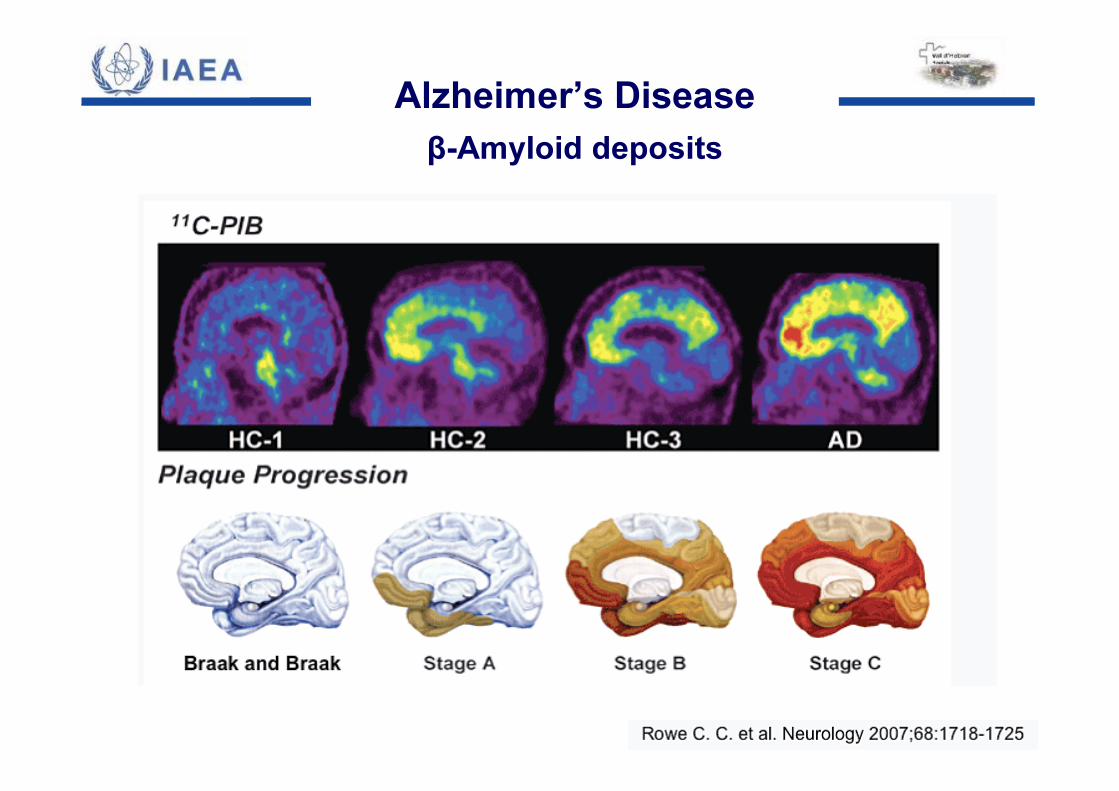
Alzheimer’s Diseaseβ-Amyloid deposits
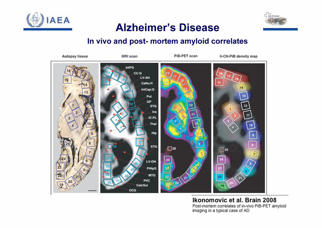
Alzheimer’s DiseaseIn vivo and post- mortem amyloid correlates
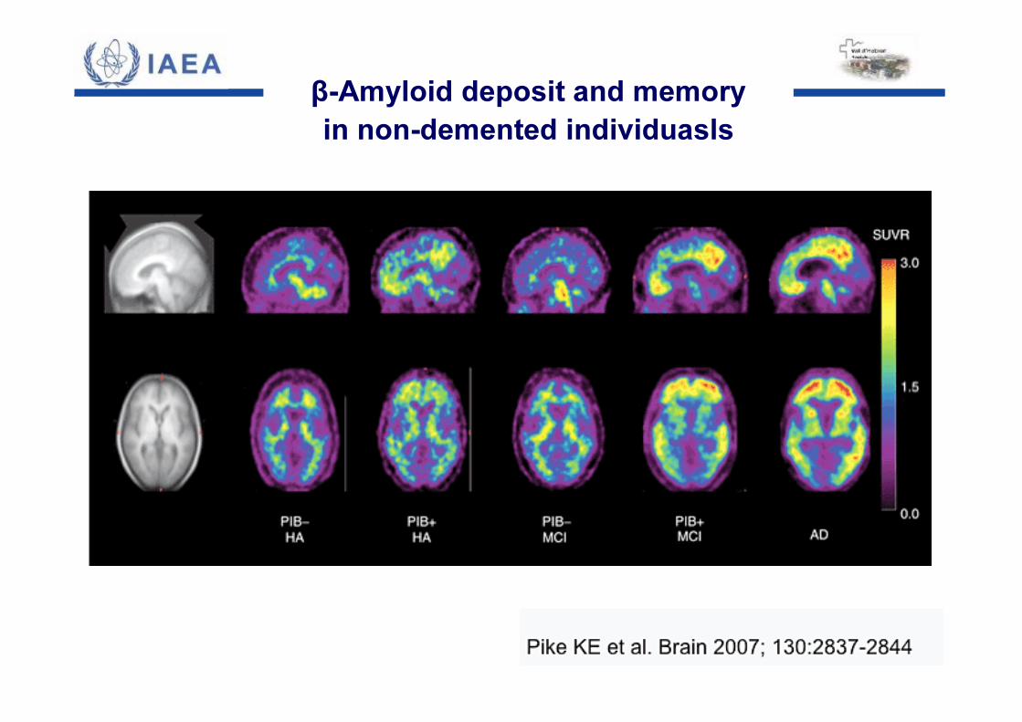
β-Amyloid deposit and memoryin non-demented individuasls
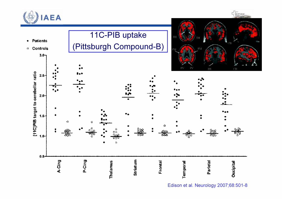
11C-PIB uptake(Pittsburgh Compound-B)
Edison et al. Neurology 2007;68:501-8
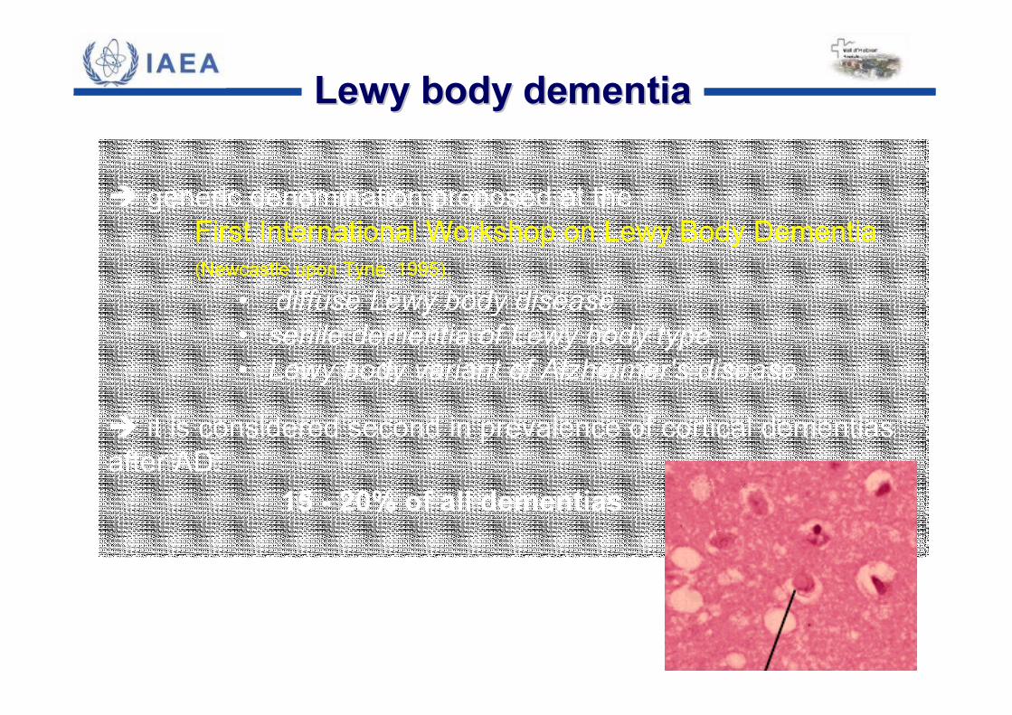
� generic denomination proposed at the First International Workshop on Lewy Body Dementia (Newcastle upon Tyne, 1995).
• diffuse Lewy body disease• senile dementia of Lewy body type• Lewy body variant of Alzheimer's disease
� it is considered second in prevalence of cortical dementiasafter AD:
15 - 20% of all dementias
Lewy body dementiaLewy body dementia
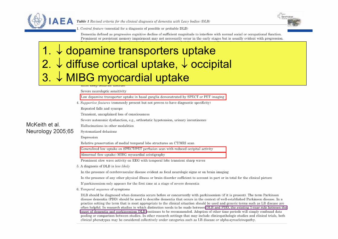
McKeith et al. Neurology 2005;65
1. ↓ dopamine transporters uptake2. ↓ diffuse cortical uptake, ↓ occipital3. ↓ MIBG myocardial uptake
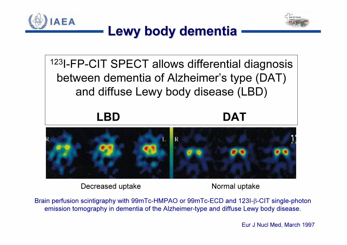
123I-FP-CIT SPECT allows differential diagnosis between dementia of Alzheimer’s type (DAT)
and diffuse Lewy body disease (LBD)LBD DAT
Brain perfusion scintigraphy with 99mTc-HMPAO or 99mTc-ECD and 123I-β-CIT single-photonemission tomography in dementia of the Alzheimer-type and diffuse Lewy body disease.
Eur J Nucl Med, March 1997
Decreased uptake Normal uptake
Lewy body dementiaLewy body dementia
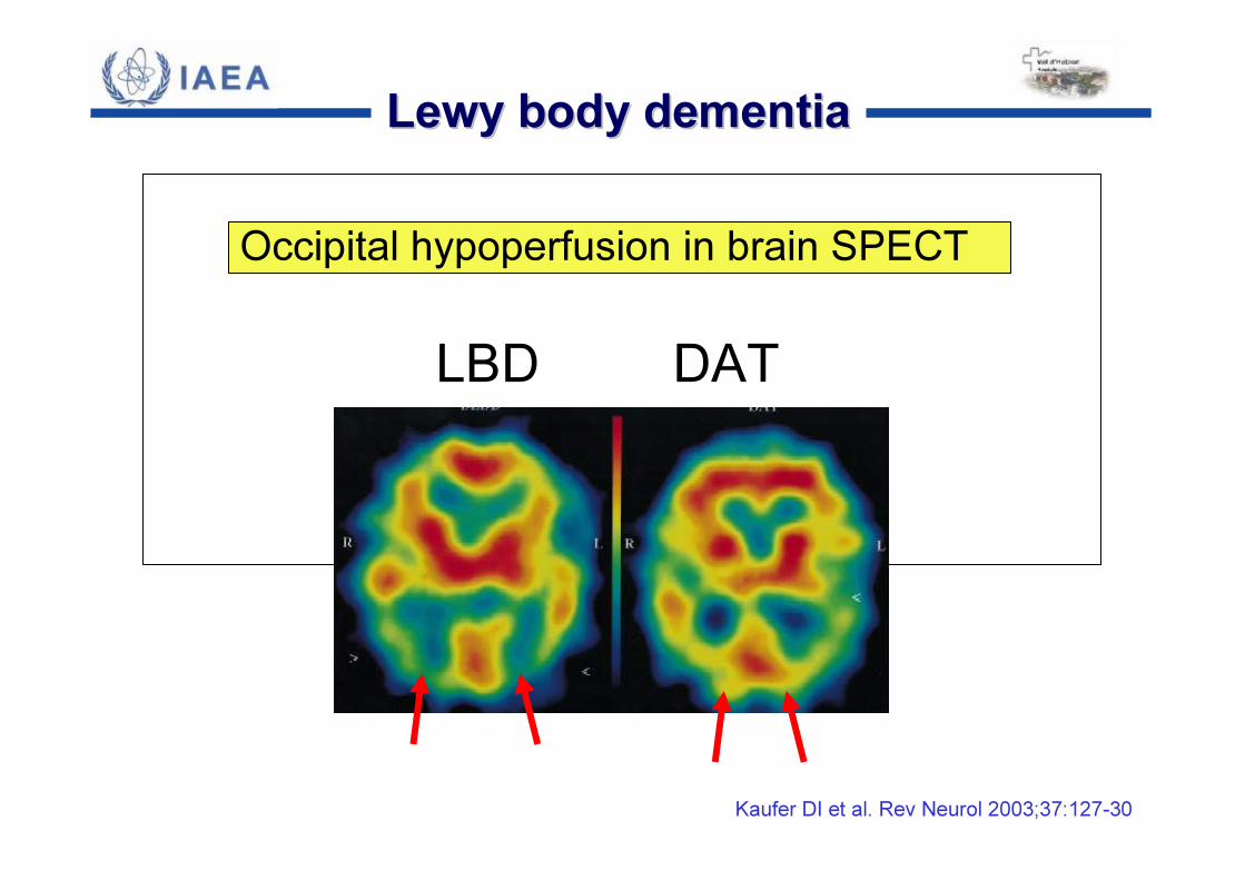
Kaufer DI et al. Rev Neurol 2003;37:127-30
LBD DAT
Occipital hypoperfusion in brain SPECT
Lewy body dementiaLewy body dementia
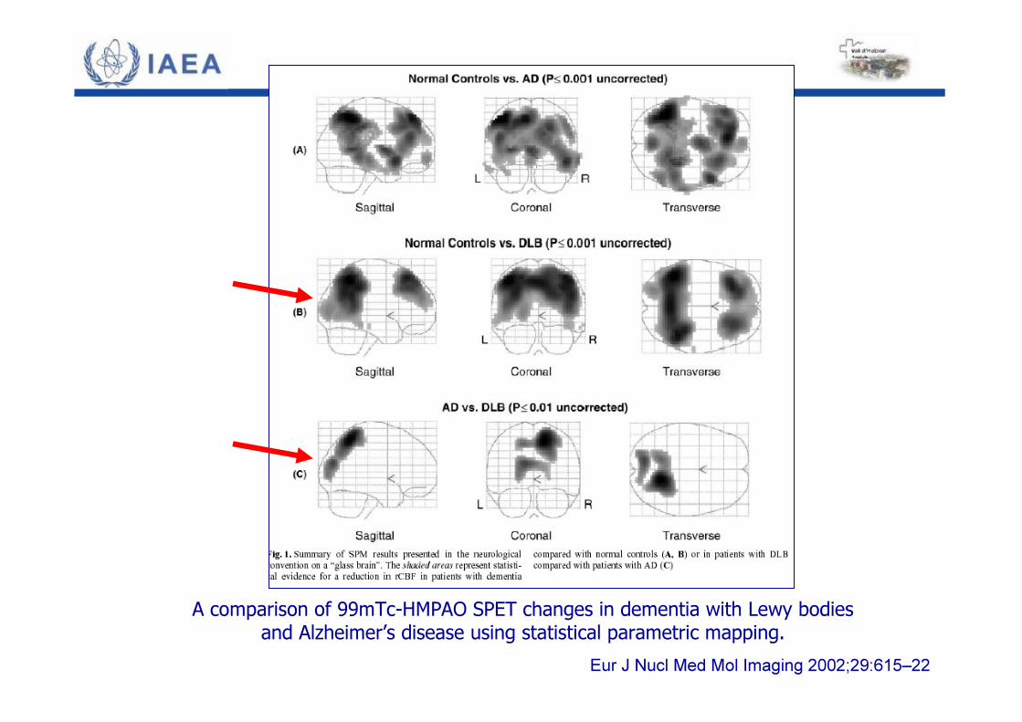
A comparison of 99mTc-HMPAO SPET changes in dementia with Lewy bodiesand Alzheimer’s disease using statistical parametric mapping.
Eur J Nucl Med Mol Imaging 2002;29:615–22

LBD DAT↓ MIBG myocardial uptake
Lewy body dementiaLewy body dementia
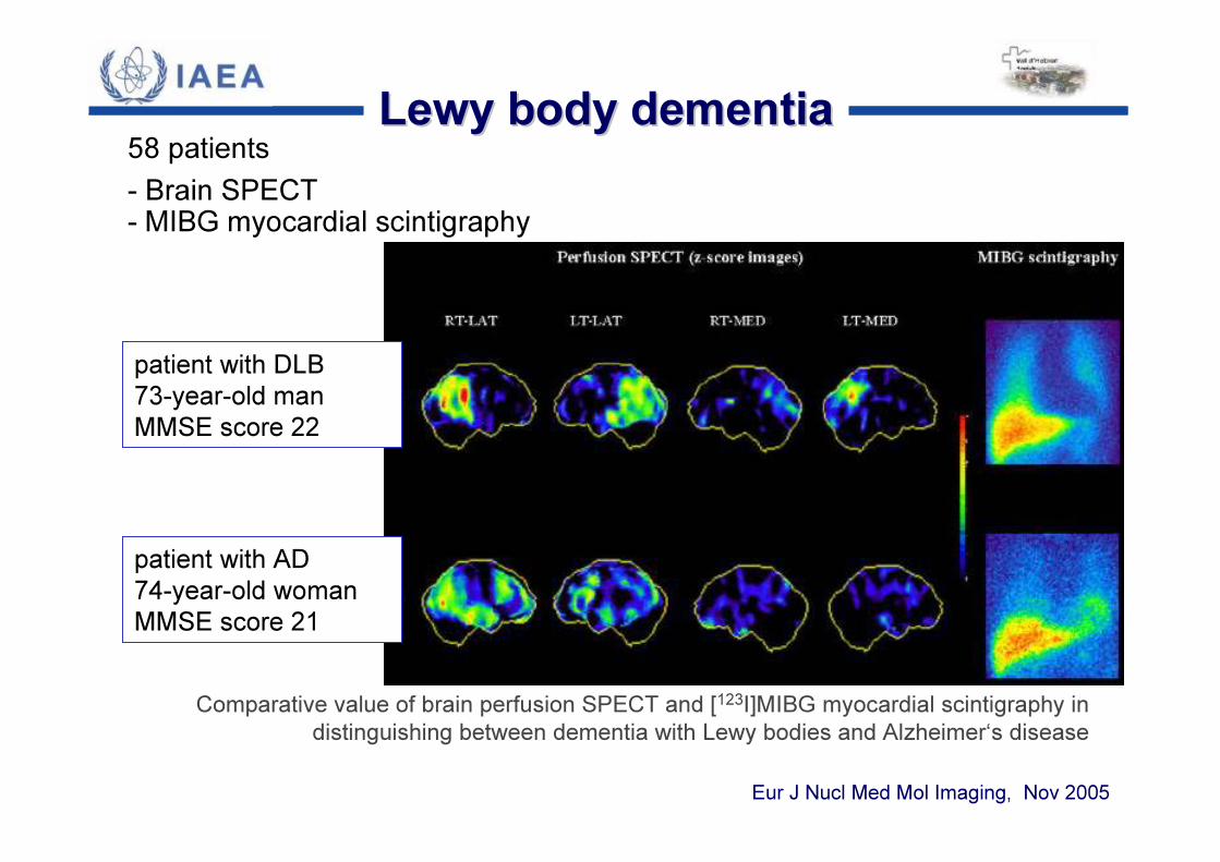
58 patients - Brain SPECT - MIBG myocardial scintigraphy
Comparative value of brain perfusion SPECT and [123I]MIBG myocardial scintigraphy in distinguishing between dementia with Lewy bodies and Alzheimer‘s disease
Eur J Nucl Med Mol Imaging, Nov 2005
patient with DLB 73-year-old man MMSE score 22
patient with AD74-year-old woman MMSE score 21
Lewy body dementiaLewy body dementia

Brain SPECTMedial occipital lobe
Myocardial MIBGH/M ratio
Comparative value…(cont.)Eur J Nucl Med Mol Imaging, Nov 2005
Lewy body dementiaLewy body dementia
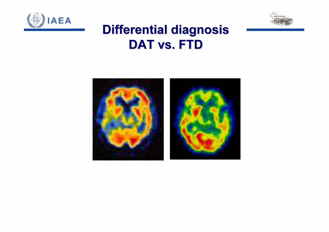
Differential diagnosisDifferential diagnosisDAT vs. FTDDAT vs. FTD
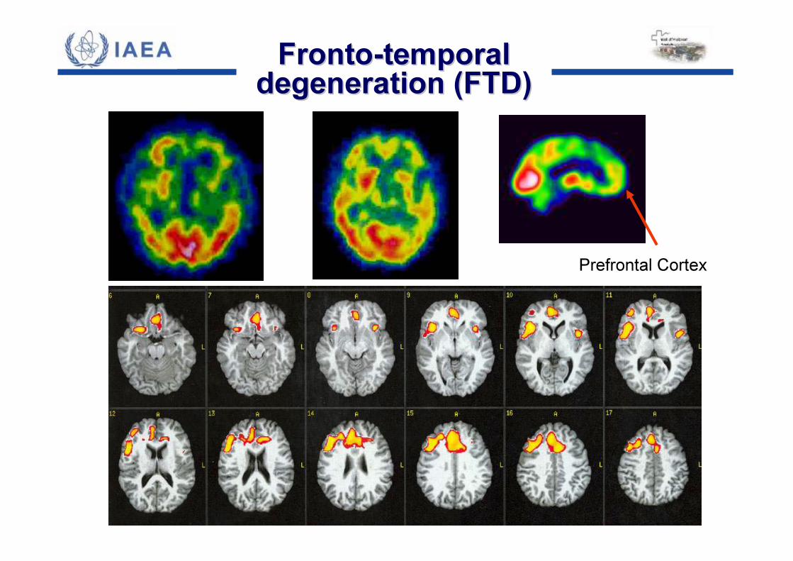
Prefrontal Cortex
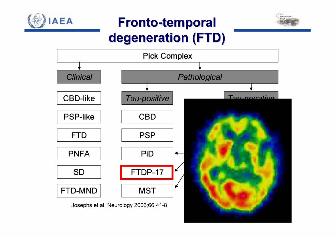
FrontoFronto--temporal temporal degeneration (FTD)degeneration (FTD)
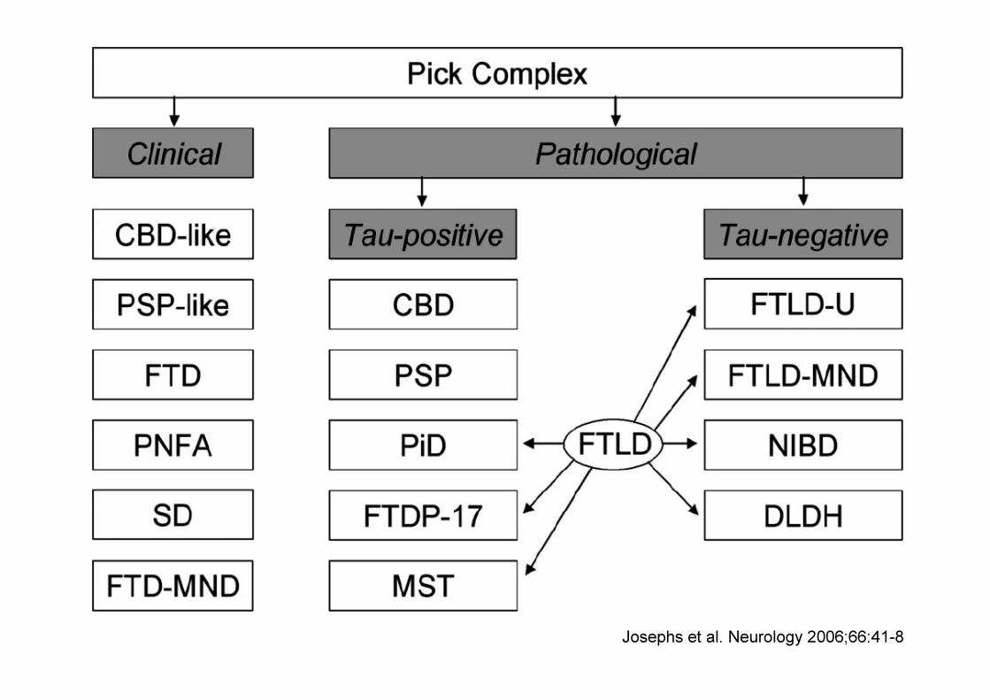
Josephs et al. Neurology 2006;66:41-8
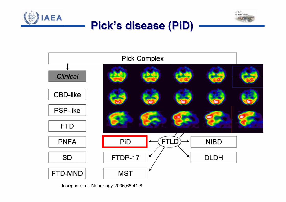
Josephs et al. Neurology 2006;66:41-8
PickPick’’s disease (PiD)s disease (PiD)
Josephs et al. Neurology 2006;66:41-8
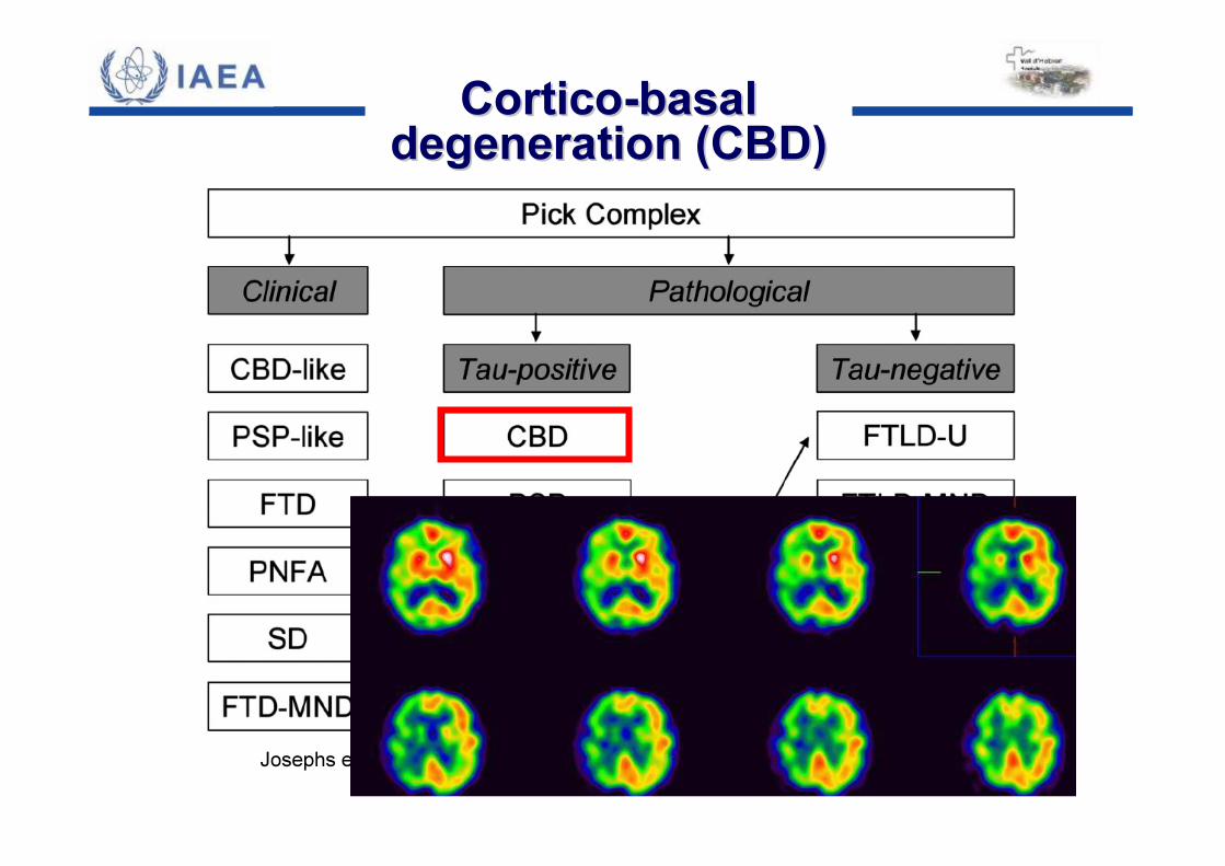
CorticoCortico--basal basal degeneration (CBD)degeneration (CBD)
Josephs et al. Neurology 2006;66:41-8
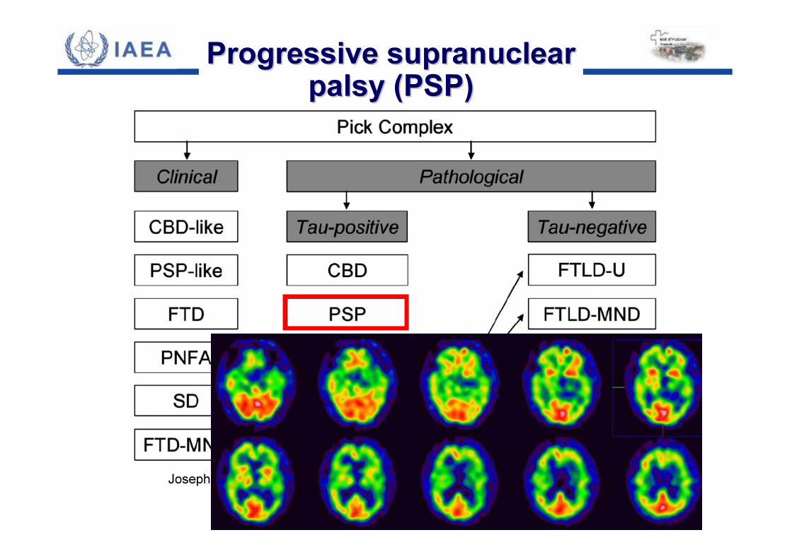
Progressive supranuclear Progressive supranuclear palsy (PSP)palsy (PSP)
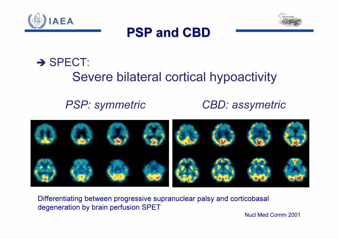
� SPECT:Severe bilateral cortical hypoactivity
PSP: symmetric CBD: assymetric
Differentiating between progressive supranuclear palsy and corticobasaldegeneration by brain perfusion SPET
Nucl Med Comm 2001
PSP and CBDPSP and CBD
Josephs et al. Neurology 2006;66:41-8
FrontoFronto--temporal temporal degeneration (FTD)degeneration (FTD)
D
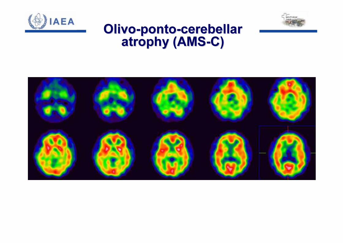
OlivoOlivo--pontoponto--cerebellar cerebellar atrophy (AMSatrophy (AMS--C)C)
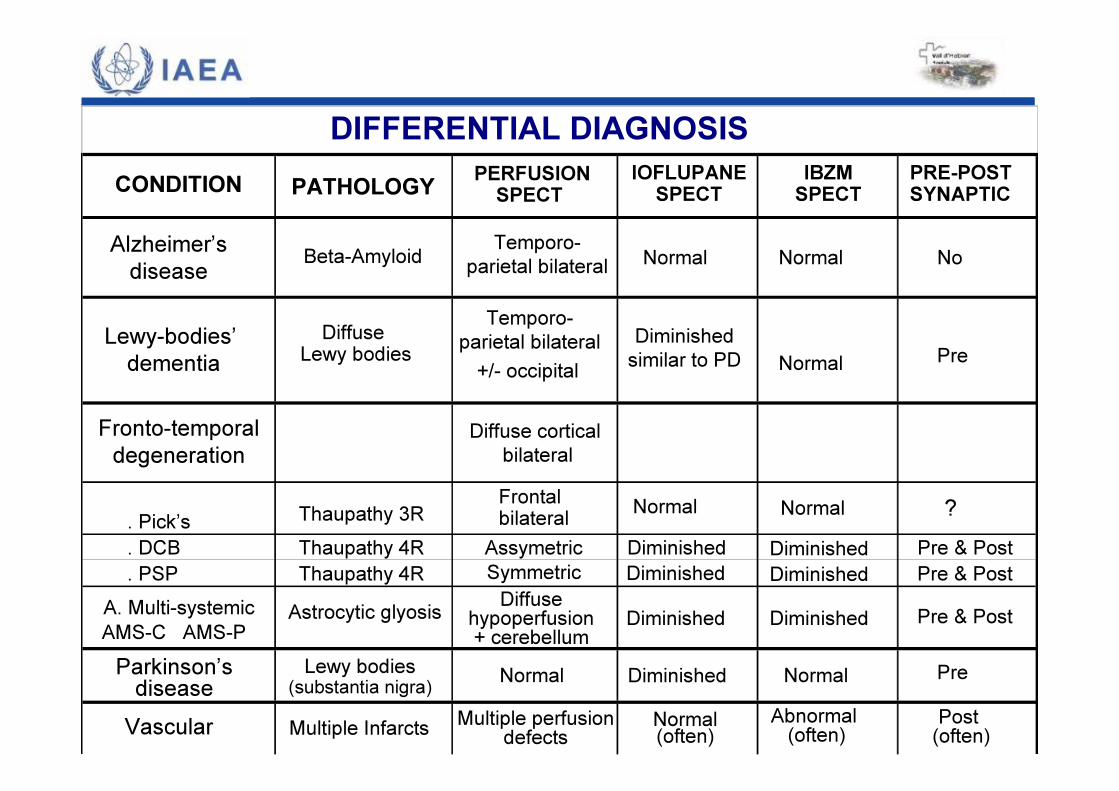
PATHOLOGY PERFUSIONSPECT
IOFLUPANESPECT
IBZMSPECT
PRE-POSTSYNAPTIC
Alzheimer’sdisease Beta-Amyloid Temporo-
parietal bilateral Normal Normal No
Lewy-bodies’dementia
Diffuse Lewy bodies +/- occipital
Diminishedsimilar to PD Normal Pre
Fronto-temporaldegeneration
Diffuse corticalbilateral
. Pick’s Thaupathy 3R Frontal bilateral Normal Normal ?
. DCB Thaupathy 4R Assymetric Diminished Pre & Post
. PSP Thaupathy 4R Symmetric Pre & PostA. Multi-systemicAMS-C AMS-P
Astrocytic glyosis Diffuse hypoperfusion+ cerebellum
Pre & Post
Parkinson’sdiseaseLewy bodies
(substantia nigra) Normal Normal Pre
Vascular Multiple Infarcts Multiple perfusiondefects Normal(often)
Abnormal (often) Post
(often)
DIFFERENTIAL DIAGNOSIS
DiminishedDiminishedDiminished
DiminishedDiminished
Diminished
CONDITION
Temporo-parietal bilateral

The End
SPECT and PET in SPECT and PET in Neurodegenerative DiseasesNeurodegenerative Diseases