somatic and special senses. receptors and sensations types of receptors chemoreceptors –...
TRANSCRIPT

Somatic and Somatic and Special SensesSpecial Senses

Receptors and Receptors and SensationsSensations Types of ReceptorsTypes of Receptors ChemoreceptorsChemoreceptors – Stimulated by – Stimulated by
changes in the chemical concentration changes in the chemical concentration of substancesof substances
Pain ReceptorsPain Receptors – stimulated by tissue – stimulated by tissue damagedamage
ThermoreceptorsThermoreceptors – stimulated by – stimulated by changes in temperaturechanges in temperature
MechanoreceptorsMechanoreceptors – stimulated by – stimulated by changes in pressure or movementchanges in pressure or movement
PhotoreceptorsPhotoreceptors – stimulated by light – stimulated by light energyenergy

Sensations (perceptions)Sensations (perceptions)
A sensation is a feeling that occurs when the A sensation is a feeling that occurs when the brain interprets sensory impulses.brain interprets sensory impulses.
Sensations depend on which region of the brain Sensations depend on which region of the brain receives an impulse.receives an impulse.
At the same time a sensation forms, the cerebral At the same time a sensation forms, the cerebral cortex causes the feeling to seem to come cortex causes the feeling to seem to come from the stimulated receptors: this process is from the stimulated receptors: this process is called called projectionprojection because the brain projects because the brain projects the sensation back to its apparent source.the sensation back to its apparent source.
Projection allows a person to pinpoint the region Projection allows a person to pinpoint the region of stimulation, thus, the eyes seem to see of stimulation, thus, the eyes seem to see and the ears seem to hear.and the ears seem to hear.
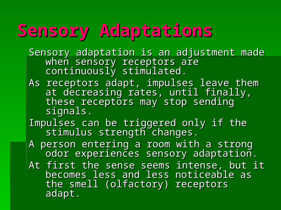
Sensory AdaptationsSensory Adaptations Sensory adaptation is an adjustment made when Sensory adaptation is an adjustment made when
sensory receptors are continuously sensory receptors are continuously stimulated.stimulated.
As receptors adapt, impulses leave them at As receptors adapt, impulses leave them at decreasing rates, until finally, these receptors decreasing rates, until finally, these receptors may stop sending signals.may stop sending signals.
Impulses can be triggered only if the stimulus Impulses can be triggered only if the stimulus strength changes. strength changes.
A person entering a room with a strong odor A person entering a room with a strong odor experiences sensory adaptation.experiences sensory adaptation.
At first the sense seems intense, but it becomes At first the sense seems intense, but it becomes less and less noticeable as the smell less and less noticeable as the smell (olfactory) receptors adapt.(olfactory) receptors adapt.

Somatic SensesSomatic Senses

Touch and Pressure Touch and Pressure SensesSenses
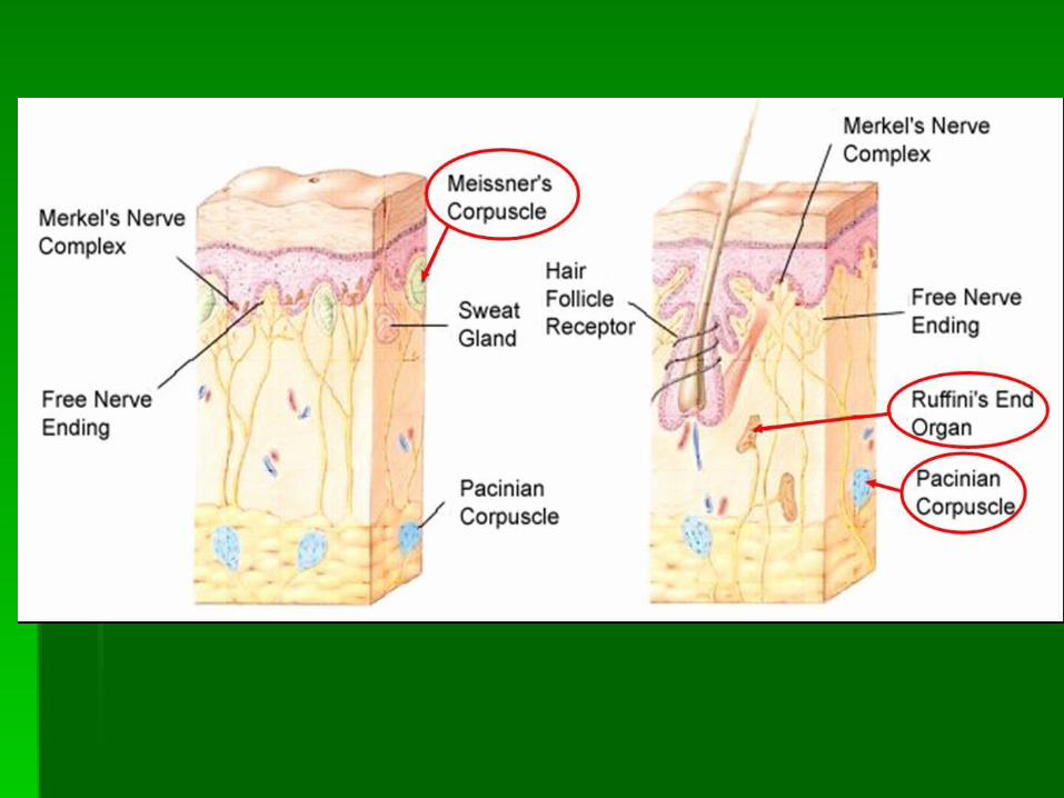
Sensory nerve fibersSensory nerve fibers – common in – common in epithelial tissues epithelial tissues where their free where their free ends are between ends are between epithelial cells; epithelial cells; associated with the associated with the sensation of touch sensation of touch and pressureand pressure

Touch and Pressure Touch and Pressure SensesSenses
Meissner’s corpusclesMeissner’s corpuscles – small, oval masses of flattened – small, oval masses of flattened connective tissue cells within connective tissue connective tissue cells within connective tissue sheathssheaths
Two or more sensory nerve fibers branch into each Two or more sensory nerve fibers branch into each corpuscle and end within it as tiny knobs.corpuscle and end within it as tiny knobs.
Abundant in the hairless portions of the skin, such as the Abundant in the hairless portions of the skin, such as the lips, fingertips, palms, soles, nipples and external lips, fingertips, palms, soles, nipples and external genital organs.genital organs.
Respond to the motion of objects that barely contact the Respond to the motion of objects that barely contact the skin, interpreting impulses from them as sensation of skin, interpreting impulses from them as sensation of light touch.light touch.
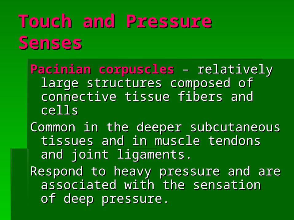
Touch and Pressure Touch and Pressure SensesSenses
Pacinian corpusclesPacinian corpuscles – relatively large – relatively large structures composed of connective tissue structures composed of connective tissue fibers and cellsfibers and cells
Common in the deeper subcutaneous Common in the deeper subcutaneous tissues and in muscle tendons and joint tissues and in muscle tendons and joint ligaments.ligaments.
Respond to heavy pressure and are Respond to heavy pressure and are associated with the sensation of deep associated with the sensation of deep pressure.pressure.

Touch and Pressure Touch and Pressure SensesSenses
Ruffini’s corpuscle Ruffini’s corpuscle – deeply located in the dermis – deeply located in the dermis and are variant’s of Meissner’s corpuscles with a and are variant’s of Meissner’s corpuscles with a more flattened capsule.more flattened capsule.
Mediate sensations of crude and persistent touch.Mediate sensations of crude and persistent touch.
Slow adapting and permit the fingers to remain Slow adapting and permit the fingers to remain sensitive to deep pressure for long periods. (ex. sensitive to deep pressure for long periods. (ex. Grasping a steering wheel for a long time.Grasping a steering wheel for a long time.
Temperature SensesTemperature Senses
Heat receptorsHeat receptors – most sensitive to – most sensitive to temperatures above 77temperatures above 77ooF and become F and become unresponsive at temperatures above unresponsive at temperatures above 113113ooF (temperatures near or above this F (temperatures near or above this stimulate pain receptors, producing a stimulate pain receptors, producing a burning sensation)burning sensation)

Temperature SensesTemperature Senses
Cold receptorsCold receptors – most sensitive to – most sensitive to temperatures between 50temperatures between 50ooF and 68oF F and 68oF ( temperatures below 50( temperatures below 50ooF stimulate F stimulate pain recpetors, producing a freezing pain recpetors, producing a freezing sensation)sensation)
Temperature SensesTemperature Senses
Both heat and cold receptors rapidly adapt. Both heat and cold receptors rapidly adapt. Within about a minute of continuous Within about a minute of continuous stimulation, the sensation of heat or cold stimulation, the sensation of heat or cold begins to fade.begins to fade.

Sense of PainSense of Pain Sensing pain consists of free nerve endings that are Sensing pain consists of free nerve endings that are
widely distributed throughout the skin and internal widely distributed throughout the skin and internal tissues, except in the nervous tissue of the brain, tissues, except in the nervous tissue of the brain, which lacks pain receptors.which lacks pain receptors.
Functions as protection.Functions as protection.Adapts poorly, if at all.Adapts poorly, if at all.Once a pain receptor is activated, it may send impulses Once a pain receptor is activated, it may send impulses
into the central nervous system for some time.into the central nervous system for some time.It is believed that injuries promote release of certain It is believed that injuries promote release of certain
chemicals that build up and stimulate pain receptors.chemicals that build up and stimulate pain receptors.Deficiency of oxygen-rich blood (ischemia) in a tissue or Deficiency of oxygen-rich blood (ischemia) in a tissue or
stimulation of certain mechanoreceptors also trigger stimulation of certain mechanoreceptors also trigger pain sensationpain sensation

Visceral painVisceral pain
Pain receptors are the only receptors in Pain receptors are the only receptors in viscera whose stimulation produces viscera whose stimulation produces sensations.sensations.
Pain may feel as if it is coming from some Pain may feel as if it is coming from some part of the body other than the part being part of the body other than the part being stimulated (stimulated (referred pain).referred pain).
Referred pain arises from common nerve Referred pain arises from common nerve pathways that carry sensory impulses pathways that carry sensory impulses from skin areas as well as viscera. from skin areas as well as viscera.

Pain nerve fibersPain nerve fibers
Acute pain fibersAcute pain fibers
1.1. relatively thin, myelinated nerve fibersrelatively thin, myelinated nerve fibers
2.2. conduct impulses rapidly and are associated conduct impulses rapidly and are associated with the sensation of sharp pain, which with the sensation of sharp pain, which typically originates from a restricted area of typically originates from a restricted area of the skin and seldom continues after the pain-the skin and seldom continues after the pain-producing stimulus stopsproducing stimulus stops
3.3. usually sensed as coming only from the skin usually sensed as coming only from the skin

Pain nerve fibersPain nerve fibers
Chronic pain fibersChronic pain fibers
1.1. thin, unmyelinated nerve fibersthin, unmyelinated nerve fibers
2.2. conduct impulses more slowly and produce a conduct impulses more slowly and produce a dull, aching sensation that may be diffuse dull, aching sensation that may be diffuse and difficult to pinpointand difficult to pinpoint
3.3. pain may continue for some time after the pain may continue for some time after the original stimulus ceasesoriginal stimulus ceases
4.4. felt from the skin as well as in deeper tissuesfelt from the skin as well as in deeper tissues
Pain nerve fibersPain nerve fibersAn event that stimulates pain receptors An event that stimulates pain receptors
usually triggers impulses on both acute and usually triggers impulses on both acute and chronic fibers – causes a dull sensation. chronic fibers – causes a dull sensation.
Pain impulses that originate from the head Pain impulses that originate from the head reach the brain on sensory fibers of cranial reach the brain on sensory fibers of cranial nerves. nerves.
All other pain impulses travel on the sensory All other pain impulses travel on the sensory fibers of spinal nerves, and they pass into fibers of spinal nerves, and they pass into the spinal cord by way of the dorsal roots of the spinal cord by way of the dorsal roots of these spinal nerves. these spinal nerves.
Within the spinal cord, neurons process pain Within the spinal cord, neurons process pain impulses in the gray matter of the dorsal impulses in the gray matter of the dorsal horn, and the impulses are transmitted to horn, and the impulses are transmitted to the brain.the brain.

Regulation of Pain Regulation of Pain ImpulsesImpulses
Awareness of pain arises when impulses Awareness of pain arises when impulses reach the thalamus. The cerebral cortex reach the thalamus. The cerebral cortex determines pain intensity, locates the determines pain intensity, locates the pain source, and mediates emotional pain source, and mediates emotional and motor responses to the pain.and motor responses to the pain.
Regulation of Pain Regulation of Pain ImpulsesImpulses
1. Areas of gray matter in the midbrain, pons, 1. Areas of gray matter in the midbrain, pons, and medulla regulate movement of pain and medulla regulate movement of pain impulses from the spinal cord.impulses from the spinal cord.
2. Impulses from special neurons in these brain 2. Impulses from special neurons in these brain areas descend in the lateral funiculus to areas descend in the lateral funiculus to various levels of the spinal cord.various levels of the spinal cord.
3. These impulses stimulate ends of certain 3. These impulses stimulate ends of certain nerve fibers to release biochemicals that can nerve fibers to release biochemicals that can block pain signals by inhibiting presynaptic block pain signals by inhibiting presynaptic nerve fibers in the posterior horn of the spinal nerve fibers in the posterior horn of the spinal cord.cord.

Regulation of Pain Regulation of Pain ImpulsesImpulses
Inhibiting substances released in the posterior Inhibiting substances released in the posterior horn include neuropeptides called horn include neuropeptides called enkephalinsenkephalins (have morphine-like actions) and the (have morphine-like actions) and the monamine monamine serotoninserotonin (stimulates other neurons (stimulates other neurons to release enkephalins). to release enkephalins).
EndorphinsEndorphins are another group of neuropeptides are another group of neuropeptides with pain-supressing, morphine-like actions. with pain-supressing, morphine-like actions. Endorphins are in the pituitary gland and the Endorphins are in the pituitary gland and the hypothalamus. hypothalamus.
Both provide natural control.Both provide natural control.

Special SensesSpecial Senses

Sense of SmellSense of Smell
Sense of smell is associated with complex Sense of smell is associated with complex sensory structures in the upper region sensory structures in the upper region of the nasal cavity.of the nasal cavity.
Olfactory receptorsOlfactory receptors
Olfactory receptors are Olfactory receptors are chemoreceptorschemoreceptors, , which means that chemicals dissolved which means that chemicals dissolved in liquids stimulate them.in liquids stimulate them.
Receptors function closely with taste to aid Receptors function closely with taste to aid in food selectionin food selection

Olfactory OrgansOlfactory Organs
yellowish brown yellowish brown masses that cover masses that cover the upper parts of the the upper parts of the nasal cavity, the nasal cavity, the superior nasal superior nasal conchae, and a conchae, and a portion of the nasal portion of the nasal septum septum

Olfactory OrgansOlfactory Organscontains contains olfactory receptorsolfactory receptors
that are neurons that are neurons surrounded by columnar surrounded by columnar epithelial cells; hair-like cilia epithelial cells; hair-like cilia cover tiny knobs at the cover tiny knobs at the distal ends of these distal ends of these neuron’s dendrites; the cilia neuron’s dendrites; the cilia project into the nasal cavity project into the nasal cavity and are the sensitive parts and are the sensitive parts of the receptors; chemicals of the receptors; chemicals enter the nasal cavity as enter the nasal cavity as gases, but they must gases, but they must dissolve at least partially in dissolve at least partially in the watery fluids that the watery fluids that surround the cilia before surround the cilia before receptors can detect themreceptors can detect them

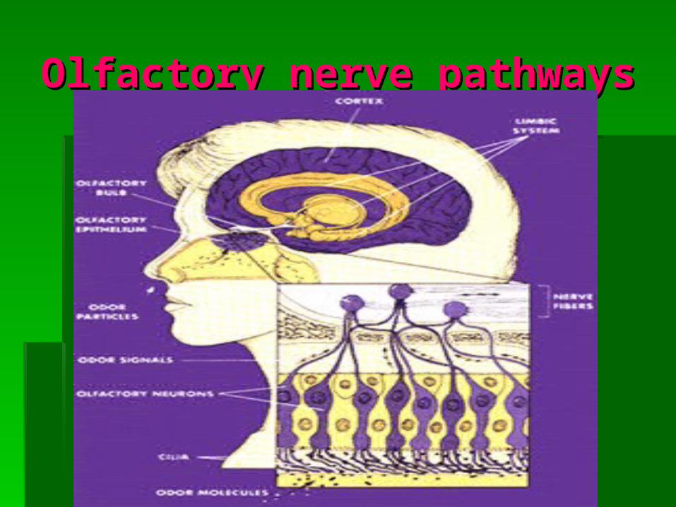
Olfactory nerve pathwaysOlfactory nerve pathways
Receptors send nerve impulses along axons of Receptors send nerve impulses along axons of the receptor cells to neurons located in the receptor cells to neurons located in enlargements called enlargements called olfactory bulbsolfactory bulbs, which lie , which lie on either side of the crista galli of the ethmoid on either side of the crista galli of the ethmoid bone, where they are analyzed.bone, where they are analyzed.
As a result, impulses travel along As a result, impulses travel along olfactory olfactory tractstracts to the limbic system. to the limbic system.
The major interpreting areas (olfactory cortex) for The major interpreting areas (olfactory cortex) for these impulses are located within the temporal these impulses are located within the temporal lobes and the base of the frontal lobes, anterior lobes and the base of the frontal lobes, anterior to the hypothalamus to the hypothalamus
Olfactory nerve pathwaysOlfactory nerve pathways

Sense of TasteSense of Taste
Taste BudsTaste Buds – – special organs of taste; special organs of taste; occur primarily on the surface of the occur primarily on the surface of the tongue and are associated with tiny tongue and are associated with tiny elevations called elevations called papillaepapillae; also found in ; also found in smaller numbers in the roof of the smaller numbers in the roof of the mouth and walls of the pharynxmouth and walls of the pharynx
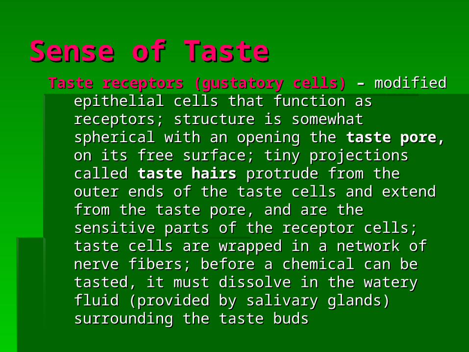
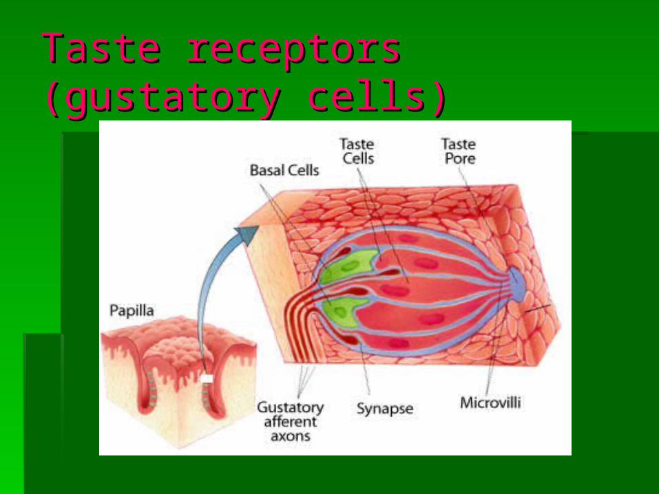
Sense of TasteSense of TasteTaste receptors (gustatory cells)Taste receptors (gustatory cells) – – modified modified
epithelial cells that function as receptors; epithelial cells that function as receptors; structure is somewhat spherical with an structure is somewhat spherical with an opening the opening the taste pore, taste pore, on its free surface; on its free surface; tiny projections called tiny projections called taste hairstaste hairs protrude protrude from the outer ends of the taste cells and from the outer ends of the taste cells and extend from the taste pore, and are the extend from the taste pore, and are the sensitive parts of the receptor cells; taste sensitive parts of the receptor cells; taste cells are wrapped in a network of nerve cells are wrapped in a network of nerve fibers; before a chemical can be tasted, it fibers; before a chemical can be tasted, it must dissolve in the watery fluid (provided by must dissolve in the watery fluid (provided by salivary glands) surrounding the taste budssalivary glands) surrounding the taste buds
Taste receptors (gustatory Taste receptors (gustatory cells)cells)

Taste sensations:Taste sensations:
1. Sweet – 1. Sweet – front front of tongueof tongue
2. Sour – 2. Sour – sides sides of tongueof tongue
3. Salty – 3. Salty – all the all the way around way around the rim of the the rim of the tonguetongue
4. Bitter – 4. Bitter – back back of tongueof tongue

Taste sensations:Taste sensations:
Flavor results from one of the primary Flavor results from one of the primary sensations or from combination of sensations or from combination of two or more. Experiencing flavors two or more. Experiencing flavors involves taste (concentration of involves taste (concentration of stimulating chemicals), as well as the stimulating chemicals), as well as the sensations of odor, texture (touch), sensations of odor, texture (touch), and temperature. Chemicals in some and temperature. Chemicals in some foods may stimulate pain receptors. foods may stimulate pain receptors. Taste receptors undergo sensory Taste receptors undergo sensory adaptation relatively rapidly.adaptation relatively rapidly.
Taste nerve pathwaysTaste nerve pathways
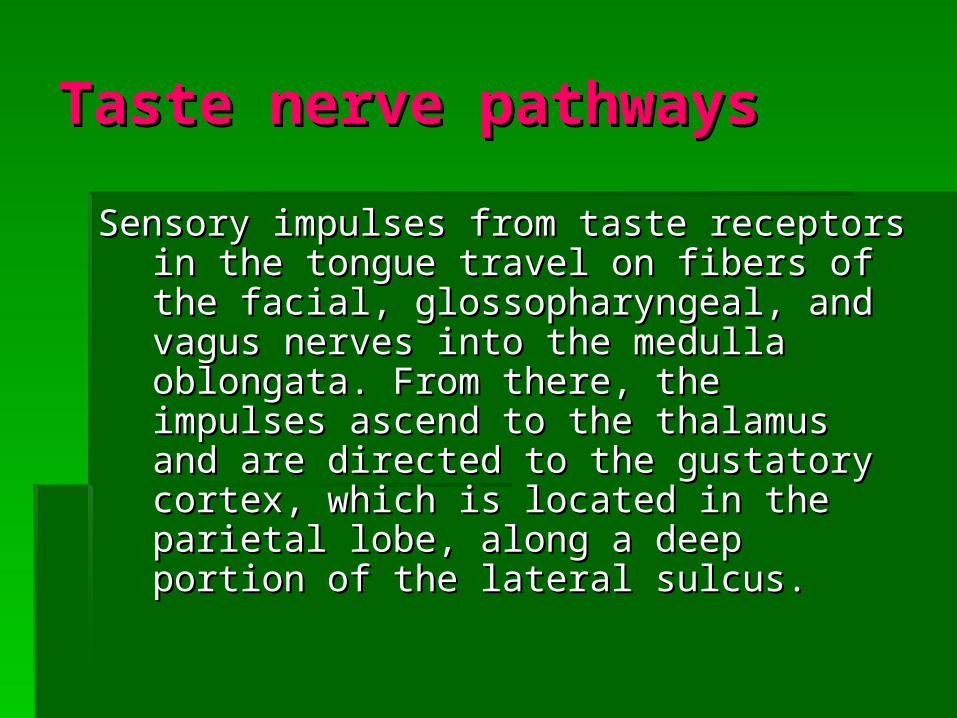
Sensory impulses from taste receptors in Sensory impulses from taste receptors in the tongue travel on fibers of the facial, the tongue travel on fibers of the facial, glossopharyngeal, and vagus nerves glossopharyngeal, and vagus nerves into the medulla oblongata. From there, into the medulla oblongata. From there, the impulses ascend to the thalamus the impulses ascend to the thalamus and are directed to the gustatory cortex, and are directed to the gustatory cortex, which is located in the parietal lobe, which is located in the parietal lobe, along a deep portion of the lateral along a deep portion of the lateral sulcus.sulcus.


Sense of HearingSense of Hearing
The ear is the organ of The ear is the organ of hearing, as well as hearing, as well as functioning in the functioning in the sense of sense of equilibrium.equilibrium.
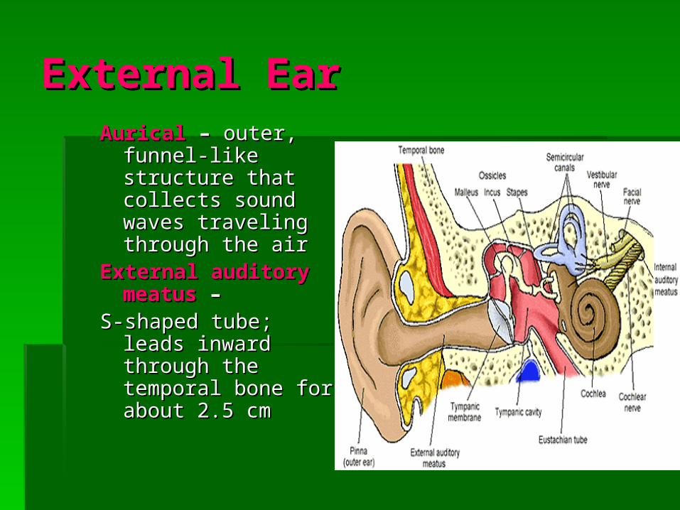
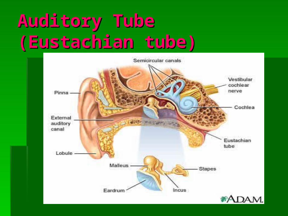
External EarExternal EarAuricalAurical – – outer, outer,
funnel-like structure funnel-like structure that collects sound that collects sound waves traveling waves traveling through the airthrough the air
External auditory External auditory meatusmeatus – –
S-shaped tube; leads S-shaped tube; leads inward through the inward through the temporal bone for temporal bone for about 2.5 cmabout 2.5 cm

Middle EarMiddle Ear
tympanic cavity – tympanic cavity – air filled space in air filled space in the temporal the temporal bonebone
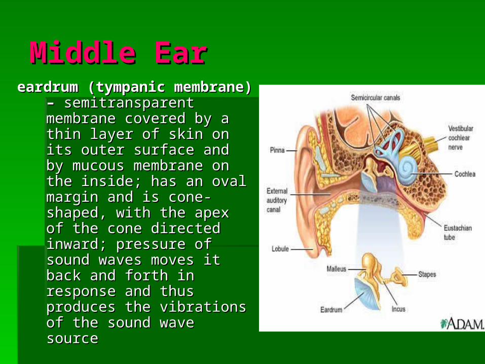
Middle EarMiddle Eareardrum (tympanic eardrum (tympanic
membrane) – membrane) – semitransparent membrane semitransparent membrane covered by a thin layer of covered by a thin layer of skin on its outer surface and skin on its outer surface and by mucous membrane on by mucous membrane on the inside; has an oval the inside; has an oval margin and is cone-shaped, margin and is cone-shaped, with the apex of the cone with the apex of the cone directed inward; pressure of directed inward; pressure of sound waves moves it back sound waves moves it back and forth in response and and forth in response and thus produces the vibrations thus produces the vibrations of the sound wave sourceof the sound wave source

Auditory ossiclesAuditory ossicles
3 bones attached by tiny ligaments to the 3 bones attached by tiny ligaments to the wall of the tympanic cavity, and they are wall of the tympanic cavity, and they are covered by mucous membranes; bridge covered by mucous membranes; bridge the eardrum and the inner ear, the eardrum and the inner ear, transmitting vibrations between these transmitting vibrations between these partsparts

Auditory ossiclesAuditory ossicles
malleusmalleus (hammer)– (hammer)– attaches to the attaches to the eardrum; when eardrum; when the eardrum the eardrum vibrates, the vibrates, the malleus vibrates malleus vibrates in unison and in unison and causes the incus causes the incus to vibrateto vibrate

Auditory ossiclesAuditory ossicles
incusincus (anvil)– (anvil)– receives receives vibrations from vibrations from the malleus and the malleus and transmits them to transmits them to the stapesthe stapes
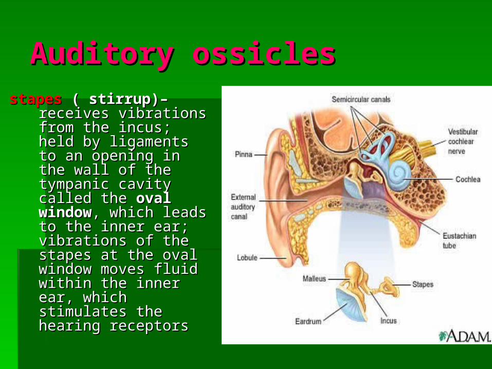
Auditory ossiclesAuditory ossiclesstapesstapes ( stirrup)– ( stirrup)– receives receives
vibrations from the vibrations from the incus; held by incus; held by ligaments to an ligaments to an opening in the wall of opening in the wall of the tympanic cavity the tympanic cavity called the called the oval oval windowwindow, which leads , which leads to the inner ear; to the inner ear; vibrations of the vibrations of the stapes at the oval stapes at the oval window moves fluid window moves fluid within the inner ear, within the inner ear, which stimulates the which stimulates the hearing receptorshearing receptors
Auditory ossiclesAuditory ossicles
The auditory ossicles also help to The auditory ossicles also help to increase (amplify) the force of increase (amplify) the force of vibrations as they pass from the vibrations as they pass from the eardrum to the oval window.eardrum to the oval window.

Auditory Tube Auditory Tube (Eustachian tube)(Eustachian tube)
connects each middle ear to the throat; connects each middle ear to the throat; conducts air between the tympanic conducts air between the tympanic cavity and the outside of the body by cavity and the outside of the body by way of the throat (nasopharynx) and way of the throat (nasopharynx) and mouth; helps maintain equal air mouth; helps maintain equal air pressure on both sides of the eardrum, pressure on both sides of the eardrum, which is necessary for normal hearing; which is necessary for normal hearing; this function is noticeable during rapid this function is noticeable during rapid changes in altitudechanges in altitude
Auditory Tube Auditory Tube (Eustachian tube)(Eustachian tube)
Inner EarInner Ear
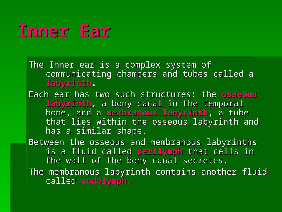
The Inner ear is a complex system of communicating The Inner ear is a complex system of communicating chambers and tubes called achambers and tubes called a labyrinthlabyrinth..
Each ear has two such structures: the Each ear has two such structures: the osseous osseous labyrinthlabyrinth, a bony canal in the temporal bone, and a , a bony canal in the temporal bone, and a membranous labyrinthmembranous labyrinth, a tube that lies within the , a tube that lies within the osseous labyrinth and has a similar shape.osseous labyrinth and has a similar shape.
Between the osseous and membranous labyrinths is a Between the osseous and membranous labyrinths is a fluid called fluid called perilymphperilymph that cells in the wall of the that cells in the wall of the bony canal secretes.bony canal secretes.
The membranous labyrinth contains another fluid called The membranous labyrinth contains another fluid called endolymph.endolymph.

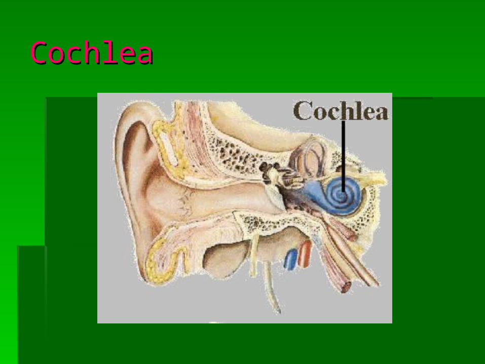
Inner EarInner EarCochleaCochlea – – functions in hearing functions in hearing
* contains a bony core and a thin, bony shelf that * contains a bony core and a thin, bony shelf that winds around the core like the thread of a screwwinds around the core like the thread of a screw
* the shelf divides the osseous labyrinth of the * the shelf divides the osseous labyrinth of the cochlea into two compartments, the upper cochlea into two compartments, the upper ((scala vestibule)scala vestibule) leads from the oval window to leads from the oval window to the apex of the spiral and the lower the apex of the spiral and the lower (scala (scala tympanitympani) extends from the apex of the cochlea ) extends from the apex of the cochlea to a membrane-covered opening in the wall of to a membrane-covered opening in the wall of the inner ear called the the inner ear called the round windowround window

CochleaCochlea
The portion of the membranous labyrinth within The portion of the membranous labyrinth within the cochlea is called the the cochlea is called the cochlear ductcochlear duct and and lies between the two bony compartments and lies between the two bony compartments and ends as a closed sac at the apex of the ends as a closed sac at the apex of the cochleacochlea
It is separated from the scala vestibule by a It is separated from the scala vestibule by a vestibular membrane vestibular membrane (Reissner’s membrane) (Reissner’s membrane) and from the scala tympani by a basilar and from the scala tympani by a basilar membrane which contains many thousands of membrane which contains many thousands of stiff, elastic fibers, whose lengths progressively stiff, elastic fibers, whose lengths progressively increase from the base of the cochlea to its increase from the base of the cochlea to its apexapex
CochleaCochlea

organ of Cortiorgan of Corti
contains hearing receptors; located on the upper contains hearing receptors; located on the upper surface of the basilar membrane and surface of the basilar membrane and stretches from the apex to the base of the stretches from the apex to the base of the cochlea; its receptor cells (hair cells) are cochlea; its receptor cells (hair cells) are organized in rows and have many hair-like organized in rows and have many hair-like processes that project into the endolymph of processes that project into the endolymph of the cochlear duct; above these hair cells is a the cochlear duct; above these hair cells is a tectorial emebranetectorial emebrane attached to the bony attached to the bony shelf of the cochlea, passing over the shelf of the cochlea, passing over the receptor cells and contracting the tips of the receptor cells and contracting the tips of the hairshairs

CochleaCochlea
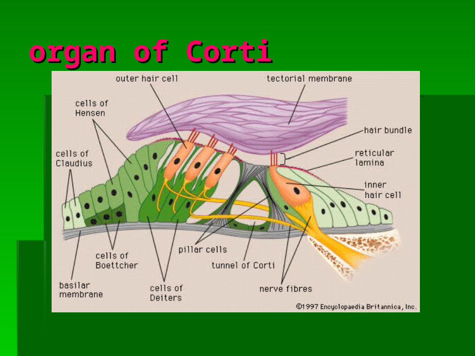
organ of Cortiorgan of Corti

Steps in the Generation of Steps in the Generation of Sensory Impulses from Sensory Impulses from the Earthe Ear
1. Sound waves enter external auditory meatus.1. Sound waves enter external auditory meatus.
2. Waves of changing pressure cause eardrum to 2. Waves of changing pressure cause eardrum to reproduce vibrations coming from sound wave reproduce vibrations coming from sound wave source.source.
3. Auditory ossicles amplify and transmit vibrations 3. Auditory ossicles amplify and transmit vibrations to end of stapes.to end of stapes.
4. Movement of stapes at oval window transmits 4. Movement of stapes at oval window transmits vibrations to perilymph in scala vestibule.vibrations to perilymph in scala vestibule.
5. Vibrations pass through vestibular membrane 5. Vibrations pass through vestibular membrane and enter endolymph of cochlear duct.and enter endolymph of cochlear duct.

Steps in the Generation of Steps in the Generation of Sensory Impulses from the Sensory Impulses from the EarEar
6. Different frequencies of vibration in endolymph 6. Different frequencies of vibration in endolymph stimulate different sets of receptor cells.stimulate different sets of receptor cells.
7. As a receptor cell depolarizes, its membrane 7. As a receptor cell depolarizes, its membrane becomes more permeable to calcium ions.becomes more permeable to calcium ions.
8. Inward diffusion of calcium ions causes vesicles at 8. Inward diffusion of calcium ions causes vesicles at the base of the receptor cell to release the base of the receptor cell to release neurotransmitter.neurotransmitter.
9. Neurotransmitter stimulates ends of nearby 9. Neurotransmitter stimulates ends of nearby sensory neurons.sensory neurons.
10. Sensory impulses are triggered on fibers of 10. Sensory impulses are triggered on fibers of cochlear branch of vestibulocochlear nerve.cochlear branch of vestibulocochlear nerve.
11. Auditory cortex of temporal lobe interprets 11. Auditory cortex of temporal lobe interprets sensory impulses.sensory impulses.
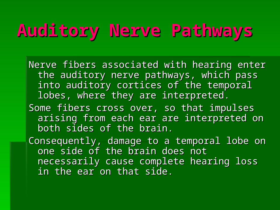
Auditory Nerve PathwaysAuditory Nerve Pathways
Nerve fibers associated with hearing enter the Nerve fibers associated with hearing enter the auditory nerve pathways, which pass into auditory nerve pathways, which pass into auditory cortices of the temporal lobes, where auditory cortices of the temporal lobes, where they are interpreted. they are interpreted.
Some fibers cross over, so that impulses arising Some fibers cross over, so that impulses arising from each ear are interpreted on both sides of from each ear are interpreted on both sides of the brain. the brain.
Consequently, damage to a temporal lobe on one Consequently, damage to a temporal lobe on one side of the brain does not necessarily cause side of the brain does not necessarily cause complete hearing loss in the ear on that side. complete hearing loss in the ear on that side.

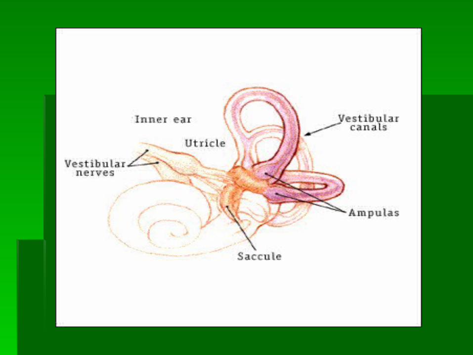
Sense of EquilibriumSense of Equilibrium

Inner EarInner Ear
semicircular canals semicircular canals - - provide a sense provide a sense of equilibriumof equilibrium

Static EqulibriumStatic Equlibrium
Senses the position of the head, maintaining stability Senses the position of the head, maintaining stability and posture when the head and body are still.and posture when the head and body are still.
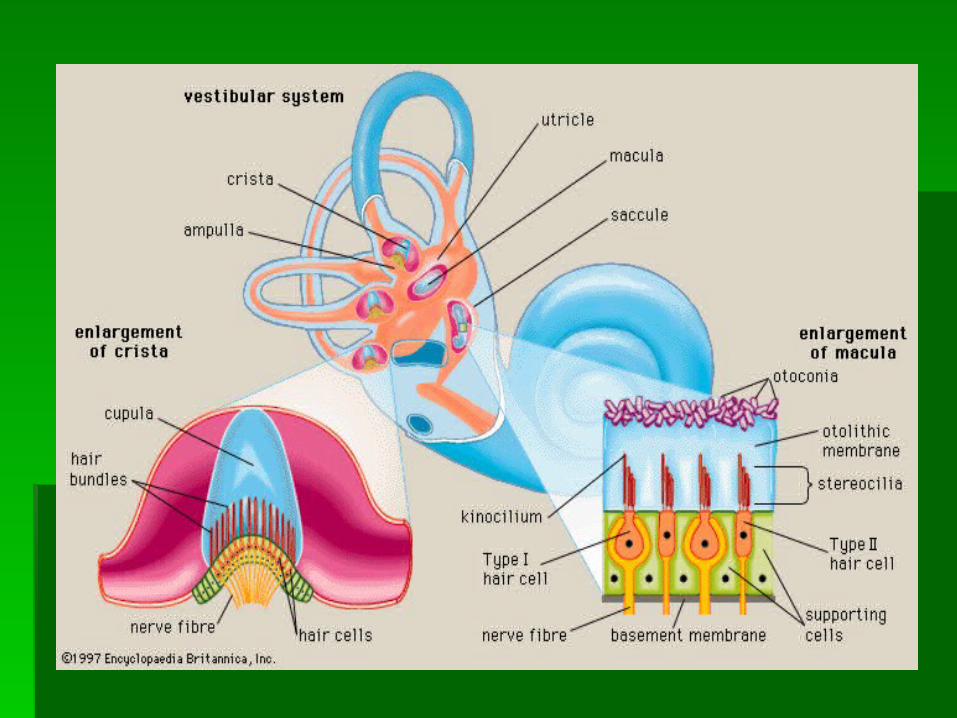
The organs for static equilibrium are located within The organs for static equilibrium are located within the the vestibulevestibule,, a bony chamber between the a bony chamber between the semicircular canals and cochlea.semicircular canals and cochlea.
The membranous labyrinth inside the vestibule The membranous labyrinth inside the vestibule consists of 2 expanded chambers: a consists of 2 expanded chambers: a utricle utricle and a and a sacculesaccule. . Each of these chambers has a tiny Each of these chambers has a tiny structure called a structure called a maculamacula on its anterior wall. on its anterior wall.
Macula contain numerous hair cells, which serve as Macula contain numerous hair cells, which serve as sensory receptors.sensory receptors.

Static EqulibriumStatic Equlibrium
The head bending forward, backward, or to the side The head bending forward, backward, or to the side stimulates hair cells. The movement tilts the gelatinous stimulates hair cells. The movement tilts the gelatinous masses of the macula bending the hair cells, which masses of the macula bending the hair cells, which signal the nerve fibers associated with them.signal the nerve fibers associated with them.
The resulting impulses travel into the CNS on the The resulting impulses travel into the CNS on the vestibular branch of the vestibulocochlear nerve.vestibular branch of the vestibulocochlear nerve.
These impulses inform the brain of the head’s position.These impulses inform the brain of the head’s position. The brain responds by sensing motor impulses to The brain responds by sensing motor impulses to
skeletal muscles, which contract or relax to maintain skeletal muscles, which contract or relax to maintain balance.balance.

Dynamic EquilibriumDynamic Equilibrium The threeThe three semicircular canalssemicircular canals detect motion of the detect motion of the
head and aid in balancing the head and body during head and aid in balancing the head and body during sudden movement.sudden movement.
The canals lie at right angles to each other and each The canals lie at right angles to each other and each corresponds to a different anatomical plane.corresponds to a different anatomical plane.
Suspended in the perilymph of the bony portion of each Suspended in the perilymph of the bony portion of each canal is a membranous canal that ends in a swelling canal is a membranous canal that ends in a swelling called an called an ampullaampulla..
The ampulla contains the sensory organs of the The ampulla contains the sensory organs of the semicircular canals, semicircular canals, crista ampullariscrista ampullaris..
Each crista ampullaris contains a number of sensory hair Each crista ampullaris contains a number of sensory hair cells and supporting cells. The hair cells extend upward cells and supporting cells. The hair cells extend upward into a dome-shaped gelatinous mass called the into a dome-shaped gelatinous mass called the capulacapula..


Dynamic EquilibriumDynamic Equilibrium
Rapid turns of the head or body stimulate the hair cells Rapid turns of the head or body stimulate the hair cells of the crista ampularis.of the crista ampularis.
The semicircular canals move with the head or body, but The semicircular canals move with the head or body, but the fluid inside the membranous canals remains the fluid inside the membranous canals remains stationary. This action bends the capula in one or more stationary. This action bends the capula in one or more of the canals in a direction opposite that of the head or of the canals in a direction opposite that of the head or body movement. The hairs in the capula also bend body movement. The hairs in the capula also bend stimulating the hair cells to signal their associated nerve stimulating the hair cells to signal their associated nerve fibers, sensing impulses to the brain.fibers, sensing impulses to the brain.
Parts of the cerebellum are important in interpreting Parts of the cerebellum are important in interpreting impulses from the semicircular canals and helping to impulses from the semicircular canals and helping to maintain balance.maintain balance.

Sense of EquilibriumSense of Equilibrium
Other sensory structures aid in maintaining Other sensory structures aid in maintaining equilibrium. equilibrium.
Certain mechanoreceptors, particularly those Certain mechanoreceptors, particularly those associated with the joints of the neck, inform associated with the joints of the neck, inform the brain about the movements. the brain about the movements.
The eyes detect changes in posture that result The eyes detect changes in posture that result from body movements. from body movements.
Such visual information is so important that even Such visual information is so important that even if the organs of equilibrium are damaged, a if the organs of equilibrium are damaged, a person may be able to maintain normal person may be able to maintain normal balance by keeping the eyes open and balance by keeping the eyes open and moving slowly.moving slowly.

VertigoVertigoVertigo is the feeling that you or your environment Vertigo is the feeling that you or your environment is moving or spinning. It differs from dizziness in is moving or spinning. It differs from dizziness in that vertigo describes an illusion of movement. that vertigo describes an illusion of movement.
When you feel as if you yourself are moving, it's When you feel as if you yourself are moving, it's called called subjective vertigosubjective vertigo, and the perception that , and the perception that your surroundings are moving is called your surroundings are moving is called objective objective vertigovertigo..
Unlike nonspecific lightheadedness or dizziness, Unlike nonspecific lightheadedness or dizziness, vertigo has relatively few causes.vertigo has relatively few causes.

Causes of VertigoCauses of Vertigo
http://www.emedicinehealth.com/vertigo/page2_em.htm

Sense of SightSense of Sight

Visual Accessory OrgansVisual Accessory Organs
Visual accessory organs are housed within Visual accessory organs are housed within the pear-shaped orbital cavity of the the pear-shaped orbital cavity of the skull.skull.
This orbit contains fat, blood vessels, This orbit contains fat, blood vessels, nerves, and connective tissue.nerves, and connective tissue.
EyelidEyelid - has four layers - has four layers
1. skin1. skin – – the thinnest skin of the body; covers the the thinnest skin of the body; covers the lid’s outer surface and fuses with its inner lining lid’s outer surface and fuses with its inner lining near the margin of the lidnear the margin of the lid
2. obicularis oculi muscle2. obicularis oculi muscle – – acts as a sphincter acts as a sphincter and closes the lids when it contractsand closes the lids when it contracts
3. levator palpebrae superioris muscle3. levator palpebrae superioris muscle – – raises raises the upper lids and thus helps open the eyethe upper lids and thus helps open the eye
4. conjunctiva4. conjunctiva – – mucous membrane that lines the mucous membrane that lines the inner surfaces of the eyelids and folds back to inner surfaces of the eyelids and folds back to cover the anterior surface of the eyeball (except cover the anterior surface of the eyeball (except for its central portion [cornea])for its central portion [cornea])
Lacrimal apparatusLacrimal apparatus1. lacrimal gland1. lacrimal gland – – located in the orbit and secretes located in the orbit and secretes
tears continuously; moistens and lubricates the tears continuously; moistens and lubricates the surface of the eye and the lining of the lids; tears exit surface of the eye and the lining of the lids; tears exit through tiny tubules and flow downward and medially through tiny tubules and flow downward and medially across the eye; tears contain an enzyme across the eye; tears contain an enzyme ((lysozymelysozyme)) that is an antibacterial agent, reducing the risk of eye that is an antibacterial agent, reducing the risk of eye infectionsinfections
2. superior and inferior canaliculi2. superior and inferior canaliculi – – two small ducts two small ducts which collect tears that flow into the which collect tears that flow into the lacrimal saclacrimal sac,, which lies deep in a groove of the lacrimal bone, and which lies deep in a groove of the lacrimal bone, and then into the then into the nasolacrimal ductnasolacrimal duct,, which empties into which empties into the nasal cavitythe nasal cavity


Extrinsic MusclesExtrinsic Muscles
Extrinsic MusclesExtrinsic Muscles arise from the bones of arise from the bones of the orbit and insert by broad tendons on the orbit and insert by broad tendons on the eye’s outer surface.the eye’s outer surface.
6 extrinsic muscles move the eye in 6 extrinsic muscles move the eye in various directions:various directions:

Extrinsic MusclesExtrinsic Muscles
1. superior rectus1. superior rectus – – rotates eye upward rotates eye upward and toward midline; oculomotor nerve IIIand toward midline; oculomotor nerve III
2. inferior rectus2. inferior rectus – – rotates eye downward rotates eye downward and toward midline; oculomotor nerve IIIand toward midline; oculomotor nerve III
3. medial rectus3. medial rectus – – rotates eye toward rotates eye toward midline; oculomotor nerve IIImidline; oculomotor nerve III
Extrinsic MusclesExtrinsic Muscles
1. lateral rectus1. lateral rectus – – rotates eye away from rotates eye away from midline; abducens nerve VImidline; abducens nerve VI
2. superior oblique2. superior oblique – – rotates eye downward rotates eye downward and away from midline; trochlear nerve IVand away from midline; trochlear nerve IV
3. inferior oblique3. inferior oblique – – rotates eye upward and rotates eye upward and away from midline; oculomotor nerve IIIaway from midline; oculomotor nerve III


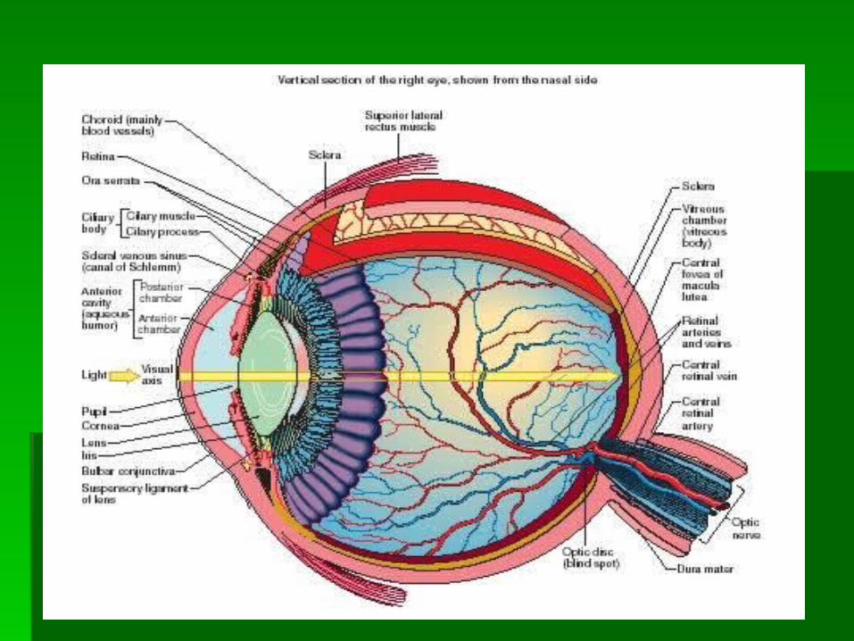
Structure of the EyeStructure of the Eye

Outer Tunic (fibrous Outer Tunic (fibrous tunic)tunic)
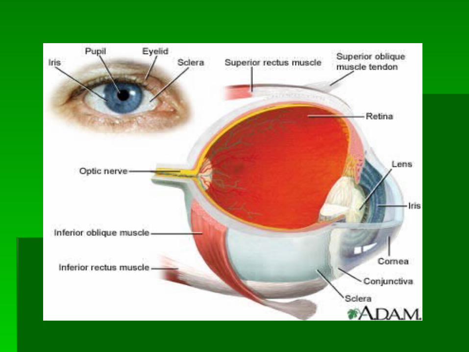
CorneaCornea comprises the anterior sixth of the outer tunic comprises the anterior sixth of the outer tunic
and bulges forwardand bulges forward window of the eye and helps focus entering window of the eye and helps focus entering
light rayslight rays composed largely of connective tissue with a composed largely of connective tissue with a
thin layer of epithelium on its surfacethin layer of epithelium on its surface transparent because it contains few cells and transparent because it contains few cells and
no blood vessels and its cells and no blood vessels and its cells and collagenous fibers form regular patternscollagenous fibers form regular patterns
Outer Tunic (fibrous Outer Tunic (fibrous tunic)tunic)
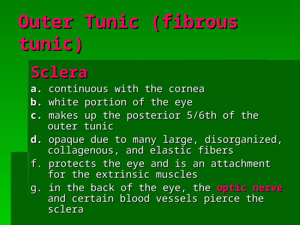
ScleraScleraa. a. continuous with the corneacontinuous with the cornea b.b. white portion of the eye white portion of the eyec.c. makes up the posterior 5/6th of the outer tunic makes up the posterior 5/6th of the outer tunicd.d. opaque due to many large, disorganized, opaque due to many large, disorganized,
collagenous, and elasticcollagenous, and elastic fibersfibersf. protects the eye and is an attachment for the f. protects the eye and is an attachment for the
extrinsic musclesextrinsic musclesg. in the back of the eye, the g. in the back of the eye, the optic nerveoptic nerve and and
certain blood vessels pierce the scleracertain blood vessels pierce the sclera
Middle Tunic Middle Tunic (vascular (vascular tunic)tunic)
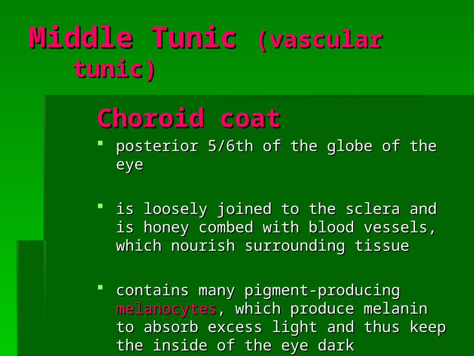
Choroid coatChoroid coat posterior 5/6th of the globe of the eyeposterior 5/6th of the globe of the eye
is loosely joined to the sclera and is honey is loosely joined to the sclera and is honey combed with blood vessels, which nourish combed with blood vessels, which nourish surrounding tissuesurrounding tissue
contains many pigment-producing contains many pigment-producing melanocytesmelanocytes, , which produce melanin to absorb excess light which produce melanin to absorb excess light and thus keep the inside of the eye darkand thus keep the inside of the eye dark

Middle Tunic Middle Tunic (vascular (vascular tunic)tunic)
Ciliary bodyCiliary bodya. a. thickest part of the middle tunicthickest part of the middle tunic
b.b. extends forward from the choroid coat and extends forward from the choroid coat and forms an internal ring around the front of the forms an internal ring around the front of the eyeeye
c. contains many radiating folds calledc. contains many radiating folds called ciliary ciliary processesprocesses and groups of muscle fibers that and groups of muscle fibers that constitute the constitute the ciliary musclesciliary muscles
Ciliary bodyCiliary bodyd. many strong, delicate fibers, called d. many strong, delicate fibers, called
suspensory ligamentssuspensory ligaments, , extend inward extend inward from the ciliary processes and hold the from the ciliary processes and hold the transparent transparent lenslens in position in position
e. the body of the lens lies directly behind e. the body of the lens lies directly behind the iris and pupil and is composed of the iris and pupil and is composed of differentiated epithelial cells called differentiated epithelial cells called lens lens fibersfibers
f. the cytoplasm of the lens fibers is the f. the cytoplasm of the lens fibers is the transparent substance of the lenstransparent substance of the lens
Ciliary bodyCiliary bodyg. g. the lens capsule is a clear, membrane-the lens capsule is a clear, membrane-
like structure composed largely of like structure composed largely of intercellular material whose elastic intercellular material whose elastic nature keeps it under constant tensionnature keeps it under constant tension
h. the suspensory ligaments attached to h. the suspensory ligaments attached to the margin of the capsule are also under the margin of the capsule are also under tension and pull outward, flattening the tension and pull outward, flattening the capsule and the lens insidecapsule and the lens inside
i. when the fibers contract, the choroid coat i. when the fibers contract, the choroid coat is pulled forward and the ciliary body is pulled forward and the ciliary body shortens relaxing the suspensory shortens relaxing the suspensory ligamentsligaments
Ciliary bodyCiliary bodyj. the lens thickens in response and is now j. the lens thickens in response and is now
focused for viewing closer objects than beforefocused for viewing closer objects than before
k. the lens thickens in response and is now k. the lens thickens in response and is now focused for viewing closer objects than beforefocused for viewing closer objects than before
l. to allow focus on more distant objects, the ciliary l. to allow focus on more distant objects, the ciliary muscles relax, tension on suspensory muscles relax, tension on suspensory ligaments increases, and the lens becomes ligaments increases, and the lens becomes thinner and less convex againthinner and less convex again
m. this ability of the lens to adjust shape to m. this ability of the lens to adjust shape to facilitate focusing is called facilitate focusing is called accommodationaccommodation

IrisIrisa. thin diaphragm composed mostly of a. thin diaphragm composed mostly of
connective tissue and smooth muscle fibersconnective tissue and smooth muscle fibersb. b. from the outside, the iris is the colored from the outside, the iris is the colored
portion of the eyeportion of the eyec. c. extends forward from the periphery of the extends forward from the periphery of the
ciliary body andciliary body and lies between the cornea and lies between the cornea and lens dividing the space (anterior cavity) lens dividing the space (anterior cavity)
d. d. separated intoseparated into an an anterior chamberanterior chamber (between the cornea and iris) and a (between the cornea and iris) and a posterior posterior chamberchamber (between the iris and vitreous body, (between the iris and vitreous body, and containing the lens)and containing the lens)
IrisIrise. the epithelium on the inner surface secretes a e. the epithelium on the inner surface secretes a
watery fluid called watery fluid called aqueous humoraqueous humor into the into the posterior chamberposterior chamber
f. f. the aqueous humor circulates from this chamber the aqueous humor circulates from this chamber through the through the pupilpupil, a circular opening in the center of , a circular opening in the center of the iris, and into the anterior chamberthe iris, and into the anterior chamber
g. g. aqueous humor fills the space between the cornea aqueous humor fills the space between the cornea and lens to nourish and aid in maintaining shape of and lens to nourish and aid in maintaining shape of the front of the eyethe front of the eye
h.h. aqueous humor leaves the anterior chamber aqueous humor leaves the anterior chamber through veins and a special drainage canal, the through veins and a special drainage canal, the scleral venous sinusscleral venous sinus ( (canal of Schlemmcanal of Schlemm)) located in located in its wallsits walls

IrisIrisi.i. the smooth muscle fibers of the iris are the smooth muscle fibers of the iris are
organized into two groups:organized into two groups:
1. circular set1. circular set – – acts as a sphincter; when it acts as a sphincter; when it contracts, the pupil gets smaller, and the contracts, the pupil gets smaller, and the amount of light entering decreasesamount of light entering decreases
2. radial set2. radial set – – when these muscles when these muscles contract, the pupil’s diameter increases, and contract, the pupil’s diameter increases, and the amount of light entering increasesthe amount of light entering increases


Inner TunicInner TunicRetinaRetinaa. contains the visual receptor cells a. contains the visual receptor cells
(photoreceptors)(photoreceptors)
b. nearly transparent sheet of tissue that b. nearly transparent sheet of tissue that is continuous with the optic nerve in the is continuous with the optic nerve in the back of the eye and extends forward as back of the eye and extends forward as the inner lining of the eyeball and ends the inner lining of the eyeball and ends just behind the margin of the ciliary just behind the margin of the ciliary bodybody
RetinaRetinac. has a number of distinct layers:c. has a number of distinct layers:
1.1. macula luteamacula lutea – – yellowish spot in the central region yellowish spot in the central region which has a depression in its center called the which has a depression in its center called the fovea fovea centralis (centralis (region of the retina that produces the region of the retina that produces the sharpest visionsharpest vision
2. Optic disk2. Optic disk – – medial to the fovea centralis; where medial to the fovea centralis; where nerve fibers from the retina leave the eye and join the nerve fibers from the retina leave the eye and join the optic nerve; a central artery and vein also pass through optic nerve; a central artery and vein also pass through the optic disk; these vessels are continous with the the optic disk; these vessels are continous with the capillary network of the retina, and with vessels in the capillary network of the retina, and with vessels in the underlying choroid coat which supply blood to the inner underlying choroid coat which supply blood to the inner tunic; since the optic disk region has no receptor cells, it tunic; since the optic disk region has no receptor cells, it is commonly known as the is commonly known as the blind spotblind spot

RetinaRetinad. the space bounded by the lens, ciliary d. the space bounded by the lens, ciliary
body, and retina is the largest body, and retina is the largest compartment of the eye and is called compartment of the eye and is called the the posterior cavityposterior cavity and is filled with a and is filled with a transparent, jelly-like fluid called transparent, jelly-like fluid called vitreous humorvitreous humor
e. vitreous humor with collagenous fibers e. vitreous humor with collagenous fibers comprise the comprise the vitreous bodyvitreous body, , which which supports the internal parts of the eye supports the internal parts of the eye and helps maintain its shapeand helps maintain its shape


Light RefractionLight Refraction
Light refraction is the bending of light rays due to Light refraction is the bending of light rays due to focusing.focusing.
When light waves pass at an oblique angle from a When light waves pass at an oblique angle from a medium of one optical density into a medium of a medium of one optical density into a medium of a different optical density light refraction occurs. different optical density light refraction occurs.
This process occurs at the curved surface This process occurs at the curved surface between the air and the cornea and the curved between the air and the cornea and the curved surface of the lens itself. surface of the lens itself.
A lens with a convex surface (such as in the eye) A lens with a convex surface (such as in the eye) causes light waves to converge. causes light waves to converge.

Light RefractionLight Refraction
The convex surface of the cornea refracts light The convex surface of the cornea refracts light waves from outside objects. waves from outside objects.
The convex surface of the lens, and to a lesser The convex surface of the lens, and to a lesser extent, the surfaces of the fluids within chambers of extent, the surfaces of the fluids within chambers of the eye then refract the light again. the eye then refract the light again.
If eye shape is normal, light waves focus sharply on If eye shape is normal, light waves focus sharply on the retina. the retina.
The image that forms on the retina is upside down The image that forms on the retina is upside down and reversed from left to right. and reversed from left to right.
The visual cortex interprets the image in its proper The visual cortex interprets the image in its proper position.position.

Visual ReceptorsVisual Receptors Visual receptors are modified neurons that are Visual receptors are modified neurons that are
located in a deep portion of the retina and are located in a deep portion of the retina and are closely associated with a layer of pigmented closely associated with a layer of pigmented epithelium. epithelium.
The epithelial pigment absorbs light waves not The epithelial pigment absorbs light waves not absorbed by the receptor cells, and together with absorbed by the receptor cells, and together with the pigment of the choroid coat, keeps light from the pigment of the choroid coat, keeps light from reflecting off surfaces inside the eye. reflecting off surfaces inside the eye.
Visual receptors are stimulated only when light Visual receptors are stimulated only when light reaches them. reaches them.
A light image focused on an area of the retina A light image focused on an area of the retina stimulates some receptors, and impulses travel stimulates some receptors, and impulses travel from them to the brain. from them to the brain.

Visual ReceptorsVisual ReceptorsThe impulse leaving each activated receptor provides The impulse leaving each activated receptor provides
only a fragment of the information required for the only a fragment of the information required for the brain to interpret a total scene. There are two distinct brain to interpret a total scene. There are two distinct kinds of receptors:kinds of receptors:
1. Rods1. Rods – – long, thin projections at their ends; hundreds of long, thin projections at their ends; hundreds of times more sensitive to light, therefore can provide times more sensitive to light, therefore can provide vision in dim light; produce colorless vision; provide vision in dim light; produce colorless vision; provide general outlines of objects give less precise images general outlines of objects give less precise images because nerve fibers from many rods converge, their because nerve fibers from many rods converge, their impulses are transmitted to the brain on the same nerve impulses are transmitted to the brain on the same nerve fiberfiber
2. Cones2. Cones – – have short, blunt projections; detect color; have short, blunt projections; detect color; provide sharp imagesprovide sharp images


Visual ReceptorsVisual Receptors
The fovea centralis, the area of sharpest vision, The fovea centralis, the area of sharpest vision, lacks rods but contains densely packed cones lacks rods but contains densely packed cones with few or no converging fibers.with few or no converging fibers. Also in the Also in the fovea centralis, the overlying layers of the fovea centralis, the overlying layers of the retina and the retinal blood vessels are retina and the retinal blood vessels are displaced to the sides, more fully exposing displaced to the sides, more fully exposing receptors to incoming light. Consequently, to receptors to incoming light. Consequently, to view something in detail, a person moves the view something in detail, a person moves the eyes so that the important part of the image eyes so that the important part of the image falls on the fovea centralis.falls on the fovea centralis.

Visual Pigments Visual Pigments
Both rods and cones contain light-sensitive Both rods and cones contain light-sensitive pigments that decompose when they pigments that decompose when they absorb light energy.absorb light energy.
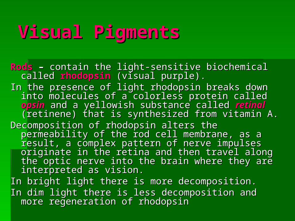
Visual PigmentsVisual Pigments
RodsRods – – contain the light-sensitive biochemical calledcontain the light-sensitive biochemical called rhodopsinrhodopsin (visual purple).(visual purple).
In the presence of light rhodopsin breaks down into molecules In the presence of light rhodopsin breaks down into molecules of a colorless protein called of a colorless protein called opsinopsin and a yellowish and a yellowish substance called substance called retinalretinal (retinene) that is synthesized from (retinene) that is synthesized from vitamin A. vitamin A.
Decomposition of rhodopsin alters the permeability of the rod Decomposition of rhodopsin alters the permeability of the rod cell membrane, as a result, a complex pattern of nerve cell membrane, as a result, a complex pattern of nerve impulses originate in the retina and then travel along the impulses originate in the retina and then travel along the optic nerve into the brain where they are interpreted as optic nerve into the brain where they are interpreted as vision.vision.
In bright light there is more decomposition. In bright light there is more decomposition. In dim light there is less decomposition and more regeneration In dim light there is less decomposition and more regeneration
of rhodopsinof rhodopsin

Visual PigmentsVisual Pigments
ConesCones – – the light-sensitive pigments are similar to the light-sensitive pigments are similar to rhodopsin in that they are composed of retinal rhodopsin in that they are composed of retinal combined with protein.The protein, however differs.combined with protein.The protein, however differs.
Three different sets of cones each contain an Three different sets of cones each contain an abundance of one of the three different visual abundance of one of the three different visual pigments.pigments.
The wavelength of light determines the color that the The wavelength of light determines the color that the brain perceives from it.brain perceives from it.
For example, the shortest wavelengths are perceived For example, the shortest wavelengths are perceived as violet, and the longest as red.as violet, and the longest as red.
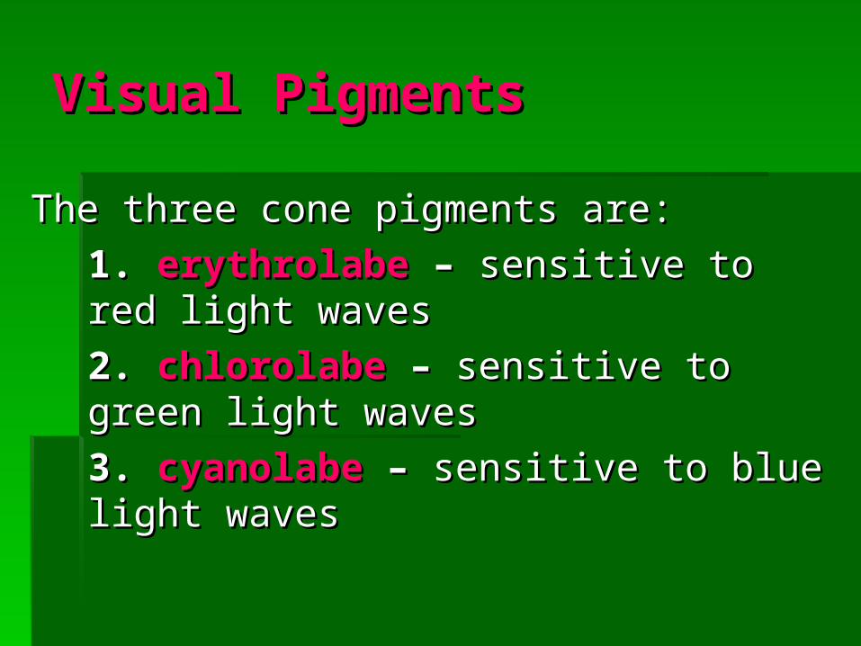
Visual PigmentsVisual Pigments
The three cone pigments are:The three cone pigments are:
1. 1. erythrolabeerythrolabe – – sensitive to red light sensitive to red light waveswaves
2. 2. chlorolabechlorolabe – – sensitive to green light sensitive to green light waveswaves
3. 3. cyanolabecyanolabe – – sensitive to blue light sensitive to blue light waveswaves

Visual PigmentsVisual Pigments
The color a person perceives depends on The color a person perceives depends on which set of cones or combination of sets the which set of cones or combination of sets the light in a given image stimulates. light in a given image stimulates.
If all three sets of cones are stimulated, the If all three sets of cones are stimulated, the person senses the light as white, and if none person senses the light as white, and if none are stimulated, the person senses black.are stimulated, the person senses black.
Different forms of colorblindness result from Different forms of colorblindness result from lack of different types of cone pigments.lack of different types of cone pigments.
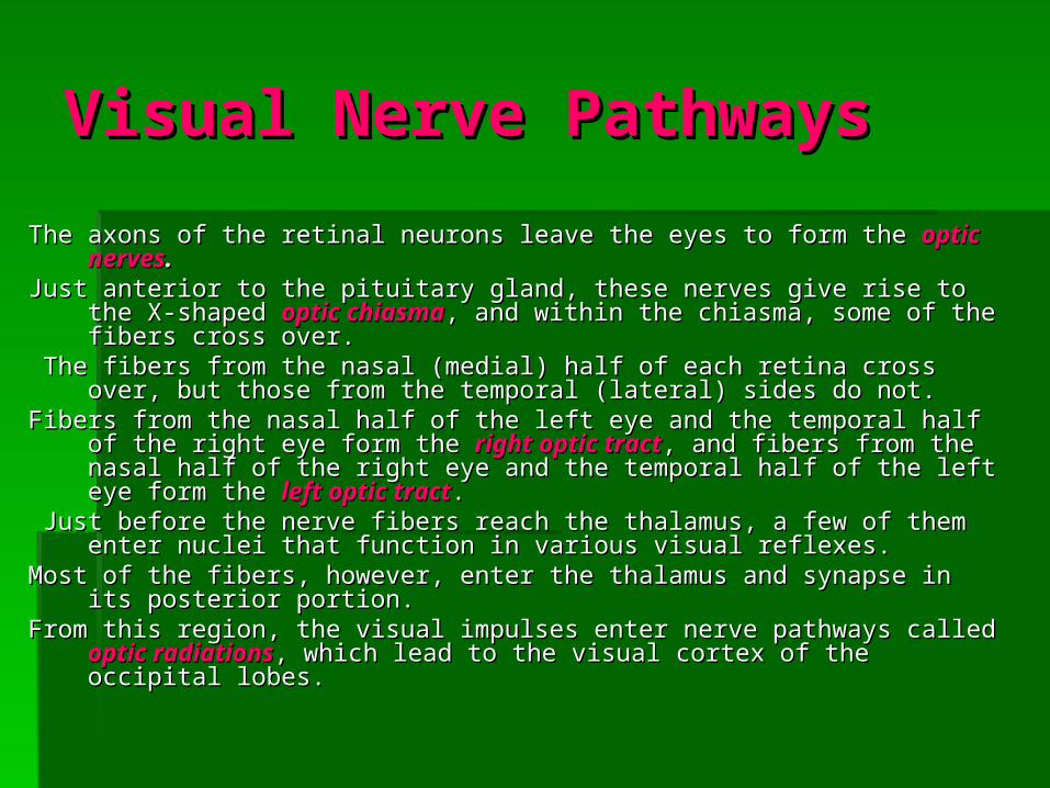
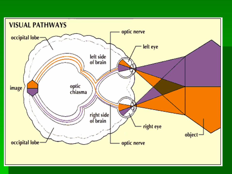
Visual Nerve PathwaysVisual Nerve Pathways
The axons of the retinal neurons leave the eyes to form the The axons of the retinal neurons leave the eyes to form the optic nervesoptic nerves.. Just anterior to the pituitary gland, these nerves give rise to the X-shaped Just anterior to the pituitary gland, these nerves give rise to the X-shaped
optic chiasmaoptic chiasma, and within the chiasma, some of the fibers cross , and within the chiasma, some of the fibers cross over.over.
The fibers from the nasal (medial) half of each retina cross over, but those The fibers from the nasal (medial) half of each retina cross over, but those from the temporal (lateral) sides do not. from the temporal (lateral) sides do not.
Fibers from the nasal half of the left eye and the temporal half of the right Fibers from the nasal half of the left eye and the temporal half of the right eye form the eye form the right optic tractright optic tract, and fibers from the nasal half of the , and fibers from the nasal half of the right eye and the temporal half of the left eye form the right eye and the temporal half of the left eye form the left optic tractleft optic tract..
Just before the nerve fibers reach the thalamus, a few of them enter nuclei Just before the nerve fibers reach the thalamus, a few of them enter nuclei that function in various visual reflexes. that function in various visual reflexes.
Most of the fibers, however, enter the thalamus and synapse in its posterior Most of the fibers, however, enter the thalamus and synapse in its posterior portion. portion.
From this region, the visual impulses enter nerve pathways called From this region, the visual impulses enter nerve pathways called optic optic radiationsradiations, which lead to the visual cortex of the occipital lobes., which lead to the visual cortex of the occipital lobes.

Visual Nerve PathwaysVisual Nerve Pathways
Just before the nerve fibers reach the thalamus, a Just before the nerve fibers reach the thalamus, a few of them enter nuclei that function in various few of them enter nuclei that function in various visual reflexes. visual reflexes.
Most of the fibers, however, enter the thalamus and Most of the fibers, however, enter the thalamus and synapse in its posterior portion. synapse in its posterior portion.
From this region, the visual impulses enter nerve From this region, the visual impulses enter nerve pathways called pathways called optic radiationsoptic radiations, which lead to , which lead to the visual cortex of the occipital lobes.the visual cortex of the occipital lobes.