soman and sarin inhibition of molecular forms of acetylcholinesterase in mice: time course of...
TRANSCRIPT
Biochemical Pharmacology, Vol. 42. No. 2, pp, 329-335, 1991. 0006-2952/91 $3.00 + 0.00 Printed in Great Britain. Pergamon Press pic
SOMAN AND SARIN INHIBITION OF MOLECULAR FORMS OF ACETYLCHOLINESTERASE IN MICE
T I M E C O U R S E O F R E C O V E R Y A N D R E A C T I V A T I O N B Y T H E O X I M E HI -6
JOHN G. CLEMENT,* SYLVESTER ROSARIO, ELAINE BESSETTE and NANCY ERHARDT Biomedical Defence Section, Defence Research Establishment Suffield, Ralston, Alberta, Canada
(Received 6 February 1990; accepted 8 February 1991)
Abstract--The in vivo sensitivity of the molecular forms of the enzyme acetylcholinesterase to inhibition by either soman or sarin, reactivation by HI-6 and the time course of recovery following inhibition by soman were investigated in mice. Administration of HI-6 (50 mg/kg, i.p.) immediately after soman (100 gg/kg, s.c.) or sarin (150 #g/kg, s.c.) resulted in an apparent selective reactivation of the 10S and 16S molecular forms of acetylcholinesterase and no reactivation of the 4S form of diaphragm acetylcholinesterase. The apparent selectivity of the reactivation of the molecular forms of the acetyl- cholinesterase was probably due to the fact that the 10S and 16S forms of acetylcholinesterase are located primarily extracellularly and the 4S form intracellularly. The HI-6 was restricted primarily to the extracellular compartment due to its quaternary, hydrophilic nature. If the administration of HI-6 was delayed until 60 min following soman (100 #g/kg, s.c.) injection, no reactivation of any of the molecular forms of acetylcholinesterase could be found in the diaphragm. The soman-inhibited acetyl- cholinesterase had probably aged and, thus, was not susceptible to reactivation by HI-6. The time course of recovery of the molecular forms in the diaphragm occurred rather quickly with the smaller 4S and 10S forms recovering to control levels faster than the larger 16S form. It took between 8 and 16 days for the 16S form to recover to normal. In the brain, hypothalamic acetylcholinesterase molecular forms such as the 4S recovered faster than the 10S form which had not recovered to control 16 days after soman administration; the 16S form of acetylcholinesterase was not detected in the brain.
The hydrolytic enzyme acetylcholinesterase inac- tivates acetylcholine at cholinergic synapses. This polymorphic enzyme is found in many neuronal and non-neuronal tissues and is usually composed of several molecular forms [1, 2]. Two general classes of the enzyme are called globular (G) and asymmetric (A). The globular class has three forms called G1, G2 and G4, based on sedimentation analysis, composed of a monomer, dimer and tetramer, respectively, while the asymmetric class has three forms called A4, A8 and A12 which consist of aggregates of 1, 2 and 3 tetramers, respectively, with a collagen tail attached. Three molecular forms of acetylcholinesterase predominate in rodent diaphragm with sedimentation coefficients of 4S, 10S and 16S which correspond to the G1, G4 and A12 forms, respectively, and two molecular forms of acetylcholinesterase predominate in rodent brain with sedimentation coefficients of 4S and 10S which correspond to the G1 and G4 forms, respectively.
Soman (pinacolyl methylphosphonofluoridate) is an extremely toxic organophosphorus compound. The locus of the toxic and lethal effects of this compound is the irreversible inhibition of the enzyme acetylcholinesterase. The recovery of acetylcholinesterase activity following poisoning with a sublethal dose of soman is through resynthesis of the enzyme [3] and not dealkylation [4]. Following
* Correspondence: Dr. J. G. Clement, Defence Research Establishment Suffield, Box 4000, Medicine Hat, Alberta, Canada, T1A 8K6.
© Government of Canada.
soman poisoning, the function of acetylcholinesterase in the periphery can be restored by parenteral administration of the bispyridinium oxime HI-6 [CAS Reg. No. 34433-31-3; 1-(((4-(aminocarbonyl)- pyridinio)methoxy)methyl) - 2 - ((hydroxyimino)- methyl)-pyridinium dichloride] [5]. HI-6 dephos- phorylates acetylcholinesterase by nucleophilic attack and the phosphylated HI-6 quickly decomposes [6], thus returning acetylcholinesterase function to normal. However, soman-inhibited acetylcho- linesterase gradually undergoes an ageing process where it is converted to a form that cannot be reactivated by oximes [7,8]. Sarin (isopropyl methylphosphonofluoridate) is another potent organophosphate anticholinesterase which acts in a manner similar to soman. However, the sarin- inhibited acetylcholinesterase is reactivated more easily by HI-6 than soman [5], and its rate of ageing is considerably slower.
The oxime HI-6 is a bisquaternary, hydrophilic compound which restricts its distribution in oioo, primarily if not totally, to the extracellular compartment [9, 10]. The purpose of the present study was 2-fold; (1) to determine if the oxime reactivator HI-6 selectively reactivated a particular molecular form of acetylcholinesterase in the mouse diaphragm following administration of a sublethal dose of an organophosphate anticholinesterase such as soman or satin; and (2) to determine the time course of the recovery of the various molecular forms of acetylcholinesterase in mouse diaphragm and brain following inhibition by a sublethal dose of soman.
329
330 J. G. CLEMENT et al.
MATERIALS AND METHODS
Materials Soman, sarin and HI-6 were prepared at Defence
Research Establishment Suffield and were in excess of 99% pure. Other substances used were obtained from various commercial sources: 4-methyl- umbelliferyl phosphate, 4-methylumbelliferyl gap actoside, acetylthiocholine and dithiobis nitrobenzoic acid (Sigma); ISO-OMPA (ICN Biochemicals); [3H]acetylcholine chloride (NEN); Triton X-100 (BDH); alkaline phosphatase (EC 3.1.3.1), mol- ecular weight of 80,000, from Escherichia coli (Sigma); catalase (EC 1.11.1.6), molecular weight of 244,000, from bovine liver (Sigma); and fl- galactosidase (EC 3.2.1.23), molecular weight of 540,000, from Escherichia coli (Sigma).
Animals CD-1 male mice (20-25 g) were obtained from
Charles River Canada Ltd., St. Constant, Quebec. The mice were housed in the vivarium at Defence Research Establishment Suffield for at least 1 week prior to use in the experiments. The light schedule was 12 hr light and 12 hr dark with the lights on at 7:00 a.m. The mice had access to food and water ad lib. The temperature was in the range of 22-24 ° .
1-11-6 reactivation of diaphragm acetylcholinesterase Mice were injected with either soman (70 or
100/zg/kg, s.c.) or sarin (150 lug/kg, s.c.) and then either immediately or 60 min later were injected with HI-6 (50mg/kg, i.p.). Appropriate controls received saline in place of HI-6. The mice were decapitated and exsanguinated 30 min following the administration of either HI-6 or saline, The diaphragm was extirpated and the homogenate prepared.
Time course of recovery of brain and diaphragm acety lcholinesterase
Mice were injected with soman (100 #g/kg, s.c.) and then at various time periods (1 ,2 ,4 ,8 and 16 days) after poisoning were decapitated and exsanguinated. The tissues (diaphragm and hypo- thalamus) were isolated and homogenates prepared.
Tissue preparation
Mice were killed and the tissues removed and rinsed in 0.9% saline, blotted dry on filter paper, and weighed. The hypothalamus homogenate was prepared by 10-20 strokes in a glass-teflon homogenizer in an extraction buffer (4 ° ) containing 1 M NaC1, 0.05 M MgCI2, 0.01 M Tris-HC1 and 1% Triton X-100, pH 7.4 [1]. The diaphragm, from individual mice, was homogenized in the same extraction buffer (4 °) using a Brinkmann Polytron with a micro probe for a period of 20-30 sec. The tissue concentration of the final homogenate was 10 and 100 mg wet weight of tissue/mL of extraction buffer for the hypothalamus and diaphragm, respectively. The homogenates were then centrifuged at 20,000 g for 20 rain at 4 ° in a Beckman model J2- 21 M centrifuge and a JA 21 rotor. Fresh, unfrozen tissue was used in all experiments since the use of frozen tissue produced changes in the relative
proportions of the various molecular forms of acetylcholinesterase detected ([1, 11]; Clement JG and Bessette E, unpublished observations).
Sucrose density gradient centrifugation Sucrose density gradients (5-30%) were formed
by manual layering and were allowed to diffuse overnight in the cold room (4 °) to form a continuous gradient. The linearity of the gradient was confirmed in preliminary experiments by determining the sucrose concentrations of the various fractions by refractometry. Tissue homogenate supernatant (200/tL) was layered on top of the sucrose gradient, and the tubes (Polyallomer Bell-top Quick Seal tubes, Beckman Instruments) were sealed using the Beckman tube sealer. The tubes were centrifuged at 65,000 rpm for 2 hr at 4 ° in a Beckman L8-55M ultracentrifuge and a VTi 65.1 rotor. The tubes were pierced at the bottom of the tube and fractionated (0.2-mL fractions collected from the top of the tube) using an ISCO (model 640) fraction collector. For the time course of recovery of the various molecular forms following soman poisoning, a 5-20% sucrose gradient was used.
To determine the sedimentation coefficients of the various molecular forms of acetylcholinesterase, alkaline phosphatase (6.1S), catalase (11.4S) and fl- galactosidase (16S) were used as markers. The marker enzymes were layered on gradients and centrifuged using the same conditions as those used in the separation of the various molecular forms of acetylcholinesterase. The tubes were fractionated and the enzyme activities of the various fractions were determined. The marker enzymes sediment in the same way whether they are added to the homogenate and layered on the gradient or layered directly on the gradient [12]. Therefore, the positions of the marker enzymes were determined in separate experiments, and this positioning was used as the indicator in the remaining experiments examining the molecular forms of acetylcholinesterase.
Data analysis The acetylcholinesterase activity for the various
fractions was determined and plotted, and the area under the curve (AUC) of the various molecular forms was determined using a LOTUS 123 template. All fractions were corrected for nonenzymatic hydrolysis using fractions from a run where extraction buffer was used as the blank. The 4S form included fractions 1-14, the 10S form fractions 15-23, and the 16S form fractions 24-32. The data were analyzed by Student's t-test. A P < 0.05 was considered statistically significant.
Enzyme assays Acetylcholinesterase. The difference in the pro-
cedures for the measurement of acetylcholinesterase activity over the course of these studies was due to the evolution of the analysis techniques used in our laboratory and the introduction of laboratory robot technology.
(1) Radiometric assay. In the study of the time course of recovery of the various molecular forms of acetylcholinesterase in the diaphragm and hypothalamus, each fraction was analyzed for
HI-6 and acetylcholinesterase molecular forms 331
acetylcholinesterase activity by the method of Johnson and Russell [13] using [3H]acetylcholine chloride as the substrate. In each sample, ISO- OMPA (10/~M) was present to selectively inhibit pseudocholinesterase activity [14]. The radioactivity was quantitated by liquid scintillation counting in a Beckman liquid scintillation counter model LS9800.
(2) Spectrophotornetric assay. In the reactivation experiments, each fraction was analyzed for acetylcholinesterase activity using the method of Ellman et al. [15] with acetylthiocholine as the substrate. Each fraction contained ISO-OMPA (10/~M) to selectively inhibit pseudocholinesterase activity [14].
Catalase. The activity of catalase was determined by following the disappearance of H202 spectro- photometrically [16].
Alkaline phosphatase. A 10-/~L sample was added to a well in a 96-well immunoassay plate. The reaction was started by the addition of 100/~L of enzyme substrate 4-methylumbelliferyl phosphate (100/~M) in 0 .1M diethylamino buffer, pH 9.8, containing 20 mM MgC12 and incubated at room temperature in the dark. The relative fluorescence was measured in a MicroFluor microplate reader (Dynatech Laboratories) at an excitation wavelength of 365 nm and an emission wavelength of 450 nm at 5 min intervals.
~-Galactosidase. A 10-/~L sample was added to a well of a 96-well immunoassay plate. The reaction was started by the addition of 100/~L of the enzyme substrate 4-methylumbelliferyl galactoside (100/~M) prepared in 10 mM sodium phosphate buffer, pH 7.0, which contained 0.1 M NaC1, 0.1 mM MgC12 and 0.1% (w/v) bovine serum albumin (BSA), and incubated at room temperature in the dark. The relative fluorescence was measured in a MicroFluor microplate reader at an excitation wavelength of 365 nm and an emission wavelength of 450 nm at 5 min intervals.
RESULTS
The doses of soman and sarin administered to mice were sublethal (LDs0 values of soman and sarin used in this study were 130--140 and 170/~g/ kg, respectively) and produced typical signs of anticholinesterase poisoning such as salivation, tremors, lacrimation, and diarrhea. The adminis- tration of HI-6 typically reduced the degree of the signs of poisoning and also increased the speed of recovery. No quantitation of the degree of poisoning was performed on these animals.
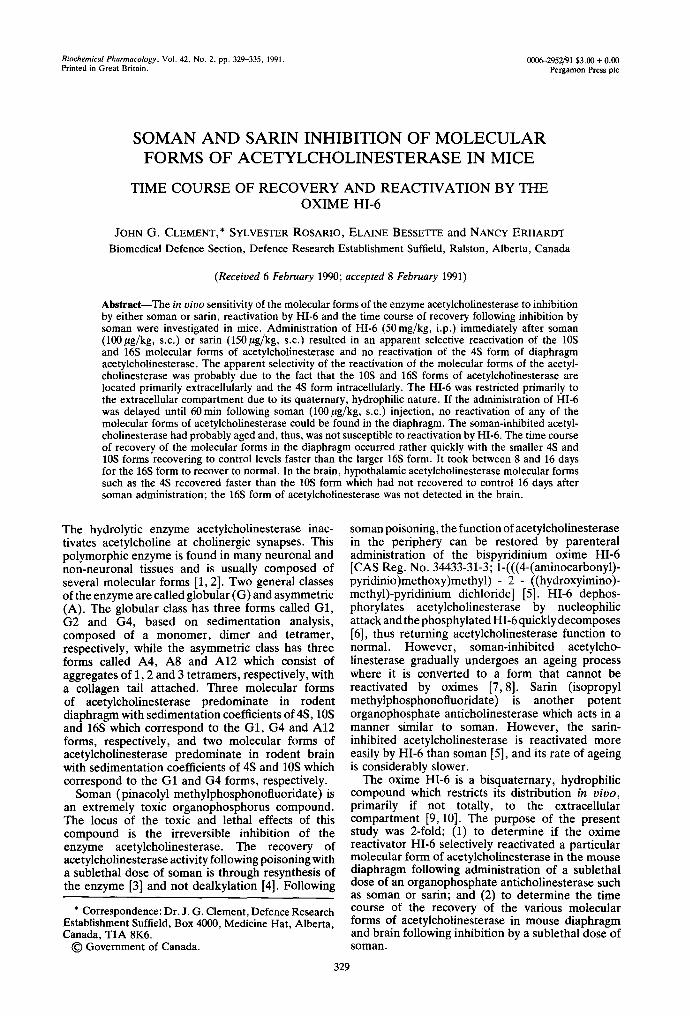
Diaphragm acetylcholinesterase activity was decreased to approximately 34--40% of control activity at 30 and 90 min after soman administration (Fig. 1; Table 1), respectively. There did not appear to be any selectivity in the inhibition of the various molecular forms of acetylcholinesterase. Administration of HI-6 immediately following soman resulted in reactivation of soman-inhibited acetyleholinesterase as indicated by an increase in total acetylcholinesterase activity to 47% of control at 30 min following soman and HI-6 administration (Table 1). The reactivation of acetylcholinesterase was isolated to the 10S and 16S forms of the enzyme
8~ A d~ ~ ~ 0 Control "1" f ~ T \ / / '~'~-"
- 5 10 15 20 25 - " 30 " "
o
v
<
o
5 I 0 15 20 25 30
5 10 15 20 25 30 Fraction N u m b e r
Fig. 1. Reactivation of the various molecular forms of acetylcholinesterases by HI-6 following inhibition by soman. (A) Mice were administered soman (100 ~ug/kg, s.c.) and were injected immediately with HI-6 (50 mg/kg, i.p.). The mice were killed 30min following the administration of HI-6. In the saline group saline was injected in place of HI-6. The symbol key in this panel applies to all panels of the figure. (B) Soman (100/~g/kg, s.c.) was administered and 60 min later HI-6 (50 mg/kg, i.p.) was injected. The mice were killed 30 min following administration of the oxime. In the saline group, saline was administered in place of HI-6. (C) Soman (70/zg/kg, s.c.) was administered and HI-6 (50mg/kg, i.p.) was injected immediately thereafter. The mice were killed 30 min following oxime administration. In the saline group, saline was administered in place of HI-6. The position of the marker proteins is as follows: left arrow, alkaline phosphatase (6.1S); middle arrow, catalase (11.4S); and right arrow, fl-galactosidase (16S). The number of observations is indicated in Table 1. All samples were
analyzed in duplicate.
with significant reactivation of the 4S form not occurring (Fig. 1A; Table 1).
If the administration of HI-6 was delayed until 60 min after soman poisoning, there was no significant reactivation of total diaphragm acetylcholinesterase or the various molecular forms of the enzyme (Fig. 1B; Table 1), indicating that the soman- acetylcholinesterase complex had aged by this time.
When the dose of soman was decreased to 70/~g/ kg, the reactivation produced by HI-6 was significantly greater (Fig. 1C; Table 1) than that following the higher dose of soman. There was significant reactivation of all of the molecular forms of acetylcholinesterase with the largest increases in the 10S and 16S forms.

332 J. G. CLEMErCr et al.
Table 1. Reactivation of soman-inhibited molecular forms of acetylcholinesterase by HI-6*
Acetylcholinesterase activity (nmol/min/mL)
Treatment Total 4S 10S 16S N
Untreated control 97.91 -+ 10.07t 41.03 +-- 3.00 31.98 -+ 5.50 24.90 ± 5.40 9 (100) (100) (100) (100)
Soman (100/~g/kg) + HI-6 at "0" time Saline 33.31 - 6.09 17.25 -+ 5.43 8.27 -+ 2.25 7.80 -- 1.97 8
(34) (42) (26) (31) HI-6 46.47 --- 5.01:~ 21.88 -+ 3.17 14.29 +- 3.86§ 10.29 -- 2.3411 7
(47) (53) (45) (41) Soman (100 #g/kg) + HI-60 rain later
Saline 39.38 +- 14.53 18.65 -+ 7.09 10.24 --- 4.46 10.48 ± 3.97 6 (40) (45) (32) (42)
HI-6 28.49 - 2.60 17.41 --- 3.33 6.78 ± 1.26 4.31 --- 1.14§ 5 (29) (42) (21) (17)
Soman (70/~g/kg) + HI-6 at "0" time Saline 36.16 -+ 6.48 16.90 -+ 2.97 10.53 + 2.09 8.74 + 2.24 8
(37) (41) (33) (35) HI-6 57.47 ± 2.765 22.44 -+ 1.68§ 17.99 ± 2.03:~ 17.04 --- 1.665 4
(59) (55) (56) (68)
* Mice were treated with soman (100 or 70/~g/kg, s.c.) and either at the same time or 60 min later received HI-6 (50 mg/kg, i.p.). The mice were killed 30 rain after administration of the oxime and the homogenate was prepared.
t Values are means --- SD with percent control given in parentheses. $-11 Significantly different from the appropriate saline control group: :~ P < 0.001, § P < 0.01, and [I P < 0.05.
x=
(a
|
"d
/ \ _~ ~ o co.t,o,
5 10 15 20 25 30
Fract ion Number
Fig. 2. Reactivation of the various molecular forms of acetylcholinesterase by HI-6 following inhibition by sarin. Mice were administered sarin (150/tg/kg, s.c.) and were injected immediately with HI-6 (50 mg/kg, i.p.). The mice were killed 30 min following administration of the oxime. In the saline group, saline was injected in place of HI-6. The position of the marker enzymes is as in Fig. 1. The number of observations is indicated in Table 2. All samples
were analyzed in duplicate.
The effect of sat in and HI-6 administrat ion on the activity of the various molecular forms of acetylcholinesterase in the mouse diaphragm was also investigated. Satin produced a profound inhibit ion of all of the various molecular forms with no apparent selectivity of inhibit ion (Fig. 2 and Table 2). Adminis t ra t ion of HI-6 immediately after satin resulted in significant reactivation of the 10S and 16S but not the 4S form of acetylcholinesterase.
The time course of recovery of the various
molecular forms of acetylcholinesterase in the diaphragm and hypothalamus is presented in Fig. 3 and 4. In the mouse diaphragm, three molecular forms, the 4S, 10S and 16S, were detected. The various forms appeared to be affected to the same degree following soman poisoning (Fig. 3). One day after poisoning all of the molecular forms of acetylcholinesterase had started to recover activity with the smaller forms, 4S and 10S, recovering to control levels faster than the 16S form. By day 8 after soman, the 4S and 10S forms had recovered to control levels, whereas the 16S form took between 8 and 16 days to recover to control level.
In the hypothalamus (Fig. 4), only two molecular forms of acetylcholinesterase were detected, the 4S and 10S forms. The 4S form did not appear to be inhibited to as great a degree as the 10S form, and it had recovered by 24hr . The time course of recovery of the 10S form was slower than that found in the diaphragm, and it was not totally recovered even at 16 days after soman poisoning.
DISCUSSION
The results of this study demonstrate that the various molecular forms of acetylcholinesterase can be separated by the use of a vertical tube rotor. The time for the completion of the centrifugation run was shortened from the 16-18 hr it would normally take using a swinging bucket rotor to 2 hr using the vertical tube rotor, thus reducing the t ime that the tissue had to be handled prior to the enzyme being separated into its various molecular forms and fractionated, which, in turn, reduced the possibility of proteolytic-induced changes in the relative proportions of the various molecular forms of acetylcholinesterase [1, 11].

HI-6 and acetylcholinesterase molecular forms
Table 2. Reactivation of sarin-inhibited molecular forms of acetylcholinesterase by HI-6*
333
Acetylcholinesterase activity (nmol/min/mL)
Treatment Total 4S 10S 16S N
Untreated control 97.91 - 10.07t 41.03 --- 3.00 31.98 ± 5.50 24.90 - 5.40 9 (100) (100) (100) (100)
Saline 47.74 ± 9.53 21.22 -- 4.26 14.32 ± 4.46 12.20 -- 1.78 5 (49) (52) (45) (49)
HI-6 64.80 ± 12.525 24.49 ± 5.81 23.57 ± 5.315 16.74 -- 2.35§ 5 (66) (60) (74) (67)
* Mice were treated with sarin (150 #g/kg, s.c.) and immediately thereafter (i.e. within 10 sec) received HI-6 (50 mg/kg, i.p.). The mice were killed 30 min after oxime administration.
t Values are means --+ SD with percent control given in parentheses. ~:, § Significantly different from saline control group: ~: P < 0.05, and § P < 0.01.
%
o
c
_o >~
0 4 I 12 10 ~ ~ U
Fraction Number
Fig. 3. Time course of recovery of the various molecular forms of mouse diaphragm acetylcholinesterase following poisoning with soman. Mice were killed at various times after the administration of soman (100 ~ug/kg, s.c.) and the molecular forms of acetylcholinesterase separated. Each point is the mean of at least three separate observations. The control acetylcholinesterase activity is included in each
panel of the figure as a dotted line.
<
<
* - - e Day2
s - - e I~y4
2- " ".
o - - e 0oy 16 l B
2- ."/" ". .
* • T: ,J ~ ~ as == Fraction Number
Fig. 4. Time course of recovery of the various molecular forms of mouse hypothalamic acetylcholinesterase following poisoning with soman. Mice were killed at various times after the administration of soman (100/~g/kg, s.c.) and the molecular forms of hypothalamic acetylcholinesterase separated. Each point is the mean of at least three separate observations. The control acetylcholinesterase activity is
included in each panel of the figure as a dotted line.
The effects of soman and satin on the molecular forms of acetylcholinesterase in the mouse diaphragm were investigated. The three major peaks found, the 4S, 10S and 16S, correspond to the G1, G4 and A12 forms, respectively. The A12 form is primarily located extracellularly, bound to the membrane [17, 18] and concentrated at the endplate region in the mouse diaphragm [19], the G1 form is primarily located intracellularly [17, 20] and the G4 form is
distributed approximately equally between the intracellular and extracellular compartments [18]. Both soman and sarin produced a profound inhibi t ion of all molecular forms of acetylcholinesterase in the mouse diaphragm without any apparent selectivity. This is not surprising since soman and sarin, both being very lipid soluble compounds, would interact with target proteins in the intracellular and extracellular compartments.

334 J. G. CLEMENT et al.
HI-6 is a powerful reactivator of soman-inhibited acetylcholinesterase [21, 22]. When the oxime was administered immediately following soman or satin, there appeared to be a selectivity with regards to the reactivation of the individual molecular forms of acetylcholinesterase, with the larger forms, the 10S and 16S, being more susceptible to reactivation by the oxime. The reason for the apparent selectivity may be due to a definite selectivity of the particular molecular form of acetylcholinesterase to reactivation by HI-6 or simply due to the accessibility of HI-6 to the phosphorylated acetylcholinesterase. The latter seems the more plausible explanation. The various molecular forms of acetylcholinesterase react in a similar manner to various inhibitors [20, 23--26]; thus, it would be unlikely that the various phosphorylated acetylcholinesterase molecular forms would interact with the oxime in a selective fashion. Since HI-6 is a hydrophilic, bispyridinium, quaternary ammonium compound with limited ability to pass plasma membranes, HI-6 could only reactivate the "unaged" molecular forms of acetylcholinesterase that it came in contact with, primarily, in the extracellular compartment. Based on the distribution of the various molecular forms of acetylcholinesterase [17-20], the results of this study suggest that in the mouse diaphragm the 4S form is located primarily intracellularly and the 10S and 16S forms are located primarily extracellularly. The lack of reactivation of the soman-inhibited acetylcholinesterase when the HI-6 was administered 60 min after soman was probably due to the fact that the inhibited enzyme had aged and thus was not susceptible to reactivation by the oxime HI-6. It was reported that the half-time for ageing of soman-inhibited acetylcholinesterase in rodents is in the range of 20-30 min [22]. The reactivation of the 4S form of acetylcholinesterase at the lower soman dose (70/~g/kg) suggests that there is some movement of HI-6 across cell membranes but at the higher dose of soman the degree of inhibition is too great for the small concentration of HI-6 to reactivate the 4S form of acetylcholinesterase.
In the mouse brain region examined, only the 4S and 10S forms of acetylcholinesterase were found with the 10S form predominating and the 16S form absent in agreement with the results of others [1, 2, 27]. The recovery of the molecular forms in the brain was much slower than that of the diaphragm and is characteristic of brain tissue where the recovery of acetylcholinesterase activity to control levels following organophosphate exposure takes from 16 to 32 days depending upon the region examined [27-30]. The differences in the time course of recovery of the molecular forms of acetylcholinesterase following soman poisoning may be an expression of the rate of synthesis and assembly of the molecular forms in the various regions examined.
Acknowledgements--The authors would like to thank Dr. R. Bhatti and Mr. Y. Siddiqui for their assistance in performing the assays for fl-galactosidase and alkaline phosphatase.
REFERENCES
1. Massoulie J and Bon S, The molecular forms of
cholinesterase and acetylcholinesterase in vertebrates. Annu Rev Neurobiol 5: 57-106, 1982.
2. Inestroso NC and Perelman A, Distribution and anchoring of molecular forms of acetylcholinesterase. Trends Pharmacol Sci 10: 325-329, 1989.
3. Harris LW, Yamamura HI and Fleisher JH, De novo synthesis of acetylcholinesterase in guinea pig retina after inhibition by pinacolyl methylphosphono- fluoridate. Biochem Pharrnacol 20: 2927-2930, 1971.
4. Coult DB, Marsh DJ and Read G, Dealkylation studies on inhibited acetylcholinesterase. Biochem J 98: 869- 873, 1966.
5. Clement JG, HI-6: Reactivation of central and peripheral acetylcholinesterase following inhibition by soman, sarin and tabun in vivo in the rat. Biochem Pharmaco131: 1283-1287, 1982.
6. Boskovic B, The treatment of soman poisoning and its perspectives. Fundam Appl Toxicol 1: 203-213, 1981.
7. Aldridge WN and Reiner E, Enzyme inhibitors as substrates: Interactions of esterases with esters of organophosphorus and carbamic acids. In: Frontiers o f Biology Eds. Neuberger A and Tatum EL), pp. 1-328. North-Holland Publishing Co., Amsterdam, 1975.
8. Berends F, Mechanism of ageing of organophosphate- inhibited esterases. In: Selectivity and Molecular Mechanisms of Toxicity (Eds. DeMatteis F and Lock EA), pp. 125-152. Macmillan Press, New York, 1987.
9. Ligtenstein DA and Kossen SP, Kinetic profile in blood and brain of the cholinesterase reactivating oxime HI- 6 after intravenous administration to the rat. Toxicol Appl Pharmacol 71: 177-183, 1983.
10. Ligtenstein DA, Moes GWH and Kossen SP, In vivo distribution of organophosphate antidotes: Autoradiography of [14C]HI-6 in the rat. Toxicol Appl Pharmacol 92: 324-329, 1988.
11. Payner TD, Drake RL, Saker DM and Shipley MT, Determination of molecular forms of brain acetylcholinesterase: Technical considerations. Brain Res Bull 19: 287-290, 1987.
12. Dreyfus PA, Friboulet A, Trans LH and Rieger F, Polymorphism of acetylcholinesterase and identification of new molecular forms after sedimentation analysis. Biol Cell 51: 35-42, 1984.
13. Johnson CD and Russell RL, A rapid, simple radiometric assay for cholinesterase, suitable for multiple determinations. Anal Biochem 64: 229-238, 1975.
14. Wilson BW, Walker CR and Nieberg PS, Tissue acetylcholinesterase in plasma of chick embryos and dystrophic chickens. J Neurol Sci 18: 333-350, 1973.
15. Ellman GL, Courtney KD, Andres V and Featherstone RM, A new and rapid colorimetric determination of acetylcholinesterase activity. Biochern Pharmacol 7" 88-95, 1961.
16. Beers RF and Sizer IW, A spectrophotometric method for measuring the breakdown of hydrogen peroxide by catalase. J Biol Chern 195: 133-140, 1952.
17. Skau KA, Mammalian acetylcholinesterase molecular forms. Comp Biochem Physiol 83C: 225-227, 1986.
18. Skau KA and Brimijoin S, Abnormal distribution of skeletal muscle acetylcholinesterase molecular forms in dystrophic mice. Exp Neurol 74: 111-121, 1981.
19. Goudou D, Verdiere-Sahuque M and Rieger F, External and internal acetylcholinesterase in rat sympathetic neurones in vivo and in vitro. FEBS Lett 186: 54-58, 1985.
20. Lazar M and Vigny M, Modulation of the distribution of acetylcholinesterase molecular forms in a murine neuroblastoma x sympathetic ganglion cell hybrid cell line. J Neurochem 35: 1067-1079, 1980.
21. DeJong LPA and Kossen SP, Stereospecific reactivation of human brain and erythrocyte acetylcholinesterase
HI-6 and acetylcholinesterase molecular forms 335
inhibited by 1,2,2-trimethylpropyl methylphosphono- fluoridate (soman). Biochim Biophys Acta 830: 345- 348, 1985.
22. DeJong LPA and Wolring GZ, Aging and stereospecific reactivation of mouse erythrocyte and brain acetyl- cholinesterases inhibited by soman. Biochem Phar- maco134: 142-145, 1985.
23. Groswald DE and Dettbam W-D, Characterization of acetylcholinesterase molecular forms in slow and fast muscle of rat. Nerochem Res 8: 983-995, 1983.
24. Adamson ED, Ayers SE, Deussen ZA and Graham CF, Analysis of the forms of acetylcholinesterase from adult mouse brain. Biochem J 147: 205-214, 1975.
25, Skau KA, Acetylcholinesterase molecular forms in serum and erythrocytes of laboratory animals. Comp Biochem Physiol 80: 207-210, 1985.
26, Taylor PB, Shelanski ML and Greene LA, Cellular localization of the multiple forms of acetylcholinesterase in cultured neuronal cells. J Biol Chern 256: 3827- 3830, 1981.
27. Grubi6 Z, Sketelj J, Klinar B and Brzin M, Recovery of acetylcholinesterase in the diaphragm, brain and plasma of the rat after irreversible inhibition by soman: A study of cytochemical localization and molecular forms of the enzyme in the motor end plate. J Neurochem 37: 909--916, 1981.
28. Clement JG, Survivors of soman poisoning: Recovery of the soman LDs0 to control value in the presence of extensive acetylcholinesterase inhibition. Arch Toxicol 63: 869--873, 1989.
29. Sung SC and Ruff BA, Intracellular distribution of molecular forms of acetylcholinesterase in rat brain and changes after diisopropylfluorophosphate treatment. Neurochem Res 12: 15-19, 1987.
30. Michalek H, Meneguz A and Bisso GM, Molecular forms of rat brain acetylcholinesterase in DFP intoxication and subsequent recovery. Neurobehav Toxicol Teratol 3: 302-312, 1981.