solubility, immunochemical, and lipoprotein binding ... · solubility to this apolipoprotein that...
TRANSCRIPT

Solubility, immunochemical, and lipoprotein binding properties of apoB-100-apo[a], the protein moiety of lipoprotein [a]
Gunther M. Fless? Ditta J. Pfaffinger? James D. Eisenbart? and Angelo M. Scanu**t
Departments of Medicine. and Biochemistry and Molecular Biology, t The Pritzker School of Medicine, The University of Chicago, Chicago, IL 60637
Abstract The protein moiety of Lp[a] consisting of apoB and apo[a] covalently linked to each other, once freed of lipids by delipidation at pH 8.0 with mixtures of diethyl ether and ethanol, is freely water-soluble at pH values above 6.4. This is in contrast to apoB which, if prepared by similar delipidation techniques, is only soluble at alkaline pH, indicating that the coupling of the carbohydrate-rich apo[a] to apoB confers water solubility to this apolipoprotein that it does not possess on its own. When probed in a sandwich ELISA with antibodies spe- cific to apo[a], the results suggest that some apo[a] epitopes in Lp[a] are masked by lipid but are freely accessible to antibodies in the lipid-free apoB-apo[ a] complex. Examination of apoB- apo(a] with an ELISA specific for apoB showed a decreased and altered immunoreactivity of apoB when compared to either low density lipoprotein (LDL) or Lp[a]. These results are consistent with a model in which the hydrophobic lipid binding domains of apoB in apoB-apo[a] self-associate and are shielded from the aqueous environment by the hydrophilic portions of apoB and by an envelope of apo[a]. The apoB-apo[a] complex has lipo- philic properties as shown by its interaction with the phospho- lipid-stabilized triglyceride emulsion, Intralipid. In addition, it has an avidity for all types of lipoproteins although displaying a preference for triglyceride-rich particles. In the presence of plasma, the interaction of apoB-apo[a] with all lipoproteins is reduced. Neither iodinated apo[a] nor iodinated Lp[a] nor LDL had an affinity for lipoproteins, suggesting that the lipophilic properties of apoB-apo[a] are probably due to apoB since apo[a] is rather hydrophilic and is unable to bind to lipids. Thus, the apoB-apo[ a] complex has amphipathic properties with apo[a] providing the hydrophilic capacity to interact with the aqueous environment and apoB providing the hydrophobic in- teractions necessary to bind lipids. -Ness, G. M., D. J. Pfafhger, J. D. Eisenbart, and A. M. Scanu. Solubility, im- munochemical, and lipoprotein binding properties of apoB- lOO-apo[a], the protein moiety of lipoprotein[a]. J. Lipid Res. 1990. 31: 909-918.
Supplementary key words triglyceride-rich particles lipoprotein binding
apoB apo[a] delipidation ELISA
Lipoprotein[a] (Lp[a]) has a protein moiety consisting of apoB-100 which is disulfide-linked to apo[a], a glyco- protein that is constructed of structural domains that bear
a striking similarity to those found in plasminogen (1-5). However, its lipid content is very similar to LDL and it is cholesterylester-rich (6) . The apoB-apo[a] complex is found not only in Lp[a] but may also be present in certain triglyceride-rich particles (TRP) (7).
The origin of apo[a] containing TRP is not clear, but since small amounts of apparently lipid-free apoB-apo[ a] exist in plasma (1, 8-10) the possibility exists that upon the entry of TRP into the bloodstream, apoB-apo[a] is trans- ferred to these particles. In previous studies we deter- mined that the surface arrangement of the protein moiety of Lp[a] is different from that of LDL in that apoB ap- pears to be sufficient to stabilize the hydrophobic lipid constituents of the Lp[a] particle, whereas apo[a] might have more contact with the aqueous environment than with the lipid surface (11). These results implied that, with the exception of its covalent attachment through disulfide bonds to apoB, apo[a] may be loosely bound to Lp[a]. In fact, upon reduction of Lp[ a], the carbohydrate-rich apo[a] is readily separated from the lipoprotein by mild procedures and is water-soluble (12, 13). This evidence suggested that apo[a] may impart considerable water solubility to the apoB-apo[a] complex. To answer these questions we have prepared apoB-lOO-apo[a] free of lipids and studied its properties in solution and i ts interaction with the main classes of plasma lipoproteins.
Abbreviations: Lp[a], lipoprotein[a]; apo[a], apolipoprotein[a]; apoB, apolipoprotein B-100; apoB-apo[a], apolipoprotein B-100-apolipopro- tein[a]; LDL, low density lipoprotein; VLDL, very low density lipo- protein; HDLs, high density lipoprotein-3; TRP, triglyceride-rich lipoprotein; ELISA, enzyme-linked immunosorbent assay; EDTA, ethylenediaminetetraacetic acid; SDS, sodium dodecyl sulfate; BSA, bovine serum albumin.
Journal of Lipid Research Volume 31, 1990 909
by guest, on March 15, 2019
ww
w.jlr.org
Dow
nloaded from
MATERIALS AND METHODS
Blood samples
Blood was obtained from normo- and hypertriglyceri- demic individuals after an overnight fast (14-16 h). Hyperlipidemic plasma was obtained from patients with familial hypertriglyceridemia and combined familial hy- perlipidemia seen at the Lipid Clinic of the University of Chicago. All subjects gave informed consent and gave blood prior to drug treatment. Venous blood was drawn into Vacutainer tubes (5 ml) containing 0.05 ml 15% EDTA. The plasma was promptly separated by low speed centrifugation at 4OC and stored at 4OC.
Isolation of lipoproteins
Blood from healthy male or female donors (500 ml) was drawn into sterile bottles that were immersed in wet ice and contained, at a final concentration, 0.15% Na2EDTA and 0.4 mM soybean trypsin inhibitor. The plasma was separated immediately by low speed centrifugation at 4OC and was made 1 mM with respect to diisopropyl fluorophosphate to minimize proteolysis. Total lipopro- teins were obtained by adjusting plasma to d 1.21 g/ml with solid NaBr followed by centrifugation in a 60 Ti rotor at 55,000 rpm for 20 h at 15OC. LDL and Lp[a] were isolated from the total lipoproteins using a combina- tion of rate zonal and density gradient ultracentrifuga- tions in the 60 Ti rotor as previously described (11). HDL obtained from the rate zonal step was purified further by density gradient centrifugation (0-20% NaBr) in the SW-40 rotor at 39,000 rpm, 48 h, 20OC. HDL3 was pooled from fractions between d 1.125 g/ml and d 1.21 g/ml.
To obtain chylomicrons, one normolipidemic volunteer subject was given a breakfast containing 100 g fat. One unit (500 ml) of blood was obtained 3 h later and treated as indicated above. Tubes containing plasma were sub- jected to 1 x lo6 g-min in the SW-40 rotor at 15OC. Chylomicrons were aspirated and washed twice by layer- ing under 0.15 M NaCl and centrifuging for 1 x lo6 g- min. VLDL was isolated from the chylomicron-free plasma by centrifugation in the 60 Ti rotor at 55,000 rpm for 20 h at 15OC. Intralipid was obtained from Cutter Labo- ratories and washed three times before use with 0.15 M NaCl, 0.01% Na,EDTA, and NaN3, pH 7.4, in the SW-40 rotor for 1 x lo6 g-min 15,000 rpm, 25 min, 15°C (14).
Isolation of apo[a] from Lp[a]
Apo[a] was isolated from Lp[a] after reduction and car- boxymethylation by rate zonal ultracentrifugation as pre- viously described (6, 12).
Preparation of water-soluble apoB-lOO-apo[a] and apoB-100
Lp[a], containing apo[a] isoform with molecular weight 280,000 (6), was delipidated at 4OC in an aqueous system
using mixtures of ether and ethanol by a procedure slight- ly modified from one originally developed for the prepara- tion of water-soluble apoB by Fless (15) and Karlin et al. (16). Lp[a] was desalted by dialysis against 0.2 mM N- ethylmorpholine, pH 8.0. Depending on the experiment, Lp[a] was either delipidated at pH 8.0 or at pH 11. In the latter case, Lp[a] was extracted immediately after the pH had been adjusted to pH 11 with NaOH. In either case delipidation was carried out at 4OC in 10-ml graduated glass centrifuge tubes equipped with Teflon-lined screw caps. Five milliliters of lipoprotein solution was extracted with an equal volume of diethyl ether-ethanol 1:l (vh). It is important that the ether-ethanol solution be injected vigorously with a syringe just below the meniscus so that good mixing takes place. This avoids aggregation of the denatured apoprotein at the water-organic solvent inter- face. The tube was inverted several times in order to mix the contents and then subjected to low speed centrifuga- tion in order to separate the phases. The lipid-containing upper phase was removed by aspiration and replaced with an equal volume of diethyl ether-ethanol 3:l (v/v) (1.4 ml) which was vigorously injected as before. The tubes were mixed, centrifuged, and the upper phase was removed and discarded. This procedure was repeated until the volume of the aqueous phase was reduced from 8.6 ml to about 5 ml. A total of six to seven extractions with 1.4-ml aliquots of diethyl ether-ethanol 3:l was required. After delipidation, which took about 1 h, the aqueous phase containing apoB-lOO-apo[a] was dialyzed against 0.5 mM N-ethyl morpholine, pH 8.0, in order to remove the or- ganic solvent. LDL was extracted using the same proce- dure. However, to prevent apoB-100 from precipitating, the organic solvent had to be removed by dialysis against pH 10, 0.5 mM N-ethyl morpholine. Lp[a] and LDL solutions (about 2 mg/ml) in 0.2 mM NEM, pH 8.0, were also delipidated with 50 volumes of ethanol (95%)-diethyl ether 3:l at -2OOC (17). This procedure resulted in the complete precipitation of either apolipoprotein. Upon centrifugation the organic phase was removed and re- placed with diethyl ether. Extraction was continued over- night and the diethyl ether was removed after centrifuga- tion. The protein precipitate was freed of remaining diethyl ether under a stream of nitrogen. Precipitated apoB-lOO-apo[a] was freely soluble in 0.15 M NaCl, 0.01% Na2EDTA, and NaN3, pH 7.4. ApoB-100 was in- soluble in this solvent.
Iodination of lipoproteins
Radioiodination of Lp[a] and LDL was performed using the iodine monochloride method of McFarlane (18) as modified by Bilheimer, Eisenberg, and Levy (19). Radio- iodinated apoB-lOO-apo[ a] and apoB-100 were prepared by delipidation using the procedures outlined above. Apo[a] in 0.2 mM N-ethyl morpholine, pH 7.4, was iodi- nated using IODO-GEN (Pierce, Rockford, IL) which
910 Journal of Lipid Research Volume 31, 1990
by guest, on March 15, 2019
ww
w.jlr.org
Dow
nloaded from

was coated on the wall of a glass tube at a ratio of 10 mg per 100 mg protein. Iodination of apo[a] (0.8 mg) was ini- tiated with 0.2 mCi Nalz5I at 4OC. After 30 min, un- reacted iodide was removed by gel filtration over Sepha- dex G-25. Radioiodinated apo[a] was dialyzed versus 0.2 mM N-ethyl morpholine, pH 7.4, to remove residual iodide.
Fractionation of plasma by single-spin centrifugation
Incubation mixtures were fractionated by single-spin centrifugation on a step-density gradient as previously described (11). Briefly, to a SW-40 tube was added 0.5 g sucrose, followed by 5 ml 4 M NaCl. This was followed with the plasma or lipoprotein mixtures and the tube was filled to the top with 0.67 M NaC1. Centrifugation proceeded for 66 h, at 15OC and 39,000 rpm in the SW-40 rotor.
ELISA procedures for apoB-apo[a] and apoB The procedure for the immunochemical characteriza-
tion of apoB-apo[a] and apoB was essentially that de- scribed previously with some minor modifications (10). The antibodies specific to apo[a] and apoB were the same ones used previously. The wash buffer was altered by eliminating the detergent Tween-20; the buffer consisted of 0.1 M NaHC03, 0.5 M NaCl containing 1% BSA, pH 8.1. The assays were shortened by eliminating the tertiary antibody-alkaline phosphatase step. Instead, alkaline phosphatase was coupled to the secondary antibody directly. One mg activated calf intestinal phosphatase (InFerGene Go., Benicia, CA) was incubated with either 0.3 mg affinity-purified anti-apo[a]-IgG or anti-apoB-IgG overnight at 4OC according to the manufacturer's instruc- tions. The reaction was blocked with 0.2 M lysine for 2 h at room temperature. The enzyme-antibody was stabilized with BSA (0.7% final concentration) and dialyzed against 2 mM MgC12, 50 mM Tris-HC1, pH 8.0, containing 0.15 M NaCl and 0.02% NaN3.
For the apoB-apo[a] characterization, microtiter plates were coated either with 200 ng/well affinity-purified anti- apo[a] or with 400 ng/well affinity-purified anti-apoB-IgG overnight at room temperature. The plates were blocked with 200 pllwell 10 mM Tris-saline-1% BSA, pH 7.6, for 2 h. The washed plates were incubated with the appropri- ate standard and apolipoprotein for 2 h at 37OC. After washing, 100 pl of either anti-apo[a] or anti-apoB alkaline phosphatase conjugate diluted 1:lOOO in wash buffer was added to the plates. After a 1-h incubation at 37OC, color was developed with 1 mg/ml p-nitrophenyl phosphate (Sigma, St. Louis, MO) for 30 min in the dark at room temperature. The reaction was stopped with 1 N NaOH and the absorbance was read at 405 nm.
The sigmoidal dose-response curves were transformed using the logit function as outlined by Tijssen (20) and analyzed by least square regression using the Statview
program (Brain Power, Inc., Calbasa, CA). Apparent affinity constants (Kd) were obtained at half maximal ab- sorbance at which the logit function is zero. Determina- tion of statistical significance was carried out by Student's t test.
Electrophoretic methods Gradient gel electrophoresis in the presence of SDS on
2-16% polyacrylamide gels (Pharmacia) was carried out according to the methods outlined by Pharmacia and de- scribed by us previously (11). Electrophoresis on precast 1% agarose films (Corning ACI) using a Corning unit for 35 min was done according to the manufacturer's instruc- tions. The films were stained with Coomassie Blue R-250, or autoradiography was performed for 2 h at -8OOC using approximately 100,000 cpm per sample and Kodak XAR-5 film.
Chemical analyses
Protein content was determined by the method of Lowry et al. (21) as modified by Markwell et al. (22) using bovine serum albumin as standard. Phospholipid (23), total cholesterol (24, 25), and triglyceride (26) analyses were also performed as previously described (11).
Radial immunodiffusion ApoB and apoA-I were quantitated in plasma by radial
immunodiffusion using the procedures established by Albers et al. (27, 28).
RESULTS
Delipidation of Lp[a]
The apoB-lOO-apo[a] complex was isolated from Lp[a] without precipitation by extracting an aqueous solution of the lipoprotein with mixtures of diethyl ether and ethanol. We followed a previously established procedure for pre- paring soluble apoB based on charge repulsion caused by conditions of low ionic strength and high pH (15). So- lutions of Lp[a], usually less than 2 mg/ml protein, were dialyzed against 0.2 mM N-ethyl morpholine, pH 8.0. Upon raising the pH to 11.0 with NaOH, Lp[a] was im- mediately delipidated with diethyl ether-ethanol 1:l fol- lowed by diethyl ether-ethanol 3:l. After removal of the organic solvent by dialysis against 0.5 mM N-ethyl mor- pholine, pH 8.0, solutions of apoB-apo[a] were com- pletely transparent and virtually lipid free. The residual lipid content of the apoB-apo[a] complex was only 1.4% cholesterol and 2.3% phospholipid. When LDL was de- lipidated by this procedure, apoB contained less than l% total cholesterol and phospholipid and remained in solu- tion only if the dialysate was kept above pH 10. Lower pH values caused progressive precipitation of apoB.
Fless et al. Water-soluble apoB-apo[a] 911
by guest, on March 15, 2019
ww
w.jlr.org
Dow
nloaded from

Because of the good solubility of apoB-apo[a] at neutral pH, we tested the possiblity that Lp[a] could be delipi- dated directly at the more neutral pH of 8.0 without precipitation of its protein moiety. This was indeed the case and the apoB-apo[a] preparations so obtained were those studied. In contrast, when LDL was delipidated at the same pH there was a massive and immediate precipi- tation of apoB (see Table 1). The solubility properties of apoB-apo[a] differed as a function of pH from those of apoB (Fig. 1). At neutral pH, apoB-apo[a] is completely soluble in contrast to apoB. Visible precipitation of apoB- apo[a] occurs only after the pH is lowered to 6.0. Precipi- tated apoB-apo[a], obtained by extraction of Lp[a] with 50 volumes of ethanol-ether 3:l (v/v) followed by diethyl ether at -20°C, was also soluble in 0.15 M NaC1, 0.01% Na2EDTA, pH 7.4. ApoB obtained by the same procedure is insoluble at this pH.
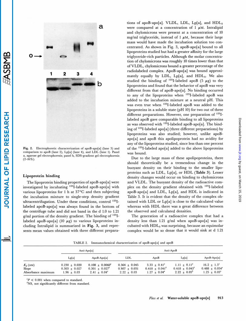
The water-soluble apoB-apo[a] complex obtained at neutral pH was characterized electrophoretically and compared to the corresponding properties of Lp[a] and apoB. Agarose gel electrophoresis confirmed the solubility properties of apoB-apo[a] in that all the material applied to the sample well entered the gel matrix (see Fig. 2a). In contrast, almost all of the apoB applied to the sample well did not enter the gel probably because of extensive self- association. ApoB-apo[a] did not exhibit a discrete band like Lp[a] upon agarose electrophoresis but had a broad, diffuse band with lesser mobility than that of Lp[a]. Elec- trophoresis of apoB-apo[a] and apoB on 2-16% poly- acrylamide gradient gels under nondenaturing conditions indicated that neither apolipoprotein entered the gel matrix which was in contrast to Lp[a] and LDL. How- ever, in the presence of SDS, both apoB-apo[a] and apoB had the same mobility as their respective parent lipo- proteins on 2-16% gradient gels (Fig. 2b).
Immunochemical characterization
ApoB-apo[a] was characterized immunochemically using two ELISAs that were specific for the two different apolipoproteins of Lp[a]. In one assay, apoB-apo[a] was compared to Lp[a] using both primary and secondary an-
TABLE 1 . Solubility of apoB-lOO-apo[a] as compared to apoB-100
Delipidation Procedure ApoB- 100- Aporal ApoB-100
Ethanol-ether 3: 1 (v/v)
Aqueous extraction, precipitation, - 2OoC Soluble Insoluble
ethanol-ether 1 : 1 + 1 :3 (v/v), pH 8.0, 4OC Soluble Insoluble
ethanol-ether 1 : 1 + 1 :3 (v/v), pH 11.0, 4OC Soluble Soluble
Aqueous extraction,
5 0.20
0 In
6.0 6.8 7.6 8.4 9.2 10.0
PH Fig. 1. Solubility properties of apoB-apo[a] and apoB as a function of pH. Various concentrated phosphate and Tris buffers were added to the apolipoproteins to an ionic strength of 0.05. Protein concentration was 0.5 mg/ml. Starting pH of apoB-apo[a] was pH 8.0 and that of apoB was pH 10. The solutions were allowed to sit for 1 h at room temperature before their turbidity was measured at 500 nm. ApoB-apo[a], circles; apoB, squares.
tibodies specific to apo[a]. In the second assay, apoB- apo[a], apoB, and Lp[a] were compared to LDL using primary and secondary antibodies specific to apoB. The results of both assays are summarized in Table 2. The water-soluble apoB-apo[a] complex had an avidity for anti-apo[a] IgG that was actually twofold greater than that of Lp[a]. The slopes of the transformed titration curves were identical indicating that the epitopes recog- nized by anti-apo[a] on Lp[a] and apoB-apo[a] are simi- lar. In conjuction with the higher avidity, the 23% higher absorbance maximum attained with apoB-apo[ a] suggests that on delipidation of Lp[a] additional epitopes on apo[a] were unmasked that were inaccessible in the native molecule. In marked contrast to apo[a], the immunoreac- tivity of the apoB portion of the complex with anti-apoB was greatly reduced. Thus, the avidity of apoB-apo[a] for anti-apoB was approximately 30 times lower than LDL, 15 times lower than Lp[a], and even 5 times lower than that of apoB. The latter finding was surprising since apoB is not soluble at neutral pH and was kept in 0.5 mM N- ethyl morpholine, pH 10, before being serially diluted into 0.1 M NaHC03, 0.5 M NaC1, 1% BSA, pH 8.1. The slope of the transformed dose-response curve obtained with apoB-apo[a] also differed from those obtained with LDL, Lp[a], and apoB which were not significantly differ- ent. This altered dose-response may be the result of con- formational changes affecting some of the apoB epitopes upon delipidation of Lp[a]. In addition, the almost two- fold lower absorbance maximum attained with apoB-apo[ a] in comparison to LDL or Lp[a] indicates that a large number of apoB epitopes are either inaccessible or altered to such an extent that they are no longer recognizable by antibodies. A similar decrease in the absorbance maxi- mum was obtained with apoB.
912 Journal of Lipid Research Volume 31, 1990
by guest, on March 15, 2019
ww
w.jlr.org
Dow
nloaded from
1 2 3 4 " -
b
Fig. 2. Electrophoretic characterization of apoB-apo[a] (lane 3) and comparison to apoB (lane 2). LpIa] (lane 4), and LDL (lane 1). Panel a, agarose g e l electrophoresis; panel b. SDS-gradient 6 1 electrophoresis (2-16%).
Lipoprotein binding
The lipoprotein binding properties of apoB-apo[a] were investigated by incubating '251-labeled apoB-apo[a] with various lipoproteins for 1 h at 37OC and then subjecting the incubation mixture to single-step density gradient ultracentrifugation. Under these conditions, control '"I- labeled apoB-apo[a] was always found in the bottom of the centrifuge tube and did not band in the d 1.0 to 1.21 g/ml portion of the density gradient. The binding of '"I- labeled apoB-apo[a] (10 pg) to various lipoproteins in- cluding Intralipid is summarized in Fig. 3, and repre- sents mean values obtained with three different prepara-
tions of apoB-apo[a]. VLDL, LDL, Lp[a], and HDL3 were compared at a concentration of 1 pM. Intralipid and chylomicrons were present at a concentration of 10 mg/ml triglyceride, instead of 1 pM, because their large mass would have made the incubation solution too con- centrated. As shown in Fig. 3, apoB-apo[a] bound to all lipoproteins studied but had a greater affinity for the large triglyceride-rich particles. Although the molar concentra- tion of chylomicrons was roughly 10 times lower than that of VLDL, chylomicrons bound a greater percentage of the radiolabeled complex. ApoB-apo[a] was bound approxi- mately equally by LDL, Lp[a], and HDL,. We also studied the binding of '251-labeled apoB (5 pg) to the lipoproteins and found that the behavior of apoB was very different from that of apoB-apo[a]. No binding occurred to any of the lipoproteins when '251-labeled apoB was added to the incubation mixture at a neutral pH. This was even true when 1251-labeled apoB was added to the lipoproteins in a soluble state (pH 10) for two out of three different preparations. However, one preparation of '"1- labeled apoB gave comparable binding to all lipoproteins as was observed with '251-labeled apoB-apo[a]. The bind- ing of '251-labeled apo[a] (three different preparations) by lipoproteins was also studied; however, unlike apoB- apo[a] and apoB this apolipoprotein had no avidity for any of the lipoproteins studied, since less than one percent of the '251-labeled apo[a] added to the above lipoproteins was bound.
Due to the large mass of these apolipoproteins, there should theoretically be a tremendous change in the buoyant density on their binding to the smaller lipo- proteins such as LDL, Lp[a], or HDL (Table 3). Lesser density changes would occur on binding to chylomicrons and VLDL. The buoyant density of the radioactive com- plex on the density gradient obtained with 1251-labeled apoB-apo[a] and LDL, Lp[a], and HDL is indicated in Table 3. It is evident that the density of the complex ob- tained with LDL or Lp[a] is close to the calculated value whereas with HDL there was a great difference between the observed and calculated densities.
The generation of a radioactive complex that had a density less than 1.21 g/ml when apoB-apo[a] was in- cubated with HDL, was surprising, because an equimolar complex would be so dense that it would sink at d 1.21
TABLE 2. Immunochemical characterization of awB-awolal and a w B
K d ('"1 0.230 f 0.020 0.108 * 0.0060' 0.564 f 0.045 3.33 f 0.41' 1.11 f 0.11. 16.2 f 1.5' Slope 0.303 * 0.027 0.301 + 0.027' Or407 f 0.031 0.410 f 0.041' 0.418 f 0.043' 0.488 f 0.054' Absorbance maximum 1.96 f 0.03 2.41 f 0.04' 2.22 + 0.03 1.27 f 0.04' 2.22 f 0.05' 1.25 f 0.05'
'P < 0.001 when compared to standard. 'NS, not significantly different from standard.
Flm et at. Water-soluble apoB-apo[a] 913
by guest, on March 15, 2019
ww
w.jlr.org
Dow
nloaded from

Intralipid Chylomicron VU)L LDC Lp(a) WL
Fig. 3. Comparative binding of apoB-apo[a] to various lipoproteins and the phospholipid-triglyceride emulsion, Intralipid. 'Z51-labeled apoB-apo[a] (10 pg) was added to various lipoproteins that were present at a concentration of 1 p~ (VLDL, LDL, Lp[a], and HDL). Chylo- microns and Intralipid had a triglyceride concentration of 10 mg/ml. Final volume was 1 ml. The mixtures were incubated for 1 h at 37°C before they were subjected to density gradient centrifugation. Mean f SD of three separate experiments, each with a different prepa- ration of '251-laheled apoB-apo[a].
g/ml. Alternative explanations that could account for this anomaly are the formation of a complex consisting of several HDL particles and 1 mole apolipoprotein, or the displacement of the constitutive apolipoproteins from several HDL particles and their replacement with apoB-
When '251-labeled apoB-apo[ a] was incubated with normotriglyceridemic plasma, binding to lipoproteins was detected by density gradient ultracentrifugation. How- ever, compared to the binding observed with purified lipoproteins, the amount of apoB-apo[a] was substantially less in plasma. The mean percentage bound to lipopro- teins (d < 1.21 g/ml) was 23.0 17.7, which is two to
apo[aI.
three times less than that obtained with purified lipopro- teins (see Fig. 3). This was in spite of the fact that the con- centration of LDL and HDL in 1 ml of normolipidemic plasma was roughly two and ten times greater, respec- tively. When the binding properties of plasma obtained from a number of hypertriglyceridemic individuals were examined, it was discovered that the amount of lZ5I- labeled apoB-apo[a] bound to total lipoproteins increased substantially (see Table 4). This 2.5-fold increase was caused by additional binding to T R P and not to LDL or HDL since mean apoB and apoA-I levels were statistically unchanged, probably indicating that the concentration of LDL and HDL was not very different. TRP-bound '251-labeled apoB-apo[a] was strongly correlated with the log of the plasma triglyceride concentration (Fig. 4B) whereas the correlation of binding to total lipoprotein (d < 1.21 g/ml) (Fig. 4A) was weaker. Thus, T R P have an avidity for apoB-apo[a] both in the absence and pres- ence of plasma. In contrast to the behavior of apoB- apo[a], radioiodinated Lp[a] did not bind to TRP. Only 1.4 0.3% of a 10 pg dose bound to VLDL and 1.2% bound to chylomicrons. Binding of '251-labeled Lp[a] to T R P in plasma was even less; only 0.3 f 0.2% bound to T R P of normotriglyceridemic plasma (n = 5) and 0.6 ~f. 0.4% bound to T R P of hypertriglyceridemic plasma (n = 10).
The inhibiting effect of plasma on the binding of Iz5I- labeled apoB-apo[a] to lipoproteins was studied with VLDL and Intralipid in a series of incubation experi- ments. When these preparations were preincubated with '251-labeled apoB-apo[a] for 1 h at 37°C before the addi- tion of normolipidemic plasma (total cholesterol 163 mg/dl; triglyceride 61 mg/dl) binding of the radioactive complex to these lipid-containing particles was of the same order as shown in Fig. 3. However, when 1251- labeled apoB-apo[ a] was incubated first with normolipi- demic plasma and then followed by VLDL (1 p M ) or
TABLE 3. Alteration of lipoprotein density caused by the binding of one mole of apoB-lOO-apoIa] per mole lipoprotein
Density of Lipoprotein-ApoB-Apo[a] Complex
Hydrated Density Molecular Protein Lipoprotein of Lipoprotein Weixht Content Calculated" Measured
f/ml x 10.6 % x/ml
Chylomicronsb 0.93 100 2 0.934 < 1.020 VLDL~ 0.97 10 10 1.014 < 1.020 L D L ~ 1.035 2.5 20 1.148 1.120 Lp[aI' 1.064 3.8 25 1.155 1.143 HDL; 1.15 0.18 55 1.365 1.172
O h calculating the density of the lipoprotein apoB-apo[a] complex (with a molar ratio of l ) , lipoprotein-protein density was taken to he 1.35 g/ml with the exception of Lp[a] whose protein moiety had a density calculated to he 1.40 g/ml. The mass of the apoB-apo[a] complex was 1.1 x lo6 daltons (6).
'Data taken from (29). 'Data taken from (1 1) .
914 Journal of Lipid Research Volume 31, 1990
by guest, on March 15, 2019
ww
w.jlr.org
Dow
nloaded from

TABLE 4. Binding of 1251-labeled apoB-apo[a] to lipoproteins in the presence of normo- and hypertriglyceridemic plasma
I O 0
901 nn
Hyper (n = 10)
Plasma triglycerides Plasma total cholesterol Plasma apoB Plasma apoA-I
mg/dl
113 i 33 1809 i 3218 230 + 39 283 i 117 133 i 16 107 i 32 116 f 11 96 f 2.5
% ApoB-apo[a] bound to lipoproteins of d < 1.21 g/ml 61.3 i 19.1 ApoB-apo[a] bound to TRP 2.3 f 4.5 34.2 i 28.8
23.0 f 17 .7
1251-Labeled apoB-apo[a] (10 pg) was incubated with 1 ml plasma at 37OC. The incubation mixture was separated by single-step density gradient centrifugation. Lipoprotein-bound apoB-apo[a] was determined in the fractions with d < 1.21 g/ml. TRP-bound apoB-apo[a] represents that floating in the top two tubes (0.8 ml) of the density gra- dient. Values are given as mean f SD.
2c
A 100-
90.
80.
7 0.
r = 0.762
I75 2 225 2 5 275 3 325 3 5 375 4 425
Log Plasma Triglyceride
Fig. 4. Correlation of lipoprotein bound 1Z51-labeled apoB-apo[a] with the log of the plasma triglyceride concentration. Plasma (1 ml) from vari- ous normal and hypertriglyceridemic individuals was incubated for 1 h at 37OC with 10 pg 9-labeled apoB-apo[a] and then subjected to single- step density gradient centrifugation for 66 h, at 15OC and 39,000 rpm in the SW-40 rutor. The gradient was fractionated and lipoprotein- bound 1Z51-labeled apoB-apo[a] was determined in the fractions with density <1.21 g/ml (panel A). The top two tubes (0.8 ml) containing VLDL- or chylomicron-bound 1251-labeled apoB-apo[a] are represented in panel B.
Intralipid (10 mg/dl), then its binding was reduced from 76.3 to 12.2% for VLDL and in the case of Intralipid from 98.8 to 25.7%. This indicates that once lz5I-labeled apoB-apo[a] is bound to the lipoprotein surface it is difficult to displace by competition with other plasma components. Thus, the reduced binding appears to be caused by some interaction of lZ5I-labeled apoB-apo[a] with plasma that reduces its avidity for lipoproteins.
DISCUSSION
The results of the present studies have shown that the protein moiety of Lp[a], the apoB-apo[a] complex, is water-soluble at neutral pH after the lipid complement is removed by extraction with organic solvents. This is in contrast to apoB which, when delipidated by traditional methods using organic solvents, forms a precipitate that is insoluble in neutral buffers and can be solubilized only with the aid of denaturants, detergents, or chemical modifying agents. Physical characterization of apoB- apo[a] was very difficult because of its extremely large mass which is approximately 1 x lo6 daltons. Its failure to enter a 2-16% polyacrylamide gradient gel in the ab- sence of detergent, which is readily penetrable by Lp[a] itself, may indicate that its conformation in the lipid-free state is more extended than on the lipoprotein surface. ApoB-apo[a] was able to enter an agarose gel where it produced a diffuse band with a lower mobility than Lp[a]. It is not clear, however, whether this electrophoretic be- havior is due to self-association, conformation, or interac- tion of apoB-apo[a] with the gel matrix.
The immunochemical characterization of apoB-apo[a] provided important additional results on the physical state of the complex. When probed with antibodies spe- cific to apo[a], the parallelism of the dose-response curves
Fless et al. Water-soluble apoB-apo[a] 915
by guest, on March 15, 2019
ww
w.jlr.org
Dow
nloaded from

obtained with apoB-apo[a] and Lp[a] indicated that apo[a] has a similar conformation in these two particles. In fact, delipidation of Lp[a] appears to expose additional antigenic determinants resulting in a higher absorbance maximum and a greater avidity of anti-apo[a]-IgG for apoB-apo[a] than for Lp[a]. However, when the apoB moiety of the apoB-apo[a] complex was probed with apoB-specific antibodies, the lack of parallelism and the low absorbance maximum in comparison to LDL or Lp[aj indicate that a large number of epitopes are either inaccessible or altered.
The surprising finding of the present study was the poor immunoreactivity of apoB in apoB-apo[a] which was about 30 times lower than LDL, 15 times lower than Lp[a], and 5 times lower than that of apoB itself despite the fact that the solubility of apoB is much less than that of apoB-apola]. It is possible that this reduced immuno- reactivity is due to a greater degree of interference of apo[a] with the interaction of anti-apoB and apoB epi- topes in the apolipoproteins as compared to the native lipoproteins. Since apo[a] confers water solubility to apoB in the complex, the apoB epitopes near the disulfide link- ages may be the ones that are not altered or rendered in- accessible due to self-association of the hydrophobic lipid binding regions upon delipidation of Lp[a]. The relative proportion of these still immunoreactive epitopes would increase upon delipidation and be exactly those epitopes that are sterically hindered or conformationally altered by the attachment of apo[a]. Overall, the evidence presented here is consistent with a model of apoB-apo[a] in which the hydrophobic lipid binding regions of apoB are self- associated, and are shielded from the aqueous environ- ment by the hydrophilic regions of apoB and apo[a], with the freely accessible and very hydrophilic apo[a] provid- ing water solubility.
The immunoreactivity of the apoB epitopes in native Lp[a] was modestly diminished in relation to that in LDL. This finding is consistent with those of Zawadzki et al. (30) and Gries et al. (31) who compared the immuno- reactivity of LDL and Lp[aj with monoclonal antibodies to apoB. Both groups found enhanced expression of some apoB epitopes in Lp[a], and diminished immunoreactiv- ity in others relative to those in LDL. However, because monoclonal antibodies probe only a limited portion of apoB, the comparison of the two lipoproteins with poly- clonal antibodies gives a better overall picture of the im- munoreactivity of apoB in Lp[a].
When the lipid-free apoB-apo[a] complex was in- cubated with various lipoproteins and the washed phospholipid- triglyceride emulsion Intralipid, we were able to demonstrate a variable degree of binding that was greatest with the triglyceride-rich particles. The ability of apoB-apo[a] to associate with lipoproteins persisted even in the presence of plasma and after density gradient cen-
trifugation for approximately 2 x lo7 g-h. Plasma reduced the avidity of apoB-apo[a] for lipoproteins and Intralipid through a process which may involve the coat- ing of lipid binding regions of apoB with unknown plasma components. Possible candidates that might occupy these sites are the amphipathic apoC peptides, apoA-I, apoA- IV, and apoE, which have all been shown to be either dis- placeable from, or exchangeable among, lipoproteins. Conversely, they could also saturate to some extent the lipoprotein or lntralipid surface and thereby reduce the binding of apoB-apo[ a]. Since neither radioionated LDL, Lp[a], nor apo[a] bound to these particles, it is likely that apoB is responsible for the binding. Ye et al. (32) have suggested that apo[a] may interact with the lysine-rich domains of apoB by means of its kringle-4 domains. However, inclusion of the lysine analogue 6-amino- hexanoic acid (20 mM) in the incubation mixture did not affect the binding, indicating that the lysine binding sites of apo[a] were not involved.
This study was undertaken in part by the observation of Bersot et al. (7) that fat-feeding induces the appearance of apo[a] in postprandial chylomicron remnants. The ac- tual quantity of apoB-apo[a] in these particles was not de- termined in that study. We, however, have found that the amount of apoB-apo[a] present in TRP of individuals we studied is measurable but is so small that it does not con- tribute significantly to the total plasma Lp[a] (ref. 10; and G. Fless, M. Snyder, and A. M. Scanu, unpublished ob- servations). Nevertheless, it is apparent that the apoB- apo[a] complex can be affiliated either with triglyceride- rich lipoproteins or the classic Lp[a] that has a core made predominantly of cholesteryl esters. It is possible that these two distinct classes of lipoproteins are separately as- sembled and secreted by the liver, which is the main source of Lp[a] synthesis (4, 33). There is evidence that Lp[a] may be secreted by the liver directly (34). Alterna- tively, the liver could also produce apoB-apo[a] contain- ing TRP in response to the intestinal absorption of fats. These species could be rapidly taken up by the liver through the remnant-receptor pathway without giving rise to Lp[a] upon their catabolism. The low apoB-apo[a] content of TRP would be in favor of such a mechanism. Relevant to this discussion is the issue relating to the exis- tence of lipid-free or lipid-poor apoB-apo[a] in plasma. Variable amounts of apoB-apo[a] have been detected in the d > 1.21 g/ml ultracentrifugal bottom fraction (1, 8-10); however, due to stability problems such as proteoly- sis, the meaning of this observation is difficult to assess. Since there is no evidence that apoB-apo[a] can be dis- sociated from Lp[a] during ultracentrifugation, it is tempting to speculate that the lipid-poor apoB-apo[a], if it does exist in plasma, represents a nascent complex newly secreted by the liver that is destined to undergo fur- ther remodeling in the blood. Thus, it is possible that dur-
916 Journal of Lipid Reseamh Volume 31, 1990
by guest, on March 15, 2019
ww
w.jlr.org
Dow
nloaded from

ing postprandial hyperlipidemia a portion of this lipid- poor apoB-apo[ a] complex could become incorporated into TRP upon their entry into the circulation. Whether this hypothetical mechanism actually occurs in vivo re- mains to be established. M
HL
1.
2.
3.
4.
5.
6.
7.
8.
9.
10.
11.
12.
The authors wish to acknowledge the technical assistance of Margaret L. Snyder. We also thank Susan Hutchison and Barbara Kass for the preparation of the manuscript. This research was supported by USPHS Program Project Grant
18577. Manuscrzpt received 2 October 1989 and in revised form 2 January 1990.
REFERENCES
Gaubatz, J. W., C. Heideman, A. M. Gotto, Jr., J. D. Mor- risett, and G. H. Dahlen. 1983. Human plasma lipoprotein [a]. J. Biol. Chem. 258: 4582-4589. Utermann, G., and W. Weber. 1983. Protein composition of Lp[a] lipoprotein from human plasma. FEBS Lett. 154:
Eaton, D. L., G. M. Fless, W. J. Kohr, J. W. McLean, Q-T. Xu, C. T. Miller, R. M. Lawn, and A. M. Scanu. 1987. Partial amino acid sequence of apolipoprotein[ a] shows that it is homologous to plasminogen. Proc. Natl. Acad Sci. USA
McLean, J. W., J. E. Tomlinson, W-J. Kuang, D.L. Eaton, E. Y. Chen, G. M. Fless, A. M. Scanu, and R. M. Lawn. 1987. cDNA sequence of human apolipoprotein[a] is homologous to plasminogen. Nature. 330 132-137. Kratzin, H., V. W. Armstrong, M. Niehaus, N. Hilsch- mann, and D. Seidel. 1987. Structural relationship of an apolipoprotein[a] phenotype (570 kDa) to plasminogen: homologous kringle domains are linked by carbohydrate- rich regions. Biol. Chem. Hoppe-Syler. 368: 1533-1544. Fless, G. M., M. E. ZumMallen, and A. M. Scanu. 1986. Physiochemical properties of apolipoprotein[ a] and lipo- protein[a- ] derived from the dissociation of human plasma Lp[a]. J. Biol. Chem. 261: 8712-8718. Bersot, T. P., T. L. Innerarity, R. E. Pitas, S. C. Rall, L. J. Weisgraber, and R. W. Mahley. 1986. Fat feeding in humans induces lipoproteins of density less than 1.006 that are enriched in apolipoprotein[a] and that cause lipid ac- cumulation in macrophages. J. Clin. Invest. 77: 622-630. Duvic, C. R., G. Smith, W. E. Sledge, L. T. Lee, M. D. Murray, P. S. Roheim, W. R. Gallaher, and J. J. Thomp- son. 1985. Identification of a mouse monoclonal antibody, LHLP-1, specific for human Lp[a]. J. Lipid Res. 26:
Gries, A., J. Nimpf, M., Nimpf, H. Wurm, and G. M. Kostner. 1987. Free and apoB-associated Lp[a]-specific pro- tein in human serum. Clin. Chim. Acta. 164: 93-100. Fless, G. M., M. L. Snyder, and A. M. Scanu. 1989. Enzyme-linked immunoassay for Lp[a]. J. Lipid Res. 30: 651-662. Fless, G. M., C. A. Rolih, and A. M. Scanu. 1984. Heter- ogeneity of human plasma lipoprotein[a]. J. Biol. Chem. 259: 11470-11478. Fless, G. M., M. E. ZumMallen, and A. M. Scanu. 1985. Isolation of apolipoprotein [a] from lipoprotein[a]. J. Lipid
357-361.
84: 3224-3228.
540-548.
Res. 26: 1224-1229.
13. Armstrong, V. W., A. K. Walli, and D. Seidel. 1985. Isola- tion, characterization, and uptake in human fibroblasts of an apo[a]-free lipoprotein obtained on reduction of lipo- protein[a]. J. Lipid Res. 26: 1314-1323.
14. Weinberg, R. W., and A. M. Scanu. 1983. Isolation and characterization of human apolipoprotein A-IV from lipo- protein-depleted serum. J. Lipid Res. 24: 52-59.
15. Fless, G. M. 1971. Preparation and physico-chemical characterization of apo-low density lipoprotein of human plasma. PhD Thesis, University of Illinois, Urbana- Champaign. 47-48.
16. Karlin, J., D. Juhn, G. Fless, A. M. Scanu, and A. H. Rubenstein. 1978. Measurement of rhesus monkey (Macaca mulatta) apolipoprotein B in serum by radioimmunoassay: comparison of immunoreactivities of rhesus and human low density lipoproteins. J. Lipid Res. 19: 197-206. Scanu, A. M., and C. Edelstein. 1971. Solubility in aqueous solutions of ethanol of the small molecular weight peptides of serum very low density and high density lipoproteins. Anal. Biochem. 41: 576-588.
18. McFarlane, A. S. 1958. Efficient trace-labeling of proteins with iodine. Nature. 182: 53-57.
19. Bilheimer, D. W., S. Eisenberg, and R. I. Levy. 1972. The metabolism of very low density lipoprotein proteins. I. Preliminary in vitro and in vivo observations. Biochim. Biophys. Acta. 260, 212-221.
20. Tijssen, P. 1985. Practice and Theory of Enzyme Im- munoassays. Elsevier, Amsterdam.
21. Lowry, 0. H., N. J. Rosebrough, A. L. Farr, and R. J. Ran- dall. 1951. Protein measurement with the Folin phenol reagent. J. Biol. Chem. 193: 265-275.
22. Markwell, M. A. K., S. M. Haas, L. L. Bieber, and N. E. Tolbert. 1987. A modification of the Lowry procedure to simplify protein determination in membrane and lipopro- tein samples. Anal. Biochem. 87: 206-210.
23. Bartlett, G. R. 1959. Phosphorus assay in column chro- matography. J. Biol. Chem. 234: 466-468.
24. Allain, C. C., L. S. Poon, C. S. Chan, W. Richmond, and P. C. Fu. 1974. Enzymatic determination of total serum cholesterol. Clin. Chem. 20: 470-475.
25. Gallo, L. L., R. Atasoy, G. V. Vahouny, and C. R. Tread- well. 1978. Enzymatic assay for cholesterol ester hydrolase activity. J. Lipid Res. 19: 913-916.
26. Wahlefeld, A. W. 1974. Triglycerides: determination after enzymatic hydrolysis. In Methods of Enzymatic Analysis. H. U. Bergmeyer, editor. 2nd English edition. Academic Press, New York. 1831-1835.
27. Albers, J. J., V. G. Cabana, and W. Hazzard. 1975. Im- munoassay of human plasma apolipoprotein B. Metabolism.
28. Albers, J. J., P. W. Wahl, V. G. Cabana, W. R. Hazzard, and J. J. Hoover. 1976. Quantitation of apolipoprotein A-I of human plasma high density lipoprotein. Metabolism. 25:
29. Shen, B. W., A. M. Scanu, and E J. Kezdy. 1977. Structure of human serum lipoproteins inferred from compositional analysis. Proc. Natl. Acad. Sci. USA. 74: 837-841.
30. Zawadzki, Z., E Terce, L. J. Seman, R. T. Theolis, W. C. Breckenridge, R. W. Milne, and Y. L. Marcel. 1988. The linkage with apolipoprotein[a] in lipoprotein[a] modifies the immunochemical and functional properties of apolipo- protein B. Biochemistv. 27: 8474-8481. Gries, A., C. Fievet, S. Marcovina, J. Nimpf, H. Wurm, H. Mezdour, J. C. Fruchart, and G. M. Kostner. 1988. Inter-
17.
24: 1339-1351.
633-644.
31.
Fless et al. Water-soluble apoB-apo[a] 917
by guest, on March 15, 2019
ww
w.jlr.org
Dow
nloaded from

action of LDL, Lp[a], and reduced Lp[a] with monoclonal antibodies against apoB. J Lipid Res. 29: 1-8.
32. Ye, S. Q, V. N. Trieu, D. L. Stiers, and W. J. McConathy. 1988. Interactions of low density lipoproteinsz and other apolipoprotein B-containing lipoproteins with lipoprotein[a] . J. Biol. Chem. 263: 6337-6343.
33. Kraft, H. G., H. J. Menzel, E Hoppichler, W. Vogel, and G. Utermann. 1989. Changes of genetic apolipoprotein phenotypes caused by liver transplantation. Implications for apolipoprotein synthesis. J. Clin. Invest. 83: 137-142.
34. Rainwater, D. L., and R. E. Lanford. 1989. Production of lipoprotein[a] by primary baboon hepatocytes. Biochim. Biophys. Acta. 1003: 30-35.
918 Journal of Lipid Research Volume 31, 1990
by guest, on March 15, 2019
ww
w.jlr.org
Dow
nloaded from