monoclonal antibodies and immunochemical techniques
TRANSCRIPT
I Government Gouvernementof Canada du Canada
Canadian serviceForestry canadien desService forets
Monoclonal antibodiesand immunochemicaltechniques: applicationsin forestry researchLeslie Ann Mitchell
Information Report BC-X-258Pacific Forest Research Centre
Monoclonal antibodies and immunochemical techniques:
applications in forestry research
Leslie Ann Mitchell1
Canadian Forestry ServicePacific Forest Research Centre
BC-X-258
1985
1 Leslie Ann Mitchell is a Natural Sciences and Engineering Research Council Visiting Fellow at the Pacific Forest Research Centre

Canadian Forestry ServicePacific Forest Research Centre
506 West Burnside RoadVictoria, B.C.
V8Z IM5
© Minister of Supply & Services Canada, 1985ISSN 0705-3274
ISBN 0-662-13593-8Cat. No. Fo46-17/258E
Abstract
Immunologic techniques employing polyclonalantisera and monoclonal antibodies as sensitive,highly specific probes for molecules in complexbiological systems are currently in use in manyresearch laboratories. For those forestry researchers unfamiliar with these techniques, thisreport describes polyclonal antisera and monoclonal antibodies and the principles behind their usein sensitive techniques such as radioimmunometric assay (RIA), enzyme-linked immunosorbentassay (ELISA), immunofluorescence, immunochemical localization of structures in light or electron microscopy, and immunoaffinity chromatography. The use of antisera and monoclonal antibodies in conjunction with other high-resolution techniques (such as single- and two-dimensional polyacrylamide gel electrophoresis) in immunoprecipitation and immunoblotting (Western blotting) techniques is also described. Thecontribution of monoclonal antibodies and antisera to gene identification and localization ingenetic engineering is discussed. Examples ofcurrent and ·potential applications of these immunologic techniques in agricultural and forestryresearch are given.
3
Resume
Des techniques immunologiques utilisant desimmuns-serums polyclonaux et des anticorpsmonoclonaux comme outils sensibles et extremement specifiques d'analyse des moleculesdans des systemes biologiques complexes sontemployees dans de nombreux laboratoires derecherche. Nous decrivons a l'intention des chercheurs dans Ie domaine forestier qui ne seraientpas familiers avec ces techniques ce que sont cesimmuns-serums et ces anticorps et les principesde leur utilisation dans des techniques sensiblescomme les dosages radio-immunologiques(RIA), les tests immuno-enzymatiques(ELISA), l'immunofluorescence, la localisationimmunochimique de structures sous microscopeoptique ou electronique et l'immunochromatographie d'affinite. Nous traitons egalement destechniques d'immunoprecipitation et des techniques immunologiques avec transfert sur papier("Western blotting techniques") qui font appelad'autres techniques ahaute resolution (commeI'electrophorese unidimensionnelle et bidimensionnelle en gel de polyacrylamide). Nous examinons l'apport des anticorps monoclonaux et desimmuns-serums al'identification et a la localisation des genes en genie genetique. Nous etudionsdes exemples d'applications actuelles et potentielles de ces techniques dans la recherche interessant les domaines agricole et forestier.
4
Table of Contents
Page
Introduction 6
The immune response....................................................................... 6Antigens 6Antibodies 7Antisera 9
Monoclonal antibodies 10Monoclonal antibodies vs. antisera 10Procedure for producing monoclonal antibodies 10Advantages of monoclonal antibodies 13
Immunochemical techniques and their applications 13Radioimmunometric assay (RIA) 14Enzyme-linked immunosorbent assay (ELISA) 14Other immunoassays............................. 14Immunofluorescence and immunohistochemistry 18Immunoaffinity chromatography..... 19Immunoblotting techniques........................................................ 19Immunoprecipitation 20Two-dimensional polyacrylamide gel electrophoresis 21Genetic engineering.................................................................... 23
Conclusion 23
References........................................................................................ 24
5
Figures
Page
1 Antigens................................................................................... 62 Antibody structure.................................................................... 83 Clonal selection: An antibody response to a multivalent
antigen.................................................................................. 84 Clonal expansion...................................................................... 95 Preparing an antiserum............................................................. 96 Monoclonal antibodies vs. antisera 117 Procedure for producing monoclonal antibodies 128 Radioimmunometric assays (RIAs) 149 Enzyme-linked immunosorbent assays (ELISAs) 151Q Sandwich assays........................................................................ 1611 Competitive inhibition assays 1612 Immunoaffinity chromatography 1813 Immunoblotting 1914 Immunoprecipitation................................................................ 2015 Two-dimensional polyacrylamide gel electrophoresis of
mouse serum proteins........................................................... 2116 Monoclonal antibodies and genetic engineering 22
Tables
Page
1 Some properties of human antibody classes............................... 7
Introduction
Forest biology researchers often require sensitive, rapid and inexpensive methods for locating,identifying and measuring both ~icroorganisms
and biochemicals in plant and insect tissues, soilsor the environment. For example, accurate detection methods are needed for pathogens suchas bacteria and fungi in seeds, seedlings andmature tree tissues so that early and appropriatecontrol measures may be taken and also for seedcertification. Sensitive methods are also requiredfor monitoring levels of pathogens such as fungior levels of biologic control agents such asviruses in insect tissues for the evaluation andmanagement of insect-borne disease or biologicalcontrol programs. Forestry researchers are alsoseeking high-resolution techniques to facilitatestudies of species relationships (for instance,among insect pests, seeds of different origin,. pollens and races of fungal pathogens differing invirulence or host range) or to quantitate growthregulators (e.g., cytokinins influencing coneproduction) or other biochemical changes suchas the synthesis of phytoalexins in host tissues inresponse to fungal, bacterial or other pathogens,Forest and agriculture biologists are also developing techniques which permit the identification ofproteins (or other biological substances) associated with disease resistance to facilitate the classification of superior genetic stock and ultimately toidentify and isolate resistance genes for use ingenetic engineering.
Sensitive, high-resolution techniques originallydeveloped for the study of human and animal systems are being adapted for use in agriculture andforestry research. Immunologic techniquesemploying antisera or monoclonal antibodies,used alone or in conjunction with other highresolution systems (such as polyacrylamide gelelectrophoresis) constitute one group of thesenewly adapted technologies. Although they arenot without some problems and limitations,these techniques are very powerful and extremelysensitive.
This information report, which is directed atforestry researchers who are unfamiliar with immunologic techniq~es, describes the biochemicalnature and derivation of antisera and monoclonalantibodies and their use as sensitive and specificprobes in a variety of procedures.
6
The immune response
Immunology made its first impact on hunlanitywhen it was observed that vaccination with liveor heat-killed microorganisms would protectpeople from the diseases caused by these pathogens. The word "immunology" derives from theLatin "immunitas" meaning "freedom frompublic service" which later came to be associatedwith freedom from disease. Although immunology originally dealt with the body's response to disease organisms, it soon became evident that allvertebrates (and some invertebrates) are capableof recognizing foreign substances and of makingspecific responses against them.
Antigens
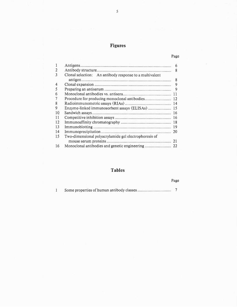
Foreign substances that induce immune reosponses are called antigens. Almost any substance may be antigenic (immunogenic) if theanimal's immune system perceives it as foreign.Antigens may be proteins, lipids, carbohydratesor even small molecules. For practical purposes,an antigen may be considered as any substancethat a researcher might wish to detect using immunological techniques (e.g., a virus, fungus,enzyme, growth regulator or insect component).
Most antigens are multivalent, i.e., they containseveral regions called antigenic sites (Fig. 1).Each antigenic site on the antigen molecule iscapable of stimulating a different aspect of theimmune system.
Antigenic sites
t \
Antigenmolecule
Fig. 1. Antigens. An antigen is a foreign moleculewhich, upon entering the body, induces animmune response. Most antigens are multivalent, i.e., they have several antigenic subregions called antigenic sites.
Antibodies
White blood\cells called lymphocytes respond toforeign substances entering our bodies. Thesecells are found in lymphoid organs such as thespleen, thymus gland, lymph nodes, tonsils andappendix as well as in t'he blood.
Antigens cause some lymphocytes to proliferateand change into cells which synthesize and secrete substances called antib09ies (or immunoglobulins, abbreviated "Ig"). The antibodiesbind specifically to the foreign molecule andeither neutralize its activity or clear it from t.hebody. Antibodies are fairly large, multichaincarbohydrate-containing proteins ranging inmolecular weight from 150 000 to 900 000 Daltons. There are several different classes ofantibodies. Some physical and biological properties of human antibodies are listed in Table 1.Ihe immunoglobulin classes differ in molecularweight, carbohydrate content and serum half-life.
Antibodies are found in various places within thebody: in the blood, lymphoid organs, gut and respiratory tract linings. Different classes of antibodies function somewhat differently in the clearanceof foreign substances from the body. Also, morethan one class of antibody can be generated in animmune response. However, all antibody classes
7
have one thing in common: their absolute specificity for the antigenic substance which elicitedtheir formation.
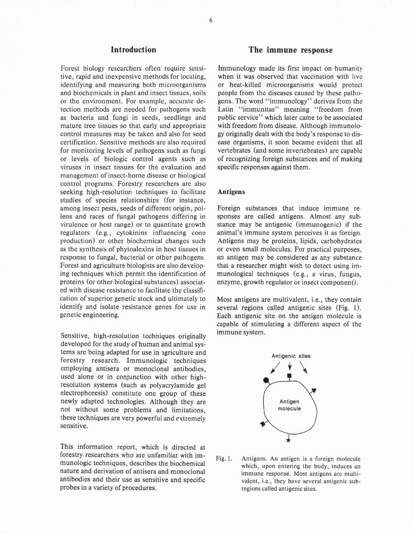
Figure 2 is a schematic representation of an antibody molecule. Antibodies of all classes aremade up of similar unit structures. For exampleIgG antibodies resemble the structure shown inFigure 2, while IgM antibodies contain five suchunits linked together as a pentamer. An, antibodymolecule is composed of two light and two heavypolypeptide chains held together by disulfidebonds. The sequence of amino acids in theamino-terminals (NT) of the heavy and lightchains are unique for a given species of antibodywhile the remaining amino acids are constant fora given heavy chain class or light chain type. Theunique sequences of amino acids form the antigen binding site of the antibody. There are twoidentical binding sites per antibody monomer asrepresented in Figure 2. They are folded in sucha way that they are complementary to a particularantigenic site on the antigen to which they bind.Thus, the size, shape and amino acid lining of theantigen binding site are different for everyantibody. This provides antibody molecules withtheir unique specificites. Therefore, differentmolecular antibody species are able to recognizeunique regions on foreign substances which arenot distinguishable by other means. For exam-
Table 1. Some properties of human antibody classes
Molecular ConcentrationAntibody weight Carbohydrate in serum Half-life
class (Daltons) content (%) (mg/ml) (days)
IgG1 146000 2-3 9 12±5IgG2 146 boo 2-3 3 20±2IgG3 165 000 2-3 1 7±1IgG4 146000 2-3 0.5 21±2.5IgM 970000 9-12 1.2 5IgA 160000 7-11 2.0 6IgD 170000 10-12 0.06 3IgE 188 000- 12 0.0002 2.5-4
196000
Adapted from Nisonoff (1982)
8
Antigen-bindingsites
Fig. 2. Antibody structure. The immunoglobulin G(IgG) molecule represented here is the prototype of antibody structure. It consists oftwo light chains (L) and two heavy chains(H) covalently-linked. The amino-terminal(NT) regions of both light and heavy chainsare highly variable (V) in amino acid sequences, while the carboxy-terminal (CT)ends of the chains are relatively constant (C)for a given class of antibody molecule.
~/ Lymphocyte1
c> ·~Antigen Ly mPhOCy te
2
Lymphocyte3
----~
Antigen Clonal ~ Specific antibodycommitment ~ expansion synthesis
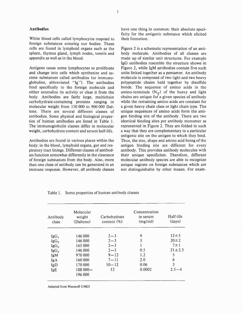
Fig. 3. Clonal selection: an antibody response to a multivalent antigen.When a multivalent antigen is injected into an animal, each antigenic site is recognized by a different lymphocyte which bearson its surface (as an antigen receptor) antibody moleculeswhich fit onto that particular antigenic site. When the surfaceantibody (receptor) on the lymphocyte binds to the antigenicsite; the lymphocyte is triggered into cell division (clonalexpansion) and antibody synthesis.
Antigen
9
1. Inject antigen (e.g.Sirococcus) emulsifiedwith adj uvant
Lymphocyte
Cell division
2. Wait 2-4 weeks and repeatthe injection
3. Wait 2-4 weeks and bleedrabbit
.~
~ X~
1~. ~
)' 1 X X)'1 )' ;rAntibodies
~Serum :
4. Allow blood to clot thenremove seru m and testfor antibody content
Anti-Sirococcus
Antiserum
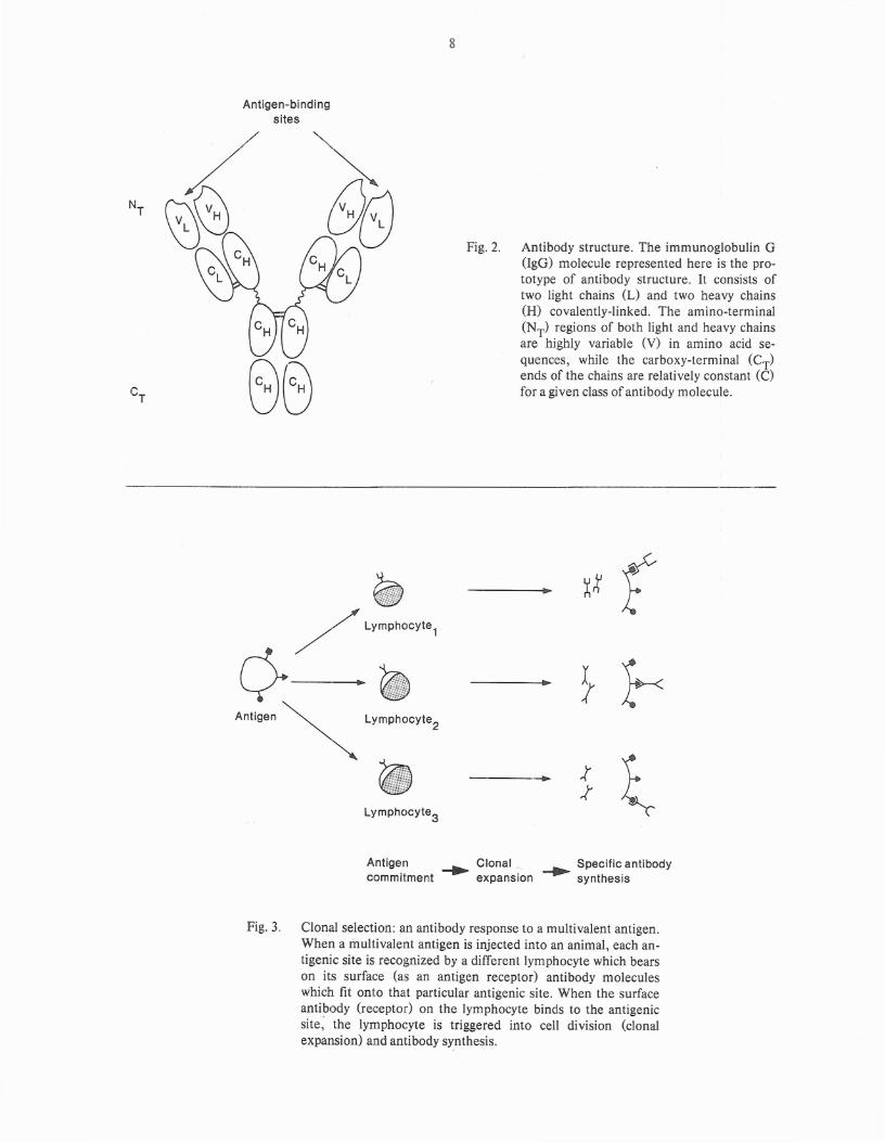
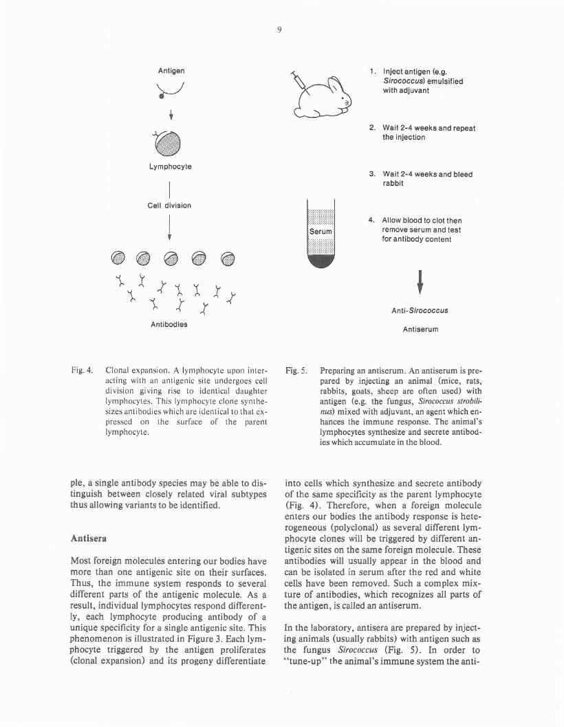
Fig. 4. Clonal expansion. A Iynlphocyte upon interacting with an antigenic site undergoes celldivision giving rise to identical daughterIynlphocytes. This Iynlphocyte clone synthesizes antibodies which are identical to that expressed on the surface of the parentIynlphocyte.
pIe, a single antibody species may be able to distinguish between closely related viral subtypesthus allowing variants to be identified.
Antisera
Most foreign molecules entering our bodies havemore than one antigenic site on their surfaces.Thus, the immune system responds to severaldifferent parts of the antigenic molecule. As aresult, individual lymphocytes respond differently, each lymphocyte producing antibody of aunique specificity for a single antigenic site. Thisphenomenon is illustrated in f'igure 3. Each lymphocyte triggered by the antigen proliferates(clonal expansion) and its progeny differentiate
Fig. 5. Preparing an antiserum. An antiserum is prepared by injecting an animal (mice, rats,rabbits, goats, sheep are often used) withantigen (e.g. the fungus, Sirococcus strobi/inus) mixed with adjuvant, an agent which enhances the immune response. The animal'slymphocytes synthesize and secrete antibodies which accumulate in the blood.
into cells which synthesize and secrete antibodyof the same specificity as the parent lymphocyte(Fig. 4). Therefore, when a foreign moleculeenters our bodies the antibody response is heterogeneous (polyclonal) as several different lymphocyte clones will be triggered by different antigenic sites on the same foreign molecule. Theseantibodies will usually appear in the blood andcan be isolated in serum after the red and whitecells have been removed. Such a complex mixture of antibodies, which recognizes all parts ofthe antigen, is called an antiserum.
In the laboratory, antisera are prepared by injecting animals (usually rabbits) with antigen such asthe fungus Sirococcus (Fig. 5) . In order to"tune-up" the animal's immune system the anti-
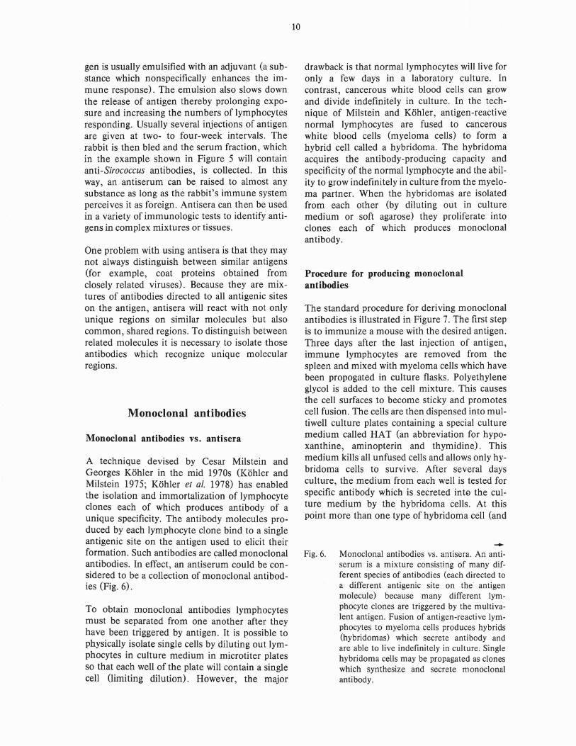
gen is usually emulsified with an adjuvant (a substance which nonspecifically enhances the immune response). The emulsion also slows downthe release of antigen thereby prolonging exposure and increasing the numbers of lymphocytesresponding. Usually several injections of antigenare given at two- to four-week intervals. Therabbit is then bled and the serum fraction, whichin the example shown in Figure 5 will containanti-Sirococcus antibodies, is collected. In thisway, an antiserum can be raised to almost anysubstance as long as the rabbit's immune systemperceives it as foreign. Antisera can then be usedin a variety of immunologic tests to identify antigens in complex mixtures or tissues.
One problem with using antisera is that they maynot always distinguish between similar antigens(for example, coat proteins obtained fromclosely related viruses). Because they are mixtures of antibodies directed to all antigenic siteson the antigen, antisera will react with not onlyunique regions on similar molecules but alsocommon, shared regions. To distinguish betweenrelated molecules it is necessary to isolate thoseantibodies which recognize unique molecularregions.
Monoclonal antibodies
Monoclonal antibodies vs. antisera
A technique devised by Cesar Milstein andGeorges Kohler in the mid 1970s (Kohler andMilstein 1975; Kohler et al. 1978) has enabledthe isolation and immortalization of lymphocyteclones each of which produces antibody of aunique specificity. The antibody molecules produced by each lymphocyte clone bind to a singleantigenic site on the antigen used to elicit theirformation. Such antibodies are 9alled monoclonalantibodies. In effect, an antiserum could be con~
sidered to be a collection of monoclonal antibodies (Fig. 6).
To obtain monoclonal antibodies lymphocytesmust be separated from one another after theyhave been triggered by antigen. It is possible tophysically isolate single cells by diluting out lymphocytes in culture medium in microtiter platesso that each well of the plate will contain a singlecell (limiting dilution). However, the major
10
drawback is that normal lymphocytes will live foronly a few days in a laboratory culture. Incontrast, cancerous white blood cells can growand divide indefinitely in culture. In the technique of Milstein and Kohler, antigen-reactivenormal lymphocytes are fused to cancerouswhite blood cells (myeloma cells) to form ahybrid cell called a hybridoma. The hybridomaacquires the antibody-producing capacity andspecificity of the normal lymphocyte and the ability to grow indefinitely in culture from the myeloma partner. When the hybridomas are isolatedfrom each other (by diluting out in culturemedium or soft agarose) they proliferate intoclones each of which produces monoclonalantibody.
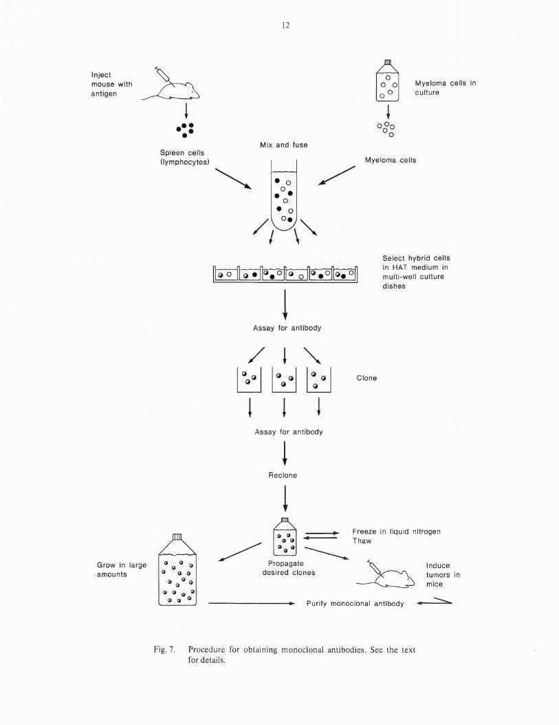
Procedure for producing monoclonalantibodies
The standard procedure for deriving monoclonalantibodies is illustrated in Figure 7. The first stepis to immunize a mouse with the desired antigen.Three days after the last injection of antigen,immune lymphocytes are removed from thespleen and mixed with myeloma cells which havebeen propogated in culture flasks. Polyethyleneglycol is added to the cell mixture. This causesthe cell surfaces to become sticky and promotescell fusion. The cells are then dispensed into multiwell culture plates containing a special culturemedium called HAT (an abbreviation for hypoxanthine, aminopterin and thymidine). Thismedium kills all unfused cells and allows only hybridoma cells to survive. After several daysculture, the medium from each well is tested forspecific antibody which is secreted into the culture medium by the hybridoma cells. At thispoint more than one type of hybridoma cell (and
.......Fig. 6. Monoclonal antibodies vs. antisera. An anti
serum is a mixture consisting of many different species of antibodies (each directed toa different antigenic site on the· antigenmolecule) because many different lymphocyte clones are triggered by the multivalent antigen. Fusion of antigen-reactive lymphocytes to myeloma cells produces hybrids(hybridomas) which secrete antibody andare able to live indefinitely in culture. Singlehybridoma cells may be propagated as cloneswhich synthesize and secrete monoclonalantibody.
Antigen
11
Antigenicsites
Inject mouse with antigen
Myeloma cells
In vivo
Lymphocytes
I I IAntibody synthesis
Fuse
+ o
I~
o
I~
o
I~
In vitro
Antibodies of3 specificitiessecreted intoblood serum
Antiserum
Hybridoma cells
2 3
Clones
Propagate in cultureRelease antibody intoculture fluid
Each antibodyspecies reactswith its specificantigenic site
Antigen Monoclonal antibodies
12
Injectmouse withantigen
•••••
Myeloma cells inculture
Mix and fuseSpleen cells(lymphocytes)
• °0 •• o
• 0
/ o. '"I \
Myeloma cells
Select hybrid cellsin HAT medium inmulti-well culturedishes
Assay for antibody
Clone
Assay for antibody
Reclone
Inducetumors inmice
• :::=:-,.,
t~
• Freeze in liquid nitrogen~ ~~~~ .. Thaw
~ ~ ~ ~ -------..Propagate ~~
desired clones ~
---------... Purify monoclonal antibody
~ ~ ~ Q
Q ~ ~
Q ~ Q ~
~ ~ ~ Q~ ~ ~
Grow in largeamounts
Fig. 7. Procedure for obtaining nlonoclonal antibodies. See the textfor details.
therefore, more than one species of antibody)may be in each well. Cells from each positive wellare then cloned by limiting dilution in culturemedium. The cells are allowed to proliferate andthe clones are. retested for antibody production.At this point it is highly probable that each positive clone is deriv~d from a single hybridomacell. However, to be sure, the cloning process isrepeated once more and the wells are retestedfor antibody production. At this point themonoclonal antibodies from each clone shouldbe characterized biochemically to make sure theyare truly different species. After the secondcloning, stable clones of interest are grown in culture flasks. Portions are frozen in liquid nitrogenfor later use.
Hybridoma cells secrete monoclonal antibody inlarge quantities into the culture medium. This antibody can be separated from the culture fluidwith relative ease. Alternatively, hybridoma cellsmay be injected into the perHoneal cavities ofmice. These cells grow into tumors secretingmonoclonal antibody which accumulates as ascites fluid. The ascites may be collected and useddirectly as a source of monoclonal antibody oncethe cells have been removed.
Advantages of monoclonal antibodies
The advantages of using monoclonal antibodiesover conventional antisera are numerous. Relatively small amounts of antigen are required toimmunize a mouse which becomes the donor ofthe immune lymphocytes and it is not essentialthat the antigen be pure. These are attractive features when the antigen is in short supply or difficult to purify. Once the hybridoma is established,antigen is no longer required. Therefore, with hybridomas it is possible to produce large amountsof antibody from very small amounts of antigen.In contrast, the production of antisera requiresthat animals be repetitively immunized with pureantigen.
Frozen hybridoma cells may be thawed and restarted in culture anytime. Monoclonal antibodies can therefore be produced in large quantitieswhenever needed. In contrast, raising antisera isconsiderably more expensive and it is necessaryto re-immunize the animals at various intervalsto maintain a constant supply.
13
Monoclonal antibodies are singular molecularspecies and thus are chemically well-defined,pure reagents. In contrast, antisera may showconsiderable variability. Also, an antiserumraised to one antigen may be partially reactive(crossreactive) with other biochemically relatedantigens and, as a result, may have to be absorbedby incubation with several crossreactive antigensto render the antiserum monospecific. Thus, theconsistency and availability of monoclonal antibodies permits standardized assays to beestablished.
The greatest advantage of a monoclonal antibodyis its specificity. Monoclonal antibodies may beused to identify even minor components in complex mixtures. They are capable of distinguishingbetween proteins differing in a single amino acidin the antigenic site recognized by the antibody.Such minor differences would probably go undetected with antisera, although the broader specificity of an antiserum may be more desirable forsome purposes.
Immunochemical techniques andtheir applications
Once an antiserum or a monoclonal antibody hasbeen obtained it may be used in a variety of immunologic procedures to detect, isolate or purifyantigen in complex mixtures. Two of the mostwidely used immunologic methods for detectingspecific components in complex mixtures areradioimmunometric assay (RIA) and enzymelinked immunosorbent assay (ELISA). Theseassays are also used to measure specificantibodies. Immunofluorescence and immunohistochemistry are also used to detect antigen inmicroscopic procedures, while immunoprecipitation and immunoblotting techniques are used inconjunction with gel electrophoresis to locateand study antigenic substances in biologicalsystems. Antibodies may be used as vehicles forpurifying antigen from associated components byimmunoaffinity chromatography and also as ameans of gene localization in genetic engineeringprocedures. The principles behind several ofthese immunologic procedures are detailed below, followed by examples of their applications inplant research.
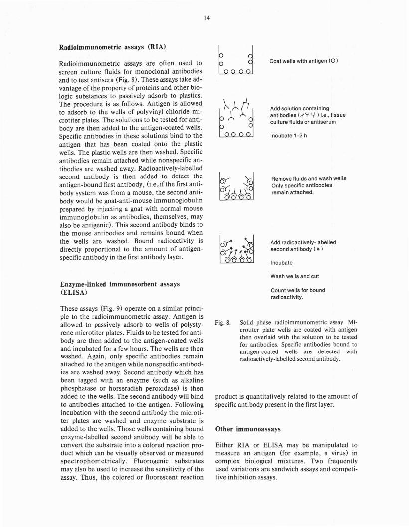
Radioimmunometric assays (RIA)
Radioimmunometric assays are often used toscreen culture fluids for monoclonal antibodiesand to test antisera (Fig. 8). These assays take advantage of the property of proteins and other biologic substances to passively adsorb to plastics.The procedure is as follows. Antigen is allowedto adsorb to the wells of polyvinyl chloride microtiter plates. The solutions to be tested for antibody are then added to the antigen-coated wells.Specific antibodies in these solutions bind to theantigen that has been coated onto the plasticwells. The plastic wells are then washed. Specificantibodies remain attached while nonspecific antibodies are washed away. Radioactively-labelledsecond antibody is then added to detect theantigen-bound first antibody, (Le.,if the first antibody system was from a mouse, the second antibody would be goat-anti-mouse immunoglobulinprepared by injecting a goat with normal·mouseimmunoglobulin as antibodies, themselves, mayalso be antigenic). This second antibody binds tothe mouse antibodies and remains bound whenthe wells are washed. Bound radioactivity isdirectly proportional to the amount of antigenspecific antibody in the first antibody layer.
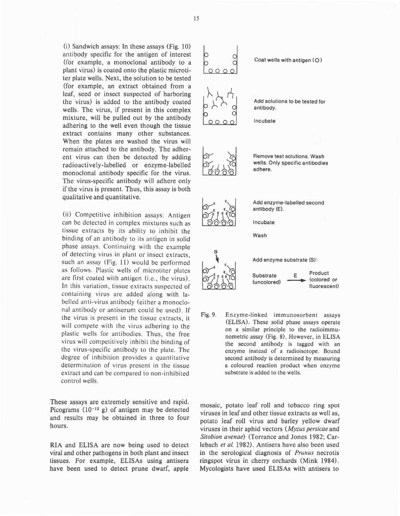
Enzyme-linked immunosorbent assays(ELISA)
14
u Coat wells with antigen (0 )
Add solution containingantibodies (~Y Y) i.e., tissueculture fluids or antiserum,
Incubate 1-2 h
Remove fluids and wash wells.Only specific antibodiesremain attached.
Add radioactively-labelledsecond antibody (*)
Incubate
Wash wells and cut
Count wells for boundradioactivity.
These assays (Fig. 9) operate on a similar principle to the radioimmunometric assay. Antigen isallowed to passively adsorb to wells of polystyrene microtiter plates. Fluids to be tested for antibody are then added to the antigen-coated wellsand incubated for a few hours. The wells are thenwashed. Again, only specific antibodies remainattached to the antigen while nonspecific antibodies are washed away. Second antibody which hasbeen tagged with an enzyme (such as alkalinephosphatase or horseradish peroxidase) is thenadded to the wells. The second antibody will bindto antibodies attached to the antigen. Followingincubation with the second antibody the microtiter plates are washed and enzyme substrate isadded to the wells. Those wells containing boundenzyme-labelled second antibody will be able toconvert the substrate into a colored reaction product which can be visually observed or measuredspectrophometrically. Fluorogenic substratesmay also be used to increase the sensitivity of theassay. Thus, the colored or fluorescent reaction
Fig. 8. Solid phase radioimmunometric assay. Microtiter plate wells are coated with antigenthen overlaid with the solution to be testedfor antibodies. Specific antibodies bound toantigen-coated wells are detected withradioactively-labelled second antibody.
product is quantitatively related to the amount ofspycific antibody present in the first layer.
Other immunoassays
Either RIA or ELISA may be manipulated tomeasure an antigen (for example, a virus) incomplex biological mixtures. Two frequentlyused variations are sandwich assays and competitive inhibition assays.
15
Fig. 9. Enzyme-linked immunosorbent assays(ELISA). These solid phase assays operateon a similar principle to the radioimmunometric assay (Fig. 8). However, in ELISAthe second antibody is tagged with anenzyme instead of a radioisotope. Boundsecond antibody is determined by measuringa coloured reaction product when enzymesubstrate is added to the wells.
E Product~ (colored or
fluorescent)
Coat wells with antigen (0 )
Add solutions to be tested forantibody.
InCUbate
Remove test solutions. Washwells. Only specific antibodiesadhere.
Add enzyme-labelled secondantibody (E).
InCUbate
Wash
Add enzyme substrate (S):
Substrate(u ncolored)
5
~
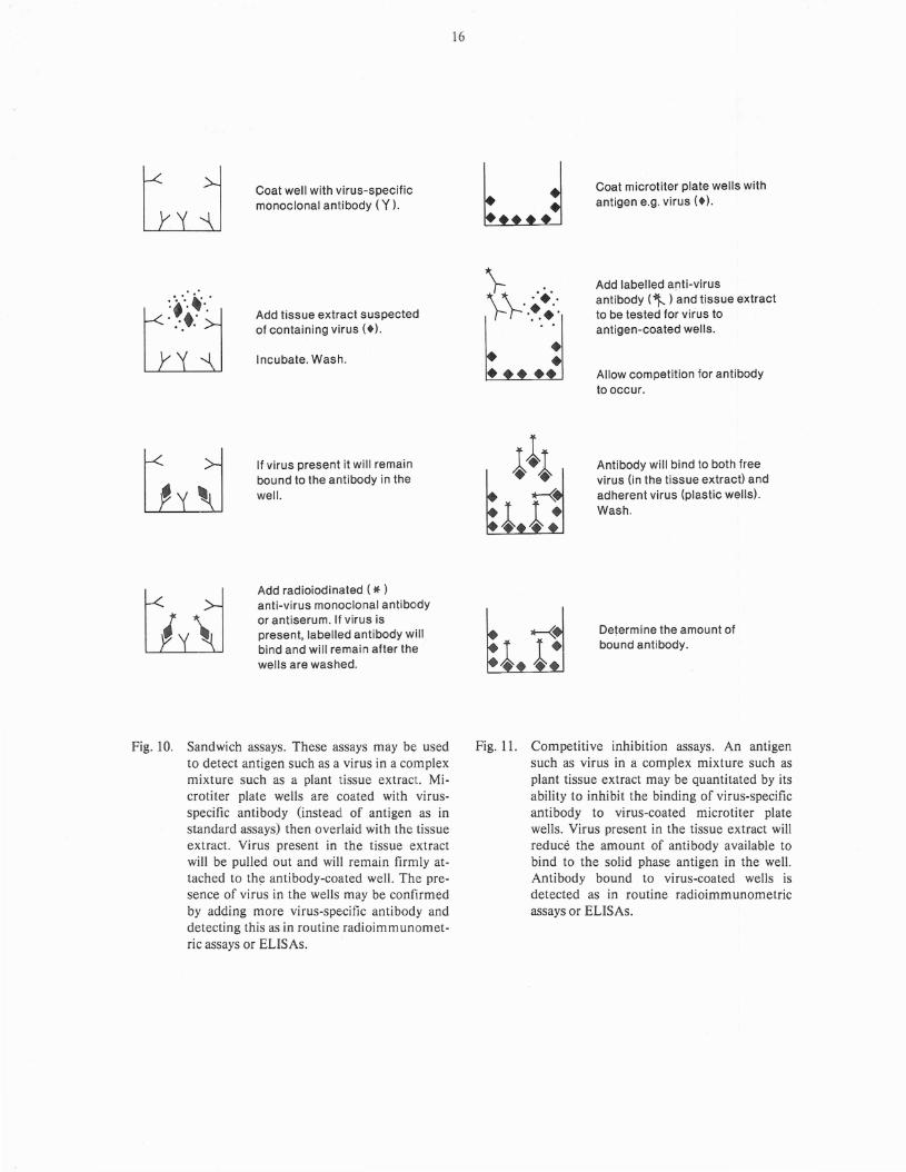
0) Sandwich assays: In these assays (Fig. 10)antibody specific for the antigen of interest(for example, a monoclonal antibody to aplant virus) is coated onto the plastic microtiter plate wells. Next, the solution to be tested(for example, an extract obtained from aleaf, seed or insect susp~cted of harboringthe virus) is added to the antibody coatedwells. The virus, if present in this complexmixture, will be pulled out by the antibodyadhering to the well even though the tissueextract contains many other substances.When the plates are washed the virus willremain attached to the antibody. The adherent virus can then be detected by addingradioactively-labelled or enzyme-labelledmonoclonal antibody specific for the virus.The virus-specific antibody will adhere onlyif the virus is present. Thus, this assay is bothqualitative and quantitative.
(ii) Conlpetitive inhibition assays: Antigencan be detected in conlplex mixtures such astissue extracts by its ability to inhibit thebinding of an antibody to its antigen in solidphase assays. Continuing with the exampleof detecting virus in plant or insect extracts,such an assay (Fig. 11) would be perfornledas follows. Plastic wells of nlicrotiter platesare first coated with antigen (i~e., the virus).In this variation, tissue extracts suspected ofcontaining virus are added along with labelled anti-virus antibody (either a monoclonal anti body or an tiserum could be used). Ifthe virus is present in the tissue extracts, itwill conlpete with the virus adhering to theplastic wells for antibodies. Thus, the freevirus will conlpetiti vely inhi bit the binding ofthe virus-specific antibody to the plate. Thedegree of inhibition provides a quantitativedeternlination of virus present in the tissueextract and can be conlpared to non-inhibitedcontrol wells.
These assays are extremely sensitive and rapid.Picograms (10-12 g) of antigen may be detectedand results may be obtained in three to fourhours.
RIA and ELISA are now being used to detectviral and other pathogens in both plant and insecttissues. For example, ELISAs using antiserahave been used to detect prune dwarf, apple
mosaic, potato leaf roll and tobacco ring spotviruses in leaf and other tissue extracts as well as,potato leaf roll virus and barley yellow dwarfviruses in their aphid vectors (Myzus persicae andSitobion avenae) (Torrance and Jones 1982; Carlebach et at. 1982). Antisera have also been usedin the serological diagnosis of Prunus necrotisringspot virus in cherry orchards (Mink 1984).Mycologists have used ELISAs with antisera to
~+ .'.•••}-: '.~•...0, 0o •••t: 0
Coat well with virus-specificmonoclonal antibody (Y ).
Add tissue extract suspectedof containing virus (+).
Incubate. Wash.
16
•
••
••
••
Coat microtiter plate wells withantigen e.g. virus (+).
Add labelled anti-virusantibody ("t )and tissue extractto be tested for vir·us toantigen-coated wells.
Allow competition for antibodyto occur.
If virus present it will remainbound to the antibody in thewell.
Add radioiodinated (* )anti-virus monoclonal antibodyor antiserum. If virus ispresent, labelled antibody willbind and will remain after thewells are washed.
Antibody will bind to both freevirus (in the tissue extract) andadherent virus (plastic wells).Wash.
Determ ine the amou nt ofbound antibody.
Fig. 10. Sandwich assays. These assays may be usedto detect antigen such as a virus in a complexmixture such as a plant tissue extract. Microtiter plate wells are coated with virusspecific anti body (instea of antigen as instandard assays) then overlaid with the tissueextract. Virus present in the tissue extractwill be pulled out and will remain firmly attached to th~ antibody-coated well. The presence of virus in the wells may be confirmedby adding more virus-specific anti body anddetecting this as in routine radioimmunometric assays or ELISAs.
Fig. 11. Competitive inhibition assays. An antigensuch as virus in a complex mixture such asplant tissue extract may be quantitated by itsability to inhibit the binding of virus-specificantibody to virus-coated microtiter platewells. Virus present in the tissue extract willreduce the amount of antibody available tobind to the solid phase antigen in the well.Antibody bound to virus-coated wells isdetected as in routine radioimm unometricassays or ELISAs.
detect fungal structural elements and toxic products such as: Epichloe typhina in tall fescue(Johnson et ale 1983; Johnson et ale 1982), phytotoxins of Ceratocystis ulmi in elm (Scheffer andElgersma 1981) , and Phonia tracheiphila, thecausative agent of Malsecco disease in lemon andother citrus trees (Nachmias et ale 1979). Monoclonal ELISAs are being developed to detectTristeza virus in citrus trees (Dr. D.J. Gumpf,Dept. of Plant Pathology, University of Californiaat Riverside, personal communication). Dorworth, et ale 1977; Dorworth 1974; Dorworthand Krywienczyk 1975) and others (Wendler etale 1981) have used other less sensitive serologictechniques to study races of Gremmeniella abietina (the causative agent of scleroderris canker ofconifers) differing in virulence.
Antisera or monoclonal antibodies have beenemployed in solid phase assays to distinguish between viral subtypes and different species offungi. For example, ELISAs using antisera havedistinguished between species of carlaviruses(Adams and Barbara 1982) and different speciesof endomycorrhizal fungi (Aldwell et ale 1983).Monoclonal antibodies have been used successfully in ELISA to distinguish plant viruses whichare poorly immunogenic in rabbits and thus,cannot be studied with conventional antisera(Dr. E. L. Halk, American Type - Culture Collection, Rockville, MD., personal communication).Similarly, monoclonal antibodies directed toFusarium oxysporum have been able to distinguishthis fungal species from other Fusarium species(Ianelli et ale 1983). However, closely related species are not always distinguishable using immunologic techniques. Banowetz and his coworkers (Banowetz et ale 1984) were unable todetect immunologic differences (with antiseraand monoclonals) between Tilletia controversaand T. caries, two closely related wheat fungiwhich are significant in wheat exportation.
Monoclonal antibodies and immunoassays havebeen used to study molecular changes duringplant development and to determine biochemicalphylogenetic relationships. For example, monoclonal antibodies have been used to compare andquantitate soluble and membrane forms of Avenasativa phytochrome (Partis et ale 1984) and toshow differential expression of these two formsin seedlings grown at different light intensities.Yee and his associates (Yee et ala 1984) haveused ELISA to compare bacterial and plant chlo-
17
roplast thioredoxins, while others (Singh andPreiss 1984) have employed monoclonal antibodies and polyclonal antisera in ELlSAs to studystarch and glycogen branching enzymes in bacteria and maize.
Monoclonal antibodies are now being used insolid phase assays to measure levels of plantgrowth regulators and to evaluate host biochenlical responses to disease. For exanlple, nlonoclonal antibodies have been used in immunoassaysto measure abscissic acid, indole-3-acetic acidand various other cytokinins in plant extracts(Mertens ef al.1984). In forestry research,antibodies prepared to zeatin riboside and isopentenyladenosine extracted fronl Douglas-firvegetative buds are being used in immunoassaysto measure levels of these cytokinins as predictors of cone production (Morris et al. 1984).Hahn and coworkers (Hahn et al. 1984) haveused radioimmunoassays to Il}easure the phytoalexin, glyceollin I in soybean roots infected withthe fungus Phytophthora megasperma.
Immunoassays such as those described abovecould be readily adapted for the detection offorest pathogens in seeds, seedlings or maturetrees, as well as in insect vectors. Immunologicassays afford several advantages over currentlyused tests. For example, the identification ofseed-borne pathogens using immunologic techniques would be advantageous for seed certification. Seed-borne pathogens are presently detected by germinating seed samples or testing seedlings (for instance, by inoculating tissue extractsinto appropriate indicator hosts). These tests areboth time consuming and expensive. In contrast,testing seed or seedlings for the pathogen usingsolid phase assays takes a matter of hours and isrelatively inexpensive. Furthermore, currenttesting methods detect pathogens in embryosand seedlings only, whereas immunologic techniques would permit pathogen detection in ungerminated seed. A further advantage is that testing may be done at any time during the year.
Immunoassays may be used to monitor levels ofinsect-borne pathogens or to measure levels ofvarious insect pathogens used in biologic control.The speed and sensitivity of these assays wouldfacilitate the management of insect-borne disease. Their application in monitoring the levelsof such biologic control agents as nucleopolyhedrosis virus in Douglas-fir tussock moth larvae
might aid in determining optimum times andamounts of virus application.
Immunofluorescence andimmunohistochemistry
18
'< yy
Antibody
+ooo
Sepharose Antibody coupled
to sepharose
Antisera or monoclonal antibodies may be usedas probes to detect any antigenic substance inplant or insect tissues using fluorescencemicroscopy. The specific antibody itself may betagged with a fluorochrome such as fluoresceinor rhodamine (direct immunofluorescence) or afluorescent second antibody may be used (indirect immunofluorescence). Alternatively, thespecific antibody may be tagged with an enzyme,such as peroxidase, which will convert a substrateto a colored reaction product. Antibodies taggedwith electron-dense materials such as ferritin areuseful for ultrastructural studies with electronmicroscopy.
Polyclonal antisera and monoclonal antibodieshave been used in a variety of immunohistochemical applications in plant research. Forexample, immunofluorescence has been used todistinguish vesicular-arbuscular mycorrhizal(VAM) fungi from other fungal species (Wilsonet a/. 1983) and to differentiate several species ofVAM fungi (Kough et a/. 1983). Antisera havealso been employed in immunofluorescence microscopy to localize Ceratocystis u/mi toxins inelm (Ipsen and Abul-Hajj 1982) and Phaeo/usschweinitzii (conifer root and butt rot fungus) propagules in soil samples (Dewey et a/. 1984). Immunohistochemistry has been used both to visualize fungal hyphae and to localize phytoalexinsin host tissue in soybeans infected with Phytophthora megasperma (Hahn et al. 1984). Antiseraand monoclonal antibodies have been employedin immunohistochemical studies of plant development. For example, intracellular transport ofthe lectin, phytohemagglutinin 'has been followedimmunocytochemically in the axes and cotyledons of Phaseolus vulgaris seeds (Greenwood andChrispeels 1984). Monoclonal antibodies havebeen used in immunofluorescence tests to studycell wall development in algae (Vreeland andLaetsch 1984) and phytochrome synthesis inAvena chloroplasts under differing light conditions (McCurdy and Pratt 1984). Also, antiserahave been employed in immunohistochemicalstudies of legume protein processing (Hermanand Shannon 1984)) and Avena globulin localiza-
Fig. 12.
Complex mixturecontaining antigen
Discard effluent
Wash column with:low pH
or high [salt]or chaotropic
agent
Pure antigen
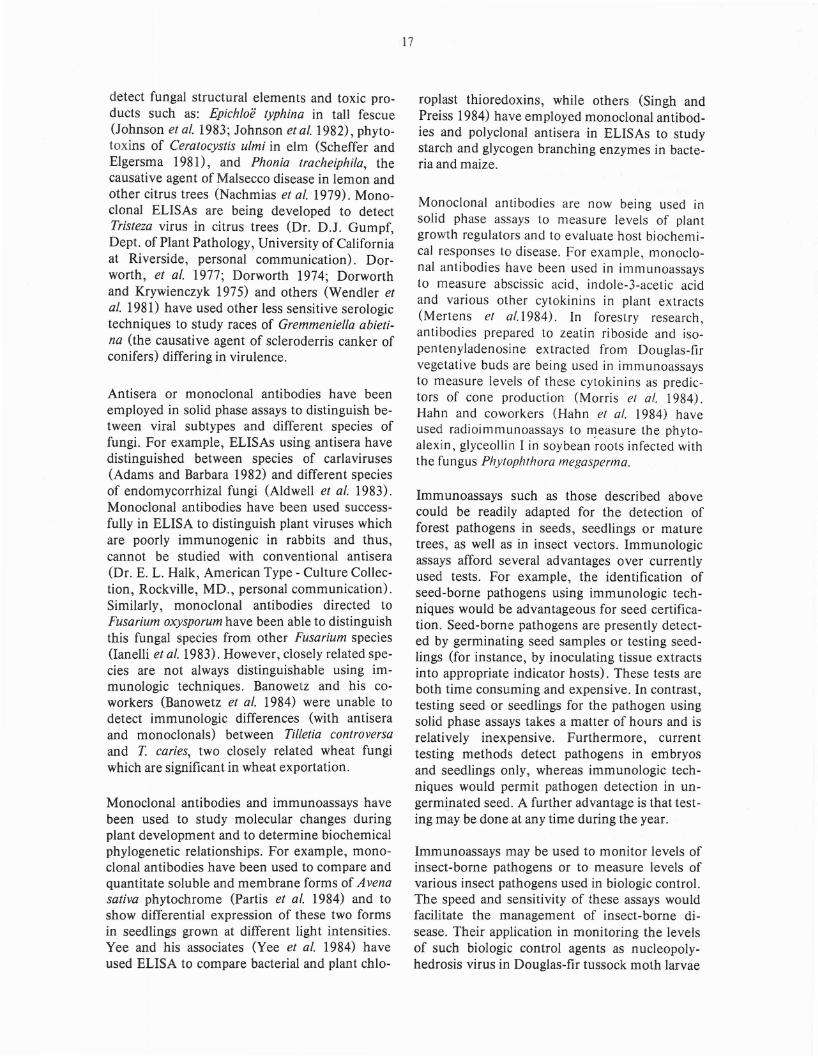
Immunoaffinity chromatography. Antibodiesmay be used to selectively isolate their antigens from complex mixtures by affinitychromatography. Specific antibodies arecoupled to a chromatographic medium suchas agarose and poured into a column. Whenthe complex mixture is passed through thecolumn, antigen adheres to the Sepharosebound antibodies while all other elementspass on through. Antigen can then be elutedfrom the column.
tion (Chan and Zee 1984) during several developmental stages.
Immunoaffinity chromatography
Monoclonal antibodies may be used in the isolation and purification of minor components incomplex mixtures using affinity chromatography(Fig. 12). In this technique, antibody is chemically coupled to agarose beads (Sepharose 4B)which are then poured into a chromatographiccolumn. The complex mixture containing theantigen to which the monoclonal antibody isdirected is poured over the column. The antigenwill bind to the antibody on the beads while theremainder of the mixture passes through thecolumn and is discarded with the effluent. Theantigen is then dissociated from the beads bywashing the column with a low-pH or high-saltbuffer or a chaotropic agent. Using affinity chromatography it is possible to achieve a 1000-foldenrichment. This method could be applied to thepurification and enrichment of any substance towhich an antibody has been prepared.
In both forestry and agricultural research thismethod has been used to purify growth regulators, enzymes or other interesting proteins forfurther study and characterization. For example,cytokinins from Douglas-fir vegetative budshave been purified by immunoaffinity chromatography (Morris et ale 1984) with specificantisera. Similarly, monoclonal antibodies arebeing employed in the purification of severalother plant growth regulators (Mertens et ale1984).
Immunoblotting techniques
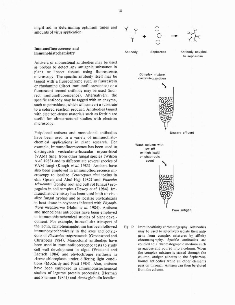
Antibodies may be used in conjunction with polyacrylamide gel electrophoresis in the study ofproteins (and other biologic substances) using atechnique called immunoblotting (Fig. 13) or"Western blotting" (Burnett 1981).
In the first step, a mixture of proteins such as acell homogenate or tissue extract is applied to aslab of polyacrylamide gel. When an electricalcurrent is passed through the gel each protein inthe mixture will migrate according to its chargeand/or molecular size and the mixture will be effectively resolved into separate protein bands.
19
:;:;:;:;=;:;:::::::::
Protein mixture is layered onto a tubeor slab of polyacrylamide gel andelectrophoresed.
:;:;:;:;:;:;:;:;:;:; Individual proteins separate into}}~{:}~:. bands according to their charge
and/or molecular size.
Proteins bands are electrophoreticallytransferred to nitrocellulose paper(NCP)
~~~t~t~~r NCP "Blot" is then developed withspecific antibody, then radiolabelledor enzyme-labelled second antibody.
NCP
Fig. 13. Immunoblotting. Proteins (or other substances) separated by gel electrophoresismay be transferred electrophoretically to nitrocellulose paper (NCP). The NCP is thenwashed with antiserum. Specific antibodieswill adhere to their respective protein bands(as originally obtained in gel electrophoresis). The NCP-bound antibodies may thenbe detected by enzyme-labelled or radioactively-labelled second antibodies.
The polyacrylamide gel is then placed on a pieceof nitrocellulose paper (NCP) and electrical current applied. The protein bands migrate onto theNCP in exactly the same relative positions theyoccupied within the gel- and stick. The "NCPblot" is then washed with antibody (usually anantiserum) which will bind to the band (s) representing the antigen. The bands containing boundantibody can then be detected by washing theNCP blot with radioactively-labelled second antibody and laying the washed and dried nitrocellulose paper against X-ray film. The radioactivebands show up as dark spots when the film isdeveloped. Alternatively, enzyme-labelled
second antibody can be used and the bandsdetected colorimetrically (as in ELISA) .
This technique has a great potential for the studyof protein biochemical relationships and for theidentification of specific proteins in complexmixtures. This technique has recently been applied to the study of genetic relationships amongseeds from Pinus strobus (Eckert and Eckert1984) and in comparative studies of A vena phytochrome (Partis et at. 1984) and plant acyl carrierproteins (Ohlrogge and Kuo 1984).
Immunoprecipitation
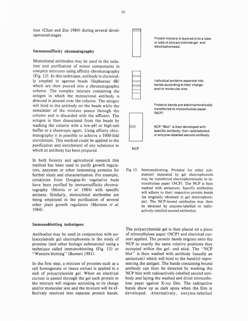
20
Specific antibody
~
Cell or tissuehomogenate
Staphylococci
Add specific antibody to cell ortissue homogenate.
Antigen-antibody complexesform.
Add Staphylococci
An alternate approach to locating antigens incomplex mixtures is immunoprecipitation (Fig.14). When specific antibody is mixed with a cellor tissue homogenate containing the antigen ofinterest the antibody will pull its antigen out ofthe mixture. In order to separate the antigenantibody complexes ,C)taphytococci are added tothe mixture. These bacteria contain on their surfaces a substance called Protein A which binds toall rabbit antibody classes and some clas~es ofmouse antibodies. The antigen-antibody cornplexsticks to the bacterial surface and can be centrifuged out of the mixture along with the bacteria.The antigenic substance binding to the antibodiescan then be analyzed by polyacrylamide gel electrophoresis. The precipitated antigen can be seenby staining the gels or alternatively, radioactiveprecursors such as radioactively-labelled aminoacids or sugars may be used to label cell constituents before immunoprecipitation. In the lattercase the precipitated antigens can then be detected by autoradiography.
This technique was used to measure pero idaserelease of peanut ceHs growing in culture (VanHuystee and Lobarzewski 1982). In this particular study biosynthesis and release of peroxidasewere monitored by the incorporation of radioactively-labeHed methionine and immunoprecipitation with anti-peroxidase antisera.
[n forestry research similar techniques might beused to monitor the synthesis of growth regulators in vegetative buds under various environmental conditions or to measure the concentration of maturational hormones in insects at various developmental stages.
Centrifuge.
Pellet:Staphylococci + antigen-antibodycomplexes.
Remove pellet.
Do polyacrylamide gelelectrophoresis.
.. Antigen precipitated byspecific antibody.
Fig. 14. Kmnlunoprecipitation. Antisera or monoclonal anti bodies may be used to puB their respective antigens out of complex mixturesby immunoprecipitation. Protein A-coatedStaphylococci bacteria are added to facilitateprecipitation of the antigen:antibodycomplexes. The antigenic components maythen be visualized by doing gel electrophoresis of the precipitated material.
Two-dimensional polyacrylamide gelelectrophoresis
High-resolution two-dimensional polyacrylamidegel electrophoresis is a powerful technique foranalyzing components of complex mixtures andcan be used in conjunction with immunoprecipitation and Western blotting (Pearson and Anderson 1983~ Pearson and Anderson 1982). It canalso detect changes which occur in any tissue inresponse to disease (for instance, the increasedsynthesis of constituitive proteins or expressionof new proteins, phytoalexins, etc.).
[n the first dimension, the components of a complex mixture such as a cell or tissue homogenateor serum are separated according to charge byisoelectric focusing in tube gels. A pH gradient isgenerated in the gel and each component of themixture migrates to its isoelectric point (the loca-tion in the at has a a which the cnent has a 1 t c (rg f z .. tu
21
then placed at the top of a slab gel and electricalcurrent is reapplied. The components migrate inthis second dimension according to their molecular size.
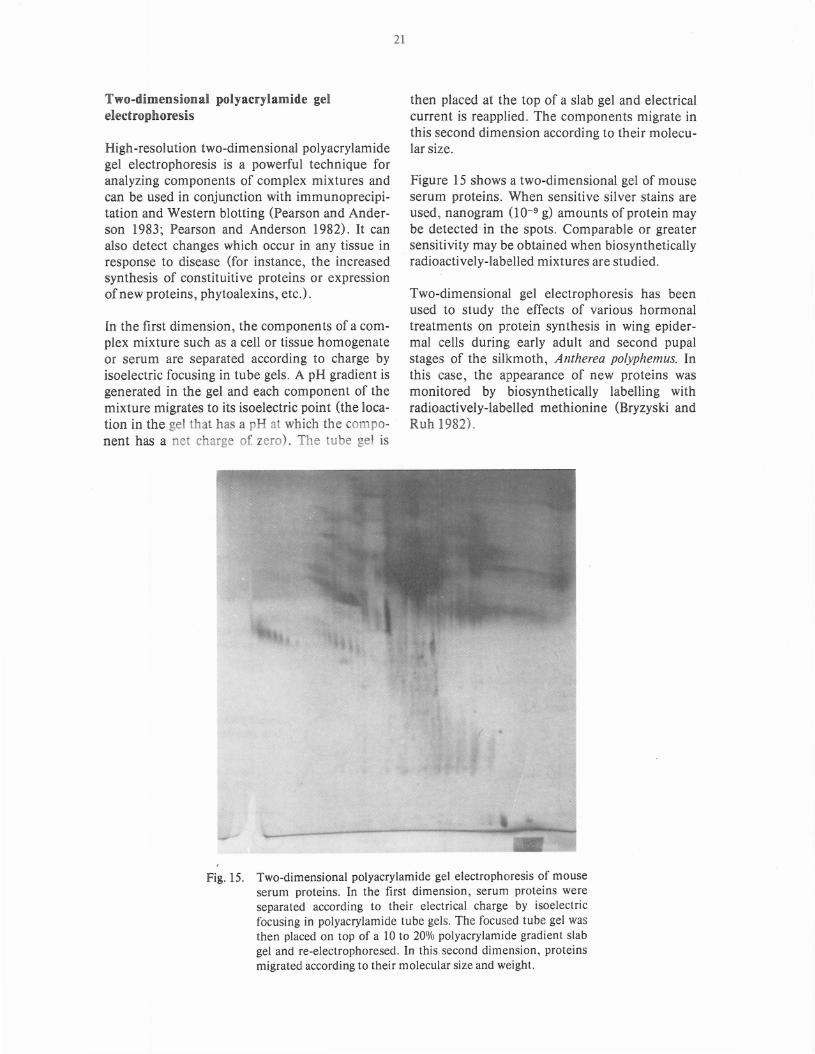
Figure 15 shows a two-dimensional gel of mouseserum proteins. When sensitive silver stains areused, nanogram (10-9 g) amounts of protein maybe detected in the spots. Comparable or greatersensitivity may be obtained when biosyntheticallyradioactively-labelled mixtures are studied.
Two-dimensional gel electrophoresis has beenused to study the effects of various hormonaltreatments on p otein synthesis in wing epidermal cells during early adult and second pupalstages of the silkmoth, Antherea polyphemus. Inthis case, the a pearance of new proteins wasmonitored by biosynthetically labelling withradioactively-labelled methionine (Bryzyski andRuh 1982).
Fig. 15. Two-dimensional polyacrylamide gel electrophoresis of mouseserum proteins. In the first dimension, serum proteins wereseparated according to their electrical charge by isoelectricfocusing in polyacrylamide tube gels. The focused tube gel wasthen placed on top of a 10 to 20% polyacrylamide gradient slabgel and re-electrophoresed. In this second dimension, proteinsmigrated according to their molecular size and weight.
22
Polyribosomesfrom resistantplant
+ Y yi Y
Antibodies toresistance protein (e)
Antibodies precipitatepolyribosomes makingresistance protein
Isolate and radioactively label (*)mRNA (~) from polyribosome
DNA isolatedfrom resistantplant, fragmentedand separated byagarose gelelectrophoresis
Radioactive mRNA is used as aprobe to locate the gene for theresistance protein on DNAfragment (----) in the electrophoretogram
Infect bacteriumwith plasmid
Insert DNA fragmentinto plasmid
o
Isolate DNA fragment containingthe gene for the resistance protein
o\
\
Use bacterium to infectand transform nonresistant plant cell.
/Transfer DNAfragment to non-resistantplant cellby microinjection
Genetic engineering
New proteins or other biologic substances havebeen associated with superior genetic characteristics such as disease resistance. For example, newsoluble proteins appear in tobacco plants whenresistance is induced either by a hypersensitivityreaction to infection with TMV or by chemicals.Studies using classical genetic techniques indicatea single gene regulates the production of theseresistance proteins (Ahl et ale 1982).
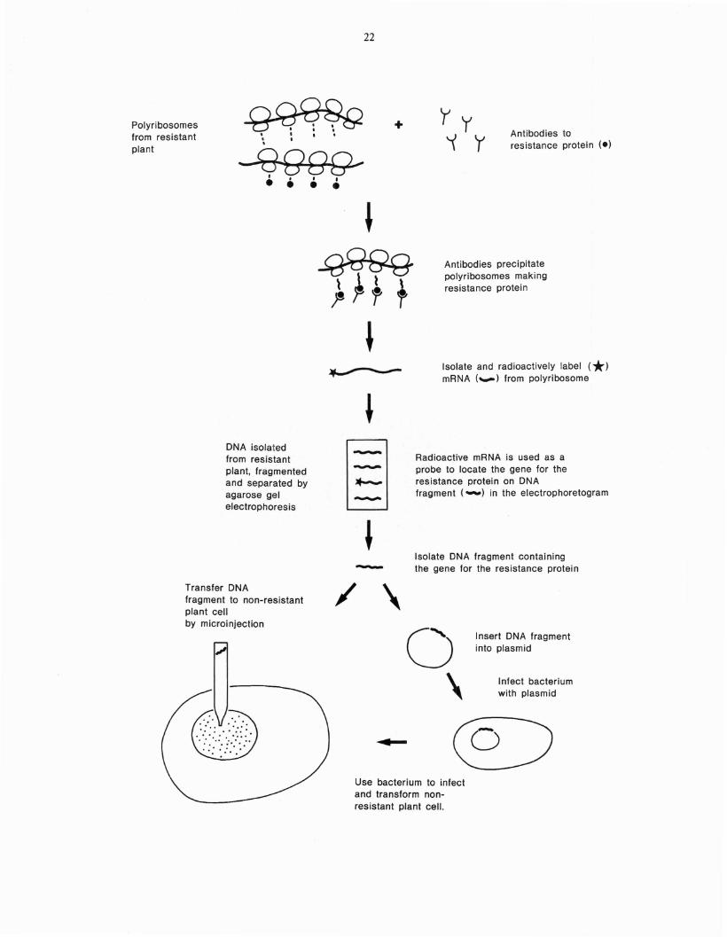
Once biologic substances such as resistance proteins in plants have been identified, specific antisera or monoclonal antibodies may be used tolocate the genes for these proteins using a technique presently employed in genetic engineeringin several other systems. In brief, specific antibody is used to precipitate the polyribosomes engaged in the synthesis of the protein of interest(Fig. 16). Once these polyri bosomes have beenseparated from the rest of the cell's proteinsynthesizing machinery, the messenger ribonucleic acid (mRNA) associated with the polyribosonle nlay be isolated. This mRNA, a genetic4'mirror inlage" of the deoxyribonucleic acid(DNA) coding for the protein, may be radioactively-labelled, then used as a probe to locate the
Fig. 16. Monoclonal antibodies and genetic engineering. Monoclonal antibodies recognizing abeneficial element, such as a resistanceprotein, may be used to selectvely immunoprecipitate polyribosomes engaged in itssynthesis as they will react with the nascentprotein chains on the ribosomes. MessengerRNA may then be removed from the polyribosomes, radioactively-labelled and used asa probe to locate the gene coding for theresistance protein on DNA fragments thathave been separated by agarose gel electrophoresis. When the resistance gene has beenlocalized on one of these fragments, thatfragment may be excised from the agarosegel and transferred by microinjection to anonresistant plant cell growing in tissueculture. Alternatively, the DNA fragmentmay be inserted into a plasmid which maythen be used as a vector to transfer the gene,via a transforming bacterial species such asAgrobacter tumefaciens, into a nonresistantplant cell.
23
gene on fragments of DNA isolated from thecell. Once the gene has been located on a DNAfragment, the fragment may be directly transferred to plant protoplasts by microinjection(Rasmussen et ale 1984). Alternatively, theDNA fragment carrying the gene may be insertedinto plant cells in the following way. The isolatedgene fragment is coupled with a plasmid, a smallpiece of extrachromosomal DNA which is able toinfect bacterial cells such as Agrobacter tumefaciens (Rempel et ale 1984; Caplan et ale 1983). Thebacteria containing the altered plasmid are then,in turn, used to transform (infect) plant cells. Inthis way a new gene may be integrated into theplant cell DNA. Thus, with genetic engineeringtechniques genes for disease resistance might betransferred to plant or tree cells growing in tissueculture. Presently') several laboratories are actively engaged in the construction of vectors for thetransmission of new DNA into plant cells (Rempel et ale 1983).
A recent example of the contribution of geneticengineering to plant resistance is in the identification and localization of the structural gene for theinsecticidal agent a-endotoxin in Bacillus thuringiensis (Gonzalez et ale 1982). This gene is conveniently located on a plasmid in B. thuringiensis.Under certain conditions this plasmid will infectother bacteria. In this way the a-endotoxin genemay be transferred to residual bacteria normallyfound on plants and trees. Of course, success isdependent upon the ability of these bacteria toexpress the gene and to secrete a-endotoxin.
Conclusion
The fine specificities of antibodies - especiallymonoclonal antibodies - for their antigensmake them excellent probes for detecting substances which may be present in minisculeamounts in complex biological systems.
This information report has described the principles and methodology involved in generating antisera and monoclonal antibodies. The theorybehind several immunologic techniques employing these sensitive reagents has also been given.Current and potential applications of these rapidand economical techniques to research problemsin forestry and agriculture have been described.
References
Adams A.N. and D.J. Barbara 1982. The use ofF(ab')2-based ELISA to detect serologicalrelationships amongst carlaviruses. Ann.appl. BioI. 101: 495-500.
Ahl P., A. Cornu and S. Gianinazzi 1982. Solubleproteins as genetic markers in studies ofresistance and phylogeny in Nicotiana. Phytopathology 72: 80-85.
Aldwell F.E.B., I.R. Hall and J.M.B. Smith 1983.Enzyme-linked immunosorbent assay(ELISA) to identify endomycorrhizal fungi.Soil BioI. Biochem. 15: 377-378.
Banowetz G.M .., E.l. Trione and B.B. Krygier1984. Inlmunological conlparisons of teliospores of two wheat bunt fungi., Tilletiaspecies., using monoclonal antibodies andantisera. Mycologia 76: 51-62.
Bryzyski R.G. and M.F. Ruh 1982. Proteinsynthesis during early adult and second pupaldevelopment in the silkmoth Antherea polyphemus. Insect Biochem. 12: 687-698.
Burnett W.N. 1981. "Western Blotting": Electrophoretic transfer of proteins from sodiumdodecyl sulfate-polyacrylamide gels to unmodified nitrocellulose and radiographic detection with antibody and radioiodinated Protein A. Anal. Biochem. 112: 195-203.
Caplan A., L. Herrera-Estrella, D. Inze, E. VanHaute, M. Van Montagu, J. Schell and P.Zambryski 1983. Introduction of geneticmaterial into plant cells. Science 222:815-821.
Carlebach R., B. Raccah and G. Loebenstein1982. DetectioI). of potato virus Y in theaphid Myzus persicae by enzyme-linked immunosorbent assay (ELISA). Ann. app. BioI.101: 511-516.
Chan H. T. and S. Y. Zee 1984. Immunofluorescent localization and immunochemical studies of the globulin of oat (A vena Sativa L.)seeds. Plant Physiol. 75, suppl: 159 (Abstr.).
Dewey F.M., D.K. Barrett, I. . Vose and C.J.Lamb 1984. Immunofluorescence microsco-
24
py for the detection and identification of propagules of Phaeolus schweinitzii in infestedsoil. Phytopathology 74: 291-296.
Dorworth C. E., J. Krywienczyk and D. D. Skilling 1977. New York isolates of Gremmeniellaabietina (Schleroderris lagerbergii) identical inimmunogenic reation to European isolates.Plant Dis. Rep. 61: 887-890.
Dorworth C. E. 1974. Comparison of soluble proteins of Ascocalyx abietis and Gremmeniellaabietina by serology and electrophoresis Can.J. Bot. 52: 912-922.
Dorworth C. E. and J. Krywienczyk 1975. Comparisons among isolates of Gremmeniellaabietina by means of growth rate, conidiameasurement and immunogenic reaction.Can. J. Bot. 53: 2506-2525.
Eckert R.T. and B.S. Eckert 1984. A procedurefor study of immunochemical properties ofmegagametophyte tissue of Pinus strobus L.Can. J. For. Res. 14: 142-145.
Greenwood J. S. and M. J. Chrispeels 1984. Immunocytochemical localization of phytohemagglutinin in the axes and cotyledons ofPhaseolus vulgaris seeds. Plant Physiol. 75suppl.: 46 (Abstr.).
Gonzalez J.M., B.J. Brown and B.C. Carlton1982. Transfer of Bacillus thuringiensis plasmids coding for a-endotoxin among strainsof B. thuringiensis and B. cereus. Proc. Natl.Acad. Sci. U.S.A. 79: 6951-6955.
Hahn, M. G., A. Bonhoff and H. Grisebach1984. Localization and quantitation of glyceollin I using a specific radioimmunoassayin soybean roots infected with Pyhtophthoramegasperma F. sp. glycinea. Plant Physiol. 75SUppl.: 166 (Abstr.).
Herman E. M. and L. M. Shannon 1984. Immunocytochemical and lectin cytochemical evidence for Golgi apparatus mediated deposition of legume body proteins. Plant Physiol.75, suppl.: 145 (Abstr.).
Ianelli D., R. Capparelli, F. Marzioan, F. Scalaand C. Noviollo 1983. Production ofhybridomas secreting antibodies to the genus
Fusarium. Mycotaxon 18: 523-532.
Ipsen J.D. and Y.J. Abul-Hajj. 1982. Fluorescentantibody technique as a means of localizingCeratocystis ulmi toxins in elm. Can. J. Bot.60: 724-729.
Johnson M.C., R.L. Anderson, R.J. Kryscio andM.R. Siegel 1983. Sampling procedures fordetermining endophyte content in TallFescue seedlots by ELISA. Phytopathology73: 1406.
Johnson M.C., T.P. Pirone, M.R. Siegel andD.R. Varney 1982. Detection of Epichloetyphina in Tall Fescue by means of enzymelinked immunosorbent assay. Phytopathology 72: 647-650.
Kohler G. and C. Milstein 1975. Continuous cultures of fused cells secreting antibody of predefined specificity. Nature 256: 495-497.
Kohler G., H. Hengartner and M.J. Shulman1978. Immunoglobulin production by lymphocyte hybridomas. Eur. J. Immunol. 8:82-88.
Kough, J., N. Malajczuk and R.G. Linderman1983. Use of the indirect immunofluorescenttechnique to study the vesicular-arbuscularfungus Glomus epigaeum and other Glomusspecies. New Phytol 94: 57-62.
McCurdy D. and L. H. Pratt 1984. Immunocytochemical detection of phytochrome duringgreening in A vena. Plant Physiol. 75, suppl.:89 (Abstr.)
Mertens R., J. Eberle and E. W. Weiler 1984.Production and use of monoclonal antibodiesfor endogenous plant growth regulators.Plant Physiol. 75 SUppl. 137 (Abstr.).
Mink G.1. 1984. Use of enzyme-linked immunosorbent assay results in efforts to control orchard spread of Cherry Rugose Mosaic Disease in Washington. Plant Dis. 68: 207-210.
Morris J. W., R. C. Durley, D. P. Lavender andJ. B. Zaerr 1984. Cytokinins in vegetativebuds of Douglas-fir: Correlation with coneproduction. Plant Physiol. 75, suppl: 90(Abstr.) .
25
Nachmias, A., M. Bar-Joseph, Z. SoleI and I.Barash 1979. Diagnosis of Mal Secco diseasein Lemon by enzyme-linked immunosorbentassay. Phytopathology 69: 559-561.
Nisonoff A., Introduction to Molecular Immunology, p. 47, 1982, Sinauer and Associates,Inc., Sunderland, Massachusetts.
Ohlrogge J. B. and T. M. Kuo 1984. "WesternBlot" Analysis of plant acyl carrier proteins.Plant Physiol. 75, suppl.: 100 (Abstr.).
Partis M. D., B. R. Jordan and B. Thomas 1984.Comparison of soluble and membrane phytochrome in Avena. Plant Physiol. 75(1) suppl:74 (Abstr.)
Pearson T.W. and N.L. Anderson 1983. The useof high-resolution two-dimensional gel electrophoresis for analysis of monoclonal antibodies and their specific antigens. in Methodsin Enzymology, Vol. 92, ImmunochemicalTechniques Part E. Monoclonal Antibodiesand General Immunoassay Methods pp. 196219. J.J. Langone and H. Van Vunakis, eds.Academic Press, New York.
Pearson T. and N.L. Anderson 1980. Analyticaltechniques for cell fractions. XXVIII. Dissection of comple~ antigenic mixtures usingmonoclonal antibodies and two-dimensionalgel electrophoresis. Anal. Biochem. 101:377-386.
Rasmussen R., B. Ward and V. Knauf 1984. Microinjection of immobilized protoplasts.Plant Physiol. 75, supp!.: 141 (Abstr.).
Rempel H., R. C. Tait and C. I. Kado 1984. Anovel foreign gene transfer system using amini-Ti plasmid for genetic modification ofplants. Plant Physiol. 75, suppl.:31 (Abstr.).
Scheffer R.J. and D.M. Elgersma 1981. Detectionof a phytotoxic glycopeptide produced by Ophiostoma ulmi in elm by enzyme-linked immunospecific assay (ELISA). Physiol. PlantPathol. 18: 27-32.
Singh B. K. and J. Preiss 1984. Immunologicalcharacterization of starch branching enzymesin maize and comparison with enzyme from
Escherichia coli B. Plant Physiol. 75 suppl.160 (Abstr.).
Torrance L. and R.A.C. Jones 1982. Increasedsensitivity of detection of plant viruses obtained by using a fluorogenic substrate inenzyme-linked immunosorbent assay. Ann.appl. BioI. 101: 501-509.
Van Huystee R.B. and J. Lobarzewski 1982. Animmunological study of peroxidase releaseby cultured peanut cells. Plant Sci. Lett. 27:59-67.
Vreeland V. and W. M. Laetsch 1984. Intracellular synthesis of Fucus cell wall antigens. Plant
26
Physio!. 75, suppl.: 62 (Abstr.).
Wendler P. B., A. R. Gotlieb and D. R. Bergdahl1981. A newly discovered serotype of Gremmeniella abietina. Can. J. Bot. 58: 1923-1928.
Wilson J.M., M.J. Trinick and C.A. Parker 1983.The identification of vesicular-arbuscularmycorrhizal fungi using immunofluorescence. Soil BioI. Biochem. 15: 439-445 ..
Yee B. C., N. A. Crawford and B. B. Buchanan1984. Immunological and affinity studies onC3 and C4 thioredoxins. Plant Physiol. 75 (1)suppl: 152 (Abstr.).