single-nucleotide polymorphisms in chromosome 3p14.1- 3p14
TRANSCRIPT

Single-nucleotide polymorphisms in chromosome 3p14.1- 3p14.2are associated with susceptibility of Type 2 diabetes with cataract
Hui-Ju Lin,1,2,3 Yu-Chuen Huang,1,2 Jane-Ming Lin,1,2,3 Jer-Yuarn Wu,4 Liuh-An Chen,1 Chao-Jen Lin,5
Yung-Ping Tsui,2,3 Chih-Ping Chen,6 Fuu-Jen Tsai1,2
(The first two authors contributed equally to the work)
1Department of Medical Genetics, China Medical University Hospital, Taichung, Taiwan; 2School of Chinese Medicine, Collegeof Chinese Medicine, China Medical University, Taichung, Taiwan; 3Department of Ophthalmology, China Medical UniversityHospital, Taichung, Taiwan; 4National Genotyping Center, Academia Sinica, Taipei, Taiwan; 5Department of Pediatrics, ChanghuaChristian Hospital, Taiwan; 6Departments of Obstetrics and Gynecology and Medical Research, Mackay Memorial Hospital, Taipei,Taiwan
Purpose: Type 2 diabetes (T2D) is highly prevalent worldwide and cataracts are of high incidence in T2D patients. Inthis study, we identify genetic variants that predispose type 2 diabetes (T2D) patients to cataracts in the Han-Chineseresiding in Taiwan.Methods: We conducted a genome-wide association study with a total of 1,715 cases and 2,000 random controls. In thehaplotype study, we defined haplotype 1 (Ht 1) to haplotype 4 (Ht 4) as the alternative alleles of the DM and cataractrelated chromosome 3p14.1- 3p14.2 polymorphisms.Results: The most significant association was detected with rs11129182, rs17047573, and rs17047586 in chromosome3p14.1- 3p14.2 (p value=3.52×10⁻⁷, 8.35×10⁻⁸, and 7.65×10⁻⁸, respectively). In genotype analysis, the “CT” genotype ofrs11129182, the ‘GG’ genotype of rs17047573, and the ‘GG’ genotype of rs17047586 were significantly different in theT2D and cataract groups (OR=3.03, 7.47, and 7.51, individually; 95% confidence index (CI): 1.97–4.65, 3.36–16.6, and3.38–16.7, individually). In the haplotype study, the distribution of the Ht3 and Ht4 between the DM and cataract groupand the control group differed significantly between the two groups (p=0.0004). The odds ratio (OR) of Ht4 was 1.89 andthe 95% confidence interval (CI) was 1.36–2.65.Conclusions: The major functions of the genes are voltage-dependent anion-selective channel proteins, long myosin lightchain kinase, adenylyl cyclase-associated proteins and retinoic acid receptors and are all closely related with thepathogenesis of T2D and cataractogenesis. This has helped us understand the pathogenesis of T2D patients with cataracts.
Diabetes mellitus (DM) is one of the major causes of sightloss. Cataracts, considered a complication of DM, are a causeof visual impairment that can affect individuals at all ages[1,2]. Among the various complications of diabetes mellitusin the eyes, cataracts are only less critical then diabeticretinopathy as cause of blindness. Although chronichyperglycemia and the duration of DM are considered to bethe major risk factors for this diabetic complication [3,4], thereare some patients with severe cataracts while others presentwith few risk factors.
A cataract is a clouding that develops in the crystallinelens of the eye or in its envelope; varying in degree from slightto complete opacity and obstructing the passage of light. Age-related cataracts are responsible for 48% of world blindness,which represents about 18 million people, according to the
Correspondence to: Fuu-Jen Tsai, M.D., Ph.D., Department ofMedical Genetics and Pediatrics, China Medical University Hospital,No. 2 Yuh Der Road, Taichung 404, Taiwan; Phone:886-4-22052121 ext. 2041; FAX: 886-4-22033295; email:[email protected]
World Health Organization (WHO) [5]. In many countries,surgical services are inadequate and cataracts remain theleading cause of blindness. Cataracts are a clinically andgenetically heterogeneous group of eye disorders that causesvisual impairment. At least 34 loci and mutations in 22 geneshave been reported to be linked with different forms ofcataracts. Type 2 diabetes (T2D) affects at least 6% of theworld’s population, and the prevalence is expected to doubleworldwide by the year 2025 [6]. T2D is a complex diseasecharacterized by hyperglycemia that results from impairedpancreatic β-cell function, decreased insulin action at targettissues, and increased glucose output by the liver [7]. Bothgenetic components and environmental factors contribute tothe pathogenesis of T2D. This disease is considered to be apolygenic disorder that each genetic variance confers a partialand additive effect. Only 5%–10% of T2D are monogenicdiabetes with single gene defects. A large amount of effort hasbeen devoted to finding common T2D genes, includinggenome-wide linkage, candidate-gene and genome-wideassociation studies [8-10]. However, these genes cannot beheld solely responsible for the development of T2D includes
Molecular Vision 2010; 16:1206-1214 <http://www.molvis.org/molvis/v16/a134>Received 1 March 2010 | Accepted 24 June 2010 | Published 1 July 2010
© 2010 Molecular Vision
1206
the wide variability of the prevalence of T2D in differentethnic groups. Cataracts are more common in people withdiabetes than in the general population under the age of 40years and they are morphologically similar to senile cataracts.The exact correlation between cataracts and T2D is without adefinite conclusion. Cataracts are a significant complicationof T2D [8-10]. In this study, we aimed to uncover diabetessusceptibility loci that increase the risk for T2D, as well ascataracts, in the Chinese population residing in Taiwan.
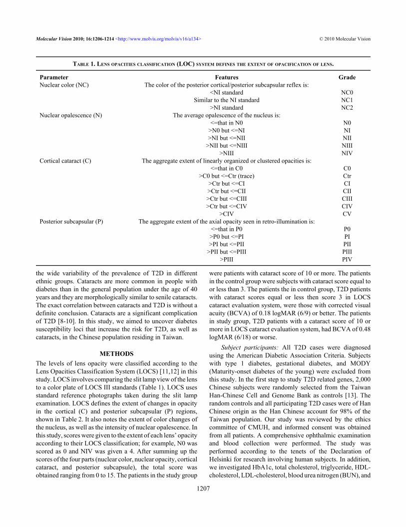
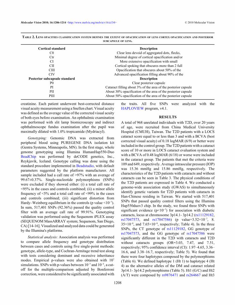
METHODSThe levels of lens opacity were classified according to theLens Opacities Classification System (LOCS) [11,12] in thisstudy. LOCS involves comparing the slit lamp view of the lensto a color plate of LOCS III standards (Table 1). LOCS usesstandard reference photographs taken during the slit lampexamination. LOCS defines the extent of changes in opacityin the cortical (C) and posterior subcapsular (P) regions,shown in Table 2. It also notes the extent of color changes ofthe nucleus, as well as the intensity of nuclear opalescence. Inthis study, scores were given to the extent of each lens’ opacityaccording to their LOCS classification; for example, N0 wasscored as 0 and NIV was given a 4. After summing up thescores of the four parts (nuclear color, nuclear opacity, corticalcataract, and posterior subcapsule), the total score wasobtained ranging from 0 to 15. The patients in the study group
were patients with cataract score of 10 or more. The patientsin the control group were subjects with cataract score equal toor less than 3. The patients the in control group, T2D patientswith cataract scores equal or less then score 3 in LOCScataract evaluation system, were those with corrected visualacuity (BCVA) of 0.18 logMAR (6/9) or better. The patientsin study group, T2D patients with a cataract score of 10 ormore in LOCS cataract evaluation system, had BCVA of 0.48logMAR (6/18) or worse.
Subject participants: All T2D cases were diagnosedusing the American Diabetic Association Criteria. Subjectswith type 1 diabetes, gestational diabetes, and MODY(Maturity-onset diabetes of the young) were excluded fromthis study. In the first step to study T2D related genes, 2,000Chinese subjects were randomly selected from the TaiwanHan-Chinese Cell and Genome Bank as controls [13]. Therandom controls and all participating T2D cases were of HanChinese origin as the Han Chinese account for 98% of theTaiwan population. Our study was reviewed by the ethicscommittee of CMUH, and informed consent was obtainedfrom all patients. A comprehensive ophthalmic examinationand blood collection were performed. The study wasperformed according to the tenets of the Declaration ofHelsinki for research involving human subjects. In addition,we investigated HbA1c, total cholesterol, triglyceride, HDL-cholesterol, LDL-cholesterol, blood urea nitrogen (BUN), and
TABLE 1. LENS OPACITIES CLASSIFICATION (LOC) SYSTEM DEFINES THE EXTENT OF OPACIFICATION OF LENS.
Parameter Features GradeNuclear color (NC) The color of the posterior cortical/posterior subcapsular reflex is: <NI standard NC0 Similar to the NI standard NC1 >NI standard NC2Nuclear opalescence (N) The average opalescence of the nucleus is: <=that in N0 N0 >N0 but <=NI NI >NI but <=NII NII >NII but <=NIII NIII >NIII NIVCortical cataract (C) The aggregate extent of linearly organized or clustered opacities is: <=that in C0 C0 >C0 but <=Ctr (trace) Ctr >Ctr but <=CI CI >Ctr but <=CII CII >Ctr but <=CIII CIII >Ctr but <=CIV CIV >CIV CVPosterior subcapsular (P) The aggregate extent of the axial opacity seen in retro-illumination is:
<=that in P0 P0>P0 but <=PI PI>PI but <=PII PII
>PII but <=PIII PIII>PIII PIV
Molecular Vision 2010; 16:1206-1214 <http://www.molvis.org/molvis/v16/a134> © 2010 Molecular Vision
1207
creatinine. Each patient underwent best-corrected distancevisual acuity measurement using a Snellen chart. Visual acuitywas defined as the average value of the corrected visual acuityof both eyes before examination. An ophthalmic examinationwas performed with slit lamp biomicroscopy and indirectophthalmoscope fundus examination after the pupil wasmaximally dilated with 1.0% tropicamide (Mydriacyl).
Genotyping: Genomic DNA was extracted fromperipheral blood using PUREGENE DNA isolation kit(Gentra Systems, Minneapolis, MN). In the first stage, wholegenome genotyping using Illumina HumanHap550-DuoBeadChip was performed by deCODE genetics, Inc.,Reykjavík, Iceland. Genotype calling was done using thestandard procedure implemented in Beadstudio, with defaultparameters suggested by the platform manufacturer. Allsample included had a call rate of >97% with an average of99.67±0.37%. Single-nucleotide polymorphisms (SNPs)were excluded if they showed either: (i) a total call rate of<95% in the cases and controls combined; (ii) a minor allelefrequency of <5% and a total call rate of <99% in the casesand controls combined; (iii) significant distortion fromHardy–Weinberg equilibrium in the controls (p value <10−7).In sum, 517,401 SNPs (92.36%) passed the quality controlfilter with an average call rate of 99.91%. Genotypingvalidation was performed using the Sequenom iPLEX assay(SEQUENOM MassARRAY system; Sequenom, San Diego,CA) [14-16]. Visualized and analyzed data could be generatedby the Illumina's platforms.
Statistical analysis: Association analysis was performedto compare allele frequency and genotype distributionbetween cases and controls using five single-point methods:genotype, allele-type, and Cochran-Armitage trend test alongwith tests considering dominant and recessive inheritancemodes. Empirical p-values were also obtained with 107
simulations. SNPs with p-values between 10−7 and 10−5, a cut-off for the multiple-comparison adjusted by Bonferronicorrection, were considered to be significantly associated with
the traits. All five SNPs were analyzed with theHAPLOVIEW program, v4.1.
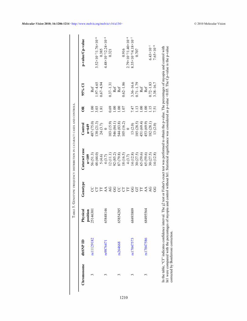
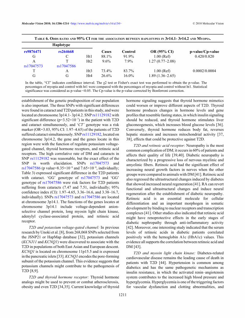
RESULTSA total of 968 unrelated individuals with T2D, over 20 yearsof age, were recruited from China Medical UniversityHospital (CMUH), Taiwan. The T2D patients with a LOCScataract score equal to or less than 3 and with a BCVA (bestcorrected visual acuity) of 0.18 logMAR (6/9) or better wereincluded in the control group. The T2D patients with a cataractscore of 10 or more in LOCS cataract evaluation system andwith a BCVA of 0.48 logMAR (6/18) or worse were includedin the cataract group. The patients that met the criteria were109 and 649, respectively. Average intraocular pressure (IOP)was 15.36 mmHg and 15.86 mmHg, respectively. Thecharacteristics of the T2D patients with cataracts and withoutcataracts can be seen in Table 3. The physical conditions ofthe T2D patients are expressed in Table 4. We conducted agenome-wide association study (GWAS) to simultaneouslyidentify genetic variants for T2D patients with cataracts inHan-Chinese residing in Taiwan. We started with 517,401SNPs that passed quality control filters using the IlluminaHap550duov3 chip. In the study, we found three SNPs withsignificant evidence (p<10−7) for association with diabeticcataracts, locus at chromosome 3p14.1- 3p14.2 (rs11129182,rs17047573, and rs17047586) (p value=3.52×10⁻⁷, 8.35×10⁻⁸, and 7.65×10⁻⁸, respectively; Table 4). In the threeSNPs, the CT genotype of rs11129182, GG genotype ofrs17047573, and the GG genotype of rs17047586 weresignificantly different in the T2D with cataracts and T2Dwithout cataracts groups (OR=3.03, 7.47, and 7.51,respectively; 95% confidence interval (CI): 1.97–4.65, 3.36–16.6, and 3.38–16.7, respectively; Table 5). We found thatthere were four haplotypes composed by the polymorphisms(Table 6). We defined haplotype 1 (Ht 1) to haplotype 4 (Ht4) as the alternative alleles of the DM and cataract related3p14.1- 3p14.2 polymorphisms (Table 5). Ht1 (G/C) and Ht2(A/T) were composed by rs9876471 and rs264667 and Ht3
TABLE 2. LENS OPACITIES CLASSIFICATION SYSTEM DEFINES THE EXTENT OF OPACIFICATION OF LENS CORTEX OPACIFICATION AND POSTERIORSUBCAPSULE OF LENS.
Cortical standard DescriptionC0 Clear lens devoid of aggregated dots, flecks,Ctr Minimal degree of cortical opacification and/orCI More extensive opacification with smallCII Cortical spoking that obscures more than 2 fullCIII Opacification that obscures about 50% of theCIV Advanced opacification filling about 90% of the
Posterior subcapsule standard DescriptionP0 Clear posterior capsulePI Cataract filling about 3% of the area of the posterior capsulePII About 30% opacification of the area of the posterior capsulePIII About 50% opacification of the area of the posterior capsule
Molecular Vision 2010; 16:1206-1214 <http://www.molvis.org/molvis/v16/a134> © 2010 Molecular Vision
1208

(T/A) and Ht4 (G/G) were composed by rs17047573 andrs17047586. The distribution of the Ht3 and Ht4 between theDM and cataract group and the control group differedsignificantly between the two groups (p=0.0004). The oddsratio (OR) of Ht4 was 1.89 and the 95% confidence interval(CI) was 1.36–2.65.
DISCUSSIONThe cut down of cataract level were LOCS 3 and 10 and theBCVA were 0.48 and 0.18 logMAR in this study. The patientswho matched both the criteria of cataract level and visualacuity were included in the control and study groups. Becausethe patients with LOCS cataract scores of less than 3 orbetween LOCS 3–10 all had some form of cataract, the BCVAof the patients was also the including criteria. In addition, theprogression of their cataracts might occur soon, the includingcriteria of cataracts and the control group was not a cut point.The patient with LOCS cataract scores of 3–10 were excludedfrom this study. In this study, we selected the patients withcataracts and poor BCVA as the study group and the patientswith contrary criteria as the control group. The patient withsenile cataracts could not be completely excluded; this wasthe limitation of our study. Nevertheless, the severity of thecataracts was the major criteria of the two groups. That is, thepatients with the similar physical conditions and variousseverity of cataract were compared. A large amount of effortshas been devoted to finding common T2D genes, includinggenome-wide linkage, candidate-gene and genome-wideassociation studies [17]. Whole-genome linkage scans haveidentified chromosomal regions linked to T2D [18]. There are
now at least 20 loci containing genes that increase the risk ofT2D, including potassium channels (KCNJ11 [potassiuminwardly-rectifying channel, subfamily J, member 11] andKCNQ1 [potassium voltage-gated channel, KQT-likesubfamily, member 1]) [19], cell cycle regulators (CDKAL1[regulatory subunit associated protein 1-like 1],CDKN2A-2B [cyclin-dependent kinase inhibitor 2A-2B], andCDC123-CAMK1D [cell division cycle 123 homolog-calcium/calmodulin-dependent protein kinase ID]) [20],nuclear hormone receptor (PPARG [peroxisome proliferator-activated receptor gamma]) [21], melatonin receptor(MTNR1B [melatonin receptor 1B]) [22], transcription factors(TCF7L2 [transcription factor 7-like 2], TCF2 or HNF1B[hepatocyte nuclear factor 1 beta], HHEX [hematopoieticallyexpressed homeobox], and JAZF1 [JAZF zinc finger 1])[23], translational regulator (IGF2BP2 [insulin-like growthfactor 2 mRNA binding protein 2]) [24], zinc transporter(SLC30A8 [solute carrier family 30 zinc transporter member8]) [25], apoptosis components (THADA [thyroid adenomaassociated]) [26], metalloprotease (ADAMTS9 [ADAMmetallopeptidase with thrombospondin type 1 motif, 9]) [27],calcium homeostasis component (WFS1 [Wolfram syndrome1 homolog]) [28], fat mass and obesity-associated gene(FTO [fat mass and obesity associated]) [29], pancreasorganogenesis (NOTCH2 [Notch homolog 2]) [30], andsurface glycoprotein (TSPAN8 [tetraspanin-8]) [31]. Variantsin these genes are almost exclusively identified in populationof European descent, and individually confer a modest risk(odds ratio=1.1–1.25) of developing T2D [31]. The ethnicdifferences in the genetic component are remarkable and the
TABLE 3. CHARACTERISTICS OF T2D PATIENTS WITH AND WITHOUT CATARACT.
Characteristics T2D with cataract T2D without cataractAge (y/o) 62±2.35 (20–70) 63±1.75 (20–70)
F/M 57/52 332/317BCVA 1.53±0.13 (0.48–2) 0.04±0.03 (0–0.18)
IOP (mmHg) 12.5±0.3 (7–21) 13.2±0.2 (7–21)
In the table, the term “F/M” indicates female/male and “BCVA” indicates best corrected visual acuity.
TABLE 4. THE PHYSICAL CONDITION OF THE T2D PATIENTS.
Variable Mean C/N Std Dev C/N Min C/N Max C/NBUN 17.42/17.32 8.17/8.15 5.80/5.70 87.10/82.20
Creatinine 0.91/0.90 0.58/0.62 0.23/0.29 13.80/12.95total-cholesterol 189.02/187.10 41.23/41.08 66.00/68.20 560.00/550.20
triglyceride 163.82/163.84 125.63/126.58 58.90/21.00 1380.00/1327.60HDL-C 48.75/48.67 13.75/13.82 8.25/8.00 111.00/108.50LDL-C 118.52/117.64 35.77/34.68 3.75/3.20 338.20/335.20HbA1C 7.94/7.92 1.45/1.49 4.20/4.50 16.20/15.90
In the table, “HDL-C” indicates high-density lipoprotein cholesterol and “LDL-C” indicates low-density lipoprotein cholesterol.In the column headers, “C/N” indicates T2D patient with cataract/T2D patient without cataract.
Molecular Vision 2010; 16:1206-1214 <http://www.molvis.org/molvis/v16/a134> © 2010 Molecular Vision
1209
TAB
LE 5
. GEN
OTY
PIC FR
EQU
ENC
Y D
ISTR
IBU
TIO
N IN
CA
TAR
AC
T C
ASE
S AN
D C
ON
TRO
LS.
Chr
omos
ome
dbSN
P ID
Phys
ical
posi
tion
Gen
otyp
eC
atar
act c
ase
n=10
9C
ontr
oln=
649
OR
95%
CI
p-va
lue/
Cp-
valu
e
3rs
1112
9182
2514
6301
CC
56 (5
1.3)
487
(75.
0)1.
00R
ef
C
T48
(44.
0)13
8 (2
1.3)
3.03
1.97
–4.6
53.
52×1
0−7/1
.76×
10−6
TT
5 (4
.6)
24 (3
.7)
1.81
0.67
–4.9
40.
385
3rs
9876
471
6584
8146
AA
4 (3
.7)
0
4.
48×1
0−5/2
.24×
10−4
A
G12
(11.
1)10
3 (1
5.9)
0.69
0.37
–1.3
10.
321
G
G92
(85.
2)54
6 (8
4.1)
1.00
Ref
3
rs26
4668
6585
4285
CC
87 (7
9.8)
544
(83.
8)1.
00R
ef
C
T18
(16.
5)10
5 (1
6.2)
1.07
0.62
–1.8
60.
916
TT
4 (3
.7)
0
2.
79×1
0−5/1
.40×
10−4
3rs
1704
7573
6849
3809
GG
14 (1
2.8)
13 (2
.0)
7.47
3.36
–16.
68.
35×1
0−8/4
.18×
10−7
G
T30
(27.
5)18
5 (2
8.5)
1.13
0.71
–1.7
90.
707
TT
65 (5
9.6)
451
(69.
4)1.
00R
ef
3rs
1704
7586
6849
5564
AA
65 (5
9.6)
453
(69.
9)1.
00R
ef
A
G30
(27.
5)18
2 (2
8.1)
1.15
0.72
–1.8
36.
43×1
0−1
G
G14
(12.
8)13
(2.0
)7.
513.
38–1
6.7
7.65
×10−8
In th
e ta
ble,
“C
I” in
dica
tes c
onfid
ence
inte
rval
. The
χ2
test
or F
ishe
r's e
xact
test
was
per
form
ed to
obt
ain
the
p-va
lue.
The
per
cent
ages
of m
yopi
a an
d co
ntro
l with
ht1
wer
e co
mpa
red
with
the
perc
enta
ges o
f myo
pia
and
cont
rol w
ithou
t ht1
. Sta
tistic
al si
gnifi
canc
e w
as c
onsi
dere
d as
p-v
alue
<0.
05. T
he C
p-va
lue
is th
e p-
valu
eco
rrec
ted
by B
onfe
rron
i cor
rect
ion.
Molecular Vision 2010; 16:1206-1214 <http://www.molvis.org/molvis/v16/a134> © 2010 Molecular Vision
1210
establishment of the genetic predisposition of our populationis also important. The three SNPs with significant differenceswere found in cataract and T2D patients in this study, and werelocated at chromosome 3p14.1- 3p14.2. SNP rs11129182 withsignificant difference (p=3.52×10⁻⁷) in the patient with T2Dand cataract simultaneously, and ‘CT’ genotype was a riskmarker (OR=3.03, 95% CI: 1.97–4.65) of the patients of T2Dsuffered cataract simultaneously. SNP rs11129182, located onchromosome 3p14.2, the gene and the genes locate in thisregion were with the function of regulate potassium voltage-gated channel, thyroid hormone receptors, and retinoic acidreceptors. The high correlative rate of DM and cataracts inSNP rs11129182 was reasonable, but the exact effect of theSNP is worth elucidation. SNPs rs17047573 andrs17047586 (p value=8.35×10⁻⁸ and 7.65×10⁻⁸, individually;Table 3) expressed significant difference in the T2D patientswith cataract. ‘GG’ genotype of rs17047573 and ‘GG’genotype of rs17047586 were risk factors for T2D patientssuffering from cataracts (7.47 and 7.51, individually; 95%confidence index (CI): 1.97–4.65, 3.36–16.6, and 3.38–16.7,individually). SNPs rs17047573 and rs17047586 are locatedat chromosome 3p14.1. The functions of the genes locates atchromosome 3p14.1 include voltage-dependent anion-selective channel protein, long myosin light chain kinase,adenylyl cyclase-associated protein, and retinoic acidreceptor.
T2D and potassium voltage-gated channel: In previousresearch by Unoki et al. [8], from 268,068 SNPs selected fromthe JSNP21 or HapMap database [32], potassium channels(KCNJ11 and KCNQ1) were discovered to associate with theT2D in populations of both East Asian and European descent.KCNQ1 is located on chromosome 11p15.5 and is expressedin the pancreatic islets [33]. KCNQ1 encodes the pore-formingsubunit of the potassium channel. This evidence suggests thatpotassium channels might contribute to the pathogenesis ofT2D [8,9].
T2D and thyroid hormone receptor: Thyroid hormoneanalogs might be used to prevent or combat atherosclerosis,obesity and even T2D [34,35]. Current knowledge of thyroid
hormone signaling suggests that thyroid hormone mimeticscould worsen or improve different aspects of T2D. Thyroidhormone produces changes in hormone levels and geneprofiles that resemble fasting states, in which insulin signalingshould be reduced, and thyroid hormone stimulates livergluconeogenesis, which increases blood glucose levels [36].Conversely, thyroid hormone reduces body fat, reverseshepatic steatosis and increases mitochondrial activity [37,38], effects that could be protective against T2D.
T2D and retinoic acid receptor: Neuropathy is the mostcommon complication of DM; it occurs in 60% of patients andaffects their quality of life [39,40]. Diabetic neuropathy ischaracterized by a progressive loss of nervous myelinic andamyelinic fibers. Retinoic acid had the significant effect ofincreasing neural growth factors in nerves when the othergroups were compared to animals with DM [41]. Retinoic acidalso regressed the ultrastructural changes induced by diabetesthat showed increased neural regeneration [41]. RA can revertfunctional and ultrastructural changes and induce neuralregeneration after the establishment of diabetic neuropathy.Retinoic acid is an essential molecule for cellulardifferentiation and an important morphogen in somaticdevelopment by binding to nuclear receptors and transcriptioncomplexes [41]. Other studies also indicated that retinoic acidmight have renoprotective effects in the early stages ofdiabetic nephropathy through anti-inflammatory activity[42]. Moreover, one interesting study indicated that the serumlevels of retinoic acids in diabetic patients correlatedpositively with the hemoglobin A1c (HbA1c) values. Thisevidence all supports the correlation between retinoic acid andDM [43].
T2D and myosin light chain kinase: Diabetes-relatedcardiovascular disease remains the leading cause of death inpatients with T2D [44]. Hypertension is common amongdiabetics and has the same pathogenetic mechanisms asinsulin resistance, in which the activated renin–angiotensinsystem contributes to the increased high blood pressure andhyperglycemia. Hyperglycemia is one of the triggering factorsfor vascular dysfunction and clotting abnormalities, and
TABLE 6. ODDS RATIO AND 95% CI FOR THE ASSOCIATION BETWEEN HAPLOTYPES IN 3P14.1- 3P14.2 AND MYOPIA.Haplotype
rs9876471 rs264668 Cases Control OR (95% CI) p-value/Cp-valueG C Ht1 88.1% 91.9% 1.00 (Ref) 0.420/0.820A T Ht2 9.6% 7.9% 1.27 (0.77–2.08)
rs17047573 rs17047586 T A Ht3 73.4% 83.7% 1.00 (Ref) 0.0002/0.0004G G Ht4 26.6% 16.0% 1.89 (1.36–2.65)
In the table, “CI” indicates confidence interval. The χ2 test or Fisher's exact test was performed to obtain the p-value. Thepercentages of myopia and control with ht1 were compared with the percentages of myopia and control without ht1. Statisticalsignificance was considered as p-value <0.05. The Cp-value is the p-value corrected by Bonferroni correction.
Molecular Vision 2010; 16:1206-1214 <http://www.molvis.org/molvis/v16/a134> © 2010 Molecular Vision
1211

therefore, for accelerated atherosclerosis in diabetes [44,45].Furthermore, angiotensin-converting enzyme inhibitorsmight offer additional cardioprotection to diabetics beyondthat provided by blood pressure reduction. Thephosphorylation of myosin light chain in cardiac muscles bycalmodulin-dependent kinase and has the modulatory role inthe activation of myofibrillar adenosine triphosphatase(ATPase) and the process of force generation [44-47]. Themyosin light chain is an important molecule in the diabetes-related cardiovascular disease [44-47].
T2D and adenylyl cyclase-associated protein: Giaproteins are associated with functions in the aorta of thestreptozotocin-induced diabetic rats [48]. A previous study byHashim et al. [48] indicated that decreased levels and activityof Gi proteins and adenylyl cyclase signaling induced byhyperglycemia may be one of the important mechanismscontributing to the cardiovascular complications associatedwith diabetes [48,49].
Cataract and the related proteins: The voltage-gatedchannel influences the intracellular iron concentrations thatare closely related to denature of crystalline and influencedthe cataract formation [50,51]. Thyroid hormones can confera protective effect against oxidative damage, which is one ofthe main causes of damages to cells in the eye's lens and isrelated to the formation of cataracts [52-54]. Retinoic acid(RA)-mediated inhibition of deregulated calpains has effectson the development of cataracts [55]. RA inhibits Ca2+
elevation and subsequent overactivation of calpains,suggesting the potential feasibility of calpain-targetingtherapies mediated by RA for cataracts [39-41]. MLC (myosinlight chain) phosphorylation is noted to play an important rolein maintaining lens function by regulating Rho kinase [55,56]. Adenylyl cyclase-associated protein was identified as amost efficient substrate of matrix metalloproteinases (MMP)[57]. Lens crystallins illustrates that intracellular structuralproteins are MMP substrates. Adenylyl cyclase-associatedprotein can induce the rapid turnover rate of MMP-9 [57].Lens opacity may be related to adenylyl cyclase-associatedprotein [57]. These protein all are related to thecataractogenesis and the pathogenesis of T2D. In thehaplotype study, Ht4 was significantly higher in the casesgroup and with an odd ratio: 1.89 and 95% CI:1.36–2.65. Thismeant that Ht4 was a risk factor for susceptibility to cataractsand DM. This evidence makes us realize the correlationbetween rs11129182, rs17047573, and rs17047586 with T2Dand cataract and helps us in predicting and designing newdiagnoses and treatment methods for T2D patients withcataracts.
ACKNOWLEDGMENTSThis study was supported by Academia Sinica GenomicMedicine Multicenter Study. This study was also supportedby grants from the China Medical University Hospital(CMU-95–141, CMU-96–081) and China Medical University(DMR-96–068, DMR-98–076 and DMR-99–093).
REFERENCES1. Falck A, Laatikainen L. Diabetic cataract in children. Acta
Ophthalmol Scand 1998; 76:238-40. [PMID: 9591961]2. Bron AJ, Cheng H. Cataract and retinopathy: screening for
treatable retinopathy. Clin Endocrinol Metab 1986;15:971-99. [PMID: 3096617]
3. Klein BE, Klein R, Moss SE. Incidence of cataract surgery inthe Wisconsin Epidemiologic Study of Diabetic Retinopathy.Am J Ophthalmol 1995; 119:295-300. [PMID: 7872389]
4. Di Benedetto A, Aragona P, Romano G, Romeo G, Di CesareE, Spinella R, Ferreri G, Cucinotta D. Age and metaboliccontrol influence lens opacity in type I, insulin-dependentdiabetic patients. J Diabetes Complications 1999;13:159-62. [PMID: 10509876]
5. Dua HS, Said DG, Otri AM. Are we doing too many cataractoperations? Cataract surgery: a global perspective. Br JOphthalmol 2009; 93:1-2. [PMID: 19098039]
6. Zimmet P, Alberti KG, Shaw J. Global and societal implicationsof the diabetes epidemic. Nature 2001; 414:782-7. [PMID:11742409]
7. Stumvoll M, Goldstein BJ, van Haeften TW. Type 2 diabetes:principles of pathogenesis and therapy. Lancet 2005;365:1333-46. [PMID: 15823385]
8. Unoki H, Takahashi A, Kawaguchi T, Hara K, Horikoshi M,Andersen G, Ng DP, Holmkvist J, Borch-Johnsen K,Jørgensen T, Sandbaek A, Lauritzen T, Hansen T, NurbayaS, Tsunoda T, Kubo M, Babazono T, Hirose H, Hayashi M,Iwamoto Y, Kashiwagi A, Kaku K, Kawamori R, Tai ES,Pedersen O, Kamatani N, Kadowaki T, Kikkawa R,Nakamura Y, Maeda S. SNPs in KCNQ1 are associated withsusceptibility to type 2 diabetes in East Asian and Europeanpopulations. Nat Genet 2008; 40:1098-102. [PMID:18711366]
9. Liu Y, Zhou DZ, Zhang D, Chen Z, Zhao T, Zhang Z, Ning M,Hu X, Yang YF, Zhang ZF, Yu L, He L, Xu H. Variants inKCNQ1 are associated with susceptibility to type 2 diabetesin the population of mainland China. Diabetologia 2009;52:1315-21. [PMID: 19448982]
10. McDonough CW, Hicks PJ, Lu L, Langefeld CD, Freedman BI,Bowden DW. The influence of carnosinase genepolymorphisms on diabetic nephropathy risk in African-Americans. Hum Genet 2009; 126:265-75. [PMID:19373489]
11. Chylack LT Jr, Wolfe JK, Singer DM, Leske MC, BullimoreMA, Bailey IL, Friend J, McCarthy D, Wu SY. The LensOpacities Classification System III. The Longitudinal Studyof Cataract Study Group. Arch Ophthalmol 1993;111:831-6. [PMID: 8512486]
12. Hall AB, Thompson JR, Deane JS, Rosenthal AR. LOCS IIIversus the Oxford clinical cataract classification and gradingsystem for the assessment of nuclear, cortical and posteriorsubcapsular cataract. Ophthalmic Epidemiol 1997;4:179-94. [PMID: 9500153]
13. Pan WH, Fann CS, Wu JY, Hung YT, Ho MS, Tai TH, ChenYJ, Liao CJ, Yang ML, Cheng AT, Chen YT. Han Chinesecell and genome bank in Taiwan: purpose, design and ethicalconsiderations. Hum Hered 2006; 61:27-30. [PMID:16534213]
Molecular Vision 2010; 16:1206-1214 <http://www.molvis.org/molvis/v16/a134> © 2010 Molecular Vision
1212
14. Camargos S, Scholz S, Simón-Sánchez J, Paisán-Ruiz C, LewisP, Hernandez D, Ding J, Gibbs JR, Cookson MR, Bras J,Guerreiro R, Oliveira CR, Lees A, Hardy J, Cardoso F,Singleton AB. DYT16, a novel young-onset dystonia-parkinsonism disorder: identification of a segregatingmutation in the stress-response protein PRKRA. LancetNeurol 2008; 7:207-15. [PMID: 18243799]
15. Duncan AJ, Bitner-Glindzicz M, Meunier B, Costello H,Hargreaves IP, López LC, Hirano M, Quinzii CM, SadowskiMI, Hardy J, Singleton A, Clayton PT, Rahman S. A nonsensemutation in COQ9 causes autosomal-recessive neonatal-onset primary coenzyme Q10 deficiency: a potentiallytreatable form of mitochondrial disease. Am J Hum Genet2009; 84:558-66. [PMID: 19375058]
16. Dunning MJ, Barbosa-Morais NL, Lynch AG, Tavaré S, RitchieME. Statistical issues in the analysis of Illumina data. BMCBioinformatics 2008; 9:85. [PMID: 18254947]
17. Ban HJ, Heo JY, Oh KS, Park KJ. Identification of Type 2diabetes-associated combination of SNPs using SupportVector Machine. BMC Genet 2010; 11:26. [PMID:20416077]
18. Takeuchi F, Serizawa M, Yamamoto K, Fujisawa T, NakashimaE, Ohnaka K, Ikegami H, Sugiyama T, Katsuya T, MiyagishiM, Nakashima N, Nawata H, Nakamura J, Kono S,Takayanagi R, Kato N. Confirmation of multiple risk Lociand genetic impacts by a genome-wide association study oftype 2 diabetes in the Japanese population. Diabetes 2009;58:1690-9. [PMID: 19401414]
19. Zhao J, Bradfield JP, Zhang H, Annaiah K, Wang K, Kim CE,Glessner JT, Frackelton EC, Otieno FG, Doran J, ThomasKA, Garris M, Hou C, Chiavacci RM, Li M, Berkowitz RI,Hakonarson H, Grant SF. Examination of all type 2 diabetesGWAS loci reveals HHEX-IDE as a locus influencingpediatric BMI. Diabetes 2010; 59:751-5. [PMID: 19933996]
20. Gouda HN, Sagoo GS, Harding AH, Yates J, Sandhu MS,Higgins JP. The association between the peroxisomeproliferator-activated receptor-gamma2 (PPARG2)Pro12Ala gene variant and type 2 diabetes mellitus: a HuGEreview and meta-analysis. Am J Epidemiol 2010;171:645-55. [PMID: 20179158]
21. Liu C, Wu Y, Li H, Qi Q, Langenberg C, Loos RJ, Lin X.MTNR1B rs10830963 is associated with fasting plasmaglucose, HbA1C and impaired beta-cell function in ChineseHans from Shanghai. BMC Med Genet 2010; 11:59. [PMID:20398260]
22. Stancáková A, Kuulasmaa T, Paananen J, Jackson AU,Bonnycastle LL, Collins FS, Boehnke M, Kuusisto J, LaaksoM. Association of 18 confirmed susceptibility loci for type 2diabetes with indices of insulin release, proinsulinconversion, and insulin sensitivity in 5,327 nondiabeticFinnish men. Diabetes 2009; 58:2129-36. [PMID: 19502414]
23. Chauhan G, Spurgeon CJ, Tabassum R, Bhaskar S, KulkarniSR, Mahajan A, Chavali S, Kumar MV, Prakash S, DwivediOP, Ghosh S, Yajnik CS, Tandon N, Bharadwaj D, ChandakGR. Impact of common variants of PPARG, KCNJ11,TCF7L2, SLC30A8, HHEX, CDKN2A, IGF2BP2 andCDKAL1 on the risk of type 2 diabetes in 5164 Indians.Diabetes. 2010 [PMID: 20424228]
24. Jing YL, Sun QM, Bi Y, Shen SM, Zhu DL. SLC30A8polymorphism and type 2 diabetes risk: Evidence from 27
study groups. Nutr Metab Cardiovasc Dis. 2010 [PMID:20167458]
25. Simonis-Bik AM, Nijpels G, van Haeften TW, Houwing-Duistermaat JJ, Boomsma DI, Reiling E, van Hove EC,Diamant M, Kramer MH, Heine RJ, Maassen JA, SlagboomPE, Willemsen G, Dekker JM, Eekhoff EM, de Geus EJ, 'tHart LM. Gene variants in the novel type 2 diabetes lociCDC123/CAMK1D, THADA, ADAMTS9, BCL11A, andMTNR1B affect different aspects of pancreatic beta-cellfunction. Diabetes 2010; 59:293-301. [PMID: 19833888]
26. Boesgaard TW, Gjesing AP, Grarup N, Rutanen J, Jansson PA,Hribal ML, Sesti G, Fritsche A, Stefan N, Staiger H, HäringH, Smith U, Laakso M, Pedersen O, Hansen T. EUGENE2Consortium. Variant near ADAMTS9 known to associatewith type 2 diabetes is related to insulin resistance in offspringof type 2 diabetes patients--EUGENE2 study. PLoS One2009; 4:e7236. [PMID: 19789630]
27. Fawcett KA, Wheeler E, Morris AP, Ricketts SL, Hallmans G,Rolandsson O, Daly A, Wasson J, Permutt A, Hattersley AT,Glaser B, Franks PW, McCarthy MI, Wareham NJ, SandhuMS, Barroso I. Detailed investigation of the role of commonand low-frequency WFS1 variants in type 2 diabetes risk.Diabetes 2010; 59:741-6. [PMID: 20028947]
28. Liu Y, Liu Z, Song Y, Zhou D, Zhang D, Zhao T, Chen Z, YuL, Yang Y, Feng G, Li J, Zhang J, Liu S, Zhang Z, He L, XuH. Meta-analysis Added Power to Identify Variants in FTOAssociated With Type 2 Diabetes and Obesity in the AsianPopulation. Obesity (Silver Spring). 2010 [PMID: 20057365]
29. Omori S, Tanaka Y, Horikoshi M, Takahashi A, Hara K, HiroseH, Kashiwagi A, Kaku K, Kawamori R, Kadowaki T,Nakamura Y, Maeda S. Replication study for the associationof new meta-analysis-derived risk loci with susceptibility totype 2 diabetes in 6,244 Japanese individuals. Diabetologia2009; 52:1554-60. [PMID: 19455301]
30. Grarup N, Andersen G, Krarup NT, Albrechtsen A, Schmitz O,Jørgensen T, Borch-Johnsen K, Hansen T, Pedersen O.Association testing of novel type 2 diabetes risk alleles in theJAZF1, CDC123/CAMK1D, TSPAN8, THADA,ADAMTS9, and NOTCH2 loci with insulin release, insulinsensitivity, and obesity in a population-based sample of 4,516glucose-tolerant middle-aged Danes. Diabetes 2008;57:2534-40. [PMID: 18567820]
31. McCarthy MI. Casting a wider net for diabetes susceptibilitygenes. Nat Genet 2008; 40:1039-40. [PMID: 19165915]
32. The international HapMap Consortium. A second generationhuman haplotype map of over 3.1 million SNPs. Nature 2007;449:851-61. [PMID: 17943122]
33. Yan L, Figueroa DJ, Austin CP, Liu Y, Bugianesi RM,Slaughter RS, Kaczorowski GJ, Kohler MG. Expression ofvoltage-gated potassium channels in human and rhesuspancreatic islets. Diabetes 2004; 53:597-607. [PMID:14988243]
34. Grover GJ, Mellström K, Malm J. Therapeutic potential forthyroid hormone receptor-beta selective agonists for treatingobesity, hyperlipidemia and diabetes. Curr Vasc Pharmacol2007; 5:141-54. [PMID: 17430219]
35. Baxter JD, Webb P. Thyroid hormone mimetics: potentialapplications in atherosclerosis, obesity and type 2 diabetes.Nat Rev Drug Discov 2009; 8:308-20. [PMID: 19337272]
Molecular Vision 2010; 16:1206-1214 <http://www.molvis.org/molvis/v16/a134> © 2010 Molecular Vision
1213

36. Crunkhorn S, Patti ME. Links between thyroid hormone action,oxidative metabolism, and diabetes risk? Thyroid 2008;18:227-37. [PMID: 18279023]
37. Ribeiro MO. Effects of thyroid hormone analogs on lipidmetabolism and thermogenesis. Thyroid 2008; 18:197-203.[PMID: 18279020]
38. Videla LA, Fernandez V, Tapia G, Varela P. Thyroid hormonecalorigenesis and mitochondrial redox signaling:upregulation of gene expression. Front Biosci 2007;12:1220-8. [PMID: 17127375]
39. Leinninger GM, Vincent AM, Feldman EL. The role of growthfactors in diabetic peripheral neuropathy. J Peripher Nerv Syst2004; 9:26-53. [PMID: 14871451]
40. Said G. Diabetic neuropathy. Nat Clin Pract Neurol 2007;3:331-40. [PMID: 17549059]
41. Hernández-Pedro N, Ordóñez G, Ortiz-Plata A, Palencia-Hernández G, García-Ulloa AC, Flores-Estrada D, Sotelo J,Arrieta O. All-trans retinoic acid induces nerve regenerationand increases serum and nerve contents of neural growthfactor in experimental diabetic neuropathy. Transl Res 2008;152:31-7. [PMID: 18593635]
42. Han SY, So GA, Jee YH, Han KH, Kang YS, Kim HK, KangSW, Han DS, Han JY, Cha DR. Effect of retinoic acid inexperimental diabetic nephropathy. Immunol Cell Biol 2004;82:568-76. [PMID: 15550114]
43. Yamakoshi Y, Fukasawa H, Yamauchi T, Waki H, KadowakiT, Shudo K, Kagechi H. Determination of endogenous levelsof retinoic acid isomers in type II diabetes mellitus patients.Possible correlation with HbA1c values. Biol Pharm Bull2002; 25:1268-71. [PMID: 12392076]
44. Clements RT, Sodha NR, Feng J, Boodhwani M, Liu Y, MienoS, Khabbaz KR, Bianchi C, Sellke FW. Impaired coronarymicrovascular dilation correlates with enhanced vascularsmooth muscle MLC phosphorylation in diabetes.Microcirculation 2009; 16:193-206. [PMID: 19152178]
45. Monnier L, Colette C, Owens DR. Integrating glycaemicvariability in the glycaemic disorders of type 2 diabetes: amove towards a unified glucose tetrad concept. DiabetesMetab Res Rev 2009; 25:393-402. [PMID: 19437415]
46. Zhu H, Zhang X, Zuo L, Zhou Q, Gui S, Wei W, Wang Y.Expression of Myosin Light Chain Kinase in Kidney ofStreptozotocin-Induced Diabetic Rats. Int J Mol Sci 2006;7:510-51.
47. Liu X, Takeda N, Dhalla NS. Myosin light-chainphosphorylation in diabetic cardiomyopathy in rats.Metabolism 1997; 46:71-5. [PMID: 9005973]
48. Hashim S, Li Y, Nagakura A, Takeo S, Anand-Srivastava MB.Modulation of G-protein expression and adenylyl cyclasesignaling by high glucose in vascular smooth muscle.Cardiovasc Res 2004; 63:709-18. [PMID: 15306227]
49. Shpakov AO, Kuznetsova LA, Plesneva SA, Bondareva VM,Guryanov IA, Vlasov GP, Pertseva MN. Decrease infunctional activity of G-proteins hormone-sensitive adenylatecyclase signaling system, during experimental type II diabetesmellitus. Bull Exp Biol Med 2006; 142:685-9. [PMID:17603670]
50. Long SB, Tao X, Campbell EB, MacKinnon R. Atomicstructure of a voltage-dependent K+ channel in a lipidmembrane-like environment. Nature 2007; 450:376-82.[PMID: 18004376]
51. Jensen MØ, Borhani DW, Lindorff-Larsen K, Maragakis P,Jogini V, Eastwood MP, Dror RO, Shaw DE. Principles ofconduction and hydrophobic gating in K+ channels. Proc NatlAcad Sci USA 2010; 107:5833-8. [PMID: 20231479]
52. Gredilla R, Barja G, López-Torres M. Thyroid hormone-induced oxidative damage on lipids, glutathione and DNA inthe mouse heart. Free Radic Biol Med 2001; 35:417-25.[PMID: 11697138]
53. Guerrero A, Pamplona R, Portero-Otin M, Barja G, López-Torres M. Effect of thyroid status on lipid composition andperoxidation in the mouse liver. Free Radic Biol Med 1999;261:73-80. [PMID: 9890642]
54. Das K, Chainey GB. Modulation of rat liver mitochondrialantioxidant defence system by thyroid hormone. BiochimBiophys Acta 2001; 1537:1-13. [PMID: 11476958]
55. Nishikiori N, Osanai M, Chiba H, Kojima T, Ohguro H, SawadaN. Inhibitory effects of retinoic acid receptor alpha stimulantson murine cataractogenesis through suppression ofderegulated calpains. Invest Ophthalmol Vis Sci 2007;48:2224-9. [PMID: 17460283]
56. Harper JM, Wolf N, Galecki AT, Pinkosky SL, Miller RA.Hormone levels and cataract scores as sex-specific, mid-lifepredictors of longevity in genetically heterogeneous mice.Mech Ageing Dev 2003; 124:801-10. [PMID: 12875743]
57. Cauwe B, Martens E, Van den Steen PE, Proost P, Van Aelst I,Blockmans D, Opdenakker G. Adenylyl cyclase-associatedprotein-1/CAP1 as a biological target substrate of gelatinaseB/MMP-9. Exp Cell Res 2008; 314:2739-49. [PMID:18671965]
Molecular Vision 2010; 16:1206-1214 <http://www.molvis.org/molvis/v16/a134> © 2010 Molecular Vision
The print version of this article was created on 28 June 2010. This reflects all typographical corrections and errata to the articlethrough that date. Details of any changes may be found in the online version of the article.
1214