selective inhibition of ezh2 by epz-6438 leads to potent ... · pdf fileactivity in...
TRANSCRIPT

2014;13:842-854. Published OnlineFirst February 21, 2014.Mol Cancer Ther Sarah K. Knutson, Satoshi Kawano, Yukinori Minoshima, et al.
-Mutant Non-Hodgkin LymphomaEZH2Activity in Selective Inhibition of EZH2 by EPZ-6438 Leads to Potent Antitumor
Updated version
10.1158/1535-7163.MCT-13-0773doi:
Access the most recent version of this article at:
Material
Supplementary
http://mct.aacrjournals.org/content/suppl/2014/02/20/1535-7163.MCT-13-0773.DC1.html
Access the most recent supplemental material at:
Cited Articles
http://mct.aacrjournals.org/content/13/4/842.full.html#ref-list-1
This article cites by 32 articles, 11 of which you can access for free at:
E-mail alerts related to this article or journal.Sign up to receive free email-alerts
Subscriptions
Reprints and
To order reprints of this article or to subscribe to the journal, contact the AACR Publications Department at
Permissions
To request permission to re-use all or part of this article, contact the AACR Publications Department at
on April 14, 2014. © 2014 American Association for Cancer Research. mct.aacrjournals.org Downloaded from
Published OnlineFirst February 21, 2014; DOI: 10.1158/1535-7163.MCT-13-0773
on April 14, 2014. © 2014 American Association for Cancer Research. mct.aacrjournals.org Downloaded from
Published OnlineFirst February 21, 2014; DOI: 10.1158/1535-7163.MCT-13-0773

Small Molecule Therapeutics
Selective Inhibition of EZH2 by EPZ-6438 Leads to PotentAntitumor Activity in EZH2-Mutant Non-HodgkinLymphoma
Sarah K. Knutson1, Satoshi Kawano3, Yukinori Minoshima3, Natalie M. Warholic1, Kuan-Chun Huang2,Yonghong Xiao1, Tadashi Kadowaki2, Mai Uesugi3, Galina Kuznetsov2, Namita Kumar2, Tim J. Wigle1,Christine R. Klaus1, Christina J. Allain1, Alejandra Raimondi1, Nigel J. Waters1, Jesse J. Smith1,Margaret Porter-Scott1, Richard Chesworth1, Mikel P. Moyer1, Robert A. Copeland1, Victoria M. Richon1,Toshimitsu Uenaka3, Roy M. Pollock1, Kevin W. Kuntz1, Akira Yokoi3, and Heike Keilhack1
AbstractMutationswithin the catalytic domain of the histonemethyltransferase EZH2havebeen identified in subsets
of patients with non-Hodgkin lymphoma (NHL). These genetic alterations are hypothesized to confer an
oncogenic dependency on EZH2 enzymatic activity in these cancers. We have previously reported the
discovery of EPZ005678 and EPZ-6438, potent and selective S-adenosyl-methionine-competitive small mol-
ecule inhibitors of EZH2. Although both compounds are similar with respect to their mechanism of action and
selectivity, EPZ-6438 possesses superior potency and drug-like properties, including good oral bioavailability
in animals. Here, we characterize the activity of EPZ-6438 in preclinical models of NHL. EPZ-6438 selectively
inhibits intracellular lysine 27 of histone H3 (H3K27) methylation in a concentration- and time-dependent
manner in both EZH2wild-type andmutant lymphoma cells. Inhibition of H3K27 trimethylation (H3K27Me3)
leads to selective cell killing of human lymphoma cell lines bearing EZH2 catalytic domain point mutations.
Treatment ofEZH2-mutantNHLxenograft-bearingmicewithEPZ-6438 causes dose-dependent tumor growth
inhibition, including complete and sustained tumor regressions with correlative diminution of H3K27Me3
levels in tumors and selected normal tissues.Mice dosed orallywith EPZ-6438 for 28 days remained tumor free
for up to 63 days after stopping compound treatment in two EZH2-mutant xenograft models. These data
confirm the dependency of EZH2-mutant NHL on EZH2 activity and portend the utility of EPZ-6438 as a
potential treatment for these genetically defined cancers. Mol Cancer Ther; 13(4); 842–54. �2014 AACR.
IntroductionRegulation of gene transcription by enzyme-catalyzed,
covalent modification of histone proteins and nucleicacids is a critical biologic process (1). Posttranslationalhistone modifications that are known to contribute to theregulation of gene transcription include acetylation,
methylation, phosphorylation, and ubiquitinylation (2).Methylation events at lysine and arginine residues, cata-lyzed by histone methyltransferases (HMT), have beensubjected to intense investigations in recent years becauseHMTs represent a particularly attractive class of targetsfor drug discovery efforts, because of their disease asso-ciation and druggability (3). For instance, genetic altera-tions have been identified in a number of HMTs in humancancers where they are purported to play a causal role inmalignancies. In addition, selective small molecule HMTinhibitors have been demonstrated to cause context-spe-cific antiproliferative activity in cancers bearing geneticalterations in the targeted HMT or related pathways.
EZH2 is the catalytic subunit of the multiprotein HMTcomplexknownaspolycombrepressive complex 2 (PRC2;refs. 4 and 5). EZH2 has been implicated in several cancertypes by mutation, amplification, and/or overexpression(6). Heterozygous EZH2 mutations within the catalyticSET domain have been observed in 10% of non-Hodgkinlymphomas (NHL) at 3 hotspots (Y646, A682, and A692,referring to EZH2 variant NM_004456.3; refs. 7–9). Inter-estingly, these mutations are exclusively found in lym-phomas of germinal center origin, in line with recent data
Authors' Affiliations: 1Epizyme Inc., Cambridge; 2Eisai Inc., Andover,Massachusetts; and 3Eisai Co. Ltd., Tsukuba-shi, Ibaraki, Japan
Note: Supplementary data for this article are available at Molecular CancerTherapeutics Online (http://mct.aacrjournals.org/).
S.K. Knutson and S. Kawano contributed equally to this work.
Current address for V.M. Richon: Sanofi, 270 Albany Street, Cambridge,MA 02139.
Corresponding Authors: Akira Yokoi, Eisai Co. Ltd., 5-1-3 Tokodai,Tsukuba-shi, Ibaraki, 300-2635, Japan. Phone: 81-29-847-5748; Fax:81-29-847-2759; E-mail: [email protected]; and Heike Keilhack,Epizyme Inc., 400 Technology Square, 4th Floor, Cambridge, MA02139. Phone: 617-500-0618; Fax: 617-349-0707; E-mail:[email protected]
doi: 10.1158/1535-7163.MCT-13-0773
�2014 American Association for Cancer Research.
MolecularCancer
Therapeutics
Mol Cancer Ther; 13(4) April 2014842
on April 14, 2014. © 2014 American Association for Cancer Research. mct.aacrjournals.org Downloaded from
Published OnlineFirst February 21, 2014; DOI: 10.1158/1535-7163.MCT-13-0773

suggesting amaster role of EZH2 in B-cell germinal centerformation and maintenance (10, 11). Mutant EZH2 pro-teins change their substrate specificity and act in concertwith wild-type EZH2 to generate abnormally high levelsof trimethylated lysine 27 on histone H3 (H3K27Me3),leading to abnormal repression of PRC2 targets, whichdrives lymphomagenesis (12–16). These preclinical dataprovide the basis for EZH2 inhibition as a specific rationaltherapy for germinal center–derived B-cell lymphomas,and we and others previously reported that selectiveinhibition of EZH2 in cell culture results in selectivekilling of lymphoma cells bearing EZH2 mutations (17–20). EPZ-6438 is oneof a several potent and selective EZH2inhibitors that we and others have recently reported.These inhibitors, including our previously publishedtool compound EPZ005687, share similar in vitro proper-ties (i.e., mechanism of action, specificity, and cellularactivity) as EPZ-6438. However, EPZ-6438 is among themost potent EZH2 inhibitors described to date anddemonstrates significantly improved pharmacokineticproperties relative to EPZ005687, including good oralbioavailability in animals. Consistent with this, we pre-viously demonstrated that oral dosing of EPZ-6438 leadsto potent in vivo target inhibition and antitumor activity inaSMARCB1-deletedmalignant rhabdoid tumor xenograftmodel (21). Here we expand on the pharmacologic prop-erties of EPZ-6438 and assessed whether EZH2-mutantlymphomas are selectively sensitive to EZH2 inhibition invitro and in vivo, which would suggest its application as anovel targeted therapy in EZH2 mutant–bearing NHL.
Materials and MethodsSynthesis of EPZ-6438A synthetic route of EPZ-6438 is described in PCT
patent application publication number WO/2012/142504.
Cell culture, immunoblot, and proliferation assaysLymphoma cell lines OCI-LY19 (ACC-528), WSU-
DLCL2 (ACC-575), KARPAS-422 (ACC-32), and SU-DHL-10 (ACC-576) were obtained from DSMZ. RL(CRL-2261), Toledo (CRL-2631), Pfeiffer (CRL-2632),SU-DHL-6 (CRL-2959), and Farage (CRL-2630) cells wereobtained from American Type Culture Collection.DOHH2 (HTL99022) was obtained from BBCF. Toledo,SU-DHL-6, and KARPAS-422 cell lines were cultured inRPMI þ 20% FBS, whereas all other cell lines were cul-tured inRPMIþ 10%FBS.Cell lineswere authenticatedbySTR assay and EZH2 mutational status was verified bysequence analysis. Histone extractions, immunoblot, 11-day proliferation assays, methylation time course, cellcycle, and apoptosis experiments were performed aspreviously described (17).
High throughput proliferation assayFor the assessment of the effect of compounds on the
proliferation of the WSU-DLCL2 cell line, exponentiallygrowing cells were plated in 384-wellwhite opaque plates
at a density of 1,250 cells/mL in a final volume of 50 mL ofassay medium (RPMI 1640 supplemented with 20% v/vheat-inactivated FBS, 100 units/mLpenicillin–streptomy-cin). A compound source polypropylene 384-well platewith an assigned container number was prepared byperforming triplicate 9-point 3-fold serial dilutions indimethyl sulfoxide (DMSO), beginning at 10 mmol/L(final top concentration of compound in the assay was20 mmol/L and the final concentration of DMSO was0.2%). A 100-nL aliquot from the compound stock platewas added to its respectivewell in the cell plate. The 100%inhibition control consisted of cells treated with 200nmol/L final concentration of staurosporine and the 0%inhibition control consisted of DMSO-treated cells. Afteraddition of the compounds, assay plates were incubatedfor 6 days at 37�C, 5% CO2, relative humidity >90%. Cellviability was measured by quantitation of ATP present inthe cell cultures, by adding 35 mL of Cell Titer Glo reagentto the cell plates. Luminescence was read in the Spectra-Max M5 instrument. The concentration inhibiting cellviability by 50% was determined using a 4-parametric fitof the normalized dose–response curves.
Chromatin immunoprecipitation followed by PCR(ChIP-PCR)
WSU-DLCL2 cells were treated with either DMSO or 1mmol/L EPZ-6438 for 4 days. Cells were harvested andfixed according to publishedmethods provided byActiveMotif (http://www.activemotif.com/documents/1848.pdf). Antibodies used for ChIP include: EZH2 (ActiveMotif catalog no. 39901), SUZ12 (Abcam ab12073), andH3K27Me3 (CST 9733). Primer design and data analysiswere performed by Active Motif using the ChIP-IT qPCRAnalysis Kit published manual (catalog no. 53029).
Cell treatments for gene expression profiling, dataprocessing, and further analyses
Each of the 4 cell lines (WSU-DLCL2, KARPAS-422, SU-DHL-6, and Pfeiffer) were plated in 6-well plates at aninitial seeding density to ensure cell densities werewithinlinear logphase growth for days 2 and 4 of the time course.Cells were treated with either DMSO, or 1� or 10� LCCconcentration of EPZ-6438. EPZ-6438 LCC concentrationsfor each of the cell lines are as follows: WSU-DLCL2, 200nmol/L; KARPAS-422, 100 nmol/L; SU-DHL-6, 200nmol/L; and Pfeiffer, 0.5 nmol/L. At each time point,cells were harvested by centrifugation, washed with PBS,and cell pellets were snap frozen. A second set of cellswere treated in a similarmanner on day 0, counted on day4, split back to the original plating density, and redosedwithDMSO, 1�or 10�LCCconcentration of EPZ-6438 forthe day 6 time point. RNA extraction and amplificationand subsequentmicroarray processingwas performed byExpression Analysis, Inc. as previously described (17).Both normalized expression data andCELfiles are depos-ited in GEO under GSE49284.
RNA extraction and amplification and subsequentmicroarray processing was performed by Expression
Lymphoma Tumor Regression with EZH2 Inhibitor
www.aacrjournals.org Mol Cancer Ther; 13(4) April 2014 843
on April 14, 2014. © 2014 American Association for Cancer Research. mct.aacrjournals.org Downloaded from
Published OnlineFirst February 21, 2014; DOI: 10.1158/1535-7163.MCT-13-0773

Analysis, Inc. as previously described (17). NormalizedAffymetrixU133plus2 array expressiondatawere createdfor all samples from CEL files using the Expression FileCreator Module from GenePattern (http://genepattern.broadinstitute.org). Quantile normalization and GCRMAwere selected as the algorithms to generate normalizedexpression values. Both normalized expression data andCEL files are deposited in GEO under GSE49284.
Two sample t tests were carried out for differentiallyexpressed genes upon EPZ-6438 treatment, for each cellline at 2 doses (LCC and 10� LCC) and 3 time points (2, 4,and6days).Geneswith fold change>2 or<0.5 andP-value< 0.05 were further analyzed through the use of IngenuityPathway Analysis (Ingenuity Systems, www.ingenuity.com).
Gene set enrichment analysis (GSEA) was performedon EPZ-6438 versus DMSO-treated 2-sample comparisonfor all cell lines, each at 2 concentrations and 3 time points,using the GSEA Java-enabled desktop software (Version2.0.13, http://www.broadinstitute.org/gsea/index.jsp).Probe sets were collapsed to gene symbols (HUGOnomenclature) using maximum probe intensity collaps-ingmode. Permutationwas carried out on gene set, ratherthan phenotype, to accommodate small sample size. Cus-tomized KEGG pathway gene sets [added Ben-Porathembryonic stem (ES) cell and Velichutina CentroblastPRC2 target sets, as described (17)] aswell as transcriptionfactor binding motif gene sets from MSigDB version 4.0(http://www.broadinstitute.org/gsea/msigdb/index.jsp) were used for all GSEA analyses.
Xenograft studiesAll the procedures related to animal handling, care, and
the treatment in this study were performed according tothe guidelines approved by the Institutional Animal CareandUse Committee of CRLPiedmont, Eisai Tsukuba, andEisai Andover following the guidance of the Associationfor Assessment and Accreditation of Laboratory AnimalCare.
WSU-DLCL2 cells were harvested during mid-logphase growth, and resuspended in PBSwith 50%Matrigel(BD Biosciences). SCIDmice received 1� 107 cells (0.2mLcell suspension) subcutaneously in the right flank. After10 to 30 days, mice with 108 to 126 mm3 tumors weresorted into treatment groupswithmean tumor volumes of117 to 119mm3. EPZ-6438 or vehicle (0.5%NaCMCþ 0.1%Tween-80 in water) was administered at the indicateddoses on 3 timesdaily every 8hours, 2 times aday every 12hours, or once a day schedules for either 7 or 28 days byoral gavage. Each dose was delivered in a volume of 0.2mL/20 g mouse (10 mL/kg), and adjusted for the lastrecorded weight of individual animals. The maximaltreatment length was 28 days. On day 7 or day 28 duringthe studies, mice were sampled in a prespecified fashion.Sampling included nonterminal bleeds (0.25mL) from themandibular vein without anesthesia and full volumeblood collection via terminal cardiac puncture underCO2 anesthesia. Blood samples were processed for plas-
ma, with K2-EDTA as anticoagulant. The plasma sampleswere frozen at �80�C and stored before bioanalysis ofcompound levels. Tumors were harvested from specifiedmice under RNase-free conditions and bisected. Tumortissue fromeachanimalwas snap frozen in liquidnitrogenand pulverized with a mortar and pestle.
KARPAS-422 cells were harvested during mid-logphase growth, and resuspended in Hank balanced saltsolution with 50% Matrigel (BD Biosciences). Balb/C-numice (Charles River Laboratories Japan) received 1 � 107
cells (0.1 mL cell suspension) subcutaneously in the rightflank.Mice carrying tumors of approximately 150mm3 forefficacy studies (14 days after injection) or 250 mm3 forpharmacodynamics studies (21 days after injection) weresorted into treatment groups with similar mean tumorvolumes. EPZ-6438 or vehicle (0.5%MCþ 0.1% Tween-80in water) was administered at the indicated doses ontwice a day or once a day schedules for either 7 or 28days by oral gavage. Each dosewas delivered in a volumeof 0.2 mL/20 g mouse (10 mL/kg), and adjusted for thelast recorded weight of individual animals. Tumorvolumeswere followed throughout the experiment. Tumorvolume was measured 2 times weekly after the start oftreatment. In pharmacodynamic studies, tumors were har-vested from specified mice, soaked in ice cold lysis buffer(10 mmol/L MgCl2, 10 mmol/L Tris-HCl, 25 mmol/LKCl, 1% TritonX-100, 8.6% sucrose and protease inhibitor),and homogenized with handy microhomogenizer.
Pfeiffer donor tumors were first prepared by implant-ing 1�107 cells (0.1mLcell suspensionwith 50%Matrigel;BD Biosciences) under the right arm of NSG mice (TheJackson Laboratory) subcutaneously. Approximately 63days after implantation, donor tumors were harvestedand dissected into fragments in size of approximate 20mg. These tumor fragmentswere subsequently implantedinto 100 NSG mice subcutaneously. Thirty days afterimplantation, 45 mice with tumors ranging from 124 to680 mm3 were selected and randomized into groups forefficacy study. EPZ-6438 or vehicle (0.5% MC þ 0.1%Tween-80 in water) was administered at once a dayschedules for either 7, 12, or 28 days by oral gavage. Eachdose was delivered in a volume of 0.2 mL/20 g mouse(10 mL/kg), and adjusted for the last recorded weight ofindividual animals. Tumor volumewasmeasured 2 timesweekly after the start of treatment.
Rat studiesMale and female Sprague-Dawley rats (8 weeks old)
were treated with EPZ-6438 at various doses or vehicle(0.5% NaCMC þ 0.1% Tween-80 in water) for 28 daysonce a day. On day 22, the females from the highest dosegroup received another dose and were subsequentlyeuthanized on day 23 approximately 29 hours after thelast dose. All other animals were euthanized on day 29approximately 29 hours after the last dose administeredon day 28. At euthanasia, the full blood volume wascollected, peripheral blood mononuclear cells (PBMC)were isolated, and cell pellets were frozen and stored at
Knutson et al.
Mol Cancer Ther; 13(4) April 2014 Molecular Cancer Therapeutics844
on April 14, 2014. © 2014 American Association for Cancer Research. mct.aacrjournals.org Downloaded from
Published OnlineFirst February 21, 2014; DOI: 10.1158/1535-7163.MCT-13-0773

�80�C before analysis. A 2-mm-thick slice of skin wasformalin-fixed for 24 hours and transferred to 70% etha-nol. The fixed tissues were paraffin embedded. Bonemarrow samples were collected from femur, tibia, andhip bones, frozen and stored at �80�C before analysis.Histones extraction from tissues and H3K27Me3 ELISAwere previously described (21).
ImmunohistochemistryParaffin sections of skin were generated using a Leica
rotary microtome RM2255 and placed on charged slides(SurgiPath). Slides were baked at 60�C for 30 minutes.Thereafter, slides were washed once with Bond Dewaxsolution (Leica Microsystems, Catalog No. AR9222), fol-lowed by 3 washes with absolute ethanol and then oncewith Leica Bond Wash solution (10� Bond wash, LeicaMicrosystems,CatalogNo.AR9590,wasused to prepare a1�working solution in deionized water). Heat-mediatedepitope retrieval was performed in Leica Bond ER2retrieval solution (Leica Microsystems, Catalog No.AR9640), applied at ambient temperature 2 times fol-lowed by an incubation at 100�C for 20 minutes, and thenat ambient temperature again for 12 minutes. After thatslides were incubated with 3% H2O2 for 15 minutes,followedby 3washeswith BondWash solution. Antibodystaining was done in a Leica BondMax autostainer, LeicaMicrosystems M211518. Slides were incubated with pri-mary antibody dilutions (anti-H3K27Me3, Cell SignalingTechnology #9733; final concentration 0.07 mg/mL oranticleaved caspase-3, Epitomics #1476-1, final concentra-tion 2 mg/mL) for 60 minutes, followed by 3 washes with
Leica Bond Wash solution. Thereafter, slides were incu-bated with polymer horseradish peroxidase reagent(Leica Microsystems, Catalog No. DS9800) for 30 minutesfollowed by 4washes with Leica BondWash solution and1 wash with deionized water. Then slides were incubatedwith the DAB Refine chromogen for 10 minutes followedby 3 washes with deionized water. Slides were counter-stained with hematoxylin for 5 minutes and washed oncewith deionizedwater and Leica BondWash. Dehydrationand mounting of the stained slides were done in a LeicaST5020 autostainer andCV5030-TS5025 Coversliper (once95% ethanol, 3 times 100% ethanol, and 3 times xylene).The mounting solution was Surgipath Micromountmounting medium (Leica Microsystems, Catalog No.01730). Slidesweredigitizedusing theAperioXTScanner.Digitized slides were examined visually using AperioImage Scope and image analysis, and quantification ofthe staining (percent of positive cells or percent positivelystained area of the sample) was done using the Aperioimage algorithms.
ResultsEPZ-6438 specifically inhibits cellular H3K27methylation in cells
A preliminary description of EPZ-6438 was reportedpreviously (21). EPZ-6438 (structure shown in Fig. 1A) is apotent, selective, and orally bioavailable small moleculeinhibitor of EZH2 showing context-specific activity inSMARCB1-deleted rhabdoid tumors. Similar to its effectin rhabdoid tumor cells, EPZ-6438 treatment of the EZH2
C
EPZ-6438 (µmol/L)0 2.7
B
H3K27Me1
H3K27Me2
H3K27Me3
H3K27acetyl
H3K4Me3
H3K9Me3
H3K36Me2
H3K79 Me2
Total H3
H3K27Me3
EPZ-6438
H3
DA
mmol/L
H3K
27M
e3 (
imm
un
ob
lot)
per
cen
tag
e o
f ve
hic
le c
on
tro
l
Days
DMSO
0.00
0133
0.00
040.
001
0.00
3
0.01
0.03 0.1
0.3
0.9
2.7
NaÏve
Figure 1. Effects of EPZ-6438 oncellular global histone methylationand cell viability. A, chemicalstructure of EPZ-6438. B,concentration-dependentinhibition of cellular H3K27Me3levels in EZH2 Y646 mutant–bearing WSU-DLCL2 cells,determined by immunoblot.C, EPZ-6438 inhibits cellularH3K27Me3 in WSU-DLCL2 cells ina time-dependent manner(assessed by immunoblot anddensitometry). D, EPZ-6438selectively inhibits cellular H3K27methylation in OCI-LY-19 EZH2wild-type lymphoma cells. Cellswere treated for 4 days for eachexperiment shown at the indicatedconcentrations (1 mmol/L in B).
Lymphoma Tumor Regression with EZH2 Inhibitor
www.aacrjournals.org Mol Cancer Ther; 13(4) April 2014 845
on April 14, 2014. © 2014 American Association for Cancer Research. mct.aacrjournals.org Downloaded from
Published OnlineFirst February 21, 2014; DOI: 10.1158/1535-7163.MCT-13-0773

Y646F-mutant lymphoma cell lineWSU-DLCL2 for 4 daysinduced a concentration-dependent reduction in globalH3K27Me3 levels with an IC50 value of 9 nmol/L(H3K27Me3 levels determined by immunoblot; Fig. 1Band Table 1). Treatment of WSU-DLCL2 cells with 1mmol/L EPZ-6438 led to time-dependent global loss ofH3K27Me3 with a half-life of approximately 1 day. Con-sistent with a simple exponential decay process, weobserved �90% loss of H3K27Me3 after 3 to 4 days ofcompound treatment (Fig. 1C). To test the specificity ofEPZ-6438 in cells, we chose an EZH2 wild-type linebecause mutant cells, including WSU-DLCL2, do notcontain detectable levels of the dimethyl form of H3K27(14). When we incubated OCI-LY19 EZH2wild-type lym-phoma cellswith 2.7mmol/LEPZ-6438 for 4days, the onlymethyl marks affected were H3K27Me1, H3K27Me2, andH3K27Me3, the 3 known products of PRC2 catalysis (Fig.1D). Incubation with EPZ-6438 also resulted in a slightincrease in H3K27 acetylation. Furthermore, lysine 36dimethylation of histone H3 was not influenced.
The ability of EPZ-6438 to reduce global H3K27Me3levels was further tested in several other human lympho-ma cell lines, including lines expressing either wild-typeor mutant EZH2. EPZ-6438 reduced H3K27Me3 withsimilar potency in all cell lines, independent of the EZH2status (Table 1).
EPZ-6438 leads to selective killing of lymphoma celllines bearing EZH2 point mutations
To evaluate the phenotypic effects of compound treat-ment, we performed proliferation assays with wild-typeand mutant lymphoma cell lines. Incubation of WSU-DLCL2 EZH2 Y646F-mutant cells with EPZ-6438 inhib-ited cell proliferation with an average IC50 value of 0.28�0.14mmol/L in a 6-dayproliferation assay. To evaluate the
kinetics of the antiproliferative activity of EPZ-6438 inmore detail, additional proliferation assays were per-formed over a period of 11 days. In WSU-DLCL2 cells,the antiproliferative effect of EPZ-6438 was apparentafter 4 days of incubation (Fig. 2A). This coincides withthe time required for maximal H3K27Me3 inhibition inthis cell line (Fig. 1B) and is consistent with a mechanismin which phenotypic effects occur following inhibitionof H3K27 methylation. The IC50 value for EPZ-6438 inhi-bition of proliferation of WSU-DLCL2 cells was lowerafter 11 days of treatment than after 6 days of treatment(Table 1), likely because of increase cell killing at later timepoints. In contrast to the WSU-DLCL2 cells, the prolifer-ation of OCI-LY19 human lymphoma cells (EZH2 wild-type for residue Y646) over 11 days was not significantlyaffected (Fig. 2B), despite comparable IC50 values forH3K27Me3 inhibition in both cell lines (Table 1). The11-day proliferation assay results were used to calculatethe lowest cytotoxic concentration (LCC), for each cellline. The LCC is defined as the concentration of inhibitorat which the proliferation rate becomes zero and repre-sents the crossover point between cytostasis and cytotox-icity (17). The LCC value for WSU-DLCL2 EZH2 Y646F-mutant human lymphoma cells was 0.17 mmol/Lwhereasno cytotoxicity (LCC > 10 mmol/L) was observed in theEZH2wild-type cell lineOCI-LY19 (Table 1). This context-specific activity of EPZ-6438 was further supported byresults from 11-day proliferation assays with an extendedlymphoma cell linepanel.All cell lines harboring anEZH2mutation, with the exception of the RL cell line (EZH2Y646N), were more sensitive to the antiproliferativeeffects of EPZ-6438 than cell lines containing wild-typeEZH2 (Table 1). Farage cells had the lowest IC50 amongthe EZH2 wild-type group, but no cytotoxicity could beobserved with compound treatment (LCC > 10 mmol/L).
Table 1. IC50 values for methylation and proliferation as well as LCC values for EPZ-6438 in humanlymphoma cell lines
Cell line EZH2 statusMethylationIC50 (nmol/L)a
ProliferationIC50 (mmol/L)b LCC (mmol/L)b
DOHH-2 Wild-type 3 1.7 >10Farage Wild-type 2 0.099 >10OCI-LY19 Wild-type 8 6.2 >10Toledo Wild-type ND 7.6 >10KARPAS-422 Y646N 90 0.0018 0.12Pfeiffer A682G 2 0.00049 0.0005RL Y646N 22 5.8 >10SU-DHL-10 Y646F ND 0.0058 0.14SU-DHL-6 Y646N 20 0.0047 0.21WSU-DLCL2 Y646F 9 0.0086 0.17
Abbreviation: ND, not determined.aDerived after incubation for 4 days by immunoblot. Values represent the result from 1 experiment.bDerived after incubation for 11 days. Compound incubations for each experiment were performed in triplicate, and values represent 1experiment for all cell lines except OCI-LY19, Pfeiffer, andWSU-DLCL2. For the remaining 3 cell lines, values represent themean fromthe following number of experiments: OCI-LY19, n ¼ 9; Pfeiffer, n ¼ 2; and WSU-DLCL2, n ¼ 15.
Knutson et al.
Mol Cancer Ther; 13(4) April 2014 Molecular Cancer Therapeutics846
on April 14, 2014. © 2014 American Association for Cancer Research. mct.aacrjournals.org Downloaded from
Published OnlineFirst February 21, 2014; DOI: 10.1158/1535-7163.MCT-13-0773

The Pfeiffer cell line (EZH2 A682G) showed a 20- to 300-fold increase in sensitivity to EPZ-6438, as measured byIC50 value and LCC, respectively, over the Y646-mutantcell lines.We also investigated the minimum time of compound
exposure necessary for sustained cell killing by washoutexperiments. The LCC values on day 14 for WSU-DLCL2cells that were either incubated with EPZ-6438 for 7 days(followed by 7 days of compound washout) or continu-ously for 14 days were similar (Supplementary Table S1).Drug exposure for only 4 days with 10-day washout alsoinduced cytotoxic effects albeit with lower potency(higher LCC value).
EPZ-6438 induces G1 arrest and apoptosis in EZH2-mutant lymphoma cellsWe assessed the effects of incubation with EPZ-6438 (1
mmol/L) for 7 days on cell-cycle progression and apopto-sis inWSU-DLCL2 cells. A time-dependent increase in thepercentage of cells in G1 phase, and a decrease in the
percentage of cells in S phase and G2–M phase wasapparent with EPZ-6438 incubation with a half-life ofapproximately 2 days (Figs. 2C and Supplementary Fig.S1D); the maximum effect was achieved after 4 days.There was no apparent increase in the sub-G1 fraction,suggesting that apoptosis was not induced by EPZ-6438incubation for 7 days. This is in agreement with thegrowth curves of WSU-DLCL2 cells in the presence ofEPZ-6438, which show that more than 7 days of incuba-tion are required for robust cytotoxic effects (Fig. 2A).Analysis of apoptosis by terminal deoxynucleotidyl trans-ferase–mediated dUTP nick end labeling (TUNEL) assayin an extended incubation ofWSU-DLCL2 cells with EPZ-6438 revealed that EPZ-6438–mediated cell deathoccurred through the induction of apoptosis sometimebetween day 11 and day 14 (Fig. 2D).
To gain additional insights into the mechanism of theantiproliferative effects of EPZ-6438,weperformed ageneexpression profiling experiment with 4 different EZH2mutant–bearing lymphoma cells (WSU-DLCL2, SU-DHL-
1.E+03
1.E+04
1.E+05
1.E+06
1.E+07
1.E+08
1.E+09
0 1 2 3 4 5 6 7 8 9 10 11
Via
ble
cel
ls/m
L
Days
1.E+03
1.E+04
1.E+05
1.E+06
1.E+07
1.E+08
1.E+09
0 1 2 3 4 5 6 7 8 9 10 11
Via
ble
cel
ls/m
L
Days
0
10
20
30
40
50
60
70
80
90
Per
cen
tag
e o
f ce
ll cy
cle
Sub-G1
G1
S
G2_M
0
10
20
30
40
50
60
Per
cen
t T
UN
EL
+
Vehicle
1 µmol/L EPZ-6438
BA
DC
**
WSU-DLCL2 OCI-LY19
Day 4 Day 7 Day 11 Day 14
µmol/L EPZ-6438
0.003
0.011
0.034
0.1
0.31
0.93
2.78
8.33
25
DMSO
Figure 2. EPZ-6438 specifically inhibits the proliferation of EZH2-mutant lymphoma cells in vitro through cell-cycle arrest and induction of apoptosis.A and B, selective inhibition of proliferation of EZH2-mutant cells by EPZ-6438 in vitro (measured by flow cytometry). WSU-DLCL2 EZH2 Y646F-mutant (A)andOCI-LY19EZH2wild-type cells (B) were replated at the original seeding densities on days 4 and 7. Each point represents themean for each concentration(n ¼ 3). C, cell-cycle analysis (by flow cytometry) in WSU-DLCL2 cells during incubation with either vehicle or 1 mmol/L EPZ-6438 for up to 7 days.Cells were split and replated on day 4 at the original seeding density. G1 arrest is observed. D, determination of apoptosis (by TUNEL assay) in WSU-DLCL2cells during incubation with either vehicle or 1 mmol/L EPZ-6438 for up to 14 days. Cells were split and replated on days 4, 7, and 11 at the original seedingdensity. Apoptosis was induced on day 14. ��, P < 0.01, Student t test.
Lymphoma Tumor Regression with EZH2 Inhibitor
www.aacrjournals.org Mol Cancer Ther; 13(4) April 2014 847
on April 14, 2014. © 2014 American Association for Cancer Research. mct.aacrjournals.org Downloaded from
Published OnlineFirst February 21, 2014; DOI: 10.1158/1535-7163.MCT-13-0773

6, Pfeiffer, and KARPAS-422) treated with compound for2, 4, and 6days at concentrations equivalent to LCCor 10�LCC for each cell line. ThemRNA levels of PRC2 complexmembers were not significantly changed with treatment(e.g., WSU-DLCL2 at 10� LCC; Supplementary Fig. S2A).Because PRC2 mainly functions as a transcriptionalrepressor, upregulation of the target genes was expected.Our results confirmed that a subset of genes previouslyidentified as PRC2/EZH2 targets in ES cells (22) or ger-minal center B cells (23) were upregulated in one or moreof the EZH2-mutant cell lines upon EPZ-6438 treatment(Supplementary Fig. S2B–S2D). In addition, downregula-tion of genes in some cell lines was also observed, par-ticularly at later time points (Supplementary Fig. S2E); afinding consistent with studies reported in the literatureusing EI1, another EZH2 inhibitor (20).
To assess the effects of EPZ-6438 on H3K27Me3 atindividual gene loci, we performed ChIP-PCR at 5 knownPRC2 target gene promoters (23) and observed a range ofH3K27Me3 diminution (Supplementary Fig. S1A). Con-sistentwith alleviation of the repressiveH3K27Me3mark,these 5 target genes were also shown to increase inexpression upon treatment with EPZ-6438 (Supplemen-tary Fig. S1A). In addition, we evaluated the effects ofEPZ-6438 on PRC2 complex promoter occupancy by per-forming ChIP-PCR on both EZH2 and SUZ12 (Supple-mentary Fig. S1B and S2C). At 3 of the 5 gene promotersPRC2 member occupancy was reduced, but not eliminat-ed. PRC2 occupancy actually increased at the remaining2 promoters. Interestingly, for those promoters withincreased PCR2 member occupancy after compoundtreatment, H3K27Me3 reduction was more limited. In allcases tested, the change in EZH2 versus SUZ12 promoteroccupancy was similar in direction and magnitude, con-sistent with the data that EPZ-6438 inhibits EZH2 by aSAM-competitive mechanism, rather than by disruptingthe PRC2 complex.
Interestingly, very few gene changes were observedin the Pfeiffer cell line, compared with the 3 EZH2Y646-mutant cell lines despite our use of an EPZ-6438concentration that gave profound phenotypic effects. Therelatively limited gene expression changes observed inPfeiffer cells meant that a post dose gene expressionsignature common to all 4 cell lines could not be derived.Even when we considered the EZH2 Y646-mutant celllines alone, only 13 genes were commonly upregulated(Supplementary Table S2A, groupA). This is similar to thefindings of McCabe and colleagues using GSK126 (anoth-er EZH2 inhibitor),where only 35 genes overlapped in 4 of5 cell lines profiled (19). The most altered (up- or down-regulated) genes in each cell line or genes commonlyaltered in pairs of cell lines are presented SupplementaryTable S2A. In addition to analyzing mRNA levels ofspecific genes, we also performed pathway and gene setlevel analysis to gainmechanistic insight. Ingenuity Path-wayAnalyses on the sets of differentially expressed genesupon EPZ-6438 treatment pointed to many pathwaysbeing significantly enriched in each cell line with the
exception of Pfeiffer; however, they were not commonamong all cell lines. When we performed gene set enrich-ment analysis of the data, we found that gene sets ofthe cell cycle and spliceosomepathways, aswell as groupsof genes containing E2F-binding motifs, were negativelyenriched (i.e., downregulated with treatment) amongall cell lines, including Pfeiffer (Supplementary TablesS2B–S2D). This was observed after only 2 days of com-pound treatment in some cell lines, which is consistentwith the timing of cell cycle changes after compoundaddition (Fig. 2C). Similar changes were also observedwith our previously published EZH2 inhibitor tool com-pound EPZ005687 (17). Genes that were commonlyaltered in all cell lines that correlated with the cell-cycleeffects mediated by EPZ-6438 are shown in Supplemen-tary Fig. S1E.
Oral administration of EPZ-6438 leads to targetinhibition in EZH2-mutant xenograft models in mice
Thepharmacokinetic properties of EPZ-6438were char-acterized in both mouse and rat following intravenousand oral administration. EPZ-6438 showed low clearancein mouse with a steady-state volume of distribution(VDss) 1.5-fold higher than total body water, whereas inrat, clearance was moderate with a VDss 3.3-fold higherthan total body water (Supplementary Fig. S3). Followingoral administration in both species, EPZ-6438was rapidlyabsorbed with maximal concentrations achieved in 15 to18 minutes after dose. In addition, the mean residencetimewas 2hours in both specieswith an elimination t1/2 of4 and 5 hours in mouse and rat, respectively. The oralbioavailability of a suspension formulationwas 15% in ratand55% inmouse.Wenext evaluatedwhether oral dosingof EPZ-6438 leads to in vivo target inhibition in micebearing EZH2-mutant lymphoma xenografts. SCID miceimplanted subcutaneously with WSU-DLCL2 xenograftswere orally dosed with EPZ-6438 for 7 days. Determina-tion of EPZ-6438 plasma levels 5 minutes before and 3hours after the last dose revealed a clear dose-dependentexposure (Supplementary Fig. S4A). Only animals dosedat 160 mg/kg 3 times a day or 213 mg/kg twice a daymaintained mean compound plasma levels above theWSU-DLCL2 cell LCC (1,652 ng/mL, adjusted for mouseplasma protein binding) throughout the dosing cycle.EPZ-6438 concentrations in plasma and in tumor homo-genates measured 3 hours after the last dose werecomparable, especially in the highest dose groups (Sup-plementary Fig. S4B). This indicates that EPZ-6438 dis-tributes effectively into tumor tissue. When we analyzedH3K27Me3 levels in tumors, dose-dependent EZH2 targetinhibition was observed (Fig. 3A and B). H3K27Me3inhibition was less in tumors from mice dosed at 213mg/kg once a day compared with 160 mg/kg 3 times aday or 213 mg/kg twice a day, suggesting that maintain-ing a plasma concentration above LCC throughout adosing cycle is optimal for maximum target inhibition.We performed a similar 7-day study in nude miceimplanted subcutaneously with KARPAS-422 xenografts
Knutson et al.
Mol Cancer Ther; 13(4) April 2014 Molecular Cancer Therapeutics848
on April 14, 2014. © 2014 American Association for Cancer Research. mct.aacrjournals.org Downloaded from
Published OnlineFirst February 21, 2014; DOI: 10.1158/1535-7163.MCT-13-0773

assessing both twice a day and once a day dosing sche-dules. EPZ-6438 induced a dose-dependent reduction oftumorH3K27Me3 levels with both regimenswith an EC50
value of 23 nmol/L (Fig. 3C–E).
EPZ-6438 induces significant antitumor effects,including complete and sustained regressions, inmultiple EZH2-mutant lymphoma xenograftsWe next performed a series of 28-day lymphoma xeno-
graft efficacy studies in mice to assess in vivo antitumoractivity of EPZ-6438. EPZ-6438 induced dose-dependenttumor growth inhibition (TGI) in SCID mice bearingWSU-DLCL2 EZH2 Y646F-mutant xenograft tumors. Thehighest dose of 160 mg/kg 3 times a day gave the max-imumTGI of 58% in thismodel (Fig. 4A). Thiswas also theonly dose group inwhich animalsmaintainedmean EPZ-6438 plasma levels above LCC for WSU-DLCL2 cellsthroughout the dosing cycle (Supplementary Fig. S4C).EPZ-6438 levels in tumor tissue homogenates were alsodetermined (Supplementary Fig. S4D). ELISA analysis ofH3K27Me3 levels in histones from tumors collected on
day 28 indicated dose-dependent target inhibition withmaximal inhibition achieved at 150 mg/kg 3 times a day(Supplementary Fig. S5A). H3K27Me3 levels in WSU-DLCL2 xenografts were lower in mice dosed for 28 dayscompared with 7 days indicating that prolonged admin-istration of EPZ-6438 increased the degree of target inhi-bition. More dramatic antitumor activity was seen whenEPZ-6438 was tested in a KARPAS-422 EZH2 Y646N-mutant xenograft model. EPZ-6438 was administeredmore than a 28-day period on a twice a day schedule.TGI of 87%was observed at the lowest dose of 80.5mg/kgtwice a day (Fig. 4B). Strikingly, both of the higher doses(161 and 322 mg/kg twice a day) completely eradicatedthe xenograft tumors. A second KARPAS-422 xenograftstudywas performed inwhich EPZ-6438was dosed at 322and 644 mg/kg twice a day for 28 days followed by anextended observation period (Supplementary Fig. S5Band S5C). As in the first study, tumors were completelyeradicated at bothdoses byday28. Furthermore, no tumorregrowthwas observed for up to 63 days after cessation ofdosing until the study was terminated on day 92. We also
Figure 3. Target inhibition in lymphoma xenograft tumors frommice treatedwith EPZ-6438 for 7 days,measured by ELISA. A andB, each point shows the ratioof H3K27Me3 to total H3 of histones extracted from WSU-DLCL2 xenograft tumors from SCID mice dosed as indicated. Tumors were harvested3 hours after the last dose on day 7. Horizontal lines represent group mean values. Two independent ELISAs were performed containing comparisons fordifferent groups. Histones extracted from WSU-DLCL2 cells incubated with 25 mmol/L EPZ-6438 or DMSO for 4 days in vitro were included as controls.C and D, each point shows the ratio of H3K27Me3 to total H3, normalized to the mean of the respective vehicle control, of histones extracted fromKARPAS-422 xenograft tumors from balb/c-nu mice dosed as indicated. Tumors were harvested 3 hours after the last dose on day 7. Horizontallines represent group mean values. E, during the KAPRAS-422 study (C and D) plasma drug concentrations were determined before the last dose onday 7 (Ctrough) and plotted against individual tumor H3K27Me3 levels to calculate the EC50 value for methylation inhibition in vivo. BID, twice a day; QD, oncea day; TID, 3 times a day.
Lymphoma Tumor Regression with EZH2 Inhibitor
www.aacrjournals.org Mol Cancer Ther; 13(4) April 2014 849
on April 14, 2014. © 2014 American Association for Cancer Research. mct.aacrjournals.org Downloaded from
Published OnlineFirst February 21, 2014; DOI: 10.1158/1535-7163.MCT-13-0773
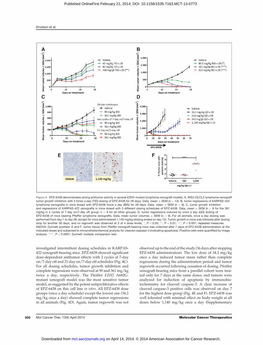
investigated intermittent dosing schedules in KARPAS-422 xenograft-bearing mice. EPZ-6438 showed significantdose-dependent antitumor effects with 2 cycles of 7-dayon/7-day off and 21-day on/7-day off schedules (Fig. 4C).For all dosing schedules, tumor growth inhibition andcomplete regressions were observed at 90 and 361mg/kgtwice a day, respectively. The Pfeiffer EZH2 A682G-mutant xenograft model was the most sensitive tumormodel, as suggested by the potent antiproliferative effectsof EPZ-6438 on this cell line in vitro. All EPZ-6438 dosegroups (once a day schedule) except the lowest one (34.2mg/kg once a day) showed complete tumor regressionsin all animals (Fig. 4D). Again, tumor regrowth was not
observedup to the endof the study (36days after stoppingEPZ-6438 administration). The low dose of 34.2 mg/kgonce a day induced tumor stasis rather than completeregressions during the administration period and tumorregrowth occurred following cessation of dosing. Pfeifferxenograft-bearing mice from a parallel cohort were trea-ted only for 7 days at the same doses, and tumors wereanalyzed for induction of apoptosis by immunohis-tochemistry for cleaved caspase-3. A clear increase ofcleaved caspase-3 positive cells was observed on day 7for the highest dose group (Fig. 4E and F). EPZ-6438 waswell tolerated with minimal effect on body weight at alldoses below 1,140 mg/kg once a day (Supplementary
Figure 4. EPZ-6438 demonstrates strong antitumor activity in several EZH2-mutant lymphoma xenograft models. A, WSU-DLCL2 lymphoma xenografttumor growth inhibition with 3 times a day (TID) dosing of EPZ-6438 for 28 days. Data, mean � SEM (n ¼ 12). B, tumor regressions of KARPAS-422lymphoma xenografts in mice dosed with EPZ-6438 twice a day (BID) for 28 days. Data, mean � SEM (n ¼ 9). C, tumor growth inhibitionand regressions of KARPAS-422 xenografts in mice dosed with 3 different dosing schedules of EPZ-6438. Data, mean � SEM (n ¼ 8 for the 361mg/kg in 2 cycles of 7-day on/7-day off group; n ¼ 9 for all other groups). D, tumor regressions induced by once a day (QD) dosing ofEPZ-6438 of mice bearing Pfeiffer lymphoma xenografts. Data, mean tumor volumes � SEM (n ¼ 9). For all animals, once a day dosing wasperformed from day 1 to day 28, except for mice administered 1,140 mg/kg (dosing ended on day 12). Tumor growth in mice was followed after dosingstop for another 36 days, and no regrowth was observed at 3 of 4 dose levels. �, P < 0.05; ��, P < 0.01; ���, P < 0.001; repeated measuresANOVA, Dunnett posttest. E and F, tumor tissue from Pfeiffer xenograft-bearing mice was collected after 7 days of EPZ-6438 administration at theindicated doses and subjected to immunohistochemical analysis for cleaved caspase-3 (indicating apoptosis). Positive cells were quantified by imageanalysis. ����, P < 0.0001; Dunnett multiple comparison test.
Knutson et al.
Mol Cancer Ther; 13(4) April 2014 Molecular Cancer Therapeutics850
on April 14, 2014. © 2014 American Association for Cancer Research. mct.aacrjournals.org Downloaded from
Published OnlineFirst February 21, 2014; DOI: 10.1158/1535-7163.MCT-13-0773

Fig. S6). Because of body weight loss dosing was stoppedon day 12 for mice administered 1,140 mg/kg once a day;nevertheless, complete and durable regressions wereobserved in this group, despite being exposed to EPZ-6438 for only 12 days.
EPZ-6438 inhibits H3K27 methylation in nontumortissues in a dose-dependent mannerThe profound and sustained regressions in the preclin-
ical models described above suggest that EPZ-6438 mayrepresent an exciting new treatment modality for subsetsof NHL. Measuring pharmacodynamic biomarker mod-ulation postdose is often performed in early clinical trialsto assess the degree of target inhibition/engagementrequired to produce a clinical or biologic response, basedon data from preclinical models. Because the collection ofpostdose tumor biopsies is often not possible, easilyaccessible surrogate tissues, such as PBMCs, skin or bonemarrow are often collected instead. To test EZH2 targetinhibition in surrogate tissues, male and female SpragueDawley rats were orally administered 100, 300, or 1,000mg/kg EPZ-6438 once a day for 28 days, and PBMCs,bone marrow, and skin samples were collected at studyend. Because of body weight loss, females in the 1,000mg/kg group had to be euthanized on day 23. Dose-dependent target inhibition was observed in PBMCs and
bone marrow from rats dosed with EPZ-6438, as mea-sured by ELISA (Fig. 5A and B). The degree of targetinhibition was less pronounced for PBMCs from femalesthat were dosed for 22 days compared with males thatwere dosed for 28 days (same dose of 1,000 mg/kg). Adose-dependent reduction in H3K27Me3 positive cellswas observed in the epidermis of skin of EPZ-6438-dosedrats, as assessed by an immunohistochemistry assay (Fig.5C). The maximum effect was observed at the highestdose, and was already evident after 22 days of EPZ-6438administration. Compound exposure was higher infemale rats compared with males (Fig. 5D), which mayexplain the higher degree of target inhibition in females,especially at 100 and 300 mg/kg.
DiscussionThe critical role of genetic alterations for oncogenesis
has been well established through a combination of DNAmolecular analysis of primary tumor tissue (e.g., sequenc-ing and copy number analysis) and correlated functionalstudies in preclinical models (24, 25). Recently, there hasbeen a growing appreciation for the impact of changes ofthe epigenome—of paramount importance in regulationof gene expression—as a similarly important hallmark ofcancer (26, 27).
Figure 5. EZH2 target inhibition innormal tissues from ratsadministered with EPZ-6438 for 22or 28 days. Inhibition of H3K27Me3levels in rat peripheral bloodmononuclear cells (A) and rat bonemarrow (B) after administrationof EPZ-6438, measured by ELISA.Individual symbols represent theratio of H3K27Me3 to totalH3 for histones extracted fromindividual tissues; horizontal linesrepresent mean values. C, targetinhibition in rat skin (epidermalcompartment) after dosing withEPZ-6438, measured byimmunohistochemistry. Individualsymbols represent the percentageof H3K27Me3-positive cells fortissue from individual animals;horizontal lines represent meanvalues. D, exposures on day 28 orday 22 (1,000 mg/kg females).Individual symbols representAUC values for individual animals;horizontal lines represent meanvalues. Black symbols are males,gray symbols are females. �, P <0.05; ��, P < 0.01; ���, P < 0.001;����, P < 0.0001, one-way ANOVA,Bonferroni posttest. QD, once aday.
Lymphoma Tumor Regression with EZH2 Inhibitor
www.aacrjournals.org Mol Cancer Ther; 13(4) April 2014 851
on April 14, 2014. © 2014 American Association for Cancer Research. mct.aacrjournals.org Downloaded from
Published OnlineFirst February 21, 2014; DOI: 10.1158/1535-7163.MCT-13-0773
Epigenetic regulation of gene expression is based onspatial and temporal orchestration of a spectrum of cova-lent modifications to chromatin. Of particular importancein regulating the fidelity of gene expression is site-specifichistone protein methylation at lysine and arginine aminoacids, a group of reactions catalyzed by the proteinmethyltransferase (PMT) class of group-transfer enzymes(28). A number of PMTs have been shown to be altered ingenetically defined human cancers, and these alterationshave further been shown to be drivers of the specificcancers (i.e., the alterations are oncogenic; ref. 3).
The PRC2 enzyme complex is the only PMT in humansknown to catalyze the site-specific methylation of theH3K27 site (4, 5). Several members of the PRC2 complex,including the catalytic subunit EZH2, have been reportedto be altered in a spectrum of human tumors (6). Asdescribed earlier, a cluster of point mutations within thecatalytic SETdomain of EZH2have been found in approx-imately 10% of patients with NHL, and these have beenshown to alter substrate utilization in such a way as todrive a hyper-trimethylated H3K27 phenotype in themutant-bearing cells that is suggested to aid in lympho-magenesis (8, 14). We and others have reported selectivetool compound inhibitors of wild-type and mutant EZH2as chemical probes with which to demonstrate the depen-dence of EZH2-mutant NHL cell lines on PRC2 activity incell culture and in vivo (17–20). These various tool com-pounds, however, lacked suitable pharmacologic proper-ties for advancement into human clinical trials.
EPZ-6438 is a pharmacologically improved EZH2inhibitor with in vitro properties similar to our previouslyreported tool compound EPZ005687 (17), but with super-ior potency and oral bioavailability (Supplementary Fig.S3). A brief description of EPZ-6438 was provided in arecent report where we demonstrated the utility of thiscompound in treating malignant rhabdoid tumors defi-cient in the SWI/SNF complex component SMARCB1(also referred to as INI1 or hSNF5; ref. 21). In this report,we have demonstrated that treatment with EPZ-6438leads to a global and specific inhibition of PRC2 substratemethylation (H3K27) in a range of NHL cells. Cytotoxic,antiproliferative effects were specifically observed inNHL cells bearing EZH2mutations, and gene expressionprofiling studies confirmed thatEPZ-6438 treatment led tode-repression of known PRC2 target genes. ChIP-PCR ofthe promoters of 5 knownPRC2 targets revealed that theirde-repression correlatedwith overall decreased promoterH3K27Me3 levels. The effect of EPZ-6438 treatment onPRC2 complex occupancy varied across the genes ana-lyzed and included increases, decreases, and no changesin occupancy. However, for any given gene promoteranalyzed, the effect of EPZ-6438 treatment on individualPRC2 complex members was similar (in direction andmagnitude). This observation is consistent with the enzy-matic data demonstrating that EPZ-6438 inhibits EZH2through a SAM-competitive mechanism and is counter tothe notion that this inhibitor disrupts the formation of thePRC2 complex itself. Notably, at those promoters where
H3K27Me3 inhibition is partial, PRC2 occupancy isincreased with treatment. One could speculate that atsuch promoters, catalytic inhibition of EZH2 inducescompensatory recruitment of more PRC2 complexes,delaying demethylation.
Although de-repression of known EZH2 target genesoccurred in the cell lines tested, there was little overlapbetween gene expression signatures between differentmutant lymphoma cell lines as reported in a previousstudy (19). Possible explanations could be cell line epige-netic diversification in vitro or inappropriate dynamicranges of the gene expression assays used to detect smallexpression changes, as previously discussed (29). Ourobservation of very modest effects on gene expression inPfeiffer cells contrasts with the results of McCabe andcolleaguesusingGSK126 (anotherEZH2 inhibitor; ref. 19),in which robust gene expression changes were observedin the same cell line. This discrepancy may be because oftheir use of an inhibitor concentration equivalent to 50�the cellular H3K27Me3 IC50 comparedwith the use in thisstudy of a much lower inhibitor concentration equivalentto 2.5� the cellular H3K27Me3 IC50. Nevertheless, the factthat this concentration is 10� the Pfeiffer LCC suggeststhat a relatively small gene expression changes in Pfeiffercells can trigger a robust antiproliferative response in thishighly EZH2 pathway addicted cell line. EPZ-6438 treat-ment also caused downregulation of genes involved incell-cycle and spliceosome pathways, as well as groupsof genes containing E2F-binding motives. These geneexpression changes could be indirect effects downstreamof the relief of PRC2 inhibition, but may also be a directeffect of inhibiting an EZH2-activating function indepen-dent of the PRC2 complex, as recently suggested by othersfor STAT3 signaling in glioblastoma and androgen recep-tor signaling in prostate cancer (30, 31). This will be thefocus of future studies. In addition, it has been reportedthat EZH2 is regulated by E2F itself in metastatic prostatecancer and in lymphomas, suggesting the existence offeed-forward loops (32).
Dosing of EZH2-mutant NHL xenograft-bearing micewith EPZ-6438 led to significant antitumor effects rangingfrom marked and dose-dependent tumor growth inhibi-tion (e.g.,WSU-DLCL2 xenografts) to complete and dura-ble eradication of tumors (e.g., KARPAS-422 and Pfeifferxenografts; Fig. 4). The EZH2 A682G mutant–bearingPfeiffer cell line was particularly sensitive to EZH2 inhi-bition, both in vitro and in vivo; relatively limited doses orshort time dosing of EPZ-6438 resulted in complete andpermanent elimination ofPfeiffer cell tumors in themousexenograft model. These preclinical data suggest thatpotent and selective EZH2 inhibitors, such as EPZ-6438,may be ideal candidates for development as a noveltargeted therapy for treatment of patients with NHLwithcancers bearing A682G-mutated EZH2.
The apparent differences in the in vivo response ofWSU-DLCL2 andKARPAS-422 tumorswas not predictedby the similar sensitivity of these 2 cell lines to EPZ-6438treatment in cell culture (i.e., the 2 lines displayed similar
Knutson et al.
Mol Cancer Ther; 13(4) April 2014 Molecular Cancer Therapeutics852
on April 14, 2014. © 2014 American Association for Cancer Research. mct.aacrjournals.org Downloaded from
Published OnlineFirst February 21, 2014; DOI: 10.1158/1535-7163.MCT-13-0773

LCC values, as summarized in Table 1). This may suggestthat in the in vivo context of the xenograft models, WSU-DLCL2 can utilize alternative pathways for cell growthand survival that partially circumvent the impact of EZH2inhibition. Similarly, the reasons for the insensitivity ofthe EZH2 Y646N-mutant lymphoma cell line RL in vitroare not understood at present, and may suggest thepresence of parallel escape routes from inhibition of theEZH2 pathway. The insensitivity of RL cells to pharma-cologic EZH2 inhibition was also observed by McCabeand colleagues with GSK126 (19). These observationsrequire more in-depth study to determine the molecularorigins of the differential effects of EZH2 inhibition onthese cells. Interestingly, we also failed to induce signif-icant resistance to EPZ-6438 by treating several inhibitorsensitive cell lines for >7 months in vitro.Beguelin and colleagues recently suggested that ger-
minal center derived DLBCLs are addicted to EZH2independent of its mutational state, as impaired enzymeactivity, for instance induced by EZH2 inhibitor treat-ment, showed antiproliferative activity in both EZH2wild-type and mutant cell lines (11). Our data suggestthat EZH2 inhibition may affect EZH2 wild-type GCBDLBCL cells by slowing their growth in a limited fashion(with micromolar IC50 values, except the Farage cell line);however, only the mutant cell lines are driven into celldeath during the 11-day compound incubation period.This suggests EZH2 oncogene addiction and a superiorpotential for antitumor activity of EZH2 inhibitors inpatients with EZH2-mutated GCB lymphoma comparedwith wild-type cases.Taken together, the preclinical data presented here
support the further development of EPZ-6438 as a newtreatment modality for genetically defined subsets ofNHL. This compound is currently under study asE7438 in a phase I trial of relapsed and refractory malig-nancies that have failed all standard therapy. The primarygoal of the phase I trial is to establish the safety and define
the maximal tolerated dose of the drug. The ability tomeasure dose-dependent changes in H3K27Me3 levels inskin, PBMCs, and bone marrowmay also portend the useof signal from these surrogate tissues as a noninvasivepharmacodynamics biomarker during human clinicaltrials.
Disclosure of Potential Conflicts of InterestS.K. Knutson, C.R. Klaus, A. Raimondi, N.J.Waters, R. Chesworth, R.A.
Copeland, V.M. Richon, K.W. Kuntz, and H. Keilhack have ownershipinterest (including patents) in Epizyme. No potential conflicts of interestwere disclosed by the other authors.
Authors' ContributionsConception and design: S.K. Knutson, T.J.Wigle, J.J. Smith, R. Chesworth,R.A. Copeland, V.M. Richon, T. Uenaka, R.M. Pollock, K.W. Kuntz,A. Yokoi, H. KeilhackDevelopment of methodology: S.K. Knutson, N. Kumar, C.R. Klaus,A. Raimondi, M. Porter-ScottAcquisition of data (provided animals, acquired and managed patients,provided facilities, etc.): S.K. Knutson, S. Kawano, N.M. Warholic, K.-C.Huang, G. Kuznetsov, N. Kumar, T.J. Wigle, C.J. Allain, A. Raimondi,A. YokoiAnalysis and interpretation of data (e.g., statistical analysis, biostatis-tics, computational analysis): S.K. Knutson, S. Kawano, Y. Minoshima,N.M. Warholic, K.-C. Huang, Y. Xiao, T. Kadowaki, M. Uesugi, G. Kuz-netsov, T.J. Wigle, C.J. Allain, A. Raimondi, N.J. Waters, J.J. Smith,M. Porter-Scott, R.A. Copeland, V.M. Richon, K.W. Kuntz, H. KeilhackWriting, review, and/or revision of the manuscript: S.K. Knutson,S. Kawano, K.-C. Huang, Y. Xiao, N.J. Waters, J.J. Smith, R. Chesworth,M.P. Moyer, R.A. Copeland, V.M. Richon, R.M. Pollock, K.W. Kuntz,A. Yokoi, H. KeilhackAdministrative, technical, or material support (i.e., reporting or orga-nizing data, constructing databases): S.K. Knutson, C.J. Allain,H. KeilhackStudy supervision: S.K. Knutson, R.A. Copeland, T. Uenaka, H. Keilhack
Grant SupportThisworkwas fundedbyEisai through collaboration betweenEpizyme
and Eisai.The costs of publication of this article were defrayed in part by the
payment of page charges. This article must therefore be hereby markedadvertisement in accordance with 18 U.S.C. Section 1734 solely to indicatethis fact.
Received September 16, 2013; revised December 19, 2013; acceptedJanuary 12, 2014; published OnlineFirst February 21, 2014.
References1. Badeaux AI, Shi Y. Emerging roles for chromatin as a signal integration
and storage platform. Nat Rev Mol Cell Biol 2013;14:211–24.2. Copeland RA, Solomon ME, Richon VM. Protein methyltransferases
as a target class for drug discovery. Nat Rev Drug Discov2009;8:724–32.
3. Copeland RA, Moyer MP, Richon VM. Targeting genetic alterations inprotein methyltransferases for personalized cancer therapeutics.Oncogene 2013;32:939–46.
4. Margueron R, Reinberg D. The polycomb complex PRC2 and its markin life. Nature 2011;469:343–9.
5. O'Meara MM, Simon JA. Inner workings and regulatory inputs thatcontrol polycomb repressive complex 2. Chromosoma 2012;121:221–34.
6. Chase A, Cross NC. Aberrations of EZH2 in cancer. Clin Cancer Res2011;17:2613–8.
7. Lohr JG, Stojanov P, Lawrence MS, Auclair D, Chapuy B, Sougnez C,et al. Discovery and prioritization of somatic mutations in diffuse largeB-cell lymphoma (DLBCL) by whole-exome sequencing. Proc NatlAcad Sci U S A 2012;109:3879–84.
8. Morin RD, Johnson NA, Severson TM, Mungall AJ, An J, Goya R,et al. Somatic mutations altering EZH2 (Tyr641) in follicular anddiffuse large B-cell lymphomas of germinal-center origin. Nat Genet2010;42:181–5.
9. Morin RD, Mendez-Lago M, Mungall AJ, Goya R, Mungall KL, CorbettRD, et al. Frequent mutation of histone-modifying genes in non-Hodgkin lymphoma. Nature 2011;476:298–303.
10. Alizadeh AA, EisenMB, Davis RE,MaC, Lossos IS, Rosenwald A, et al.Distinct types of diffuse large B-cell lymphoma identified by geneexpression profiling. Nature 2000;403:503–11.
11. Beguelin W, Popovic R, Teater M, Jiang Y, Bunting KL, RosenM, et al.EZH2 is required for germinal center formation and somatic EZH2mutations promote lymphoid transformation. Cancer Cell 2013;23:677–92.
12. Majer CR, Jin L, Scott MP, Knutson SK, Kuntz KW, Keilhack H, et al.A687V EZH2 is a gain-of-function mutation found in lymphomapatients. FEBS Lett 2012;586:3448–51.
13. McCabe MT, Graves AP, Ganji G, Diaz E, Halsey WS, Jiang Y, et al.Mutation of A677 in histone methyltransferase EZH2 in human B-cell
Lymphoma Tumor Regression with EZH2 Inhibitor
www.aacrjournals.org Mol Cancer Ther; 13(4) April 2014 853
on April 14, 2014. © 2014 American Association for Cancer Research. mct.aacrjournals.org Downloaded from
Published OnlineFirst February 21, 2014; DOI: 10.1158/1535-7163.MCT-13-0773

lymphoma promotes hypertrimethylation of histone H3 on lysine 27(H3K27). Proc Natl Acad Sci 2012;109:2989–94.
14. Sneeringer CJ, Scott MP, Kuntz KW, Knutson SK, Pollock RM, RichonVM, et al. Coordinated activities of wild-type plus mutant EZH2 drivetumor-associated hypertrimethylation of lysine 27 on histone H3(H3K27) in human B-cell lymphomas. Proc Natl Acad Sci 2010;107:20980–5.
15. Wigle TJ, Knutson SK, Jin L, Kuntz KW, Pollock RM, Richon VM,et al. The Y641C mutation of EZH2 alters substrate specificity forhistone H3 lysine 27 methylation states. FEBS Lett 2011;585:3011–4.
16. Yap DB, Chu J, Berg T, Schapira M, Cheng SW, Moradian A, et al.Somatic mutations at EZH2 Y641 act dominantly through a mecha-nism of selectively altered PRC2 catalytic activity, to increase H3K27trimethylation. Blood 2011;117:2451–9.
17. Knutson SK, Wigle TJ, Warholic NM, Sneeringer CJ, Allain CJ, KlausCR, et al. A selective inhibitor of EZH2 blocks H3K27 methylation andkills mutant lymphoma cells. Nat Chem Biol 2012;8:890–6.
18. Konze KD,MaA, Li F, Barsyte-Lovejoy D, Parton T,Macnevin CJ, et al.An orally bioavailable chemical probe of the lysine methyltransferasesEZH2 and EZH1. ACS Chem Biol 2013;8:1324–34.
19. McCabe MT, Ott HM, Ganji G, Korenchuk S, Thompson C, Van AllerGS, et al. EZH2 inhibition as a therapeutic strategy for lymphoma withEZH2-activating mutations. Nature 2012;492:108–12.
20. QiW, Chan H, Teng L, Li L, Chuai S, Zhang R, et al. Selective inhibitionof Ezh2 by a small molecule inhibitor blocks tumor cells proliferation.Proc Natl Acad Sci U S A 2012;109:21360–5.
21. Knutson SK, Warholic NM, Wigle TJ, Klaus CR, Allain CJ, Raimondi A,et al. Durable tumor regression in genetically altered malignant rhab-doid tumors by inhibition of methyltransferase EZH2. Proc Natl AcadSci U S A 2013;110:7922–7.
22. Ben-Porath I, Thomson MW, Carey VJ, Ge R, Bell GW, Regev A, et al.An embryonic stem cell-like gene expression signature in poorlydifferentiated aggressive human tumors. Nat Genet 2008;40:499–507.
23. Velichutina I, Shaknovich R, Geng H, Johnson NA, Gascoyne RD,Melnick AM, et al. EZH2-mediated epigenetic silencing in germinalcenter B cells contributes to proliferation and lymphomagenesis.Blood 2010;116:5247–55.
24. Simon R, Roychowdhury S. Implementing personalized cancer geno-mics in clinical trials. Nat Rev Drug Discov 2013;12:358–69.
25. Vogelstein B, Papadopoulos N, Velculescu VE, Zhou S, Diaz LA Jr.,Kinzler KW. Cancer genome landscapes. Science 2013;339:1546–58.
26. Sandoval J, Esteller M. Cancer epigenomics: beyond genomics. CurrOpin Genet Dev 2012;22:50–5.
27. Timp W, Feinberg AP. Cancer as a dysregulated epigenome allowingcellular growth advantage at the expense of the host. Nat Rev Cancer2013;13:497–510.
28. Richon VM, Johnston D, Sneeringer CJ, Jin L, Majer CR, Elliston K,et al. Chemogenetic analysis of human protein methyltransferases.Chem Biol Drug Des 2011;78:199–210.
29. Melnick A. Epigenetic therapy leaps ahead with specific targeting ofEZH2. Cancer Cell 2012;22:569–70.
30. KimE, KimM,WooDH, Shin Y, Shin J, ChangN, et al. Phosphorylationof EZH2 activates STAT3 signaling via STAT3 methylation and pro-motes tumorigenicity of glioblastoma stem-like cells. Cancer Cell2013;23:839–52.
31. Xu K, Wu ZJ, Groner AC, He HH, Cai C, Lis RT, et al. EZH2 oncogenicactivity in castration-resistant prostate cancer cells is polycomb-inde-pendent. Science 2012;338:1465–9.
32. Bracken AP, Pasini D, Capra M, Prosperini E, Colli E, Helin K. EZH2 isdownstream of the pRB-E2F pathway, essential for proliferation andamplified in cancer. EMBO J 2003;22:5323–35.
Knutson et al.
Mol Cancer Ther; 13(4) April 2014 Molecular Cancer Therapeutics854
on April 14, 2014. © 2014 American Association for Cancer Research. mct.aacrjournals.org Downloaded from
Published OnlineFirst February 21, 2014; DOI: 10.1158/1535-7163.MCT-13-0773