selective and sensitive turn-on chemosensor for arsenite ion at the ppb level in aqueous media...
TRANSCRIPT

Subscriber access provided by Linnaeus University - University Library
Analytical Chemistry is published by the American Chemical Society. 1155 SixteenthStreet N.W., Washington, DC 20036Published by American Chemical Society. Copyright © American Chemical Society.However, no copyright claim is made to original U.S. Government works, or worksproduced by employees of any Commonwealth realm Crown government in the courseof their duties.
Article
Selective and sensitive turn-on chemosensor for arseniteion at ppb level in aqueous media applicable in cell staining
Somenath Lohar, Siddhartha Pal, Buddhadeb Sen, ManjiraMukherjee, Samya Banerjee, and Pabitra Chattopadhyay
Anal. Chem., Just Accepted Manuscript • DOI: 10.1021/ac503255f • Publication Date (Web): 14 Oct 2014
Downloaded from http://pubs.acs.org on October 18, 2014
Just Accepted
“Just Accepted” manuscripts have been peer-reviewed and accepted for publication. They are postedonline prior to technical editing, formatting for publication and author proofing. The American ChemicalSociety provides “Just Accepted” as a free service to the research community to expedite thedissemination of scientific material as soon as possible after acceptance. “Just Accepted” manuscriptsappear in full in PDF format accompanied by an HTML abstract. “Just Accepted” manuscripts have beenfully peer reviewed, but should not be considered the official version of record. They are accessible to allreaders and citable by the Digital Object Identifier (DOI®). “Just Accepted” is an optional service offeredto authors. Therefore, the “Just Accepted” Web site may not include all articles that will be publishedin the journal. After a manuscript is technically edited and formatted, it will be removed from the “JustAccepted” Web site and published as an ASAP article. Note that technical editing may introduce minorchanges to the manuscript text and/or graphics which could affect content, and all legal disclaimersand ethical guidelines that apply to the journal pertain. ACS cannot be held responsible for errorsor consequences arising from the use of information contained in these “Just Accepted” manuscripts.

Selective and sensitive turn-on chemosensor for arsenite ion at ppb
level in aqueous media applicable in cell staining
Somenath Lohar,†
Siddhartha Pal,† Buddhadeb Sen,
† Manjira Mukherjee,
† Samya Banerjee
‡ and
Pabitra Chattopadhyay*,†
† Department of Chemistry, The University of Burdwan, Burdwan-713104, West Bengal, India,
E-mail: [email protected]. Fax: +91-342-2530452. ‡ Department of Inorganic and Physical Chemistry, Indian Institute of Science, Bangalore, 560012, India
ABSTRACT. A newly designed and structurally characterized cell permeable receptor (HL) selectively senses the AsO33-
ion upto
ca. 4.1 ppb in aqueous media over the other competitive ions at biological pH through intermolecular H-bonding induced CHEF
process, established by detailed experimental and theoretical studies. This bio-friendly probe is highly competent to recognize the
existence of AsO33-
ions in living organism by developing image under fluorescence microscope and useful to estimate the amount
of arsenite ions in various water samples.
KEYWORDS. Chemosensor; AsO33-
; Cell staining
INTRODUCTION
Arsenic is a teratogenic and carcinogenic toxic element
and it being present in natural waters supports to propagate the
serious health problems allied with dermal toxicity, cardiovas-
cular disease, and neurodegenerative disorders etc.1,2
The
physical status and chemical structure of the involved com-
pound demonstrate the degree of arsenic toxicity, which is the
highest in case of the inorganic form of arsenite(III) state over
that of arsenate(V) and organic form of this element.3,4
In natural waters, especially groundwater, arsenic (As) ex-
ists as both arsenite (AsIII
) and arsenate (AsV), and the ratio of
AsV to As
III has been found to be in the range of 10-100 de-
pending on the chemical environment.5-7
Especially, high lev-
els of AsIII
(> 0.3 mg.L-1
) were found in groundwater in Bang-
ladesh, India, Laos, Cambodia and Pakistan8 and the oxidation
of AsIII
by dissolved oxygen is extremely slow. The carcino-
genic effect due to arsenite ions has already been confirmed by
the recent experimental outcome, where it has been found that
in vitro exposure of 5 μM arsenite [As3+(
OH)3] to normal stem
cells, result in cancer stem cell phenotype by 18 weeks.9
In
2001 the WHO, EPA and U.S. have already advised to lower
the maximum contamination level (MCL) of arsenic com-
pounds in drinking water to 10 ppb from 50 ppb owing to the
high toxicity of arsenicals.10
For this real urgency, the deter-
mination of ppb level arsenic species in aqueous media is to be
effectively developed in proper way.
Detection of arsenicals by colorimetric methods resulting
from the Gutzeit reaction suffers from the deadly poisonous
arsine gas (AsH3) and byproducts of mercury compoounds.11
ICP-MS being a sensitive detection strategy recommended by
the EPA is an expensive process and required an extensive
methodology for sample preparation.12
Thus, fluorescence
spectroscopy meets the requirements in developing the simple
field-effective methods to detect the arsenicals of very low
level as it has been widely utilized for numerous analytes.13-16
Till date, a few chemosensors selective for arsenate ions17
and
for AsIII
cation,18-20
including some bioluminescents for arse-
nite ion21-23
have been reported. However, AsO33–
ion selective
chemosensor is still unexplored. Herein, we report an easy to
make simple Schiff base system (HL) which senses selectively
the most hazardous AsO33-
ion as low as 54.91×10⁻⁹
M (ca.
4.12 ppb lower than MCL) in aqueous media through inter-
molecular hydrogen bonding assisted CHEF process colori-
metrically and fluorimetrically.
EXPERIMENTAL SECTION
Materials and methods
All the analytical reagent grade solvents and the other rea-
gent grade chemicals consumed in this work were procured
from commercial sources and used as received. Here, through-
out the experiments Milli-Q 18 Ω water was employed. 2,6-
diformyl-p-cresol was synthesized following the literature
procedure.24
Tetrabutylammonium salts of dihydrogen phos-
phate, acetate, halides (F-, Cl
-, Br
-, I
-); and sodium salts of
arsenate, arsenite, bicarbonate, nitrate, chlorate, monohydro-
gen phosphate, azide, cyanide, thiocyanate, sulphate, etc were
used for the selectivity study of the receptor towards different
anions.
A Perkin Elmer 2400 CHN elemental analyzer was used
for performing the elemental analyses. The electronic and
FTIR spectra were obtained on Shimadzu made UV-1800 and
Prestige-21 FTIR spectrophotometers, respectively. A Bruker
Avance DPX 500 MHz spectrometer for recording 1HNMR
spectra using DMSO-d6 solution and a Qtof Micro YA263
mass spectrometer for electrospray ionization (ESI) mass
spectra were used. To measure the pH of the solution on ad-
justing the pH of the solution using either 50 mM NaOH or
HCl solution, a Systronics pH meter (model 335) was taken. A
Hitachi F-4500 FL spectrophotometer was employed to record
the steady-state fluorescence spectra, whereas a HORIBA
JOBIN Yvon (TCSPC) spectrometer from IBH (UK) at λex=
Page 1 of 6
ACS Paragon Plus Environment
Analytical Chemistry
123456789101112131415161718192021222324252627282930313233343536373839404142434445464748495051525354555657585960
438 nm and MCP-PMT as a detector was used to perform the
time-resolved fluorescence lifetime measurements experi-
ments.
A HEPES buffer (1 mM, pH 7.4) (DMSO-Water 1 : 9) co-
solvent at 25 °C temperature with an HL concentration of 10
μM was used for doing all the UV−visible and Fluorescence
spectroscopic experiments (detail in the Supporting Infor-
mation). The relevant Benesi-Hildebrand equation25
was used
for calculating the apparent binding constant of the produced
HL-AsO33-
ensemble species; the estimation of the detection
limit and Job’s plot are shown in the Supporting Information.
The preparation of cells for in vitro cellular imaging and the
cell viability test with HL to determine the cytotoxicity assay
are carefully carried out and the details in the Supporting In-
formation.
A suitable single crystal was mounted on a Bruker’s Apex-
II CCD diffractometer using MoKα (λ = 0.71069) to collect the
data to solve the structure of HL, and necessary corrections
were applied using SADABS from Bruker. The direct methods
using SIR-92 duly refined by full-matrix least squares refine-
ment methods based on F2, using SHELX-97
26 was used to
solve the structure. Wingx package27,28
was taken to perform
all the calculations. Deposition of all the crystallographic data
of HL as the CCDC no. of 1008294 has been performed.
Theoretical Calculation
DFT calculations of the probe (HL), and its corresponding
HL-AsO33-
ensemble species were done with the help of
Gaussian-09 software over a Red Hat Linux IBM cluster with
the B3LYP/6-31G (d,p)29-31
functional model and basis set to
describe the understanding of the electronic configurations, the
interactions in the molecular level and the ground state charac-
ter.
RESULTS AND DISCUSSION
Synthesis of the Probe (HL)
The AsO33–
ions selective probe (HL) was synthesized by
condensation of 2,6-diformyl-p-cresol with 4-aminoantipyrine
in 1:2 mole ratio (Scheme 1) and it was characterized by spec-
troscopic and physico-chemical tools including single crystal
X-ray crystallographic analysis (Figures S-1 (a) and (b) in the
supporting information).
Scheme 1 Schematic representation of synthesis of the probe
HL
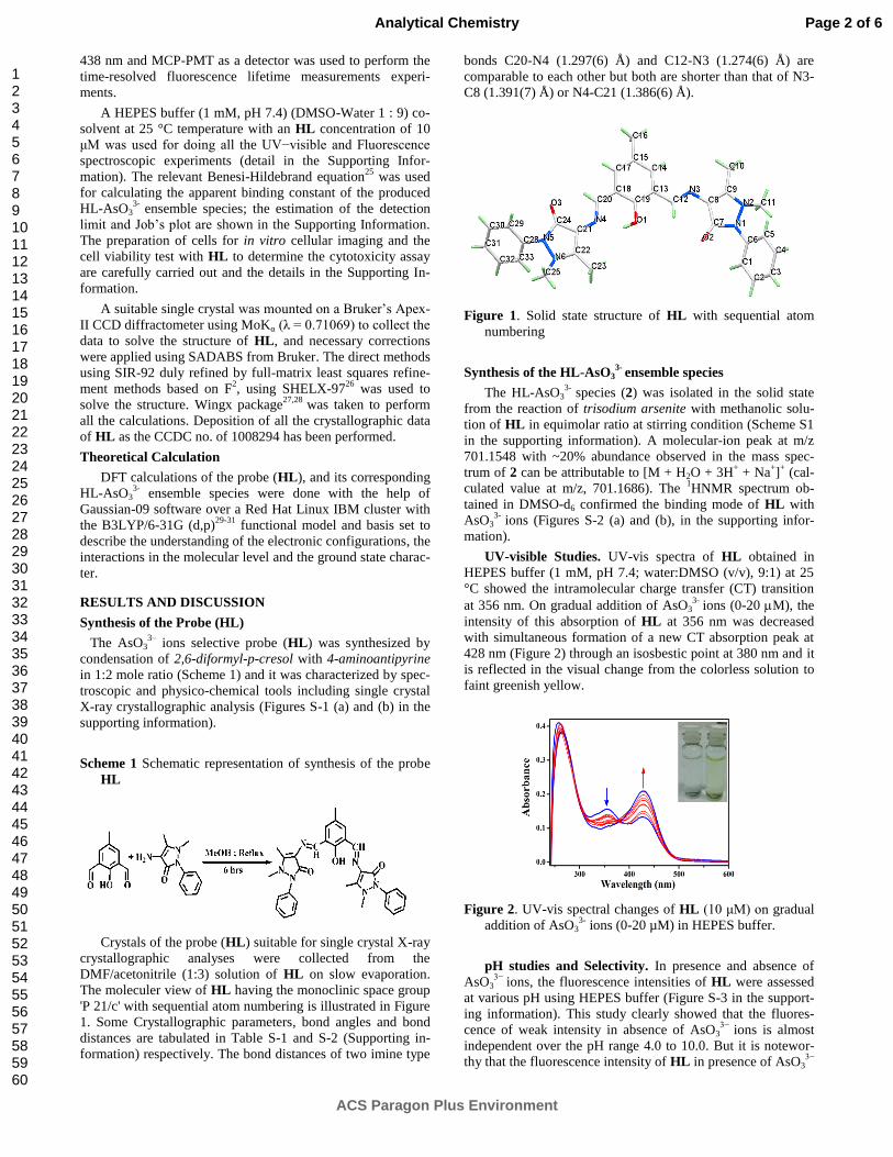
Crystals of the probe (HL) suitable for single crystal X-ray
crystallographic analyses were collected from the
DMF/acetonitrile (1:3) solution of HL on slow evaporation.
The moleculer view of HL having the monoclinic space group
'P 21/c' with sequential atom numbering is illustrated in Figure
1. Some Crystallographic parameters, bond angles and bond
distances are tabulated in Table S-1 and S-2 (Supporting in-
formation) respectively. The bond distances of two imine type
bonds C20-N4 (1.297(6) Å) and C12-N3 (1.274(6) Å) are
comparable to each other but both are shorter than that of N3-
C8 (1.391(7) Å) or N4-C21 (1.386(6) Å).
Figure 1. Solid state structure of HL with sequential atom
numbering
Synthesis of the HL-AsO33-
ensemble species
The HL-AsO33-
species (2) was isolated in the solid state
from the reaction of trisodium arsenite with methanolic solu-
tion of HL in equimolar ratio at stirring condition (Scheme S1
in the supporting information). A molecular-ion peak at m/z
701.1548 with ~20% abundance observed in the mass spec-
trum of 2 can be attributable to [M + H2O + 3H+ + Na
+]
+ (cal-
culated value at m/z, 701.1686). The 1HNMR spectrum ob-
tained in DMSO-d6 confirmed the binding mode of HL with
AsO33-
ions (Figures S-2 (a) and (b), in the supporting infor-
mation).
UV-visible Studies. UV-vis spectra of HL obtained in
HEPES buffer (1 mM, pH 7.4; water:DMSO (v/v), 9:1) at 25
°C showed the intramolecular charge transfer (CT) transition
at 356 nm. On gradual addition of AsO33-
ions (0-20 M), the
intensity of this absorption of HL at 356 nm was decreased
with simultaneous formation of a new CT absorption peak at
428 nm (Figure 2) through an isosbestic point at 380 nm and it
is reflected in the visual change from the colorless solution to
faint greenish yellow.
Figure 2. UV-vis spectral changes of HL (10 μM) on gradual
addition of AsO33-
ions (0-20 µM) in HEPES buffer.
pH studies and Selectivity. In presence and absence of
AsO33−
ions, the fluorescence intensities of HL were assessed
at various pH using HEPES buffer (Figure S-3 in the support-
ing information). This study clearly showed that the fluores-
cence of weak intensity in absence of AsO33−
ions is almost
independent over the pH range 4.0 to 10.0. But it is notewor-
thy that the fluorescence intensity of HL in presence of AsO33−
Page 2 of 6
ACS Paragon Plus Environment
Analytical Chemistry
123456789101112131415161718192021222324252627282930313233343536373839404142434445464748495051525354555657585960

ion is higher than that in the absence of AsO33−
ions. This ob-
servation is ascertained by the formation of HL-AsO33-
species
(2) through intermolecular H-bonding. The fluorescence fea-
tures of HL (10 μM) were also validated upon the addition of
10 equivalents in excess of different anions i.e. AsO33−
, NO3−,
Mo43-
, Br
−, F
−, HCO3
-, N3
-, ClO4
−, H2PO4
−, HPO4
2−, Cl
−, PO4
3-,
CO32-
, OAc−, SO4
2−, HSO4
- and HAsO4
2− but only AsO3
3- selec-
tively enhanced the fluorescence intensity of HL system (Fig-
ure S-4 in the supporting information).
It is remarkable to note that the presence of other competi-
tive anions does not affect the enhancement of fluorescence.
Moreover, in presence of 50 equivalents of different tested
anions in excess with HL and AsO33-
, almost no undesirable
effect on intensity was recorded (Figure S-5 (a) in the support-
ing information). It is also notable to mention that the interfer-
ence coming from metal ions is also negligible though this
Schiff base has an affinity to form complexes.32,33
Fluorescence Studies. On addition of various concentra-
tions of AsO33-
ions (0-20 µM), significant enhancement of the
fluorescence intensity at 532 nm was observed. On addition of
2.0 equivalent of AsO33-
ions, HL displayed ca. 9-fold increase
of its fluorescence intensity (Figure 3), which is also reflected
in the values of the calculated quantum yields (). The value
of increases by ca. 5.67 times due to the addition of addition
of AsO33-
ions from = 0.00228 to = 0.01294. This spectral
feature was also evidenced by the color change in fluorescence
from colorless to green in presence of light in the UV region
(Figure S-6 in the supporting information).
With the help of the fluorescence study, Job’s plot (Figure
S-7 in the supporting information) clearly demonstrates the 1:1
in stoichiometric ratio of HL and AsO33-
to form the ensemble
2. This fact of 1:1 stoichiometry is also validated by the spec-
troscopic and physico-chemical data of the isolated solid HL-
AsO33-
ensemble species. The strong binding affinity of HL
towards the AsO33-
ions has also been strengthened by the
binding constant (K) value (2.5267 x 10⁵ M
-1) calculated from
the slope/intercept using the plot of (F∞-F0)/(Fx- F0) against 1/[
AsO33-
] (Figure S-8 in the supporting information).
Figure 3. Fluorescence spectra of HL (10 μM) as a function
of added AsO33-
ions [0-20 µM] in DMSO-water (1 : 9,
v/v) HEPES buffer (1 mM) at 25 °C [λex = 438 nm].
To validate this method the detection limit (LOD) was ob-
tained by drawing the calibration curve (Figure S-9 in the sup-
porting information) in the lower region. The detection limit
was estimated to be 54.91x10⁻⁹
M i.e. 4.12 ppb using the equa-
tion 3/S (S = slope of the calibration curve and is the zero
level standard deviation.34
This method is significantly unique
one as the LOD of HL for AsO33-
ions (4.12 ppb) is remarka-
bly lower than that of MCL.6
1H NMR Studies.
1H NMR titration was carried out in
dmso-d6 (Figure 4) in support of the formation of the HL-
AsO33−
species in solution state. This study clearly indicates
that the signal of the phenolic O-H proton shifted to upfield by
0.2856 ppm probably because of the interaction of phenolic O-
H proton with AsO33−
ions.
Figure 4. 1H NMR titration of HL upon gradual addition of
AsO33- ions in dmso-d6 (A) HL; (B) HL:AsO3
3- (1:0.5) and
(C) HL:AsO33- (1:1).
The average fluorescence life time (av) of HL was calcu-
lated to be 4.5250 ns at λem= 532 nm. The av values of 2 (at
λem= 532 nm) was enhanced from 4.7917 ns (HL : AsO3
3- = 1 :
0.5) to 4.9163 ns (HL : AsO3
3- = 1 : 1) on incremental addition
of AsO33−
ions to the HL solution (Figure 5). This observation
is in favour of the CHEF process through intermolecular hy-
drogen bonding.
Figure 5. Time resolved fluorescence decay of HL (10 μM) in the
absence and presence of added AsO33- ions (λem = 532nm).
The strong binding affinity of AsO33-
ion with HL ex-
tremely affects the PET process, which facilitates to enhance
the fluorescence intensity in favour of the selective detection
of AsO33−
ions (Scheme 2). The radiative rate constant kr and
Page 3 of 6
ACS Paragon Plus Environment
Analytical Chemistry
123456789101112131415161718192021222324252627282930313233343536373839404142434445464748495051525354555657585960

nonradiative rate constant knr of HL and HL-AsO33-
ensemble
(2) were calculated using the equations:16
-1
= kr + knr and kr
= f/ (viz. Table S-3 in the supporting information). This
table of the data clearly indicates that the fluorescent en-
hancement is principally attributed to the more than 5 times
increase of kr due to the binding of AsO33-
ion with HL, which
is substantiated by the large binding constant value (as men-
tioned above). Upon selective binding of AsO33-
ion, the elec-
tron lone pair on the phenolic-O atom of HL is no longer ac-
cessible for PET process, making possible to the fluorescence
enhancement.
Scheme 2 Plausible mechanism of fluorescence enhancement
of HL in presence of AsO33-
ions
Density Functional Theoretical (DFT) Studies. For under-
standing of the sort of interaction of HL with AsO33-
ion DFT
calculations were performed to attain the optimised geometry
of HL and HL-AsO33-
ensemble (2) (Figure 6). From this
study it is clear that the energy gap between HOMO-LUMO in
2 is 0.63 eV is reasonably lower than that of free HL (3.13
eV) giving rise a more stable species produced by binding the
guest (AsO33-
ion) with the host (HL) through H-bonding.
Figure 6. Geometry optimisation and theoretical calculation of
HL and 2
Cell Imaging Studies. The chemosensor (HL) was exam-
ined by applying HL to human cervical cancer HeLa cell to
explore the utility in biological system. Here, the cells of in-
terest were permitted to feed both HL and AsO33−
and the
images of the cells were acquired using the fluorescence mi-
croscopy at ex 438 nm. The distribution of the probe within
the cells with a very faint fluorescence was observed after
incubation with HL (10 μM) for 30 min. However, the cells
displayed intense fluorescence after the introduction of exoge-
nous AsO33−
into the cell via incubation with Na3AsO3 (details
in Figure 7). These cellular images obtained by adding various
concentrations of added AsO33−
ions are the interesting signa-
ture of the fluorescence response ability of the probe.
Moreover, the in vitro study demonstrated that 10 μM of
HL showed almost full cell viability in support of non-
cytotoxic effect to cell upto 6 h (Figure S-10 in the supporting
information).35
These results clearly indicate that the probe is
an excellent bio-friendly candidate for both in vivo and in vitro
purpose as AsO33−
ions sensor and imaging in both ways for
living cell staining.
Figure 7. Fluorescence images and phase contrast image of HeLa
cells after incubation for 30 min. with HL only (1, 1) and HL
plus arsenite ions (2, 2) 3 μM; (3, 3) 5 μM; (4, 4) 7 µM and
(5, 5’) 10 µM at 37 °C
In addition of the above fact, it is also noteworthy to men-
tion that the developed method was also applicable to estimate
the trace level arsenite ions in different drinking water samples
and the water of the arsenic affected zone of Purbasthali,
Burdwan, West Bengal, India36
using standard addition tech-
nique (Table S-4 in the supporting information). The result
presented in Table S4 had revealed the accuracy of the devel-
oped method in case of drinking water and the determination
of the amount of the arsenite ions in the contaminated water.
In another experiment these arsenic-contaminated water sam-
ples were treated with Fenton's reagent for 12 h maintaining
the appropriate condition37-39
to oxidise the arsenite ions to
arsenate ions and then the probe (HL) was added to these
treated water samples taking all precautions. Here, it was no-
ticed that the emission intensity was remarkably decreased
(viz. Figure S-11 in the supporting information). The results
indicated that the fluorescence due to the arsenite ions was
almost diminished because of the oxidation of arsenite ions to
the arsenate ions through Fenton's reactions. These observa-
tions clearly reveal that the developed method is highly specif-
ic for arsenite ions and useful for selectively quantitative de-
termination of arsenite ions in presence of arsenate ions in
water samples.
CONCLUSION
In conclusion, herein, a new easy to make simple Schiff
base duly structurally characterized has been successfully es-
tablished as a first report of a novel chemosensor for the most
toxic AsO33-
ions upto as low as 4.1 ppb in aqueous media at
biological pH through the intermolecular H-bonding induced
Page 4 of 6
ACS Paragon Plus Environment
Analytical Chemistry
123456789101112131415161718192021222324252627282930313233343536373839404142434445464748495051525354555657585960

CHEF process based on thorough experimental and theoretical
studies. This non-cytotoxic probe is a biomarker to detect the
intracellular AsO33-
ions concentrations under a fluorescence
microscope and is efficient to determine the amount of AsO33-
ions in various water samples.
AUTHOR INFORMATION
Corresponding Author
*E-mail: [email protected]. Tel: +91-342-2558554 extn.
424. Fax: +91-342-2530452.
Present Addresses † Department of Chemistry, The University of Burdwan, Golap-
bag, Burdwan-713104, West Bengal, India ‡ Department of Inorganic and Physical Chemistry, Indian Insti-
tute of Science, Bangalore, 560012, India
ACKNOWLEDGMENT
The authors sincerely acknowledge Council of Scientific and
Industrial Research (CSIR), New Delhi, India for financial assis-
tance. S. Lohar wishes to thank to UGC, New Delhi, India for
offering him the fellowship. The authors are indebted to USIC,
The University of Burdwan for the single crystal X-ray diffrac-
tometer facility under PURSE program. The authors thank to Prof.
Samita Basu and Mr. Ajay Das, Chemical Science Division,
SINP, Kolkata for collecting data using TCSPC instrument.
Supporting Information. The material having experimental sec-
tion including characterization and spectral data, schemes, spec-
tra, figures, tables, crystallographic data for CCDC No.1008294,
is available free of charge via the Internet at http://pubs.acs.org.
REFERENCES
(1) Wang, X.; Mandal, A.K.; Saito, H.; Pulliam, J.F.; Lee, E.Y.; Ke,
Z.J.; Lu, J.; Ding, S.; Li, L.; Shelton, B.J.; Tucker, T.; Evers, B.M.;
Zhang, Z.; Shi, X. Toxicol. Appl. Pharmacol. 2012, 262, 11-21.
(2) Rahman, M.M.; Ng, J.C.; Naidu, R. Environ. Geochem. Health
2009, 31, 189-200.
(3) Kochhar, T.S.; Howard, W.; Hoffman, S.; Brammer-Carleton, L.
Toxicol. Lett. 1996, 84, 37-42.
(4) Cui, X.; Kobayashi, Y.; Akashi, M.; Okayasu, R. Curr. Med.
Chem. 2008, 15, 2293-2304.
(5) Peterson, M.L.; Carpenter, R. Mar. Chem. 1983, 12, 295-321.
(6) Sadiq, M. Mar. Chem. 1990, 31, 285-297.
(7) Aurilio, A.C.; Mason, R. P.; Hemond, H.F. Environ. Sci. Tech.
1994, 28, 577-585.
(8) Hug, S.J.; Canonica, L.; Wegelin, M.; Gechter, D.; von Gunten, U.
Environ. Sci. Tech. 2001, 35, 2114-2221.
(9) Tokar, E.J.; Diwan, B.A.; Waalkes, M.P. Environ. Health Per-
spect. 2010, 118, 108-115.
(10) United States Environmental Protection Agency Publication.
EPA816-F-01-004, http://www.epa.gov/safewater/arsenic/compliance
.html (Sept 24, 2011).
(11) Melamed, D. Anal. Chim. Acta 2005, 532, 1-13.
(12) Goessler, W. Kuehnelt, D. Analytical Methods for the Determi-
nation of Arsenic and Arsenic Compounds in the Environment: In
Environmental Chemistry of Arsenic; Frakenberger, W.T.J., Ed.;
Mercel Dekker Inc.: New York, 2002.
(13) Santos-Figueroa, L.E.; Moragues, M.E.; Climent, E.; Agostini,
A.; Martınez-Manez, R. Sancenon, F. Chem. Soc. Rev. 2013, 42,
3489-3613.
(14) Wenzel, M.; Hiscock, J.R.; Gale, P.A. Chem. Soc. Rev. 2012, 41,
480-520.
(15) Dhara, K.; Saha, U.C.; Dan, A.; Sarkar, S.; Manasserod, M.;
Chattopadhyay, P. Chem. Commun. 2010, 46, 1754-1756.
(16) Mukherjee, M.; Pal, S.; Sen, B.; Lohar, S.; Banerjee, S.;
Banerjee, S.; Chattopadhyay, P. RSC Adv. 2014, 4, 27665- 27673 and
refs. therein.
(17) Das, S.; Banerjee, A.; Lohar, S.; Sarkar, B.; Mukhopadhyay, S.
K.; Matalobos, J.S.; Sahana A.; Das, D. New J. Chem. 2014, 38,
2744-2747 and references therein.
(18) Dey, B.; Sahab, R.; Mukherjeea, P. Chem. Commun. 2013, 49,
7064-7066.
(19) Baglan, M.; Atılgan, S. Chem. Commun. 2013, 49, 5325-5327.
(20) Ezeh, V. C.; Harrop, T. C. Inorg. Chem. 2013, 52, 2323-2334.
(21) Ramanathan, S.; Shi, W.; Rosen, B.P.; Daunert, S. Anal. Chem.
1997, 69, 3380-3384.
(22) Liao, V.H.C.; Ou, K.L. Environ. Toxicol. Chem. 2005, 24, 1624-
1631.
(23) Tani, C.; Inoue, K.; Tani, Y.; Harun-ur-Rashid, M.; Azuma, N.;
Ueda, S.; Yoshida, K.; Maeda, I. J. Biosci. Bioeng. 2009, 108, 414-
420.
(24) Saha, U.C.; Chattopadhyay, B.; Dhara, K.; Mandal, S.K.; Sarkar,
S.; Khuda-Bukhsh, A.R.; Mukherjee, M.; Helliwell, M.; Chattopadh-
yay, P. Inorg. Chem. 2011, 50, 1213-1219.
(25) Benesi, H.A.; Hildebrand, J.H. J. Am. Chem. Soc. 1949, 71,
2703-2707.
(26) Altomare, A.; Cascarano, G.; Giacovazzo, C.; Guagliardi, A. J.
Appl. Crystallogr. 1993, 26, 343-350.
(27) Sheldrick, G.M. Acta Cryst A 2008, 64, 112-122.
(28) Farrugia, L.J.; J. Appl. Cryst. 1999, 32, 837-838.
(29) Becke, A.D. J. Chem. Phys. 1993, 98, 5648- 5652.
(30) Lee, C.; Yang, W.; Parr, R.G. Phys. Rev. B 1988, 37, 785-789.
(31) Hehre, W.J.; Radom, L.; Schleyer, P.V.R.; Pople, J.A. Ab initio
Molecular Orbital Theory; Wiley: New York, 1988.
(32) Liu, H.; Shi, X.; Xu, M.; Li, Z.; Huang, L.; Bai, D.; Zeng, Z.
Eur. J. Med. Chem. 2011, 46, 1638-1647.
(33) Annigeri, S.M.; Sathisha, M.P.; Revankar, V.K. Transition Met.
Chem. 2007, 32, 81-87.
(34) (a) Pal, S.; Sen, B.; Mukherjee, M.; Dhara, K.; Zangrando, E.;
Mandal, S.K.; Khuda-Bukhsh, A.R.; Chattopadhyay, P. Analyst 2014,
139, 1628-1631.
(35) Banerjee, S.; Prasad, P.; Hussain, A.; Khan, I.; Kondaiah, P.;
Chakravarty, A.R. Chem. Commun. 2012, 48, 7702-7704.
(36) Biswas, B. Int. J. Env. Sci. 2010, 1, 429-439.
(37) Wang, X.Q.; Liu, C.P.; Yuan, Y.; Li, F.B. J. Hazard. Mater.
2014, 275, 200-207.
(38) Katsoyiannis, I.A.; Ruettimann, T.; Hug, S.J. Environ. Sci. Tech-
nol. 2008, 42, 7424-7430.
(39) Hug, S.J.; Leupin, O. Environ. Sci. Technol. 2003, 37, 2734-
2742.
Page 5 of 6
ACS Paragon Plus Environment
Analytical Chemistry
123456789101112131415161718192021222324252627282930313233343536373839404142434445464748495051525354555657585960

for TOC only
Page 6 of 6
ACS Paragon Plus Environment
Analytical Chemistry
123456789101112131415161718192021222324252627282930313233343536373839404142434445464748495051525354555657585960