research article prenatal exposure to sodium arsenite...
TRANSCRIPT

Research ArticlePrenatal Exposure to Sodium Arsenite Alters PlacentalGlucose 1, 3, and 4 Transporters in Balb/c Mice
Daniela Sarahí Gutiérrez-Torres,1 Carmen González-Horta,1
Luz María Del Razo,2 Rocío Infante-Ramírez,1 Ernesto Ramos-Martínez,3
Margarita Levario-Carrillo,4 and Blanca Sánchez-Ramírez1
1Programa de Maestrıa en Ciencias en Biotecnologıa, Facultad de Ciencias Quımicas, Universidad Autonoma de Chihuahua,Circuito No. 1 Nuevo Campus Universitario, 31125 Chihuahua, CHIH, Mexico2Departamento de Toxicologıa, Centro de Investigacion y Estudios Avanzados del Instituto Politecnico Nacional (CINVESTAV),Avenida Instituto Politecnico Nacional 2508, 07360 Mexico, DF, Mexico3Departamento de Anatomıa Patologica del Hospital CIMA, Avenida Hacienda del Valle No. 7120, 31217 Chihuahua, CHIH, Mexico4Laboratorio de Embriologıa, Facultad de Medicina, Universidad Autonoma de Chihuahua, Nuevo Campus Universitario,31125 Chihuahua, CHIH, Mexico
Correspondence should be addressed to Blanca Sanchez-Ramırez; [email protected]
Received 28 November 2014; Accepted 17 April 2015
Academic Editor: Petros Gikas
Copyright © 2015 Daniela Sarahı Gutierrez-Torres et al. This is an open access article distributed under the Creative CommonsAttribution License, which permits unrestricted use, distribution, and reproduction in any medium, provided the original work isproperly cited.
Inorganic arsenic (iAs) exposure induces a decrease in glucose type 4 transporter (GLUT4) expression on the adipocytemembrane,which may be related to premature births and low birth weight infants in women exposed to iAs at reproductive age. The aim ofthis study was to analyze the effect of sodium arsenite (NaAsO
2) exposure on GLUT1, GLUT3, and GLUT4 protein expression
and on placental morphology. Female Balb/c mice (𝑛 = 15) were exposed to 0, 12, and 20 ppm of NaAsO2in drinking water from
8th to 18th day of gestation. Morphological changes and GLUT1, GLUT3, and GLUT4 expression were evaluated in placentas byimmunohistochemical and image analysis and correlated with iAs and arsenical species concentration, which were quantified byatomic absorption spectroscopy. NaAsO
2exposure induced a significant decrease in fetal and placental weight (𝑃 < 0.01) and
increases in infarctions and vascular congestion. Whereas GLUT1 expression was unchanged in placentas from exposed group,GLUT3 expression was found increased. In contrast, GLUT4 expression was significantly lower (𝑃 < 0.05) in placentas fromfemales exposed to 12 ppm.The decrease in placental GLUT4 expression might affect the provision of adequate fetal nutrition andexplain the low fetal weight observed in the exposed groups.
1. Introduction
Inorganic arsenic (iAs) is a ubiquitous element and itstoxicity has been demonstrated both in humans [1–5] andin experimental models [6]. Groundwater concentration ofAs has been documented in the literature, which revealsa very large range from less than 0.001 to 5 ppm coveringnatural As contamination found in more than 70 countries[7, 8]. Chronic exposure to iAs through contaminated waterhas been associated with reproductive disorders. Exposurehas caused spontaneous abortions, stillbirths, prematurebirths, and low birth weight infants in women of reproductive
age [9–12]. The mechanisms by which iAs negatively affectsreproductive health are, however, poorly understood. Duringpregnancy, the placenta maintains the fetal development,ensuring an adequate supply of nutrients and the removalof waste products from the fetal circulation to maternalcirculation [13, 14]. Transplacental transport of nutrients iscarried out by various proteins such as glucose transporters(GLUT) located in the cell membranes of maternal and fetalstructures [15, 16]. To date, there are reports of the expressionof the isoforms GLUT1, GLUT3, and GLUT4 in placentaltissue from both humans and mice and suggestions for thefunction of each isoform [17]. GLUT1 has been related to
Hindawi Publishing CorporationBioMed Research InternationalVolume 2015, Article ID 175025, 9 pageshttp://dx.doi.org/10.1155/2015/175025

2 BioMed Research International
the transfer of glucose from maternal circulation to theplacenta. In contrast, GLUT3 seems to function in transfer-ring glucose from the placenta to fetal blood, and GLUT4contributes to meeting the metabolic needs of the placenta[18, 19]. Although the placenta is highly selective whenpreventing the passage of toxic substances to the fetus, a rela-tionship between the levels of iAs and its metabolites foundin placenta and umbilical cord blood has been reported,indicating a considerable transfer of As from the motherto the developing fetus [20]. Transplacental exposure toarsenicals may cause alterations in fetal development thatleave the individual predisposed to diseases in adulthoodsuch as atherosclerosis, type 2 diabetesmellitus andmetabolicsyndrome, cardiovascular disease, neuropathy, and cancer[6, 21–24]. Moreover, chronic iAs exposure has a deleteriouseffect on peripheral glucoregulation. It can decrease bothexpression and secretion of insulin in the body [25], thetranslocation of GLUT4 toward the surface of the membranein adipose tissue cells [26], and glucose uptake so that glucoselevels are increased in peripheral blood.
iAs is extensivelymetabolized by humans andmany otherspecies to yield twomajor methylated metabolites, methyl As(MAs) and dimethyl As (DMAs) [27]. Because the action ofiAs as a toxin is fundamentally influenced by its metabolism,placental patterns of iAs and its metabolites are relevant toassessing the risk of toxicity by this metalloid.
The aim of this study was to analyze the expressionof GLUT1, GLUT3, and GLUT4 transporters in placentasfrom mice exposed to 0, 12, and 20 ppm of sodium arsenite(NaAsO
2) from the 8th to 18th day of gestation. Additionally,
we conducted a histopathology study in the three zones ofthe placenta (decidua basalis, junction zone, and labyrinth)to describe the lesions and their relationship with iAs-exposure.
2. Materials and Methods
We obtained acetone, ethanol, methanol, potassium chloride,potassium phosphate monobasic, sodium chloride, sodiumhydroxide, sodium phosphate dibasic, disodium hydrogenarsenate heptahydrate, phosphoric acid (Ultrex II), andxylene from JT Baker (Estado de Mexico, Mexico). Tris–HCl was purchased from Gibco BRL (Rockville, Maryland,USA) andmonomethylarsenate from Supelco (St. Louis,Mis-souri, USA). 3-Aminopropyl triethoxysilane, hydrogen per-oxide, paraformaldehyde, polyoxyethylene sorbitan mono-laurate (Tween 20), dimethylarsonic acid, sodium arsen-ite (NaAsO
2), and sodium borohydride were purchased
from Sigma-Aldrich (St. Louis, Missouri, USA). Entel-lan mounting resin and sodium hydroxide were obtainedfrom Merck (Darmstadt, Germany). For the immunohis-tochemistry (IHC) analysis, polyclonal anti-human GLUT1,GLUT3, and GLUT4 antibodies were purchased from SantaCruz Biotechnology, Inc. (Santa Cruz, California, USA). AHistostain-plus kit and hematoxylin were obtained fromZymed Lab., Inc. (San Francisco, California, USA).Mice werefed pellets of Mouse Diet 5015 fish meal free from LabDiet,Purina Mills, Inc. (Richmond, Indiana, USA).
2.1. Animals and Treatment Conditions. Briefly, Balb/c adultfemalemicewith a bodyweight of 20 g (𝑛 = 15)were obtainedfrom the Bioterium of the Faculty of Chemical Sciences(Autonomous University of Chihuahua). The InstitutionalAnimal Care and Use Committee approved all animal pro-cedures, in compliance with the Mexican Official Technicalspecifications for the production, care, and use of laboratoryanimals [28]. Animals were randomly assigned to one ofthree groups and treated with 0, 12, or 20 ppm of NaAsO
2
(concentration of the salt) dissolved in deionized water.NaAsO
2concentrations were selected according to the lowest
observed adverse effect level (LOAEL) of 5 ppm, which hada slight decrease in the median life span and no effect ongrowth [29]. The estrous cycle was monitored by vaginalexamination; females in estrous phase weremated withmales(5 : 1) for a period of 48 hr. The following day was consideredto be day 1 of gestation. Females were observed daily duringand after exposure, and body weight and water consumptionwere also recorded. On the 8th day of gestation, the pregnantfemales began to receive the treatment orally ad libitum untilthe 18th gestation day. The control group (0 ppm) was givenonly deionized water. At the end of treatment, the animalswere euthanized by asphyxiation; the placentas and fetuseswere dissected and weighed. Each placenta was divided intwo, one part was fixed in 3.7% phosphate-buffered formalin,and embedded in paraffin, and another part was snap-frozenin liquid nitrogen until determination of arsenical species.
2.2. Histopathology Study. Paraffin sections (4 𝜇m thick)were obtained and stained with hematoxylin-eosin (H&E).Morphological alterations were evaluated in the three mainareas of the placenta (decidua basalis, junction zone, andlabyrinth) by optical microscopy (10x and 60x). The analysisof the effects of treatment consisted of recording fibrosis,hemorrhage, and infarcts for the decidua zone; documentingalterations in phagocytosis or abnormal nuclei of cells inthe junction zone, and observing vascular congestion andinfarcts in the labyrinth zone. The morphological alterationswere included in a binary database; qualitative variableswere transformed into quantitative data by assigning thenumber “1” if the alteration was present and the number“0” for the absence of morphological alterations. These datawere captured in a spreadsheet, and the percentages ofmorphologic alterations found in each groupwere calculated.
2.3. Immunohistochemical Analysis (IHC). The IHC tech-nique was carried out according to the protocol implementedand described by our research group [30, 31] with the fol-lowing modifications: after blocking (PBS pH 7.4, containing10% nonfat milk), the slides were incubated separately for 1 hrat 37∘C with the corresponding polyclonal goat anti-humanantiserum GLUT1, GLUT3, or GLUT4 (1 : 750 dilution inPBS at pH 7.4, containing 1% non-fat milk). The slideswere washed and exposed to the secondary (affinity-purifiedbiotinylated rabbit anti-goat IgG) antibody for 1 hr at roomtemperature.The signal was detected using avidin-peroxidaseand freshly prepared diaminobenzidine substrate. Stainedslides were dehydrated for permanent cover slipping with
BioMed Research International 3
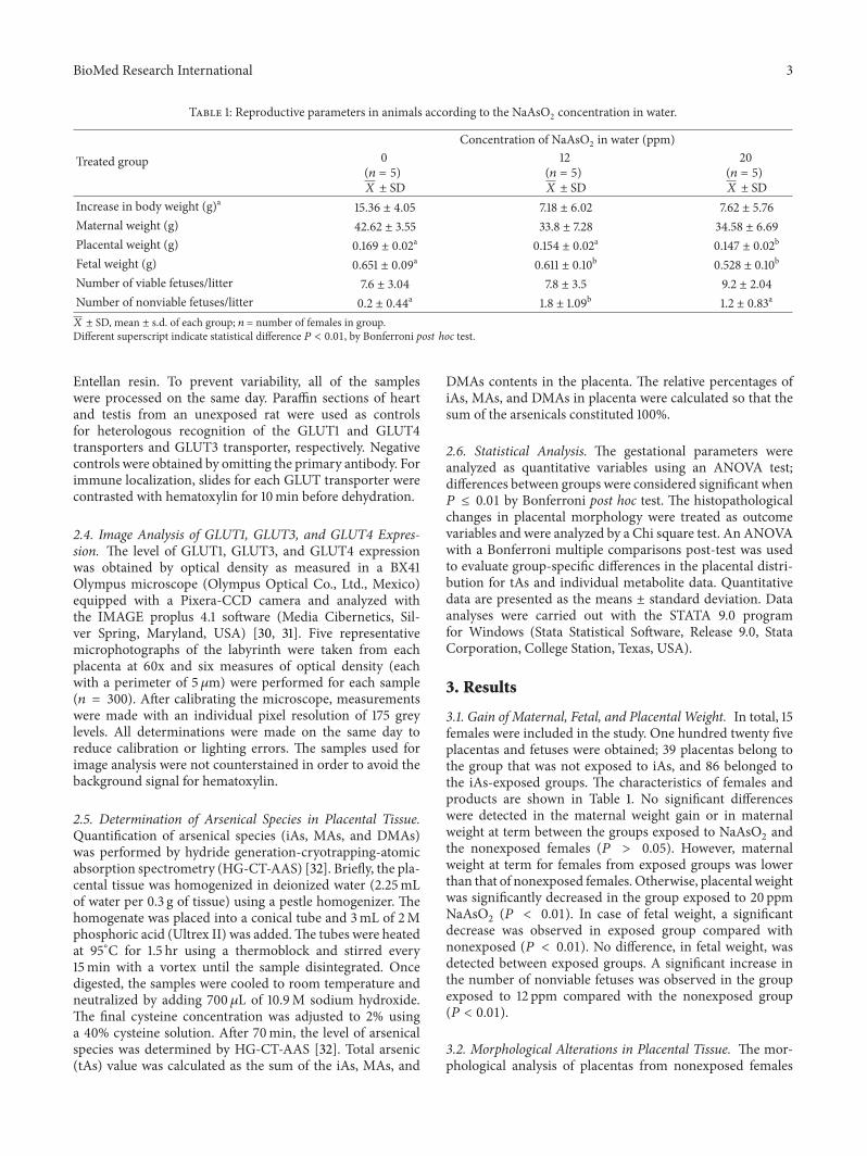
Table 1: Reproductive parameters in animals according to the NaAsO2 concentration in water.
Treated group
Concentration of NaAsO2 in water (ppm)0
(𝑛 = 5)𝑋 ± SD
12(𝑛 = 5)𝑋 ± SD
20(𝑛 = 5)𝑋 ± SD
Increase in body weight (g)a 15.36 ± 4.05 7.18 ± 6.02 7.62 ± 5.76Maternal weight (g) 42.62 ± 3.55 33.8 ± 7.28 34.58 ± 6.69Placental weight (g) 0.169 ± 0.02a 0.154 ± 0.02a 0.147 ± 0.02b
Fetal weight (g) 0.651 ± 0.09a 0.611 ± 0.10b 0.528 ± 0.10b
Number of viable fetuses/litter 7.6 ± 3.04 7.8 ± 3.5 9.2 ± 2.04Number of nonviable fetuses/litter 0.2 ± 0.44a 1.8 ± 1.09b 1.2 ± 0.83a
𝑋 ± SD, mean ± s.d. of each group; 𝑛 = number of females in group.Different superscript indicate statistical difference 𝑃 < 0.01, by Bonferroni post hoc test.
Entellan resin. To prevent variability, all of the sampleswere processed on the same day. Paraffin sections of heartand testis from an unexposed rat were used as controlsfor heterologous recognition of the GLUT1 and GLUT4transporters and GLUT3 transporter, respectively. Negativecontrols were obtained by omitting the primary antibody. Forimmune localization, slides for each GLUT transporter werecontrasted with hematoxylin for 10min before dehydration.
2.4. Image Analysis of GLUT1, GLUT3, and GLUT4 Expres-sion. The level of GLUT1, GLUT3, and GLUT4 expressionwas obtained by optical density as measured in a BX41Olympus microscope (Olympus Optical Co., Ltd., Mexico)equipped with a Pixera-CCD camera and analyzed withthe IMAGE proplus 4.1 software (Media Cibernetics, Sil-ver Spring, Maryland, USA) [30, 31]. Five representativemicrophotographs of the labyrinth were taken from eachplacenta at 60x and six measures of optical density (eachwith a perimeter of 5𝜇m) were performed for each sample(𝑛 = 300). After calibrating the microscope, measurementswere made with an individual pixel resolution of 175 greylevels. All determinations were made on the same day toreduce calibration or lighting errors. The samples used forimage analysis were not counterstained in order to avoid thebackground signal for hematoxylin.
2.5. Determination of Arsenical Species in Placental Tissue.Quantification of arsenical species (iAs, MAs, and DMAs)was performed by hydride generation-cryotrapping-atomicabsorption spectrometry (HG-CT-AAS) [32]. Briefly, the pla-cental tissue was homogenized in deionized water (2.25mLof water per 0.3 g of tissue) using a pestle homogenizer. Thehomogenate was placed into a conical tube and 3mL of 2Mphosphoric acid (Ultrex II) was added.The tubes were heatedat 95∘C for 1.5 hr using a thermoblock and stirred every15min with a vortex until the sample disintegrated. Oncedigested, the samples were cooled to room temperature andneutralized by adding 700 𝜇L of 10.9M sodium hydroxide.The final cysteine concentration was adjusted to 2% usinga 40% cysteine solution. After 70min, the level of arsenicalspecies was determined by HG-CT-AAS [32]. Total arsenic(tAs) value was calculated as the sum of the iAs, MAs, and
DMAs contents in the placenta. The relative percentages ofiAs, MAs, and DMAs in placenta were calculated so that thesum of the arsenicals constituted 100%.
2.6. Statistical Analysis. The gestational parameters wereanalyzed as quantitative variables using an ANOVA test;differences between groups were considered significant when𝑃 ≤ 0.01 by Bonferroni post hoc test. The histopathologicalchanges in placental morphology were treated as outcomevariables and were analyzed by a Chi square test. An ANOVAwith a Bonferroni multiple comparisons post-test was usedto evaluate group-specific differences in the placental distri-bution for tAs and individual metabolite data. Quantitativedata are presented as the means ± standard deviation. Dataanalyses were carried out with the STATA 9.0 programfor Windows (Stata Statistical Software, Release 9.0, StataCorporation, College Station, Texas, USA).
3. Results
3.1. Gain of Maternal, Fetal, and Placental Weight. In total, 15females were included in the study. One hundred twenty fiveplacentas and fetuses were obtained; 39 placentas belong tothe group that was not exposed to iAs, and 86 belonged tothe iAs-exposed groups. The characteristics of females andproducts are shown in Table 1. No significant differenceswere detected in the maternal weight gain or in maternalweight at term between the groups exposed to NaAsO
2and
the nonexposed females (𝑃 > 0.05). However, maternalweight at term for females from exposed groups was lowerthan that of nonexposed females. Otherwise, placental weightwas significantly decreased in the group exposed to 20 ppmNaAsO
2(𝑃 < 0.01). In case of fetal weight, a significant
decrease was observed in exposed group compared withnonexposed (𝑃 < 0.01). No difference, in fetal weight, wasdetected between exposed groups. A significant increase inthe number of nonviable fetuses was observed in the groupexposed to 12 ppm compared with the nonexposed group(𝑃 < 0.01).
3.2. Morphological Alterations in Placental Tissue. The mor-phological analysis of placentas from nonexposed females

4 BioMed Research International
Table 2: Histological findings in mice placentas according to theNaAsO2 concentration.
Concentration of NaAsO2 in water (ppm)0 12 20𝑛 (%) 𝑛 (%) 𝑛 (%)
DeciduaFibrosis 41 (100) 25 (100) 46 (100)Hemorrhage 29 (71) 18 (72) 40 (87)
Junction zoneInfarct 24 (58) 20 (80) 39 (85)∗
Phagocytosis in TGC 41 (100) 25 (100) 46 (100)Abnormal nucleus 0 0 0Degenerative GC 41 (100) 25 (100) 46 (100)
LabyrinthInfarct 0 0 1 (2.2)Vascular congestion 27 (66) 25 (100)∗ 45 (98)∗
𝑛= number of placentas where findings were detected by microscopicobservation.TGC: trophoblast giant cells; GC: glycogenic cells.∗𝑃 < 0.01 between NaAsO2 concentrations, by Chi square analysis.
showed normal cellular architecture (Figure 1(a)); no micro-scopic alterations were observed in the labyrinth (Fig-ure 1(d)). However, fibrosis and hemorrhagic processes wereobserved in the decidua basalis area. In placentas fromNaAsO
2exposed females, it was common to detect hem-
orrhagic zones (Figure 1(c)) and infarct lesions in deciduabasalis, where the cells were replaced by fibrinoid material(Figure 1(b)). In the labyrinth zone, vascular congestion wasevident (Figure 1(e)) in placentas from females in the groupsexposed to any concentration of NaAsO
2. Microinfarction
with cellular infiltration was detected in the labyrinth in oneplacenta from a NaAsO
2exposed female (Figure 1(f)).
To analyze the differences in placental histological find-ings among groups, the percentages obtained for each alter-ation were compared. As shown in Table 2, a significantincrease in infarcts in the junction zone was detected inplacentas from NaAsO
2exposed females, as well as vascular
congestion in the labyrinth zone.
3.3. Immunelocalization of GLUT1, GLUT3, and GLUT4 inMurine Placenta. GLUT1, GLUT3, and GLUT4 were ex-pressed in the labyrinth (Figure 2); in the case of GLUT1,protein expression was identified on both sides of thematernal-fetal interface, mainly in the brush border of thesyncytiotrophoblast layer (Figure 2(a)) andweakly in the fetalendothelium. In the case of GLUT3, protein expression waslocalized mainly in syncytiotrophoblast layer (Figure 2(c)).Finally, GLUT4 expression showed the same pattern as thatobserved for GLUT1; the protein was expressed on bothsides of thematernal-fetal interface (Figure 2(e)). No changesin the localization of GLUT transporters were detected inplacentas from females exposed toNaAsO
2(data not shown).
The controls for antibody heterologous recognition using ratheart (GLUT1; Figure 2(b) and GLUT4; Figure 2(f)) and rat
testis (GLUT3; Figure 2(d)) showed strong signal for eachprotein.
3.4. Effect of NaAsO2Exposure on GLUT1, GLUT3, and
GLUT4 Expression. To analyze the expression of GLUTtransporters among groups, image analysis was performedusing the methodology previously reported by our researchgroup [30, 31].The expression ofGLUT1 showednodifferencebetween placentas in female controls and those that wereexposed to iAs (Figure 3). Although a nonsignificant increasewas detected in GLUT3 expression in placentas from femalemice exposed to 12 ppm of NaAsO
2, the expression of this
transporter was quite similar in placentas from femalesexposed to 20 ppm and in nonexposed females.
The expression of GLUT4 was significantly lower (𝑃 <0.05) in placentas exposed to 12 ppm of NaAsO
2compared
with the expression in placentas from control females andthose exposed to 20 ppm iAs (Figure 3).
When iAs and its metabolites were quantified in placentasamples (Table 3), higher concentrations of iAs, MAs, andDMAs were found in placentas from exposed females thanin those from control mice. The main arsenical speciesfound in placental tissue was DMAs, which agrees with theresults reported by Devesa et al. (2006). However, we didnot find a significant difference in iAs or in arsenical speciesconcentrations relative to the dose used for iAs exposure.Thismay be, in part, due to significantly lower water consumptionby mice in the 20 ppm as compared to the 12 ppm group.Similar results were observed in Paul et al. 2007where controlmice consumed an average of 5.0mL of water per day (mL/d).Mice in the 25 ppmand 50 ppmgroup consumed significantlyless water: 3.8mL and 2.5mL per day, respectively [33].
4. Discussion
In the present study, we used an environmentally relevantconcentration of NaAsO
2. It is recognized that mice metabo-
lize iAs and clear iAsmetabolites from tissuesmore efficientlythan humans and that significantly higher exposure levelsor longer exposure times are needed in mice to producesymptoms of chronic As toxicity found in humans [33].Prenatal exposure to NaAsO
2in Balb/c strain mice can
cause maternal toxicity, which presents as a decrease inbody weight of exposed females relative to nonexposed mice[34]. In our study, no maternal toxicity was detected asmaternal weight showed no significant difference from thenonexposed group. Although the existing studies are highlydiverse, a nonsignificant decrease in body weight in miceexposed to iAs has been reported by another groups [24, 35,36]. As a result of prenatal exposure, fetuses from exposedlitters showed lower body weights compared with fetusesfrom nonexposed litters, and although fetuses from exposedfemales were not measured, some of them were smaller thanthose from nonexposed litters (data not shown). In addition,decrease in placental weight in As exposed females could bedue to an impairment in placental vasculogenesis [36], reduc-tion of cytotrophoblastic plugging, and syncytium formationcaused by oxidative stress which could generate placental

BioMed Research International 5
D
JZ
(a)
FM
(b)
H
(c)
BBBBBBBBBBBBBBBBBBBBBBBBBBBBBBBBBBBB
L
(d)
VC
(e)
FM
(f)
Figure 1: Morphologic alterations found in placentas from female mice exposed to NaAsO2in water. Microphotographs show the decidua
(D), junction zone (JZ), and labyrinth (L) zones of murine placenta. (a) Normal morphology of murine placenta and the trophoblast giantcells (arrows) and clusters of glycogen trophoblast cells (arrowheads) are shown in JZ and near to L.Magnification 10x. (b) Infarct and depositsof fibrinoid material (FM) present in JZ and L. (c) Hemorrhagic lesion located in D zone found in placenta from a NaAsO
2exposed female.
Magnification 10x. (d) Normal appearance of the L zone present in placenta from a nonexposed female. Magnification 60x. (e) Vascularcongestion (VC) located in L zone present in the placenta from a NaAsO
2exposed female. Magnification 60x. (f) Magnification of fibrinoid
material (FM) in the placenta from a female exposed to NaAsO2. Magnification 60x. H&E stain.
pathology and/or preeclampsia [37–39].These results suggestthat arsenic exposuremay induce placental tissue damage andcompromise the supply of nutrients, oxygen, hormones, andother growth factors that are necessary for fetal welfare.
The histopathological study revealed morphologicalchanges in placental tissue such as fibrosis, hemorrhage,infarcts, and vascular congestion.These results are consistentwith those reported by Levario-Carrillo et al. (2004), whoevaluated the effect of methyl parathion, an organophosphatepesticide, on placental morphology in pregnant rats and
reported large areas of fibrosis and hemorrhage in deciduabasalis as a consequence of exposure. The authors suggestedthat congestion detected in the labyrinth might act as a com-pensatory mechanism to dilute the toxic agent and replacethe dead cells [40]. However, vascular congestion could berelated with deficiencies in vasculogenesis as was reportedby Paul et al. [35]. The presence of fibrosis in the junctionzone has been suggested as a normal indicator of placentalaging [41], and the fibrinoid material found in placentas withinfarct lesions suggests a remodeling mechanism to replace

6 BioMed Research International
(a) (b)
ME
FE
(c) (d)
FE
(e) (f)
Figure 2: Immunelocalization of glucose transporters in placenta of female mice. The photomicrographs represent immunelocalization ofGLUT transporters in placenta from nonexposed females. Samples were counterstained with hematoxylin after an immunohistochemicalprocedure. (a), (c), and (e) show a positive signal for GLUT1, 3, and 4, respectively, (arrows) in mouse placenta. GLUT1 was identifiedon both sides of the maternal-fetal interface while GLUT3 was localized mainly in the fetal endothelium. GLUT4 was detected in bothsyncytiotrophoblast and stromal cells. Positive controls were mounted in rat tissues: (b) heart for GLUT1; (d) testis for GLUT3; and (f) heartfor GLUT4. The signal for GLUT1 and GLUT4 was localized on myocytes sarcoplasm and GLUT3 in cytosol of Sertoli cells. ME: maternalerythrocyte; FE: fetal erythrocyte. Magnification 60x.
the damaged tissue. Considering these findings, we suggestthe existence of a general mechanism whereby the placentaresponds to aggression regardless of the toxic agent (arsenic,lead, methyl parathion, etc.). In addition, histological alter-ations found in placentas from exposed females were notsevere enough to explain the diminution in placental and fetalweight, which underscores the need to analyze these changesat the molecular level.
The placental expression of GLUT1, GLUT3, and GLUT4has been reported previously [15, 42], and our findings are
consistent with these results. GLUT1 is a ubiquitous isoformexpressed in almost all of the tissues examined [18]. TheGLUT1 transporter was located in both the junction zoneand the labyrinth. Expression in the junction zone could benecessary to satisfy the metabolic demands of the placenta,whereas its expression in the labyrinth could be involvedin the maternal-fetal transfer of glucose [42]. The specificexpression of GLUT3 in the labyrinth suggests that thistransporter is more important for the regulation of glucosetransport than GLUT1. GLUT4 expression was localized by

BioMed Research International 7
Table 3: Concentration and relative proportion of iAs and methylated species in placental tissue according to NaAsO2 concentration.
ArsenicalArsenite concentration in drinking water (ppm)
0 12 20ngAs/g of tissuea % ngAs/g of tissuea % ngAs/g of tissuea %
iAs 1.85 ± 0.05 100 7.425 ± 3.31 21 8.573 ± 4.23 25MAs UD 0 1.945 ± 0.54 6 1.592 ± 0.49 5DMAs UD 0 25.55 ± 3.87 73 24.21 ± 4.84 70tAs 1.85 ± 0.05 100 34.92 ± 5.56∗ 100 34.38 ± 8.69∗ 100amean ± s.d.; % of arsenical species in relation to total arsenic (tAs).UD: undetected concentration.∗Difference between exposed versus control group; 𝑃 < 0.01, one way ANOVA test.
0
1
2
3
4
5
6
7
GLUT1 GLUT3 GLUT4
A.D.U
∗
Figure 3: Expression of GLUT1, 3, and 4 in placenta from femalemice exposed during gestation to NaAsO
2through water. Each bar
indicates the mean ± s.d. of arbitrary units of optical density (ADU)obtained for each transporter (𝑛 = 300 for each protein), accordingto NaAsO
2water concentration: White, 0 ppm; Gray, 12 ppm; and
Black, 20 ppm. Comparisons among groups were performed usingthe Wilcoxon range test; ∗𝑃 < 0.05.
Xing et al. (1998) in the stromal cells of human and rodentterm placentas using indirect IHC [15]. Nevertheless, weidentified the expression in both syncytiotrophoblast andstromal cells. GLUT4 is already synthesized and maintainedin subplasmalemmal vesicles. It is primarily responsible forthe rapid upregulation of transport activity observed insome tissues (fat and muscle) in response to insulin, whichis supplemented by a slower phase of transcription andtranslation [18]. In the placenta, the syncytial GLUT4 couldhelp improve glucose transport in response to any insulinstimulus (maternal or fetal).
Instead, we observed an increase in the expression ofGLUT3 in placentas exposed to 12 ppm of NaAsO
2(although
the difference was not statistically significant). These resultsare consistent with those reported by Boileau et al. (1995),who analyzed the expression of GLUT1 and GLUT3 inplacentas from diabetic rats and demonstrated an increasein GLUT3 expression, whereas the expression of GLUT1remained unchanged [43]. Considering this evidence, wesuggest that exposure to NaAsO
2induces a state of maternal
hyperglycemia in which GLUT3 seems to be more sensi-tive than GLUT1 to blood glucose levels. Additionally, theexpression of the placental GLUT4 transporter was decreased
in the group exposed to 12 ppm. Decrease of GLUT4 isrelated to a mechanism to protect the fetus from high levelsof maternal glucose [44]. Although we did not determineglucose concentrations in the blood of females during Asexposure, hyperglycemia, high levels of insulin, and impairedglucose tolerance in pregnant mice exposed to As havebeen reported [24, 35]. Alterations in the transfer of glucoseare common in many abnormalities of fetal growth (fetalmacrosomia and intrauterine growth restriction), and thisrelationship could explain the adverse pregnancy outcomesassociated with iAs exposure. However, in the group exposedto 20 ppm this regulation does not seem to occur becauseexpression did not differ from the control group. Thesedifferences are most likely related to the fact that femalesexposed to this concentration consumed a lower volume ofwater, as was previously described by Paul et al. [35].
On the other hand, studies performed in a S. cerevisiaemodel have demonstrated that GLUT1 and GLUT4 partici-pate in the uptake of arsenite andMAs, by a different pathwayused for glucose, which might contribute to As toxicity [45–47]. In addition, some studies show that glucose uptake canbe inhibited by As, even with high levels of insulin [46, 48],these findings suggest that iAs exposure during pregnancycould impair placental capacity for nutrient transfer resultingin pregnancy complications and/or restricted fetal growth[17].
To our knowledge, this is the first report concerningthe expression of GLUT4 in placentas from mice exposedto low levels of NaAsO
2, and our findings are consistent
with the mechanism proposed by Paul et al. (2007) toexplain the diabetogenic potential of As. Paul et al. (2007)suggest that arsenical trivalent species disrupt the activityof (3-phosphoinositide-dependent) kinases 1 and 2, blockingthe intracellular signaling cascade triggered by the insulinreceptor and resulting in the blockage of GLUT4 translo-cation from endosomal vesicles to the plasma membrane,which consequently decreases GLUT4 expression in cells[35]. In our study, the decrease in GLUT4 expression inplacentas might explain the low fetal weight observed inthe exposed groups. However, more research is requiredto analyze whether the signaling mechanism of GLUT4 issimilar to that reported in adipocyte or muscle cells.
These results are important as they provide evidence that,in pregnant women, even low concentrations of iAs in watermay damage the placenta and the fetus, not only affecting its
8 BioMed Research International
survival in utero but alsomodifying the expression of its genesand increasing the risk of disease in the future.
Abbreviations
As: ArsenicDMAs: Dimethyl arsenicGLUT: Glucose transporter(HG-CT-AAS): Hydride generation-cryotrapping-atomic
absorption spectrometryiAs: Inorganic arsenicIHC: Immunohistochemical analysisMAs: Methyl arsenicNaAsO
2: Sodium arsenite
tAs: Total arsenic.
Conflict of Interests
The authors have no other relevant affiliations or financialinvolvement with any organization or entity with a financialinterest in or financial conflict with the subject matter ormaterials discussed in the paper apart from those disclosed.
Acknowledgments
This work was partially supported by the Mixed Fund of theGovernment of the State of Chihuahua (FOMIX) and theNational Council of Science and Technology (CONACyT)(CHIH-2008-C01-92074). Daniela Sarahı Gutierrez-Torreswas the recipient of an M.Sc. scholarship from CONACyT(Register no. 213781).
References
[1] A. H. Smith, C. Hopenhayn-Rich, M. N. Bates et al., “Cancerrisks from arsenic in drinking water,” Environmental HealthPerspectives, vol. 97, pp. 259–267, 1992.
[2] A. Hernandez-Zavala, L. M. Del Razo, C. Aguilar, G. G. Garcıa-Vargas, V. H. Borja, and M. E. Cebrian, “Alteration in bilirubinexcretion in individuals chronically exposed to arsenic inMexico,” Toxicology Letters, vol. 99, no. 2, pp. 79–84, 1998.
[3] M. Rahman, M. Tondel, S. A. Ahmad, I. A. Chowdhury, M. H.Faruquee, and O. Axelson, “Hypertension and arsenic exposurein Bangladesh,” Hypertension, vol. 33, no. 1, pp. 74–78, 1999.
[4] D. N. Guha Mazumder, R. Haque, N. Ghosh et al., “Arseniclevels in drinking water and the prevalence of skin lesions inWest Bengal, India,” International Journal of Epidemiology, vol.27, no. 5, pp. 871–877, 1998.
[5] V. M. Rodrıguez, M. E. Jimenez-Capdeville, and M. Giordano,“The effects of arsenic exposure on the nervous system,”Toxicology Letters, vol. 145, no. 1, pp. 1–18, 2003.
[6] M. P. Waalkes, J. M. Ward, and B. A. Diwan, “Induction oftumors of the liver, lung ovary and adrenal in adult mice afterbrief maternal gestational exposure to inorganic arsenic: pro-motional effects of postnatal phorbol ester exposure on hepaticand pulmonary, but not dermal cancers,”Carcinogenesis, vol. 25,no. 1, pp. 133–141, 2004.
[7] P. Ravenscroft, H. Brammer, and K. Richards,Arsenic Pollution:A Global Synthesis, John Wiley & Sons, West Sussex, UK, 2009.
[8] ATSDR,Toxicological Profile for Arsenic, Division of Toxicology,Agency for Toxic Substances and Disease Registry, Atlanta, Ga,USA, 2007.
[9] S. A. Ahmad, M. H. Salim Ullah Sayed, S. Barua et al., “Arsenicin drinking water and pregnancy outcomes,” EnvironmentalHealth Perspectives, vol. 109, no. 6, pp. 629–631, 2001.
[10] M. J. Leroy, G. Tanguy,M. Vial,W. Rostene, A.Malassine, and F.Ferre, “The effect of vasoactive intestinal peptide (VIP) on thecontractile activity of human uterine smooth muscle,” Clinicaland Experimental Pharmacology and Physiology, vol. 18, no. 4,pp. 205–215, 1991.
[11] K. K. Saha, A. Engstrom, J. D. Hamadani, F. Tofail, K. M.Rasmussen, and M. Vahter, “Pre- and postnatal arsenic expo-sure and body size to 2 years of age: a cohort study in ruralBangladesh,” Environmental Health Perspectives, vol. 120, no. 8,pp. 1208–1214, 2012.
[12] H. Guan, F. Piao, X. Zhang et al., “Prenatal exposure to arsenicand its effects on fetal development in the general populationof dalian,” Biological Trace Element Research, vol. 149, no. 1, pp.10–15, 2012.
[13] P. Georgiades, A. C. Fergyson-Smith, and G. J. Burton, “Com-parative developmental anatomy of the murine and humandefinitive placentae,” Placenta, vol. 23, no. 1, pp. 3–19, 2002.
[14] A. Malassine, J.-L. Frendo, and D. Evain-Brion, “A comparisonof placental development and endocrine functions between thehuman andmouse model,”Human Reproduction Update, vol. 9,no. 6, pp. 531–539, 2003.
[15] A. Y. Xing, J. C. Challier, J. Lepercq et al., “Unexpectedexpression of glucose transporter 4 in villous stromal cells ofhuman placenta,” The Journal of Clinical Endocrinology andMetabolism, vol. 83, no. 11, pp. 4097–4101, 1998.
[16] G. T. Knipp, K. L. Audus, and M. J. Soares, “Nutrient transportacross the placenta,” Advanced Drug Delivery Reviews, vol. 38,no. 1, pp. 41–58, 1999.
[17] T. Jansson and T. L. Powell, “Human placental transport inaltered fetal growth: Does the placenta function as a nutrientsensor? A review,” Placenta, vol. 27, pp. 91–97, 2006.
[18] N. P. Illsley, “Glucose transporters in the human placenta,”Placenta, vol. 21, no. 1, pp. 14–22, 2000.
[19] S. Lager and T. L. Powell, “Regulation of nutrient transportacross the placenta,” Journal of Pregnancy, vol. 2012, Article ID179827, 14 pages, 2012.
[20] G. Concha, G. Vogler, D. Lezcano, B. Nermell, and M. Vahter,“Exposure to inorganic arsenic metabolites during early humandevelopment,” Toxicological Sciences, vol. 44, no. 2, pp. 185–190,1998.
[21] J. C. States, A. V. Singh, T. B. Knudsen et al., “Prenatal arsenicexposure alters gene expression in the adult liver to a proinflam-matory state contributing to accelerated atherosclerosis,” PLoSONE, vol. 7, no. 6, Article ID e38713, 2012.
[22] M. E. Davila-Esqueda, J. M. Morales, M. E. Jimenez-Capdevilleet al., “Low-level subchronic arsenic exposure from prenataldevelopmental stages to adult life results in an impaired glucosehomeostasis,” Experimental and Clinical Endocrinology andDiabetes, vol. 119, no. 10, pp. 613–617, 2011.
[23] S. F. Farzan, M. R. Karagas, and Y. Chen, “In utero and early lifearsenic exposure in relation to long-term health and disease,”Toxicology and Applied Pharmacology, vol. 272, no. 2, pp. 384–390, 2013.
[24] D. S. Hill, B. J. Wlodarczyk, L. E. Mitchell, and R. H. Finnell,“Arsenate-induced maternal glucose intolerance and neural

BioMed Research International 9
tube defects in a mouse model,” Toxicology and Applied Phar-macology, vol. 239, no. 1, pp. 29–36, 2009.
[25] A. Dıaz-Villasenor, M. C. Sanchez-Soto, M. E. Cebrian, P.Ostrosky-Wegman, and M. Hiriart, “Sodium arsenite impairsinsulin secretion and transcription in pancreatic 𝛽-cells,” Tox-icology and Applied Pharmacology, vol. 214, no. 1, pp. 30–34,2006.
[26] F. S. Walton, A. W. Harmon, D. S. Paul, Z. Drobna, Y. M. Patel,andM. Styblo, “Inhibition of insulin-dependent glucose uptakeby trivalent arsenicals: possible mechanism of arsenic-induceddiabetes,” Toxicology and Applied Pharmacology, vol. 198, no. 3,pp. 424–433, 2004.
[27] L. M. Del Razo, M. Styblo, W. R. Cullen, and D. J. Thomas,“Determination of trivalent methylated arsenicals in biologicalmatrices,” Toxicology and Applied Pharmacology, vol. 174, no. 3,pp. 282–293, 2001.
[28] Normalizacion, “Especificaciones tecnicas para la produccion,cuidado y uso de los animales de laboratorio,” Tech. Rep. NOM-062-ZOO-1999, Normalizacion, 2001.
[29] D. M. Opresko, B. E. Sample, and G. W. Suter, ToxicologicalBenchmarks to Wildlife, edited by Division PbtRAPHSR, U.S.Department of Energy, Oak Ridge, Tenn, USA, 1996.
[30] B. Gonzalez-Garcıa, M. E. Olave, E. Ramos-Martınez, C.Gonzalez-Horta, M. Levario-Carrillo, and B. Sanchez-Ramırez,“Decrease of muscarinic cholinergic receptors expression inplacenta from rats exposed to methyl parathion,” Human &Experimental Toxicology, vol. 27, no. 3, pp. 241–246, 2008.
[31] E. Gonzalez-Puebla, C. Gonzalez-Horta, R. Infante-Ramırez,L. H. Sanin, M. Levario-Carrillo, and B. Sanchez-Ramırez,“Altered expressions of MMP-2, MMP-9, and TIMP-2 in pla-centas from women exposed to lead,” Human & ExperimentalToxicology, vol. 31, no. 7, pp. 662–670, 2012.
[32] V. Devesa, B. M. Adair, J. Liu et al., “Arsenicals in maternaland fetal mouse tissues after gestational exposure to arsenite,”Toxicology, vol. 224, no. 1-2, pp. 147–155, 2006.
[33] D. S. Paul, A. Hernandez-Zavala, F. S. Walton et al., “Exam-ination of the effects of arsenic on glucose homeostasis incell culture and animal studies: development of a mousemodel for arsenic-induced diabetes,” Toxicology and AppliedPharmacology, vol. 222, no. 3, pp. 305–314, 2007.
[34] A. Wang, S. D. Holladay, D. C. Wolf, S. A. Ahmed, and J.L. Robertson, “Reproductive and developmental toxicity ofarsenic in rodents: a review,” International Journal of Toxicology,vol. 25, no. 5, pp. 319–331, 2006.
[35] D. S. Paul, A. W. Harmon, V. Devesa, D. J. Thomas, andM. Styblo, “Molecular mechanisms of the diabetogenic effectsof arsenic: inhibition of insulin signaling by arsenite andmethylarsonous acid,” Environmental Health Perspectives, vol.115, no. 5, pp. 734–742, 2007.
[36] W. He, R. J. Greenwell, D.M. Brooks, L. Calderon-Garciduenas,H. D. Beall, and J. D. Coffin, “Arsenic exposure in pregnantmice disrupts placental vasculogenesis and causes spontaneousabortion,”Toxicological Sciences, vol. 99, no. 1, pp. 244–253, 2007.
[37] E. Jauniaux, L. Poston, and G. J. Burton, “Placental-related dis-eases of pregnancy: involvement of oxidative stress and impli-cations in human evolution,”Human Reproduction Update, vol.12, no. 6, pp. 747–755, 2006.
[38] M. Vahter, “Effects of arsenic on maternal and fetal health,”Annual Review of Nutrition, vol. 29, pp. 381–399, 2009.
[39] S. Ahmed, S. M.-E. Khoda, R. S. Rekha et al., “Arsenic-associated oxidative stress, inflammation, and immune disrup-tion in human placenta and cord blood,” Environmental HealthPerspectives, vol. 119, no. 2, pp. 258–264, 2011.
[40] M. Levario-Carrillo, M. E. Olave, D. C. Corral, J. G. Alderete,S. M. Gagioti, and E. Bevilacqua, “Placental morphology ofrats prenatally exposed to methyl parathion,” Experimental andToxicologic Pathology, vol. 55, no. 6, pp. 489–496, 2004.
[41] E. P. C. T.DeRijk, E. VanEsch, andG. Flik, “Pregnancy dating inthe rat: placental morphology and maternal blood parameters,”Toxicologic Pathology, vol. 30, no. 2, pp. 271–282, 2002.
[42] B.-C. Shin, K. Fujikura, T. Suzuki, S. Tanaka, and K. Takata,“Glucose transporter GLUT3 in the rat placental barrier: apossible machinery for the transplacental transfer of glucose,”Endocrinology, vol. 138, no. 9, pp. 3997–4004, 1997.
[43] P. Boileau, C. Mrejen, J. Girard, and S. Hauguel-de Mouzon,“Overexpression of GLUT3 placental glucose transporter indiabetic rats,” Journal of Clinical Investigation, vol. 96, no. 1, pp.309–317, 1995.
[44] T. Hahn, S. Barth, U. Weiss, W. Mosgoeller, and G. Desoye,“Sustained hyperglycemia in vitro down-regulates the GLUT1glucose transport system of cultured human term placentaltrophoblast: a mechanism to protect fetal development?” TheFASEB Journal, vol. 12, no. 12, pp. 1221–1231, 1998.
[45] Z. Liu, E. Boles, and B. P. Rosen, “Arsenic trioxide uptake byhexose permeases in Saccharomyces cerevisiae,” The Journal ofBiological Chemistry, vol. 279, no. 17, pp. 17312–17318, 2004.
[46] Z. Liu,M.A. Sanchez, X. Jiang, E. Boles, S.M. Landfear, andB. P.Rosen, “Mammalian glucose permease GLUT1 facilitates trans-port of arsenic trioxide and methylarsonous acid,” Biochemicaland Biophysical Research Communications, vol. 351, no. 2, pp.424–430, 2006.
[47] X. Jiang, J. R. McDermott, A. Abdul Ajees, B. P. Rosena, and Z.Liu, “Trivalent arsenicals and glucose use different translocationpathways in mammalian GLUT1,”Metallomics, vol. 2, no. 3, pp.211–219, 2010.
[48] M. Bazuine, D. M. Ouwens, D. S. Gomes De Mesquita, and J.A. Maassen, “Arsenite stimulated glucose transport in 3T3-L1adipocytes involves both Glut4 translocation and p38 MAPKactivity,” European Journal of Biochemistry, vol. 270, no. 19, pp.3891–3903, 2003.

Submit your manuscripts athttp://www.hindawi.com
Hindawi Publishing Corporationhttp://www.hindawi.com Volume 2014
Anatomy Research International
PeptidesInternational Journal of
Hindawi Publishing Corporationhttp://www.hindawi.com Volume 2014
Hindawi Publishing Corporation http://www.hindawi.com
International Journal of
Volume 2014
Zoology
Hindawi Publishing Corporationhttp://www.hindawi.com Volume 2014
Molecular Biology International
GenomicsInternational Journal of
Hindawi Publishing Corporationhttp://www.hindawi.com Volume 2014
The Scientific World JournalHindawi Publishing Corporation http://www.hindawi.com Volume 2014
Hindawi Publishing Corporationhttp://www.hindawi.com Volume 2014
BioinformaticsAdvances in
Marine BiologyJournal of
Hindawi Publishing Corporationhttp://www.hindawi.com Volume 2014
Hindawi Publishing Corporationhttp://www.hindawi.com Volume 2014
Signal TransductionJournal of
Hindawi Publishing Corporationhttp://www.hindawi.com Volume 2014
BioMed Research International
Evolutionary BiologyInternational Journal of
Hindawi Publishing Corporationhttp://www.hindawi.com Volume 2014
Hindawi Publishing Corporationhttp://www.hindawi.com Volume 2014
Biochemistry Research International
ArchaeaHindawi Publishing Corporationhttp://www.hindawi.com Volume 2014
Hindawi Publishing Corporationhttp://www.hindawi.com Volume 2014
Genetics Research International
Hindawi Publishing Corporationhttp://www.hindawi.com Volume 2014
Advances in
Virolog y
Hindawi Publishing Corporationhttp://www.hindawi.com
Nucleic AcidsJournal of
Volume 2014
Stem CellsInternational
Hindawi Publishing Corporationhttp://www.hindawi.com Volume 2014
Hindawi Publishing Corporationhttp://www.hindawi.com Volume 2014
Enzyme Research
Hindawi Publishing Corporationhttp://www.hindawi.com Volume 2014
International Journal of
Microbiology