sec6 mutations and the drosophilaexocyst complex
TRANSCRIPT

IntroductionStudies of the exocyst complex in yeast have benefited from anabundance of mutations in each member of the complex. Theeight subunits of the exocyst (Sec3, Sec5, Sec6, Sec8, Sec10,Sec15, Exo70 and Exo84) were first identified in a screen thatisolated conditional mutations in genes required for exocytosis.Mutations of each have been shown to prevent exocytosis andto arrest growth of the daughter cell and cytokinesis (Fingerand Novick, 1998; Novick et al., 1980). The similarities of thephenotypes and extensive biochemical characterization haveled to a model in which the complex functions as an integralunit that can interact with both plasma membranes andtransport vesicles, and that, as a unit, marks sites of membraneinsertion (Finger et al., 1998; Finger and Novick, 1997; Haareret al., 1996; Mondesert et al., 1997).
In higher organisms, the investigation of the exocyst hasbeen hampered by a lack of mutations. A mutation in murinesec8 causes lethality shortly after gastrulation of the embryo,precluding a detailed analysis of the role of the complex(Friedrich et al., 1997). Recently, we have characterizedDrosophila sec5 mutations. As in yeast, Sec5 localization inDrosophila undergoes dynamic changes correlating with thesites at which it is required for the traffic of membrane proteinsduring oogenesis and cellularization. In homozygous sec5larvae and germ-line clones of sec5 alleles, we observeddefects in trafficking proteins to the plasma membrane (Murthyet al., 2003; Murthy and Schwarz, 2004).
In contrast to these genetic studies, investigation of othercomponents of the exocyst has depended on the introductionof antibodies and the overexpression of wild-type or mutatedforms of the proteins in wild-type genetic backgrounds. Fromthese studies, some discrepancies in the localization of exocystproteins and their phenotypes have emerged. Drosophila Sec5concentrates specifically at sites of membrane addition in both
ovaries and embryos but, in normal rat kidney (NRK) cells,different monoclonal antibodies to Sec6 and Sec8 recognizedthe exocyst complex at either the trans-Golgi network (TGN)or the plasma membrane (Yeaman et al., 2001). Furthermore,Exo70 associates with microtubules at the microtubule-organizing center in undifferentiated PC12 cells (Vega andHsu, 2001), and Sec10 has been found both at the endoplasmicreticulum (ER) (Lipschutz et al., 2003) and on tubulo-vesicularextensions of the TGN and recycling endosomes (Prigent et al.,2003). Finally, an interaction between Sec8, SAP102 and theN-methyl-D-aspartate receptor (NMDAR) in mammalianneurons was found in the ER (Sans et al., 2003).
Indeed, although biochemical studies in yeast and neuronssuggest the presence of only one copy of each subunit percomplex (Hsu et al., 1996; TerBush et al., 1996) and theisolation of the exocyst complex from all yeast exocyst mutantsshows that its structure is altered (TerBush and Novick, 1995),there is growing evidence that the members of the exocystmight not always act as a complex. For example, whereasDrosophila sec5 mutations blocked the transport of manyproteins to the plasma membrane of neurons and developingoocytes, the addition of antibodies specific for TGN-boundexocyst complexes to semi-intact NRK cells resulted in cargoaccumulation in a perinuclear region (Yeaman et al., 2001).Also, the introduction of a dominant negative Sec10 or Sec5small interfering RNA to NRK cells causes morphologicalchanges and phenotypes at the recycling endosome (Prigent etal., 2003). Finally, the overexpression of Sec10 affects proteinsynthesis in MDCK cells by an interaction with an ERtranslocon (Lipschutz et al., 2000; Lipschutz et al., 2003) andyeast Sec10p and Sec15p might form a subcomplex (Guo etal., 1999). These findings raise the possibility that differentcomplex members have different functions within the cell andmight not always function as a unit.
1139
To allow a detailed analysis of exocyst function inmulticellular organisms, we have generated sec6 mutantsin Drosophila. We have used these mutations to comparethe phenotypes of sec6 and sec5 in the ovary and nervoussystem, and we find them to be similar. We also find thatSec5 is mislocalized in sec6 mutants. Additionally, we havegenerated an epitope-tagged Sec8 that localized with Sec5on oocyte membranes and was mislocalized in sec5 and sec6
germ-line clones. This construct further revealed a geneticinteraction of sec8 and sec5. These data, taken together,provide new information about the organization of theexocyst complex and suggest that Sec5, Sec6 and Sec8 actas a complex, each member dependent on the others forproper localization and function.
Key words: Sec6, Sec5, Sec8, Oogenesis, Membrane trafficking
Summary
Sec6 mutations and the Drosophila exocyst complexMala Murthy1,*, Ravi Ranjan1,‡, Natalie Denef2, Misao E. L. Higashi1, Trudi Schupbach2
and Thomas L. Schwarz1,§
1Division of Neuroscience, Children’s Hospital, Harvard Medical School, Boston, MA 02115, USA2Howard Hughes Medical Institute, Molecular Biology Department, Princeton University, Princeton, NJ 08544, USA*Present address: Division of Biology, MC 139-74, Caltech, Pasadena, CA 91125, USA‡Present address: Department of Pharmacology, University of Texas Health Sciences Center, San Antonio, TX 78229, USA§Author for correspondence ([email protected])
Accepted 11 November 2004Journal of Cell Science 118, 1139-1150 Published by The Company of Biologists 2005doi:10.1242/jcs.01644
Research Article
Jour
nal o
f Cel
l Sci
ence

1140
The present uncertainty about the significance of exocystsubunits in multicellular organisms might, in part, arise from alack of loss-of-function mutations that can be directlycompared. In the present study, we report the isolation of a sec6mutation in Drosophila whose phenotype is comparable to thatof sec5. Moreover, with antibodies to Sec5 and an epitope-tagged sec8 transgene, we determine the interdependency ofthese complex members for their subcellular localization.
Materials and MethodsIsolation of sec6 allelesFrom the P-element-carrying line w/w; EP2021/EP2021, virginfemales were crossed to males of the transposase-expressing line w;CyO/+; ∆2-3, Sb/+. From the progeny of this cross, 400 male w;EP2021/CyO; ∆2-3, Sb/+ progeny were selected and individuallymated to w; Sco/CyO females. Then, 64 white-eyed, curly-wingedmale progeny (Ex/CyO) from independent lines were selected andcrossed to w; Sco/CyO females to establish balanced stocks. For eachline, Ex/CyO males and females were mated and 15 lines wereidentified in which Ex/Ex was lethal. Three of these (Ex15, Ex212 andEx228) were also lethal over Df(2R)PC4. The molecular characteriza-tion of the Ex15, Ex228 and Ex212 alleles (Fig. 1) was performed withthe following primers: 1, 5′-ATGGAGAATCTGAAGCAC-3′ in sec6;2, 5′-TAGGAGGTCAGGAAGGTGTT-3′ in Eip55E; 3, 5′-GAATG-GATGACCAAGGCCGC-3′ in sec6; 4, 5′-CGCTGTATCCGTATGC-CTGCTC-3′ in Eip55E; 5, 5′-CCCTAAGCTTTTGTATGTTCTTAT-GCCTTC-3′ in CG30332; 6, 5′-GAATCCGAAAAGGAAAAG-GACAGGTC-3′ in CG30122.
Drosophila stocks and clonesThe following genotypes were used.w; FRT42D sec6Ex15/Cyo, GFP[Kr-Gal4, UAS-GFP]w; FRT42D sec6Ex212/Cyo, GFPw; FRT40 ovoD/FRT40 sec5E10 or sec5E13; nanos-Gal4/UAS-FLPw; FRT42B ovoD/FRT42D sec6Ex15; nanos-Gal4/UAS-FLPw, hs-FLP; FRT42D Ubi-GFP/FRT42D sec6Ex15
y, w, hs-FLP; FRT40 ovoD/FRT40 sec5E13; nanos-Gal4/UAS-HA-Sec8w, hs-FLP; FRT42D Ubi-GFP/FRT42D sec6Ex15; nanos-Gal4/UAS-HA-Sec8y, w, hs-FLP; FRT40 sec5E13/Cyo; nanos-Gal4/UAS-HA-Sec8UAS-HA-sec8 (III)
To generate the plasmid p[UASp-HA-sec8], we amplified the openreading frame of sec8 from expressed sequence tag clone GM30905using the 5′ primer 5′-TTTTCTAGAATGGACGCCCCACCGCC-CACG-3′ and the 3′ primer 5′-TTTGCGGCCGCCTACACAAC-TACTCCCTTCGAGGG-3′, and digested it with XbaI and NotI. Theresultant fragment was cloned into pBS-HA, and then cut with KpnIand NotI. The HA-sec8 fragment [N-terminal fusion of a triplehemagglutinin (HA) tag and sec8] was cloned into p[UASp].
Germ-line clones were generated both by using the dominantfemale sterile technique, involving ovoD, and by looking for green-fluorescent-protein-deficient (GFP–) clones. We did this because ovoD
had been recombined onto a FRT42B chromosome, and our sec6mutations had been recombined onto FRT42D. Because achromosomal deletion between 42B and 42D might cause a phenotypeon its own, we also generated germ-line clones using FRT42D Ubi-GFP. In this way, GFP– egg chambers were homozygous mutant onlyfor sec6, with no other chromosomal deletion. The phenotypes usingboth methods were identical, indicating that heterozygosity for the42B-42D deletion is of no consequence to egg chambers within thecontext of the assays below and others (T. S., unpublished). Forgenerating germ-line clones by heat shock, vials were placed for ~30minutes per day at 37°C during larval and pupal development.
The trafficking assay used the following stocks.w; FRT40 sec5E10/CyO, GFP; UAS-CD8-GFPw; FRT40 sec5E10/CyO, GFP; elav-Geneswitchw; sec6Ex15/Cyo, GFP; UAS-CD8-GFPw; sec6Ex15/Cyo, GFP; elav-GeneswitchUAS-CD8-GFP (III)elav-Geneswitch (III)
Larvae were isolated by collecting eggs on grape caps with yeastpaste for 2 hours and then raised at room temperature. At 24 hoursafter egg laying (AEL), homozygous mutant larvae were chosen byan absence of the GFP marker and transferred to fresh yeast paste until72 hours AEL. Control larvae were raised similarly and were y,wunless otherwise indicated.
Immunocytochemistry and microscopyOvaries from 1-4-day-old females were dissected in PBS and kept onice. Ovaries were fixed in 6:1 heptane:FIX [FIX=4 volumes H2O, 1volume buffer B (100 mM potassium phosphate pH 6.8, 450 mM KCl,150 mM NaCl, 20 mM MgCl2), and 1 volume 37% formaldehyde] for15 minutes. They were stained in PBS containing 0.5% bovine serumalbumin, 0.1% Triton X-100 and 5% normal goat or donkey serum.Larvae were attached to Sylgard with Nexaband glue (VeterinaryProducts Laboratories) and dissected in PBS with pulled-glassdissecting needles, and subsequently fixed with 3.7% formaldehydein buffer B. For the trafficking assay, Triton X-100 was omitted fromwashes and antibody incubations.
The following stains and primary antibodies were used: Texas Red-X phalloidin, Alexa Fluor 568 phalloidin, Hoechst 33342, Alexa-Fluor-488-conjugated mouse anti-HA (Molecular Probes), mouseanti-HA at 1:500 (BabCO), mouse anti-Gurken-1D12 (DevelopmentalStudies Hybridoma Bank), mouse anti-Sec5 (ascites) 22A2 at 1:200(Murthy et al., 2003), rat monoclonal anti-CD8α at 1:200 (CALTAGLaboratories), Cy5-conjugated goat anti-horseradish-peroxidase (anti-HRP) at 1:200 (Jackson Immunoresearch). Secondary antibodies usedwere: Cy3-conjugated goat anti-mouse and FITC-conjugated goatanti-mouse (Jackson Immunoresearch), Alexa Fluor 647 goat anti-mouse (Molecular Probes). For images of ovaries, confocal data wereacquired as single images or as image stacks of multitracked separatechannels with a Zeiss LSM 510 microscope.
The amount of Sec5 in the neuropil (Fig. 2) was estimated bydrawing an approximately 900 µm2 box within the neuropil of theventral nerve cord, on one side of the commissures in single confocalsections and obtaining a value for mean pixel intensity.
Trafficking assaysWe modified the methods for this trafficking assay from our earlierpublished version of the experiment performed on sec5E10 larvae(Murthy et al., 2003). Originally, we were not aware of leakage fromthe elav-Geneswitch driver and therefore did not take into account themCD8-GFP that was expressed before introduction to the drug andbefore Sec5 maternal contribution ran down in the mutants. In thisversion, we aged larvae to the appropriate time points and then fedhalf of the larvae 0.5 mM RU486 (mifepristone, Sigma) dissolved inwet yeast paste and mixed with instant fly food and sucrose. The otherhalf were fed regular yeast paste. We were then able to subtractuninduced values for GFP and surface mCD8 from induced values toobtain final values that reflected the expression and trafficking ofmCD8-GFP after 72 hours AEL. In addition, originally for sec5E10
larvae, we had collected ten sections per image stack at variable stepintervals. This method might have led to an overestimate of theamount of mCD8-GFP expression. In this version of the traffickingassay, we instead collected confocal sections at 2 µm intervals, withvariable numbers of sections per image stack. The experiment was inall other ways identical to the published version. Finally, in ourprevious analysis, some values were inadvertently calculated as the
Journal of Cell Science 118 (6)
Jour
nal o
f Cel
l Sci
ence
1141sec6 mutants and exocyst interactions
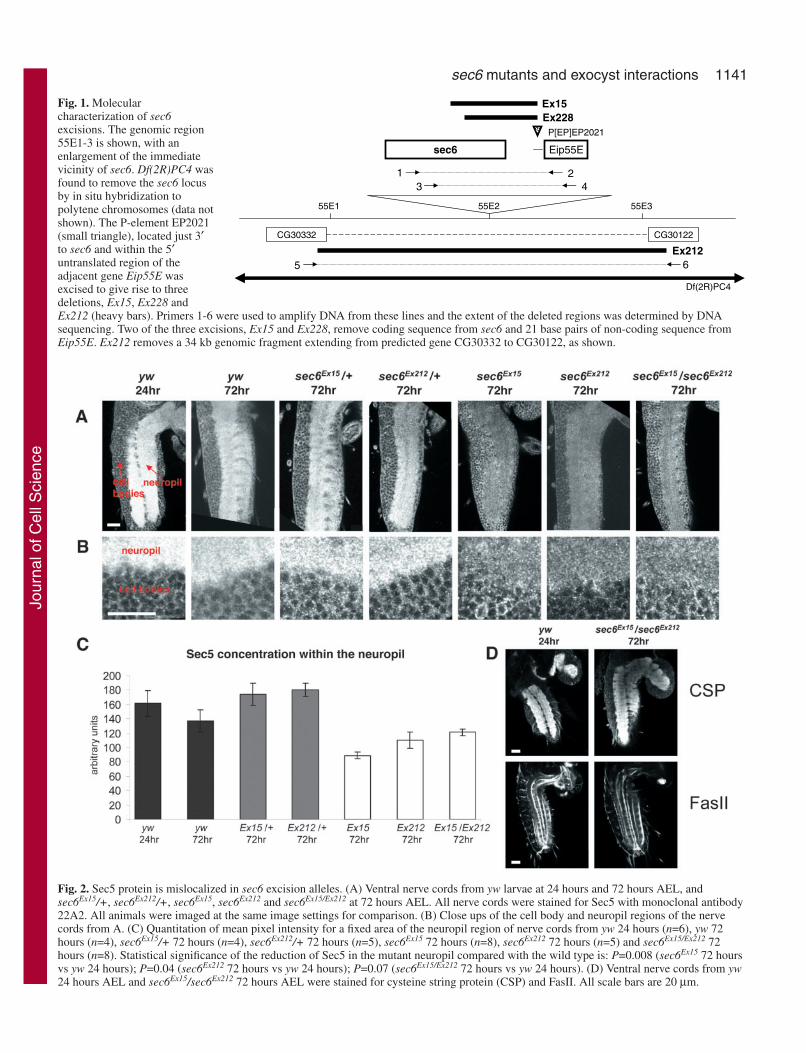
Fig. 1. Molecularcharacterization of sec6excisions. The genomic region55E1-3 is shown, with anenlargement of the immediatevicinity of sec6. Df(2R)PC4 wasfound to remove the sec6 locusby in situ hybridization topolytene chromosomes (data notshown). The P-element EP2021(small triangle), located just 3′to sec6 and within the 5′untranslated region of theadjacent gene Eip55E wasexcised to give rise to threedeletions, Ex15, Ex228 andEx212 (heavy bars). Primers 1-6 were used to amplify DNA from these lines and the extent of the deleted regions was determined by DNAsequencing. Two of the three excisions, Ex15 and Ex228, remove coding sequence from sec6 and 21 base pairs of non-coding sequence fromEip55E. Ex212 removes a 34 kb genomic fragment extending from predicted gene CG30332 to CG30122, as shown.
E55piE6ces
1202PE]PE[P
822xE51xE
1 243
4CP)R2(fD
1E55 3E552E55
23303GC 22103GC
212xE5 6
Fig. 2. Sec5 protein is mislocalized in sec6 excision alleles. (A) Ventral nerve cords from yw larvae at 24 hours and 72 hours AEL, andsec6Ex15/+, sec6Ex212/+, sec6Ex15, sec6Ex212 and sec6Ex15/Ex212 at 72 hours AEL. All nerve cords were stained for Sec5 with monoclonal antibody22A2. All animals were imaged at the same image settings for comparison. (B) Close ups of the cell body and neuropil regions of the nervecords from A. (C) Quantitation of mean pixel intensity for a fixed area of the neuropil region of nerve cords from yw 24 hours (n=6), yw 72hours (n=4), sec6Ex15/+ 72 hours (n=4), sec6Ex212/+ 72 hours (n=5), sec6Ex15 72 hours (n=8), sec6Ex212 72 hours (n=5) and sec6Ex15/Ex212 72hours (n=8). Statistical significance of the reduction of Sec5 in the mutant neuropil compared with the wild type is: P=0.008 (sec6Ex15 72 hoursvs yw 24 hours); P=0.04 (sec6Ex212 72 hours vs yw 24 hours); P=0.07 (sec6Ex15/Ex212 72 hours vs yw 24 hours). (D) Ventral nerve cords from yw24 hours AEL and sec6Ex15/sec6Ex212 72 hours AEL were stained for cysteine string protein (CSP) and FasII. All scale bars are 20 µm.
Jour
nal o
f Cel
l Sci
ence

1142
maximum intensity pixel at each point within a projected z-axis stack.In the present analysis, we summed the pixels in the z-axis stack toobtain total pixel intensity for each channel.
Confocal data for these experiments were acquired as 2 µm intervalimage stacks of multitracked separate channels with a Zeiss LSM 510microscope. Identical gain, offset, pinhole and laser settings were usedfor the mutant and control for each experiment. For quantification oftotal pixel intensity in cell bodies of bipolar dendrite (bd) sensoryneurons, a region of interest (ROI) was drawn around the two cellbodies that lie next to one another for a given bd neuron in a peripheralsegment, found in the Cy5 channel, and the sum of pixel intensitiesfor that ROI across the z-axis stack calculated for each of threechannels, Cy5 (HRP), Cy3 (CD8) and GFP. For backgroundsubtraction, an identical ROI was drawn outside the cell bodies.
Under these modified conditions, we also repeated the traffickingassay in the axons of bd neurons and at the boutons of theneuromuscular junction (NMJ). As before, we found reductions insurface mCD8 in sec5E10 larvae. Surface mCD8 immunoreactivitywas reduced in axons to 8% of control (9% when normalized to HRP;P=0.0001) and at the NMJ to 2% of control (1% normalized to HRP;P=0.0002). However, in both axons and the NMJ, the amount ofmCD8-GFP expression was also reduced compared with the wildtype. For sensory axons, an ROI was drawn along a portion of theaxon and the sum of pixel intensities for each channel obtained afterbackground subtraction. This sum was divided by the length in µm ofthe axon segment measured. For quantification of pixel intensity atsynaptic boutons of the NMJ, an ROI was drawn around the entireendplate and total pixel intensity values for each channel calculatedafter background subtraction.
StatisticsAll P values reported in this study are two-tailed values and derivedfrom Student’s t test, assuming unequal variances. For calculatingstandard error after subtracting two averages from each other, eachwith a standard error (as in Fig. 2F), we took the square root of thesum of the squares of the standard error from each average. To obtainthe standard error after dividing two averages, i.e. for valuesnormalized to either HRP- or GFP-intensity, we applied the followingformula for the error on the ratio of two values, A and B:
standard error of the mean on A/B = 1/B (standard error on A) +A/B2 (standard error on B).
We determined whether the normalized values were significantlydifferent by finding values for test statistic (t) and degrees of freedom(df ) according to the following formulae:
t = (q1 – q2) ÷ (v12/n1 + v2
2/n2)0.5
df = (v12/n1 + v2
2/n2)2 ÷ [(v12/n1)2/n1–1 + (v2
2/n2)2/n2–1],
where q1 and q2 are the normalized values for fluorescent intensity, v1and v2 are the variances of those values, and n is the number of valuesin the data sets for the determination of the average intensities. Fromthese values, we calculated a two-tailed P value by using thecalculator at http://www.graphpad.com/quickcalcs/PValue1.cfm.
ResultsDeletions of sec6The Drosophila homolog of sec6 is located at 55E (Murthy etal., 2003) and has a predicted open reading frame of 739 aminoacids that are 21% identical to human sec6 and 37% identicalto yeast sec6. No other sec6 gene is present in the sequencedDrosophila genome. This gene is removed by Df(2R)PC4. Toobtain a specific mutation in sec6, we excised a P element,EP2021, located 1.2 kb downstream of the 3′ end of sec6 andupstream of the adjacent predicted open reading frame, Eip55E
(Fig. 1). We obtained three excision lines from this screen,Ex15, Ex212 and Ex228. Amplification of genomic DNAisolated from homozygous Ex15, Ex212 and Ex228 larvae withthe polymerase chain reaction primer pairs outlined in Fig. 1allowed us to sequence across the deletions created by theimprecise excision of the P element. The Ex15 deletion beginsat amino acid 397 of Sec6 and continues to the 21st base pairin the 5′ untranslated region of the neighboring gene Eip55E.Ex228 is a smaller deletion, extending from amino acid 541 ofSec6 to the 21st base pair of the 5′ untranslated region ofEip55E. Ex212 carries a 38 kb deletion surrounding sec6. Thedeletion begins in the last exon of the gene CG30332 and endsafter the fifth intron of CG30122. This deletion removes eightgenes, including sec6, between CG30332 and CG30122.Because available anti-Sec6 antibodies do not work inDrosophila, we cannot determine directly whether a truncatedform of Sec6 remains in the sec6 excision alleles. However, thelethal periods and phenotypes of sec6Ex15, sec6Ex228 andsec6Ex212 as homozygotes are identical to those of each alleleover Df(2R)PC4 or over one another; therefore, by geneticcriteria, the three excision alleles are likely to be null. Inaddition to disrupting sec6, these excision are likely to interferewith expression of the Eip55E gene, an ecdysone-inducedcystathionine γ-lyase expressed in fat bodies, salivary glandsand lymph glands (Andres et al., 1993). The lethality of theexcisions and the phenotypes discussed below are notattributable to Eip55E, because Eip55E mutants are viable andfertile.
Although flies homozygous for the original P element insertEP2021 are both viable and fertile, larvae either homozygousfor sec6Ex15 or trans-heterozygous for sec6Ex15 and Df(2R)PC4or sec6Ex15 and sec6Ex212 die from growth arrest at 96 hoursAEL, similar to the phenotype for sec5E10, a null allele (Murthyet al., 2003). The sec6 larvae, however, are much smaller thanthe sec5 larvae. Whereas sec5 larvae at the end of their lifespanare the size of wild-type larvae at 48 hours AEL, sec6 larvaeare never larger than wild-type larvae at 24 hours AEL. Thesec6Ex15 larvae, like the sec5E10 larvae, probably survive to 96hours AEL owing to the persistence of maternally contributedRNA and protein.
Sec5 is mislocalized in sec6 mutantsNormally, Sec5 concentrates in the neuropil, the synapse-richregion of the nerve cord of first-instar larvae. We therefore useda previously characterized Sec5-specific antibody (Murthy etal., 2003) to determine whether Sec5 localization depends onthe presence of Sec6. In 72-hour-AEL sec6Ex15/sec6Ex15,sec6Ex212/sec6Ex212 or sec6Ex15/sec6Ex212 larvae, Sec5 is presentbut greatly reduced in the neuropil when compared with eitherwild-type or heterozygous larvae (Fig. 2A,B). We quantifiedSec5 in the neuropil and found it to be significantly reduced inthe mutants compared with wild-type controls (Fig. 2C). Thismislocalization is not due to a general disruption of thearchitecture of the central nervous system (CNS), whichappeared to be grossly normal when sec6Ex15/sec6Ex212 larvaewere stained for the synaptic vesicle marker cysteine stringprotein or for FasII, which labels axon tracts within the CNS.The mislocalization of Sec5 in the nerve cord of sec6 mutantsis therefore likely to be a specific effect on members of theexocyst complex.
Journal of Cell Science 118 (6)
Jour
nal o
f Cel
l Sci
ence

1143sec6 mutants and exocyst interactions
Defects in vesicle trafficking in sec6 mutantsBecause sec5 mutations in Drosophila, like exocyst mutationsin yeast, prevent membrane traffic to the cell surface, wehypothesized that a similar defect might occur in sec6 mutants.To analyse sec5 mutations, we developed an acute assay ofmembrane transport so as to examine the transport of newlysynthesized proteins at the end of the mutants’ lifespan, whenmaternally contributed Sec5 was no longer sufficient forcellular function (Murthy et al., 2003). The neuron-specificelav promoter was used to express the Geneswitch product, aninactive form of Gal4, in the nervous system. Upon feedinglarvae RU486, Geneswitch is rendered active, which causes
transcription of an mCD8-GFP-encoding transgene. mCD8-GFP is a transmembrane protein whose cytoplasmic GFPdomain serves as a reporter of protein synthesis and whoseCD8 epitopes (expressed on the surface of the cell) can be usedto quantify transport to the surface; in the absence of Triton X-100, anti-mCD8 antibody binds only the subset of CD8expressed on the cell surface, whereas the GFP fluorescencerepresents both surface and internal pools of the protein. Weexamined the lateral bd neurons in the peripheral nervoussystem because they were accessible to the antibody in theabsence of Triton X-100. Animals were also stained with ananti-HRP antibody that labels a neuronal surface antigen sothat the GFP and mCD8 signals could be normalized to thesurface area of the cell.
Our previous use of this method (Murthy et al., 2003) mighthave overestimated the amount of transgene expression in thesec5 mutant and also did not take into account the leakiness ofthe Geneswitch system, which permits a low level of transgene
Fig. 3. Vesicle trafficking defects in sec5E10.(A) A lateral bd sensory neuron in a wild-typelarva that had been fed RU486 at 48 hours AELand dissected at 59 hours AEL. Anti-HRPimmunostaining (gray) was used to find bdsensory neurons in each animal. (B) A lateral bdneuron as in A but from a sec5E10 mutant fedRU486 at 72 hours AEL and dissected at 83hours AEL. sec5E10 larvae show less cellsurface CD8 than the control. (C,D) Additionalwild-type and sec5E10 neurons as in A and B.(E) From quantitative fluorescent microscopy ofthe transport assay, surface-expressed CD8immunofluorescence, total GFP fluorescenceand cell surface area (measured by HRPimmunoreactivity) were expressed asfluorescent units after background subtraction.In the sensory neuron soma [n=7 for wild-typelarvae fed RU486 (white) and uninduced (blue);n=6 for sec5E10 larvae fed RU486 (gray) anduninduced (black)], there are comparable levelsof anti-HRP labeling at the cell surface. TotalGFP fluorescence is reduced in the RU486-fedmutants (gray) compared with RU486-fedcontrols (white), but there is a significantinduction of the GFP signal, which is a measureof transgene expression, in the RU486-fed(gray) compared with uninduced (black)mutants. The amount of mCD8 at the cellsurface is reduced in the RU486-fed mutantscompared to RU486-fed controls, and a 50%reduction is observed when surface mCD8 isnormalized to GFP in order to control for thelevel of transgene expression (P=0.004). (F) Tomeasure the amount of transgene induction byfeeding RU486, the uninduced averages (blueand black bars in E) were subtracted from theRU486-induced averages (white and gray barsin E). To control for differences in cell size,induced GFP and induced surface CD8 werethen normalized to the anti-HRP signal. Theamount of induced mCD8 trafficked to the cellsurface was also normalized to the induced GFPsignal, revealing the disruption of transport ofthe newly synthesized protein to the surface.
Jour
nal o
f Cel
l Sci
ence

1144
expression even in the absence of RU486. Based on thisinformation, we have made corrections to the methods andused the modified assay for a re-examination of sec5E10 and anexamination of sec6Ex15.
We repeated the trafficking analysis on sec5E10 larvae. Theselarvae were fed either plain or RU486-containing yeast pastefrom 72-83 hours AEL. Two types of controls were used forcomparison: similarly sized larvae that had been fed eitherplain or RU486-containing food from 48-59 hours AEL (Fig.3) or similarly aged larvae that had been fed from 72-83 hoursAEL (data not shown). We observed a strong induction of thetransgene in the somata of lateral bd neurons in both mutantand wild-type larvae (Fig. 3A-D). Although we noticed thepresence of a moderate leak from the Geneswitch driver insec5E10 and wild-type animals (such that, even when not fedRU486, both mutant and wild-type larvae showed somemCD8-GFP expression), there was a substantial induction ofthe transgene over this baseline in both mutant and control(Fig. 3E). When normalized to the HRP signal, the totalinduced GFP in the cell body and the total induced anti-mCD8labeling at the surface of the cell body were reduced in themutant to 41% (P=0.0019) and 10% (P<0.0001) of control,respectively (Fig. 3F). The amount of induced mCD8 at the cellsurface, even when normalized to the amount of induced GFPin the soma, was still reduced in the mutant to 15% of control(P=0.0526; even if un-induced ‘leak’ is not subtracted, a 50%reduction is measured, P=0.004) (Fig. 3E). Thus, the reduction
of mCD8 at the cell surface in the mutant was not due simplyto a decrease in the amount of mCD8-GFP induction. Rather,confirming our earlier result, between 72 hours and 83 hoursAEL in sec5E10 mutants, much less of the newly synthesizedmCD8 is inserted at the membrane and a defect in themembrane-trafficking pathway is indicated.
The sec6Ex15 mutant larvae at 72 hours AEL were also fedeither plain or RU486-containing yeast paste for 12 hours, andcompared with similarly-sized control larvae fed at 24 hoursAEL. Both control and mutant larvae, after introduction to thedrug, showed expression of the transgene in the cell bodies oflateral bd neurons (Fig. 4A,B), but the amount of GFPfluorescence was greatly reduced in the mutant cells whencompared to wild type.
The transport to the surface of the mCD8 reporter-geneproduct was measured as above (Fig. 4A-D). Although therewas a 79% induction (P=0.01) of mCD8-GFP in sec6 animalsfed RU486 compared with animals that were not induced (Fig.4C), there was no significant induction of mCD8 at the cellsurface, indicating a defect in trafficking the protein to the cellsurface at 72 hours AEL.
When we subtracted the levels of expression without RU486from the RU486-induced levels for both the mutants and thewild type, and normalized these values to the HRP levels (Fig.4D), the total induced GFP in the cell body and the totalinduced anti-mCD8 labeling at the surface of the cell bodywere reduced in the mutant to 7% (P<0.0001) and 3%
Journal of Cell Science 118 (6)
Fig. 4. Vesicle traffickingdefects in sec6Ex15 larvae at 72hours AEL. (A) Fromquantitative fluorescentmicroscopy of the transportassay in neuronal cell bodies,surface-expressed CD8immunofluorescence, total GFPfluorescence and cell surfacearea (measured by HRPimmunoreactivity) wereexpressed as fluorescent unitsafter background subtraction.n=10 for wild-type larvae fedRU486 (white); n=8 for wild-type larvae uninduced (blue);n=13 for sec6Ex15 larvae fedRU486 (grey); and n=9 foruninduced sec6Ex15 larvae(black). (B) Representativelateral bd sensory neurons asused in the assay in A. Inparticular, a wild-type larva thathad been fed RU486 at 24 hoursAEL and dissected and stainedin the absence of Triton X-100at 36 hours AEL is comparedwith a sec6Ex15 mutant fedRU486 at 72 hours AEL similarly stained at 84 hours AEL. Anti-HRP immunostaining (gray) was used to find bd sensory neurons in eachanimal. The sec6Ex15 larvae show less cell surface CD8 than the control, but also showa large decrease in GFP fluorescence. (C) Whennormalized to the anti-HRP signal, there was a significant induction of the mCD8-GFP transgene in the sec6Ex15 mutant after feeding RU486(P=0.01), but the amount of mCD8 at the soma surface was not significantly increased. (D) To measure the amount of transgene induction inthe cell body by feeding RU486, the uninduced averages (blue and black bars in A) were subtracted from the RU486 averages (white andgray bars in A). To control for differences in cell size, GFP and surface CD8 were also normalized to the anti-HRP signal, as in C. Theamount of induced mCD8 trafficked to the cell surface, when normalized to the corrected GFP signal, is decreased in the mutant comparedwith control.
Jour
nal o
f Cel
l Sci
ence

1145sec6 mutants and exocyst interactions
(P<0.0001) of control, respectively. The amount of inducedmCD8 at the cell surface, normalized to the induced amountof GFP, was reduced in the mutant to 37% of control (P=0.1903).The overall reduction of mCD8 at the cell surface in the mutantstherefore probably arises from two factors: a decrease in theamount of mCD8-GFP produced in response to RU486 and adefect in trafficking the protein to the plasma membrane.
Although the results of this assay suggest that there is atrafficking defect in sec6 larvae, the amount of mCD8-GFPexpression was so low in the mutants that the specificity andsignificance of the defect was uncertain. The poor expression ofthe reporter might be due either to a problem with consumptionof the RU486 or to a defect in protein synthesis. We thereforefed sec6 larvae RU486 at 48 hours AEL, instead of at 72 hoursAEL, in the hope that the mutants would be healthier. However,in animals that were fed RU486 at 48 hours AEL for 12 hours,we did not observe a significant trafficking defect (n=7 for bothmutant and wild type; data not shown), possibly because therewas sufficient maternally contributed Sec6 at this earlier stage.Overall, the phenotypes of sec5 and sec6 are fundamentallysimilar with regard to the trafficking of this reporter protein.
The sec6 germ-line clones phenocopy sec5 in the ovaryWe generated clones of the sec6Ex15 allele in the female germline in order to compare the ovarian phenotype with that of
sec5E10 (Murthy and Schwarz, 2004). As with sec5, thesemothers did not lay eggs, because the sec6 allele was lethal tothe developing germ-line cells. Moreover, the cellular phenotypewas examined with phalloidin to visualize F-actin on membranesand found to be very similar, although less severe, than thesec5E10 phenotype (Fig. 5B-G). Egg chambers normally consistof 16 germ-line cells linked by cytoplasmic bridges termed ringcanals and enclosed within an epithelium of follicle cells. Insec6Ex15 germ lines, however, egg chambers formed and exitedthe germarium but, after stage 3, cell membranes were frequentlyabsent between the cells and ring canals that should reside inthose membranes were instead clumped together in the middleof a large multinucleate cell (Fig. 5F,G).
When individual cells could be distinguished within the eggchamber, defects were observed in the positioning of the oocyte.In the wild type, the oocyte invariably assumes the posteriormostposition among the group of 16 germ cells (Fig. 5H). Similar tosec5E10, when the germ line is mutant for sec6Ex15, the oocyte isoften mispositioned (Fig. 5I-L).
Sec5 protein is mislocalized in sec6Ex15 germ lines andHA-Sec8 is mislocalized in both sec6Ex15 and sec5E13
germ linesBecause Sec5 is dynamically localized to particular regionsof the developing oocyte plasma membrane (Murthy and
Fig. 5. The sec5E10 and sec6Ex15 mutants have similar phenotypes in the ovary. (A-G) Egg chambers labeled with phalloidin (red), Hoechst33342 (blue) and GFP (green in G). Unlike in the wild type (A), egg chambers from sec5E10 germ-line clones (B,C) lack membranes (markedwith phalloidin) between nuclei and have ring canals clumped together (arrows). Egg chambers from sec6Ex15 germ-line clones (D-G) exit thegermarium and progress through stage 3 (D,E), initially resembling the control. However, after stage 3 (F,G), ring canals clump together(arrows) and phalloidin-marked membranes between nuclei are absent. FRT42D sec6Ex15 homozygous germ lines were generated by mitoticrecombination in combination with either FRT42B ovoD (D-F) or FRT42D Ubi-GFP and imaging egg chambers that lacked GFP in the germline (G). (H-L) Egg chambers labeled with Texas Red-phalloidin (red), Hoechst 33342 (blue) and anti-Gurken antibody (green). Gurkenaccumulates only in the oocyte, which resides at the posterior end of the egg chamber throughout oogenesis, contacting the posterior folliclecells in the wild type (H). In egg chambers from sec5E10 (I,J) and sec6Ex15 (K,L) germ-line clones, the oocyte is often mispositioned anteriorly(arrows). Anterior (A) and posterior (P) ends of the chamber are marked. All scale bars are 20 µm.
Jour
nal o
f Cel
l Sci
ence

1146
Schwarz, 2004), the ovary provided an additional opportunityto test the hypothesis that Sec5 localization depends on Sec6function. At early stages of wild-type oogenesis, prior to thetime when development arrests in sec6Ex15 germ lines, Sec5 isfound principally on the plasma membrane of the 16 cells (Fig.6A). Moreover, Sec5 is enriched on the oocyte membrane atits posterior edge. In the sec6Ex15 germ lines Sec5 wasmislocalized, with an excess of punctate staining within thecytoplasm of the cells (Fig. 6B-E). In sec5E10 mutant germlines, membrane markers such as anti-syntaxin antibody and afluorescently tagged lectin also label cytoplasmic punctawithin the egg chamber, and these puncta are likely to representfragments of membrane amidst the disrupted cells (Murthy andSchwarz, 2004). The punctate Sec5 immunoreactivity in sec6germ lines might similarly represent membrane fragments.Sec5 staining is not, however, completely lost from theboundary of the sec6 germ-line cells, although much of thismight represent Sec5 in the follicle cells, which areheterozygous for the sec6 mutation.
To compare further the distribution of exocyst components,we generated an HA-tagged Sec8 upstream activation sequence(UAS) construct and expressed this transgene specifically inthe Drosophila germ line using the nanos-GAL4 promoter. Thetransgene did not prevent normal oocyte development and thedistribution of the tagged Sec8 was then compared with that ofSec5 (Murthy and Schwarz, 2004). Although little expressionof the transgene was detectable at early stages of oogenesis,the two components of the exocyst had parallel distributions at
later stages. During stages 7 and 8,Sec5 is localized along the oocyte andnurse-cell membranes, with a slightconcentration at anterior corners of theoocyte (i.e. where the lateralmembranes of the oocyte meet theanterior surface) (Fig. 7A); at thisstage, HA-Sec8 is localized similarly(Fig. 7B). By stage 10, Sec5 is highlyenriched at the anterior corners of theoocyte membrane, and we observed asimilar distribution for HA-Sec8 (Fig.7C-E). However, whereas a double lineof staining is visible with anti-Sec5antibody, representing both follicle-cell and oocyte membranes, HA-Sec8is present only on the oocytemembranes, because the nanospromoter does not transcribe thetransgene in the follicle cells. Whenexpressed exclusively in the folliclecells of stage-9 egg chambers (using aGR1-GAL4 driver), HA-Sec8 wasagain concentrated on the plasmamembrane, particularly at the apicalend (data not shown). Thus, thedistribution of this exocyst componentparalleled that of Sec5 in both theoocyte and follicle cells. As with Sec5,the anterior localization of HA-Sec8 inthe oocyte was not dependent onmicrotubules, because it persisted incolcemid-treated females (data not
shown). In one regard, however, the distribution of HA-Sec8differed from that of endogenous Sec5: we frequently observedthat individual nurse cells (but not the oocyte) would containan exceptionally high concentration of HA-Sec8 (Fig. 7B).This might be due to abnormal overexpression of the transgeneby nanos-Gal4. Indeed, we have observed the same irregularaccumulation of several other proteins in nurse cells whenexpressed from transgenes under the control of nanos-Gal4(N.D. and T.S., unpublished).
To examine the dependence of Sec8 localization on Sec5,we expressed the transgene in a sec5 mutant background. Wewished to examine stages 7-10, when the HA-Sec8 localizationis clearest, and so looked at germ lines homozygous for thehypomorphic allele sec5E13 (Fig. 7F-H). In doing so, weuncovered a genetic interaction between Sec8 and Sec5.Females with sec5E13 germ lines can lay eggs, althoughmembrane trafficking defects in these oocytes result in aberrantdorsal appendages (Murthy and Schwarz, 2004). Theoverexpression of HA-Sec8, which had no deleterious effecton wild-type oogenesis, enhanced the phenotype of sec5E13
alone by arresting egg-chamber development between stages 7and 9, such that no eggs were deposited by these females.Within these germ lines, the oocyte nucleus had migratedappropriately to the anterior end of the cell (at stage 7).However, the oocytes failed to enlarge properly, remainingcomparable in size to the nurse cells. Moreover, some disorderof the nurse-cell membranes was apparent, with some cellsfused and ring canals clustered together.
Journal of Cell Science 118 (6)
Fig. 6. Sec5 protein is mislocalized in sec6Ex15 germ-line clones. (A) Control egg chamber,stages 3 and 4, labeled with anti-Sec5 antibody (green) and Hoechst 33342 (blue). Sec5concentrates on membranes and is enriched at the boundary between the oocyte and posteriorfollicle cells (arrow). (B) In sec6Ex15 germ lines, labeled as in A, Sec5 protein is found in puncta(arrow), many of which are clustered towards the center of the egg chamber. (C,D) Sec5localization (green) is compared with the submembranous actin cytoskeleton (gray) in sec6Ex15
germ lines. (E1) GFP– sec6Ex15 germ lines (generated as in Fig. 5G) stained for Sec5 (red) andHoechst 33342 (blue). GFP is shown in green. (E2) GFP– sec6Ex15 germ lines stained for Sec5(green) and actin (red). (B-E) Sec5 accumulates in intracellular puncta (arrows) and any germ-line membranes separating nuclei have a patchy, discontinuous distribution of Sec5. The Sec5puncta often accumulate near the clump of ring canals. All scale bars are 20 µm.
Jour
nal o
f Cel
l Sci
ence

1147sec6 mutants and exocyst interactions
HA-Sec8 no longer concentrated at the membrane in sec5E13
oocytes and was instead uniformly localized throughout thecytoplasm (Fig. 7G,H). This was seeneven in those oocytes whose structurewas otherwise most normal, with ananterior nucleus and normal membrane-associated actin. By contrast, HA-Sec8in wild-type oocytes was mostlymembrane-associated throughoutoocyte development (Fig. 7B).
To examine the dependence of Sec8localization on Sec6, we expressed theHA-Sec8 transgene, under the controlof nanos-GAL4, in a sec6Ex15 mutantbackground, by generating sec6homozygous germ-line clones. In theseclones, HA-Sec8 accumulated in punctawithin the egg chamber cytosol (Fig.7I,J), akin to the Sec5 mislocalizationphenotype in sec6Ex15 egg chambers(Fig. 6). We did not observe anenhancement of the sec6Ex15 phenotypewhen HA-Sec8 was overexpressed, butthis might be because the sec6Ex15
phenotype is already quite severe.
DiscussionWhereas the description of exocystfunction in Drosophila was previouslylimited to mutations in and antibodies toSec5, the sec6 mutations and epitope-tagged form of Sec8 presented hereallow a comparison of the distributionand phenotype of additional componentsof the complex. As summarized below,the data generally favor a model inwhich these components function as aunit and depend on one another for theirlocalization.
The localization of complex membersis consistent with their function as anintegral unit. The distribution of Sec5has been examined most closely in theovary. In this tissue, it was present on allmembranes early in the development ofthe egg chamber. At late stages,however, Sec5 acquired a characteristicdistribution not reported for any othercellular component – a progressiveenrichment at the anterior end of thelateral oocyte membranes. HA-Sec8 hasnow been found to be similarlyconcentrated in this area, suggesting thatseveral (and perhaps all) exocystcomponents will be similarly localized.
We also find that mutations in onecomplex member appear to disrupt thelocalization of others. Thus, in sec5E13
homozygous oocytes, HA-Sec8 was nolonger membrane bound or concentrated
at the anterior sites. Instead, it appeared to fill the cytoplasmdiffusely. Similarly, Sec5 was mislocalized within the nervous
Fig. 7. HA-Sec8 localization in late-stage egg chambers. HA-Sec8 was expressed exclusivelyin the germ line under the control of nanos-Gal4. (A,B) Egg chambers from sec5E13/+heterozygous controls at stage 7 are labeled with anti-Sec5 antibody (A) or anti-HA antibodyand Texas-Red/phalloidin (B). Sec5 and HA-Sec8 both concentrate along the oocyte (*)membrane. Probably owing to overexpression, HA-Sec8 often accumulates in the cytoplasmof some nurse cells. (C-E) Egg chambers from sec5E13/+ heterozygous controls at stage 10 arelabeled with Hoechst 33342 and anti-Sec5 antibody (C) or anti-HA antibody and Texas Red-phalloidin (D,E). Sec5 and HA-Sec8 concentrate at anterior corners (arrows) of the oocytemembrane. (F-H) Egg chambers from sec5E13 homozygous germ lines are labeled withHoechst 33342 and Texas Red-phalloidin (F-H) and with anti-HA antibody (G,H). HA-Sec8no longer concentrates at the oocyte (*) membrane but rather fills the oocyte cytosol whenSec5 function is compromised by the mutation. In addition, the sec5E13 phenotype (F) isenhanced by the presence of the HA-sec8 transgene (G,H), stunting the oocyte, disruptingnurse cell membranes and causing ring canals to clump together. (I,J) GFP– egg chambersfrom FRT42D sec6Ex15 homozygous germ lines, generated by mitotic recombination withFRT42D Ubi-GFP. GFP is shown in green (I1,J1). HA-Sec8 (red in I1,J1 and green in I2,J2)accumulates in puncta within the mutant egg chambers and in proximity to actin-rich ringcanals (red in I2,J2). All scale bars are 20 µm.
Jour
nal o
f Cel
l Sci
ence

1148
system of sec6 mutant larvae and Sec5 and HA-Sec8 were bothmislocalized within germ lines homozygous for sec6. Themislocalization of Sec5 and HA-Sec8 in sec6 germ lines,however, was not identical to the mislocalization of HA-sec8 insec5 germ lines. Whereas the latter involved a diffuse filling ofthe cytoplasm with immunoreactivity, the mislocalized Sec5 andHA-Sec8 remained punctate within the sec6 egg chambers.Because these puncta resembled syntaxin and lectin-staining insec5 germ lines, it seems likely that they represent fragments ofmembrane or transport vesicles that have not fused with theplasma membrane. The difference in these two phenotypesmight arise from any of several causes, including the perduranceof some Sec6 in the sec6Ex15 mutant germ lines. It is temptingto speculate, however, that the difference reflects theorganization of proteins within the complex (Fig. 8). Sec3p hasbeen shown in yeast to bind to the plasma membrane at the budtip even when other complex members are absent (Finger et al.,1998). This has been interpreted as indicating that Sec3p bindsdirectly to a membrane protein and that the localization of othercomplex members is dependent on Sec3p. Sec5p is thought tobind directly to Sec3p (Guo et al., 1999) and so it is plausiblethat, in the present study, Sec5 remained membrane bound viaits direct interaction with Sec3 even in the absence of Sec6. Sec8,however, is not thought to interact directly with Sec3. BecauseSec8 appears to remain membrane-associated in sec6 but notsec5 mutants, we hypothesize that a partial complex consistingof Sec3, Sec5 and Sec8 remains on the membrane even in theabsence of Sec6. The disposition of the remaining complexmembers in the sec5 and sec6 mutants must remain speculativeuntil suitable reagents have been obtained for their localization.
The interdependence of the complex members is alsoevident in the genetic interaction of Sec8 and Sec5: althoughgerm-line expression of HA-tagged Sec8 had no phenotype ofits own, it enhanced the germ-line phenotype of sec5E13,making this partial loss-of-function allele more similar to thenull allele. This observation requires that the epitope-taggedtransgene be used with caution, because its expression mightinterfere with exocyst function owing either to an influence ofthe epitope tag or to unphysiological expression levels. Indeed,phenotypes have been associated with the overexpression ofSec10, another complex member (Lipschutz et al., 2000;Lipschutz et al., 2003).
The phenotypes of sec6 and sec5 mutants can be comparedin several regards. Like sec5, sec6 caused lethality at
approximately 96 hours AEL and these larvae were stunted intheir growth and did not progress beyond the first instar. In anassay of membrane-protein transport to the cell surface ofidentified neurons, we found trafficking defects for sec6 thatwere akin to those of sec5. In the germ line, we found thatmembranes between cells disintegrate in sec6 clones, aphenotype we previously observed for the null allele of sec5(Murthy and Schwarz, 2004). For sec5, we hypothesized that,as the cells of the germ line grow and expand, membraneaddition cannot keep pace, and that membranes between nursecells and the oocyte consequently fall apart. A similarexplanation is likely for sec6. We also observed themispositioning of the oocyte within the sec6 germ line.Previously, we had shown that this phenotype occurred wheneither the germ line or the posterior follicles were mutant forsec5. Because the positioning of the oocyte is dependent on E-cadherin and cell-cell signaling between the oocyte and folliclecells (Godt and Tepass, 1998; Gonzalez-Reyes and StJohnston, 1998), it is likely that this phenotype arises from adefect in the expression of E-cadherin or other signalingmolecules on the oocyte surface. In fact, E-cadherin and Nectin2a have been recently shown to be binding partners for theexocyst complex in MDCK cells (Yeaman et al., 2004).
Although the similarities of their phenotypes suggest thatSec5 and Sec6 share functions, we observed some differencesin the mutant phenotypes. sec6Ex15 larvae are smaller thansec5E10 larvae but germ-line clones of sec5E10 have a moresevere phenotype in the ovary, arresting earlier and with fewerremaining membranes. The most intriguing difference arose inthe mCD8-GFP expression assay: whereas sec5E10 larvae werecapable of synthesizing the protein but not of expressing it atthe cell surface, sec6Ex15 larvae expressed only low levels ofthe protein, which also appeared to be blocked in their transportto the surface. Finally, whereas HA-Sec8 protein wasmislocalized in both sec5 and sec6 germ-line clones, thepatterns of mislocalized protein were distinct. The differencesin the mutant phenotypes might arise from minor factors suchas the degree of perdurance of protein in the homozygousgerm-line clones or the amount or stability of maternal proteindeposited in the egg. However, they might also representlegitimate functional distinctions. The most pronounceddifference, the different levels of expression of the mCD8-GFPreporter protein, might reflect the fact that Sec6 is required atan earlier step in the synthesis of membrane proteins, in
Journal of Cell Science 118 (6)
3ceS
8ceS
6ceS
51ceS
07oxE
48oxE
01ceS
5ceS
? enarbmem amsalp
3ceS
?
51ceS
01ceS
8ceS
6ceS
07oxE
48oxE
3ceS
8ceS
51ceS
07oxE
48oxE
01ceS
5ceS
?
Fig. 8. Organization of the exocyst complex.Interactions are depicted for members of theexocyst complex with each other and with anunknown receptor on the plasma membrane (left),based on a slight modification of the model derivedfrom studies in yeast (Guo et al., 1999) and inaccordance with the data from Figs 6, 7. In sec5mutants, Sec8 is no longer associated with themembrane and other subunits might also becomecytosolic (center). In sec6 mutants, however, bothSec5 and Sec8 can remain membrane associated(right).
Jour
nal o
f Cel
l Sci
ence

1149sec6 mutants and exocyst interactions
addition to its requirement (along with Sec5) for insertion atthe plasma membrane. Such a role would be consistent withfindings that Sec6 and Sec8 have been observed in the TGN,that Sec8 and Sec10 associate with proteins at the TGN andER, and that overexpression of Sec10 alters membrane-proteinsynthesis (Lipschutz et al., 2000; Lipschutz et al., 2003; Sanset al., 2003; Yeaman et al., 2001). The general similaritiesbetween and severity of the sec6 and sec5 phenotypes also donot exclude the possibility that other components will havemore restricted roles, particularly given that several GTPaseshave emerged as binding partners of particular members of thecomplex and might be either effectors or regulators of thosecomponents (Adamo et al., 1999; Brymora et al., 2001; Inoueet al., 2003; Moskalenko et al., 2002; Prigent et al., 2003;Robinson et al., 1999; Sugihara et al., 2002; Walch-Solimenaet al., 1997; Zhang et al., 2001).
In contrast to the cell lethality of the sec5 and sec6phenotypes, a Sec10 RNA-interference construct in Drosophilawas reported to have very little effect in most tissues, possiblyaffecting only the secretions of the ring gland cells (Andrewset al., 2002). However, because no antibody is available forDrosophila Sec10 and because maternally contributed proteinwould be unaffected by this construct, the RNA interferencemight have been ineffective at reducing endogenous Sec10levels. In light of the broad phenotypes of dominant negativeand overexpressed Sec10 in other cell types (Lipschutz et al.,2000; Lipschutz et al., 2003; Prigent et al., 2003), this is alikely explanation of the discrepancy.
In summary, the similarity of localization of Sec5 and HA-Sec8, the interdependency of the complex members for properlocalization in this study, the genetic interaction between HA-Sec8 and sec5, and the general similarity of the sec5 and sec6phenotypes suggest that Sec5, Sec6 and Sec8 associate as acomplex in Drosophila, acting in concert, and that each iscrucial for the function of the complex at the membrane. It willbe important to examine the localization and phenotypes of theother complex members to determine whether all the complexmembers do indeed function primarily as part of the intactexocyst. Furthermore, the mutations in sec5 and sec6 shouldprovide a useful genetic background for structure functionstudies with which to test the significance of their individualbinding partners and regulators.
This work was supported by NIH grant NS41062 (T.L.S.) and theHoward Hughes Medical Institute (T.S.). N.D. is supported by afellowship from the Human Frontier Science Program. We thank Y.Nasrullah for technical support and also M. Salanga and the MRRCImaging Core.
ReferencesAdamo, J. E., Rossi, G. and Brennwald, P. (1999). The Rho GTPase Rho3
has a direct role in exocytosis that is distinct from its role in actin polarity.Mol. Biol. Cell 10, 4121-4133.
Andres, A. J., Fletcher, J. C., Karim, F. D. and Thummel, C. S. (1993).Molecular analysis of the initiation of insect metamorphosis: a comparativestudy of Drosophila ecdysteroid-regulated transcription. Dev. Biol. 160,388-404.
Andrews, H. K., Zhang, Y. Q., Trotta, N. and Broadie, K. (2002).Drosophila Sec10 is required for hormone secretion but not generalexocytosis or neurotransmission. Traffic 3, 906-921.
Brymora, A., Valova, V. A., Larsen, M. R., Roufogalis, B. D. and Robinson,P. J. (2001). The brain exocyst complex interacts with RalA in a GTP-dependent manner: identification of a novel mammalian Sec3 gene and asecond Sec15 gene. J. Biol. Chem. 276, 29792-29797.
Finger, F. P. and Novick, P. (1997). Sec3p is involved in secretion andmorphogenesis in Saccharomyces cerevisiae. Mol. Biol. Cell 8, 647-662.
Finger, F. P. and Novick, P. (1998). Spatial regulation of exocytosis: lessonsfrom yeast. J. Cell Biol. 142, 609-612.
Finger, F. P., Hughes, T. E. and Novick, P. (1998). Sec3p is a spatial landmarkfor polarized secretion in budding yeast. Cell 92, 559-571.
Friedrich, G. A., Hildebrand, J. D. and Soriano, P. (1997). The secretoryprotein Sec8 is required for paraxial mesoderm formation in the mouse. Dev.Biol. 192, 364-374.
Godt, D. and Tepass, U. (1998). Drosophila oocyte localization is mediatedby differential cadherin-based adhesion. Nature 395, 387-391.
Gonzalez-Reyes, A. and St Johnston, D. (1998). The Drosophila AP axis ispolarised by the cadherin-mediated positioning of the oocyte. Development125, 3635-3644.
Guo, W., Grant, A. and Novick, P. (1999). Exo84p is an exocyst proteinessential for secretion. J. Biol. Chem. 274, 23558-23564.
Haarer, B. K., Corbett, A., Kweon, Y., Petzold, A. S., Silver, P. and Brown,S. S. (1996). SEC3 mutations are synthetically lethal with profilin mutationsand cause defects in diploid-specific bud-site selection. Genetics 144, 495-510.
Hsu, S. C., Ting, A. E., Hazuka, C. D., Davanger, S., Kenny, J. W., Kee, Y.and Scheller, R. H. (1996). The mammalian brain rSec6/8 complex. Neuron17, 1209-1219.
Inoue, M., Chang, L., Hwang, J., Chiang, S. H. and Saltiel, A. R. (2003).The exocyst complex is required for targeting of Glut4 to the plasmamembrane by insulin. Nature 422, 629-633.
Lipschutz, J. H., Guo, W., O’Brien, L. E., Nguyen, Y. H., Novick, P.and Mostov, K. E. (2000). Exocyst is involved in cystogenesisand tubulogenesis and acts by modulating synthesis and delivery ofbasolateral plasma membrane and secretory proteins. Mol. Biol. Cell 11,4259-4275.
Lipschutz, J. H., Lingappa, V. R. and Mostov, K. E. (2003). The exocystaffects protein synthesis by acting on the translocation machinery of theendoplasmic reticulum. J. Biol. Chem. 278, 20954-20960.
Mondesert, G., Clarke, D. J. and Reed, S. I. (1997). Identification of genescontrolling growth polarity in the budding yeast Saccharomyces cerevisiae:a possible role of N-glycosylation and involvement of the exocyst complex.Genetics 147, 421-434.
Moskalenko, S., Henry, D. O., Rosse, C., Mirey, G., Camonis, J. H. andWhite, M. A. (2002). The exocyst is a Ral effector complex. Nat. Cell Biol.4, 66-72.
Murthy, M. and Schwarz, T. L. (2004). The exocyst component Sec5 isrequired for membrane traffic and polarity in the Drosophila ovary.Development 131, 377-388.
Murthy, M., Garza, D., Scheller, R. H. and Schwarz, T. L. (2003).Mutations in the exocyst component Sec5 disrupt neuronal membranetraffic, but neurotransmitter release persists. Neuron 37, 433-447.
Novick, P., Field, C. and Schekman, R. (1980). Identification of 23complementation groups required for post-translational events in the yeastsecretory pathway. Cell 21, 205-215.
Prigent, M., Dubois, T., Raposo, G., Derrien, V., Tenza, D., Rosse, C.,Camonis, J. and Chavrier, P. (2003). ARF6 controls post-endocyticrecycling through its downstream exocyst complex effector. J. Cell Biol.163, 1111-1121.
Robinson, N. G., Guo, L., Imai, J., Toh, E. A., Matsui, Y. and Tamanoi, F.(1999). Rho3 of Saccharomyces cerevisiae, which regulates the actincytoskeleton and exocytosis, is a GTPase which interacts with Myo2 andExo70. Mol. Cell. Biol. 19, 3580-3587.
Sans, N., Prybylowski, K., Petralia, R. S., Chang, K., Wang, Y. X., Racca,C., Vicini, S. and Wenthold, R. J. (2003). NMDA receptor traffickingthrough an interaction between PDZ proteins and the exocyst complex. Nat.Cell Biol. 5, 520-530.
Sugihara, K., Asano, S., Tanaka, K., Iwamatsu, A., Okawa, K. and Ohta,Y. (2002). The exocyst complex binds the small GTPase RalA to mediatefilopodia formation. Nat. Cell Biol. 4, 73-78.
TerBush, D. R. and Novick, P. (1995). Sec6, Sec8, and Sec15 are componentsof a multisubunit complex which localizes to small bud tips inSaccharomyces cerevisiae. J. Cell Biol. 130, 299-312.
TerBush, D. R., Maurice, T., Roth, D. and Novick, P. (1996). The exocystis a multiprotein complex required for exocytosis in Saccharomycescerevisiae. EMBO J. 15, 6483-6494.
Vega, I. E. and Hsu, S. C. (2001). The exocyst complex associates withmicrotubules to mediate vesicle targeting and neurite outgrowth. J. Neurosci.21, 3839-3848.
Jour
nal o
f Cel
l Sci
ence

1150
Walch-Solimena, C., Collins, R. N. and Novick, P. J. (1997). Sec2p mediatesnucleotide exchange on Sec4p and is involved in polarized delivery of post-Golgi vesicles. J. Cell Biol. 137, 1495-1509.
Yeaman, C., Grindstaff, K. K., Wright, J. R. and Nelson, W. J. (2001).Sec6/8 complexes on trans-Golgi network and plasma membrane regulatelate stages of exocytosis in mammalian cells. J. Cell Biol. 155, 593-604.
Yeaman, C., Grindstaff, K. K. and Nelson, W. J. (2004). Mechanism ofrecruiting Sec6/8 (exocyst) complex to the apical junctional complex duringpolarization of epithelial cells. J. Cell Sci. 117, 559-570.
Zhang, X., Bi, E., Novick, P., Du, L., Kozminski, K. G., Lipschutz, J. H.and Guo, W. (2001). Cdc42 interacts with the exocyst and regulatespolarized secretion. J. Biol. Chem. 276, 46745-46750.
Journal of Cell Science 118 (6)
Jour
nal o
f Cel
l Sci
ence