role of therapeutic blockade of ccl2 in a mouse model of ... · complement components (tumlin...
TRANSCRIPT

Aus der Medizinischen Poliklinik – Innenstadt der Ludwig-Maximilians-Universität München
Komm. Direktor: Prof. Dr. med. Martin Reincke
Role of therapeutic blockade of CCL2 in a mouse model of SLE and lupus
nephritis
Dissertation
zum Erwerb des Doktorgrades der Humanbiologie
an der Medizinischen Fakultät der
Ludwig-Maximilians-Universität zu München
vorgelegt von
Onkar P. Kulkarni
Solapur, India
2010

Mit Genehmigung der Medizinischen Fakultät der Universität München
1. Berichterstatter: Prof. Dr. med. Hans-Joachim Anders 2. Berichterstatter: Prof. Dr. Dr. h.c. Thomas Ruzicka Mitberichterstatter: Prof. Dr. Hartmut Wekerle Mitberichterstatter: Priv. Doz. Dr. Udo Kummer Dekan: Prof. Dr. med. Dr. h. c. M. Reiser, FACR, FRCR Tag der mündlichen Prüfung: 16.09.2010

Onkar Prakash Kulkarni, M. Pharm. Med. Poliklinik, Klinische Biochemie, Ludwig-Maximillians University (LMU), Schiller straße-42, Munich- 80336, Germany [email protected] .
DECLARATION
I here by declare that the present work embodied in this thesis was carried out by me
under the supervision of OA PD Dr. Hans Joachim Anders, Internist-Nephrologe-
Rheumatologie, Medizinische Poliklinik-Innenstadt Klinikum der Universität
München. This work has not been submitted in part or full to any other university or
institute for any degree or diploma.
Onkar P. Kulkarni Date:

ACKNOWLEDGEMENTS
I can not resist myself from expressing my heart felt deep sense of gratitude and respect for my PhD supervisor PD Dr. Hans-Joachim Anders, for his keen interest in my research, constant encouragement, concrete suggestions and meticulous guidance that helped me at each and every step of my research work during my PhD. Above all his kindness and support to me through out my tenure at Klinische Biochemie, LMU. I feel myself extremely lucky to be one of his students.
I would like to acknowledge Prof. S. Klussmann and Dr. D. Eulberg (Noxxon Pharma, Berlin) for providing me experimental drug molecules for the research work carried out and constructive discussions during my PhD work.
My sincere thank goes to Dr. Bruno Luckow and Dr. Peter Nelson for their constant encouragement of my research work and constructive suggestions throughout my stay at Klinische Biochemie.
I wish to express my profound gratitude to Ewa Radomska, Dan Draganovici and Jana Mandelbaum for providing skillful technical assistance to carry out the research work successfully.
I am really grateful to all my friends who always cared for me and made my stay a delightful and helped me at every stage of my PhD. To name the few which I really hold close to my heart Rahul, Anji, Sufyan, Julia, Mi, Stephie, Pati, Anil, Anela, Maciej, Lilly, Murthy. The list is long but for those who are missed I would like to mention that you will always be close to my heart.
I wish to express my heartiest thanks to my lab colleagues for their delightful and stimulating companionship during my stay at Klinische Biochemie, LMU.
I would like to take this opportunity to mention here few of the best pals during my stay in Munich, who were and are always there whenever I called them for any kind of help and support namely, Pandu, Pallavi, Jas, Ebru, Ravi, Saritha, most importantly Shiva (my roomy who could bear me for these long years).
There are no words to express my feeling, love and affectionate gratitude to my parents, my brother Nilesh, sister in-law Ashwini and family members for their faith, love, inspiration, selfless sacrifices and constant encouragement throughout my life.
My special thanks to Supriya for inspiring me to do better in my life.
Date: Onkar P. Kulkarni
Place: München

Dedicated to
‘My loving parents’
Without whom i would have not reached so far
Table of content 1. Introduction 3 1.1 Lupus nephritis 3 1.1.1 Histology 4 1.1.2 Pathogenesis of lupus nephritis 5 1.1.2.1 Predisposing factors associated with lupus nephritis 6 1.1.2.2 Apoptosis 8 1.1.2.3 Role of T cells 9 1.1.2.4 Role of B cells 11 1.1.2.5 Anti-DNA antibodies 12 1.1.2.6 Nucleosomes 14 1.1.2.7 Complements 15 1.1.2.8 Role of cytokines 16 1.1.2.9 Other inflammatory mediators of chronic inflammation 18 1.1.2.10 Role of chemokines 20 1.1.3 Biology behind therapy of lupus nephritis 22 1.2 Spiegelmers-Next generation aptamers 26 1.3 MRL lpr/lpr mice- mouse model of lupus nephritis 30 2. Research hypothesis/objectives 31 3. Materials and methods 32 3.1 Materials 32 3.2 Methods 36 3.2.1 Methods part-I 36 3.2.2 Methods part-II 51 3.2.3 Mehtods part-III 55 4. Results 58 4.1 Results part-I 58 4.1.1 Pharmacokinetics of anti-CCL2 Spiegelmer in MRL lpr/lpr mice 58 4.1.1.1 Bioavailability 58 4.1.1.2 Distribution 59 4.1.2 Survival rate 60 4.1.3 Renal parameters 61 4.1.3.1 Albumin/creatinine ratio 61 4.1.3.2 Glomerular filtration rate 62 4.1.3.3 Renal histology 63 4.1.4 Extra renal autoimmune tissue injury in MRL lpr/lpr mice 68 4.1.4.1 Skin lesion 68 4.1.4.2 Lung histology 69 4.1.4.3 Splenomegaly and lymphadenopathy 70 4.1.5 Systemic parameters 71 4.1.5.1 Plasma IgGs 71 4.1.5.2 Plasma CCL2 72 4.1.6 Emigration of monocytes from bone marrow 72 4.1.7 RT-PCR analysis 74 4.1.8 Body weight development 74 4.1.9 Immunostimulatory effect of Spiegelmer (in vitro) 75 4.2 Results part-II 76 4.2.1 Pharmacokinetics of anti-CCL2 Spiegelmer in MRL lpr/lpr mice 76
1

2
4.2.2 Renal parameters 77 4.2.2.1 Renal histology 77 4.2.3 Extra renal autoimmune tissue injury in MRL lpr/lpr mice 82 4.2.3.1 Lung histology 82 4.2.3.2 Splenomegaly and lymphadenopathy 84 4.2.4 Plasma cytokines 85 4.2.4.1 Plasma IL12p40 and TNF- 85 4.2.4.2 Plasma CCL2 86 4.2.5 Body weight development 86 4.3 Results part-III 87 4.3.1 Splenomegaly and lymphadenopathy 87 4.3.2 Effect on blood and bone marrow 89 4.3.3 CFU-GM colony assay 90 5. Discussion 91 6. Summary and Conclusion 98 7. Zusammenfassung 101 8. References 104 9. Abbreviations 114 Appendix 117 Resume 120

1. Introduction Systemic lupus erythematosus (SLE) is characterised by the production of antibodies
to components of the cell nucleus in association with a diverse array of clinical
manifestations. The basic pathological features of SLE are that of inflammation and
blood vessel abnormalities, which include band or occlusive vasculopathy, vasculitis,
and immune complex deposition. The best characterised organ pathology is in the
kidney. By light and immunofluorescence microscopy, renal biopsies in patients with
SLE display mesangial cell proliferation, inflammation, basement membrane
abnormalities, and immune complex deposition, with immunoglobulins and
complement components (Tumlin JA.2008). On electron microscopy, these deposits
can be visualised in the mesangium and the subendothelial or subepithelial surface of
the basement membrane. Current therapeutic regimen for lupus nephritis mainly
comprised of medications which target abnormalities of immune regulation e.g.
immunosuppressant B and T cell targeting drugs. Corticosteroids, antimalarial drugs
and other therapies are being practiced along with immunosuppressants (Houssiau FA,
Ginzler EM.2008).
Even though we are yet to narrow down the exact aetiology of lupus pathogenesis,
immunologists have enough idea about how the end stage organ damage happens?
Researchers have been able to identify some of the inflammatory mediators which
play important role in chronic tissue inflammation in lupus nephritis. With this
information, we assume that reducing inflammatory mediators derived tissue
destruction along with low exposure to immunosuppressants; can be an excellent
approach to lupus nephritis treatment. In this study we evaluated the effectiveness of
blocking CC-chemokine ligand 2 (CCL2) in a murine model of lupus nephritis using a
novel tool to neutralize CCL2 in vivo. Before going in to details of the study, we will
try to summarise known details of Lupus nephritis, about its histology, pathogenesis,
role of different immune cell types (T cells, B cells), cytokines, chemokines and brief
information about lupus therapy in this introductory segment.
1.1 Lupus nephritis Lupus nephritis is one form of immune complex glomerulonephritis. The major
immunological features of lupus are, loss of self tolerance to autoantigens, the
presence of autoreactive B and T cells, with polyclonal activation of B-cells, the
consequent production of autoantibodies by plasma cells, and the release of cytokines.
3

Anti-double stranded DNA antibodies (anti-dsDNA) are probably the most pathogenic
type of antibody (Ab) produced. Other antibodies which bind to nucleosomes, laminin
and collagen type IV also contribute to nephritis. The formation of immune
complexes, the activation of the complement pathway and the defective clearance of
immune complexes are also likely to play an important part in disease pathogenicity.
1.1.1 Histology
The International Society of Nephrology (ISN) and the World Health Organization
(WHO) systems classify the various forms of lupus nephritis according to light,
immunofluorescent and electron microscopic changes.
As shown in Table 1, the WHO system of nomenclature identifies six different classes
of lupus nephritis, with classes III and IV being the “proliferative” forms of the
disease. By definition, patients with WHO class III have less than 50% of the volume
of an individual glomerulus or less than 50% of the total number of Glomeruli with
endocapillary proliferation. While many patients with severe class III may exhibit
focal necrosis (karyorrhexis) or extracapillary proliferation (crescents), these findings
are not required for staging a particular biopsy as class III or class IV. The overall
prevalence of class III is 25% to 30%. In general, class III is associated with higher
titers of anti-DNA antibodies, low complement levels, and active extra-renal
manifestations of SLE. WHO class IV (diffuse proliferative) lupus nephritis shares
many similarities with class III but generally demonstrates more extensive and
aggressive histopathology.
At the histology level, class IV is defined by the presence of endocapillary
proliferation in greater than 50% of glomeruli. While not required for the diagnosis,
patients with class IV lupus nephritis often demonstrate extensive crescents and
karyorrhexis. At the clinical level, patients with class IV lupus nephritis frequently
demonstrate severe extra-renal manifestations, including lupus cerebritis and lupus
pneumonitis.
4

Table 1: World Health Organization (WHO) nomenclature for classifying the various forms of lupus nephritis.
Adopted from Tumlin JA 2008
WHO Class Class I Class II Class III Class IV Class V Class VI
Name Normal Mesangial Expansion
Focal proliferative
Diffuse proliferative
Membranous Sclerosing
Light microscopy
Normal Mesangial proliferation
<50% Glomeruli endocapillary proliferation
>50% Glomeruli endocapillary proliferation
Thickened capillary loops
Interstitial Fibrosis
Normal IgG mesangial
+/- Karyorrhexis Crescents
+/- Karyorrhexis Crescents
Absent proliferation/ crescents
Glomerulo-sclerosis
Immuno-fluorescent microscopy
Immune complex deposits
IgG/IgM mesangial staining
IgG-IgM to full house
IgG-IgM to full house
IgG mesangial Subepithelial
IgG/IgM mesangial
Electron microscopy
Immune complex deposits
Mesangial dense deposits
Mesangial subendothelial deposits
Mesangial subendothelial subepithelial
Mesangial Subepithelial
Variable
Membranous lupus nephritis (WHO class V) is present in between 10% to 20% of
renal biopsies and is characterized by thickened capillary loops and mesangial
expansion, but without significant crescent formation or endocapillary proliferation.
However, the histopathology of class V is more diffuse than other forms of lupus
nephritis and can be subdivided into three other forms. Patients with class Vb exhibit
membranous features in conjunction with mesangial proliferation, while class Vc and
Vd demonstrate focal or diffuse endocapillary proliferation.
1.1.2 Pathogenesis of lupus nephritis
Search of the precise immunopathogenesis of lupus nephritis has remained a challenge
to many research groups all around the globe. This has led us to lots of conceptual
theories which are published in recent times. Progression to lupus nephritis in SLE is
thought to be dependent on the loss of self-tolerance and the formation of
autoantibodies that deposit in the kidney to induce nephritis. Mechanistic studies in
human and several murine models of SLE have shown that a variety of predisposing
factors in the host must be present for these events to result in renal pathology.
5

Figure 1: Pathogenesis of systemic lupus erythematosus. (Adopted from Mok CC, Lau CS 2003)
1.1.2.1 Predisposing factors associated with lupus nephritis
Genetical factors
Genetic susceptibility to lupus is inherited as a complex trait and studies have
suggested that several genes could be important. Genes that contribute to the
pathogenesis of systemic lupus are classified as follows: 1) Genes that cause break in
tolerance for the self-antigens. 2) Genes that lead to immune dysregulation (loss of
control of regulatory lymphocytes over the autoreactive lymphocytes). It is also
suggested that multiple mutations, inherited or somatic, may be needed before a self-
reactive clone B and T lymphocytes bypasses sequential tolerance check points
resulting in emergence of autoimmune disease (Goodnow.2007).
6
Hormonal factors
SLE is a disease affecting women of childbearing age and there have been many
anecdotal reports of exogenous oestrogens exacerbating lupus or increasing the risk of
developing this disorder. Oral contraceptive use in the Nurses Health Study (Sanchez-
Guerrero J.1997) was associated with a slightly increased risk of disease with a relative
risk for users versus never users of 1·9. Sex hormones are shown to affect T cells and
B cells. Oestrogen has multiple effects of immune system. Oestrogen upregulates Bcl2,
thus blocking tolerance induction of naive B cells (Bynoe MS. et al.2000). Either an
increase in oestrogen or prolactin can break tolerance of high-affinity DNA-reactive B
cells. Oestrogen, in this model, promotes the survival and activation of the T-
independent marginal zone B-cell subset (Grimaldi CM. 2006a). Thus, oestrogen may
facilitate the maturation of pathogenic naive autoreactive B cells, whereas hampering a
potentially protective autoreactive B-cell repertoire (Grimaldi CM. et al.2006b). In
SLE T cells oestrogen increases expression of CD40L (Rider V. et al.2000). Oestrogen
can activate dendritic cells (Hughes GC., Clark EA.2007). Prolactin has some effects
that mirror oestrogen in the immune system and other effects that oppose it. For
example, an increase in oestrogen or prolactin can break tolerance of high-affinity
DNA-reactive B cells (Grimaldi CM.2006a). In murine SLE models,
hyperprolactinemic mice have elevated albuminuria, regardless of oestrogen levels
(Elbourne KB. et al.1998).
Environmental factors
Ultraviolet (UV), UV-A and UV-B, light has been classified as the classic
environmental precipitant of SLE. Drugs like minocycline, and anti-TNF biologics,
can lead to drug-induced lupus erythematosus (DILE). Sulfonamide antibiotics can
induce idiopathic SLE. Subacute cutaneous lupus erythematosus (SCLE) is associated
with thiazides, calcium channel blockers and angiotensin-converting enzyme
inhibitors. Epstein-Barr viral infection has been strongly associated with SLE, in both
children and adults in multicase SLE families. Amongst the other factors which are
also associated with SLE are toxic exposures to silica and mercury (Mok et al.2003).
We just had a brief overview of various predisposing factors of lupus nephritis, but
what triggers the immune dysregulation? We know that SLE T cells and B cells lose
the tolerance for self antigen, but what is the source of these self antigens? To
understand this theory we will review the phenomena of apoptosis in the next section.
7

1.1.2.2 Apoptosis
Cell death is the most likely phenomenon to supply autoantigens. There are two main
forms of cell death, namely apoptosis and necrosis. Whether cells die through
apoptosis or necrosis is determined by the initial stimulus and the microenvironment.
Apoptosis is an active, programmed and regulated cellular process which appears
under both physiological and pathological conditions in all tissues. In states of high-
rate tissue turnover like embryogenesis and development, apoptosis plays a critical
role in the maintenance of a balance between old and new cells. Morphological and
biochemical changes of dying cells are extremely important for their clearance from
tissues by the scavenger system. If these cells are not cleared on time, they lose their
membrane integrity and become secondarily necrotic; thereby releasing high amounts
of modified nuclear and cytoplasmic material. Apoptosis is a tightly regulated process
of programmed cell death that regulates the late phase of immune responses.
Disordered regulation of both apoptosis and the clearance of apoptotic products have
been implicated in the pathogenesis of SLE and lupus nephritis (Kamradt et al.2001).
Under normal immune circumstances, activity against self antigens is prevented by
several mechanisms, including the Apo-1/Fas pathway of apoptosis, which was shown
to be involved in the process of immune tolerance by deletion of unwanted
autoreactive T cells and B cells (Nagata S., Golstein P.1995). Defective Fas function
therefore has the potential to lead to accumulation of lymphocytes, particularly
autoreactive lymphocytes. This is the basic mechanism by which MRLlpr/lpr mice
develop autoimmune syndrome.
Cell death by necrosis, on the other hand, occurs when external factors strike cells. A
violent interruption of their vital functions and finally a disruption of the plasma
membrane are the consequences. This phenomenon is often triggered by an infectious
agent, heat, ischaemia, low ATP levels or a mechanical injury (Rahman A., Isenberg
DA.2008). No matter if cells die through apoptosis or necrosis, they must quickly be
eliminated from tissues in order to prevent further damage. Early apoptotic cells are
cleared by phagocytosis without eliciting either inflammation or immune response.
Necrotic cells induce inflammation and favour the initiation of immune responses
(Green DR. et al. 2009). There is growing evidence for a clearance deficiency of early
apoptotic cells in mouse models of SLE (Taylor PR. et al.2000, Licht R. et al.2004)
and in humans (Herrmann et al. 1998, Baumann et al. 2002). In later stage of
apoptosis, apoptotic cells are called as secondary necrotic cells. Secondary necrotic
8

cells can release DNA-containing nucleosomes together with dangerous inflammatory
signals towards immune system cells (Rovere P. et al.2000). It has been shown that
the high mobility group B1 (HMGB1) protein, which is attached to the chromatin of
apoptotic cells, remains immobilized even under conditions of secondary necrosis,
while in the case of primary necrotic cells it is released and acts as an inflammatory
cytokine (Voll RE. et al.2008). In clinical observations human SLE subjects had
increased levels of circulating apoptotic mononuclear cells and dermal keratinocytes.
The SLE patients had greater levels of circulating products of apoptosis such as
nucleosomes than the controls. Autoantibodies associated with SLE reacted against the
granzyme-cleaved nuclear products presented in the surface membrane blebs of
apoptotic cells. While increased rates of apoptosis and/or reduced clearance of
neoantigens created by apoptosis might lead to increased autoantibody production, one
mechanism by which direct renal damage might occur in lupus nephritis is by
increased rates of apoptosis among resident cells. In a murine model of lupus
nephritis, caspase inhibitor therapy reduced glomerular injury (Seery JP. et al.2001).
These combined observations suggest that disordered regulation of apoptosis might
contribute to the lupus nephritis phenotype at different stages in the progression of the
disease.
So we know that apoptotic bodies are one of the major sources of auto antigens. If we
look at the physiology of cell, every cell undergoes apoptosis at one point of time in its
life cycle. But at the same time in a healthy individual clearing mechanisms are
appropriately placed to clear the debris. In addition to that healthy immune system is
tolerant to self. But in SLE the immune system is deregulated and increased generation
of autoreactive B and T cells takes place along with the reduction of regulatory T cells.
The regulatory T cells lose its control on the expansion of T cells. Circulating
autoantigens or immune-complexes which are non antigenic in healthy conditions, are
recognised as danger signals which further lead to excessive activation of immune
system in SLE. (Goodnow.2005, 2007; Marshak-Rothstein.2006, Mok et al.2003). In
the next parts of the introduction we will try to have an overview of the T cells and B
cells characteristics in lupus nephritis.
1.1.2.3 Role of T cells
Among the cells that participate in the initiation, progression and perpetration of the
disease, T lymphocytes play a key role in all stages, also because the production of
9

pathogenic autoantibodies in SLE is a T-cell-dependent process (Datta SK, et al.
1987). As major contributors to the disease, T cells in SLE display multiple
abnormalities that reflect and partly explain some aspects of the complex disease
process.
T cells are functionally and phenotypically heterogeneous, and it has therefore been
useful to classify and divide these cells into subgroups. One major distinction is based
on the surface expression of CD8 on the T cells that have cytotoxic functions and CD4
on the T cells that provide help to other cells (although each subset may also display
the opposite function, under specific circumstances). Another important distinction for
T cells is between naive (or virgin) T cells and memory T cells, in reference to the
experience of the cell with the antigen after its migration from thymus. T cells can also
be divided into regulatory T cells and effector T cells. Regulatory T cells are typically
hyporesponsive to stimulation with antigen and suppress the activation or effector
activities of other immune cells. Differently from regulatory T cells, effector T cells
proliferate in response to antigen stimulation, secrete cytokines and help the function
of cytotoxic T cells or B cells for the production of antibodies. Effector T cells are
divided into T helper (Th)1, Th2 and Th17 cells, depending on the major cytokines
that they produce. Th1 cells mainly make IL-2 and IFN-γ (which favour the
elimination of pathogens), Th2 cells produce IL-4 and IL-13 (which associate with
allergy and parasitic infection) and Th17 cells make the proinflammatory cytokine IL-
17.
The CD8+ T cells in SLE are impaired, and activated CD4+ helper T cells produce
elevated amounts of cytokines and help B cells to secrete autoantibodies that form
immune complexes which can bind to and/or remain trapped in tissue, with subsequent
inflammation and organ damage. Most of the disturbed T cell homeostasis in SLE
seems to depend on aberrant mechanisms of peripheral control, and it is generally
thought that central tolerance (that during thymic development allows the deletion of
autoreactive T cells before they migrate to the periphery) may not be affected or can
only partly influence T-cell autoimmunity in SLE (Wither J., Vukusic B.1998,
Fatenejad et al.1998). On the contrary, several mechanisms of peripheral immune
tolerance are abnormal in SLE T cells, including: a) resistance to the induction of
anergy (Xu L. et al.2004), b) reduced apoptosis and impaired clonal deletion of
autoreactive T cells ( Budagyan et al. 1998), c) increased spontaneous signalling and
decreased threshold for the activation of T cells, and d) reduced number and/or
10

function of regulatory T cells (in addition to indirect factors such as an abnormal
cytokine production that contribute to immune deviation).
SLE T cells display spontaneously increased activation associated with a reduced
threshold of activation to self antigens, yet they are hyporesponsive to further
antigenic stimulation (Murashima et al.1990, Dawisha et al.1994), furthermore they
are resistant to apoptosis (Budagyan et al. 1998), show increased survival after
activation, and have many altered intracellular signalling pathways. At the molecular
level, T cell recepator (TCR) stimulation in lupus T cells associates with an increased
signalling protein phosphorylation and a sustained increase in free intracellular Ca++
(Vassilopoulos et al.1995). Lupus T-cell deficiencies in signaling pathways include a
decreased expression of the CD3 ζ chain, a decreased expression of protein kinase
(PK)C, a reduced expression of the p65-RelA subunit of the transcription factor NF-
κB, a decreased activity of PKA, a decreased phosphatase activity of CD45 (Takeuchi
et al.1997), a reduced levels of the intracellular signalling protein Lck in lipid rafts
(Jury et al.2003) and a defective phosphorylation of Cbl-b, a factor that negatively
regulates transmembrane signaling (Yi et al.2000). Other abnormalities include an
increased binding of the transcriptional inhibitor cyclic AMP response element
modifier (pCREM) to the IL-2 promoter (Tenbrock et al.2003). IL-2 abnormalities
have a central relevance in the activity and function of T cells in SLE. IL-2 is a pivotal
regulator of T-cell responses, and it is reduced in lupus mice and in some patients with
SLE. Additional molecular mechanisms that could contribute to impaired T-cell
functions in SLE include histone acetylation and methylation, as shown by the finding
that treatment with histone deacetylase inhibitors can suppress murine lupus (Mishra
et al.2003).
1.1.2.4 Role of B cells
The role of the B cell in the pathogenesis of immune mediated glomerulonephritis
(GN) has traditionally been viewed as limited to that of antibody producer. However,
it is increasingly appreciated that B cells contribute to the pathogenesis of GNs in
many other ways. They can function as potent antigen-presenting cells (APCs), and in
this role, their ability to clonally expand makes them highly efficient activators of
antigen-specific T cells. More recent evidence suggests that B cells also play a role in
the production of lymphangiogenic factors; thus, the B cell may orchestrate the local
expansion of lymphatics required to support a florid immune response. Furthermore, B
11

cells may regulate T cells and dendritic cells (DCs) through the production of
cytokines or regulatory antibodies.
Evidence for an antibody-independent role for B cells is suggested by animal models,
that is, an MRLlpr/lpr lupus prone mouse with nonsecretory plasma cells developed
evidence of autoimmune disease (Chan OT. et al.1999), yet B-cell-deficient MRLlpr/lpr
mice did not (Shlomchik et al.1994). B cells producing autoantibodies in SLE have
undergone extensive clonal expansion, suggesting that the antibodies are produced in
response to chronic stimulation of B cells by antigen and costimulatory autoreactive
CD4þ T cells—therefore suggesting an important role for the autoreactive T cell in
addition to B lymphocytes. Another B-cell-related functions likely to be important in
the pathogenesis of SLE is cytokine release, particularly proinflammatory IL-10,
tumor necrosis factor (TNF)-, and IL-6, all of which are produced in high levels in
SLE, and BlyS/BAFF (Blymphocyte stimulator/B-cell activating factor; a TNF-family
cytokine that promotes B-cell maturation and survival and plasma cell differentiation)
(Martin F., Chan AC. 2006). The role of the B lymphocyte as an APC is also likely to
be essential in the development of autoimmunity. In experimental models of
autoimmune arthritis, the APC function of B cells was essential for the development of
disease, while the antibody-secreting function was not (O’Neill SK. et al.2005, Chan
OT. et al.1999). Ultimately, activated B cells can aggregate into ectopic lymph node-
like structures containing plasmablasts, memory B cells, and plasma cells are observed
in sites with chronic inflammation.
1.1.2.5 Anti-DNA antibodies
Antibodies to DNA were first described in 1957 (Holborow EJ. et al 1957). They
constitute a subgroup of antinuclear antibodies that bind ssDNA, dsDNA, or both.
They might be IgM antibodies or any of the subclasses of IgG antibodies. Anti-dsDNA
antibodies are thought to play a crucial role in the pathogenesis of lupus nephritis
(Hahn BH. 1998). In many patients with SLE, increased renal disease activity is
associated with rising titres of anti-DNA antibodies. Antibodies to ssDNA and dsDNA
are part of the normal repertoire of natural autoantibodies; most of these are low-
affinity IgM antibodies that react weakly with several self-antigens. However, these
natural antibodies can undergo an isotype switch (from IgM to IgG) that increases
their potential to be pathogenic. In addition, somatic mutations in the encoding
12
immunoglobulin genes might result in the production of high-affinity IgG antibodies
to DNA.
It is indisputable from histopathological analyses that immune aggregates are present
at sites of injury in glomeruli. Whether these are derived from circulating immune
complexes or from an in situ combination of antigen and antibody were once
debatable. Although anti-dsDNA was once thought to cause glomerulonephritis by
forming complexes with DNA that are passively trapped in the glomeruli, many
investigators now believe that anti dsDNA antibodies are pathogenic to the kidney via
direct (cross-reactivity) or indirect (via a nuclear antigen bridge) binding to glomerular
structures. A series of studies by Chan et al (1995, 1997, and 2002) have shed light on
the potential pathogenicity of anti-DNA binding to glomerular cells in lupus nephritis.
Subsets of anti-DNA antibodies from patients with SLE were shown to bind to human
mesangial cells and endothelial cells, and the cellular binding of these autoantibodies
correlated with disease activity (Chan et al 1995, 1997, and 2002). In addition to cell
binding, there is convincing evidence to show that anti-dsDNA penetrate into living
cells. Administration of certain anti-dsDNA antibodies to nonautoimmune mice in
vivo leads to cell penetration and intranuclear Ig deposits in the kidney and other
organs (Madaio et al 1998). Apart from glomerular cells, renal tubular cells,
hepatocytes, neuronal cells, fibroblasts and mononuclear cells are all susceptible to
penetration by anti-dsDNA antibodies. One intracellular effect of anti-DNA antibodies
is to enhance cell growth and proliferation, or conversely induce apoptosis. Madaio et
al (1996) reported that nuclear localizing anti-DNA antibodies bind to DNAse I in
living cells and inhibit the activity of this enzyme, making the cells more resistant to
apoptosis. This observation might explain the finding of glomerular hypercellularity in
mice injected with penetrating antibodies. In contrast, the cytopathic effects of anti-
dsDNA antibodies to induce cell death have been demonstrated by others. (Hsieh et al
2001). Up-regulation of apoptosis by anti-DNA antibodies would be consistent with
the observation that anti-DNA antibodies can enhance cleavage of DNA (Kubota et al
1996). Apart from their effect on cell viability; anti-dsDNA antibodies might influence
pro-inflammatory pathways upon their binding to cell membrane antigens and/or entry
into the cell cytosol.
13
1.1.2.6 Nucleosomes
Accumulating evidence suggests that autoantibody interactions occur with
nucleosomes (complexes of DNA and histone-containing pairs of histone peptides
around which dsDNA are wrapped twice. Antibodies reactive to nucleosomes have
been detected both in SLE patients and in murine lupus models, even prior to the
development of anti-dsDNA and antihistone antibodies (Monestier M.1997). These
antibodies are immunoglobulin G in isotype, and bind to glomerular basement
membrane via nuclesomes (Tax et al.1995). Generation of nuclesomes in vivo requires
apoptosis (Casciola-Rosen L., Rosen A.1997). These nucleosomes and intracellular
debris appear as blebs on apoptotic cell surfaces, and might incite T cell-driven
stimulation of B cells. The injection of syngeneic apoptotic cells into normal mice has
been shown to generate antinuclear antibodies and immune deposition in kidneys
(Mevorach et al.1998). Data generated by transmission electron microscopy, immune
electron microscopy (IEM), and co-localization IEM analyses using experimental
antibodies specific for dsDNA, histones, transcription factors, or laminin identified
exposed glomerular basement membrane-associated nucleosomes as target structures
for nephritogenic autoantibodies in vivo ( Kalaaji M. et al.2006a, 2006b, 2007).
Furthermore, terminal deoxynucleotidyl-transferase (TdT)-mediated dUTP nick end-
labeling (TUNEL) assay and activated caspase 3 staining demonstrated that murine
lupus nephritis was linked to the accumulation of apoptotic cells in glomeruli and
chromatin in glomerular capillary membranes and in the mesangial matrix (Kalaaji M.
et al.2006b, 2007). These data suggest that apoptotic nucleosomes can be released,
bound to glomerular membranes, but not cleared. In this situation, they may be
targeted by pathogenic anti-nucleosome antibodies. It would be reasonable to assume
that nucleosomes are one of the major autoantigens in lupus nephritis and might well
be the primary inciting stimulus for the ‘antigen-driven’ IgG immune response.
14

Figure 2: A, a typical transmission electron microscopy observation in lupus nephritis is glomerular basement membrane (GBM)-associated EDSs. B, bound antibodies are stained with gold particles. C, the EDSs are shown as dark unique structures. In D, it is shown how nucleosomes bind glomerular capillary membranes or the mesangial matrix (GBM) where they are observed as EDSs (Mortensen et al 2008).
1.1.2.7 Complement
An intimate but paradoxical relationship exists between the complement system and
SLE. Kidney is an important source of complement synthesis. Activation of
complements by immune complexes through classical pathway is supposed to be the
main mechanism of tissue injury. Wang et al. demonstrated decreased proteinuria and
renal disease in NZB/NZW F1 mice, a mouse model of SLE, after treatment with a
blocking anti-C5 antibody (Wang Y. et al.1996). The involvement of the complement
system in SLE occurs in three steps, known as the ‘waste disposal hypothesis’. The
first step is the failure to clear autoantigens i.e. defective waste disposal (Walport
MJ.2001). This is the stage at which complement deficiency might have a pathogenic
role. The second step is the uptake of autoantigen by immature dendritic cells in the
presence of inflammatory cytokines, which causes these cells to mature into antigen-
15

presenting cells, allowing the presentation of autoantigens to T cells. The third step is
the pathogenesis of lupus nephritis provision of help by T cells to autoreactive B cells,
which have taken up autoantigen by means of their immunoglobulin receptors. Such B
cells mature into plasma cells that secrete autoantibodies. It is likely that in the
majority of patients, SLE develops only in the presence of abnormalities in more than
one of these steps. At the same time hereditary deficiencies in the complement
components of the classical pathway increase the risk of lupus and lupus like disease.
Deficiencies in C1, C2, C4 and CR1 predispose an individual to the development of
SLE. C1q-deficient mice developed renal injury with vascular thrombosis, proteinuria
and renal failure (Robson et al.2001). Animal studies have produced conflicting data
as to whether C3 confers protective or harmful effects (Einav S. et al.2002, Sekine H.
et al.2001). At present, it is difficult to reconcile the potentially positive and negative
impacts of complement in SLE. It is difficult to ascribe the role of complement in
lupus as a bystander effect, especially when complement has several potent effector
mechanisms to induce inflammation and tissue damage.
1.1.2.8 Role of cytokines
The role of TNF-α in lupus is controversial. This cytokine may be protective in
patients with lupus, since giving TNF-α to lupus-prone NZB/W F1 mice delayed the
development of lupus (Jacob CO., McDevitt HO.1988). The protective effect is
specific to that mouse strain, and the mechanism is unknown. In some patients with
rheumatoid arthritis who were treated with anti–TNF-α antibodies, anti–dsDNA
antibodies developed (Charles PJ. et al.2000), and lupus erythematosus developed in a
few of these patients (Mohan AK. et al.2002). Serum levels of IL-10 are consistently
high in patients with lupus, and they correlate with the activity of the disease
(Houssiau FA. et al.1995). IL-10 has a number of biologic effects, including
stimulation of polyclonal populations of B lymphocytes. Blocking this cytokine could
reduce the production of pathogenic autoantibodies.
16

Figure 3: Summary of abnormal cytokine profiles in skin, kidney and neurological systems. (Dean GS. et al.2000)
Serum levels of interferon-α are also elevated in patients with active lupus (Rönnblom
L., Alm GV.2003), and microarray studies showed that 13 genes regulated by
interferon were up-regulated in peripheral-blood mononuclear cells from patients with
lupus, as compared with similar cells from healthy controls (Baechler EC. et al.2003).
In studies of lupus-prone NZB/W F1 mice, nephritis developed 15 to 20 weeks earlier
in mice continuously exposed to interferon-α from a young age than in control mice
not subject to this exposure (Mathian A. et al.2005). Anti-interferon drugs may be the
next anticytokine agents to be developed as treatments for patients with lupus.
The B-lymphocyte stimulator (BLyS) is a member of the TNF-ligand superfamily. It
promotes the proliferation and survival of B lymphocytes. Circulating levels of BLyS
are elevated in several other conditions, including rheumatoid arthritis and Sjögren’s
syndrome, as well as in lupus. The overexpression of BLyS has been detected in both
humans with lupus and lupus-prone mice (Cancro MP. et al.2009).
IL-6 is yet another important cytokine which, among many other effects, induces the
expression of acute phase proteins and also leads to increased antibody secretion by B-
lineage cells (Naka T. et al.2002).
TGF could exert a bidirectional effect similar to the effects of the classical Th2
cytokines, with less inflammation on the one hand, but more fibrosis on the other.
TGF may also play a role for regulatory T cell action. Increased glomerular TGF
17

was found present and locally produced in samples of adults as well as children with
lupus nephritis (Iwano M. et al.1994, Yamamoto T. et al.1996). In MRLlpr/lpr mice,
where TGF is likewise over expressed, additional TGF was beneficial with regard
to autoantibody formation, kidney disease and survival (Raz E. et al.1995), but its role
in fibrosis has been suggested to be critical.
1.1.2.9 Other inflammatory mediators of cell migration in chronic inflammation
So far we have seen how the immunological reaction is build up in lupus nephritis.
Apoptotic bodies are a source of self antigen, which in turn leads to release of
pathogenic antibodies into the circulation. Pathogenic antibodies then form immune
complexes and along with nucleosomes get deposited in various organs. This is where
the inflammatory mediators contribute to the pathology of lupus. Initiation of
inflammatory reaction is characterised by expression of cytokines, chemokines, and
hyper-infiltration of immune cells. In chronic inflammatory conditions like lupus
nephritis leukocytes leave the intravascular compartment and exit into the interstitium.
A series of distinct molecular interactions between the monocyte and the lining
vascular endothelium are essential in this process. Certain molecules presented by
vascular endothelial cells on their luminal surface represent the ‘signs’ that indicate
the monocyte to slow down. Transient interactions of E- and L-selectins of the
endothelium and the monocyte surface reduce the monocyte flow velocity and appears
like cells slowly rolling on a sticky surface upon flow chamber or in vivo microscopy.
These transient interactions are required to eventually enable the binding of
chemokines that are presented on the luminal surface of endothelial cells with their
respective chemokine receptors on the monocyte. This process is necessary to activate
members of the β2-integrin family such as leukocyte function-associated antigen-1
(LFA-1) (αMβ2, CD11a/CD18) and Mac-1 (αMβ2, CD11b/CD18) on the macrophage
surface. Upon activation these can interact with endothelial counter-receptors such as
ICAM-1 and surface-associated fibrinogen which enables full adhesion appearing as
monocyte arrest on microscopy. The subsequent monocyte transmigration into the
interstitium involves additional molecules that either facilitate transcellular or
paracellular migration, the latter requiring the opening and closing of endothelial cell-
endothelial cell interactions including tight junctions.
18

Table 2: Mediators of cell migration as novel anti inflammatory targets.
Target Drug Company (Status of the compound)
Refs
Selectins E-, P- and L-selectin antagonist
TBC1269 (Bimosiamose) Revotar (Phase II)
Aydt E (2002)
E-, P- and L-selectin antagonist
OC229648 Ontogen, (Preclinical 2003) Kanebo
Slee D (2001)
E-, P- and L-selectin antagonist
Efomycine M Bayer, (Preclinical 2003)
Schon MP (2002)
Humanized mAb against P- and E-selectin
HuEP5C7 (Smart™) Protein Design Labs (Phase II)
Mocco J (2002)
mAb against P-selectin CY1747 (PB 1.3) Epimmune (Preclinical 2001)
Lefer DJ (2000)
mAb against L-selectin DREG200 Boehringer (Preclinical 2001)
Lefer DJ (2000)
β integrins (CD18) 2 Humanized mAb against the β2 integrins
Rovelizumab Icos, Phase III (halted 2002)
Faxon D (2002)
Humanized mAb against β2 integrins
Erlizumab Genentech Roche Phase II (halted 2001)
Baran KW (2001)
α integrin (CD11a) L Murine mAb against the αL integrin (CD11a)
Odulimomab Aventis Pasteur Sangstat Launched in France (1997)
Dedrick RL (2003)
Humanized mAb against αL integrin
Efazulimab Genentech Xoma Awaiting FDA and EMEA (2003)
Antagonist of the αL integrin
ICI747 Icos, Biogen Phase II (2002)
Antagonist of the αL integrin
BIRT377 Boehringer (Preclinical 2001)
Yusuf H (2002)
Antagonist of the αL integrin
A304470 Abbott Preclinical (2001) Pei Z (2001)
α4 integrins (CD49d) mAb against α4 integrin (CD49d)
Natalizumab Elan, Biogen, Approved Miller DH (2003)
Small peptide antagonist of α4β1 (CD29/CD49d) and α4β7 integrins
TR14035 Tanabe Seiyaku Phase II 2003
Kudlacz E (2002)
Humanized mAb against α4β7 integrin
MLUPUS NEPHRITIS02 (LDP02)
Genentech Millenium Phase II 2003
van Assche G (2002)
Antagonist of α4β1 integrin
TBC4746 Encysive Schering Preclinical 2003
Yang G (2003)
ICAM-1 (CD54) Antisense nucleotide to ICAM-1
ISIS2302 Isis Phase III 2003 Forbes A (2003)
Inhibitors of ICAM-1 and E-selectin expression
A205804 Abbott Icos Preclinical 2002
Zhu D (2001)
Others mAb PECAM-1 mAb PECAM-1 preclinical Vaporciyan, A (1993) mAb JAM-A mAb BV-11 preclinical Khandoga, A (2005) soluble JAM-C (sJAM-C) soluble JAM-C preclinical Scheiermann (2009) anti-CD99 monoclonal antibody
mAb CD99 preclinical Dufour EM (2008)
19

1.1.2.10 Role of chemokines
All types of renal cells can express chemokines upon stimulation. The regulatory role
of chemokines for leukocyte recruitment during inflammatory tissue injury has gained
most interest over the last decade. In the kidney, all types of renal cells have been
shown to produce inflammatory chemokines upon various kinds of injury, including
CCL2, CCL3, CCL4, CCL5 or CXCL10. Local production of these chemokines
initiates recruitment of macrophages, natural killer cells and T-cell subsets, which
leads to subsequent glomerulonephritis in the glomerular compartment and to
interstitial nephritis in the tubulointerstitium. Secreted chemokines bind to endothelial
surfaces or interstitial matrix components and mediate leukocyte migration through
their corresponding CCR on the surface of the leukocyte. The specificity of recruiting
a particular leukocyte subset is provided by specific surface expression patterns of
CCR. For example, neutrophils but not macrophages express CXCR1, which
facilitates recruitment upon recognition of the neutrophil attractant chemokine
CXCL8. Glomerular and interstitial leukocyte recruitment is regulated by different
chemokines. CCL5 is critical for glomerular macrophage recruitment, as CCL5
blockade reduces glomerular macrophage counts during immune complex
glomerulonephritis (Anders HJ. et al.2003), but CCL5 blockade or lack of the CCL5
receptor CCR5 has no effect on interstitial macrophage and T-cell accumulation after
unilateral ureteral obstruction (Eis V. et al. 2004).
In contrast, CCR1 is critical for interstitial macrophage and T-cell recruitment during
progressive lupus nephritis, but CCR1 blockade does not affect the number of
glomerular macrophages in murine model of lupus nephritis (Anders HJ. et al.2004).
In this study Anders et al. (2004) could show prevention of disease progression with
late onset of CCR1 antagonist treatment which explains a role of CCR1 other than
mediating leukocyte recruitment. Another target for therapeutic intervention in kidney
disease is CCL2. Deletion of the CCL2 gene dramatically reduced tubulointerstitial
injury in mice with nephrotoxic serum nephritis or lupus nephritis of MRLlpr/lpr mice
(Tesch GH. et al. 1999a,1999b).
20

Figure 4: Hypothetical model of the role of chemokines and chemokine receptors during progressive renal diseases. (A) Normal renal tissue. (B) Initiation phase. (C) Amplification phase. (D) Progression phase. (Segerer et.al. 2000)
Gene transfer of a truncated human CCL2 protein demonstrated beneficial effects in
renal fibrosis after unilateral ureteral obstruction in mice (Wada T. et al.2004),
protein-overload disease in rats (Shimizu H. et al.2003), ischaemic acute renal failure
in mice (Furuichi K. et al.2003) and in MRLlpr/lpr mice with lupus nephritis (Hasegawa
H. et al.2003). Epidemiological studies in renal transplant recipients with mutations in
chemokine or CCR genes support the relevance of such rodent data in human kidney
disease (Fischereder M. et al.2001, Kruger B. et al.2002). These data suggest that
interstitial fibrosis, the common final pathway of most chronic nephropathies, may be
susceptible for therapeutic blockade of selected chemokines or chemokine receptors
CCL2 in nephritis
There is an increasing body of evidence that the CC chemokine monocyte
chemoattractant protein-1 (CCL2) plays a major role in the pathogenesis of
progression of renal failure. This is based on observations both in various animal
models of renal damage and in different types of human renal disease. Locally
produced CCL2 seems to be particularly involved in the initiation and progression of
21

tubulointerstitial damage. The latter has been documented in experiments using
transgenic mice with nephrotic serum-induced nephritis: compared with wild-type
mice, CCL2-deficient mice exhibit less tubulointerstitial lesions, but they exhibit no
differences in glomerular lesions (Tesch GH. et al.1999a). There is, however, evidence
that CCL2 also plays a role in the progression of glomerular lesions, since glomerular
expression of CCL2 correlates with the degree of renal damage in inflammatory
(Panzer U. et al.2001) and non-inflammatory (Taal MW. et al.2000) models of
glomerular injury. Furthermore, in humans with crescentic glomerulonephritis, CCL2
is not only expressed in tubular epithelial cells and leukocytes infiltrating the
tubulointerstitium, but also in crescents and parietal epithelium (Segerer S. et al.2000).
In experimental crescentic glomerulonephritis, administration of antibodies to CCL2
decreases the extent of proteinuria, reduces glomerulosclerosis and improves renal
dysfunction. CCL2, a potent chemoattractant for monocytes and for T cells, is secreted
from many kinds of renal cells, including endothelial, mesangial, tubular epithelial and
interstitial cells, and macrophages, in response to stimulation with proinflammatory
cytokines and immune complexes. A significant number of T cells and macrophages
infiltrate the kidneys of patients with lupus nephritis. Overexpression of CCL2 in renal
tissue parallels mononuclear cell accumulation (Tesch GH. et al.1999b, Perez de Lema
G. et al. 2001). In biopsy samples from patients with lupus nephritis, the presence of
CCL2 has been observed in endothelial cells, macrophages, mesangial cells, and
tubular epithelial cells (Rovin BH. et al.1994). In addition, it has been reported that
elevated urinary CCL2 excretion reflects lupus nephritis disease activity (Noris M et
al. 1995). These findings suggest that overexpression of CCL2 plays an important role
in the pathogenesis of lupus nephritis in humans and in animal models. In lupus
nephritis, production of CCL2 in the many types of renal cells described above is
triggered by the deposition of ICs and complement activation, and subsequently leads
to mononuclear cell infiltration. Moreover, after CCL2 production by local cells, the
infiltrating macrophages become a source of CCL2, resulting in an amplification loop.
1.1.3 Biology behind therapy for lupus nephritis
Since lupus nephritis is a chronic disease with no known cure, its treatment is
restricted to dealing with symptoms, that is, management of inflammatory reactions by
suppression of the activity of the patient’s immune system. This is done mainly with
steroids alone or in combination with other durgs. Mild or remittent disease may
22
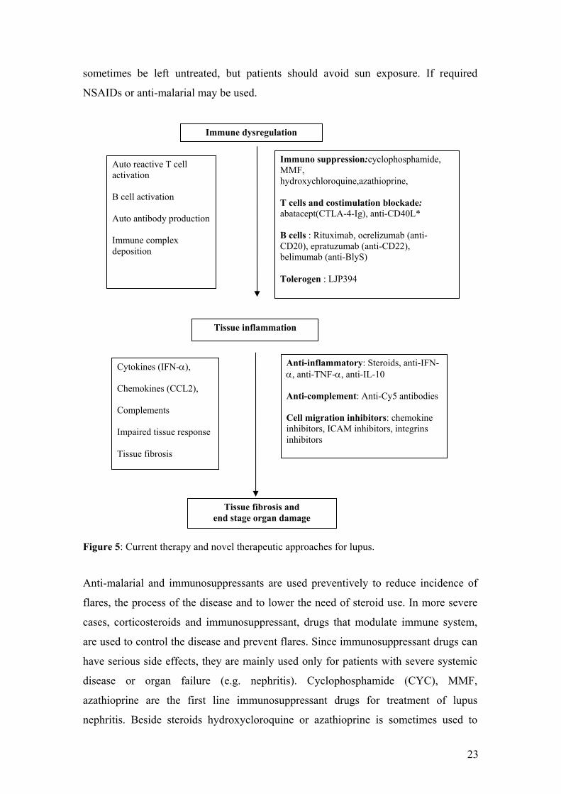
sometimes be left untreated, but patients should avoid sun exposure. If required
NSAIDs or anti-malarial may be used.
Immune dysregulation
Figure 5: Current therapy and novel therapeutic approaches for lupus.
Anti-malarial and immunosuppressants are used preventively to reduce incidence of
flares, the process of the disease and to lower the need of steroid use. In more severe
cases, corticosteroids and immunosuppressant, drugs that modulate immune system,
are used to control the disease and prevent flares. Since immunosuppressant drugs can
have serious side effects, they are mainly used only for patients with severe systemic
disease or organ failure (e.g. nephritis). Cyclophosphamide (CYC), MMF,
azathioprine are the first line immunosuppressant drugs for treatment of lupus
nephritis. Beside steroids hydroxycloroquine or azathioprine is sometimes used to
Tissue inflammation
Immuno suppression:cyclophosphamide, MMF, hydroxychloroquine,azathioprine, T cells and costimulation blockade: abatacept(CTLA-4-Ig), anti-CD40L* B cells : Rituximab, ocrelizumab (anti-CD20), epratuzumab (anti-CD22), belimumab (anti-BlyS) Tolerogen : LJP394
Auto reactive T cell activation B cell activation Auto antibody production Immune complex deposition
Anti-inflammatory: Steroids, anti-IFN-, anti-TNF-, anti-IL-10 Anti-complement: Anti-Cy5 antibodies Cell migration inhibitors: chemokine inhibitors, ICAM inhibitors, integrins inhibitors
Cytokines (IFN-), Chemokines (CCL2), Complements Impaired tissue response Tissue fibrosis
Tissue fibrosis and end stage organ damage
23

control lupus during pregnancy since these drugs appear to have fewer risks to the
foetus than other drugs.
Lupus patients are more susceptible to infections, since both lupus and its treatments,
in particular corticosteroids and immunosuppressive drugs, affect the immune system.
Infections can induce lupus flare, which further increases the risk of infection. Finally
lupus appears to increase the risk of cancer, especially non-Hodgkin’s lymphoma,
which affects the lymphoid organs. Immunosuppressive drugs used to treat lupus can
also enhance the risk of cancer. Thus the main goal of research is to find drugs to treat
lupus more specifically, without systemically suppressing the immune system.
Treatments, that are more specific in modifying particular subsets of immune cells
(e.g. T/B cells), or the activity of cytokine they secret have been gaining attention.
Biological agents targeting B cells are under development including a fully humanised
anti-CD20 antibody (Rituximab), an anti-CD22 antibody (Epratuzumab) and a number
of molecules blocking BlyS have been developed. The human anti-BlyS antibody
(Belimumab) is currently in a phase III study. BlyS (also called BAFF) is a soluble
mediator that plays a role in the regulation of B-cell homeostasis and differentiation.
The three known receptors for BlyS are differentially expressed at various stages of B-
cell and plasma cell development suggesting that the consequences of BlyS blockade
may differ from B-cell depletion by anti-CD20 treatment (Treml et al.2006).
A variety of co-stimulatory ligand/receptor pairs have been described that modulate
the interaction of dendritic cells and T cells, or T cells and B cells, respectively. The
first family of these to be discovered was the B7 family. CTLA-4-Ig is a drug that
blocks the interaction of CD80 and CD86 on antigen-presenting cells with CD28 on T
cells and has negative regulatory effects. CTLA-4-Ig has been approved for the
treatment of RA after recent successful RCTs (Todd et al.2007). It has also been
demonstrated to block nephritis in a mouse model of SLE (Cunnane et al. 2004) and
human studies are ongoing. There were 3 trials with anti-CD40L mAb IDEC-131,
enrolling in global 110 lupus patients, although not all had with nephritis (Huang et
al.2002, Davis Jr et al.2001) and one with BG9588 which included 28 patients with
active proliferative lupus nephritis (Boumpas et al. 2003). In summary the results
failed to demonstrate significant drug benefit compared to placebo in terms of time to
renal flare, SLE disease activity index (SLEDAI) and serologic markers (complement
and anti-DNA Ab) (Kalunian et al.2002). The study with BG9588 was prematurely
24

terminated due to the increased occurrence of thromboembolic events despite
concurrent prophylactic anticoagulation (Boumpas et al.2003, Kawai et al.2000).
A very different approach is to try modulating the autoimmune process in SLE in an
antigen-specific way. Abetimus is a molecule composed of a series of linked
oligonucleotides. The concept behind Abetimus is that the drug will block the binding
of anti dsDNA antibodies to their autoimmune targets and/ or will tolerise B cell with
antigen-specificity for DNA (Alarcon-Segovia et al.2003). Results in clinical trials
have been very modest so far. Edratide represents a similar approach. Edratide is a
peptide derived from the antigen-binding region of a human monoclonal anti-dsDNA
antibody (Sharabi et al. 2007). It has been proposed that this molecule can modulate
the function of DNA-reactive B cells through idiotype–anti-idiotype interactions.
Even though T cells and B cells targets seem to promising, clinical trail results for
these therapies do not look encouraging (see table). This leaves scope for the
development of new strategies to encounter lupus nephritis.
Table 3: SLE trail data for new biological therapies
Target Treatment In mouse mdoel In humans T cells CD28/B7 costimulation blockade
Abatacept Effective (Ramanujam M. et al. 2004)
End points not met (Westhovens R. et al. 2008)
CD40 and CD40-L BJ9588 Anti-CD154
Effective (Wang X. et al. 2003)
Trial stopped for side effects (Boumpas DT. et al. 2003) End points not met (Kalunian KC. et al. 2002)
ICOS and ICOS-L Anti-ICOS L antibody Effective (Iwai H et al. 2003)
B cells CD22 Epratuzumab Effective
(Dorner T. et al. 2006) CD20 Rituximab End points not met
(Explorer Trail) Anti metabolite MMF End points not met
(ALMS study) Multivalent DNA
Abetimus End points not met (Cardiel MH. et al. 2008)
Anti-BAFF Belimumab End points not met (Wallace DJ. et al. 2009)
Targeting CCL2 in lupus
As we have reviewed in early part of this introduction CCL2 presents an exciting
target to attenuate lupus nephritis particularly in controlling the inflammatory cascade
of the lupus pathogenesis. To block CCL2 and its receptor (CCR2) signalling pathway
Shimizu et al. (2004) used the strategy of anti-CCL2 gene therapy involving the
25
transfection of a mutant human CCL2 gene into skeletal muscle. This mutant human
CCL2, designated 7ND, lacks NH2-terminal amino acids 2–8 and has been shown to
work as a dominant-negative inhibitor of human CCL2. Histological findings of
kidneys in treated mice with 7ND showed that protection against renal injury resulted
from reduced infiltration of leucocytes. 7ND gene therapy was shown to prolong the
life span of MRLlpr/lpr mice (Shimizu S et al 2004). Another group (Hagesawa et al.
2003) injected CCL2 antagonist- or TARC antagonist-transfected MRL/N-1 cells
subcutaneously into MRLlpr/lpr mice ages 7 weeks (before the onset of lupus nephritis)
and 12 weeks (at the early stage of the disease). After 8 weeks, mice bearing the CCL2
antagonist showed markedly diminished infiltration of macrophages and T cells,
glomerular hypercellularity, glomerulosclerosis, crescent formation, and vasculitis
compared with control mice. Authors concluded that the effect was due to decreased
production of interferon- and IL-2 in the kidney. However, such experimental
approaches cannot be used in humans because of irrepressible antagonist production
and tumor formation (Shimizu S et al.2004, Hagesawa et al.2003).
Following these impressive findings we thought of targeting CCL2 blockade with a
clinically applicable approach. Spiegelmer technology based aptamers rose to the
occasion as these are safe, biostable and specifically binding to the target.
1.2. Spiegelmers: Next generation aptamers
To achieve CCL2 antagonism we used a RNA-aptamers (Spiegelmer), a patented
technology of NOXXON Pharma (Berlin). RNA-aptamer binds to the active site of
target chemokine and makes them biologically non-functional.
An aptamer is a nucleic acid structure that can bind to a target molecule conceptually
similar to an antibody that recognizes an antigen. Aptamers have binding
characteristics similar to peptides or antibodies, with affinities in the low nanomolar to
the picomolar range. However, there are several drawbacks to aptamers as useful
therapeutic products. As relatively small molecules, aptamers demonstrate circulating
half-lives in vivo in the order of minutes. This situation can be addressed by attaching
large inert molecules to aptamers (e.g. polyethylene glycol) to reduce their elimination
via the kidney and increase their presence in the circulation. Still, aptamers, as natural
nucleic acid polymers, are prone to rapid degradation by nucleases that are present in
all tissues in the body.
26

Spiegelmers are biostable aptamers, that have all of the diverse characteristics of
aptamers and possess a structure that prevents enzymatic degradation. While
conventional aptamers are created from the natural D-nucleotides, which are
recognized by the nucleic acid degrading enzymes, Spiegelmers are synthesized from
mirror image L-oligonucleotide and hence they can not be degraded by any nucleases
since there are no such enzymes in the body capable of interacting with these
unnatural molecules. Spiegelmer technology is based on the simple concept that if an
aptamer binds its natural target, the mirror image of the aptamer will identically bind
the mirror image of the natural target (Figure 6). The process of aptamer selection is
carried out against the mirror image target protein; an aptamer against this unnatural
mirror image is obtained. More important, this Spiegelmer is now resistant to nuclease
degradation. Spiegelmers should not be confused with antisense RNAs in that they do
not directly interfere with transcription or translation of their target molecules. They
are designed to bind specifically to extracellular proteins, either a receptor or its
ligand, similar to the behavior of a monoclonal antibody, aptamer or peptide.
Spiegelmers appear to be non-immunogenic, even under the most inductive conditions
for antibody formation in rabbits. These molecules are termed “Spiegelmer” from the
German word “Spiegel” meaning “mirror”.
Spiegelmers possess the high affinity binding characteristics of the best aptamers and
antibodies in the low nanomolar and picomolar range, while defying enzymatic
degradation that severely limits the utility of aptamers (Klussmann S. et al.1996). Data
indicate that Spiegelmers are stable in human plasma for over 60 hours at 37 °C
(Figure 7.) while non-modified RNA aptamers are degraded in seconds under the same
conditions (left panel, Figure. 7). Results in animals indicate that a similar stability can
be expected in vivo as well
27

Figure 6: Representation of Spiegelmer generation. L-enantiomer of the target is processed to get the selective binding D-aptamer. Highly selective D-aptamer for mirror image (L-target) is then amplified and then mirrored to get L-aptamer which has the selective binding property for natural D-target. (Taken from www.noxxon.net)
Figure 7: Representation of Spiegelmer stability. Left panel with D-enantiomeric RNA which degrades in seconds when incubated in human plasma at 37°C., while L-enantiomeric RNA is stable even at 60 hrs of incubation in human plasma (right panel)
28

Summary of Spiegelmer Properties
• high binding specificity to their target
• comparable binding affinity as antibodies (low nanomolar-picomolar)
• stable in human plasma and after injection into animals
• non-immunogenic
• low toxicity (target-based)
• synthesized using standard chemistry (scalable)
• no biological contaminants
• easy to formulate (polar) with excellent solubility
29
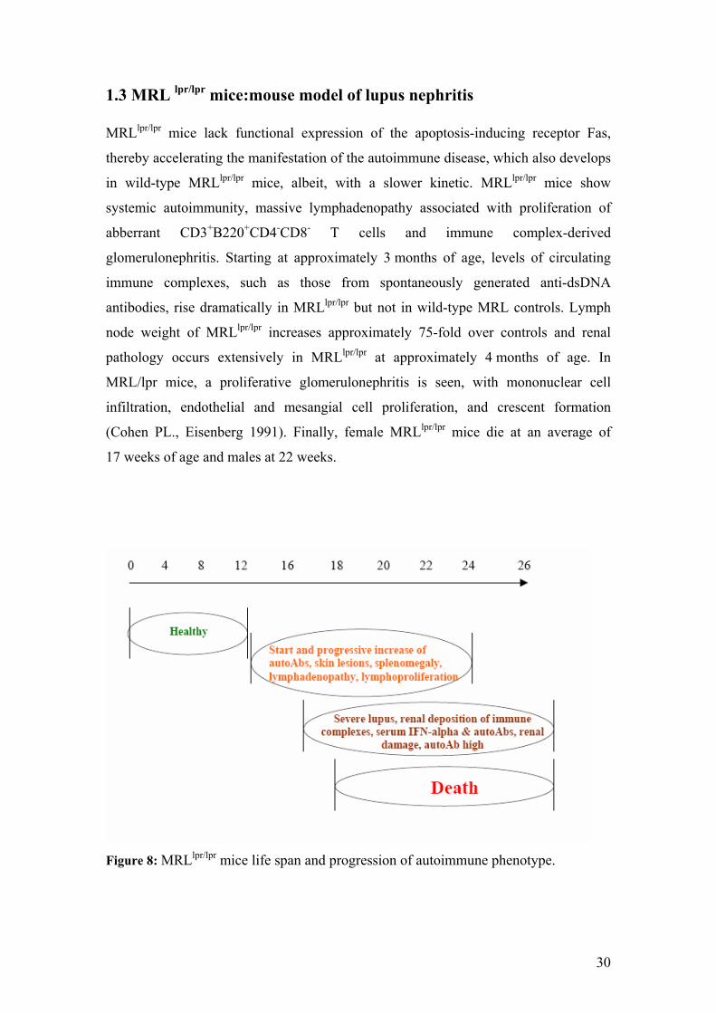
1.3 MRL lpr/lpr mice:mouse model of lupus nephritis MRLlpr/lpr mice lack functional expression of the apoptosis-inducing receptor Fas,
thereby accelerating the manifestation of the autoimmune disease, which also develops
in wild-type MRLlpr/lpr mice, albeit, with a slower kinetic. MRLlpr/lpr mice show
systemic autoimmunity, massive lymphadenopathy associated with proliferation of
abberrant CD3+B220+CD4-CD8- T cells and immune complex-derived
glomerulonephritis. Starting at approximately 3 months of age, levels of circulating
immune complexes, such as those from spontaneously generated anti-dsDNA
antibodies, rise dramatically in MRLlpr/lpr but not in wild-type MRL controls. Lymph
node weight of MRLlpr/lpr increases approximately 75-fold over controls and renal
pathology occurs extensively in MRLlpr/lpr at approximately 4 months of age. In
MRL/lpr mice, a proliferative glomerulonephritis is seen, with mononuclear cell
infiltration, endothelial and mesangial cell proliferation, and crescent formation
(Cohen PL., Eisenberg 1991). Finally, female MRLlpr/lpr mice die at an average of
17 weeks of age and males at 22 weeks.
Figure 8: MRLlpr/lpr mice life span and progression of autoimmune phenotype.
30

2. Research hypothesis/objectives
1. The role of CCL2/CCR2 axis in chronic inflammatory conditions is well
known. But so far there is no therapy in the clinics which attenuate the inflammation
mediated by CCL2, even though lots of efforts are being made. Gene therapy against
CCL2 can suppress nephritis. in murine model of lupus nephritis (Hagesawa et
al.2004). But these kinds of approaches are not clinically applicable, as these
represent irreversible antagonism and have been reported for side effects.
Considering these facts in mind our first ojective was to evaluate the efficacy of an
anti-CCL2 Spiegelmer (mNOX-E36).
2. Second objective was to compare the efficacy of Anti-CCL2 Spiegelmer
(mNOX-E36) with that of cyclophosphamide and MMF.
3. Immunosuppressants like CYC and MMF effectively control rapidly
progressing lupus. But with high exposure of immunotherapies there is always a
possiblity of adverse effects. In clinical practice lots of patients experienced
infections, gonadal toxicities and other side effects which are even fatal in some
cases. That is why lots of efforts are being made to discover novel ways to combat
lupus nephritis. T cell and B cells based therapies did not show any promising
results. Considering all these findings we thought of combining anti-CCL2
Spiegelmer with low dose of CYC and then comparing it high dose of CYC for its
efficacy as well as safety.
31

3. Materials and Methods
3.1 Materials
Equipments
Balances:
Analytic Balance, BP 110 S Sartorius, Göttingen, Germany
Mettler PJ 3000 Mettler-Toledo, Greifensee,Switzerland
Cell Incubators:
Type B5060 EC-CO2 Heraeus Sepatech, München, Germany
Centrifuges:
Heraeus, Minifuge T VWR Internationl, Darmstadt, Germany
Heraeus, Biofuge primo Kendro Laboratory Products GmbH,
Hanau, Germany
Heraeus, Sepatech Biofuge A Heraeus Sepatech, München, Germany
ELISA-Reader
Tecan, GENios Plus Tecan, Crailsheim, Germany
Fluorescence Microsocopes
Leica DC 300F Leica Microsystems, Cambridge, UK
Olympus BX50 Olympus Microscopy, Hamburg,
Germany
Spectrophotometer
Beckman DU® 530 Beckman Coulter, Fullerton, CA, USA
TaqMan Sequence Detection System
ABI prism ™ 7700 sequence
detector PE Biosystems, Weiterstadt, Germany
Other Equipments
Cryostat RM2155 Leica Microsystems, Bensheim,
Germany
Cryostat CM 3000 Leica Microsystems, Bensheim,
Germany
Homogenizer ULTRA-TURRAX
T25 IKA GmbH, Staufen, Germany
Microtome HM 340E Microm, Heidelberg, Germany
32

pH meter WTW WTW GmbH, Weilheim, Germany
Thermomixer 5436 Eppendorf, Hamburg, Germany
Vortex Genie 2™ Bender&Hobein AG, Zurich,
Switzerland
Water bath HI 1210 Leica Microsystems, Bensheim,
Germany
Chemicals and materials
Chemicals for the molecular biology techniques
RNeasy Mini Kit Qiagen GmbH, Hilden, Germany
RT-PCR primers PE Biosystems, Weiterstadt, Germany
Cell culture
DMEM-medium Biochrom KG, Berlin, Germany
RPMI-1640 medium GIBCO/Invitrogen, Paisley, Scotland, UK
FSC Biochrom KG, Berlin, Germany
Dulbecco’s PBS (1×) PAA Laboratories GmbH,
Cölbe,Germany
Trypsine/EDTA (1×) PAA Laboratories GmbH,
Cölbe,Germany
Penicillin/Streptomycin (100×) PAA Laboratories GmbH,
Cölbe,Germany
Antibodies
rat anti-Mac2 Cederlane, Ontario, Canada
anti-CD3 BD Pharmingen, Hamburg, Germany
anti-CD4 BD Pharmingen, Hamburg, Germany
anti-CD8 BD Pharmingen, Hamburg, Germany
anti-CD25 BD Pharmingen, Hamburg, Germany
anti-CD45 BD Pharmingen, Hamburg, Germany
anti-7/4 Abd-Serotec,
anti-Ly6G BD Pharmingen, Hamburg, Germany
33
Miscellaneous
Needles BD Drogheda, Ireland
Pipette’s tip 1-1000μL Eppendorf, Hamburg, Germany
Plastic histosettes NeoLab, Heidelberg, Germany
Preseparation filters Miltenyi Biotec, Bergish Gladbach,
Germany
SuperFrost Plus
microscope slides Menzel-Gläser, Braunschweig,
Germany
Silver Impregnation Kit Bio-Optica, Milano, Italy
Syringes Becton Dickinson GmbH, Germany
Chemicals
Aceton Merck, Darmstadt, Germany
AEC Substrat Packung Biogenex, San Ramon, USA
Ether Merck, Darmstadt, Germany
Bovines Serum Albumin Roche Diagnostics, Mannheim,Germany
Cyclophosphamide Sigma-Aldrich Chemicals,
Steinheim,Germany
DEPC Fluka, Buchs, Switzerland
DMSO Merck, Darmstadt, Germany
EDTA Calbiochem, SanDiego, USA
Ethanol Merck, Darmstadt, Germany
Formalin Merck, Darmstadt, Germany
Hydroxyethyl cellulose Sigma-Aldrich, Steinheim, Germany
HCl (5N) Merck, Darmstadt, Germany
Isopropanol Merck, Darmstadt, Germany
Kaliumchlorid Merck, Darmstadt, Germany
Kaliumdihydrogenphosphat Merck, Darmstadt, Germany
Kaliumhydroxid Merck, Darmstadt, Germany
MACS-Puffer Miltenyi Biotec, Bergisch Gladbach,
Germany
Merkaptoethanol Roth, Karlsruhe, Germany
34

Natriumacetat Merck, Darmstadt, Germany
Natriumchlorid Merck, Darmstadt, Germany
Natriumcitrat Merck, Darmstadt, Germany
Natriumdihydrogenphosphat Merck, Darmstadt, Germany
Penicillin Sigma, Deisenhofen, Germany
Roti-Aqua-Phenol Carl Roth GmbH, Karlsruhe, Germany
SSC (Saline-sodium citrate Puffer) Sigma, Deisenhofen, Germany
Tissue Freezing Medium Leica, Nussloch, Germany
Trypan Blue Sigma, Deisenhofen, Germany
Oxygenated water DAKO, Hamburg, Germany
Xylol Merck, Darmstadt, Germany
35
3.2. Methods
3.2.1 Methods Part I
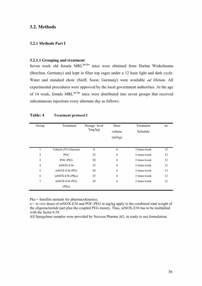
3.2.1.1 Grouping and treatment
Seven week old female MRLlpr/lpr mice were obtained from Harlan Winkelmann
(Borchen, Germany) and kept in filter top cages under a 12 hour light and dark cycle.
Water and standard chow (Sniff, Soest, Germany) were available ad libitum. All
experimental procedures were approved by the local government authorities. At the age
of 14 week, female MRLlpr/lpr mice were distributed into seven groups that received
subcutaneous injections every alternate day as follows:
Table: 4 Treatment protocol I
Group Treatment Dosage level a(mg/kg)
Dose
volume
(ml/kg)
Treatment
Schedule
no
1 Vehicle (5% Glucose) 0 4 3 times/week 12
2 POC 25 4 3 times/week 12
3 POC-PEG 50 4 3 times/week 12
4 mNOX-E36 25 4 3 times/week 12
5 mNOX-E36-PEG 50 4 3 times/week 12
6 mNOX-E36 (PKa) 25 4 3 times/week 12
7 mNOX-E36-PEG
(PKa)
50 4 3 times/week 12
Pka = Satellite animals for pharmacokinetics; a = in vivo doses of mNOX-E36 and POC-PEG in mg/kg apply to the combined total weight of the oligonucleotide part plus the coupled PEG moiety. Thus, mNOX-E36 has to be multiplied with the factor 0.29. All Spiegelmer samples were provided by Noxxon Pharma AG, in ready to use formulation.
36

3.2.1.2 Route and rationale of test material administration
The chosen route of administration was subcutaneous, as the animals tolerate a large
number of subcutaneous administrations better than numerous intraperitoneal
injections, handling is easier and because by this route unPEGylated Spiegelmer is
present longer in the circulation.
3.2.1.3 Test substance and formulation
Spiegelmer Sequence
mNOX-E36 5`-GGCGACAUUG GUUGGGCAUG
AGGCGAGGCC CUUUGAUGAA
UCCGCGGCCA-3`
mNOX-E36-PEG 40 kD PEG
5`-GGCGACAUUG GUUGGGCAUG
AGGCGAGGCC CUUUGAUGAA
UCCGCGGCCA-3`
Control Spiegelmer Sequence
POC 5`-UAAGGAAACU CGGUCUGAUG
CGGUAGCGCU GUGCAGAGCU-3`
POC-PEG 40 kD PEG
5`-UAAGGAAACU CGGUCUGAUG
CGGUAGCGCU GUGCAGAGCU-3`
For in vivo application, the mNOX-E36 and the non-functional control Spiegelmer
POC were used non-modified or modified with 40-kD PEG at the 3` and 5` terminus,
respectively. The test substance (Spiegelmer solution) was dissolved in isotonic 5 %
glucose solution
3.2.1.4 Mortality, clinical signs, skin score and body weight
Mortality was recorded every week throughout the study. Animals were observed daily
for clinical signs. In addition, MRLlpr/lpr mice of all groups were checked daily for
cutaneous lupus manifestations, which typically occur in the facial or neck area. The
severity was graded from 0 – 0.5 (no lesion), 1 (mild lesion), 2 (moderate) to 3 (severe)
After 10 weeks of treatment, satellite animals for pharmacokinetics were kept alive in
order to monitor survival rate. Body weight was recorded once a week during weeks 14
to 24 of age (10 weeks of treatment).
37

3.2.1.5 Pharmacokinetic analysis
In week 1, 5 and 10 of treatment, blood samples for pharmacokinetic analysis were
collected in satellite animals at the following times:
- mNOX-E36 (25 mg/kg) at 0.083, 0.33, 0.66, 1, 3, 9, 12 and 24 hours after injection;
- mNOX-E36-P (50 mg/kg) at 3, 6, 9, 12, 24, 30, 36 and 48 hours after injection.
To correlate the pharmacokinetic alteration with the progression of the disease,
additional samples were taken 3 hours post administration for unPEGylated mNOX-
E36 (25 mg/kg) and 24 hours post administration for mNOX-E36 (50 mg/kg), after the
first dose of the week. All samples were stored at –20 ºC until analysed by method
described below.
For immobilization of the Spiegelmer an L-RNA capture probe (CP) which is
complemantary (base-pairing) to one end of the analyte Spiegelmer is covalently
coupled to a 96-well plate. After hybridization (base-pairing) of the analyte Spiegelmer
to this capture probe, a second (biotinylated) L-RNA probe ("detect probe", DP) is
hybridized (base-pairing) to the second end of the analyte. After unbound complexes
have been removed, the complex of Spiegelmer and detect probe is detected by a
streptavidine/alkaline phosphatase conjugate converting a chemiluminescence
substrate.
3.2.1.6 Glomerular filtration rate
Preparation of 5% FITC-inulin solution
5% FITC-inulin was dissolved in two ml of 0.9% NaCl -- facilitated by heating the
solution in boiling water.
Intravenous injection and blood collection
Mice were anesthetized using Isoflurane, which for approximately 20 seconds. 5%
FITC-inulin (3.74μl /g body weight) was injected retroorbitally under anesthesia within
10 seconds. Under general anaesthesia, blood was drawn from the retro orbital plexus
at 5, 10, 15, 20, 35, 60 and 90 minutes post administration.
Determination of fluorescence of the sampled plasma
Since pH significantly affects FITC fluorescence value, each plasma sample was
buffered to pH 7.4, by mixing 10 μl of plasma with 40 μl of 500 mM HEPES (pH 7.4).
The titrated samples were then loaded onto a 96-well plate, 50μl sample/well.
Fluorescence was determined with 485 nm excitation, and read at 538 nm emission.
38
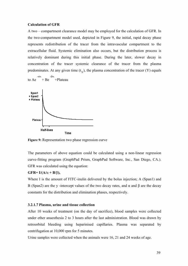
Calculation of GFR
A two – compartment clearance model may be employed for the calculation of GFR. In
the two-compartment model used, depicted in Figure 9, the initial, rapid decay phase
represents redistribution of the tracer from the intravascular compartment to the
extracellular fluid. Systemic elimination also occurs, but the distribution process is
relatively dominant during this initial phase. During the later, slower decay in
concentration of the tracer systemic clearance of the tracer from the plasma
predominates. At any given time (tX
), the plasma concentration of the tracer (Y) equals
to Ae -αtx
+ Be -βtx
+Plateau
Figure 9: Representation two phase regression curve
The parameters of above equation could be calculated using a non-linear regression
curve-fitting program (GraphPad Prism, GraphPad Software, Inc., San Diego, CA.).
GFR was calculated using the equation:
GFR= I/(A/α + B/β),
Where I is the amount of FITC-inulin delivered by the bolus injection; A (Span1) and
B (Span2) are the y -intercept values of the two decay rates, and α and β are the decay
constants for the distribution and elimination phases, respectively.
3.2.1.7 Plasma, urine and tissue collection
After 10 weeks of treatment (on the day of sacrifice), blood samples were collected
under ether anaesthesia 2 to 3 hours after the last administration. Blood was drawn by
retroorbital bleeding using heparinised capillaries. Plasma was separated by
centrifugation at 10,000 rpm for 5 minutes.
Urine samples were collected when the animals were 16, 21 and 24 weeks of age.
39

At study termination (at the end of the 10-week treatment period), organ weights of
spleen and lymph nodes were determined. Spleen, lymph nodes, kidney, lung, liver,
and salivary gland tissue samples were collected from each mouse for histology, RNA
analysis.
3.2.1.8 Histology of kidney and lungs
Morphological and histological processing
From all mice, the tissues were isolated from kidneys or spleens and placed in plastic
histocassettes and immediately fixed in 10% buffered formalin (formaldehyde in PBS)
for overnight and next day processed, with automatic tissue-processor (Thermo-
Shandon), which processes tissues in following manner- 70% ethanol (for 5 hours), 96
% ethanol (2 hrs), 100 % ethanol 3.5 hrs, xylene (2.5 hrs), paraffin (4 hrs). Then the
histocassettes were taken out and the paraffin blocks were prepared with hot liquid
paraffin using a machine (Microm- EC 350). The prepared blocks were allowed to cool
down and later separated from the steel holders. These blocks were later used for
making fine sections for different stainings (PAS or immunohisto). The fine sections
(5μm) were prepared using the microtome (Microm- HM340E) and mounted gently on
a glass frosted slide. Such slides were later allowed to dry for 2 hours in an oven at
50°C temperature. Deparaffinisation followed by dehydration was carried out by
incubating the sections in xylene, 100% absolute ethanol, 95%, 80% and 50% ethanol
followed by rinsing with PBS (2 changes, 3 minutes each). 5 μm sections for silver and
periodic acid-Schiff stains were prepared following routine protocols for
histopathology. From each mouse tissue collected as mentioned above, 2 μM thick
paraffin-embedded sections were cut and processed for immunohistochemical staining
performed on paraffin-embedded sections.
Analysis of glomerulonephritis
The severity of the renal lesions was graded using the indices for activity and
chronicity as described for human lupus nephritis (Austin et al, 1984). Activity index
was considered as the sum of individual scores of the following items considered to
represent measures of adaptive lupus nephritis: glomerular proliferation, leucocyte
exudation, karyorrhexis/fibrinoid necrosis (X2), cellular crescents (X2), hyaline
deposits, and interstitial inflammation. The maximum score was 24 points for activity
index. Chronicity index was considered to be the sum of individual scores of the
following items considered to represent measures of chronic irreversible lupus
40
nephritis: glomerular sclerosis, fibrous crescents, tubular atrophy and interstitial
fibrosis. The maximum score was 12 points for the chronicity index.
Immunostaining
For immunostaining sections of formalin-fixed and paraffin-embedded tissues were
dewaxed and rehydrated. Endogenous peroxidase was blocked by 3 % hydrogen
peroxide and antigen retrieval was performed in Antigen Retrieval Solution (Vector,
Burlingame, CA USA) in an autoclave oven. Biotin was blocked using the Avidin /
Biotin blocking kit (Vector, Burlingame, CA USA). Slides were incubated with the
primary antibodies for one hour, followed by biotinylated secondary antibodies (anti-
rat IgG, Vector, Burlingame, CA USA), and the ABC reagent (Vector, Burlingame, CA
USA). Slides were washed in phosphate buffered saline between the incubation steps.
3’3’Diaminobenzidine (DAB, Sigma, Taufkirchen, Germany) with metal enhancement
was used as detection system, resulting in a black coloured product. Methyl green was
used as counterstain, slides were dehydrated and mounted in Histomount (Zymed
Laboratories, San Francisco, CA USA). The following primary antibodies were used:
rat anti-MAC-2 (macrophages, Cederlane, Ontario, Canada, 1:50), anti-mouse CD3
(1:100, clone 500A2, BD), anti-mouse IgG1 (1:100, M32015, Caltag Laboratories,
Burlingame, CA USA), anti-mouse IgG2a (1:100, M32215, Caltag Laboratories,
Burlingame, CA USA). Negative controls included incubation with a respective isotype
antibody. For quantitative analysis glomerular cells were counted in 15 cortical
glomeruli per section. Glomerular Ig deposits were scored from 0 – 3 on 15 cortical
glomerular sections. The indices for interstitial volume, interstitial collagen deposition,
tubular cell damage, and tubular dilatation were determined by superposing a grid
containing 100 (10 x 10) sampling points on photographs of 10 nonoverlapping cortical
fields of silver-stained tissue (x 20) of each kidney.
3.2.1.9 Biochemical analysis of plasma and urine samples
Urine albumin (Bethyl Laboratories Inc, Montgomery, TX USA) and creatinine were
determined using diagnostic kits. Plasma samples were analysed for cytokines (BD
Pharmingen kits for IL12p40, IL6 and CCL2) and immunoglobulin (Bethyl
Laboratories Inc, Montgomery, USA) using ELISA kits. Plasma creatinine, albumin,
urea nitrogen and cholesterol were analysed by SynLab GmbH & Co. KG, Augsburg,
Germany.
41

Analysis of plasma autoantibodies
Serum dsDNA autoantibodies were determined by using commercial ELISA kits
(Bethyl Labs, Montgomery, TX, USA) using the following antibodies: anti-mouse
IgG1 and IgG2a following the manufacturer’s protocol with some modifications as
follows:
1) Sample wells in NUNC maxisorp 96 well flat bottom ELISA plate were coated
with 50 μg/ml (dilute in PBS) of the aqueous solution of 100 μl of Poly-L-Lysine
(Trevigen, Gaithersburg, MD, USA) for 1 hour at room temperature.
2) Washed once with washing buffer, tap dried on blotting paper and coated the
sample wells with 1 μg/ml of dsDNA, which was diluted in SSC buffer (1X), pH 7 .
3) For standard wells, capture antibody in coating buffer was added as described in
the protocol.
4) The plate was incubated overnight at 4°C.
5) Next day the plate was washed with the wash buffer 3 times and then blocked
with blocking solution and the protocol was followed as provided by the bethyl labs
from this blocking step onwards.
6) Standard dilutions and samples were added in respective wells in required
dilution and incubated at room temperature for 2 hours.
7) Plate was washed 3 times and then secondary antibody at given dilution was
added as per the protocol and incubated for 1 hour at room temperature.
8) Plate was again washed as before and TMB substrate (1:1 mixture of substrate
A and B) was added in each well and incubated in dark for 5-20 minutes.
9) The reaction was stopped with 1 M H2SO4 and absorbance was measured at
450 nm.
Protocol for ELISAs (in brief)
1) The NUNC ELISA plate wells were captured overnight at 4°C with the capture
antibody in coating buffer.
2) Next day the plates were washed 3 times with the washing buffer as given in
protocol for 3 times and blocked with the blocking solution or assay diluent for 1 hour
or as specified.
3) Again the washings were repeated 3 times followed by addition of standards;
samples and sample diluent (blank) into the wells of tap dried plate and incubated at
RT for 2 hours
42

4) This was followed by washings for 5 times or as specified
5) Then HRP/AP conjugated secondary antibody diluted in assay diluent was
added. Incubate the plate as specified.
6) The wells were washed again for 5-7 times or as specified and incubated with
the 100 μl of substrate A and B (1:1 mixture) for 25-30 min in dark to develop colour.
The reaction was stopped by addition of 100 μl 1 M H2SO4.
7) The reading of the absorbance was taken at 450 nm and the reference
wavelength was 620 nm using a spectrophotometer (TECAN-Genios Plus).
3.2.1.10 RNA isolation, cDNA synthesis and real time RT-PCR
Renal tissue from each mouse was snap frozen in liquid nitrogen and stored at -80 °C.
Real time RT-PCR was performed in pooled samples (5 animals from each group) on a
TaqMan ABI 7700 Sequence Detection System (PE Biosystems, Weiterstadt,
Germany) using a heat activated TaqDNA polymerase (Amplitaq Gold, PE
Biosystems). Controls comprising ddH2O were negative for target and housekeeper
genes. Primers and probes were from PE Biosystems, Weiterstadt, Germany.
Oligonucleotide primer (300 nM) and probes (100 nM) were used (primers and probes
used are mentioned below). From each animal total renal RNA preparation and reverse
transcription were performed as described.
Isolation of RNA from tissues
When animals were sacrificed on termination of the study, small parts of tissue from
each mouse were preserved in RNA-later and stored at -20 0C until processed for RNA
isolation. RNA isolation was carried out using RNA isolation kit from Qiagen
(Germany). In short, tissues (30 mg) preserved in RNA-later were homogenized using
blade homogenizer for 30 seconds at 14500 rpm in lysis buffer (600 µl) containing β-
mercaptoethanol (10 µl/ml). The homogenate was centrifuged at 15000 rpm for 3 min.
and 350 µl of supernatant was transferred to fresh DEPC-treated tube to this equal
amount (350 µl) of 70 % ethanol was added and whole mixture was loaded on RNA
column and processed for RNA isolation as per the manufacturer’s instruction. Isolated
RNA was stored at -80 °C until further used.
RNA quantification and purity check
For quantification isolated RNA samples were diluted in DEPC water (2 μl of RNA +
98 μl of DEPC water, 50 times dilution) and absorbance was measured at two
wavelengths as 260 nm and 280 nm.
43

Amount of RNA (μg/μl) = O.D. at 260 nm * 40 * 50 (dilution factor)/ 1000
The ratio of optical densities at 260 nm and 280 nm is an indicator for RNA purity
(indicative of protein contamination in the RNA samples). Only samples with a ratio of
1.8 or more were considered to be of acceptable quality.
RNA integrity check
Further quality check (if necessary) was performed using a denaturing RNA gel. In
short 2 % Agarose gel with Ethidium-bromide was casted, RNA samples were mixed
with RNA loading buffer (4:1 ratio) (Sigma) and were loaded on the gel.
Electrophoresis was carried out at constant volt (70-100 V) using MOBS running
buffer for 1 hour and the gel was read on a gel documentation apparatus under UV
lamp. RNA samples showing a single bright band were considered to be of good
quality. Loss of RNA integrity could be detected as smear formation in the agarose gel
(Figure 10).
Figure 10: Representative agarose gel for RNA integrity check.
cDNA synthesis and real-time RT-PCR
The RNA samples isolated according to the procedure detailed above were diluted in
DEPC water to a concentration of 1μg/20μl. A master mix was prepared with reagents
including 9 μl of 5x buffer (Invitrogen, Karlsruhe, Germany), 1μl of 25mM dNTP
mixture (Amersham Pharmacia Biotech, Freiburg, Germany), 2 μl of 0.1 M DTT
(Invitrogen, Karlsruhe, Germany), 1 μl of 40U/μl RNasin (Promega, Mannheim,
Germany), 0.5μl of 15μg/ml linear acrylamide (Ambion Ltd, Cambridgeshire, UK),
0.5μl of Hexanucleotide (Roche, Mannheim, Germany), 1μl of Superscript (Invitrogen,
Karlsruhe, Germany) or ddH2O in case of the controls. The master mix was made to a
volume of 15μl and added to 2μg/30μl RNA samples were taken in separate DEPC
44
treated microcentrifuge tubes, which were mixed and placed at 42 °C on a thermal
shaker incubator for 1 hour. After 1 hour the cDNA samples were collected and stored
at -20 °C until use for real-time RT-PCR analysis.
The cDNA samples prepared as described above were diluted 1:10 a dilution for the
real-time RT-PCR. The real-time RT-PCR was performed on a TaqMan® ABI Prism
7000 or 7700 (Applied Biosystems, Darmstadt, Germany). The quantitative PCR for
mRNA is based on the employment of sequencespecific primers and likewise
sequence-specific probes. The latter is tagged at both ends with a fluorescent molecule.
The quencher absorbs TAMRA (at the 3'-End) the fluorescence of the other reporter
tagged material such as FAM or VIC at the 5'-End. The TaqMan® universal PCR
master mix (Applied Biosystems, Darmstadt, Germany) contained Taq polymerase
possessing a 5' _ 3' polymerase activity and a 5' _ 3 ' exonuclease activity. During the
elongation phase of the PCR, specifically bound probe was hydrolyzed by the
exonuclease and the 5'-tag was set free. With every newly synthesized DNA strand
fluorescent tag material was set free and the resulting fluorescence was measured at
488nm. The resulting fluorescence signal is directly proportional to the quantity of
DNA synthesized. The CT value (Cycle Threshold value) was computed for each
sample. This is the cycle number, with which the reporter fluorescence signal breaks
through a user-defined threshold. The TaqMan® universal PCR master mix containing,
the forward primers and Reverse primers (final concentration of 300 nM) and the probe
(final concentration of 100 NM) was placed on ice. TaqMan® universal PCR master
mix contained PCR buffers, dNTPs and the AmpliTaqGold® previously mentioned
(Taq polymerase without 3' _ 5' exonuclease activity). 18μl of the mastermix was
pipetted into each well of a 96-well plate and 2μl of template (DNA dilution) was
added to each of these wells. The plate was sealed and centrifuged at 280 xg and
analyzed using TaqMan® ABI PRISM 7000 or 7700. For the TaqMan® RT-PCR the
following temperature settings were used: The first incubation was carried out for 2
minutes at 50°C followed by 95°C for 10 minutes so as to activate the polymerase.
Templates were amplified during 40 cycles each comprising 15 seconds incubation at
95°C followed by 1 minute incubation at 60°C. The RT-PCR for the housekeepers (18S
rRNA or GAPDH) was carried out under similar conditions. The CT values were
recorded using the ABI PRISM Sequence Detection software (version 1.0) and the
results were evaluated in relation the respective housekeepers. In all cases controls
consisting of ddH2O were negative for target and housekeeper genes. Oligonucleotide
45
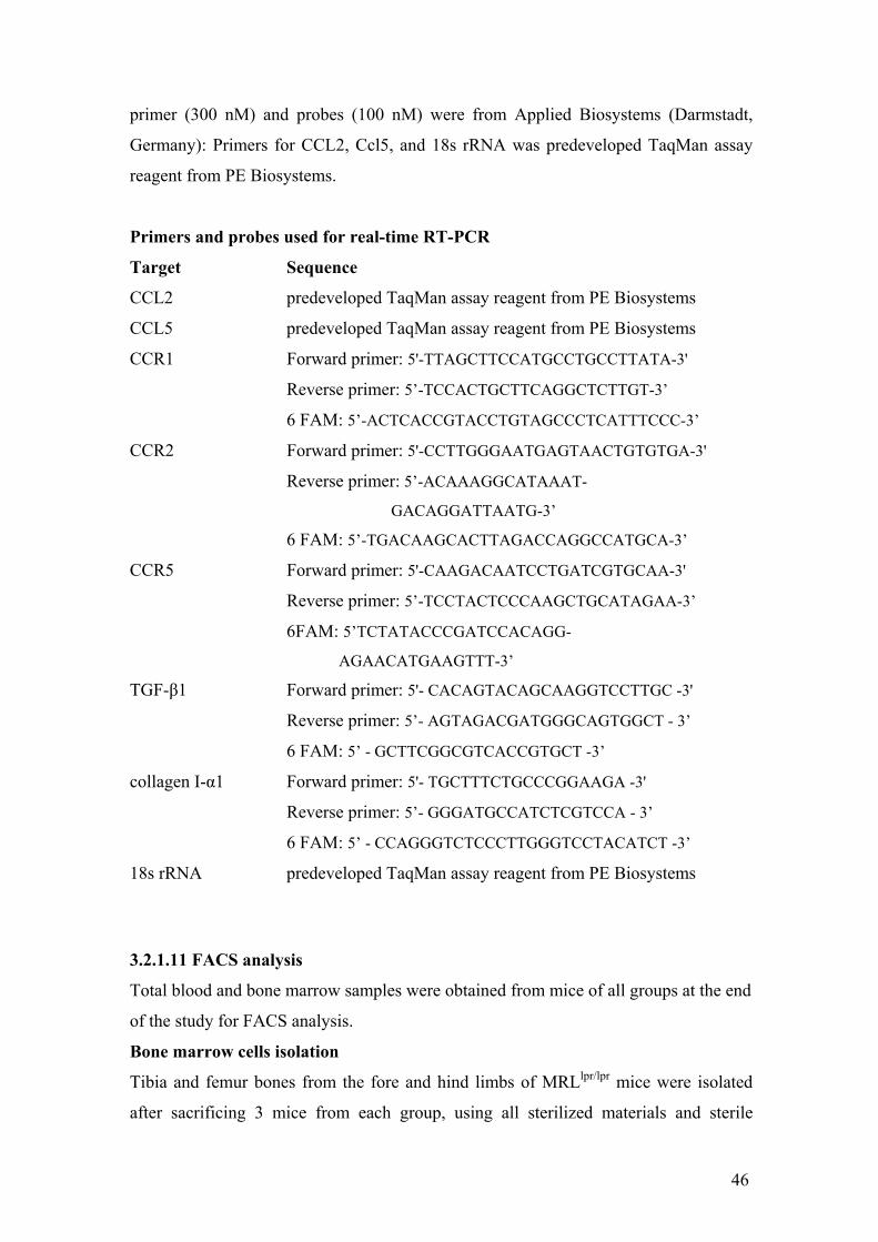
primer (300 nM) and probes (100 nM) were from Applied Biosystems (Darmstadt,
Germany): Primers for CCL2, Ccl5, and 18s rRNA was predeveloped TaqMan assay
reagent from PE Biosystems.
Primers and probes used for real-time RT-PCR
Target Sequence
CCL2 predeveloped TaqMan assay reagent from PE Biosystems
CCL5 predeveloped TaqMan assay reagent from PE Biosystems
CCR1 Forward primer: 5'-TTAGCTTCCATGCCTGCCTTATA-3'
Reverse primer: 5’-TCCACTGCTTCAGGCTCTTGT-3’
6 FAM: 5’-ACTCACCGTACCTGTAGCCCTCATTTCCC-3’
CCR2 Forward primer: 5'-CCTTGGGAATGAGTAACTGTGTGA-3'
Reverse primer: 5’-ACAAAGGCATAAAT-
GACAGGATTAATG-3’
6 FAM: 5’-TGACAAGCACTTAGACCAGGCCATGCA-3’
CCR5 Forward primer: 5'-CAAGACAATCCTGATCGTGCAA-3'
Reverse primer: 5’-TCCTACTCCCAAGCTGCATAGAA-3’
6FAM: 5’TCTATACCCGATCCACAGG-
AGAACATGAAGTTT-3’
TGF-β1 Forward primer: 5'- CACAGTACAGCAAGGTCCTTGC -3'
Reverse primer: 5’- AGTAGACGATGGGCAGTGGCT - 3’
6 FAM: 5’ - GCTTCGGCGTCACCGTGCT -3’
collagen I-α1 Forward primer: 5'- TGCTTTCTGCCCGGAAGA -3'
Reverse primer: 5’- GGGATGCCATCTCGTCCA - 3’
6 FAM: 5’ - CCAGGGTCTCCCTTGGGTCCTACATCT -3’
18s rRNA predeveloped TaqMan assay reagent from PE Biosystems
3.2.1.11 FACS analysis
Total blood and bone marrow samples were obtained from mice of all groups at the end
of the study for FACS analysis.
Bone marrow cells isolation
Tibia and femur bones from the fore and hind limbs of MRLlpr/lpr mice were isolated
after sacrificing 3 mice from each group, using all sterilized materials and sterile
46

conditions for the isolation in an aseptic hood with HEPA filters. Bones were freed
from the adhering tissues and both the ends of each bone were punctured with the 24
gauge needle and then the RPMI media was flushed (using sterile surgical syringe with
needle) through the bones to isolate bone marrow in a sterile petri plate containing the
2-3 ml of ice-cold media (RPMI+ 10 %FCS + 1 % PS) on ice. Then it was
homogenously resuspended using 1 ml pipette. This cell suspension was taken in a 15
ml Falcon tube using 1 ml pipette and centrifuged at 1600 rpm at 4°C for 6 min. and
washed with 1x PBS (cell culture grade) for 2 times. Then it was passed through the 40
μm sterile filters to obtain single cell suspension and centrifuged as before. The
supernatant was decanted and the pellet was resuspended in 4 ml of sterile 0.3M
NH4Cl and kept at room temperature for 5 min. Then it was centrifuged as above and
supernatant was decanted and pellet was resuspended in ice cold (cell culture use) PBS
and washed 2 times with PBS as before. The pellet was resuspended in RPMI media (1-
2 ml).
Whole blood collection
Blood sample was drawn after 2-3 hrs of last injection of Spiegelmer and vehicle in
respective groups in an ependrof tube containing EDTA.
Staining bone marrow and blood samples for FACS
Bone marrow the cells were resuspended to approximately 1-5x10⁶ cells/ml in ice cold
PBS, 10%FCS, 1% sodium azide. Use ice cold reagents/solutions and at 4oC as low
temperature and presence of sodium azide prevented the modulation and internalization
of surface antigens which could produce a loss of fluorescence intensity. 100µl of
whole blood sample was used for staining.
1) 100 μl of cell suspension /100µl of whole blood was added to each tube.
2) 0.1-10 μg/ml of the primary antibody (MC21, Mack et.al 2001) was added.
Dilutions for primary antibody were made in 3% BSA/PBS.
3) Incubated for at least 30 min at 4oC in the dark.
4) Samples were then washed 3-times by centrifugation at 400 g for 5 min and
resuspend them in ice cold PBS.
4) Biotinylated anti-rat IgG antibody (BD Biosciences GmbH, Heidelberg,
Germany) diluted in 3% BSA/PBS at the optimal dilution was added in the samples
(secondary antibody) and then resuspended the cells in this solution.
5) Incubated for at least 40 minutes at 4oC in the dark.
47

6) The cells were then washed 3-times by centrifugation at 400 g for 5 min and
resuspend them in ice cold PBS, 3% BSA, 1% sodium azide.
7) Then the cells were treated with optimum amount of streptavidin-PE.
8) Store the samples for 40 min at 4oC in the dark.
9) The cells were then washed 3-times by centrifugation at 400 g for 5 min and
resuspend them in ice cold PBS, 3% BSA, 1% sodium azide.
10) Bone marrow cells were ready for the analysis. Blood samples were then lysed
with BD lysis buffer (BD Biosciences). Followed by two washings before analysis.
FACS acquisition
Stained samples were gated for monocyte region according FSC and SSC plot. Gated
cells were then plotted against FL2 (for PE fluroscence).
3.2.1.12 Immunostimulatory effect of Spiegelmer (in vitro)
Isolation and preparation of murine bone marrow derived dendritic cells
(BMDCs)
Tibia and femur bones from the fore and hind limbs of MRLlpr/lpr mice were isolated
after sacrificing 4-5 mice, using all sterilized materials and sterile conditions for the
isolation in an aseptic hood with HEPA filters. Bones were freed from the adhering
tissues and both the ends of each bone were punctured with the 24 gauge needle and
then the RPMI media was flushed (using sterile surgical syringe with needle) through
the bones to isolate bone marrow in a sterile Petri plate containing the 2-3 ml of ice-
cold media (RPMI+ 10 %FCS + 1 % PS) on ice. Then it was homogenously
resuspended using 1 ml pipette. This cell suspension was taken in a 15 ml Falcon tube
using 1 ml pipette and centrifuged at 1600 rpm at 4°C for 6 min. and washed with 1x
PBS (cell culture grade) for 2 times. Then it was passed through the 40 μm sterile
filters to obtain single cell suspension and centrifuged as before. The supernatant was
decanted and the pellet was resuspended in 4 ml of sterile 0.3M NH4Cl and kept at
room temperature for 5 min. Then it was centrifuged as above and supernatant was
decanted and pellet was resuspended in ice cold (cell culture use) PBS and washed 2
times with PBS as before. The pellet was resuspended in RPMI media (2-4 ml) and the
no. of cells were counted using Neubar’s chamber under the microscope after suitable
dilution. Adequate no. of cells (5 million) in 12 well plates (NUNC cell culture plates),
each well containing 1.5 ml of complete RPMI media containing 100 ng/ml
recombinant murine Flt3 ligand (Immunotools, Friesoyth, Germany). The plates were
48

incubated at 37°C for 8 days to obtain a lineage of mixture of plasmacytoid dendritic
cells and conventional dendritic cells (pDC and cDC). After 4 days of incubation 1 ml
of fresh media (with 100 ng/ml Flt3 ligand) was added in each well. After 8 days of the
incubation, 2 ml of fresh media was replaced for the old media by careful removal only
from the top of each well.
Stimulation of BMDCs
Then the cells were stimulated with ligands or media as followed. 10 μg D- and L-
enantiomeric phosphorothioates RNA40 (TLR7 agonistic sequence), RNA41 and
RNA42 (control sequences), and various concentrations of mNOX-E36 (12.5, 25 and
50 μg/mL) and POC-PEG control (25 and 50 mg/mL) in 1,2-dioleoyel-3-trimethyl-
ammonium-propane (DOTAP; Roche, Mannheim, Germany.The plates were again
incubated for 24 hrs at 37°C. After incubation the supernatants were carefully isolated
in microcentrifuge tubes. The supernatants were separated from the residual amount of
cells by centrifuging it at 6000 rpm for 10 min. And immediately stored them at –20°C.
These supernatants were analyzed for the murine IFN- by (ELISA (PBL, Biomedical
Labs, and Piscataway, NJ USA)
3.2.1.13 In-situ hybridization (distribution of mNOX-E36)
The paraffin-embedded tissue sections were deparaffinised in glass coplin jars at room
temperature (RT) in used xylene, fresh xylene, used 100% ethanol, fresh 100% ethanol,
used 95%, 70%, 50% ethanol and phosphate buffered saline (PBS), respectively (1 5
min each). Sections were fixed again 20 min in 4% (w/v) paraformaldehyde in PBS at
RT and flushed in PBS. The tissue then was partially digested for 30 min with 20 g/ml
proteinase K in TE buffer at RT and flushed with 2 mg/ml glycine in PBS at RT and
PBS, respectively. Pre-hybridisation was carried out in a humidified container for 2 h at
40°C in pre-hybridisation buffer. Slides were washed once in pre-warmed 2 SSC for
5 min. The hybridisation with biotinylated L-RNA probes was performed in a
humidified container overnight with 0.2 M L-RNA probe in pre-hybridisation buffer
at 40°C.
The next day slides were washed three times for 15 min (1) with pre-warmed 2 SSC,
(2) with 2 SSC at RT, and (3) with TBS. Hybridised probes were detected with
streptavidin alkaline phosphatase 1:1000 in TBS for 2 h at RT. Slides were washed
again twice for 10 min (1) with TBS and (2) with 10 mM Tris/HCl, pH 9.8; 1 mM
MgCl2. 5-Bromo-4-chloro-3-indolyl phosphate/nitro blue tetrazolium (BCIP/NBT)
49

alkaline phosphatase substrate solution (Sigma B1911) was then added and slides were
incubated for 30-120 min at RT. The reaction was stopped with deionized H2O and
tissue sections were counterstained with Nuclear Fast Red (Sigma N3020) for 10 min at
RT. The tissue sections were then dehydrated and embedded in Canada balm or
Entellan (Merck) for long-term storage.
3.2.1.14 Statistical analysis
Statistics were done using GraphPad Prism (4.03 version). Data were expressed as
mean ± SEM. Data were analysed using unpaired two-tailed t-test for comparison
between two groups. One-way ANOVA followed by post-hoc Bonferroni`s test was
used for multiple comparisons. For nonparametric analysis of two groups Two-tailed
Mann Whitney U test were performed.
50

3.2.2 Methods Part II
3.2.2.1 Grouping and treatment
Seven week old female MRLlpr/lpr mice were obtained from Harlan Winkelmann
(Borchen, Germany) and kept in filter top cages under a 12 hour light and dark cycle.
Water and standard chow (Sniff, Soest, Germany) were available ad libitum. All
experimental procedures were approved by the local government authorities. At the age
of 14 week, female MRLlpr/lpr mice were distributed into seven groups that received
injections as follows:
Table:5 Treatment protocol II
Group Treatment Dosage level a
(mg/kg) Dose volume
(ml/kg)
Treatment
Schedule
no
1 Vehicle (5% Glucose) 0 4 3times/week 16
2 mNOX-E36-PEG 50 4 3times/week 12
3 revmNOX-E36-PEG
50
4
3times/week 16
4 CYC ¼ th full dose
30
4
once/ 4 weeks 16
5 CYC full dose 30 4 once weekly 12
6 mNOX-E36-PEG
plus
CYC ¼ th full dose
50
30
4
3times/week
once/4 weeks
12
7 MMF 100 4 once daily 12
a = in vivo doses of mNOX-E36 and revmNOX-E36-PEG in mg/kg apply to the combined total weight of the oligonucleotide part plus the coupled PEG moiety. Thus, mNOX-E36 has to be multiplied with the factor 0.29. All Spiegelmer samples were provided by Noxxon Pharma AG, in ready to use formulation. Cyclophsphamide (CYC) was procured from sigma Aldrich. Mycophenolate mofetil (MMF) was purchased from Roche (Manheim, Germany)
3.2.2.2 Route and rationale of test material administration
The chosen route of administration was subcutaneous, as the animals tolerate a large
number of subcutaneous administrations better than numerous intraperitoneal
injections, handling is easier and because by this route unPEGylated Spiegelmer is
present longer in the circulation. So mNOX-E36 and controls were administered
51

subcutaneously (4 mL/kg) three times a week (Monday, Wednesday and Friday), CYC
intraperitoneally once per month or once weekly and MMF orally at daily intervals.
3.2.2.3 Test substance and formulation
Spiegelmer Sequence
mNOX-E36-PEG 40 kD PEG
5`-GGCGACAUUG GUUGGGCAUG
AGGCGAGGCC CUUUGAUGAA
UCCGCGGCCA-3`
Control Spiegelmer Sequence
revmNOX-E36-PEG 40 kD PEG
5`-ACCGGCGCCUAAGUAGUUUC
CCGGAGCGGAGUACGGGUUG
GUUACAGCGG-3`
For in vivo application, the mNOX-E36 and the non-functional control Spiegelmer
were used as modified with 40-kD PEG at the 5` terminus. The test substance
(Spiegelmer solution) was dissolved in isotonic 5 % glucose solution
3.2.2.4 Pharmacokinetic analysis
In weeks 1, 3, 7 and 10 of treatment, blood samples for pharmacokinetic analysis were
collected 24 hours after injection from mNOX-E36 treated groups and determined as
previously described
3.2.2.5 Plasma, urine and tissue collection
After 10 weeks of treatment (on the day of sacrifice), blood samples were collected
under ether anaesthesia 2 to 3 hours after the last administration. Blood was drawn by
retroorbital bleeding using heparinised capillaries. Plasma was separated by
centrifugation at 10,000 rpm for 5 minutes.
Urine samples were collected when the animals were 24 weeks of age.
At study termination (at the end of the 10-week treatment period), organ weights of
spleen and lymph nodes were determined. Spleen, lymph nodes, kidney, lung, liver,
and salivary gland tissue samples were collected from each mouse for histology, RNA
analysis and Cryo analysis.
52

3.2.2.6 Biochemical analysis of plasma and urine samples
Urine albumin (Bethyl Laboratories Inc, Montgomery, TX USA) and creatinine were
determined using diagnostic kits. Plasma samples were analysed for cytokines (BD
Pharmingen kits for IL12p40, IL6 and CCL2), TNF- (BioLegend Ltd, San Diego, CA,
USA) and immunoglobulin (Bethyl Laboratories Inc, Montgomery, USA) using ELISA
kits.
3.2.2.7 FACS analysis
Spleen cells were obtained from mice of all groups at the end of the study for FACS
analysis.
Isolation and preparation of murine spleen cells
Spleen was isolated from the MRLlpr/lpr mice after sacrificing it, using all sterilized
materials and sterile conditions for the isolation in an aseptic hood with HEPA filters.
Spleen was collected in a sterile petri plate containing 2-3 ml of ice-cold media
(RPMI+ 10 %FCS + 1 % PS) on ice. Then it was mashed using forceps in the petri
plate itself and broken into fine pieces of tissues. Then the cell suspension was passed
through 70 μm plastic filter (BD biosciences) using the 1 ml pipette and collected in
another sterile plate. This single cell suspension was later collected in a 15 ml Falcon
tube with pipette and centrifuged at 1600 rpm at 4°C for 6 min. The supernatant was
decanted and the pellet was resuspended in 4 ml of sterile 0.3M NH4Cl and kept at
room temperature 5 min. Then it was centrifuged at 1600 rpm at 4°C for 6 min.
Supernatant was decanted and pellet was resuspended in ice cold (cell culture use) PBS
and centrifuged at 1600 rpm at 4°C for 6 min.
Staining of spleen cells for FACS
Spleen cells were resuspended to approximately 1-5x10⁶ cells/ml in ice cold PBS,
10%FCS, 1% sodium azide. Use ice cold reagents/solutions and at 4oC as low
temperature and presence of sodium azide prevented the modulation and internalization
of surface antigens which could produce a loss of fluorescence intensity.
1) 100 μl of cell suspension was added to each tube.
2) 0.1-10 μg/ml of the flurochrome labelled antibody was added. Dilutions for
primary antibody were made in 3% BSA/PBS. following antibodies were used ; anti-
mouse CD3-FITC, anti-mouse CD45-PE, anti-mouse CD4-APC, anti-mouse CD8-
PerCp and anti-mouse CD25-PerCp (all from BD Pharmingen, Heidelberg, Germany)
53

3) Incubated for at least 45 min at 4oC in the dark.
4) Samples were then washed 3-times by centrifugation at 400 g for 5 min and
resuspend them in ice cold PBS.
5) Store the samples for 40 min at 4oC in the dark.
FACS acquisition
Stained samples were gated for lymphocytes region according FSC and SSC plot.
Gated cells were then plotted against FITC (FL1) to identify CD3 positive cells
population. These cells then gated and plotted in another dot plot (FL3-CD8-PerCp vs
FL4-CD4.APC) to identify CD3+CD4-CD8- (autoreactive T cells). In other staining
gated CD3 positive cells were plotted (FL3-CD25-PerCp vs FL4-CD4.APC) to identify
CD3+CD4+CD25+ (regulatory T cells).Cells number / suspension was determined by
using Caltag counting beads as per the manufacture’s method.
3.2.2.8 Statistical analysis
Statistics were done using GraphPad Prism (4.03 version). Data were expressed as
mean ± SEM. Data were analysed using unpaired two-tailed t-test for comparison
between two groups. One-way ANOVA followed by post-hoc Bonferroni`s test was
used for multiple comparisons. For nonparametric analysis of two groups Two-tailed
Mann Whitney U test were performed.
54

3.2.3 Methods Part III
3.2.3.1 Grouping and treatment
Seven week old female MRLlpr/lpr mice were obtained from Harlan Winkelmann
(Borchen, Germany) and kept in filter top cages under a 12 hour light and dark cycle.
Water and standard chow (Sniff, Soest, Germany) were available ad libitum. All
experimental procedures were approved by the local government authorities. At the age
of 8 week, female MRLlpr/lpr mice were distributed into four groups that received
injections as follows:
Table: 6 Treatment protocol III
Group Treatment Dosage level a
(mg/kg)
Dose
volume
(ml/kg)
Treatment
Schedule
no
1 Vehicle (5% Glucose) 0 4 3 times/week 5
2 mNOX-E36-PEG 50 4 3 times/week 5
3 CYC full dose 30 4 once/week 5
4 mNOX-E36-PEG
plus
CYC low
50
30
4 3 times/week
once/4 weeks
5
a = in vivo doses of mNOX-E36 in mg/kg apply to the combined total weight of the oligonucleotide part plus the coupled PEG moiety. Thus, mNOX-E36 has to be multiplied with the factor 0.29. All Spiegelmer samples were provided by Noxxon Pharma AG, in ready to use formulation. Cyclophsphamide (CYC) was procured from sigma Aldrich.
3.2.3.2 Route and rationale of test material administration
The chosen route of administration was subcutaneous, as the animals tolerate a large
number of subcutaneous administrations better than numerous intraperitoneal
injections, handling is easier and because by this route unPEGylated Spiegelmer is
present longer in the circulation. So mNOX-E36 and controls were administered
subcutaneously (4 mL/kg) three times a week (Monday, Wednesday and Friday), CYC
intraperitoneally once per month or once weekly.
55

3.2.3.3 Test substance and formulation
Spiegelmer Sequence
mNOX-E36-PEG 40 kD PEG
5`-GGCGACAUUG GUUGGGCAUG
AGGCGAGGCC CUUUGAUGAA
UCCGCGGCCA-3`
For in vivo application, the mNOX-E36 was used as modified with 40-kD PEG at the
5` terminus. The test substance (Spiegelmer solution) was dissolved in isotonic 5 %
glucose solution
3.2.3.4 Blood samples for FACS
To evaluate the effect of treatments of blood cell count, blood samples were drawn
every week.
3.2.3.5 FACS analysis
Bone Marrow cells, Spleen cells and whole blood samples were obtained from mice of
all groups at the end of the study for FACS analysis.
Isolation and preparation of bone marrow cells and murine spleen cells:
Bone marrow cells were isolated as described in methods part-I. Spleen cells were
isolated as described in methods part-II
Staining of procedure for FACS
Blood samples and Bone marrow samples were stained for monocytes and neutrophills
by using anti mouse 7/4 –PE and anti mouse Ly6G-FITC antibodies.
Spleen cells were resuspended to approximately 1-5x10⁶ cells/ml in ice cold PBS, 10%
FCS, 1% sodium azide. Use ice cold reagents/solutions and at 4oC as low temperature
and presence of sodium azide prevented the modulation and internalization of surface
antigens which could produce a loss of fluorescence intensity.
1) 100 μl of cell suspension was added to each tube.
2) 0.1-10 μg/ml of the flurochrome labelled antibody was added. Dilutions for
primary antibody were made in 3% BSA/PBS. following antibodies were used ; anti-
mouse CD3-FITC, anti-mouse CD45-PE, anti-mouse CD4-APC, anti-mouse CD8-
PerCp and anti-mouse CD25-PerCp (all from BD Pharmingen, Heidelberg, Germany)
3) Incubated for at least 45 min at 4oC in the dark.
56
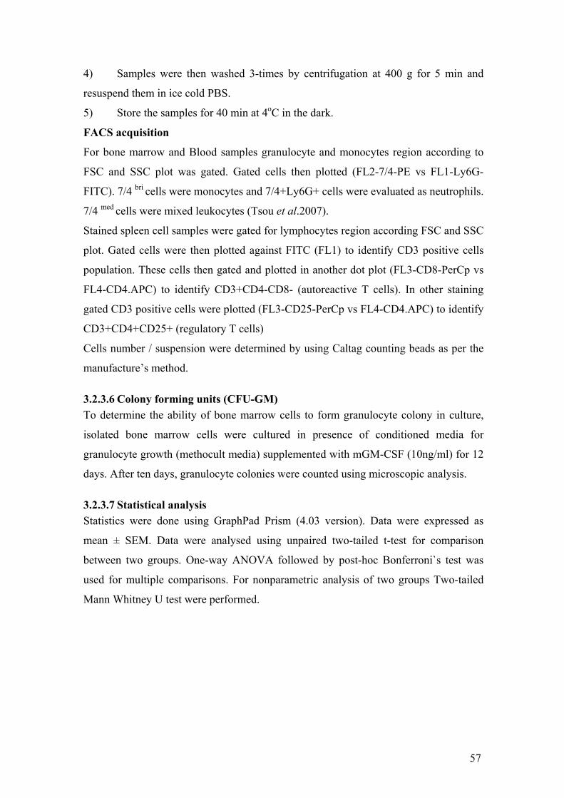
4) Samples were then washed 3-times by centrifugation at 400 g for 5 min and
resuspend them in ice cold PBS.
5) Store the samples for 40 min at 4oC in the dark.
FACS acquisition
For bone marrow and Blood samples granulocyte and monocytes region according to
FSC and SSC plot was gated. Gated cells then plotted (FL2-7/4-PE vs FL1-Ly6G-
FITC). 7/4 bri cells were monocytes and 7/4+Ly6G+ cells were evaluated as neutrophils.
7/4 med cells were mixed leukocytes (Tsou et al.2007).
Stained spleen cell samples were gated for lymphocytes region according FSC and SSC
plot. Gated cells were then plotted against FITC (FL1) to identify CD3 positive cells
population. These cells then gated and plotted in another dot plot (FL3-CD8-PerCp vs
FL4-CD4.APC) to identify CD3+CD4-CD8- (autoreactive T cells). In other staining
gated CD3 positive cells were plotted (FL3-CD25-PerCp vs FL4-CD4.APC) to identify
CD3+CD4+CD25+ (regulatory T cells)
Cells number / suspension were determined by using Caltag counting beads as per the
manufacture’s method.
3.2.3.6 Colony forming units (CFU-GM)
To determine the ability of bone marrow cells to form granulocyte colony in culture,
isolated bone marrow cells were cultured in presence of conditioned media for
granulocyte growth (methocult media) supplemented with mGM-CSF (10ng/ml) for 12
days. After ten days, granulocyte colonies were counted using microscopic analysis.
3.2.3.7 Statistical analysis
Statistics were done using GraphPad Prism (4.03 version). Data were expressed as
mean ± SEM. Data were analysed using unpaired two-tailed t-test for comparison
between two groups. One-way ANOVA followed by post-hoc Bonferroni`s test was
used for multiple comparisons. For nonparametric analysis of two groups Two-tailed
Mann Whitney U test were performed.
57

4. Results
4.1. Results part- I
Lupus nephritis is a chronic renal disease. Loss of self tolerance and impaired clearance
of autoantigens leads to deposition of immune complexes in renal tissue, which
initiates the inflammatory cascade. Lupus nephritis is characterised by the induced
expression of cytokines and chemokines in renal tissue. So far the therapy for lupus
nephritis is targeted towards modulating immune reaction, but we hypothesised that
controlling inflammatory cascade in combination with immunomodulators will help to
reduce the side effects of therapy without compromising its effect. In this study first we
looked at the efficacy of blocking CCL2 mediated tissue injury in mouse model of
lupus nephritis (MRLlpr/lpr). We looked at anti-CCL2 Spiegelmer for its
pharmacokinetics, and pharmacodynamic effects. Results of the first part are
summarised below.
4.1.1 Pharmacokinetics of the anti-CCL2 Spiegelmer in MRLlpr/lpr mice
4.1.1.1 Bioavailability
In intervention studies for chronic renal disease determination of plasma levels of
injected substance is essential as kidney being one of the important excretory organs. In
order to monitor drug exposure in MRLlpr/lpr mice, Spiegelmer plasma levels were
determined at weekly intervals. The median plasma levels of mNOX-E36 and mNOX-
E36-3'PEG were approx. 300 nM and 1 µM throughout the study, respectively (Figure
11). Thus, PEGylation increased the plasma levels of mNOX-E36 and the progressive
kidney disease of MRLlpr/lpr mice did not modulate the pharmacokinetics of both
Spiegelmers. In addition, neither drug accumulation nor metabolic induction or
reduction was obvious.
58

10
100
1000
10000
0 1 2 3 4 5 6 7 8 9 10
study duration [week]
pla
sma
leve
l [n
M]
mNOX-E36-PEG
mNOX-E36
Figure 11: Pharmacokinetics of anti-mCCL2 Spiegelmers in chronic kidney disease. Concomitant pharmacokinetic studies were performed for mNOX-E36 and mNOX-E36-PEG in female MRLlpr/lpr mice during the course of treatment from 14 to 24 wk of age. Plasma sampling was carried out 3 and 24 h after the first weekly injection for the Spiegelmers. Spiegelmer plasma levels were determined by modification of a sandwich hybridization procedure.
4.1.1.2 Distribution
Anti-CCL2 Spiegelmer showed an excellent bioavailability profile. Bio-distribution is
one of the key factors which can predict untoward effects or non specific activity of
injected drug. To determine distribution of mNOX-E36, tissues were fixed in formalin
and paraffin-embedded. From these fixed tissues 5 m sections were prepared and
mounted on silanized glass slides. These sections were then processed to detect
Spiegelmer by in situ hybridisation. Spiegelmer was found to be distributed mainly in
tubulointerstitial area of renal tissue (Figure 12). CCL2 has been shown to be expressed
in this region. Selective binding of the Spiegelmer with its target was seen even
systemically when we looked at the plasma levels of CCL2 (data shown in later parts of
the results). This explains the selective binding of mNOX-E36 to murine CCL2 and its
distribution to high CCL2 expressed area of tissue.
59
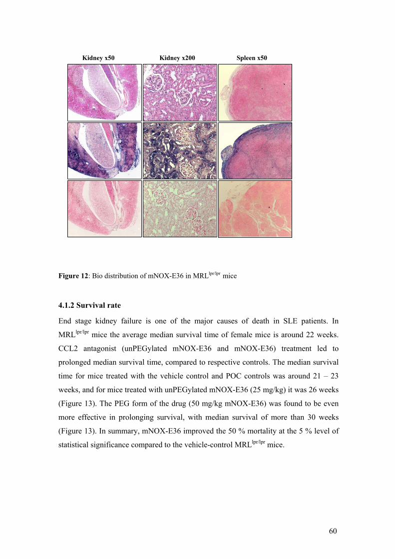
Kidney x50 Kidney x200 Spleen x50
Figure 12: Bio distribution of mNOX-E36 in MRLlpr/lpr mice
4.1.2 Survival rate
End stage kidney failure is one of the major causes of death in SLE patients. In
MRLlpr/lpr mice the average median survival time of female mice is around 22 weeks.
CCL2 antagonist (unPEGylated mNOX-E36 and mNOX-E36) treatment led to
prolonged median survival time, compared to respective controls. The median survival
time for mice treated with the vehicle control and POC controls was around 21 – 23
weeks, and for mice treated with unPEGylated mNOX-E36 (25 mg/kg) it was 26 weeks
(Figure 13). The PEG form of the drug (50 mg/kg mNOX-E36) was found to be even
more effective in prolonging survival, with median survival of more than 30 weeks
(Figure 13). In summary, mNOX-E36 improved the 50 % mortality at the 5 % level of
statistical significance compared to the vehicle-control MRLlpr/lpr mice.
60

0 5 10 15 20 25 300
25
50
75
100
Vehicle control
POC-PEG (50mg/kg)
mNOX-E36-PEG(50mg/kg)
mNOX-E36(25mg/kg)
POC (25mg/kg)
P=0,05
Spiegelmer Treatment period
Age in weeks
Pe
rce
nt
su
rviv
al
Figure 13: Survival of mice of the various treatment groups was calculated by Kaplan-Meier
analysis.
4.1.3 Renal parameters
4.1.3.1 Albumin / creatinine ratio
Albumin / creatinine ratio is an important clinical parameter for renal disease. In lupus
nephritis it increases with the progression of kidney inflammation. Treatment with a
CCL2 antagonist was expected to inhibit progression of renal disease in MRLlpr/lpr mice,
which can be indicated by the reduction of albumin / creatinine ratio, when compared
to untreated or POC-treated groups. In week 16 of age, the CCL2 antagonist
unPEGylated mNOX-E36 at a dose of 25 mg/kg, administered three times per week,
delayed the onset of renal inflammation, indicated by a statistically significant (p <
0.05, Figure 14) reduction in proteinuria compared to vehicle control. In week 24 of
age, there was no significant reduction in the albumin / creatinine ratio, but a definite
trend indicating protection in all groups treated with mNOX-E36 or unPEGylated
mNOX-E36 could be seen when compared to the vehicle control (Figure 14). Some of
the diseased mice from control group died before the final estimation of proteinuria
might explain, why the protective effect could not reach the significance at week 24.
61
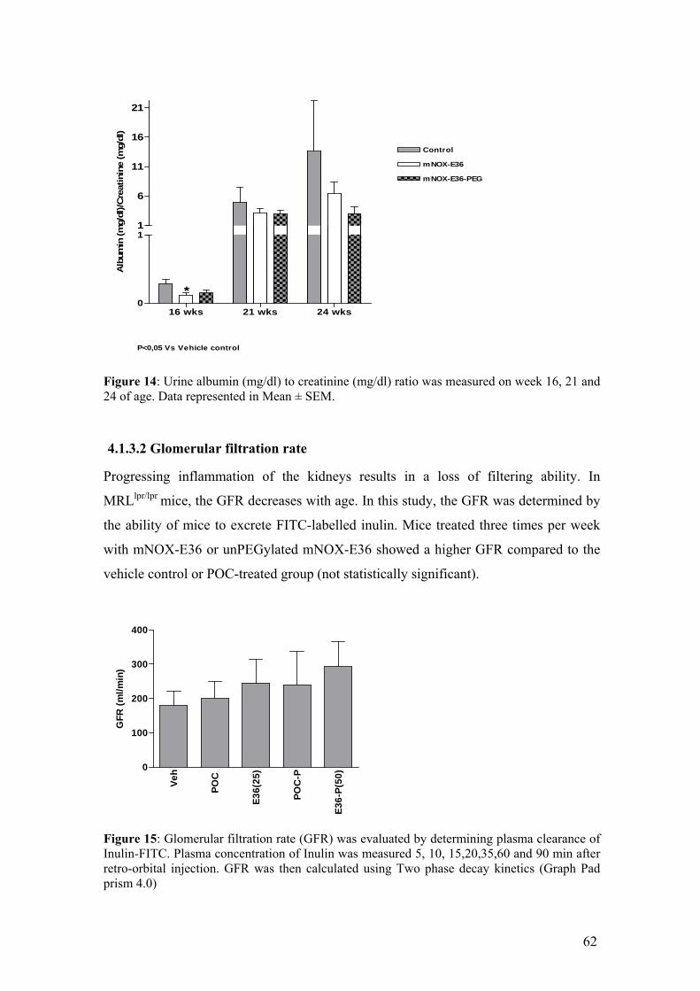
16 wks 21 wks 24 wks0
1
mNOX-E36
mNOX-E36-PEG
1
6
11
16
21
Control
*
P<0,05 Vs Vehicle control
Alb
um
in (m
g/d
l)/C
reatinin
e (m
g/d
l)
Figure 14: Urine albumin (mg/dl) to creatinine (mg/dl) ratio was measured on week 16, 21 and 24 of age. Data represented in Mean ± SEM.
4.1.3.2 Glomerular filtration rate
Progressing inflammation of the kidneys results in a loss of filtering ability. In
MRLlpr/lpr mice, the GFR decreases with age. In this study, the GFR was determined by
the ability of mice to excrete FITC-labelled inulin. Mice treated three times per week
with mNOX-E36 or unPEGylated mNOX-E36 showed a higher GFR compared to the
vehicle control or POC-treated group (not statistically significant).
Ve
h
PO
C
E3
6(2
5)
PO
C-P
E3
6-P
(50
)0
100
200
300
400
GF
R (
ml/
min
)
Figure 15: Glomerular filtration rate (GFR) was evaluated by determining plasma clearance of Inulin-FITC. Plasma concentration of Inulin was measured 5, 10, 15,20,35,60 and 90 min after retro-orbital injection. GFR was then calculated using Two phase decay kinetics (Graph Pad prism 4.0)
62

4.1.3.3 Renal histology
Female MRLlpr/lpr mice develop and subsequentially die from diffuse proliferative
immune complex glomerulonephritis, with striking similarities to diffuse proliferative
lupus nephritis in humans. MRLlpr/lpr mice showed diffuse proliferative
glomerulonephritis characterised by glomerular macrophage infiltration and a mixed
periglomerular and interstitial inflammatory cell infiltrate consisting of glomerular and
interstitial MAC-2 positive macrophages and interstitial CD3-positive lymphocytes.
Activity and chronicity index
Even at 14 wk of age, MRLlpr/lpr mice revealed proliferative glomerulonephritis with an
activity index of 4.1 ± 1.1 and a chronicity index of 0.1 ± 0.2. At all dose groups,
treatment with mNOX-E36 and unPEGylated mNOX-E36 improved the activity and
chronicity index (Figure 16) of lupus nephritis, markers of renal inflammation. The
improvement of activity and chronicity index was statistically significant at the 5 %
level (p = < 0.05, vs. respective POC, Mann Whitney test) after treatment with 50
mg/kg of mNOX-E36 or 25 mg/kg of unPEGylated mNOX-E36. Administration of 25
mg/kg of unPEGylated mNOXE36 was less effective on the chronicity index than
treatment with 50 mg/kg of mNOX-E36 (Figure 16).
63

Figure 16: Renal histopathology in MRLlpr/lpr mice. Renal sections of 24-wk-old MRLlpr/lpr mice were stained with periodic acid-schiff. Disease activity and chronicity was evaluated by scoring method established by Austin et.al. 1984 Images are representative for seven to 12 mice in each group. § p < 0.05 versus respective control Spiegelmer. Magnifications: x100
Cellular infiltration
CD3 positive cells (interstitial)
Immune cell infiltrates contribute to renal damage in lupus nephritis and CCL2
mediates the recruitment of T cells and macrophages to MRLlpr/lpr mice (Tesch et al.,
1999). Cellular infiltration is characteristic for glomerulonephritis in MRLlpr/lpr mice.
Treatment with 50 mg/kg of mNOX-E36 or 25 mg/kg of unPEGylated mNOX-E36
three times per week reduced CD3 positive cell infiltration significantly (p < 0.01 or <
0.05, Figure 17) when compared to vehicle control, respectively.
8
Ve
h
PO
C
E3
6(2
5)
PO
C-P
E3
6-P
(50
)0
2
4
6
10
§
§
Ch
ron
icit
y I
nd
ex
PoC
E36
E36-Peg
20
15
10
25
§§
Ac
tiv
ity
In
de
x
5
0
Ve
h
PO
C
E3
6(2
5)
PO
C-P
E3
6-P
(50
)
64

Macrophages (interstitial and glomerular)
Macrophages are important inflammatory cells, responsible for tissue destruction.
Lupus nephritis in MRLlpr/lpr mice leads to significant renal macrophage infiltration.
Treatment with mNOX-E36 and unPEGylated mNOX-E36 at all dose groups reduced
the macrophage infiltration (MAC-2 positive cells) in interstitial as well as glomerular
compartment. This effect was statistically significant for 50 mg/kg of mNOX-E36 (p <
0.05, interstitial and glomerular, Figure 18) and for 25 mg/kg of unPEGylated mNOX-
E36 (p < 0.05, glomerular, Figure 18) when compared to vehicle control.
PoC
E36
E36-Peg
50
40
30
20
60
***
CD
3(i
nte
rsti
tia
l)
10
0
Ve
h
PO
C
E3
6(2
5)
PO
C-P
E3
6-P
(50
)
Figure 17: Renal histopathology in MRLlpr/lpr mice. Renal sections of 24-wk-old MRLlpr/lpr mice were stained with antibodies for CD3 T cells (interstitial). Images are representative for seven to 12 mice in each group. * P< 0.05, ** P<0.01 versus respective control Spiegelmer. Magnifications: x100
65
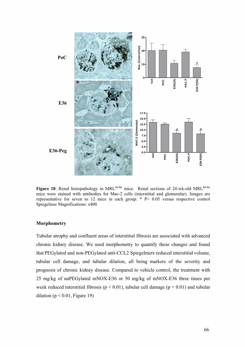
Figure 18: Renal histopathology in MRLlpr/lpr mice. Renal sections of 24-wk-old MRLlpr/lpr mice were stained with antibodies for Mac-2 cells (interstitial and glomerular). Images are representative for seven to 12 mice in each group. * P< 0.05 versus respective control Spiegelmer Magnifications: x400
Morphometry
Tubular atrophy and confluent areas of interstitial fibrosis are associated with advanced
chronic kidney disease. We used morphometry to quantify these changes and found
that PEGylated and non-PEGylated anti-CCL2 Spiegelmers reduced interstitial volume,
tubular cell damage, and tubular dilation, all being markers of the severity and
prognosis of chronic kidney disease. Compared to vehicle control, the treatment with
25 mg/kg of unPEGylated mNOX-E36 or 50 mg/kg of mNOX-E36 three times per
week reduced interstitial fibrosis (p < 0.01), tubular cell damage (p < 0.01) and tubular
dilation (p < 0.01, Figure 19)
Ve
h
PO
C
E3
6(2
5)
PO
C-P
E3
6-P
(50
)0.0
2.5
5.0
7.5
10.0
12.5
15.0
17.5
* *
MA
C-2
(G
lom
eru
lar)
20
Ve
h
PO
C
E3
6(2
5)
PO
C-P
E3
6-P
(50
)0
10
30
*
Ma
c-2
(in
ters
titi
al)
PoC
E36
E36-Peg
66

Vehicle E36
IgG deposits
CCL2 plays an important role in chronic inflammation as in lupus nephritis. But it has
no effect on autoimmune parameters which supports our aim to develop an small
molecule antagonist which attenuates the inflammation without affatcing the immunity.
Treatment with mNOX-E36 or unPEGylated mNOX-E36 reduced the cellular
infiltration and severity of glomerular alterations at the light microscopic level but there
was no change in extent of IgG depositions in glomeruli of all dose groups (data shown
in Figure 20).
Ve
hic
le
PO
C
E3
6(2
5)
PO
C-P
E3
6-P
(50
)0
5
10
15
20
25
30
35
******
Inte
rsti
tia
l V
olu
me
in
de
x
Ve
hic
le
PO
C
E-3
6(2
5)
PO
C-P
E3
6-P
(50
)0
5
10
15
20
****
Tu
bu
lar
Ce
ll D
am
ag
e i
nd
ex
20
15
10
5
25
******T
ub
ula
r D
ila
tati
on
in
de
x
0V
eh
icle
PO
C
E3
6(2
5)
PO
C-P
E3
6-P
(50
)
Figure 19: Renal histopathology in MRLlpr/lpr mice. Morphometry was performed on silver-stained sections of mice from all groups as described in the concise methods section. The interstitial volume index, tubular cell damage index, and tubular dilation index were calculated as percentage of high-power field and are expressed as means ± SEM. **P < 0.01, ***P<0.001 versus respective control Spiegelmer
67

IgG2a IgG10.0
0.5
1.0
1.5
2.0
VehiclePOC
POC-PEGmNOX-E36-PEG
mNOX-E36G
lom
eru
lar
Sc
ore
Figure 20: Renal histopathology in MRLlpr/lpr mice. Renal sections of 24-wk-old MRLlpr/lpr mice were stained for IgG. Images are representative for seven to 12 mice in each group.
4.1.4 Extra-renal autoimmune tissue injury in MRLlpr/lpr mice
4.1.4.1 Skin lesion
Cutaneous lupus manifestations, which typically occur in the facial or neck area, were
less common in mNOX-E36 (50 mg/kg) or unPEGylated mNOX-E36-treated (25
mg/kg) mice being statistically significant at the 1 % level (p < 0.01, vs. POC-PEG, see
Figure 21). The percentage of animals with skin lesions was determined to demonstrate
variability within the group. In control groups, more than 60 % (80 % in 50 mg/kg
POC-PEG) of the animals showed skin lesions in Week 24. The severity was graded
from 0 (no lesion), 1 (mild lesion), 2 (moderate) to 3 (severe). The percentage of
animals with skin lesions was determined to see the variability within the group.
68

4.1.4.2 Lung histology
After sacrifice, lung tissue of all mice was prepared and scored semi-quantitatively.
Treatment with unPEGylated mNOX-E36 and mNOX-E36 at all dose groups reduced
peribronchiolar inflammation with mean severity grades from 0.42 to 1.2 (controls,
severity grades from 1.4 to 1.6) in MRLlpr/lpr mice. After the administration of 50 mg/kg
of mNOX-E36 this effect was statistically significant at the 5 % level when compared
to the vehicle control (Figure 22).
VehicleE36
2
1
3
** **
Sk
in S
co
res
0
Ve
h
PO
C
E3
6(2
5)
PO
C-P
E3
6-P
(50
)
Vehicle E36 Figure 21: Extrarenal autoimmune tissue injury in MRLlpr/lpr mice. MRLlpr/lpr mice of all groups were regularly checked for cutaneous lupus manifestations, which typically occur at the facial or neck area and were less common in anti–mCCL2 Spiegelmer–treated mice The severity was graded from 0 (no lesion), 1 (mild lesion), 2 (moderate) to 3 (severe).** p< 0.01 vs control
69

Figure 22: Extrarenal autoimmune tissue injury in MRLlpr/lpr mice. At 24 wk of age, lung tissue was prepared from MRLlpr/lpr mice of all groups and scored semiquantitatively as described in the concise methods section. Treatment with anti-CCL2 Spiegelmers reduced peribronchiolar inflammation in MRLlpr/lpr mice. Images are representative of seven to 11 mice in each group. Magnification: x100. * p<0.05 vs control group.
4.1.4.3 Splenomegaly and lymphadenopathy
Female MRLlpr/lpr mice are known to develop a lymphoproliferative syndrome
characterized by massive splenomegaly and bulks of cervical, axillary, inguinal, and
mesenterial lymph nodes. Splenomegaly and lymphadenopathy are two of the typical
morphological clinical signs for lupus in MRLlpr/lpr mice. Treatment with unPEGylated
mNOX-E36 and mNOX-E36 did not show any effect on spleen (Figure 23) or lymph
node weights (Figure 23). Thus, treatment with Spiegelmers did not affect
lymphoproliferation in MRLlpr/lpr mice.
E36
E36-PEGVehicle
1.5
1.0
0.5
2.0
*
Lu
ng
His
tolo
gy
In
de
x
0.0
Ve
h
PO
C
E3
6(2
5)
PO
C-P
E3
6-P
(50
)
70

Ve
h
PO
C
E3
6(2
5)
PO
C-P
E3
6-P
(50
)0
1
2
3
Ly
mp
h N
od
e w
eig
ht/
10
0g
Bw
Ve
h
PO
C
E3
6(2
5)
PO
C-P
E3
6-P
(50
)0.0
0.5
1.0
1.5
2.0
2.5S
ple
en
we
igh
t /1
00
g B
W
Figure 23: At week 24 of age MRLlpr/lpr mice show massive splenomegaly and lymphadenopathy. Data represents organ weight/100 gm body weight of spleen and mesenteric lymphnodes of the groups under observation.
4.1.5 Systemic parameters
4.1.5.1 Plasma IgGs
Autoimmunity in MRLlpr/lpr mice is characterised by the production of autoantibodies
against multiple nuclear antigens including dsDNA. In 24-week old MRLlpr/lpr, plasma
dsDNA IgG, IgG1, IgG2a, and IgG2b autoantibodies were present at high levels.
unPEGylated mNOX-E36 and mNOX-E36 both had no effect on either of these DNA
autoantibodies. Thus, treatment with mNOX-E36 did not affect anti-dsDNA IgG
production in MRLlpr/lpr mice.
Table 7: Plasma IgG subtypes.
[μg/ml] Vehicle
mNOX-PoC mNOX-E36 mNOX-PoC-P mNOX-E36-P
IgG1 11.7 ± 3.6 7.5 ± 2.8 11.7 ± 3.4 8.2 ± 1.2 6.6 ± 1.2
IgG2a 2.0 ± 0.3 1.9 ± 0.3 2.1 ± 0.1 2.0 ± 0.2 1.9 ± 0.2
IgG2b 17.6 ± 5.6 18.9 ± 5.2 22.6 ± 3.3 28.3 ± 2.5 20.6 ± 3.8
Data presented as Mean ± SEM
71

4.1.5.2 Plasma CCL2
UnPEGylated mNOX-E36 and mNOX-E36, but none of the control Spiegelmers, were
found to increase the plasma concentrations of CCL2, indicating that CCL2-specific
Spiegelmers retain soluble CCL2 in the intravascular compartment. Plasma CCL2 was
measured in samples collected 3 hours post administration on the day of sacrifice.
These data showed that pegylation increases the plasma levels of unPEGylated mNOX-
E36, which most effectively retains soluble CCL2 in the intravascular compartment
(Figure 24). It also explains the selectivity of the treatment for murine CCL2.
Veh
icle
PO
C
E36
(25)
PO
C-P
E36
-P(5
0)
0
2500
5000
7500
10000
12500
***
***
Pla
sma
CC
L2
(pg
/ml)
Figure 24: Plasma CCL2 levels in MRLlpr/lpr mice. Plasma CCL2 levels were determined in mice of all groups at the end of the study. Data are expressed as means ± S.E.M.
4.1.6 Emigration of monocytes from the bone marrow
Monocyte emigration from bone marrow during bacterial infection was shown to
involve chemokine receptor CCR2 (Serbina et al.2006), but the role of CCR2 in the
context of autoimmunity remains hypothetical. To verify the effect of CCL2 blockade
on monocyte migration with Spiegelmer treatment, we performed a single-dose study of
the anti-CCL2 Spiegelmer (50 mg/kg), control Spiegelmer (50 mg/kg), and 5% glucose
(control).
72

Vehicle control mNOX-E36-PEG
A B
Figure 25 : Flow cytometry for CCR2 in peripheral blood and bone marrow of MRLlpr/lpr mice (A) To study the effects of anti-CCL2 Spiegelmer on leukocyte evasion from the bone marrow, we performed flow cytometry for CCR2 on bone marrow and peripheral blood in 24-wk-old vehicle- or anti–CCL2 Spiegelmer–treated MRLlpr/lpr mice. Data are mean percentage of CCR2+ cells ± SEM in either bone marrow or peripheral blood in five mice of each group. . (B) Effect of single dose of anti-CCL2 Spiegelmer (50 mg/kg), Spiegelmer (Co, 50 mg/kg), and 5% glucose on circulating CCR2+ cells. Data are mean percentage of CCR2+F4/80+ cells ± SEM in blood 6 h after injection.
Anti-CCL2 Spiegelmer significantly reduced the percentage of the circulating CCR2-
positive cells 6 h after injection, compared with the vehicle-treated group. By contrast,
the control Spiegelmer had no effect (Figure 25B). Furthermore, we examined the
CCR2-positive monocyte population in peripheral blood and bone marrow in 24-wk-
old MRLlpr/lpr mice that had been treated with anti-CCL2 Spiegelmer or saline from
weeks 14 to 24 of age. Anti-CCL2 Spiegelmer increased CCR2-positive cells from 13
to 26% in the bone marrow and reduced this population from 26 to 11% in the
peripheral blood, respectively (Figure 25A). These data support a novel role for CCL2
for the evasion of CCR2-positive cells from the bone marrow during autoimmune
disease of MRLlpr/lpr mice
Bone marrow Blood0
10
20
30
mNOX-E36-PEG(50mg/kg)
mNOX-E36(25mg/kg)
Vehicle Control
****
**
%G
ated
CC
R2
po
siti
ve c
ells
30
25
20
15
10
5
35
*%
Ga
ted
F4
/80
+C
CR
2+
ce
lls
in
blo
od
0Vehicle POC-P E36-P(50)
73

4.1.7 RT-PCR analysis
In order to study whether treatment with unPEGylated mNOX-E36 and mNOX-E36
affects intrarenal inflammation in MRLlpr/lpr mice, real-time RT-PCR was performed in
pooled samples for the proinflammatory chemokines CCL2 and CCL5, which are
progressively up regulated in kidneys of MRLlpr/lpr mice during progression of renal
disease. Treatment with Spiegelmers, unPEGylated mNOX-E36 and mNOX-E36, from
Week 14 to 24 of age reduced renal expression of CCL2 and CCL5 mRNA compared
to vehicle-treated controls. mRNA levels for CCL2 (Figure 26) and CCL5 (Figure 26)
were expressed per respective 18s rRNA expression.
Ve
hic
le
PO
C
E3
6(2
5)
PO
C-P
E3
6-P
(50
)
0.00000
0.00002
0.00004
0.00006
0.00008
0.00010
0.00012
CC
L2
mR
NA
/18
s r
RN
A
Figure 26: Renal chemokine expression of MRLlpr/lpr mice with experimental lupus: Renal mRNA expression for the CC-chemokines CCL2 and CCL5 was determined by real-time reverse transcriptase–PCR using total renal RNA pooled from five mice of each group. RNA levels for each group of mice are expressed per respective 18s rRNA ex
Vehic
le
PO
C
E36(2
5)
PO
C-P
E36-P
(50)0.00000
0.00025
0.00050
0.00075
CC
L5 m
RN
A/1
8s r
RN
A
pression.
4.1.8 Body weight development
After subcutaneous treatment with mNOX-E36 or unPEGylated mNOX-E36, MRLlpr/lpr
mice gained body weight at all dose groups. In week 24, the mean body weight gains of
MRLlpr/lpr mice treated with 16.66 or 50 mg/kg mNOX-E36 and with 8.33, 25 mg/kg or
25 mg/kg (once weekly) unPEGylated mNOX-E36 were +5 % or −3 % and ±0 %, −1
% or +6 % higher or lower when compared to the vehicle control .
74
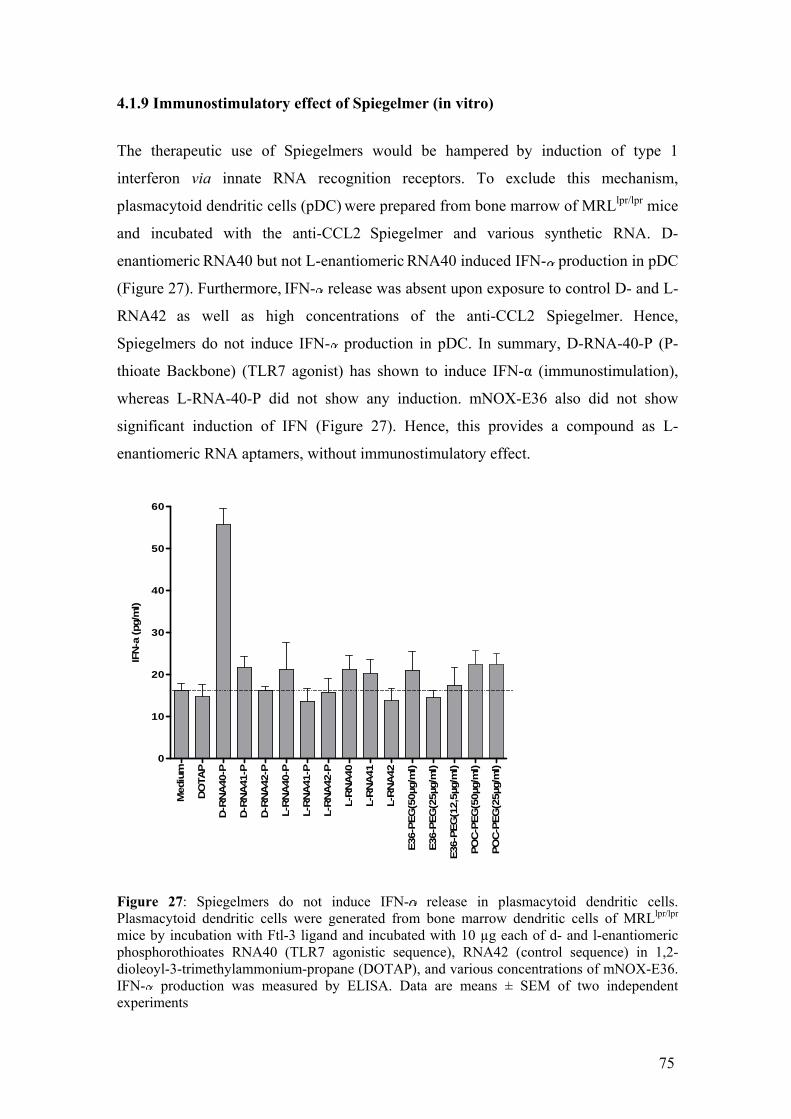
4.1.9 Immunostimulatory effect of Spiegelmer (in vitro)
The therapeutic use of Spiegelmers would be hampered by induction of type 1
interferon via innate RNA recognition receptors. To exclude this mechanism,
plasmacytoid dendritic cells (pDC) were prepared from bone marrow of MRLlpr/lpr mice
and incubated with the anti-CCL2 Spiegelmer and various synthetic RNA. D-
enantiomeric RNA40 but not L-enantiomeric RNA40 induced IFN- production in pDC
(Figure 27). Furthermore, IFN- release was absent upon exposure to control D- and L-
RNA42 as well as high concentrations of the anti-CCL2 Spiegelmer. Hence,
Spiegelmers do not induce IFN- production in pDC. In summary, D-RNA-40-P (P-
thioate Backbone) (TLR7 agonist) has shown to induce IFN-α (immunostimulation),
whereas L-RNA-40-P did not show any induction. mNOX-E36 also did not show
significant induction of IFN (Figure 27). Hence, this provides a compound as L-
enantiomeric RNA aptamers, without immunostimulatory effect.
Mediu
m
DO
TA
P
D-R
NA
40-P
D-R
NA
41-P
D-R
NA
42-P
L-R
NA
40-P
L-R
NA
41-P
L-R
NA
42-P
L-R
NA
40
L-R
NA
41
L-R
NA
42
E36-P
EG
(50µg/m
l)
E36-P
EG
(25µg/m
l)
E36-P
EG
(12,5
µg/m
l)
PO
C-P
EG
(50µg/m
l)
PO
C-P
EG
(25µg/m
l)
0
10
20
30
40
50
60
IFN
-a (pg/m
l)
Figure 27: Spiegelmers do not induce IFN- release in plasmacytoid dendritic cells. Plasmacytoid dendritic cells were generated from bone marrow dendritic cells of MRLlpr/lpr mice by incubation with Ftl-3 ligand and incubated with 10 µg each of d- and l-enantiomeric phosphorothioates RNA40 (TLR7 agonistic sequence), RNA42 (control sequence) in 1,2-dioleoyl-3-trimethylammonium-propane (DOTAP), and various concentrations of mNOX-E36. IFN- production was measured by ELISA. Data are means ± SEM of two independent experiments
75
4.2 Results Part- II
In the first part of this study we evaluated the efficacy and pharmokinetics of anti-
CCL2 Spiegelmer in MRLlpr/lpr mice. Treatment with mNOX-E36 improved the renal
phenotype in MRLlpr/lpr mice and had no effect on parameters which defines
autoimmunity. It was clear that this L-enatiomeric aptamer specifically attenuates
CCL2-derived inflammation. In the second part of our study we compared the efficacy
of the anti-CCL2 Spiegelmer with that of known immunomodulators like CYC and
MMF which are already in clinical use as a therapy for LN. Looking at the
pathogenesis of lupus nephritis, we assumed that the efficacy will not be comparable,
but we assumed if we combine the anti-CCL2 Spiegelmer with a low dose of
cyclophosphamdie, we will be able to reduce the side effects of high exposure of CYC.
But is the combination therapy as effective as high dose CYC? Results of the second
part of the study will answer this question. Results are summarised below.
4.2.1 Pharmacokinetics of Spiegelmer in MRLlpr/lpr mice
In order to monitor drug exposure in MRLlpr/lpr mice, mNOX-E36 plasma levels were
determined in the 1st
, 3rd
, 6th
, and 10th
week of treatment, i.e. the 15th
, 18th
, 21st
, and 24th
week of age. 24 hours after administration of 50 mg/kg mNOX-E36, median plasma
levels were approximately 1 μM throughout the study in both of the Spiegelmer-treated
groups (Figure 28). Apparently, the progressive kidney disease of MRLlpr/lpr mice did
not modulate Spiegelmer pharmacokinetics and neither drug accumulation nor
metabolic induction or reduction was detected.
76

100
1.000
10.000
15 18 21 24
Age [wks]
mN
OX
-E3
6 P
las
ma
Co
nc
en
tra
tio
n [
nM
] mNOX-E36-3'PEG alone
mNOX-E36-3'PEG +Cyclophosphamide
Figure 28: Plasma concentration (median) of mNOX-E36 (nM) − MRL lpr/lpr mice
4.2.2 Renal parameters
4.2.2.1 Renal Histology
Activity and chronicity index
At 14 weeks of age MRLlpr/lpr mice showed diffuse proliferative lupus nephritis with an
activity score index of 4.1 ± 1.1. At this age major abnormalities of the
tubulointerstitial compartment were absent (data not shown).
After 10 weeks of treatment, vehicle- and control Spiegelmer-treated MRLlpr/lpr mice
revealed diffuse proliferative lupus nephritis associated with glomerular
hypercellularity, expansion of glomerular matrix, focal tuft necrosis, and a mixed
periglomerular and interstitial inflammatory cell infiltrate (Figure 30). Full dose CYC
and the combination of mNOX-E36 and ¼ full dose CYC were equally potent in
improving the activity (Figure 29) and chronicity index (Figure 29) of lupus nephritis,
markers of renal inflammation. This effect was statistically significant at the 0.1 %
level (p < 0.001 vs. vehicle control). Furthermore, combination of mNOX-E36 with ¼
full dose CYC was significantly (p < 0.05) more effective when compared with ¼ full
dose CYC treatment alone. ¼ full dose CYC alone, mNOX-E36 alone as well as MMF
were less potent but still significantly improved the activity (p < 0.05 vs. vehicle
77

control), but only ¼ full dose CYC alone and mNOX-E36 alone improved the
chronicity indices (p < 0.001 or p < 0.05 vs. vehicle control) of lupus nephritis (Figure
29). Thus, adding mNOX-E36 to a low dose (30 mg/kg/4 weeks) CYC-based regimen
is as potent as full dose CYC therapy for diffuse proliferative lupus nephritis in
MRLlpr/lpr mice.
Ve
hic
le
rev
-E3
6-P
E-3
6-P
CY
C l
ow
CY
C h
igh
Co
mb
i
MM
F
0
5
10
15
20
25
*** ***
*
*+A
cti
vit
y I
nd
ex
Ve
hic
le
rev
-E3
6-P
E-3
6-P
CY
C(l
ow
)
CY
C(h
igh
)
Co
mb
i
MM
F
0
1
2
3
4
5
6
7
8
9
***
******
*
+
+C
hro
nic
ity
In
de
x
Figure 29: Markers of lupus nephritis in MRLlpr/lpr mice. The activity index (A) and chronicity index (B) for DPLN were determined on periodic acid-Schiff-stained renal sections from seven to 12 mice from each group as described by Austin et al. (1984) Control Sp, pegylated control Spiegelmer (rev-E36-P); anti-CCL2 Sp, pegylated anti-CCL2 Spiegelmer (-E36-P); CYC low, monthly 30 mg/kg cyclophosphamide; CYC high, weekly 30 mg/kg cyclophosphamide; MMF, 100 mg/kg mycophenolate mofetil. *, p < 0.05; **, p < 0.01; ***, p < 0.001 versus vehicle; +, p < 0.05 versus CYC low
78

Anti-CCL2
Vehicle
MMF
CYC full
CYC (1/4th) + Anti-CCL2 Sp Control Sp
CYC (1/4th)
Figure 30: Renal histopathology in MRLlpr/lpr mice. Renal sections of 24-week-old MRLlpr/lpr mice from all groups were stained with periodic acid Schiff. Original magnification, x200. Control Sp, pegylated control Spiegelmer (rev-E36-P); anti-CCL2 Sp, pegylated anti-CCL2 Spiegelmer (E36-P); CYC low, monthly 30 mg/kg cyclophosphamide; CYC high, weekly 30 mg/kg cyclophosphamide; MMF, 100 mg/kg mycophenolate mofetil. Crescentic or globally sclerotic glomeruli are indicated by a star, and tubular casts are indicated by an arrow
79

Cellular infiltration
Cellular infiltration is characteristic for glomerulonephritis in MRLlpr/lpr mice. Additive
effects of an anti-CCL2 Spiegelmer and ¼ full dose CYC combination may relate to
impaired macrophage and T cell recruitment in MRLlpr/lpr mice. Immunostaining was
performed for Mac2-positive macrophages and CD3-positive T cells and the number of
glomerular and interstitial macrophages as well as interstitial T cells was assessed. The
numbers of glomerular T cells were not analysed as they were very low in all groups.
CD3 positive cells (interstitial)
All treatments reduced CD3 positive cell infiltration significantly (p < 0.001 vs.
vehicle control, Figure 31), while treatment with full dose CYC or the combination of
mNOX-E36 and ¼ full dose CYC seemed to be equally potent in reducing the
numbers of interstitial CD3 positive cell in kidneys of MRLlpr/lpr mice than treatment
with ¼ full dose CYC alone and mNOX-E36 as well as MMF
Ve
hic
le
rev
-E3
6-P
E-3
6-P
CY
C(l
ow
)
CY
Ch
igh
)
Co
mb
i
MM
F
0
10
20
30
40
***
***
***
***
***
$$
CD
3(i
nte
rsti
tia
l)
Figure 31: Renal sections of 24-week-old MRLlpr/lpr mice were stained for CD3-positive T cells. Numbers of interstitial T cells were evaluated as mean numbers of CD3-positive cells in 15 high-power fields (hpf) per section. Data are expressed as means ± S.E.M. Control Sp, pegylated control Spiegelmer (rev-E36-P); anti-CCL2 Sp, pegylated anti-CCL2 Spiegelmer (E36-P); CYC low, monthly 30 mg/kg cyclophosphamide; CYC high, weekly 30 mg/kg cyclophosphamide; MMF, 100 mg/kg mycophenolate mofetil. *, p < 0.05; **, p < 0.01; ***, p < 0.001 versus vehicle; ##, p < 0.01 versus CYC low. .
80

Macrophages (glomerular and interstitial)
Full dose CYC and the combination of mNOX-E36 and ¼ full dose CYC were equally
potent (p < 0.001 vs. vehicle control) in reducing the numbers of glomerular Mac-2
positive macrophages in kidneys of MRLlpr/lpr mice (Figure 32). Additionally,
combination of mNOX-E36 with ¼ full dose CYC was significantly more effective in
reducing the numbers of glomerular Mac2-positive macrophages than treatment with
¼ full dose CYC alone (p < 0.01). ¼ full dose CYC alone and mNOX-E36 alone as
well as MMF were less potent but still significantly (p < 0.01 and p < 0.001 vs. vehicle
control, respectively) reduced the macrophages in glomeruli and the interstitium
(Figure 32). The number of interstitial Mac2-positive macrophages in kidneys of
MRLlpr/lpr mice were significantly reduced by full dose CYC and to a lower extend
equally reduced by ¼ full dose CYC and combination therapy with mNOX-E36 (p <
0.001 vs. vehicle control, Figure 32). Treatment with mNOX-E36 alone and MMF
were less potent but still significantly at the 5 % level (p < 0.05 vs. vehicle control).
Thus, the additive effect of mNOX-E36 and ¼ full dose CYC on renal pathology of
MRLlpr/lpr mice was associated with a significant reduction of interstitial and
glomerular macrophages which was similar to the effect of full dose CYC.
Ve
hic
le
rev
-E3
6-P
CY
C(l
ow
)
E-3
6-P
CY
Ch
igh
)
Co
mb
i
MM
F
0
5
10
15
20
25
30
35
******
***
Ma
c2
(in
ters
titi
al)
Ve
hic
le
rev
-E3
6-P
E-3
6-P
CY
C(l
ow
)
CY
C(h
igh
)
Co
mb
i
MM
F
0.0
2.5
5.0
7.5
10.0
12.5
15.0
17.5
20.0
*** ***
******
**
++
Ma
c2
(glo
me
rula
r)
Figure 32: Renal sections of 24-week-old MRLlpr/lpr mice were stained for Mac-2-positive macrophages. The number of glomerular macrophages was evaluated as the mean number of Mac2-positive cells in 15 glomeruli (glom.) per section. The numbers of interstitial macrophages (D)were evaluated as mean numbers of Mac2-positive cells in 15 high-power fields (hpf) per section. Data are expressed as means ± S.E.M. Control Sp, pegylated control Spiegelmer (rev-E36-P); anti-CCL2 Sp, pegylated anti-CCL2 Spiegelmer (E36-P); CYC low, monthly 30 mg/kg cyclophosphamide; CYC high, weekly 30 mg/kg cyclophosphamide; MMF, 100 mg/kg mycophenolate mofetil. *, p < 0.05; **, p < 0.01; ***, p < 0.001 versus vehicle; ++, p < 0.01 versus CYC low.
81

4.2.3 Extra-renal autoimmune tissue injury in MRLlpr/lpr mice
4.2.3.1 Lung histology
Autoimmune peribronchitis is another manifestation of lupus-like systemic
autoimmunity in MRLlpr/lpr mice. After sacrifice, lung tissue of all mice was prepared
and scored semiquantitatively. Full dose CYC (30 mg/kg/week) was significantly (p <
0.001) more effective than ¼ full dose CYC (30 mg/kg/4 weeks) and mNOX-E36 in
reducing peribronchiolar inflammation in MRLlpr/lpr mice. However, ¼ full dose (30
mg/kg/4 weeks) CYC plus anti-CCL2 Spiegelmer were as effective as full dose of
CYC and significantly (p< 0.05) more effective than treatment with mNOX-36 alone
(Figure 33 and Figure 34). Daily oral treatment with MMF had no effect on lung injury
in MRLlpr/lpr mice. Thus, the ¼ full dose (monthly) CYC/Spiegelmer combination was
as effective as full dose (weekly) administrations of 30 mg/kg of CYC on autoimmune
lung injury in MRLlpr/lpr mice
Veh
icle
rev-
E36
-P
CY
C(l
ow
)
E-3
6-P
CY
Ch
igh
)
MM
F
Co
mb
i
0.0
0.5
1.0
1.5
2.0
* *
*****
Lu
ng
His
tolo
gy
sco
re
Figure 33: Semiquantitative scoring of lung injury was performed as described Materials and Methods. Data represent means ± S.E.M. Control Sp, pegylated control Spiegelmer (rev-E36-P); anti-CCL2 Sp, pegylated anti-CCL2 Spiegelmer (E36-P); CYC low, monthly 30 mg/kg cyclophosphamide; CYC high, weekly 30 mg/kg cyclophosphamide; MMF, 100 mg/kg mycophenolate mofetil. * p<0.05,** p<0.01,*** p<0.001 vs vehicle control.
.
82

CYC (1/4th)
Vehicle
MMF
CYC full
CYC (1/4th) + Anti-CCL2 Sp Control Sp
Anti-CCL2 Sp
Figure 34: Autoimmune lung injury in MRLlpr/lpr mice. Lung sections of 24-week-old MRLlpr/lpr mice from all groups were stained with periodic acid Schiff. Original magnification, x100. Control Sp, pegylated control Spiegelmer (rev-E36-P); anti-CCL2 Sp, pegylated anti-CCL2 Spiegelmer (E36-P); CYC low, monthly 30 mg/kg cyclophosphamide; CYC high, weekly 30 mg/kg cyclophosphamide; MMF, 100 mg/kg mycophenolate mofetil.
83

4.2.3.2 Splenomegaly and lymphadenopathy
Splenomegaly and lymphadenopathy are two of the typical morphological clinical
signs for lupus in MRLlpr/lpr mice. CYC treatment caused a dose-dependent reduction
of spleen (p < 0.01, full dose CYC) and lymph node (p < 0.001, full and ¼ full dose
CYC) weights in 24-week old MRLlpr/lpr mice as compared to vehicle-treated MRLlpr/lpr
mice (Figure 35). The effect of MMF treatment was less evident and did only reduce
significantly (p < 0.001) weight of lymph nodes. Treatment with mNOX-E36 did not
show a significant effect on spleen or lymph node weights (Figure 35) and no
additional effect of mNOX-E36 could be noted when given in combination with ¼ full
dose of CYC. Thus, mNOX-E36 did not affect lymphoproliferation in MRLlpr/lpr mice
Ve
hic
le
rev
-E3
6-P
E-3
6-P
CY
C(l
ow
)
CY
Ch
igh
)
MM
F
co
mb
i
0.0
0.5
1.0
1.5
2.0
2.5
**
+
Sp
lee
n w
t/1
00
gm
bo
dy
wt
Ve
hic
le
rev
-E3
6-P
E-3
6-P
CY
C(l
ow
)
CY
Ch
igh
)
MM
F
co
mb
i
0.0
0.5
1.0
1.5
2.0
2.5
3.0
3.5
*** ***
***
***
LN
wt/
10
0g
m B
od
y w
t
Figure 35: Lymphoproliferation in MRLlpr/lpr mice. Spleens and the bulk of mesenteric lymph nodes were harvested from 24-week-old MRLlpr/lpr mice. Weight of lymph nodes is expressed as mean weight per 100 mg body weight ± S.E.M. Spleen weights are expressed as mean weight per 100 mg body weight ± S.E.M. Control Sp, pegylated control Spiegelmer (rev-E36-P); anti-CCL2 Sp, pegylated anti-CCL2 Spiegelmer (E36-P); CYC low, monthly 30 mg/kg cyclophosphamide; CYC high, weekly 30 mg/kg cyclophosphamide; MMF, 100 mg/kg mycophenolate mofetil. *, p < 0.05; **, p < 0.01; ***, p < 0.001 versus vehicle; +, p < 0.05 versus vehicle (t Test) .
84
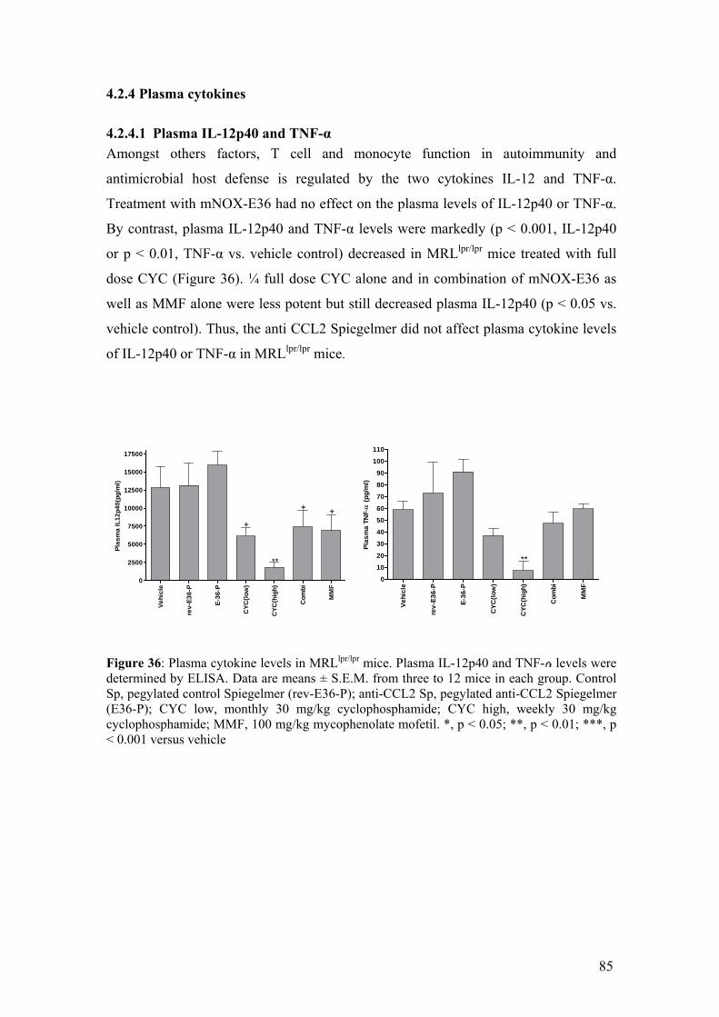
4.2.4 Plasma cytokines
4.2.4.1 Plasma IL-12p40 and TNF-α
Amongst others factors, T cell and monocyte function in autoimmunity and
antimicrobial host defense is regulated by the two cytokines IL-12 and TNF-α.
Treatment with mNOX-E36 had no effect on the plasma levels of IL-12p40 or TNF-α.
By contrast, plasma IL-12p40 and TNF-α levels were markedly (p < 0.001, IL-12p40
or p < 0.01, TNF-α vs. vehicle control) decreased in MRLlpr/lpr mice treated with full
dose CYC (Figure 36). ¼ full dose CYC alone and in combination of mNOX-E36 as
well as MMF alone were less potent but still decreased plasma IL-12p40 (p < 0.05 vs.
vehicle control). Thus, the anti CCL2 Spiegelmer did not affect plasma cytokine levels
of IL-12p40 or TNF-α in MRLlpr/lpr mice.
Ve
hic
le
rev
-E3
6-P
E-3
6-P
CY
C(l
ow
)
CY
C(h
igh
)
Co
mb
i
MM
F0
10
20
30
40
50
60
70
80
90
100
110
**
Pla
sm
a T
NF
- (
pg
/ml)
Ve
hic
le
rev
-E3
6-P
E-3
6-P
CY
C(l
ow
)
CY
C(h
igh
)
Co
mb
i
MM
F
0
2500
5000
7500
10000
12500
15000
17500
**
+
++
Pla
sm
a I
L1
2p
40
(pg
/ml)
Figure 36: Plasma cytokine levels in MRLlpr/lpr mice. Plasma IL-12p40 and TNF- levels were determined by ELISA. Data are means ± S.E.M. from three to 12 mice in each group. Control Sp, pegylated control Spiegelmer (rev-E36-P); anti-CCL2 Sp, pegylated anti-CCL2 Spiegelmer (E36-P); CYC low, monthly 30 mg/kg cyclophosphamide; CYC high, weekly 30 mg/kg cyclophosphamide; MMF, 100 mg/kg mycophenolate mofetil. *, p < 0.05; **, p < 0.01; ***, p < 0.001 versus vehicle
85

4.2.4.2 Plasma CCL2
mNOX-E36, but not the control Spiegelmer revmNOX-E36, was found to increase
statistically significant (p < 0.001 vs. vehicle control) the plasma concentrations of
CCL2 (Figure 37), indicating that CCL2-specific Spiegelmers retain soluble CCL2 in
the intravascular compartment. Plasma CCL2 was measured in samples collected 2 to 3
hours post administration on the day of sacrifice. These data showed that
pharmacologically relevant levels of circulating mNOX-E36, most likely, binds and
retains CCL2 in the intravascular compartment.
Veh
icle
rev-
E36
-P
E-3
6-P
CY
C(l
ow
)
CY
C(h
igh
)
Co
mb
i
MM
F0
500
1000
50000
100000*** ***
Pla
sm
a C
CL
2(p
g/m
l)
Figure 37: Plasma CCL2 levels in MRLlpr/lpr mice. Plasma CCL2 levels were determined in mice of all groups at the end of the study. Data are expressed as means ± S.E.M. Control Sp, pegylated control Spiegelmer (rev-E36-P); anti-CCL2 Sp, pegylated anti-CCL2 Spiegelmer (E36-P); CYC low, monthly 30 mg/kg cyclophosphamide; CYC high, weekly 30 mg/kg cyclophosphamide; MMF, 100 mg/kg mycophenolate mofetil. *** p< 0.001 vs control.
4.2.5 Body weight development
After subcutaneous treatment with mNOX-E36 or revmNOX-E36, MRLlpr/lpr mice
gained body weight at all dose groups as expected; animals in the groups treated with
immunosuppressants full dose CYC and MMF lost weight. Mice on ¼ full dose CYC
and combination therapy showed only minimal or no weight loss at all. In week 24, the
mean body weight gains of MRLlpr/lpr mice treated with 50 mg/kg mNOX-E36 were
(+3.84 g) similar to mean body weight gain in the vehicle group (+5.2 g). In contrast,
animals in the full dose CYC and MMF showed strong loss of mean body weight (-6.38
g and -6.52 g, respectively).
86

4.3 Results Part III
We have been able to show that mNOX-E36 treatment improves the renal phenotype in
MRLlpr/lpr mice. Combining anti CCL2 therapy with low dose CYC shows comparable
efficacy with that of high dose CYC. But what is the effect of combination therapy on
overall immunity? is it equally immunosuppressive? or it spares the side effects of
high dose CYC without hampering the efficacy. To evaluate this, in a separate
experiment four satellite groups of MRLlpr/lpr mice were treated for four weeks with
vehicle, Spiegelmer, Spiegelmer in combination with ¼ full dose CYC, full dose CYC
alone from 8 to 12 weeks of age in order to characterise immune cell subsets in spleen,
bone marrow and peripheral blood by flow cytometry (FACS).
4.3.1 Splenomegaly and lymphadenopathy
Ve
hic
le
E3
6-P
(50
)
Co
mb
i
CY
C(3
0)
0
10
20
30
40
50
60
*
Ce
lls
/Sp
lee
n (
in m
illi
on
s)
Veh
icle
E36
-P(5
0)
Co
mb
i
CY
C(3
0)
0.00
0.05
0.10
0.15
0.20
*
Sp
lee
n w
eig
ht(
gm
)
Figure 38:Spleen weights are expressed as mean weight per 100 mg body weight ± S.E.M. Spleen cells are expressed as million/spleen. anti-CCL2 Sp, pegylated anti-CCL2 Spiegelmer (E36-P-50); CYC(30), weekly 30 mg/kg cyclophosphamide; Combi; mNOX-E36 plus CYC (low) *, p < 0.05 versus vehicle; Full-dose CYC but not ¼ full dose CYC + mNOX-E36 significantly (p < 0.05 vs.
vehicle control) reduced the total number of spleen cells and spleen weight. (Figure
38). This was consistent for all spleen T cell subsets studied, i.e. CD3+, CD3+CD4+
CD3+CD8+, CD3+CD4-CD8-, and CD3+CD4+CD25+
cells (Figure 39). Thus,
mNOX-E36 + ¼ full dose CYC had no additive effects on T cell depletion as seen with
full dose CYC treatment in MRLlpr/lpr mice.
87

Vehicle E36-P(50) Combi CYC(30)0.0
0.5
CD3+
CD3+CD4+
CD3+CD8+
CD3+CD4+CD25+
1
2
CD3+CD4-CD8-
****
3
6
9
12
15
18
21
*
*
*
Ce
lls
(in
mil
lio
ns
)
Figure 39: T cell depletion in MRLlpr/lpr mice. Spleens were harvested from 12-week-old MRLlpr/lpr mice (n = 5) after 4 weeks of treatment as indicated. Spleen cell suspensions were quantified by flow cytometry. CD3-positive T lymphocyte subsets of all the groups are shown. Data represent mean percentages ± S.E.M. of all leukocytic cells. anti-CCL2 Sp, pegylated anti-CCL2 Spiegelmer (E36-P-50); CYC(30), weekly 30 mg/kg cyclophosphamide; Combi; mNOX-E36 plus CYC (low) *, p < 0.05 **,p<0.01 versus vehicle;
Full dose of CYC usually causes transient myelosuppression which renders patients
susceptible to life-threatening infections. Full dose CYC significantly (p < 0.05 vs.
vehicle control and ¼ full dose CYC + mNOX-E36 combination) reduced the numbers
of 7/4 (bri) monocytes in bone marrows of MRLlpr/lpr mice while mNOX-E36-treated
and combined ¼ full dose CYC + mNOX-E36-treated MRLlpr/lpr
mice had significantly
(p < 0.05 vs. vehicle control) elevated numbers of 7/4 (bri) bone marrow monocytes
(Figure 40). In peripheral blood, test item- and CYC-treated MRLlpr/lpr mice showed a
trend towards lower 7/4 (bri) monocyte counts but this did not reach statistical
significance for any of the groups (Figure 40).
88

4.3.2 Effect on blood and bone marrow
Week1 Week2 Week3 Week4
0.0
2.5
5.0
7.5
10.0
12.5
Vehicle
Combination
Cyclophsphamide(30mg/kg/week)
E36-P-50mg/kg
7/4
briL
y6
G- m
on
oc
yte
s
A
Veh
icle
E36
-P(5
0)
Co
mb
i
CY
C(3
0)
0
5
* *
***+++
5.0
7.5
10.0
7/4
briL
y6
G-m
on
oc
yte
s(B
on
e m
arr
ow
)
B
Figure 40: Myelosuppression in MRLlpr/lpr mice. Bone marrow and peripheral blood (every week) samples were harvested from 12-week-old MRLlpr/lpr mice (n = 5) after 4 weeks of treatment as indicated. 7/4high-Positive monocytes were quantified in bone marrows (B) and peripheral blood (A) of MRLlpr/lpr mice from all groups Data represent mean percentages ± S.E.M. of all leukocytic cells. anti-CCL2 Sp, pegylated anti-CCL2 Spiegelmer (E36-P-50); CYC(30), weekly 30 mg/kg cyclophosphamide; Combi; mNOX-E36 plus CYC (low). *, p < 0.05 **,p<0.01 versus vehicle; +++ p< 0.001 vs combination.
89

4.3.3 CFU-GM colony assay
Veh
icle
E36
-P(5
0)
Co
mb
i
CY
C(3
0)
40
50
60
70
80
90
100
110
**
CF
U-G
M c
olo
nie
s/
3*10
4ce
lls
Figure 41: Number of CFU-GM colonies / 3*104 BM cells. Data represented as Mean ± SEM. anti-CCL2 Sp, pegylated anti-CCL2 Spiegelmer (E36-P-50); CYC (30), weekly 30 mg/kg cyclophosphamide; Combi; mNOX-E36 plus CYC (low). ** p< 0.01 vs control.
CFU-GM colony assay in Methocult media supplemented with m-GM-CSF is a test for
myelosuppression, i.e. the growing capacity of haematopoietic stem cells and immune
cell progenitors. CFU-GM colonies in full dose CYC-treated mice were significantly
reduced on day 12 of incubation (p < 0.01 vs. vehicle control and p < 0.05 vs. ¼ full
dose CYC + mNOX-E36), while mNOX-E36 and ¼ full dose CYC + mNOX-E36
treatment did not affect CFU-GM counts (Figure 41). Thus, mNOX-E36 + ¼ full dose
CYC treatment did not cause myelosuppression as seen with full dose CYC in
MRLlpr/lpr mice.
90
5. Discussion CCL2 and Lupus nephritis
Apart from activation of auto-reactive T cells, B cells and production of auto-
antibodies, end stage tissue damage is an important aspect of lupus pathogenesis.
Tissue inflammation in chronic disease is characterised by hyper infiltration of immune
cells (T cells, macrophages). Thus targeting molecules (CCL2, CCL5, ICAM,
integrins) which participate in cell migration would be effective in preventing end stage
tissue damage. Evidence for a pathogenic role of the CCL2/CCR2 axis in lupus-
associated tissue injury already exists. CCL2- and CCR2-deficient MRLlpr/lpr mice are
protected from kidney, lung, and skin pathology (Tesch et al. 1999, Perez de Lema et
al. 2005) Thus, CCL2 and CCR2 both are crucial for the recruitment of cytokine-
producing leukocytes to peripheral tissues. A recent study found that CCR2 is also
required for the evasion of monocytes from the bone marrow into the intravascular
compartment during bacterial infection (Serbina NV, Pamer EG. 2006).
Hasegawa et al (2003) injected 5 x 10⁶ cells that were transfected with an NH2-
truncated CCL2 analogue subcutaneously into 12-wk-old female MRLlpr/lpr mice which
was associated with an improved pathology at 20 wk. Similar results were obtained by
Shimizu et al. (2004) who injected 7ND, a NH2-terminal deletion mutant of the CCL2
gene, into skeletal muscles of 16-wk-old female MRLlpr/lpr mice.In both of the studies
CCL2 blockade improves the renal pathology without affecting the systemic
autoimmunity parameters, as none of the above studies showed reduction in
autoantibodies. This makes CCL2 blockade as attractive approach to attenuate chronic
inflammation without interfering with immunity. However, the cell transfer approach
caused local tumours of considerable size (Hasegawa et al. 2003).The caveat for the use
of this gene therapy–like approach in humans is that CCL2 antagonism cannot be
abandoned once initiated.
Spiegelmers: Clinically applicable aptamers
Cell transfer approach (induction of local tumours) and gene therapy (irreversible
antagonism) are not clinically applicable in humans. Aptamers can neutralize biologic
functions of target molecules conceptually similar to antibodies. But aptamers are not
biostable and can induce immune reaction though recognition by TLRs. Development
of safer biological aptamers is motivating many groups working all over the globe.
91

Spigelmer (L-enantiomeric aptamer) is one of such invention. Unlike aptamers,
Spiegelmer-based oligonucleotides are nuclease resistant and thus biostable without
further modifications (Klussmann et al.1996), which makes them very well suited for in
vivo applications. We evaluated anti-CCL2 Spiegelmer (mNOX-E36), which binds with
high affinity to murine CCL2 and blocks its function in vitro at low nanomolar
concentrations. Spiegelmers, being L-enatiomeric, are not recognised by nucleases.
Because of these structural benefits they remain in systemic circulation for long time
and have better pharmacokinetics than conventional aptamers. We analysed systemic
Spiegelmer levels after subcutaneous injection in MRLlpr/lpr mice. Blood samples were
taken after 3hrs and 24hrs for mNOX-E36 and mNOX-E36-Peg respectively. The
PEGylated form of mNOX-E36 displayed higher plasma levels compared with the non-
PEGylated mNOX-E36. Levels determined for both form of drugs were sufficient for
the in vivo application. Progressive renal failure in MRLlpr/lpr mice did not affect their
plasma levels.
We analysed CCL2 levels at termination of the studies at 24 weeks age of MRLlpr/lpr
mice. Groups treated with anti-CCL2 Spiegelmers (not control Spiegelmers) showed
higher levels of CCL2. This defines the specificity of Spiegelmer to neutralise murine
CCL2. Spiegelmers upon interacting with systemic CCL2 neutralises it and keeps it in
circulation in biologically inactive form. This biological inactivation was seen with
decreased percentage of CCR2 positive monocytes in peripheral blood. Contrary to
peripheral blood, percentage of CCR2 positive blood cells in bone marrow was found
to be elevated in MRLlpr/lpr mice treated with anti-CCL2 Spiegelmer. We looked at the
in vivo-distribution of mNOX-E36 in MRLlpr/lpr after subcutaneous injection. mNOX-
E36 was seen in tubulointerstitial compartment of renal tissue. CCL2 is known to be
expressed in high levels in this compartment of renal tissue. All these observations
show that mNOX-E36 specifically binds to CCL2 and neutralises it in vivo.
Synthetic oligonucleotides may induce IFN- in dendritic cells (Heil et al.2004,
Hornung et al.2005) through innate pattern recognition receptors in cytosolic or
endosomal compartments such as Toll-like receptor-7, retinoic acid–inducible protein-
1, or melanoma differentiation–associated gene-5 (Heil et al.2004, Hornung et al.2005)
IFN- is an important mediator of autoimmunity (Banchereau J,Pascaul V. 2006) The
induction of IFN- would hamper the use of mNOX-36 in lupus nephritis. However,
Spiegelmers did not induce IFN- production in dendritic cells, because the L-
92

enantiomeric configuration of Spiegelmers is probably not recognized by RNA-specific
pattern recognition receptors.
All these properties make anti-CCL2 Spiegelmer (mNOX-E36), a clinically applicable
apatamer.
Anti-CCL2 Spiegelmer (mNOX-E36) in lupus nephritis
Our data demonstrate that even non prophylactic late-onset treatment with anti-CCL2
Spiegelmer at 14 wk of age, a point at which autoimmune tissue injury is already
established (Tesch et al.1999,Perez de Lema et al.2005), is effective to the same extent
as CCL2 or CCR2 deficiency, although such knockout animals lack CCL2/CCR2 from
birth. The consistency of the data from CCL2/CCR2 deficiency and therapeutic CCL2
blockade may relate to the predominant role of the CCL2/CCR2 axis during the late
leukocyte recruitment–dependent autoimmune tissue injury rather than the early
development of autoimmunity in MRLlpr/lpr mice. In fact, both CCL2-deficient and anti-
CCL2 Spiegelmer–treated MRLlpr/lpr mice did not affect the lymphoproliferative
syndrome, DNA autoantibodies, and renal immune complex deposition, all being early
disease markers (Tesch et al.1999). Treatment with anti-CCL2 Spiegelmer reduced
inflammatory cell infiltration in renal tissue, improved renal phenotype, and improved
survival in MRLlpr/lpr mice. Our findings are consistent with other studies that initiated
blockade of the CCL2/CCR2 axis after onset of autoimmunity.
CCL2 blockade is thought to target CCL2-dependent leukocyte adhesion to activated
endothelia and transendothelial migration of leukocytes at sites of inflammation (Luster
et al.2005). Our finding that CCL2 blockade has a similar effect on monocyte homing
to the bone marrow during systemic autoimmunity extends this findings two-fold: First,
monocyte homing to the bone marrow involves CCL2, possibly acting through CCR2,
and second, this mechanism seems to apply to systemic inflammation caused by
infection and autoimmunity.
Current lupus therapy regimen and novel approaches
The aim of the treatment in lupus nephritis is to suppress the inflammation in renal
tissue and to preserve the structure and function of the kidney, avoiding the
progression to renal failure. This objective must be achieved without developing
significant adverse events. Currently, the therapy for severe lupus nephritis is based on
the use of high-dose corticosteroids and immunosuppressive drugs. Several
93

randomized controlled studies demonstrated that, for patients with severe lupus
nephritis, a regimen including CYC is more effective than treatment with only
glucocorticoids (Steinberg AD et al.1991, Austin HA et al. 1986). Noteworthy, these
randomized trials of the United States National Institutes of Health (NIH) failed to
demonstrate significant differences among the different regimens containing
immunosuppressive drugs (daily oral CYC, monthly intravenous (IV) CYC, daily oral
azathioprine (AZA), combined low-dose oral CYC plus AZA). Nevertheless, monthly
IV CYC became the gold standard of treatment for severe lupus nephritis since then. A
recent study (by Houssiau et al.2002) has showed that, as far as induction of remission
and prevention of end-stage renal failure is concerned, a 12-week course of IV CYC
given at a fixed dose of 500 mg fortnightly and followed by AZA for 2 years is as
effective as an abbreviated NIH protocol consisting on six monthly IV CYC pulses of
0.5 g/m2 (increasing the dose according to the leukocyte count) followed by two
quarterly pulses, after a median follow-up of 72 months. Unfortunately, the use of IV
CYC is not exempt from pitfalls. Despite the treatment with CYC, a significant
proportion, up to 50%, of patients with lupus nephritis (Korbet SM et al.2000, Ginzler
EM et al.2005) do not achieve a complete remission. In the studies of the United
States NIH, a longer duration of the therapy with CYC reduced the cumulative
probability of relapse, when comparing 24 versus 6 months of treatment (Boumpas DT
et al.1992), but longer therapy with CYC was associated with increased gonadal
toxicity. In addition, CYC, even with intravenous administration, has a significant
toxicity in terms of hemorrhagic cystitis, gonadal failure and infections.
For the above mentioned reasons, the need for less toxic and more effective
alternatives to CYC in the treatment of lupus nephritis is clear.
Because costimulation sustains the abnormal activity of T cells and helps the
production of autoantibodies, it represents an obvious target in SLE. Thus, several
approaches aim at decreasing the deleterious effects of T cells by blocking
costimulation in SLE. CD28/CTLA-4 (Abatacept) and CD80/86, CD40 and CD40
ligand (L)(BJ9588), ICOS and ICOS-L (anti ICOS antibody) have been targeted to
normalise activity of T cells.
Several different B cell targeting strategies have been identified and are currently
under investigation. These include targets directly associated with the B cell, such as B
cell surface molecules (CD20, rituxumab) (CD22, Epratuzumab) and more indirect
targets, such as the inhibition of costimulatory ligands present on the T cell.
94

Figure 42: Mechanisms of current immunotherapies. BCDT: B-cell depletion therapy; Ag: antigen; Ab: antibody. Influence of immunotherapeutic drugs indicated by red arrows. Anti-BLyS acts on BLyS, a B-cell survival factor. Costimulatory molecules CD40 and B7 are present on both APC and B-cells. For ease of use, the thicker arrows are only indicated for anti-CD40, CTLA4-Ig on APC.
All these approaches are meant to target reduced autoantiboby production. None of the
novel approaches have shown any promising clinical data so far.
Complexities of the disease prevent us from relying solely on anti-inflammtory
treatment approach for lupus nephritis. We hypothesised that blocking inflammatory
mediators along with low exposure of immunosuppressant like cyclophophamide, can
improve the renal pathology as effectively as high dose of immunosuppressant. CCL2
plays an important role in inflammatory phase of lupus. Anti-CCL2 Spiegelmer
showed good therapeutic benefit in murine model of LN. So we combined anti-CCL2
Spiegelmer (mNOX-E36) with low dose of cyclophosphamide. This way we thought
of finding a novel way to reduce immunosuppressant exposure and it also signifies the
probable role of CCL2 blockade in LN treatment
95

Combining CCL2 blockade with low dose cyclophosphamide
Immunosuppressive treatment regimen for diffuse proliferative lupus nephritis
involving high dose CYC or MMF remain associated with serious complications in
lupus nephritis patients, i.e. infections (Katsifis et al. 2004, Appel et al.2007) Thus,
combinations of CYC with drugs that more specifically interact with autoimmune
tissue injury in lupus may overcome this problem. Although the therapeutic effect was
clearly evident, it remained unclear how the efficacy of the anti-CCL2 Spiegelmer
would compare with that of CYC or MMF. Based on the specific anti-inflammatory
mechanism of CCL2 blockade, we assumed that treatment with an anti-CCL2
Spiegelmer would not be as effective as CYC full dose for the treatment of diffuse
proliferative lupus nephritis. However, we hypothesized that therapeutic effects
equivalent to CYC full dose might be achieved with a combination of less frequent
CYC dosing plus anti-CCL2 Spiegelmer, which may avoid the toxicity of CYC full
dose.
It was demonstrated that a combination of mNOX-E36 and monthly (¼ full dose) CYC
treatment initiated at 14 weeks of age and continued for 10 weeks was as effective as
weekly (full dose) CYC treatment in suppressing diffuse proliferative lupus nephritis
and lung injury in MRLlpr/lpr mice. The effect of mNOX-E36 either given alone or in
combination with a ¼ full dose CYC had no additive effects on T cell depletion, serum
TNF-α and IL-12p40 levels, and myelosuppression. All these parameters were
significantly suppressed by full dose CYC therapy. Anti-CCL2 Spiegelmer had no anti-
proliferative effect on immune cells and bone marrow-derived blood cell progenitors as
the alkylating agent CYC. By contrast, the anti-CCL2 Spiegelmer increased the
numbers of bone marrow macrophages. As a consequence, mNOX-E36 had no effect
on the underlying autoimmune dysregulation in MRLlpr/lpr mice. Furthermore, mNOX-
E36 did not modulate the CD4/CD8 double negative ‘autoreactive’ T cell population,
indicating that mNOX-E36 did not modulate the autoimmune process in itself but
rather the local mechanisms of autoimmune tissue injury. Additionally, an additive
effect of combined mNOX-E36 / ¼ full dose CYC therapies on the numbers of renal
macrophages and T cells was observed which is consistent with the role of CCL2 in
renal macrophage and T cell recruitment. However, late onset treatment with mNOX-
E36 alone was not as effective as high dose CYC therapy on autoimmune tissue injury
in MRLlpr/lpr mice. As expected, the non-functional control Spiegelmer was not
96

effective in this lupus nephritis model. The animals tolerated the treatment well and
there was no influence on body weight development due to Spiegelmer treatment.
We could see significant renal protection with anti-CCL2 Spiegelmer treatment in
MRLlpr/lpr mice. But translation of murine efficacy in human clinical efficacy has
always been a tough task for researchers. Even lupus nephritis phenotype in MRLlpr/lpr
mice differs with that of human disease. So our data could be encouraging for us but
may/may not differ interms of efficacy in humans. Anti-CCL2 Spiegelmer (NOX-E36)
is already in clinical trials for diabetic nephropathy and lupus nephritis as indications.
Recently phase-I data for pharmacokinetic and pharmacodynamic evaluation has been
released by NOXXON AG on their website. NOX-E36 in human showed good Pka
behaviour and could be able to reduce CCR2 positive monocytes in a dose dependent
manner as pharmcodynamic parameter. It is just a data from healthy individuals from
phase-I study, data from patients study (phase-II) is still pending.
97

6. Summary and conclusion
The monocyte chemoattractant protein CCL2 is crucial for monocyte and T cell
recruitment from the vascular to the extravascular compartment at sites of
inflammation. CCL2 is expressed in human lupus nephritis and was shown to mediate
experimental lupus in murine models of lupus nephritis; therefore, CCL2 antagonists
may be beneficial for therapy. That is why our first aim for this study was to find out
the efficacy of novel tool i.e. anti-CCL2 Spiegelmer in MRLlpr/lpr mice. This study
showed the L-enantiomeric RNA oligonucleotide mNOX-E36, an anti-CCL2
Spiegelmer that binds murine CCL2 with high affinity and neutralizes its action in
vitro and in vivo. CCL2 antagonism in MRLlpr/lpr mice was shown to be effective
earlier by some groups using different approaches to block CCL2 e.g. injecting
transfected cells with mutant CCL2 (Hagesawa et al.2003), gene therapy (Shimizu et
al.2004), and by using CCL2 knockout mice in MRLlpr/lpr background (Tesch et
al.1999). Our experimental approach is more clinically suitable as 1) the mirror image
configuration of the Spiegelmer confers nuclease resistance and, thus, excellent
biostability and 2) mNOX-E36 does not induce type I IFN via Toll-like receptor-7 or
cytosolic RNA receptors, as recently shown for certain synthetic D-RNA.
Distribution of anti-CCL2 Spiegelmer was confined to the tissue regions where CCL2
is expressed in MRLlpr/lpr mice. In situ hybridisation located Spiegelmers in the
tubulointerstitial compartment of the kidney where CCL2 is known to be expressed at
high levels in these mice. Plasma levels of CCL2 where found to be high in the
circulation as Spiegelmer tend to bind CCL2 to neutralize it biologically and keep it in
the circulation. This showed the specificity of anti-CCL2 Spiegelmer for murine CCL2
in vivo.
Autoimmune-prone MRLlpr/lpr mice that were treated with a polyethylene glycol form
of mNOX-E36 from weeks 14 to 24 of age showed prolonged survival associated with
a robust improvement of lupus nephritis, peribronchial inflammation, and lupus-like
inflammatory skin lesions. Improvement of lupus nephritis was seen with reduced
activity index and chronicity index compared to control mice. As anti-CCL2
Spiegelmer acts by inhibiting the influx of inflammatory cells to injured tissues, we
found a profound reduction in macrophages and T cell infiltration in renal tissue. But
at the same time Spiegelmer treatment had no effect of autoimmunity parameters in
98

these mice. Thus, mNOX-E36-based inhibition of CCL2 represents a novel strategy
for the treatment of lupus nephritis.
Cyclophosphamide (CYC) can control diffuse proliferative lupus nephritis by potent
immunosuppression but remains associated with serious and life-threatening
complications. Drugs that specifically target mediators of diffuse proliferative lupus
nephritis may help to reduce CYC dose and side effects. In the second part of the study
we wanted, 1) To compare the efficacy of anti-CCL2 Spiegelmer with that of existing
therapies for lupus nephitis, 2) To evaluate the efficacy of combining anti-CCL2
Spiegelmer and low dose of CYC with that of high dose of CYC, 3) To compare the
safety profile of this combination with high dose CYC.
To achieve that we injected MRLlpr/lpr mice with diffuse proliferative lupus nephritis
from 14 weeks of age with vehicle, weekly 30 mg/kg CYC (full dose), monthly 30
mg/kg CYC (one-fourth full dose), pegylated control Spiegelmer, pegylated anti-CCL2
Spiegelmer (3/week), pegylated anti-CCL2 Spiegelmer plus CYC one-fourth full dose
and mycophenolate mofetil. At week 24, diffuse proliferative lupus nephritis and
autoimmune lung injury were virtually abolished with CYC full dose but not with
CYC one-fourth full dose. The CYC one-fourth full dose/Spiegelmer combination was
equipotent to CYC full dose on kidney and lung injury. Serum IL12p40 and TNF-
levels were all markedly affected by CYC full dose but not by CYC one-fourth full
dose.
In other experimental design with four groups, we injected MRLlpr/lpr mice with
vehicle, anti-CCL2 Spiegelmer, anti-CCL2 Spiegelmer plus CYC (once/4 weeks) and
CYC (once/week). We did the treatment for one month before terminating the
experiment. CYC full dose was associated with reduction in spleen weights,
CD3+CD4-CD8- T cells and CD3+CD4+CD25+ T cells. No additive effects of anti-
CCL2 Spiegelmer were noted on bone marrow colony-forming unit-granulocyte
macrophage counts and 7/4(high) monocyte counts, lymphoproliferation, and spleen T
cell depletion.
In summary, anti-CCL2 Spiegelmer permits 75% dose reduction of CYC for
controlling diffuse proliferative lupus nephritis and pneumonitis in MRLlpr/lpr mice,
sparing suppressive effects of full-dose CYC on myelosuppression and T cell
depletion.
99

Conclusion
The Spiegelmer-based inhibition of CCL2/monocyte-chemoattractant protein-1 offers a
new and promising way to treat lupus nephritis. The compound mNOX-E36 not only
shows efficacy in MRLlpr/lpr mice but also demonstrates its suitability in terms of
pharmacokinetic profile as well as its absence of immunostimulatory adverse effects..
Furthermore, combination of anti-CCL2 Spiegelmer and CYC therapy permits
significant dose reduction of CYC and could be proposed as novel strategy to reduce
CYC toxicity in the treatment of severe lupus.
100

7. Zusammenfassung CCL2 ist für die Rekrutierung von Monozyten und T-Zellen aus dem Gefäßsystem bei
Verletzung, Infektion oder Entzündung von wesentlicher Bedeutung. Das Protein ist
bei Patienten mit Lupusnephritis vermehrt exprimiert und es wurde in früheren Studien
bereits gezeigt, dass es bei der Entwicklung der Lupusnephritis im Mausmodell eine
bedeutende Rolle spielt. Demzufolge wurde in dieser Arbeit angenommen, dass CCL2-
Antagonisten neue Therapieoptionen der Lupusnephritis darstellen könnten. Das erste
Ziel dieser Studie war es daher, die Wirksamkeit des anti-CCL2-Spiegelmers in
MRLlpr/lpr-Mäusen herauszufinden. Hierfür wurde das L-enantiomere RNA-
Oligonukleotid mNOX-E36, ein anti-CCL2-Spiegelmer verwendet, welches das
murine CCL2 mit hoher Affinität binden und seine Aktivität sowohl in vitro als auch
in vivo neutralisieren kann. Die Antagonisierung von CCL2 im MRLlpr/lpr-Mausmodell
konnte bereits in früheren Studien anderer Arbeitsgruppen als effektive Methode zur
Behandlung der Lupusnephritis, beispielsweise durch Injektion transfizierter CCL2-
Mutanten (Hagesawa et al.2003), Gentherapie (Shimizu et al.2004) oder durch die
Verwendung von CCL2-Knockout-Mäusen im MRLlpr/lpr-Hintergrund (Tesch et
al.1999) nachgewiesen werden. Unser experimenteller Ansatz erweist sich für die
klinische Anwendung als geeigneter, da zum einen die spiegelbildliche Konfiguration
der Spiegelmere Nuklease-Resistenz gewährt und daher Spiegelmere eine exzellente
Biostabilität aufweisen und zum anderen, weil mNOX-E36 keine Typ-I-Interferone via
Toll-like Rezeptor-7 oder zytosolische RNA-Rezeptoren induziert, wie es kürzlich für
synthetische D-RNA gezeigt worden ist.
Die Verteilung des anti-CCL2-Spiegelmers war auf die CCL2-exprimierten Gewebe in
MRLlpr/lpr-Mäusen begrenzt. Durch in situ-Hybridisierung konnten die Spiegelmere im
tubulointerstitiellen Kompartment der Niere lokalisiert werden, wo CCL2
bekanntermaßen vermehrt exprimiert wird. Im Plasma konnten ebenfalls hohe CCL2-
Spiegel nachgewiesen werden, da Spiegelmere dazu tendieren CCL2 zu neutralisieren
und weiter zirkulieren. Dies zeigte die Spezifität des anti-Spiegelmers für murines
CCL2 in vivo.
MRLlpr/lpr-Mäuse, ein murines SLE Modell, wurden mit einer pegylierten Form des
mNOX-E36 im Alter von 14 bis 24 Wochen behandelt. Diese Mäuse zeigten ein
verlängertes Überleben, welches mit einer deutlichen Verbesserung der
101

Lupusnephritis, der peribrochialen Entzündung, und der Lupus-ähnlichen
Hautveränderungen assoziiert war. Die Verbesserung der Lupusnephritis wurde
anhand des verminderten Aktivitäts- und Chronizitätsindex im Vergleich zur
Kontrollgruppe bestimmt. Da anti-CCL2-Spiegelmere durch Behinderung des
Einstroms von Entzündungszellen in verletztes Gewebe wirken, konnten wir eine
deutliche Reduktion der Makrophagen- und T-Zell-Infiltration im Nierengewebe
nachweisen. Dennoch hatte die gleiche Spiegelmer-Behandlung keinen Einfluss auf
Parameter für Autoimmunität in den Mäusen. Somit stellt die mNOX-E36-basierte
Hemmung von CCL2 eine neue Strategie für die Behandlung der Lupusnephritis dar.
Cyclophosphamid (CYC) kann die diffuse proliferative Lupusnephrits durch potente
Immunsuppression kontrollieren. Diese Therapie ist aber mit schwerwiegenden und
lebensbedrohlichen Komplikationen verbunden. Medikamente, die spezifisch
Mediatoren der diffusen proliferativen Lupusnephrits erreichen, könnten helfen, die
CYC-Dosis und damit Nebenwirkungsrate zu reduzieren. Im zweiten Teil dieser
Studie wollten wir 1) die Wirksamkeit des anti-CCL2-Spiegelmers mit bereits
existierenden Therapien der Lupusnephritis vergleichen, und 2) die Wirksamkeit des
anti-CCL2-Spiegelmers in Kombination mit niedrig-dosiertem CYC evaluieren und 3)
die Nebenwirkungsrate dieser Kombinationsstudie mit der der hoch-dosierten CYC-
Behandlung vergleichen.
Dazu injizierten wir MRLlpr/lpr-Mäusen mit nachgewiesener diffuser proliferativer
Lupusnephritis im Alter von 14 Wochen jeweils den Trägerstoff, 30 mg/kg CYC
wöchentlich (hoch-dosiert), 30 mg/kg CYC monatlich (ein Viertel der hoch-dosierten
CYC-Gabe), pegyliertes Kontroll-Spiegelmer, pegyliertes Anti-CCL2-Spiegelmer
(3mal/Woche) sowie pegyliertes Anti-CCL2-Spiegelmer in Kombination mit CYC (ein
Viertel der hoch-dosierten CYC-Gabe) und Mycophenolat Mofetil. Im Alter von 24
Wochen war die proliferative Lupusnephritis und die autoimmune Lungenbeteiligung
durch die Behandlung mit der hoch-dosierten CYC-Gabe komplett aufgehoben, was
jedoch nicht mit der Behandlung mit niedrig-dosierten CYC (ein Viertel der hoch-
dosierten CYC-Gabe) erreicht werden konnte. Die Behandlung der Kombination aus
niedrig-dosiertem CYC (ein Viertel der hoch-dosierten CYC-Gabe) und Spiegelmer
erzielte vergleichbare Ergebnisse bezüglich der Nieren- und Lungenbeteiligung wie die
die Behandlung mit hoch-dosiertem CYC. IL12p40- und TNF--Werte im Serum
102
waren in der hoch-dosierten CYC-Gruppe deutlich beeinflusst, jedoch nicht in der
Gruppe mit der niedrig-dosierten CYC-Gabe.
In einer anderen experimentellen Studie mit vier Gruppen, injizierten wir in MRLlpr/lpr -
Mäusen jeweils den Trägerstoff, anti-CCL2-Spiegelmer, anti-CCL2-Spiegelmer mit
CYC (1mal/Monat) und CYC (1mal/Woche). Diese Behandlung erfolgte ein Monat. In
der Gruppe mit der hoch-dosierten CYC-Gabe konnte eine Reduktion des
Milzgewichts, der CD3+CD4-CD8- T-Zellen und der CD3+CD4+CD25+ T-Zellen
nachgewiesen werden. Bei der zusätzlichen Gabe von anti-CCL2-Spiegelmer konnte
kein additiver Effekt in der CFU-GM-positiven Granulozyten- und Makrophagen-
Kolonienzahl und 7/4(high)-positiven Monozyten-Zellzahl im Knochenmark sowie
Lymphoproliferation und T-Zell-Depletion in der Milz gefunden werden.
Zusammenfassend ermöglicht die anti-CCL2-Spiegelmer-Therapie eine 75%ige Dosis-
Reduzierung von CYC, um die diffuse proliferative Lupusnephritis und Pneumonitis in
MRLlpr/lpr-Mäusen zu behandeln, während die suppressiven Eigenschaften der hoch-
dosierten CYC-Gabe mit Knochenmarksuppression und T-Zell-Depletion ausgespart
werden.
Fazit
Die Spiegelmer-basierte Hemmung von CCL2 offenbart eine neue und viel-
versprechende Therapieoption für die Behandlung der Lupusnephritis. Das Präparat
mNOX-E36 zeigt nicht nur Wirksamkeit im MRLlpr/lpr-Mausmodell, sondern
demonstriert auch seine Eignung im Hinblick auf das pharmakokinetische Profil und
das Fehlen immunstimulatorischer Nebenwirkungen. Darüber hinaus ermöglicht die
Kombinationstherapie mit anti-CCL2-Spiegelmer und CYC eine signifikante
Dosisreduktion von CYC und könnte somit als neue Therapiestrategie zur Toxizitäts-
Reduktion von CYC bei der Behandlung des progressiv verlaufenden Lupus
vorgeschlagen werden.
103

8. References
1. Alarcón-Segovia D, Tumlin JA, Furie RA, McKay JD, Cardiel MH, Strand V, Bagin RG, Linnik MD, Hepburn B; LJP 394 Investigator Consortium.: LJP 394 for the prevention of renal flare in patients with systemic lupus erythematosus: results from a randomized, double-blind, placebo-controlled study. Arthritis Rheum. 2003 Feb;48(2):442-54
2. Anders HJ, Belemezova E, Eis V, Segerer S, Vielhauer V, Perez de Lema G, Kretzler M, Cohen CD, Frink M, Horuk R, Hudkins KL, Alpers CE, Mampaso F, Schlöndorff D: Late onset of treatment with a chemokine receptor CCR1 antagonist prevents progression of lupus nephritis in MRL-Fas(lpr) mice. J Am Soc Nephrol. 2004 Jun;15(6):1504-13.
3. Anders HJ, Frink M, Linde Y, Banas B, Wörnle M, Cohen CD, Vielhauer V, Nelson PJ, Gröne HJ, Schlöndorff D: CC chemokine ligand 5/RANTES chemokine antagonists aggravate glomerulonephritis despite reduction of glomerular leukocyte infiltration. J Immunol. 2003 Jun 1;170(11):5658-66.
4. Anders HJ, Vielhauer V., Frink M., Linde Y., Cohen CD, Blattner SM, Kretzler M., Strutz F., Mack M., Grone HJ, Onuffer J., Horuk R., Nelson PJ, Schlondorff D: A chemokine receptor CCR-1 antagonist reduces renal fibrosis after unilateral ureter ligation. J Clin Invest 2002; 109:251-259.
5. Appel G, Dooley MA, Ginzler EM, Isenberg D, Jayne D, Solomons N, and Wolfs D.: Mycophenolate mofetil compared with intravenous cyclophosphamide as induction therapy for lupus nephritis: Aspreva Lupus Management Study (ALMS) results (Abstract). J Am Soc Nephrol 2007; 18: SA-FC057.
6. Austin HA., Muenz LR., Joyce KM., Antonovych TT. and Balow JE.: Diffuse proliferativ lupus nephritis: Identification of specific pathologic features affecting renal outcome: Kidney Intern., 1984; 25:689−695.
7. Austin III HA, Klippel JH, Balow JE, et al.: Therapy of lupus nephritis: controlled trial of prednisone and cytotoxic drugs. N Engl J Med 1986; 314: 614-19.
8. Aydt E and Wolf G: Development of synthetic pan-selectin antagonists: a new treatment strategy for chronic inflammation in asthma, Pathobiology (2002-3); 70, 297–301.
9. Baechler EC, Batliwalla FM, Karypis G, Gaffney PM, Ortmann WA, Espe KJ, Shark KB, Grande WJ, Hughes KM, Kapur V, Gregersen PK, Behrens TW: Interferon-inducible gene expression signature in peripheral blood cells of patients with severe lupus. Proc Natl Acad Sci U S A. 2003 Mar 4;100(5):2610-5.
10. Banchereau J, Pascual V: Type I interferon in systemic lupus erythematosus and other autoimmune diseases. Immunity 2006; 25: 383 –39.
11. Baran KW et al.: Double-blind, randomized trial of an anti-CD18 antibody in conjunction with recombinant tissue plasminogen activator for acute myocardial infarction: limitation of myocardial infarction following thrombolysis in acute myocardial infarction (LIMIT AMI) study, Circulation 2001;104, 2778–2783.
12. Baumann I, Kolowos W, Voll RE, Manger B, Gaipl U, Neuhuber WL, Kirchner T, Kalden JR, Herrmann M: Impaired uptake of apoptotic cells into tingible body macrophages in germinal centers of patients with systemic lupus erythematosus.Arthritis Rheum. 2002 Jan; 46(1):191-201.
104

13. Boumpas DT, Austin III HA, Vaughn EM et al.: Controlled trial of pulse methylprednisolone versus two regimens of pulse cyclophosphamide in severe lupus nephritis. Lancet 1992; 340: 741–745.
14. Boumpas DT, Furie R, Manzi S et al.: A Short Course of BG9588 (Anti-CD40 Ligand Antibody) Improves Serologic Activity and Decreases Hematuria in Patients With Proliferative Lupus Glomerulonephritis. Arthritis Rheum 2003; 48:719-727.
15. Budagyan VM, Bulanova EG, Sharova NI, NikonovaMF, Stanislav ML, Yarylin AA: The resistance of activated T-cells from SLE patients to apoptosis induced by human thymic stromal cells. Immunol Lett 1998; 60: 1–5.
16. Bynoe,MS, Grimaldi, CM, Diamond, B: Estrogen up-regulates Bcl-2 and blocks tolerance induction of naive B cells. Proc Natl Acad Sci USA 2000; 97: 2703–2708.
17. Cancro MP, D'Cruz DP, Khamashta MA: The role of B lymphocyte stimulator (BLyS) in systemic lupus erythematosus.J Clin Invest. 2009 May;119(5):1066-73.
18. Cardiel MH, Tumlin JA, Furie RA, Wallace DJ, Joh T, Linnik MD; LJP 394-90-09 Investigator Consortium: Abetimus sodium for renal flare in systemic lupus erythematosus: results of a randomized controlled phase III trial. Arthritis Rheum. 2008 Aug;58(8):2470-80.
19. Casciola-Rosen L, Rosen A.: Ultraviolet light-induced keratinocyte apoptosis: a potential mechanism for the induction of skin lesions and autoantibody production in LE. Lupus 1997; 6: 175–80.
20. Chan OT, Hannum LG, Haberman AM et al.: A novel mouse with B cells but lacking serum antibody reveals an antibody-independent role for B cells in murine lupus. J Exp Med 1999; 189: 1639–1648.
21. Chan TM, Cheng IK.: Identification of endothelial cell membrane proteins that bind anti-DNA antibodies from patients with systemic lupus erythematosus by direct or indirect mechanisms. J. Autoimmun. 1997; 10: 433–39.
22. Chan TM, Leung JK, Ho SK, Yung S.: Mesangial cell-binding anti- DNA antibodies in patients with systemic lupus erythematosus. J. Am. Soc. Nephrol. 2002; 13: 1219–29.
23. Chan TM, Yu PM, Tsang KL, Cheng IK.: Endothelial cell binding by human polyclonal anti-DNA antibodies: Relationship to disease activity and endothelial functional alterations. Clin. Exp. Immunol. 1995; 100: 506–13.
24. Charles PJ, Smeenk RJ, De Jong J, Feldmann M, Maini RN: Assessment of antibodies to double-stranded DNA induced in rheumatoid arthritis patients following treatment with infliximab, a monoclonal antibody to tumor necrosis factor alpha: findings in open-label and randomized placebo-controlled trials.Arthritis Rheum. 2000 Nov; 43(11):2383-90.
25. Cohen PL, Eisenberg RA.: Lpr and gld: single gene models of systemic autoimmunity and lymphoproliferative disease. Annu Rev Immunol. 1991; 9:243-69.
26. Cooper GS, Dooley MA, Treadwell EL, St Clair EW, Gilkeson GS.: Hormonal and reproductive risk factors for development of systemic lupus erythematosus: results of a population-based, case-control study. Arthritis Rheum 2002; 46: 1830–39.
27. Cunnane G, Chan OT, Cassafer G et al.: Prevention of renal damage in murine lupus nephritis by CTLA-4Ig and cyclophosphamide. Arthritis Rheum 2004; 50: 1539–48.
28. Datta SK, Patel H, Berry D: Induction of a cationic shift in IgG anti-DNA autoantibodies. Role of T helper cells with classical and novel phenotypes in three murine models of lupus nephritis. J Exp Med. 1987 May 1;165(5):1252-68.
105
29. Davis Jr JC, Totoritis MC, Rosenberg J, Sklenar TA, Wofsy D.: Phase I Clinical Trial of a Monoclonal Antibody Against CD40-Ligand (IDEC-131) in Patients with Systemic Lupus Erythematosus. J Rheumatol 2001; 28:91-101.
30. Dawisha SM, Gmelig-Meyling F, Steinberg AD: Assessment of clinical parameters associated with increased frequency of mutant T cells in patients with systemic lupus erythematosus. Arthritis Rheum 1994; 37: 270–277.
31. Dean GS, Tyrrell-Price J, Crawley E, Isenberg DA.: Cytokines and systemic lupus erythematosus. Ann Rheum Dis. 2000 Apr;59(4):243-51
32. Dedrick RL: Adhesion molecules as therapeutic targets for autoimmune diseases and transplant rejection, Expert Opin. Biol. Ther. 2003; 3, 85–95.
33. Dörner T, Kaufmann J, Wegener WA, Teoh N, Goldenberg DM, Burmester GR: Initial clinical trial of epratuzumab (humanized anti-CD22 antibody) for immunotherapy of systemic lupus erythematosus.Arthritis Res Ther. 2006; 8(3):R74.
34. Dufour EM et al CD99 is essential for leukocyte diapedesis in vivo. Cell Commun Adhes. 2008 Nov; 15(4):351-63.
35. Einav S, Pozdnyakova OO, Ma M, Carroll MC: Complement C4 is protective for lupus disease independent of C3. J. Immunol. 2002; 168: 1036–41.
36. Eis V, Luckow B, Vielhauer V, Siveke JT, Linde Y, Segerer S, Perez De Lema G, Cohen CD, Kretzler M, Mack M, Horuk R, Murphy PM, Gao JL, Hudkins KL, Alpers CE, Gröne HJ, Schlöndorff D, Anders HJ: Chemokine receptor CCR1 but not CCR5 mediates leukocyte recruitment and subsequent renal fibrosis after unilateral ureteral obstruction.J Am Soc Nephrol. 2004 Feb; 15(2):337-47.
37. Elbourne, KB, Keisler, D, McMurray, RW: Differential effects of estrogen and prolactin on autoimmune disease in the NZB/NZW F1 mouse model of systemic lupus erythematosus. Lupus 1998; 7: 420–427.
38. Fatenejad S, Peng SL, Disorbo O, Craft J: Central T cell tolerance in lupus-prone mice: influence of autoimmune background and the lpr mutation. J Immunol 1998; 161: 6427–6432.
39. Faxon D et al.: The effect of blockade of the CD11/CD18 integrin receptor on infarct size in patients with acute myocardial infarction treated with direct angioplasty: the results of the HALT-MI study, J. Am. Coll. Cardiol. 2002; 40,1199–1204.
40. Fischereder M, Luckow B, Hocher B, Wüthrich RP, Rothenpieler U, Schneeberger H, Panzer U, Stahl RA, Hauser IA, Budde K, Neumayer H, Krämer BK, Land W, Schlöndorff D: CC chemokine receptor 5 and renal-transplant survival. Lancet. 2001 Jun 2;357(9270):1758-61.
41. Forbes A: Alternative immunomodulators, Eur. J. Gastroenterol. Hepatol. 2003; 15, 245–248.
42. Furuichi K, Wada T, Iwata Y, Kitagawa K, Kobayashi K, Hashimoto H, Ishiwata Y, Tomosugi N, Mukaida N, Matsushima K, Egashira K, Yokoyama H: Gene therapy expressing amino-terminal truncated monocyte chemoattractant protein-1 prevents renal ischemia-reperfusion injury.J Am Soc Nephrol. 2003 Apr;14(4):1066-71.
43. Ginzler EM, Dooley MA, Aranow C, Kim MY, Buyon J, Merrill JT, Petri M, Gilkeson GS, Wallace DJ, Weisman MH, Appel GB: Mycophenolate mofetil or intravenous cyclophosphamide for lupus nephritis. N Engl J Med. 2005 Nov 24;353(21):2219-28.
44. Goodnow CC :Multistep pathogenesis of autoimmune disease. Cell. 2007 Jul 13;130(1):25-35.
106

45. Goodnow CC, Sprent J, Fazekas de St Groth B, Vinuesa CG: Cellular and genetic mechanisms of self tolerance and autoimmunity. Nature. 2005 Jun 2;435(7042):590-7.
46. Gourley MF, Austin HA III, Scott D et al.: Methylprednisolone and Cyclophosphamide, Alone or in Combination, in Patients with Lupus Nephritis. A Randomized, Controlled Trial. Ann Intern Med 1996 125: 549-557.
47. Green DR, Ferguson T, Zitvogel L, Kroemer G: Immunogenic and tolerogenic cell death. Nat Rev Immunol. 2009 May;9(5):353-63.
48. Grimaldi, CM, Jeganathan, V, Diamond, B: Hormonal regulation of B cell development: 17 beta-estradiol impairs negative selection of high-affinity DNA-reactive B cells at more than one developmental checkpoint. J Immunol 2006b; 176: 2703–2710.
49. Grimaldi, CM. Sex and systemic lupus erythematosus: the role of the sex hormones estrogen and prolactin on the regulation of autoreactive B cells. Curr Opin Rheumatol 2006a; 18: 456–461.
50. Hahn BH. Antibodies to DNA. N. Engl. J. Med. 1998; 338 : 1359–68.
51. Hasegawa H, Kohno M, Sasaki M, Inoue A, Ito MR, Terada M, Hieshima K, Maruyama H, Miyazaki J, Yoshie O, Nose M, Fujita S: Antagonist of monocyte chemoattractant protein 1 ameliorates the initiation and progression of lupus nephritis and renal vasculitis in MRL/lpr mice. Arthritis Rheum 2003; 48: 2555 –2566.
52. Heil F, Hemmi H, Hochrein H, Ampenberger F, Kirschning C, Akira S, Lipford G, Wagner H, Bauer S: Species-specific recognition of single-stranded RNA via toll-like receptor 7 and 8. Science 2004; 303: 1526 –1529.
53. Herrmann M, Voll RE, Zoller OM, Hagenhofer M, Ponner BB, Kalden JR: Impaired phagocytosis of apoptotic cell material by monocyte-derived macrophages from patients with systemic lupus erythematosus.Arthritis Rheum. 1998 Jul;41(7):1241-50.
54. Holborow EJ, Weir DM, Johnson GD. A serum factor in lupus erythematosus with affinity for tissue nuclei. Br. Med. J. 1957; 13: 732–34.
55. Hornung V, Guenthner-Biller M, Bourquin C, Ablasser A, Schlee M, Uematsu S, Noronha A, Manoharan M, Akira S, de Fougerolles A, Endres S, Hartmann G: Sequence-specific potent induction of IFN-alpha by short interfering RNA in plasmacytoid dendritic cells through TLR7. Nat Med 2005; 11, 263 –270.
56. Houssiau FA, Ginzler EM.: Current treatment of lupus nephritis. Lupus. 2008;17(5):426-30
57. Houssiau FA, Lefebvre C, Vanden Berghe M, Lambert M, Devogelaer JP, Renauld JC: Serum interleukin 10 titers in systemic lupus erythematosus reflect disease activity. Lupus. 1995 Oct;4(5):393-5.
58. Houssiau FA, Vasconcelos C, Cruz DD et al.: Early response to immunosuppressive therapy predicts good renal outcome in lupus nephritis: Lessons From Long-Term Follow up of patients in the Euro-Lupus Nephritis Trial. Arthritis Rheum 2004; 50: 3934–3940.
59. Houssiau FA, Vasconcelos C, Cruz DD et al.: Immunosuppressive therapy in lupus nephritis: the Euro-Lupus Nephritis Trial, a randomized trial of low-dose versus high-dose intravenous cyclophosphamide. Arthritis Rheum 2002; 46: 2121–2131.
60. Hsieh SC, Sun KH, Tsai CY et al.: Monoclonal anti-double stranded DNA antibody is a leucocyte-binding protein to up-regulate interleukin-8 gene expression and elicit apoptosis of normal human polymorphonuclear neutrophils. Rheumatology (Oxford) 2001; 40: 851–58.
107

61. Huang W, Sinha J, Newman J et al.: The effect of Anti-CD40 Ligand Antibody on B Cells in Human Systemic Lupus Erythematosus. Arthritis Rheum 2002; 46:1554—1562.
62. Hughes, GC, Clark, EA.: Regulation of dendritic cells by female sex steroids: relevance to immunity and autoimmunity. Autoimmunity 2007; 40: 470–481.
63. Illei GG, Austin III HA, Crane M et al.: Combination therapy with pulse cyclophosphamide plus pulse methylprednisolone improves long-term renal outcome without adding toxicity in patients with lupus nephritis. Ann Intern Med 2001; 135: 248–257.
64. Iwai H, Abe M, Hirose S, Tsushima F, Tezuka K, Akiba H, Yagita H, Okumura K, Kohsaka H, Miyasaka N, Azuma M: Involvement of inducible costimulator-B7 homologous protein costimulatory pathway in murine lupus nephritis. J Immunol. 2003 Sep 15;171(6):2848-54.
65. Iwano M, Akai Y, Fujii Y, Dohi Y, Matsumura N, Dohi K: Intraglomerular expression of transforming growth factor-beta 1 (TGF-beta 1) mRNA in patients with glomerulonephritis: quantitative analysis by competitive polymerase chain reaction. Clin Exp Immunol. 1994 Aug;97(2):309-14.
66. Jacob CO, McDevitt HO: Tumour necrosis factor-alpha in murine autoimmune 'lupus' nephritis. Nature 1988 Jan 28; 331(6154):356-8.
67. Jury EC, Kabouridis PS, Abba A, Mageed RA, Isenberg DA: Increased ubiquitination and reduced expression of LCK in T lymphocytes from patients with systemic lupus erythematosus. Arthritis Rheum 2003; 48: 1343–1354.
68. Kalaaji M, Fenton KA, Mortensen ES, Olsen R, Sturfelt G, Alm P, Rekvig OP: Glomerular apoptotic nucleosomes are central target structures for nephritogenic antibodies in human SLE nephritis. Kidney Int 2007, 71:664–672
69. Kalaaji M, Mortensen E, Jorgensen L, Olsen R, Rekvig OP: Nephritogenic lupus antibodies recognize glomerular basement membrane associated chromatin fragments released from apoptotic intraglomerular cells. Am J Pathol 2006b, 168:1779–1792
70. Kalaaji M, Sturfelt G, Mjelle JE, Nossent H, Rekvig OP: Critical comparative analyses of anti-alpha-actinin and glomerulus-bound antibodies in human and murine lupus nephritis. Arthritis Rheum 2006a, 54:914–926.
71. Kalunian KC, Davis Jr JC, Merrill JT, Totoritis MC, Wofsy D.: Treatment of Systemic Lupus Erythematosus by Inhibition of T Cell Costimulation With Anti-CD154. Arthritis Rheum 2002; 46:3251-3258.
72. Kamradt T, Mitchison NA. Tolerance and autoimmunity. N. Engl. J. Med. 2001; 344: 655 64.
73. Katsifis GE and Tzioufas AG: Ovarian failure in systemic lupus erythematosus patients treated with pulsed intravenous cyclophosphamide. Lupus 2004:13: 673-678.
74. Kawai T, Andrews D, Colvin R, Sachs D, Cosimi A.: Thromboembolic complications after treatment with monoclonal antibody against CD40 ligand. Nat Med 2000; 6:214
75. Khandoga A., Kessler JS., Meissner H., Hanschen M., Corada M., Motoike T., Enders G., Dejana E., Krombach F: Junctional adhesion molecule-A deficiency increases hepatic ischemia-reperfusion injury despite reduction of neutrophil transendothelial migration Blood; 2005, 106,725-733.
76. Klussmann S, Nolte A, Bald R, Erdmann VA, Furste JP: Mirror-image RNA that binds D-adenosine. Nat Biotechnol 1996;14: 1112 –1115.
108

77. Korbet SM, Lewis EJ, Schwartz MM, Reichlin M, Evans J, Rohde RD: Factors predictive of outcome in severe lupus nephritis. Lupus Nephritis Collaborative Study Group.Am J Kidney Dis. 2000 May;35(5):904-14.
78. Kramers C, Hylkema MN, van Bruggen MCLR et al.: Anti-nucleosome antibodies complexed to nucleosomal antigens show anti- DNA reactivity and bind to rat glomerular basement membrane in vivo. J. Clin. Invest. 1994; 94: 568–77.
79. Krüger B, Schröppel B, Ashkan R, Marder B, Zülke C, Murphy B, Krämer BK, Fischereder M: A Monocyte chemoattractant protein-1 (MCP-1) polymorphism and outcome after renal transplantation.J Am Soc Nephrol. 2002 Oct;13(10):2585-9.
80. Kubota T, Watanabe N, Kanai Y, Stollar BD.: Enhancement of oxidative cleavage of DNA by the binding sites of two anti-double-stranded DNA antibodies. J. Biol. Chem. 1996; 271:6555–61.
81. Kudlacz E et al.: Pulmonary eosinophilia in a murine model of allergic inflammation is attenuated by small molecule alpha4beta1 antagonists, J. Pharmacol. Exp. Ther. 2002; 301, 747–752.
82. Kulkarni O., Pawar RD., Purschke W., Eulberg D., Selve N., Buchner K., Ninichuk V., Segerer S., Vielhauer V., Klussmann S., Anders HJ.: Spiegelmer Inhibition of MCP-1/MCP-1 ameliorates lupus nephritis in MRL-(Fas)lpr mice. J Am Soc Nephrol, 2007;18(8):2350-2358.
83. Lefer DJ: Pharmacology of selectin inhibitors in ischemia/reperfusion states, Annu. Rev. Pharmacol. Toxicol.2000;40, 283–294.
84. Licht R, Dieker JW, Jacobs CW, Tax WJ, Berden JH: Decreased phagocytosis of apoptotic cells in diseased SLE mice. J Autoimmun. 2004 Mar; 22(2):139-45.
85. Luster AD, Alon R, von Andrian UH: Immune cell migration in inflammation: present and future therapeutic targets. Nat Immunol. 2005 Dec;6(12):1182-90
86. Madaio MP, Fabbi M, Tiso M, Daga A, Puccetti A: Spontaneously produced anti-DNA/DNase I autoantibodies modulate nuclear apoptosis in living cells. Eur. J. Immunol. 1996; 26: 3035–41.
87. Madaio MP, Yanase K.: Cellular penetration and nuclear localization of anti-DNA antibodies: Mechanisms, consequences, implications and applications. J. Autoimmun. 1998; 11: 535–38.
88. Marshak-Rothstein A: Toll-like receptors in systemic autoimmune disease.Nat Rev Immunol. 2006 Nov;6(11):823-35.
89. Martin F, Chan AC. B cell immunobiology in disease: evolving concepts from the clinic. Annu Rev Immunol 2006; 24: 467–496.
90. Mathian A, Weinberg A, Gallegos M, Banchereau J, Koutouzov S: IFN-alpha induces early lethal lupus in preautoimmune (New Zealand Black x New Zealand White) F1 but not in BALB/c mice. J Immunol. 2005 Mar 1;174(5):2499-506.
91. Mevorach D, Zhou JL, Song X, Elkon KB: Systemic exposure to irradiated apoptotic cells induces autoantibody production. J. Exp.Med. 1998; 188: 387–92.
92. Miller DH et al.: A controlled trial of natalizumab for relapsing multiple sclerosis, New Engl. J. Med.2003; 348, 15–23.
93. Mishra N, Reilly CM, Brown DR, Ruiz P, Gilkeson GS : Histone deacetylase inhibitors modulate renal disease in the MRLlpr/lpr mouse. J Clin Invest 2003; 111: 539–552.
109

94. Mocco J et al.: HuEP5C7 as a humanized monoclonal anti-E/P-selectin neurovascular protective strategy in a blinded placebo-controlled trial of nonhuman primate stroke, Circ. Res.2002; 91, 907–914.
95. Mohan AK, Edwards ET, Coté TR, Siegel JN, Braun MM: Drug-induced systemic lupus erythematosus and TNF-alpha blockers. Lancet. 2002 Aug 24; 360(9333):646.
96. Mok CC, Lou CS: Pathogenesis of systemic lupus erythematosus. J Clin Pathol 2003; 56:481–490.
97. Monestier M.: Autoantibodies to nucleosomes and histone-DNA complexes. Methods 1997; 11: 36–43.
98. Mortensen ES, Fenton KA, Rekvig OP: Lupus nephritis: the central role of nucleosomes revealed. Am J Pathol. 2008 Feb;172(2):275-83.
99. Murashima A, Takasaki Y, Ohgaki M, Hashimoto H, Shirai T, Hirose S. Activated peripheral blood mononuclear cells detected by murine monoclonal antibodies to proliferating cell nuclear antigen in active lupus patients. J Clin Immunol 1990; 10: 28–37
100. Nagata S, Golstein P.: The Fas death factor. Science 1995; 267: 1449–56.
101. Naka T, Nishimoto N, Kishimoto T: The paradigm of IL-6: from basic science to medicine. Arthritis Res. 2002;4 Suppl 3:S233-42.
102. Noris M, Bernasconi S, Casiraghi F, Sozzani S, Gotti E, Remuzzi G, Mantovani A: Monocyte chemoattractant protein-1 is excreted in excessive amounts in the urine of patients with lupus nephritis. Lab Invest. 1995 Dec;73(6):804-9.
103. O’Neill SK, Shlomchik MJ, Glant TT et al.: Antigen-specific B cells are required as APCs and autoantibody-producing cells for induction of severe autoimmune arthritis. J Immunol 2005; 174: 3781–3788.
104. Panzer U, Thaiss F, Zahner G, Barth P, Reszka M, Reinking RR, Wolf G, Helmchen U, Stahl RA: Monocyte chemoattractant protein-1 and osteopontin differentially regulate monocytes recruitment in experimental glomerulonephritis. Kidney Int. 2001 May;59(5):1762-9.
105. Pei Z et al.: Discovery of potent antagonists of leukocyte function-associated antigen-1/intercellular adhesion molecule-1 interaction. 3. Amide (C-ring) structure–activity relationship and improvement of overall properties of arylthio cinnamides, J. Med. Chem. 2001;44, 2913–2920.
106. Perez de Lema G, Maier H, Franz TJ, Escribese M, Chilla S, Segerer S, Camarasa N, Schmid H, Banas B, Kalaydjiev S, Busch DH, Pfeffer K, Mampaso F, Schlondorff D, Luckow B: Chemokine receptor Ccr2 deficiency reduces renal disease and prolongs survival in MRL/lpr lupus-prone mice. J Am Soc Nephrol 2005; 16: 3592 –3601.
107. Pérez de Lema G, Maier H, Nieto E, Vielhauer V, Luckow B, Mampaso F, Schlöndorff D: Chemokine expression precedes inflammatory cell infiltration and chemokine receptor and cytokine expression during the initiation of murine lupus nephritis. J Am Soc Nephrol. 2001 Jul;12(7):1369-82.
108. Rahman A, Isenberg DA: Systemic lupus erythematosus. N Engl J Med. 2008 Feb 28; 358(9):929-39.
109. Ramanujam M, Wang X, Huang W, Schiffer L, Grimaldi C, Akkerman A, Diamond B, Madaio MP, Davidson A: Mechanism of action of transmembrane activator and calcium modulator ligand interactor-Ig in murine systemic lupus erythematosus. J Immunol. 2004 Sep 1;173(5):3524-34.
110
110. Raz E, Dudler J, Lotz M, Baird SM, Berry CC, Eisenberg RA, Carson DA: Modulation of disease activity in murine systemic lupus erythematosus by cytokine gene delivery. Lupus. 1995 Aug;4(4):286-92.
111. Rekvig OP, Kalaaji M, Nossent H.: Anti-DNA antibody subpopulations and lupus nephritis. Autoimmun. Rev. 2004; 3: 1–6.
112. Rider, V, Jones, SR, Evans, M, Abdou, NI: Molecular mechanisms involved in the estrogen-dependent regulation of calcineurin in systemic lupus erythematosus T cells. Clin Immunol 2000; 95: 124–134.
113. Robson MG, Cook HT, Botto M et al: Accelerated nephrotoxic nephritis is exacerbated in C1q-deficient mice. J. Immunol. 2001; 166: 6820–28.
114. Rönnblom L, Alm GV: Systemic lupus erythematosus and the type I interferon system. Arthritis Res Ther. 2003;5(2):68-75.
115. Rovere P, Fazzini F, Sabbadini MG, Manfredi AA. Apoptosis and systemic autoimmunity: the dendritic cell connection. Eur J Histochem. 2000; 44(3):229-36.
116. Rovin BH, Rumancik M, Tan L, Dickerson J: Glomerular expression of monocyte chemoattractant protein-1 in experimental and human glomerulonephritis. Lab Invest. 1994 Oct; 71(4):536-42.
117. Sanchez-Guerrero J, Karlson EW, Liang MH, Hunter DJ, Speizer FE, Colditz GA. Past use of oral contraceptives and the risk of developing systemic lupus erythematosus. Arthritis Rheum. 1997 May;40(5):804-8.
118. Scheiermann C et.al: Junctional Adhesion Molecule-C Mediates Leukocyte Infiltration in Response to Ischemia Reperfusion Injury. Arterioscler Thromb Vasc Biol. 2009 Oct; 29(10):1509-15.
119. Schon MP et al.: Efomycine M, a new specific inhibitor of selectin, impairs leukocyte adhesion and alleviates cutaneous inflammation, Nat. Med. 2002;4, 366–372 .
120. Seery JP, Cattell V, Watt FM: Cutting edge: amelioration of kidney disease in a transgenic mouse model of lupus nephritis by administration of the caspase inhibitor carbobenzoxy-valyl-alanyl-aspartyl-(beta-o-methyl)-fluoromethylketone.J Immunol. 2001 Sep 1; 167(5):2452-5.
121. Segerer S, Cui Y, Hudkins KL, Goodpaster T, Eitner F, Mack M, Schlöndorff D, Alpers CE: Expression of the chemokine monocyte chemoattractant protein-1 and its receptor chemokine receptor 2 in human crescentic glomerulonephritis. J Am Soc Nephrol. 2000 Dec;11(12):2231-42.
122. Sekine H, Reilly CM, Molano ID et al: Complement component C3 is not required for full expression of immune complex glomerulonephritis in MRL/lpr mice. J. Immunol. 2001; 166: 6444–51.
123. Serbina NV, Pamer EG: Monocyte emigration from bone marrow during bacterial infection requires signals mediated by chemokine receptor CCR2. Nat Immunol 2006; 7: 311 –317.
124. Sharabi A, Luger D, Ben-David H, Dayan M, Zinger H, Mozes E.: The role of apoptosis in the ameliorating effects of a CDR1-based peptide on lupus manifestations in a mouse model. J Immunol. 2007 Oct 15;179(8):4979-87.
125. Shimizu H, Maruyama S, Yuzawa Y, Kato T, Miki Y, Suzuki S, Sato W, Morita Y, Maruyama H, Egashira K, Matsuo S: Anti-monocyte chemoattractant protein-1 gene therapy attenuates renal injury induced by protein-overload proteinuria. J Am Soc Nephrol. 2003 Jun;14(6):1496-505.
111
126. Shimizu S, Nakashima H, Masutani K, Inoue Y, Miyake K, Akahoshi M, Tanaka Y, Egashira K, Hirakata H, Otsuka T, Harada M: Anti-monocyte chemoattractant protein-1 gene therapy attenuates nephritis in MRL/lpr mice. Rheumatology (Oxford) 2004; 43: 1121 –1128.
127. Shlomchik MJ, Madaio MP, Ni D et al.: The role of B cells in lpr/lpr-induced autoimmunity. J Exp Med 1994; 180: 1295–1306.
128. Slee D et al.: Development of potent non-carbohydrate imidazole-based small molecule selectin inhibitors with antiinflammatory activity, J. Med. Chem. 2001; 44, 2094–2107.
129. Steinberg AD, Steinberg SC: Long-term preservation of renal function in patients with lupus nephritis receiving treatment that includes cyclophosphamide versus those treated with prednisone only. Arthritis Rheum. 1991 Aug;34(8):945-50.
130. Taal MW, Zandi-Nejad K, Weening B, Shahsafaei A, Kato S, Lee KW, Ziai F, Jiang T, Brenner BM, MacKenzie HS: Proinflammatory gene expression and macrophage recruitment in the rat remnant kidney.Kidney Int. 2000 Oct;58(4):1664-76.
131. Takeuchi T, Pang M, Amano K, Koide J, Abe T: Reduced protein tyrosine phosphatase (PTPase) activity of CD45 on peripheral blood lymphocytes in patients with systemic lupus erythematosus (SLE). Clin Exp Immunol 1997; 109: 20–26.
132. Tax WJ, Kramers C, van Bruggen MC, Berden JH: Apoptosis, nucleosomes, and nephritis in systemic lupus erythematosus. Kidney Int. 1995; 48: 666–73.
133. Taylor PR, Carugati A, Fadok VA, Cook HT, Andrews M, Carroll MC, Savill JS, Henson PM, Botto M, Walport MJ.:A hierarchical role for classical pathway complement proteins in the clearance of apoptotic cells in vivo.J Exp Med. 2000 Aug 7;192(3):359-66.
134. Tenbrock K, Juang YT, Tolnay M, Tsokos GC : The cyclic adenosine 5′-monophosphate response element modulator suppresses IL-2 production in stimulated T cells by a chromatin-dependent mechanism. J Immunol 2003; 170: 2971–2976.
135. Tesch GH, Maifert S, Schwarting A, Rollins BJ, Kelley VR: Monocyte chemoattractant protein 1-dependent leukocytic infiltrates are responsible for autoimmune disease in MRL-Fas(lpr) mice. J Exp Med 1999a; 190: 1813 –1824.
136. Tesch GH, Schwarting A, Kinoshita K, Lan HY, Rollins BJ, Kelley VR: Monocyte chemoattractant protein-1 promotes macrophage-mediated tubular injury, but not glomerular injury, in nephrotoxic serum nephritis.J Clin Invest. 1999b Jan;103(1):73-80.
137. Todd DJ, Costenbader KH, Weinblatt ME.:Abatacept in the treatment of rheumatoid arthritis. Int J Clin Pract. 2007 Mar;61(3):494-500.
138. Treml LS, Crowley JE, Cancro MP.: BLyS receptor signatures resolve homeostatically independent compartments among naïve and antigen-experienced B cells. Semin Immunol. 2006 Oct;18(5):297-304.
139. Tsou CL, Peters W., Si Y., Slaymaker S., Aslanian AM., Weisberg SP., Mack M., Charo IF: Critical roles for CCR2 and MCP-3 in monocyte mobilization from bone marrow and recruitment to inflammatory sites. J Clin Invest 2007; 117(4):902–902.
140. Tumlin JA. Lupus nephritis: histology, diagnosis, and treatment. Bull NYU Hosp Jt Dis. 2008; 66(3):188-94.
141. van Assche G and Rutgeerts P: Antiadhesion molecule therapy in inflammatory bowel disease, Inflamm. Bowel Dis. 2002; 8, 291–300.
142. van Bruggen MC, Kramers C, Walgreen B et al.: Nucleosomes and histones are present in glomerular deposits in human lupus nephritis. Nephrol. Dial. Transplant. 1997; 12: 57–66.
112
143. Vaporciyan AA., DeLisser HM., Yan HC., Mendiguren II., Thom SR., Jones ML., Ward PA., Albelda SM: Involvement of platelet-endothelial cell adhesion molecule-1 in neutrophil recruitment in vivo Science 1993; 262,1580-1582.
144. Vassilopoulos D, Kovacs B, Tsokos GC: TCR/CD3 complex mediated signal transduction pathway in T cells and T cell lines from patients with systemic lupus erythematosus. J Immunol 1995; 155: 2269–2281.
145. Voll RE, Urbonaviciute V, Herrmann M, Kalden JR: High mobility group box 1 in the pathogenesis of inflammatory and autoimmune diseases. Isr Med Assoc J. 2008 Jan; 10(1):26-8.
146. Wada T, Furuichi K, Sakai N, Iwata Y, Kitagawa K, Ishida Y, Kondo T, Hashimoto H, Ishiwata Y, Mukaida N, Tomosugi N, Matsushima K, Egashira K, Yokoyama H: Gene therapy via blockade of monocyte chemoattractant protein-1 for renal fibrosis. J Am Soc Nephrol. 2004 Apr;15(4):940-8.
147. Wallace DJ, Stohl W, Furie RA, Lisse JR, McKay JD, Merrill JT, Petri MA, Ginzler EM, Chatham WW, McCune WJ, Fernandez V, Chevrier MR, Zhong ZJ, Freimuth WW: A phase II, randomized, double-blind, placebo-controlled, dose-ranging study of belimumab in patients with active systemic lupus erythematosus. Arthritis Rheum. 2009 Sep 15;61(9):1168-78.
148. Walport MJ: Complement. Second of two parts. N. Engl. J. Med.2001; 344: 1140–44.
149. Wang X, Huang W, Schiffer LE, Mihara M, Akkerman A, Hiromatsu K, Davidson A: Effects of anti-CD154 treatment on B cells in murine systemic lupus erythematosus. Arthritis Rheum. 2003 Feb;48(2):495-506.
150. Wang Y, Hu Q, Madri JA, Rollins SA, Chodera A, Matis LA : Amelioration of lupus-like autoimmune disease in NZB/WF1 mice after treatment with a blocking monoclonal antibody specific for complement component C5. Proc. Natl. Acad. Sci. U S A 1996; 93: 8563–8.
151. Westhovens R, Verschueren P: Translating co-stimulation blockade into clinical practice. Arthritis Res Ther. 2008;10 Suppl 1:S4.
152. Wither J, Vukusic B: Autoimmunity develops in lupus-prone NZB mice despite normal T cell tolerance. J Immunol 1998; 161: 4555–4562.
153. Xu L, Zhang L, Yi Y, Kang HK, Datta SK: Human lupus T cells resist inactivation and escape death by up regulating COX-2. Nat Med 2004; 10: 411–415.
154. Yamamoto T, Noble NA, Cohen AH, Nast CC, Hishida A, Gold LI, Border WA: Expression of transforming growth factor-beta isoforms in human glomerular diseases. Kidney Int. 1996 Feb; 49(2):461-9.
155. Yang G et al.: VLA-4 antagonists: Potent inhibitors of lymphocyte migration, Med. Res. Rev. 2003; 23, 369–392.
156. Yi Y, McNerney M, Datta SK: Regulatory defects in Cbl and mitogen-activated protein kinase (extracellular signal-related kinase) pathways cause persistent hyperexpression of CD40 ligand in human lupus T cells. J Immunol 2000; 165: 6627–6634.
157. Yusuf-Makagiansar H et al.: Inhibition of LFA-1/ICAM-1 and VLA-4/VCAM-1 as therapeutic approach to inflammation and autoimmune diseases, Med. Res. Rev. 2002; 22,146–167.
158. Zhu D et al.: Selective inhibition of ICAM-1 and E-selectin expression in human endothelial cells. 2. Aryl modifications of 4-(aryloxy)thieno [2,3-c]pyridines with fine-tuning at C-2 carbamides, J. Med. Chem. 2001;44, 3469–3487.
113
9. Abbreviations
BM basement membrane
BSA bovine serum albumin
BUN blood urea nitrogen
cDNA complementary DNA
CCL2 chemokine C-C motif ligand 2
CCL5 chemokine C-C motif ligand 5
CCR1 chemokine C-C motif receptor 1
CCR2 chemokine C-C motif receptor 2
CCR3 chemokine C-C motif receptor 3
CCR5 chemokine C-C motif receptor 5
CCR7 chemokine C-C motif receptor 7
CX3CL1 chemokine C-X3-C motif ligand 3
CXCL10 chemokine C-X-C motif ligand 10
CXCL12 chemokine C-X-C motif ligand 12
CXCR2 chemokine C-X-C motif receptor 2
CXCR4 chemokine C-X-C motif receptor 4
CXCR7 chemokine C-X-C motif receptor 7
CX3CR1 chemokine C-X3-C motif receptor 1
CKD chronic kidney disease
CT cycle threshold
ddH2O double distilled water
DEPC diethyl pyrocarbonate
DMEM Dulbecco’s modified Eagle’s medium
DMSO dimethyl sulfoxide
et al. et alii = and others
e.g. exempli gratia = for instance
ECM extracellular matrix
EDTA ethylendiamintetra acetic acid
ELISA Enzyme-Linked Immunosorbent Assay
ESRD end-stage renal disease
FACS fluorescence activated cell sorting
FCS fetal calf serum
114

FITC fluorescein isothiocyanate
GBM glomerular basement membrane
GFR glomerular filtration rate
HE Hematoxylin-Eosin
hpf high-power-field
ICAM-1 intercellular adhesion molecule-1
i.e. id est = in other words
IL interleukin
IFN- interferon-
kDa kilo Dalton
MCP monocyte chemoattractant protein
MCP-1 monocyte chemoattractant protein-1
MCP-2 monocyte chemoattractant protein-2
min minute/minutes
ml/min milliliter/minute
M/M monocytes / macrophages
mNOX-E36 anti-CCL2 Spiegelmer
mRNA messenger ribonucleic acid
n.d. nondeteclable
NF-B nuclear factor-B
O.D. optical density
PAS Periodic acid Schiff
PBS phosphate buffered saline
PCR polymerase chain reaction
pDCs plasma dendritic cells
POC scrambled sequence of RNA
(Control Spiegelmer for mNOX-E36)
RANTES regulated on activation normal T cell expressed and secreted
RNA ribonucleic acid
Rnase ribonuclease
RPM revolutions per minute
RPMI Medium cell culture medium
rRNA ribosomal ribonucleic acid
RT room temperature
115

RT-PCR real-time reverse transcription-polymerase chain reaction
s second
TGF- transforming growth factor-
Th1 T helper cell type 1
TNF- tumor necrosis factor-
U units
VCAM-1 vascular cell adhesion molecule-1
VEGF vascular endothelial growth factor
vs versus
v/v volume/volume
116

Appendix
1. FACS buffer :
Sterile DPBS 500 ml
Na Azide 500 mg (0.1 % )
BSA 1 g (0.2 %)
2. Paris Buffer:
20 mM Tris-HCL, 125 mM NaCl, 10 mM KCl, 10 mM Sodium acetate,
5 mM Glucose.
For 1000 ml:
Tris-HCL (MW 121.14) 2.4228 g
NaCl (MW 58.44) 7.31 g
KCl (MW 74.56) 0.74556 g
Sod. Acetate (MW 82.03) 0.8203 g
D-Glucose (MW 180.16) 0.9 g
3. 10X HBSS (Hank’s Balanced Saline Solution) with Ca, Mg:
For 1000 ml
KCl 4 g
KH2PO4 0.6 g
NaCl 80 g
Na2HPO4.2H2O 0.621 g
NaHCO3 3.5 g
CaCl2 1.4 g (or CaCl2.2H2O 1.854 g)
MgCl2.6H2O 1 g
MgSO4.7H2O 1 g
D-Glucose 10 g
Dissolve in 900 ml of distilled water and adjust to pH 7.4 with 1N HCl
or 1N NaOH. Make up the volume with distilled water to 1000 ml.
4. 10X HBSS (Hank’s Balanced Saline Solution) without Ca, Mg:
For 1000 ml
KCl 4 g
KH2PO4 0.6 g
117

NaCl 80 g
Na2HPO4.2H2O 0.621 g
Dissolve in 1000 ml and autoclave.
5. DNAse stock solution (1 mg/ml):
DNAse (type III) 15000 U/6 mg (Sigma D5025)
To prepared 1 mg/ml solution: Add 6 ml of 50 % (w/v) Glycerol in 20
mM Tris-HCl (pH 7.5), 1 mM MgCl2.
Can be kept at – 20 0C for several weeks.
Caution: Solution is stable only for 1 week at 4 0C.
6. 50 % Glycerol in 20 mM Tris-HCl (pH 7.5), 1 mM MgCl2:
a. 0.48 g of Tris-HCl in 100 ml of distilled water, adjust pH to 7.4 (= 40
mM)
b. 50 ml of Glycerol 100 % + 50 ml of 40 mM Tris-HCl (20 mM)
c. Add 100 ul of 1M MgCl2 solution.
7. Collagenase / DNAse solution:
1 mg/ml Collagenase, 0.1 mg/ml DNAse in 1X HBSS (with Ca, Mg)
For 10 ml:
Collagenase (type I) (Sigma C0130) 10 mg
1 mg/ml DNAse stock solution 1 ml
HBSS (with Ca, Mg) 9 ml
To be preheated in 37 0C water bath before use.
Caution: Prepare freshly every time (Stable only for few days)
8. Collagenase solution:
1 mg/ml Collagenase in 1X HBSS (with Ca, Mg)
For 10 ml:
Collagenase (type I) 10 mg
HBSS (with Ca, Mg) 10 ml
To be preheated in 37 0C water bath before use.
Caution: Prepare freshly every time (Stable only for few days)
118

9. EDTA 2 mM:
EDTA 7.44 mg in 10 ml HBSS (without Ca, Mg)
To be preheated in 37 0C water bath before use.
9. MACS Buffer
PBS pH 7.2
0.5% bovine serum albumin
2 mM EDTA
10. Citrate buffer 10X
110 mM Sodiumcitrate
in ddH2O
with 2N NaOH to pH 6
11. PBS
2.74 M NaCl
54 mM KCl
30 mM KH2PO4
130 mM Na2HPO4
in ddH2O
Adjust pH to 7.5 with HCl
119

Name: Mr. Onkar P. Kulkarni
Date of Birth:
Nationality:
Marital status:
20th October 1981
Indian
Single
Current Address for
correspondence:
Nephrological centre, Medical Policlinic, Ludwig-
Maximillians University (LMU), SchillerStr-42, 80336,
Munich, Germany
Curriculum vitae Mr. Onkar P.Kulkarni Ph.D.student, Research Fellow (since February 2006), Nephrological centre, Medical Policlinic, Ludwig-Maximillians University, Munich, Schillerstr-42, 80336, Germany. Email: [email protected] [email protected] Phone : 0049- 17621798168, 0049-89218075856 Fax : 0049-218075860
Current position
(Since Feb. 2006) Research Fellow: Ph.D. student, Nephrological centre, Medical
Policlinic, faculty of medicine, Ludwig Maximilians University, Munich, Germany
(Registered for Ph.D. in human biology). Principal investigator: Dr. H. J. Anders,
M.D., Asst. Prof., Med. Policlinic, LMU, Munich. Research project: “Therapeutic
Blockade of MCP-1 in mouse model of Lupus Nephritis”
Research publications
1. Kulkarni O, Eulberg D, Selve N, Zöllner S, Allam R, Pawar RD, Pfeiffer S,
Segerer S, Klussmann S, Anders HJ: Anti-Ccl2 Spiegelmer permits 75% dose
reduction of cyclophosphamide to control diffuse proliferative lupus nephritis
and pneumonitis in MRL-Fas(lpr) mice. J Pharmacol Exp Ther. 2009 Feb;
328(2):371-7.
2. Kulkarni O, Pawar RD, Purschke W, Eulberg D, Selve N, Buchner K,
Ninichuk V, Segerer S, Vielhauer V, Klussmann S, Anders HJ.: Spiegelmer
120

3. Flür K, Allam R, Zecher D, Kulkarni OP, Lichtnekert J, Schwarz M, Beutler
B, Vielhauer V, Anders HJ.: Viral RNA induces type I interferon-dependent
cytokine release and cell death in mesangial cells via melanoma-
differentiation-associated gene-5: Implications for viral infection-associated
glomerulonephritis. Am J Pathol. 2009 Nov;175(5):2014-22
4. Sayyed SG, Hägele H, Kulkarni OP, Endlich K, Segerer S, Eulberg D,
Klussmann S, Anders HJ: Podocytes produce homeostatic chemokine stromal
cell-derived factor-1/CXCL12, which contributes to glomerulosclerosis,
podocyte loss and albuminuria in a mouse model of type 2 diabetes.
Diabetologia. 2009 Nov;52(11):2445-54.
5. Pawar RD, Castrezana-Lopez L, Allam R, Kulkarni OP, Segerer S, Radomska
E, Meyer TN, Schwesinger CM, Akis N, Gröne HJ, Anders HJ: Bacterial
lipopeptide triggers massive albuminuria in murine lupus nephritis by
activating Toll-like receptor 2 at the glomerular filtration barrier. Immunology.
2009 Sep;128(1 Suppl):e206-21.
6. Lichtnekert J, Vielhauer V, Zecher D, Kulkarni OP, Clauss S, Segerer S,
Hornung V, Mayadas TN, Beutler B, Akira S, Anders HJ: Trif is not required
for immune complex glomerulonephritis: dying cells activate mesangial cells
via Tlr2/Myd88 rather than Tlr3/Trif. Am J Physiol Renal Physiol. 2009
Apr;296(4):F867-74
7. Clauss S, Gross O, Kulkarni O, Avila-Ferrufino A, Radomska E, Segerer S,
Eulberg D, Klussmann S, Anders HJ: Ccl2/Mcp-1 blockade reduces glomerular
and interstitial macrophages but does not ameliorate renal pathology in
collagen4A3-deficient mice with autosomal recessive Alport nephropathy. J
Pathol. 2009 May;218(1):40-7
8. Allam R, Pawar RD, Kulkarni OP, Hornung V, Hartmann G, Segerer S, Akira
S, Endres S, Anders HJ: Viral 5'-triphosphate RNA and non-CpG DNA
aggravate autoimmunity and lupus nephritis via distinct TLR-independent
immune responses. Eur J Immunol. 2008 Dec; 38(12):3487-98.
121

9. Lech M, Kulkarni OP, Pfeiffer S, Savarese E, Krug A, Garlanda C, Mantovani
A, Anders HJ: Tir8/Sigirr prevents murine lupus by suppressing the
immunostimulatory effects of lupus autoantigens. J Exp Med. 2008 Aug
4;205(8):1879-88.
10. Kulkarni O, Anders HJ.:Chemokines in lupus nephritis. Front Biosci. 2008
May 1;13:3312-20
11. Kulkarni O, Anders HJ: [CCL2/MCP1: a novel target in systemic lupus
erythematosus and lupus nephritis] Z Rheumatol. 2008 May; 67(3):220-4.
12. Ninichuk V, Clauss S, Kulkarni O, Schmid H, Segerer S, Radomska E,
Eulberg D, Buchner K, Selve N, Klussmann S, Anders HJ: Late onset of Ccl2
blockade with the Spiegelmer mNOX-E36-3'PEG prevents glomerulosclerosis
and improves glomerular filtration rate in db/db mice. Am J Pathol. 2008
Mar;172(3):628-37.
13. Ninichuk V, Kulkarni O, Clauss S, Anders HJ : Tubular atrophy, interstitial
fibrosis, and inflammation in type 2 diabetic db/db mice. An accelerated model
of advanced diabetic nephropathy. Eur J Med Res. 2007 Aug 16;12(8):351-5.
14. Patole PS, Pawar RD, Lichtnekert J, Lech M, Kulkarni OP, Ramanjaneyulu
A, Segerer S, Anders HJ: Coactivation of Toll-like receptor-3 and -7 in
immune complex glomerulonephritis. J Autoimmun. 2007 Aug;29(1):52-9.
15. Pawar RD, Ramanjaneyulu A, Kulkarni OP, Lech M, Segerer S, Anders HJ:
Inhibition of Toll-like receptor-7 (TLR-7) or TLR-7 plus TLR-9 attenuates
glomerulonephritis and lung injury in experimental lupus. J Am Soc Nephrol.
2007 Jun;18(6):1721-31.
Previous education
1. (Jul 2002- May 2004) Master of science (Pharmacology) Master’s Project –
Anti-inflammatory, Anti-oxidant, and Anti-pyretic activity of medicinal
mushroom Ganoderma lucidum- in chronic and acute animal models, in
supervision of N. Gopalan Kutty, PhD, Additional Professor, MCOPS, Manipal
University. Project completed at Zydus Research Center, Ahemdabad, India.
Under the guidance of Dr.Mukul Jain, GM, Deptpartment of Pharmacology
ZRC.
122
123
2. (1998-2002) Bachelor of Pharmaceutical sciences was completed at College
of Pharmacy Solapur, Shivaji University, India.
Additional Research Experience in R & D
(Sep. 2004-Aug 2006) As “Research Associate” in Division of Pharmacology,
New Drug Discovery Research, Ranbaxy Research Laboratories Ltd., India.
During this, worked on the “Standardizing of the in vivo and in vitro models for
the screening of NCE’s in Rheumatoid Arthritis models using various animal
species”. Safety Pharmacology studies.
Awards and Scholarships
1. First prize at ‘Fellows night’ nephrology forum, Munich-2008.
2. Awarded with ‘Teaching assistance ship for LMU research students’ in year
2009.
Abstracts and conference proceedings
1. Paul-Martini-Workshop 2006, Toll-like receptor-based drug development,
Universität-Bonn, Bonn, attended on September 15-16 2006,
References
1. PD. Dr. Hans-Joachim Anders, MD.
Asst. Professor (Privat Dozent, Oberarzt),
Medical policlinic, LMU, Pettenkoferstr. 8a, 80336 Munich, Germany.
Phone: 0049-89 51603583; E-mail: [email protected]
Declaration -The above stated particulars are true to the best of my knowledge.
(Mr. Onkar P. Kulkarni)