role of protein in iron metabolism
TRANSCRIPT

Thirupathi RajaGroup 2

1 to 2 mg of iron enters the body each day.
Most of the iron in the body is incorporated into hemoglobin in erythroid precursors and mature red cells.
Most of the iron found in the plasma derives from the continuous breakdown of hemoglobin in senescent red cells by RE macrophages.
Each day, approximately 1 to 2 mg of iron are lost from the body.
The remaining body iron is stored, primarily in hepatocytes

Two types of iron-containing proteins:
1)Haemoproteins2)Non-haem iron proteins

Haemoproteins: contain iron in the form of haem
Haem: iron inserted in a tetrapyrrole ring

Ferritin - iron storage protein
Transferrin: iron transport protein
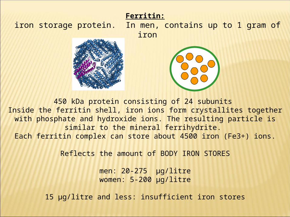
Ferritin: iron storage protein. In men, contains up to 1 gram of iron
450 kDa protein consisting of 24 subunits Inside the ferritin shell, iron ions form crystallites together with phosphate and hydroxide
ions. The resulting particle is similar to the mineral ferrihydrite. Each ferritin complex can store about 4500 iron (Fe3+) ions.
Reflects the amount of BODY IRON STORES
men: 20-275 μg/litrewomen: 5-200 μg/litre
15 μg/litre and less: insufficient iron stores

Ferritin
x 24
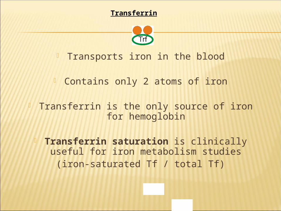
Transports iron in the blood
Contains only 2 atoms of iron
Transferrin is the only source of iron for hemoglobin
Transferrin saturation is clinically useful for iron metabolism studies
(iron-saturated Tf / total Tf)
Transferrin

Transferrin saturation:
Normal about 30-50 %
Transferrin saturation under 15 %= Iron deficiency

Transferrin

pH 5.5A) Uptake (TfR cycle)
C) Storage B) Metabolic Utilization
Extracellular Space
Cytoplasm
D) Export
Heme Iron Containing Proteins
Ferritin
Protoporphyrin IX5-Aminolevulinate
Succinyl-CoA + Glycine
Mitochondrion
The transferrin cycle
Dmt1
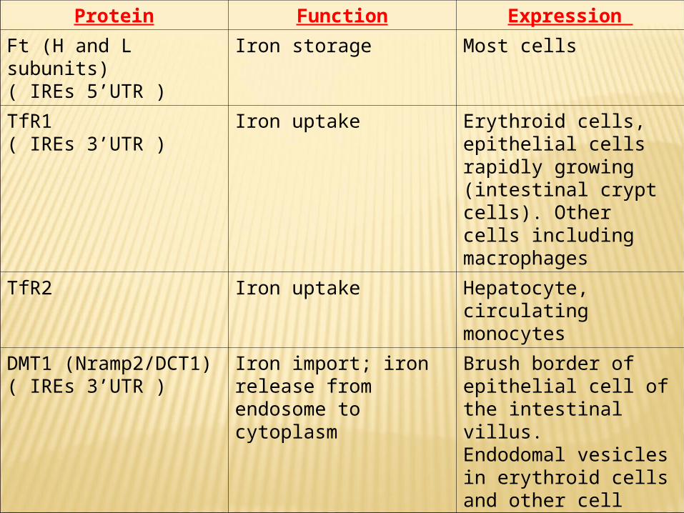
Protein Function Expression
Ft (H and L subunits)( IREs 5’UTR )
Iron storage Most cells
TfR1( IREs 3’UTR )
Iron uptake Erythroid cells, epithelial cells rapidly growing (intestinal crypt cells). Other cells including macrophages
TfR2 Iron uptake Hepatocyte, circulating monocytes
DMT1 (Nramp2/DCT1)( IREs 3’UTR )
Iron import; iron release from endosome to cytoplasm
Brush border of epithelial cell of the intestinal villus.Endodomal vesicles in erythroid cells and other cell types
FP1/IR1/MTP1( IREs 5’UTR )
Iron export Basolateral membrane of epithelial cell of the intestinal villus; placenta; other cell types

Protein, carriers, & “regulators” in iron
metabolism
Protein Function Expression
Duodenal cytochrome b Ferric reductase Brush border of enterocytes of the intestinal villus.
Ephaestin Ferroxidase Basolateral membrane and vesicles of enterocytes of the intestinal villus.
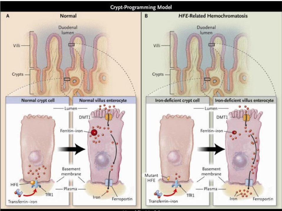
HFE (No IREs) Interacts with TfR1 Intestinal crypt cells and tissue macrophages
Hepcidin (No IREs) Antibacterial activity; iron homeostasis?
Liver; blood; urine
IRP1 AND IRP2 Posttranscriptional control of target mRNAs (Ft, TfR1, DMT1, FP1/IR1/MTP1, mitochondrial aconitase, erythroid aminolevulinate synthase)
Mainly: Liver, spleen, kidney, heart.Also: duodenum, brain


Fe 2+Tf binds to TfR1 on RBC. Fe 2+Tf/TfR1 complex localize to clathrin-coated pits which invaginate to form endosomes. A proton pump decreases pH leading to iron release. DMT1 moves iron across endosomal membrane
to cytoplasm. Apo-Tf& TfRl are recycled to the cell surface for further use. In RBC most iron is incorporateed
into protoporphyrin to make Heme (mitochondria).

specialized transport systems and membrane carriers have evolved in humans to maintain iron in a "soluble" state suitable for circulation in the
bloodstream [i.e., bound to serum transferrin (Tf)] or to transfer it across cell membranes
[through metal transporters such as divalent metal transporter 1 (DMT1) or ferroportin1/Ireg-
1/MTP-1] for tissue utilization. Since there is no mechanism for delibrate
excretion of iron through the liver or kidneys, iron homeostasis is maintained primarily by the
tight regulation of iron absorption in the intestine and the high degree of conservation of
body iron stores.

It is induced by iron deficiency. Mediates cellular iron egress in
conjunction with the ferroxidase, hephaestin.
Localizes to the basolateral membrane of polarized cells.
IREs are at the 5’UTR of FP1 mRNA.

Ferroxidase activity. It oxidizes iron as part of the
transmembrane transfer process &/or the process of loading iron onto plasma Tf.
Mice hemizygous or homozygous for sex-linked anemia (sla) mutation have partial loss of hephaestin function & resultant iron deficiency due in part to hephaestin mislocalization away from basolateral membrane.


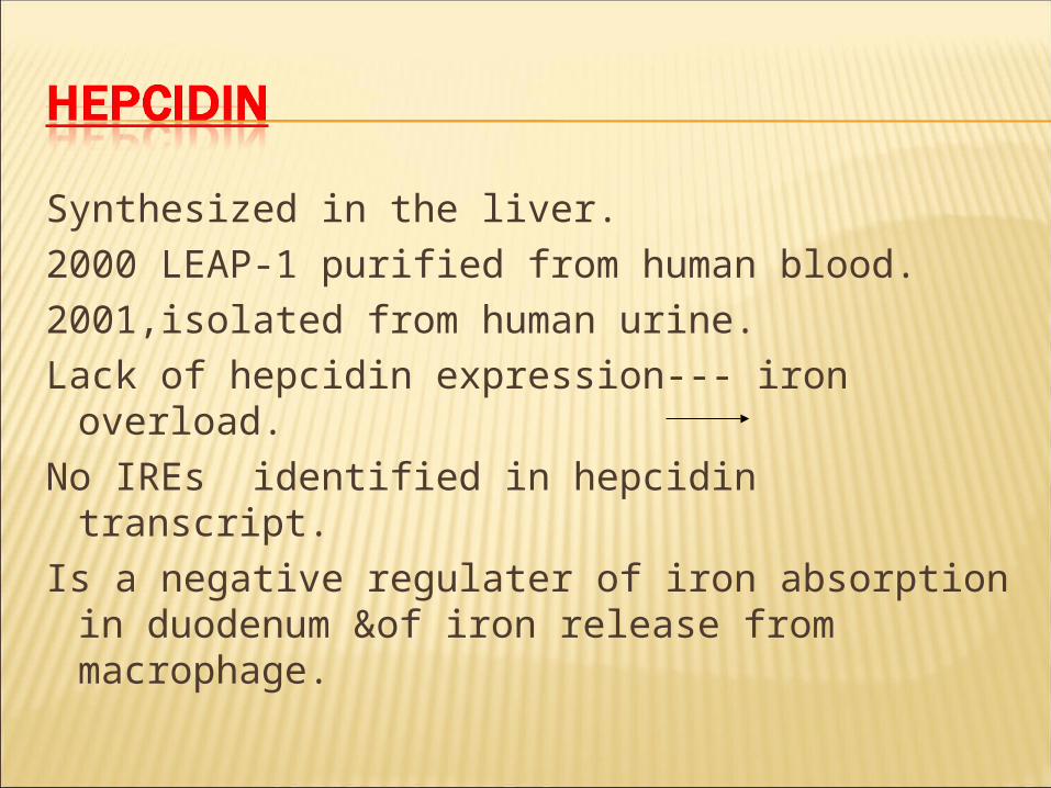
Synthesized in the liver.2000 LEAP-1 purified from human blood.2001,isolated from human urine.Lack of hepcidin expression--- iron overload.No IREs identified in hepcidin transcript.Is a negative regulater of iron absorption in
duodenum &of iron release from macrophage.

Is secreted in response to change in the ratio of diferric Tf in the circulation to TfR1.
Changes detected by TfR2&HFE-TfR1. It directly influences the expression of
DMT1 & ferroportin in enterocytes, there by regulating absorption in response to body iron requirements.

HC represent a common single-gene hereditary disorder.
C282Y mutation disrupts a critical disulfide bond in HFE ptn & abrogates its binding to B2microglobulin------reduced expression on the cell.
HFE Knockout &knockin mice recapitulate the human disease.

Paradoxically, the mRNA coding for the HC gene product HFE, does not contain IREs nor is it known to be regulated by iron status.Yet, HFE has a dramatic impact on cell iron trafficking &, indirectly, on intestinal iron absorption.

The "information" on the erythron and body iron status is transferred to the crypt cells of the distal duodenum by Tf: the extent of Tf saturation with iron acts as the "signal" that is transferred through the transferrin receptor (TfR)/HFE pathway to the stem cells of the crypts. This sets the level of "free iron pool" in the crypt cell that will also be reflected in the mature enterocytes on differentiation and migration to the villus. The free iron pool through the iron regulatory proteins (IRP) will dictate the level of expression of apical and basolateral iron transporters in the mature enterocytes of the villus and, in turn, of iron absorption.

B: in iron-deficiency anemia, the circulating iron-poor Tf will signal to RE cells the increased erythron demands prompting for iron release and diverting iron from the periphery, including intestinal cells, to the erythron. Due to low availability of circulating iron, the decreased free iron pool in crypt cells will signal the mature enterocyte to activate the iron uptake transfer machinery.

. C: during secondary iron overload (e.g., transfusion siderosis), iron-saturated Tf will cause the RE cells and crypt cells to retain iron; this will then downregulate iron carriers in the mature iron-replete enterocytes and decrease iron absorption.

D: in hereditary hemochromatosis, due to the defective HFE and the faulty HFE/TfR pathway, both RE cells and crypt cells receive an incorrect signal of "iron deficiency" despite increasing saturation of circulating Tf with iron. This, paradoxically, will lead, as in a "true" iron-deficiency state, both macrophages and duodenal enterocytes to "release" more iron. Duodenal cells will accomplish this by activating the iron uptake transfer machinery. DMT1, divalent metal transporter 1.

