iron metabolism - aiims, rishikesh
TRANSCRIPT

Iron MetabolismDR BELA GOYAL

MineralsTrace elements: Required in amounts in mg/day
Ultratrace elements: Required in amounts less than 1mg/day or ug/day
(Trace and ultratrace elements in health and disease FH Nielsen -Comprehensive therapy, 1991 - naldc.nal.usda.gov)

Learning objectives(Iron)Distribution and requirements of Iron
Metabolism of Iron
Molecular mechanisms of Regulation
Laboratory evaluation of Iron deficiency anemia
Iron overload disorders

Body Iron Distribution

Iron cycle


Daily requirement(RDA)Male: 8-10mg
Female: 10-15mg
Preg. & lactation: 20-40mg
Children: 10-15mg
liver, meat, fish, spleen, leafy veg., legumes,pulses,cereals, JAGGERY, DATESCooking in iron utensils

Types of iron
Heme Proteins: Hb, myoglobin, Cytochromes (cytb,c,c1,p450), cyt oxidase, catalase, peroxidase, tryptophan pyrrolase, nitric oxide synthase
Iron-sulphur complexes : ETC, NADH dehydrogenase, succinate dehydrogenase, xanthine oxidase
Non-heme iron containing proteins :Aconitase, Transferrin , Ferritin, Hemosiderin
Absorption10% of dietary iron is absorbed
In duodenum & jejunum
Gastric juice HCl liberates free ferric ions from proteins

Iron absorption


Factors affecting absorptionHeme iron more efficiently absorbed.
Ferrous form is absorbed
Increased by: Vit.C , cysteine , glutathione , acidic pH & iron deficient state
Decreased by: alkaline medium , tea , coffee, phytates , oxalates, Ca, gastrectomy

Transport Transferrin: non–heme iron binding glycoprotein
synthesized by liver cells
apotransferrin is apoprotein binds two atoms of iron
Iron transported to RE cells, bone marrow (immature RBCs)

Delivery of Iron to cells by transferrin

Iron Recycling
Cell. 2004 Apr 30;117(3):285-97.Balancing acts: molecular control of mammalian iron metabolism.

Regulation of systemic Iron homeostasis
Cell. 2010 Jul 9;142(1):24-38. doi: 10.1016/j.cell.2010.06.028.

Regulation of Cellular Iron Metabolism
Cell. 2010 Jul 9;142(1):24-38. doi: 10.1016/j.cell.2010.06.028.

Hepcidin based Therapy
Hepcidin agonist?
Antibodies/siRNA for hepcidin?

FerritinIron is stored in liver, spleen, bone marrow &
intestinal mucosal cells
Apoferritin – apoprotein(500 Kda), 24 subunits
4000 iron atoms stored in a ferritin molecule
Ferritin an index of total body iron stores

HemosiderinDerived from ferritin
(sec. lysosomes)
Seen in iron overload
Mobilisation of iron slower
Excretionexcreted (1-2mg/d)
Haptoglobin binds Hb to prevent its loss in urine
Hemopexin binds heme
Faeces contain unabsorbed iron & iron inside the desquamated intestinal cells
In females, more excreted during mensturation

Laboratory evaluation of Iron MetabolismPacked cell volume
Hemoglobin
Red cell count and indices
Total iron
TIBC
Percent saturation
Transferrin
Ferritin

Total Iron Content (Serum Iron)
Fe3 bound to transferrin (50-170µg/dL)
Specimen of choice: serum or plasma with heparin. (Oxalate, citrate, or EDTA binds Fe ions)
Steps:
Fe3 is released from binding proteins by acidification,
reduced to Fe2 by ascorbate
complexed with a color reagent such as ferrozine, ferene, or bathophenanthroline.
Spectrophotometric determination

Total Iron Content (Serum Iron)Decreased
Hemorrhage
Iron Deficiency anemia
Increased
Iron loading disorders
Iron poisoning

Total Iron-Binding Capacity (TIBC)The amount of iron that could be bound by saturating transferrin and other minor iron-binding proteins present in the serum or plasma sample.
one-third of the iron binding sites on transferrin are saturated

Total Iron-Binding CapacityDetermined by
Adding sufficient Fe3 to saturate the binding sites on transferrin,
Excess iron removed by addition of MgCO3 to precipitate any Fe3 remaining in solution.
After centrifugation to remove the precipitated Fe3,
The supernatant solution containing the soluble iron bound to proteins is analyzed for total iron content.
This is the TIBC, which ranges from around 250 to 425 µg/dL.


Percent Saturation

Transferrinmeasured by immunochemical methods
Increased
Iron deficiency
Decreased
iron overload
Chronic infections and malignancies
primarily monitored as an indicator of nutritional status.

Ferritin
Measured by immunochemical methods like ELISA and chemiluminescent techniques.
Reflects Iron store
Earliest marker to decline

FerritinDecreased
iron-deficiency anemia( most sensitive and earliest)
Increased
Iron overload ( used to gauge effectiveness of phlebotomy therapy).
Chronic infections, malignancy
Viral hepatitis release of ferritin from diseased liver cells

Clinical SignificanceIron Deficiency anemia
Iron overload disorders
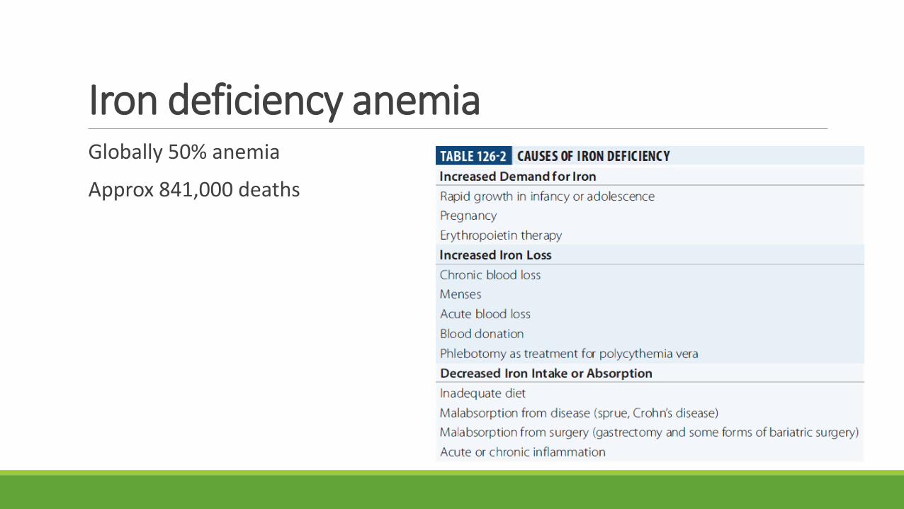
Iron deficiency anemiaGlobally 50% anemia
Approx 841,000 deaths

Clinical presentation of IDAMuscle abnormalities
Koilonychia
Pica
Anemia
Reduced work performance
Impaired cognitive development
Premature labour
Increased Perinatal deaths

Diagnosis
Microscopic examination of a blood smear : microcytic hypochromic RBCs
Bone marrow aspiration: no storage iron
serum ferritin: virtually zero
serum TIBC: elevated
serum iron saturation: less than 16%.
Treatment: •Careful examination for the cause and supplementation with iron (oral ferrous sulfate tablets)
• intravenous iron therapy
•transfusion with packed red blood cells


Iron overloadHemosiderosis
Hemochromatosis

HemosiderosisCauses: Repeated blood transfusions (Thalassemia, Hemophilia)
Hemosiderin pigments- golden brown granules in liver, spleen
No associated tissue injury

Hereditary Hemochromatosis(HHC).
Primary (Bronze Diabetes)
autosomal recessive
abnormal gene on chromosome 6
↑ iron absorption
Triad of Bronzing of skin, cirrhosis and diabetes
The liver is the first organ to be affected (hepatomegaly)
Primary hepatocellular carcinoma is 200 times more
Common


Hemochromatosis
Juvenile : ? Mutation in hepcidin
Variant: Mutation in ferroportin
Secondary : BT in Thal major, myelodysplastic syndromes
Diagnosis: Transferrin saturation and Ferritin
Treatment:
Phlebotomy
Iron chelating therapies
