iron metabolism 2013 - eth zacac1.ethz.ch/koppenol/iron_metabolism_2013_a.pdf · iron-sulfur...
TRANSCRIPT


OUTLINE
_ Introduction_General properties_Overview of iron-containing enzymes and their role
_Selected Fe-containing enzymes_Hemoglobin, myoglobin (O2-uptake and O2-transport)_Transferrin (Fe-transport), and receptor-mediated cellular iron uptake_Ferritin/hemosiderin (Fe-storage)
_ Iron metabolism_Duodenal iron uptake_Iron metabolism in cells and macrophages_Regulation of iron homeostasis
_Diseases related to the iron metabolism_Iron overload_Iron-deficiency anemia
2

OUTLINE
_Introduction_General properties_Overview of iron-containing enzymes and their role
_Selected Fe-containing enzymes_Hemoglobin, myoglobin (O2-uptake and O2-transport)_Transferrin (Fe-transport), and receptor-mediated cellular iron uptake_Ferritin/hemosiderin (Fe-storage)
_ Iron metabolism_Duodenal iron uptake_Iron metabolism in cells and macrophages_Regulation of iron homeostasis
_Diseases related to the iron metabolism_Iron overload_Iron-deficiency anemia
3

INTRODUCTION (1)
4
1) Iron is essential but toxic
2) Iron is not very bioavailable
1a) Iron is essential
_Only few bacteria can live without iron (e.g. the lyme disease pathogen Borrelia burgdorferi seems to use Mn instead of Fe)
_Involved in all essential metabolic processes_photosynthesis_respiration_electron transfer_elimination of noxious metabolites of O2
_DNA-synthesis_detoxification_fixation of N2 and H2
_hydratase_hydrolase_mineralization of theeth by invertebrates

INTRODUCTION (2)
5
1b) Iron is toxic
_FeII/FeIII/FeIV: redox processes
_e−-donor/acceptor mostly O2 or one of its derivatives
_Reactions may lead to the formation of partially reduced
oxygen species („free radicals“, „reactive oxygen species“ or ROS“)
_Fenton reaction → cellular injury
_Nature has developed strategies to avoid the presence
of „free iron“

INTRODUCTION (3)
6
2) Iron is not bioavailable
_abundant element in the universe, on the earth crust1…
_but not in the ocean (as low as 50 pM!)
_Why?
→ Solubility of Fe3+, stability of Fe3+ compelxes
→ Bioavailabilty increased via reduction to Fe2+
(1) http://en.wikipedia.org/wiki/Abundance_of_the_chemical_elements

BASIC IRON CHEMISTRY
7
„Free iron“ (aerobic environment): [Fe(H2O)6]3+
_pKa ~ 2
_neutral conditions: insoluble ferric hydroxides (rust)
Ksp(Fe(OH)3) = 10–38/10–39
at pH = 7: [Fe(H2O)6]3+ ~ 10–17/10–18 M
_Iron(III) concentration in the ocean: 50 pM (higher!)
→ chelators bind iron(III), e.g. citrate, EDTA, O-donor ligands
→ increase solubility
→ but chelated iron(III) is not available for uptake (rate of H2O
exchange, i.e. of ligand exchange is rather low)

STRATEGY TO MAKE IRON MORE BIOAVAILABLE
8
Reduce iron(III) to iron(II)
_rate of H2O exchange 3–4 orders of magnitude larger
_affinity of iron(II) to natural O-donor ligands 3–4 orders of
magnitude lower, e.g. EDTA complex 108 times lower
_ferrous hydroxide is more soluble
Ksp(Fe(OH)2) = 10–15
at pH = 7: [Fe(H2O)6]2+ ~ 0.08 M

9 Modified from: Henzte et al. Cell (2004) 117, 285–297.
SYSTEMIC IRON HOMEOSTASIS
Ferritin
Hemo-globin
Transferrin
Myoglobin (300 mg)∼100 enzymes (100 mg)

10 © Galenica Group 23.11.2013
IRON-CONTAINING PROTEINSNON-HEME-CONTAINING PROTEINS
Protein (class) Function %Fe body
Non-heme proteins
ribonucleotide reductase
Oxidative metabolism
DNA biosynthesis
Very little
Iron-sulfur proteins
aconitaseiron regulatory protein
Electron transfer
Citric acid cycleFe homeostasis
~1
Ferritin
Hemosiderin
Fe storage 20
variable
Transferrin Fe transport ~0.2

11 © Galenica Group 23.11.2013
IRON-CONTAINING PROTEINSNON-HEME-CONTAINING PROTEINS
_Non-heme containing proteins
Fe-S-proteinsRibonucleotide reductase
″rust″
Ferritin

IRON-CONTAINING PROTEINSHEME-CONTAINING PROTEINS
N
N N
N
Fe
Protein Function Localization %Fe body
Hemoglobin O2 uptake and transport Red blood cells 65
Myoglobin O2 transport and storage Heart, skeletal muscle 6
cyt c oxidase
cyt c
cyt P450
Oxidative production of cellular energy
Electron transfer
Oxidative degradation
Mitochondria
Endoplasmic reticulum
<1
Peroxidases Oxidation GranulocytesMacrophages
0.1
Catalase H2O2 breakdown e.g. liver macrophages 0.1
Cyt: cytochrome12 © Galenica Group 23.11.2013

IRON ENZYMES INVOLVED IN ENERGY METABOLISM
13
_Respiratory chain in the mitochondria:

THE RESPIRATORY CHAIN IN THE MITOCHONDRIA
14
Citric acidcycle
Glycolisis
NADHFADH2

IRON ENZYMES INVOLVED IN ENERGY METABOLISM
15
_Respiratory chain in the mitochondria:
Cytochrome c oxidase reduction of oxygen to water
Cytochromes
Ferredoxins electron transfer
Rieske proteins
_Citric acid cycle:Aconitase _dehydratase/hydratase (isomerase)
_apo-aconitase (proetin without Fe-S-cluster)acts as iron regulatory protein
→ Possible effect of iron deficiency: reduced levels of these enzymes→ impaired physical performance→ fatigue

IRON ENZYMES INVOLVED IN IMMUNE DEFENCE
16
_Nitric oxide synthase NO●-synthesis (iNOS)
_Myeloperoxidase
_Eosinophil peroxidase hypohalogenite production (HOCl, HOBr)(action against microorganisms)
_Lactoperoxidase
→ Possible effect of iron deficiency: reduced levels of these enzymes→ increased susceptibility to infectious diseases

IRON ENZYMES INVOLVED IN ANTIOXIDANT DEFENCE
17
_Peroxidases reduction of lipid peroxides_Catalase disproportionation of hydrogen peroxide_Cytochromes reduction of antioxidants, e.g. vitamin C
→ Possible effect of iron deficiency: reduced levels of these enzymes→ increased susceptibility to oxidative stress

IRON ENZYMES INVOLVEDIN NEURONAL METABOLISM AND SIGNALLING
18
_Nitric oxide synthase NO●-synthesis (nNOS)neuronal signalling function of NO●
_Hydroxylases involved in synthesis of neurotransmitters and hormones: dopamine, noradrenaline,
_Tyrosine peroxidase adrenaline, serotonine, epinephrine, norepinephrine
→ Effects of iron deficiency→ low brain iron levels→ decreased density of dopamine receptors→ decreased level of dopamine transporters

FURTHER ESSENTIAL IRON-CONTAINING ENZYMES
19
_Ribonucleotide reductase first step of DNA synthesis
_Ferrochelatase heme synthesis
_Cytochrome P450 family detoxification reactions in the liver
_Cyclooxygenase (COX) prostaglandin, thromboxane,prostacyclin synthesis
_Lipoxygenase isoenzymes prostaglandin and leukotrienemetabolism
_Lipoate synthase lipoate synthesis(precursor of different enzymes)

OUTLINE
_ Introduction_General properties_Overview of iron-containing enzymes and their role
_Selected Fe-containing enzymes_Hemoglobin, myoglobin (O2-uptake and O2-transport)_Transferrin (Fe-transport), and receptor-mediated cellular iron uptake_Ferritin/hemosiderin (Fe-storage)
_ Iron metabolism_Duodenal iron uptake_Iron metabolism in cells and macrophages_Regulation of iron homeostasis
_Diseases related to the iron metabolism_Iron overload_Iron-deficiency anemia
20

HEMOGLOBIN, MYOGLOBINAND OTHER DIOXYGEN CARRIERS
21
_Oxygen carriers needed because of low solubility of O2 in H2O
_Oxygen carriers are Fe- or Cu-containing proteins_Hemoglobin (Hb) heme_Hemerythrin (Hr) iron (non-heme)_Hemocyanin (Hc) copper
_Essential features of oxygen binding proteins_reversible binding_no redox reaction_no generation of partially reduced oxygen sepcies
Literature for Hb: any biochemistry or bioniorganic chemsitry text book!

HEMOGLOBIN AND MYOGLOBIN → BLOOD
22
_Blood: ca. 7–8% of body weight, ca. 5 L, pH 7.35−7.45_Plasma
H2O, Na+Cl− (ca. 0.9% w/w), glucose, proteins, O2,CO2/HCO3
− , …_White blood cells (immune system)_Red blood cells
_Red blood cells (RBC)_optimized form (large surface, shape)_half of the volume of human blood (2.5 ×10¹³)_most weight of RBC is hemoglobin
(5 mM, ca. 800 g in men and 550 g in women)_life span: 80−120 days
→ Fe recycled, porphyrin metabolized (CO!)_production: 1010 RBC/h
MYOGLOBIN (Mb)
23
_ca. 10% of the amount of hemoglobin
_Transports O2 from Hb to mitochondria of muscle cells (cyt c oxidase)
_Correlation in concentration: Mb and cyt c oxidase

MYOGLOBIN (Mb) STRUCTURE
24
_Monomer
_First X-ray of a protein(John Kendrew andMax Perutz,1958)
_ca.150 amino acids,mostly α-helices (globin), heme, highly conserveddistal and proximal histidine
http://en.wikipedia.org/wiki/Myoglobin
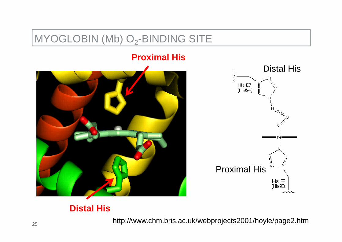
MYOGLOBIN (Mb) O2-BINDING SITE
25 http://www.chm.bris.ac.uk/webprojects2001/hoyle/page2.htm
Distal His
Proximal HisDistal His
Proximal His

MYOGLOBIN (Mb) OXYGEN BINDING CURVE
26http://www.aw-bc.com/mathews/ch07/c07obm.htm
_P50 ≈ 2 mm Hg
_Muscle ≈ 35−40 mm Hg
_Lung ≈ 80−100 mm Hg
Binding fraction

HEMOGLOBIN (Hb) STRUCTURE
27 http://en.wikipedia.org/wiki/Hemoglobin
_Tetramer (α2β2)
_Each chain:ca.150 amino acids,mostly α-helices (globin), heme, highly conserveddistal and proximalhistidine
_Cooperative oxygen binding(sigmoidal oxygen bindingcurve)
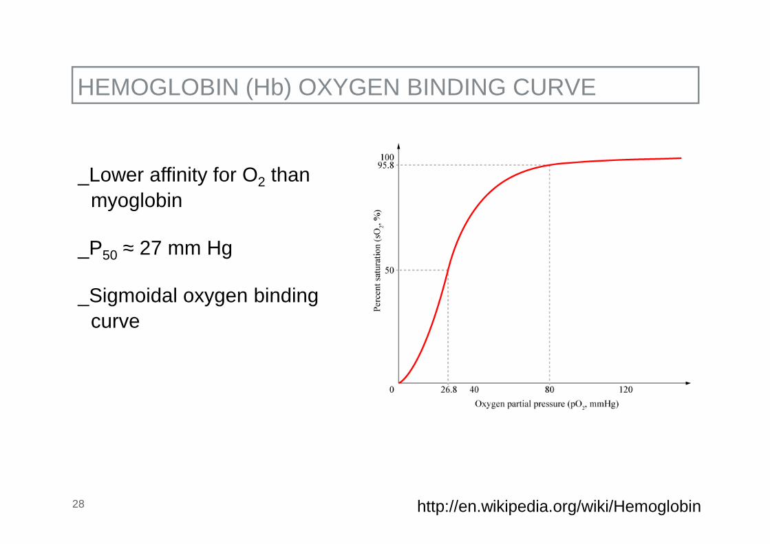
HEMOGLOBIN (Hb) OXYGEN BINDING CURVE
28 http://en.wikipedia.org/wiki/Hemoglobin
_Lower affinity for O2 thanmyoglobin
_P50 ≈ 27 mm Hg
_Sigmoidal oxygen bindingcurve

OXYGEN BINDING CURVES (Hb, Mb)
29 http://employees.csbsju.edu/hjakubowski/classes/ch331/bind/olbindhemoglobin.html

HEMOGLOBIN (Hb) OXYGEN BINDING CURVE
30 http://en.wikipedia.org/wiki/Hemoglobin
_Different (higher) affinity foreach subsequent O2-moleculebinding to the protein
_Allosteric ligands (effectors)regulate the binding of O2
to Hb: H+, CO2, andbisphosphoglycerate(see later)

MOLECULAR BASES FOR COOPERATIVE O2-BINIDING
31
_Two main conformational states:_deoxy (or T, tense) → low O2-affinity_oxy (or R, relaxed) → high O2-affinity
http://employees.csbsju.edu/hjakubowski/classes/ch331/bind/olbindhemoglobin.html

COOPERATIVE O2-BINIDING AT AN ATOMIC LEVEL (1)
32
_deoxyHb (T-state): Fe(II), high spin, 0.78 Å, d6, paramagnetic
_oxyHb (R-state): diamagnetic
_two possibilities: Fe(III)−O2●−, low spin, 0.55 Å, d5; S = ½;
antiferromagnetic coupling
Fe(II)−1O2, low spin, 0.61 Å, d6; S = 0;∆E(1O2/ 3O2) = ~22 kcal/mol (94 kJ/mol)

COOPERATIVE O2-BINIDING AT AN ATOMIC LEVEL (2)
33 http://www.ul.ie/~childsp/CinA/Issue65/TOC28_Haemoglobin.htm
Transition from T(deoxyHb)- to R(oxyHb)-state:
_conformational change of the porphyrin/heme
_cleavage of 8 salt bridges (that stabilize T-state)

CLEAVED SALT BRIDGES
34
6 of the salt bridges are between different subunits, with 4 of those involving the C- or N-terminus
http://employees.csbsju.edu/hjakubowski/classes/ch331/bind/olbindhemoglobin.html

35
_H-bonds between Tyr140 (α-chain) or Tyr145 (β-chain) and the
carbonyl O of Val93 (α-chain) or Val98 (β-chain) are cleaved
_When Fe binds O2, Fe is pulled into the plane of the heme ring
(shift of about 0.2 nm)
_This small shift leads to larger conformational changes since the
subunits are packed so tightly that compensatory changes in their
arrangement must occur
_The proximal His is pulled toward the heme, which causes the F
helix to shift, causing a change in the FG corner (the sequence
separating the F and G helices) at the alpha-beta interface
(→ cleavage of salt bridges)
SUMMARY: CHANGES T-STATE → R-STATE

MOLECULAR BASES FOR COOPERATIVE O2-BINIDING
36 http://employees.csbsju.edu/hjakubowski/classes/ch331/bind/olbindhemoglobin.html

37
_Lower pH → lower O2 affinity stabilization of T-state, moresalt bridges because Hisprotonated
_Not so relevant when PO2 high
_But in tissues PO2 low,
pH is lower
→ O2 easily dissociated
→ Hemoglobin even more effective
ALLOSTERIC EFFECTORS, BOHR EFFECT
http://www.aw-bc.com/mathews/ch07/c07be.htm
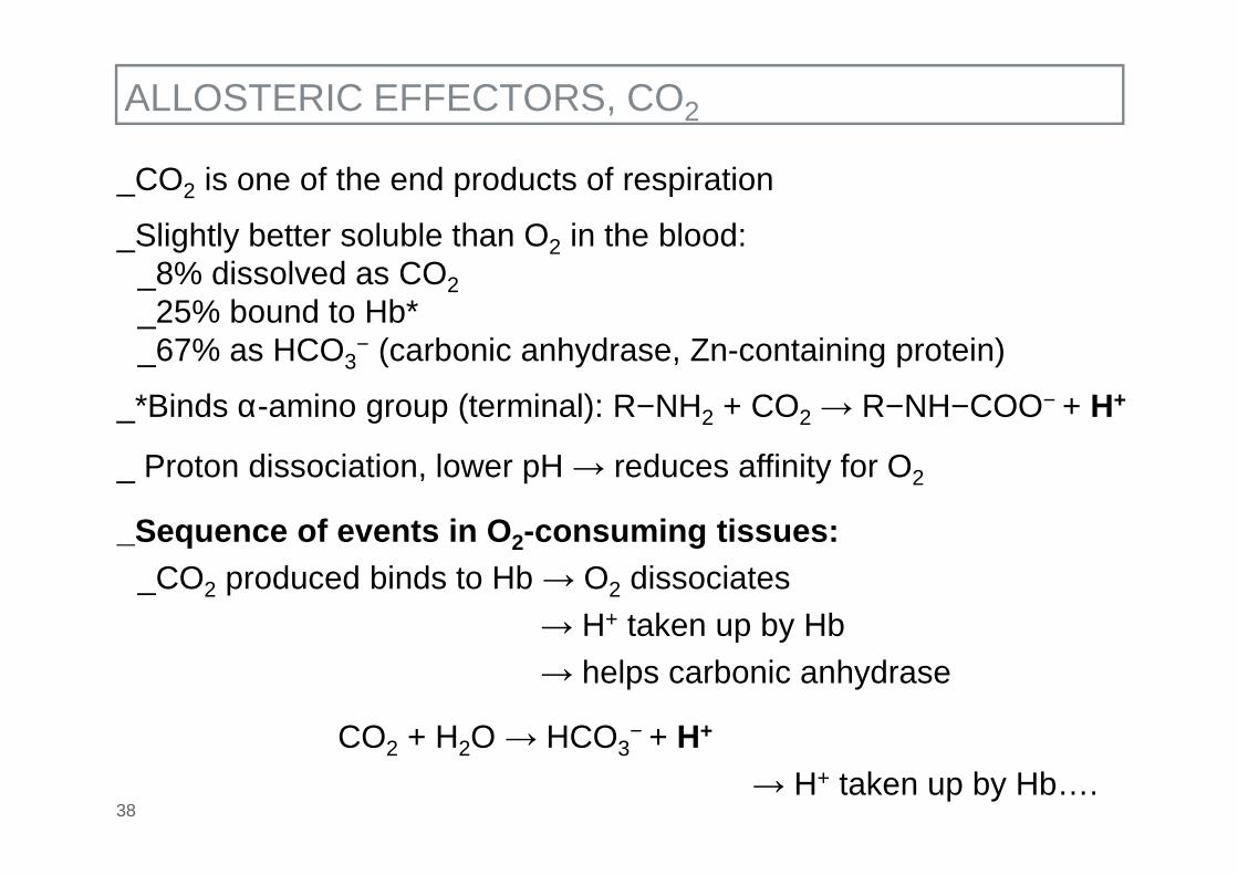
38
_CO2 is one of the end products of respiration
_Slightly better soluble than O2 in the blood:_8% dissolved as CO2_25% bound to Hb*_67% as HCO3
− (carbonic anhydrase, Zn-containing protein)
_*Binds α-amino group (terminal): R−NH2 + CO2 → R−NH−COO− + H+
_ Proton dissociation, lower pH → reduces affinity for O2
_Sequence of events in O2-consuming tissues:_CO2 produced binds to Hb → O2 dissociates
→ H+ taken up by Hb
→ helps carbonic anhydrase
CO2 + H2O → HCO3− + H+
→ H+ taken up by Hb….
ALLOSTERIC EFFECTORS, CO2

O2 AND CO2 METABOLISM
39
CO2
CO2
NH2
O2
H+
NH–COO–
Carbonic anhydraseCO2 + H2O
H+
O2
Carbonic anhydrase
NH2
CO2 + H2O
CO2
CO2NH–COO–
HCO3–
Cl–
HCO3–
Cl–
Tissues Plasma
Red blood cell
HbFeO2 HbFeII
HCO3– + H+
Lungs Plasma
Red blood cell
HbFeO2 HbFeII
HCO3– + H+

40
_Lower pH → lower O2 affinity stabilization of T-state, moresalt bridges because Hisprotonated
_Not so relevant when PO2 high
_But in tissues PO2 low,
pH is lower
→ O2 easily dissociated
→ Hemoglobin even more effective
ALLOSTERIC EFFECTORS, BOHR EFFECT
http://www.aw-bc.com/mathews/ch07/c07be.htm
Binding of CO2 to Hb also shifts the curve towards right, i.e. lowers thebinding affinity of Hb for O2

41
_2,3,-bisphosphateglycerate (BPG) binds in the opening between theβ-chains of deoxyHb
_BPG concentration in red blood cells: ~5 mM
ALLOSTERIC EFFECTORS, bis-PHOSPHOGLYCERATE (1)
http://www.aw-bc.com/mathews/ch07/c07bh.htm

42
_The opening is much smaller in oxyHb: BPG cannot bind to theoxyHb form
ALLOSTERIC EFFECTORS, bis-PHOSPHOGLYCERATE (2)
http://www.aw-bc.com/mathews/ch07/c07bh.htm

43
_The higher the BPG content in red blood cells, the more stable thedeoxyHb form
→ decrease in O2-affinity → O2-release
_At high altitudes (low PO2) the BPG levels increase within a few days
Example:_At 4500 m, PO2 in lungs drops from ~100 to ~50 mm Hg (12 to 7 kPa)
→ within 24 h the BPG concentration raises from 5 to 8 mM→ lowers O2-affinity → enables tissues to obtain O2 in spite of its diminished availability
_On returning to low altitude the concentration of BPG (half-life 6 h)returns rapidly to normal levels
ALLOSTERIC EFFECTORS, bis-PHOSPHOGLYCERATE (3)

ALLOSTERIC EFFECTORS, bis-PHOSPHOGLYCERATE (4)
44
_In the lungdecreased affinity makes littledifference to the amount of O2 bound
_In the tissuesdecreased affinity allows muchmore O2 to be released
_At sea level, with normal BPG levels,occupancy goes from 0.97 in lungs to0.50 in tissues
_At 4500 m, with 8 mM BPG, occupancygoes from 0.75 in lungs to 0.35 in tissues
http://vohweb.chem.ucla.edu/voh/classes%5Csummer08%5C153A-2ID19%5CHbMb.pdf

OTHER DIATOMIC LIGANDS THAT BIND TO THE HEME IRON
45
1) CO: degradation of RBC, heme oxygenase (heme oxidase, see later)
2) NO● (endothelium-derived relaxing factor, EDRF): produced byeNOS, responsible for relaxation of smooth muscles surroundingthe blood vessels, resulting in vasodilation and thus increasedblood flow
1) CO: competitive binding vs. O2
Heme oxygenase: 1 CO/porphyrin
Normal conditions: ca. 1% HbCO (smokers up to 20%)
Hemoglobin structure reduces affinity for CO:
Fe-Porphyrin KCO/KO2 = 25’000
Hb(Mb) KCO/KO2 = 200
Why?
http://web.virginia.edu/Heidi/chapter15/chp15.htm

CO BINDING TO Hb
46
Why reduced affinity for CO?Old (wrong!) explanation (still found in many textbooks and internetpages)
http://web.virginia.edu/Heidi/chapter15/chp15.htm
HisE7 forces the CO molecule to tilt away from a preferred perpendicular alignment with the plane of the heme (“confirmed” by first X-ray structures)
First X-ray structures of Hb(Mb)CO: 135−150°Recent, more accurate, X-ray structures of Hb(Mb)CO: 167°

CORRECT EXPLANATION FOR REDUCED AFFINITY FOR CO
47
ElectronIc, not steric effect!
O2 binding: electrostatic stabilzation
CO binding: little electrostatic stabilization
Olson et al. JBIC (1997) 2, 544–552.

48 Olson et al. JBIC (1997) 2, 544–552.

NO● BINDING TO Hb(Mb)
49
NO● binding: partial electrostatic stabilization
Olson et al. JBIC (1997) 2, 544–552.

COMPARISON OF CO AND NO● BINDING TO Hb(Mb)
http://www.inchem.org/documents/ehc/ehc/ehc213.htm#1.950
CO is significantly more toxic than NO●
_WHO, levels of CO for which HbCO maximally 2.5%(even during light or moderate exercise):
→ 87 ppm for 15 min or 9 ppm for 8 h
_NO● is used in critical care to promote capillary and pulmonarydilation to treat primary pulmonary hypertension in neonatal patients
→ up to doses of 80 ppm/h
Why?_CO binding to Hb leads to stabilization of R-state
→ high O2-affinity, O2 cannot dissociate
_ NO● binding to Hb leads to stabilization of T-state→ low O2-affinity, O2 dissociates
WHO: world health organization

SUMMARY OF REACTIONS WITH HEMOGLOBIN
51blood HbFeO2

OUTLINE
_ Introduction_General properties_Overview of iron-containing enzymes and their role
_Selected Fe-containing enzymes_Hemoglobin, myoglobin (O2-uptake and O2-transport)_Transferrin (Fe-transport), and receptor-mediated cellular iron uptake_Ferritin/hemosiderin (Fe-storage)_Iron regulatory protein/aconitase (regulation of iron metabolism/citric acid cycle)
_ Iron metabolism_Duodenal iron uptake_Iron metabolism in cells and macrophages_Regulation of iron homeostasis
_Diseases related to the iron metabolism_Iron overload_Iron-deficiency anemia
52

HEMOCYANIN
53
Dioxygen carrier in most molluscs, and some arthropods (tarantula, 24-mer), including crabs.

OTHER DIOXYGEN CARRIERS(1)
54
_Invertebrates_Hemerythrin (Hr) iron (non-heme)_Hemocyanin (Hc) copper
_Hemerythrin: large variety, several X-ray structures, from monomericto octameric (cooperativity)Active site: deoxyHr: 2 FeII; oxyHr: 2 FeIII/peroxo; metHr: 2 FeIII/oxo
http://en.wikipedia.org/wiki/Hemerythrin

OTHER DIOXYGEN CARRIERS (2)
55
_Hemocyanin_Large proteins, mostly 6–10 domains, each domain 2 Cu_Active site:
_deoxyHc: 2 CuI
_oxyHc: 2 CuII/peroxo
http://www.ml.duke.edu/projects/Magnus/images/hemocyanin_color.gif
Intense blue (ligand tometal charge transfer)