role of diffusion weighted mr-imaging in the evaluation of
TRANSCRIPT
RESEARCH Open Access
Role of diffusion weighted MR-imaging inthe evaluation of malignant mediastinallesionsYoussriah Yahia Sabri1, Eman Zaki Bassyouni Nossair2* , HebatAllah Hany Assal3 and Hisham Samir Wahba4
Abstract
Background: Conducted studies showed that the ADC (apparent diffusion coefficient) values of malignantmediastinal lesions are significantly lower than those of benign lesions. Investigators determined cut-off ADC valuesto differentiate the two; concluding that ADC value is a promising noninvasive, imaging parameter that helpsassess and characterize mediastinal tumors.Taking this a step forward, the primary objective of our prospective study was to investigate the potential of DW-MRI (diffusion-weighted magnetic resonance imaging) to characterize malignant mediastinal lesions using theirADC values.Thirty-three patients that underwent MRI of the chest with DWI and latter pathologically diagnosed with amalignant mediastinal lesion were included in this study. Lesions’ ADC values were measured and correlated withthe histopathological results. The statistical significance of differences between measurements was tested using theone-way ANOVA (analysis of variance) test; p values equal to or less than 0.05 were considered significant.
Results: There was no statistically significant difference between the ADCmean values of the histopathologicalgroups of lesions assessed with the overlap of their ADCmean values. The average ADCmean value of NHL (non-Hodgkin lymphoma) was evidently lower than that of HD (Hodgkin disease) with no overlap between theirADCmean values. DWI failed at characterizing one lesion in this study as a malignant tumor, namely an immatureteratoma (germ-cell tumor). Again DWI could not be used to evaluate a mass, latter pathologically diagnosed as anangiosarcoma, because of its overall hemorrhagic nature showing no definite non-hemorrhagic soft tissuecomponents. The aforementioned results did not differ considerably when minimum ADC was used instead ofmean ADC.
Conclusion: There was no statistically significant difference between the ADC values of the malignant mediastinallesions evaluated. However, regarding lymphoma subtypes, our limited sample study of lymphoma suggested aconsiderable difference between the ADC values of Hodgkin disease and non-Hodgkin lymphoma.
Keywords: Malignant mediastinal lesions, DWI (diffusion-weighted imaging), ADC (apparent diffusion coefficient)
BackgroundThe mediastinum comprises the thoracic compart-ment anatomically bounded by the thoracic inlet su-periorly, the diaphragm inferiorly, the posteriorsternal border anteriorly, and posteriorly by the ver-tebral column. It is an intricate segment of thethorax that contains vital intra-thoracic structures
such as the heart and great vessels, trachea and mainbronchi, esophagus, thymus, venous and lymphaticstructures, and nerve tissue. Lesions of the mediasti-num span a wide histopathological and radiologicalspectrum. Both anatomical information (location)and internal characteristics (composition) are essen-tial to formulate the differential diagnosis of a massin the mediastinum and therefore determine thera-peutic options [1–3].A cross-sectional imaging-based classification that divides
the mediastinum into anterior (prevascular), middle
© The Author(s). 2020 Open Access This article is distributed under the terms of the Creative Commons Attribution 4.0International License (http://creativecommons.org/licenses/by/4.0/), which permits unrestricted use, distribution, andreproduction in any medium, provided you give appropriate credit to the original author(s) and the source, provide a link tothe Creative Commons license, and indicate if changes were made.
* Correspondence: [email protected], National Hepatology and Tropical Medicine ResearchInstitute (NHTMRI), 10 (A) Kasr El-Aini St, Cairo, EgyptFull list of author information is available at the end of the article
Egyptian Journal of Radiologyand Nuclear Medicine
Sabri et al. Egyptian Journal of Radiology and Nuclear Medicine (2020) 51:32 https://doi.org/10.1186/s43055-020-0132-6

(visceral), and posterior (paravertebral) compartments wasrecently published by the International Thymic MalignancyInterest Group (ITMIG). Contents of each mediastinalcompartment, as well as the most commonly encounteredlesions, are reviewed in Table 1 [4].Computed tomography (CT) is considered the
imaging modality of choice for evaluating most me-diastinal masses; however, the role of magnetic res-onance (MR) imaging continues to expand given itshigh soft-tissue contrast. It is superior to CT in dif-ferentiating between cystic and solid masses, identi-fying cystic and solid components within complexlesions, and distinguishing thymic hyperplasia andnormal thymus from thymic epithelial neoplasmsand other neoplasms—the result is added diagnosticspecificity or virtual biopsy of the lesion. Assessmentof preoperative relationships with the pericardium,heart cavities, spinal cord, and vascular involvementis a common indication of MR imaging of a medias-tinal lesion [6].With the recent advances in MR systems, DW-MRI
(diffusion-weighted magnetic resonance imaging) ofthe thoracic cavity has become possible with fastimaging time that minimizes the effect of grossphysiological motion from respiration and cardiacmovement. DWI, a non-contrast functional MR im-aging technique, allows the analysis of tissue charac-teristics based on the diffusivity of water moleculeswithin tissues. Quantitative assessment of a tumor ispossible by calculating its apparent diffusion coeffi-cient (ADC) value which is inversely correlated withtissue cellularity. Hence, DWI has been proposed as a
cancer biomarker with the diagnostic potential to dis-tinguish benign from malignant tumors because of thetendency of the latter to show more restricted diffu-sion, i.e., lower ADC values (Table 2) [9–12].Studies, conducted over the past few years, showed
that the ADC values of malignant mediastinal lesionsare significantly lower than those of benign lesionsand determined cut-off ADC values to differentiatethe two; concluding that ADC value is a promisingnoninvasive, imaging parameter that helps assess andcharacterize mediastinal tumors. Further studies wererecommended [11, 13–17].Thereby, the primary objective of this prospective
study was to further investigate the potential ofDW-MRI to characterize malignant mediastinal le-sions using their ADC values; in an attempt to im-prove non-invasive approaches by which patientswith mediastinal lesions are diagnosed/managed. Onsearching the literature, no studies, aiming primarilyto differentiate between the various pathologicaltypes of malignant mediastinal lesions using DWI,were found.
MethodsSubjects
� Institutional ethical clearance was taken beforeconducting this prospective study.
� Written consent was obtained from patients or theirauthorized representatives.
� Forty-eight patients (29 males and 19 females; agerange: 2 to 73 years; mean age = 40 years) presenting
Table 1 Contents of each mediastinal compartment and the most commonly encountered lesions [1, 4, 5]
Compartment Major contents Common lesions
Anterior (prevascular) ThymusFatLymph nodesLeft brachiocephalic vein
• Thymic lesions/masses• Germ cell neoplasms• Lymphoma• Intra-thoracic goiter• Metastatic lymphadenopathy(50% of all mediastinal masses)
Middle (visceral) Non-vascular:trachea, carina, esophagus, lymph nodesVascular:heart, aorta,superior vena cava, intra-pericardial pulmonaryarteries, thoracic duct
• Lymphoma• Metastatic lymphadenopathy• Foregut duplication cysts• Tracheal lesions• Esophageal masses• Aortic aneurysms• Cardiac masses• Pericardial masses/cysts(Congenital cysts are the most common)
Posterior (para-vertebral) Paravertebral soft tissues • Benign and malignant peripheral nerve sheath tumors• Sympathetic ganglia tumors• Lateral thoracic meningocele• Extramedullary hematopoiesis(Neurogenic tumors are the most common)
Central bronchogenic carcinoma may be allocated to/involveany one or more of the three mediastinal compartments.
Sabri et al. Egyptian Journal of Radiology and Nuclear Medicine (2020) 51:32 Page 2 of 16

with a mediastinal mass recently identified on CTwere referred from the Thoracic Surgical OncologyClinic to the Radiology Department of NationalCancer Institute for MRI of the chest with DWI overa period of 20months (February 2016 to October2017). After MR imaging, patients were scheduled forbiopsy and histopathological diagnosis.
� Inclusion criteria:� Patients with a mediastinal lesion identified on CT.
� Exclusion criteria:� Patients with contraindications to MRI including
pacemakers, cochlear implants, cerebral aneurysmclips, ocular metallic foreign body, and bullets orshrapnel near great vessels or vital organs.
� History of previous biopsy/treatment of themediastinal mass.
� Patients whose images showed motion artifacts(n = 2).
� Patients with pathologically proven benignmediastinal lesions (n = 10) or indeterminatepathological diagnosis (n = 3).
MethodsMRI of the chest with DWI (image acquisition andevaluation)
1. Image acquisition: MRI of the chest with DWIwas done for all patients with a 1.5 Tesla unit(Achieva; Philips Medical Systems, Best, TheNetherlands) using a 16-channel phased array
Table 2 Qualitative (Visual) assessment of DW images: signal intensities seen in the high b value images and corresponding ADCmaps [7, 8]
High b value (DWI) ADC map
Facilitated diffusion (e.g. benign cyst)
Restricted diffusion (e.g. malignant tumor)
Pitfalls of DWI
T2 – Shine through effectE.g. a cyst will demonstrate high signal intensity on T2-weighted images and on diffusion images obtainedat high b values but also on the ADC map. Unlike a cyst, a region with truly restricted diffusion willdemonstrate low signal intensity on the ADC map.
Slow-flowing blood within a region may demonstrate the signal intensity characteristics of a highly cellular lesion. This pitfall is readily observed onDWI of a hemangioma. In this situation, interpretation of the findings obtained with other MR sequences is very helpful.
Table 3 MR imaging parameters
TR/TE (ms) Direction of frequencyencoding
Section thickness (mm) Inter-slice gap (mm) FOV (mm) Matrix
Axial T1-WI (TSE) 10/4.6 AP 9 2 420 × 325 × 306 212 × 179
Coronal T1-WI (TSE) 10/4.6 R/L 9 1.5 425 × 425 × 208 284 × 246
Axial T2-WI (TSE) 738/100 AP 9 2 420 × 325 × 306 248 × 167
Coronal T2-WI (TSE) 738/100 R/L 9 1.5 425 × 425 × 208 284 × 246
Sagittal T2-WI (TSE) 738/100 AP 8.5 1 400 × 299 × 284 268 × 195
STIR-WI 1788.3/20 AP 10 2 450 × 333 × 334 216 × 161
DWI (SS-SE-EPI-with fat suppression) DWI were acquired in a transverse plane, using three b values: low (0–50 s/mm2), intermediate (500 s/mm2) andhigh b value (1000 s/mm2).
1407/66.5 AP 9 2 420 × 324 × 306 140 × 107
ADC map ADC maps were calculated by the MR system via linear regression analysis of the natural log of signal intensityusing all three b values
Abbreviations: TR: repetition time, TE: echo time, FOV: Field-of-view, WI: Weighted image, TSE: Turbo spin echo, STIR: Short tau inversion recovery, R/L: Right to left,A/P: Antero-posterior, SS-SE-EPI: Single-shot spin-echo echo-planar imaging
Sabri et al. Egyptian Journal of Radiology and Nuclear Medicine (2020) 51:32 Page 3 of 16
torso coil (Sense XL Torso; Philips Healthcare)to acquire axial and coronal unenhanced T1-WI,axial, coronal and sagittal T2-WI, and axialSTIR and DWI. Respiratory triggering was used.MR imaging parameters are listed in Table 3.
2. Image evaluation: MR images were qualitativelyanalyzed by means of visual assessment of thedifferent pulse sequences and quantitativelyassessed by measuring the ADC values of thedepicted mediastinal lesions.2.1 Qualitative assessment:
The following was recorded:
� Lesions’ location (anterior, middle, and posteriormediastinum), morphological features (e.g., shapeand margin) as well as the extent and relations toadjacent structures.
� Signal intensity on the T1, T2, and STIR WI relativeto that of muscles in the same pulse sequence.Signal intensity on the high b value (b = 1000 s/mm2) DWI and corresponding ADC map.
� Associated MR imaging findings, e.g., pleural effusion.
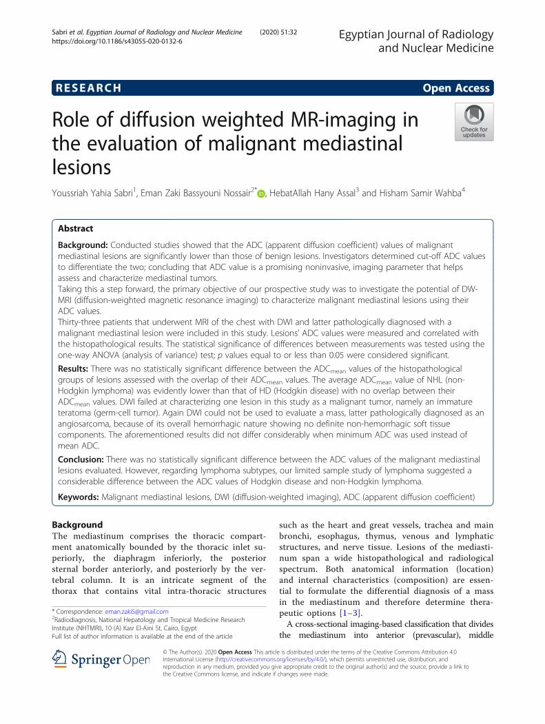
Fig. 1 Malignant B3 thymoma. A 42-year-old female patient clinically presenting with back and right upper limb pain underwent MDCT of thechest that revealed an anterior mediastinal mass. a Axial T1 WI, b axial T2 WI, and c axial STIR WI show a small well-defined oval-shaped anterior(prevascular) mediastinal mass displaying intermediate T1 and relatively bright T2 and bright STIR signal intensities. The lesion shows restricteddiffusion by being bright on e DWI and dark on the f ADC map. ADCmean = 0.895 × 10⁻³ mm2/s and ADCmin = 0.769 ×10⁻³ mm2/s. Associatedfindings: mid-dorsal sclerotic vertebra as seen on d coronal T1 WI; latter proven metastatic by bone scan. Right lobar hepatic focal lesion seen onh axial T1, T2, STIR and DWI and ADC map (from left to right) displaying low T1 and high T2 and STIR signal intensities with evident diffusionrestriction; biopsy proven to be metastatic
Sabri et al. Egyptian Journal of Radiology and Nuclear Medicine (2020) 51:32 Page 4 of 16

2.2 Quantitative assessment:
ADC values were measured by placing a ROI (re-gion of interest) within the lesion on the trace ADCmaps. ROIs were positioned within areas visuallyjudged to be the most restricted (excluding obviouslycystic/necrotic areas). To avoid image selection bias,three ROIs were placed on at least three sections(cranial, middle, and caudal portions) within the le-sion whenever possible and average mean and mini-mum ADC values (ADCmean and ADCmin) werecalculated.
Histopathological diagnosisTaking both lesion accessibility and patients’ general con-dition into account, patients underwent U/S or CT-guidedbiopsies, bronchoscopy, mediastinoscopy, or thoracotomy.Consequently, 33 patients pathologically diagnosed
with a malignant mediastinal lesion were included inthe study; 21 males (63.6%) and 12 females (36.4%)with age range 10 to 73 years (mean age 42.18 years).
Statistical/data analysisData was coded and entered using the statistical packageSPSS version 21. It was summarized using descriptive
Fig. 2 Ganglioneuroblastoma: A 16-year-old female with a posterior mediastinal (left paravertebral) mass identified on CT. The patient clinicallypresented with dorsal back pain radiating to the right upper limb and bilateral hip pain with a history of blood transfusion to correct anemia. aAxial T1 WI, b and d axial and coronal T2 WI and c STIR WI show a well-defined oval-shaped posterior mediastinal (left paravertebral) massdisplaying intermediate T1 and bright T2 and STIR signal intensities with a central scar showing bright signal on all three pulse sequences. Mass iscentered on the adjacent neural exit foramina with no definite neural foraminal or intraspinal extension. Invasion of the posterior aspects of theadjacent ribs is seen in the form of soft tissue encasement. Associated patchy altered MR marrow signal of the examined bones is seen mostevident on g axial STIR and DWI and the ADC map (from left to right). The mass shows restricted diffusion by being bright on e DWI and dark onthe f ADC map: ADCmean = 0.473 × 10⁻³ mm2/s and ADCmin = 0.327 × 10⁻³ mm2/s
Sabri et al. Egyptian Journal of Radiology and Nuclear Medicine (2020) 51:32 Page 5 of 16

statistics: number and percentage for qualitative valuesand mean and standard deviation and median and inter-quartile range for quantitative variables. One-wayANOVA (analysis of variance) test was used to comparemeans. p values equal to or less than 0.05 were consideredsignificant. Data was presented by Box-Plot graphs, barcharts, histograms, pie charts as well as tables.
Results
– Lesions were grouped according to theirhistopathological diagnosis into lymphoma
(n = 9; 27.3%), bronchogenic carcinoma (n = 10;30.3%), metastatic lymph nodes (n = 4; 12.1%),thymic tumors (n = 4; 12.1%), germ cell tumors(n = 4; 12.1%), neurogenic tumors (n = 1; 3.03 %), andangiosarcoma (n = 1; 3.03%) (Figs. 1, 2 and 3).
– Lesions were allocated to one or moremediastinal compartments according to thecross-sectional-based mediastinal classificationsystem recentlypublished by the ITMIG as shown in Table 4.
– The range of ADCmean and ADCmin values oflesions in each histopathological group, as well
Fig. 3 Seminoma: 30-year-old male with an anterior (prevascular) mediastinal mass identified on CT. The patient clinically presented with dyspnea andchest pain. a Axial T1 WI, b axial T2 WI, c axial STIR WI, and d sagittal T2 WI show a large well-defined macro-lobulated anterior mediastinal mass displayingrather homogeneous intermediate T1, relatively bright T2 and bright STIR signal intensities. The mass shows restricted diffusion; with the predominantbright areas on the e DWI corresponding to dark areas on the f ADC map: ADCmean = 0.615 × 10⁻³ mm2/s and ADCmin = 0.494 × 10⁻³ mm2/s
Table 4 Lesions allocated to mediastinal compartments
Mediastinal compartment Lymphoma Central bronchogeniccarcinoma
Metastaticlymph nodes
Thymictumors
Germ celltumors
Neurogenictumors
Angiosarcoma Total
Anterior 7 1 2 4 4 – 1 19 (57.6%)
Middle – 4 2 – – – – 6 (18.2%)
Posterior – – – – – 1 – 1 (3%)
More than one compartment 2 5 – – – – – 7 (21.2%)
Total 9 10 4 4 4 1 1 33 (100%)
Sabri et al. Egyptian Journal of Radiology and Nuclear Medicine (2020) 51:32 Page 6 of 16
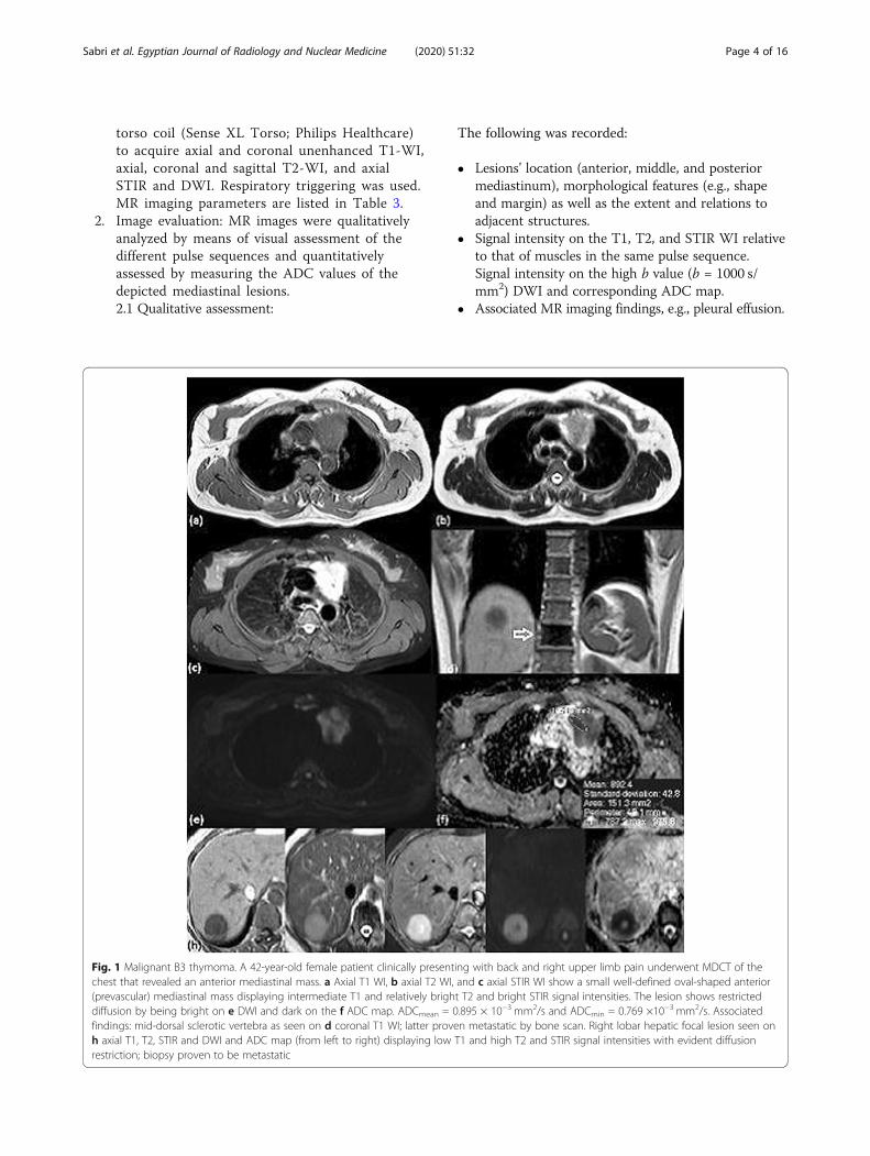
as their arithmetic means (averages), are shownin the Table 5.
– There was no statistically significant differencebetween the ADCmean values of the histopathologicalgroups of lesions assessed (p value > 0.05). Overlapof their ADCmean values is demonstrated on the Bar-and-Whisker plot (Fig. 4).
– Since lymphoma and bronchogenic carcinoma werethe two most prevalent lesions in our study, thefollowing was considered:
– Average ADCmean of lymphoma was evidently lowerthan that of bronchogenic carcinoma; however, nostatistically significant difference was found betweentheir ADCmean values (p value > 0.05).
– Average ADCmean value of lymphoma was thelowest when compared to those of bronchogeniccarcinoma, metastases, thymic, and germ celltumors. Yet, there was no statistically significantdifference between the ADCmean values oflymphoma and the ADCmean values of thesehistopathological groups conjointly (p value > 0.05).
– Similarly, there was no statistically significantdifference between the ADCmean values ofbronchogenic carcinoma and the ADCmean values ofthe other malignant mediastinal lesions all together(p value > 0.05)
– In the context of mediastinal compartments,anterior mediastinal lesions were the most prevalentin our study, representing 57.6% of lesions. Thelesions most importantly included lymphoma,thymic, and germ cell tumors (in ascending order ofaverage ADCmean values). Difference between theirADC values was, however, statistically insignificant(p value > 0.05).
Table 5 Summary of ADCmean and ADCmin values perhistopathological group
Pathologicalentity
Number of casesand percentage (%)
ADCmean rangeaverage ± SD*
ADCmin rangeaverage ± SD*
Lymphoma 9 (27.3%) (0.507–1.08)0.733 ± 0.191
(0.343–0.846)0.541 ± 0.164
Centralbronchogeniccarcinoma
10 (30.3%) (0.501–1.633)0.904 ± 0.342
(0.354–1.474)0.747 ± 0.336
Metastaticlymph nodes
4 (12.1%) (0.667–1.391)1.033 ± 0.3
(0.467–1.212)0.822 ± 0.307
Thymic tumors 4 (12.1%) (0.847–1.233)0.959 ± 0.184
(0.708–1.111)0.827 ± 0.191
Germ cell tumors 4 (12.1%) (0.568–2.44)1.058 ± 0.921
(0.431–1.701)0.776 ± 0.616
Neurogenic tumors 1 (3.03%) 0.473 0.327
Angiosarcoma 1 (3.03%) Could not be measured**
Total 33 (100%) 0.884 ± 0.396 0.699 ± 0.326
All values are expressed in units of 10−3 × mm2/s*SD Standard deviation**Elaborated later
Fig. 4 Bar-and-Whisker plot: ADCmean values of the different histopathological groups
Sabri et al. Egyptian Journal of Radiology and Nuclear Medicine (2020) 51:32 Page 7 of 16

– Each histopathological group was studied distinctlyand the following was recognized:
– Among the nine patients, pathologically diagnosedas lymphoma; five had non-Hodgkin lymphoma
(NHL) and four had Hodgkin lymphoma (HD)(Figs. 5 and 6). The average ADCmean value ofNHL was evidently lower than that of HD withno overlap between their ADCmean values on the
Fig. 5 NHL (Burkitt lymphoma): A 13-year-old male clinically presenting with constitutional symptoms (anorexia and weight loss) and leftparasternal and left lateral lower chest wall swellings. a Axial T1 WI, b axial T2 WI, and c axial STIR WI show a well-defined rather ovoid-shapedanterior mediastinal mass displaying intermediate T1, relatively bright T2 and bright STIR signal intensities. d Axial T2 WI, e axial STIR WI, and fcoronal T2 WI show another oblong-shaped posterior mediastinal (left paravertebral) mass displaying similar signal characteristics. It extends fromthe level of D9 down to D12 vertebra. Intraspinal extension in the form of a large epidural soft tissue component is seen opposite D7/8 down toD12 vertebra on the g sagittal T2 WI with evident displacement and compression of the dorsal cord. The lesions show restricted diffusion bybeing bright on the h DWI and dark on the i ADC map: ADCmean = 0.507 × 10⁻³ mm2/s and ADCmin = 0.343 × 10⁻³ mm2/s. Associated findings:left parasternal and left lateral lower chest wall soft tissue lesions (seen encasing the related ribs). The upper abdominal cuts revealedcircumferential gastric mural thickening. Both display similar signal characteristics to the aforementioned mediastinal masses. Minimal leftpleural effusion
Sabri et al. Egyptian Journal of Radiology and Nuclear Medicine (2020) 51:32 Page 8 of 16
Box-and-Whisker plot (Fig. 7). However, thestatistical significance of this important findingcould not be assessed and a cut-off value be-tween these two could not be determined be-cause of the limited number of cases involved.
– Similarly, the ten bronchogenic carcinoma casesin this study were histopathologically classifiedinto non-small-cell lung cancer (NSCLC 60%)and small-cell lung cancer (SCLC 40%) (Fig. 8).Average ADCmean value of SCLC (0.855 ±0.324x10⁻³ mm2/s) was lower than that ofNSCLC (0.936 ± 0.380 × 10⁻³ mm2/s) buttheir ADCmean values showed substantialoverlap (Fig. 9).
– DWI failed at characterizing one lesion in this studyas a malignant tumor, namely an immature teratoma(germ cell tumor) (Fig. 10). Its soft tissuecomponent showed facilitated diffusion with anADCmean (2.44 × 10⁻³ mm2/s) higher than
previously reported cut-off ADC values, hence, itwas misdiagnosed as a benign lesion.
– Again DWI could not be used to evaluate the massthat was later pathologically diagnosed as anangiosarcoma because of its overall hemorrhagicnature showing no definite non-hemorrhagic softtissue components (Fig. 11).
– Minimum ADC of each lesion was taken intoaccount when attempting to characterize thedifferent malignant mediastinal lesions included inthis study. However, the aforementioned results didnot differ considerably when minimum ADC wasused instead of mean ADC.
DiscussionInfluenced by the constantly evolving approach tonon-invasive patient management in the era of func-tional imaging, investigators demonstrated the diag-nostic potential of DW-MR images and calculated
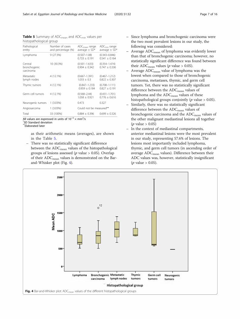
Fig. 6 Hodgkin lymphoma: A 51-year-old male with an anterior (prevascular) mediastinal mass identified on CT. The patient clinicallypresented with constitutional symptoms (unexplained fever and weight loss), dyspnea, cough, and retrosternal chest pain. a Axial T1 WI, baxial T2 WI, c axial STIR WI, and d sagittal BFFE WI show a well-defined ovoid-shaped anterior mediastinal mass displaying intermediate T1and heterogeneous T2 and STIR signal intensities. The mass shows restricted diffusion; with the predominant bright areas on the e DWIcorresponding to dark areas on the f ADC map. ADCmean = 0.890 × 10⁻³ mm2/s and ADCmin = 0.624 × 10⁻³ mm2/s
Sabri et al. Egyptian Journal of Radiology and Nuclear Medicine (2020) 51:32 Page 9 of 16

ADC values to differentiate between benign and ma-lignant tumors including those of the mediastinum.Hence, serving as a virtual biopsy and in some casespreventing unnecessary diagnostic intervention.Conducted studies showed that the ADC values of ma-
lignant mediastinal lesions are significantly lower thanthose of benign lesions and determined cut-off ADCvalues to differentiate the two.Taking this a step forward, the main purpose of our
prospective study was to investigate the potential ofDW-MRI to characterize malignant mediastinal lesionsusing their ADC values.The study included 33 patients with a mediastinal
mass identified on CT that underwent MRI of thechest with DWI and were later histopathologically di-agnosed with a malignant mediastinal lesion. Patientswere grouped according to their histopathologicaldiagnosis.There was no statistically significant difference be-
tween the ADCmean values of the histopathologicalgroups of lesions assessed. This is in concordancewith the study carried out by Tondo et al., 2011 whofound substantial overlap in ADCmean values of allexamined malignant lesions (n = 30), namely bron-chogenic carcinoma, thymic carcinoma, and malignantteratoma [16].
Moreover, the 32 malignant mediastinal lesions stud-ied by Nasr et al., 2016 included lymphoma, broncho-genic carcinoma, invasive thymoma, and metastases. Nosignificant difference between their ADCmean valueswas found as well [11].Likewise, the 30 malignant mediastinal tumors in
Abdel Razek et al., 2009s study were lymphoma, bron-chogenic carcinoma, invasive thymoma, and angiosar-coma and again difference between their ADCmeanvalues was insignificant with evident overlapping of theADC values of lymphoma, bronchogenic carcinoma,and thymoma [13].These findings may be attributed to a relatively
small sample size and/or heterogeneity of the histo-pathological subtypes of malignant mediastinal lesionsevaluated.In agreement with Nasr et al., there was no statistically
significant difference between the ADCmean values oflymphoma and the ADCmean values of the rest of thehistopathological groups conjointly. Tondo et al., 2011reported similar results to ours where no significant dif-ference was found between bronchogenic carcinoma(adenocarcinomas) and malignant mediastinal lesions intheir study [11, 16].On comparing the ADCmean values of lymphoma
and bronchogenic carcinoma, lesions most frequently
Fig. 7 Bar-and-Whisker plot: ADCmean values of NHL and HD
Sabri et al. Egyptian Journal of Radiology and Nuclear Medicine (2020) 51:32 Page 10 of 16

encountered in our study, the difference betweentheir ADCmean values was statistically insignificant aswell. Among the 26 malignant lesions prospectivelystudied by Gümüştaş et al., 2011, 11 were broncho-genic carcinoma (NSCLC) and 9 were lymphoma.When their ADCmean values were compared, no sig-nificant difference was found with an overlap betweenthese two subgroups; results similar to ours. However,Maeda et al., 2005, Abdel Razek et al., 2006, Sumiet al., 2007, Holzapfel et al., 2009 and Kato et al.2015 reported that the ADC of lymphoma is signifi-cantly lower than that of squamous cell carcinoma ofthe head and neck [14, 18–22].
Anterior mediastinal lesions “most importantlylymphoma, thymic, and germ cell tumors” in ourstudy were entitled to be evaluated separately andagain difference between their ADC values wasstatistically insignificant. Yabuuchi et al., 2015 con-ducted a retrospective study in the search of signifi-cant parameters to characterize anterior mediastinaltumors; one of the parameters evaluated was tu-mors’ ADC value. Malignant anterior mediastinallesions included in their study were thymicepithelial tumors (malignant thymoma and thymiccarcinoma), lymphoma, and germ cell tumors. Inagreement with our results, there was no significant
Fig. 8 Squamous cell carcinoma of lung origin (bronchogenic carcinoma: NSCLC): A 62-year-old female clinically presenting with cough,dyspnea and chest pain underwent MDCT of the chest that revealed an anterior mediastinal mass. a Axial T1 WI, b axial T2 WI andd axial STIR WI show a well-defined rather ovoid-shaped anterior mediastinal mass displaying intermediate T1 and relatively bright T2 andSTIR signal intensities. Small areas of breaking down exhibiting low T1 and bright T2 and STIR signal intensities are noted. The lesion(sparing the small areas of breaking down) shows restricted diffusion by being bright on c DWI and dark on the e ADC map. ADCmean =1.63 × 10⁻³ mm2/s and ADCmin = 1.47 × 10⁻³ mm2/s
Sabri et al. Egyptian Journal of Radiology and Nuclear Medicine (2020) 51:32 Page 11 of 16

difference between the ADC of these histopatho-logical subtypes at both initial and validationstudies [23].Regarding the pathological subtypes of lymphoma; in
the retrospective study of mediastinal lymphadenopathyin children by Abdel Razek et al., 2015, the averageADCmean of NHL was lower than that of HD—resultssimilar to ours—but the difference between the two wasinsignificant. On searching the literature, no attempts atusing DWI to differentiate between lymphoma subtypes,in particular, were found [24].A meta-analysis of 34 studies (conducted in the
years 2007 to 2014) involving 2086 patients withpulmonary lesions was recently performed by Shenet al., 2016. One of their objectives was to evaluatethe role of ADC in characterizing subtypes of lungcancer. Pooled ADC values of SCLC were signifi-cantly lower than those of NSCLC, which to an ex-tent agrees with our study (results more or lesssimilar to ours). The potential histopathological ra-tionale might be that SCLC has high tumoral cellu-larity, large nuclei, and almost no cytoplasm, all ofwhich restrict the diffusion of water moleculesthereby reducing ADC values [25].Limited data are available on quantitative assess-
ment of thymic epithelial tumors (TETs) by usingDW-MRI from small cohorts of studies which
considered various anterior mediastinal tumors. Notto mention that those studies did not attempt todifferentiate thymomas based on WHO andMasaoka-Koga classifications by using ADC. Re-cently, a study by Abdel Razek et al. involving 30patients with TETs has demonstrated the ability ofADC in differentiating low-risk from high-risk tu-mors, and early from advanced disease. Mean ADCvalues of high-risk thymomas and advanced stageTETs were significantly lower than those of low-risk thymomas and early-stage TETs respectively.They determined cut-off mean ADC values of 1.22mm2/s and 1.25 mm2/s below which a high-riskthymoma is indicated. In a relatively similar studyby Priola et al., accurate cut-off mean ADC valuesof 1.309 × 10⁻³ mm2/s and 1.243 × 10⁻³ mm2/swere determined below which a high-risk thymomaand an advanced stage TET are indicated respect-ively. Moreover, mean ADC of B3 thymoma wassignificantly lower than that of B2 thymoma. ADClevels were significantly associated with disease-freesurvival of patients with a recurrence rate beinghigher for patients with ADC ≤ 1.299 × 10⁻³ mm2/s. To conclude, ADC helps to differentiate high-riskfrom low-risk thymomas and may be used as aprognostic indicator of recurrence, yet further stud-ies are needed to validate these results [26, 27].
Fig. 9 Bar-and-Whisker plot: ADCmean values of SCLC and NSCLC
Sabri et al. Egyptian Journal of Radiology and Nuclear Medicine (2020) 51:32 Page 12 of 16

Nasr et al., 2016, Usuda et al.,2015, Tondo et al.,2011, Gümüştaş et al., 2011 and Abdel Razek et al.,2009 conducted prospective/retrospective studies toquantitatively assess and differentiate benign andmalignant mediastinal lesions using DWI. They de-termined highly sensitive and specific cut-off ADCvalues below which a malignant mediastinal lesionis indicated; 1.15 × 10⁻³, 2.21 × 10⁻³, 1.25 × 10−3,1.39 × 10−3, and 1.56 × 10⁻³ mm2/s respectively.One lesion, namely a malignant teratoma had anADCmean evidently higher than these cut-off ADC
values (ADCmean = 2.44 × 10⁻³ mm2/s) and washence mistaken for a benign lesion. This may beexplained by the fact that when the mass was surgi-cally excised, it proved malignant only by the pres-ence of microscopic foci of immature neuroepithelialcomponent [11, 13, 14, 16, 17].The one lesion pathologically diagnosed as an angio-
sarcoma was overall hemorrhagic in nature with nosizeable non-hemorrhagic soft tissue components seenon the conventional MR images rendering a qualitativeassessment of the DWI and ADC measurement not
Fig. 10 Grade I immature (malignant) teratoma (pathologically proven after thoracotomy and complete excision—excised lymph nodes:free): A 13-year-old male with an anterior mediastinal (prevascular) mass identified on CT. The patient clinically presented with mildchest pain (discomfort). b Axial T1 WI, c axial T2 WI, and d axial STIR WI show a well-defined rather wedge-shaped anteriormediastinal mass displaying overall heterogeneous signal on all pulse sequences by virtue of its fat and calcific components(confirmed by referring to a axial MDCT of the chest in mediastinal window). Associated findings: Lingular collapse and smallprevascular and left hilar lymph nodes, largest measures 1.5 cm. Lesion’s soft tissue component shows facilitated diffusion. It displaysbright signal on e DWI seen corresponding to bright signal on the f ADC map: ADCmean = 2.44 × 10⁻³ mm2/s and ADCmin = 1.701 × 10⁻³ mm2/s
Sabri et al. Egyptian Journal of Radiology and Nuclear Medicine (2020) 51:32 Page 13 of 16

possible. This was based on the fact stated by Qayyum,2009: blood demonstrates signal characteristics of highlycellular soft tissue lesions; a pitfall readily observed onDWI [7].A number of authors recommended that minimum
ADC is used instead of mean ADC when quantitativelyassessing a lesion since it theoretically reflects the areaof highest tumor cellularity and unlike mean ADC, itwould not underestimate tumor cellularity in the contextof a largely necrotic tumor [28]. Accordingly, a mini-mum ADC of each lesion was taken into account whenattempting to characterize the different malignant medi-astinal lesions included in this study. However, theaforementioned results did not differ considerably whenminimum ADC was used instead of mean ADC. Never-theless, most published studies that evaluated medias-tinal lesions using DWI referred to mean and notminimum ADC values.
ConclusionAlthough highly sensitive and specific cut-off ADCvalues to distinguish benign and malignant mediastinallesions were published, in this study, DWI could not beused to characterize the histopathological subtypes ofmalignant mediastinal lesions. However, regardinglymphoma subtypes, our limited sample study of lymph-oma suggested a considerable difference between theADC values of Hodgkin disease and non-Hodgkinlymphoma.
AbbreviationsADC: Apparent diffusion coefficient; ADCmean: Mean apparent diffusioncoefficient; ADCmin: Minimum apparent diffusion coefficient; ANOVA: Analysisof variance; CT: Computed tomography; DW-MRI: Diffusion-weightedmagnetic resonance imaging; HD: Hodgkin disease; ITMIG: InternationalThymic Malignancy Interest Group; MRI: Magnetic resonance imaging;n: Number; NHL: Non-Hodgkin lymphoma; NSCLC: Non-small cell lungcancer; ROI: Region of interest; SCLC: Small-cell lung cancer; SPSS: Statistical
Fig. 11 Angiosarcoma (pathologically proven after thoracotomy and complete excision): A 33-year-old male patient clinically presentedwith cough, dyspnea, chest pain, and congested neck veins. He underwent MDCT of the chest that revealed an anterior mediastinal massencroaching upon the right hemithorax. a Axial T1 WI, b and f axial and coronal T2 WI and c STIR WI show a large well-defined ratherovoid-shaped anterior mediastinal mass encroaching upon and occupying most of the right hemithorax. It displays heterogeneous signalon all pulse sequences with fluid-fluid levels reflecting different ages of blood with no sizeable non-hemorrhagic solid components. Acontralateral shift of the mediastinum is noted. Associated findings: ipsilateral pleural thickening and minimal pleural effusion. d DWI ande ADC map: not assessed because of the lesion’s overall hemorrhagic nature with no definite soft tissue components as apparent on theconventional MR images
Sabri et al. Egyptian Journal of Radiology and Nuclear Medicine (2020) 51:32 Page 14 of 16

Package for the Social Sciences; STIR: Short tau inversion recovery;TETs: Thymic epithelial tumors; U/S: Ultrasound; WI: Weighted image
AcknowledgementsNot applicable.
Authors’ contributionsYYS, EZN, and HSW reviewed all patients’ magnetic resonance images. EZNanalyzed and interpreted the patient data. EZN wrote the manuscript. Allauthors read and approved the final manuscript.
FundingNot applicable (no funding was provided).
Availability of data and materialsThe datasets used and/or analyzed during the current study are availablefrom the corresponding author on reasonable request.
Ethics approval and consent to participateInstitutional (Faculty of Medicine ‘Kasr El-Ainy Hospital’ and National CancerInstitute, Cairo University) ethical clearance was taken before conducting thisprospective study; March 2016—Reference number: not available. Writtenconsent was obtained from patients or their authorized representatives.
Consent for publicationAll patients included in this research gave written informed consent topublish the data contained within this study. If the patient was less than 16years old, deceased, or unconscious when consent for publication wasrequested, written informed consent for the publication of this data wasgiven by their parent or legal guardian.
Competing interestsThe authors declare that they have no competing interests.
Author details1Radiodiagnosis, Faculty of Medicine, Cairo University, Cairo, Egypt.2Radiodiagnosis, National Hepatology and Tropical Medicine ResearchInstitute (NHTMRI), 10 (A) Kasr El-Aini St, Cairo, Egypt. 3Pulmonology, Facultyof Medicine, Cairo University, Cairo, Egypt. 4Radiodiagnosis, National CancerInstitute, Cairo University, Cairo, Egypt.
Received: 31 October 2019 Accepted: 5 January 2020
References1. Carter BW, Okumura M, Detterbeck FC, Marom EM (2014) Approaching the
patient with an anterior mediastinal mass: a guide for radiologists. J ThoracOncol 9(9):S110–S118 https://doi.org/10.1097/JTO.0000000000000295
2. Juanpere S, Cañete N, Ortuño P, Martínez S, Sanchez G, Bernado L (2013) Adiagnostic approach to the mediastinal masses. Insights Imaging 4(1):29–52https://doi.org/10.1007/s13244-012-0201-0
3. Thacker PG, Mahani MG, Heider A, Lee EY (2015) Imaging evaluation ofmediastinal masses in children and adults: practical diagnostic approachbased on a new classification system. J Thorac Imaging 30(4):247–267https://doi.org/10.1097/RTI.0000000000000161
4. Carter BW, Benveniste MF, Madan R, Godoy MC, de Groot PM, Truong MTet al (2017) ITMIG classification of mediastinal compartments andmultidisciplinary approach to mediastinal masses. Radiographics 37(2):413–436 https://doi.org/10.1148/rg.2017160095
5. Whitten CR, Khan S, Munneke GJ, Grubnic S (2007) A diagnostic approachto mediastinal abnormalities. Radiographics 27(3):657–671 https://doi.org/10.1148/rg.273065136
6. Carter BW, Betancourt SL, Benveniste MF (2017) MR imaging of mediastinalmasses. Top Magn Reson Imaging 26(4):153–165
7. Qayyum A (2009) Diffusion-weighted Imaging in the abdomen and pelvis:concepts and applications. Radiographics 29(6):1797–1810 https://doi.org/10.1148/rg.296095521
8. Taouli B, Koh D-M (2010) Diffusion-weighted MR imaging of the liver.Radiology 254(1):47–66 https://doi.org/10.1148/radiol.09090021
9. Ackman JB (2015) MR imaging of mediastinal masses. Magn Reson ImagingClin N Am 23(2):141–164 https://doi.org/10.1016/j.mric.2015.01.002
10. Luna A, Sánchez-Gonzalez J, Caro P (2011) Diffusion-weighted imaging ofthe chest. Magn Reson Imaging Clin N Am 19(1):69–94 https://doi.org/10.1016/j.mric.2010.09.006
11. Nasr A, Elshahat H, Safwat H, Alsaif R, Alshehab D, Shebl M (2016)Diffusion weighted MRI of mediastinal masses: can measurement ofADC value help in the differentiation between benign and malignantlesions. Egypt J Radiol Nuc Med 47(1):119–125 https://doi.org/10.1016/j.ejrnm.2015.12.002
12. Shin KE, Yi CA, Kim TS, Lee HY, Choi YS, Kim HK, Kim J (2014) Diffusion-weighted MRI for distinguishing non-neoplastic cysts from solid masses inthe mediastinum: problem-solving in mediastinal masses of indeterminateinternal characteristics on CT. Eur Radiol 24(3):677–684 https://doi.org/10.1007/s00330-013-3054-0
13. Abdel Razek AAK, Elmorsy A, Elshafey M, Elhadedy T, Hamza O (2009)Assessment of mediastinal tumors with diffusion-weighted single-shotecho-planar MRI. J Magn Reson Imaging 30(3):535–540 https://doi.org/10.1002/jmri.21871
14. Gümüştaş S, Inan N, Sarisoy HT, Anik Y, Arslan A, Çiftçi E et al (2011)Malignant versus benign mediastinal lesions: quantitative assessment withdiffusion weighted MR imaging. Eur Radiol 21(11):2255–2260 https://doi.org/10.1007/s00330-011-2180-9
15. Sabri YY, Kolta MFF, Khairy MA (2017) MR diffusion imaging inmediastinal masses the differentiation between benign and malignantlesions. Egypt J Radiol Nucl Med 48(3):569–580 https://doi.org/10.1016/j.ejrnm.2017.03.015
16. Tondo F, Saponaro A, Stecco A, Lombardi M, Casadio C, Carriero A (2011)Role of diffusion-weighted imaging in the differential diagnosis of benignand malignant lesions of the chest–mediastinum. Radiologia Medica 116(5):720–733 https://doi.org/10.1007/s11547-011-0629-1
17. Usuda K, Maeda S, Motono N, Ueno M, Tanaka M, Machida Y et al (2015)Diffusion weighted imaging can distinguish benign from malignantmediastinal tumors and mass lesions: Comparison with positron emissiontomography. Asian Pac J Cancer Prev 16(15):6469–6475 https://doi.org/10.7314/APJCP.2015.16.15.6469
18. Abdel Razek AAK, Soliman NY, Elkhamary S, Alsharaway MK, Tawfik A (2006)Role of diffusion-weighted MR imaging in cervical lymphadenopathy. EurRadiol 16(7):1468–1477 https://doi.org/10.1007/s00330-005-0133-x
19. Holzapfel K, Duetsch S, Fauser C, Eiber M, Rummeny EJ, Gaa J (2009) Valueof diffusion-weighted MR imaging in the differentiation between benignand malignant cervical lymph nodes. Eur J Radiol 72(3):381–387 https://doi.org/10.1016/j.ejrad.2008.09.034
20. Kato H, Kanematsu M, Watanabe H, Kawaguchi S, Mizuta K, Aoki M (2015)Differentiation of extranodal non-Hodgkins lymphoma from squamous cellcarcinoma of the maxillary sinus: a multimodality imaging approach.Springerplus 4(1):228 https://doi.org/10.1186/s40064-015-0974-y
21. Maeda M, Kato H, Sakuma H, Maier SE, Takeda K (2005) Usefulness ofthe apparent diffusion coefficient in line scan diffusion-weightedimaging for distinguishing between squamous cell carcinomas andmalignant lymphomas of the head and neck. AJNR Am J Neuroradiol26(5):1186–1192
22. Sumi M, Ichikawa Y, Nakamura T (2007) Diagnostic ability of apparentdiffusion coefficients for lymphomas and carcinomas in the pharynx. EurRadiol 17(10):2631–2637 https://doi.org/10.1007/s00330-007-0588-z
23. Yabuuchi H, Matsuo Y, Abe K, Baba S, Sunami S, Kamitani T et al(2015) Anterior mediastinal solid tumors in adults: characterizationusing dynamic contrast-enhanced MRI, diffusion-weighted MRI, andFDG-PET/CT. Clin Radiol 70(11):1289–1298 https://doi.org/10.1016/j.crad.2015.07.004
24. Abdel Razek AAK, Gaballa G, Elashry R, Elkhamary S (2015) Diffusion-weighted MR imaging of mediastinal lymphadenopathy in children. Jap JRadiol 33(8):449–454 https://doi.org/10.1007/s11604-015-0434-1
25. Shen G, Jia Z, Deng H (2016) Apparent diffusion coefficient values ofdiffusion-weighted imaging for distinguishing focal pulmonary lesions andcharacterizing the subtype of lung cancer: a meta-analysis. Eur Radiol 26(2):556–566 https://doi.org/10.1007/s00330-015-3840-y
26. Abdel Razek AAK, Khairy M, Nada N (2014) Diffusion-weighted MR imagingin thymic epithelial tumors: correlation with World Health OrganizationClassification and Clinical staging. Radiology 273(1):268–275 https://doi.org/10.1148/radiol.14131643
27. Priola AM, Gned D, Veltri A, Priola SM (2016) Chemical shift and diffusion-weighted magnetic resonance imaging of the anterior mediastinum in
Sabri et al. Egyptian Journal of Radiology and Nuclear Medicine (2020) 51:32 Page 15 of 16

oncology: current clinical applications in qualitative and quantitativeassessment. Crit Rev Oncol Hematol 98:335–357 https://doi.org/10.1016/j.critrevonc.2015.11.012
28. Subhawong TK, Jacobs MA, Fayad LM (2014) Diffusion-weighted MRimaging for characterizing musculoskeletal lesions. Radiographics 34(5):1163–1177 https://doi.org/10.1148/rg.345140190
Publisher’s NoteSpringer Nature remains neutral with regard to jurisdictional claims inpublished maps and institutional affiliations.
Sabri et al. Egyptian Journal of Radiology and Nuclear Medicine (2020) 51:32 Page 16 of 16