diffusion-weighted mr imaging of unusual white matter ... · diffusion-weighted mr imaging of...
TRANSCRIPT
82 Korean J Radiol 8(1), February 2007
Diffusion-Weighted MR Imaging ofUnusual White Matter Lesion in a Patientwith Menkes Disease
We report here on the diffusion-weighted imaging of unusual white matterlesions in a case of Menkes disease. On the initial MR imaging, the white matterlesions were localized in the deep periventricular white matter in the absence ofdiffuse cortical atrophy. The lesion showed diffuse high signal on the diffusion-weighted images and diffuse progression and persistent hyperintensity on the fol-low up imaging. Our case suggests that the white matter lesion may precede dif-fuse cortical atrophy in a patient with Menkes disease.
enkes disease is an X-linked disorder that’s caused by impaired intracellu-lar transport of copper (1, 2). The clinical manifestations include failure tothrive, mental retardation, seizure and characteristic hypopigmented
“kinky hair”, and all this could be explained by dysfunction of the copper dependentenzymes (2). The previous neuroimaging reports on this disorder have been describeddiffuse brain atrophy, subdural effusion or hematoma, white matter changes andvascular tortuosity (3 6). However, the diffusion-weighted imaging (DWI) of thecerebral lesions in this disease has rarely reported on and the previous report has beenlimited to the deep gray matter lesion (7).
We describe here the DWI findings of unusual and progressive white matter lesionsin a case of Menkes disease.
CASE REPORT
A 10-month-old male was admitted with a few days history of fever. He wasdelivered at 38 weeks gestation following an unremarkable pregnancy and labor, andhis birth weight was 2,960 g. He had a sister and there was no significant familialhistory. A cephalhmatoma over the parietooccipital region was detected at delivery. Askull radiograph showed numerous Wormian bones in the lambdoid suture. At twomonths of age, he underwent inguinal herniorrhaphy under the diagnosis of bilateralinguinal hernia. He had a history of recurrent pneumonia and urinary tract infection.The retrograde vesicogram demonstrated the presence of multiple bladder diverticula.
On presentation, he was found to be very delayed in growth and development. Hisgrowth parameters were less than the 3rd percentile (weight: 7,500 g and length: 73.7cm). He had pale skin and very sparse hair, which was lightly pigmented and curly.Microscopic examination of the hair showed fragile and brittle filaments curled up intheir own axis, which is called in pili torti. Neurologic examination revealed diffusehypotonia with poor head and trunk support. He blinked to bright light stimulation,but he did not fix or follow. The deep tendon reflexes were hyperreflexic bilaterally,with clonus at the ankles. The Barbinski responses were bilateral extension. The serum
Eun Shin Lee, MD1,3
Jae Wook Ryoo, MD2,3
Dae Seob Choi, MD2,3
Jae Min Cho, MD2,3
Soo Hyun Kwon, MD1
Hee Suk Shin, MD1,3
Index terms:BrainMagnetic Resonance (MR)Menkes diseaseWhite MatterDiffusion study
Korean J Radiol 2007;8:82-85Received August 17, 2006; accepted after revision November 11, 2006.
Department of 1Rehabilitation Medicineand 2Radiology, and the 3Institute ofHealth Science, Gyeongsang NationalUniversity College of Medicine, Jinju 660-702, Korea
Address reprint requests to:Jae Wook Ryoo, MD, Department ofRadiology, Gyeonsang NationalUniversity, College of Medicine, 90Chilam-dong, Jinju 660-702, KoreaTel. (8255) 750-8818Fax. (8255) 758-1568e-mail: [email protected]
M

copper level was 27 g/dL (normal = 70 130 g/dL) andthe ceruloplasmin was 9.07 g/dL (normal = 20 60 g/dL).The clinical and biochemical data confirmed the diagnosisof Menkes disease.
Magnetic resonance (MR) imaging was obtained using a1.5T clinical scanner (Sonata, Siemens, Germany). Theimaging protocol included DWI, axial pre- and post T1-weighted and T2-weighted spin echo images, and fluidattenuated inversion recovery images. On the initial MRexamination, the T2-weighted images demonstratedsymmetric hyperintense lesions in both deep cerebralwhite matters (Fig. 1A). There was mild atrophy of thecerebellum and brainstem, yet atrophy of the cerebral
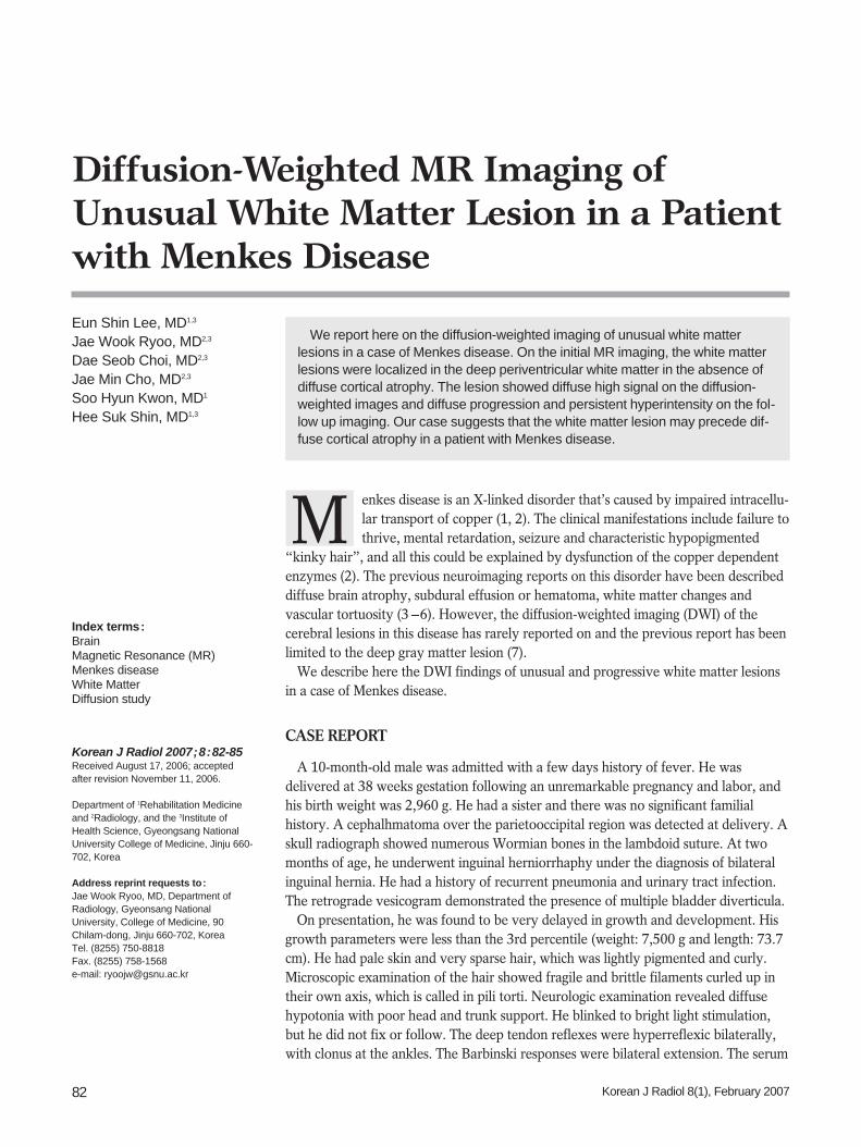
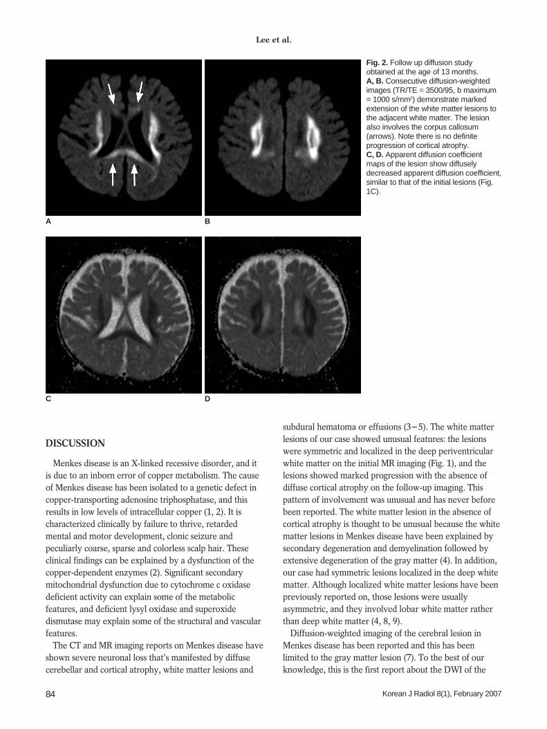
hemispheres was not prominent. DWI demonstrated highsignal intensity of the lesions, and their apparent diffusioncoefficients (ADCs) were decreased on the ADC maps(Figs. 1B, C). Three dimensional MR angiography (Fig. 1D)showed markedly tortuous intracranial and extracranialvessels, which are characteristic of Menkes disease. Thefollow up MR imaging at 13 months of age showed markedprogression of white matter lesions without definiteprogression of the cortical atrophy. The lesions extendedto the deep white matter and corpus callosum (Figs. 2A,B). The extended lesions also showed high signal intensityon DWI and decreased ADC, similar to those of the initiallesions (Figs. 2C, D).
Diffusion-Weighted MR Imaging of Menkes Disease
Korean J Radiol 8(1), February 2007 83
A B
C
D
Fig. 1. Initial MR images obtained at ageof 10 months.A. T2-wieghted axial image (TR/TE =4700/104 msec, 5 mm thickness)demonstrates symmetric hyperintenselesion in both deep periventricular whitematters (arrows).B. Diffusion weighted image (TR/TE =3500/92, b maximum = 1000 s/mm2,three directions, 5 mm thickness) showsthe bright signal intensity of the lesion. C. On the apparent diffusion coefficientmap, the lesion shows diffuse low signalintensities, suggesting restricteddiffusion.D. Three dimensional MR angiographyshows markedly tortuous intracranialand extracranial vessels, which arecharacteristics of Menkes disease.
DISCUSSION
Menkes disease is an X-linked recessive disorder, and itis due to an inborn error of copper metabolism. The causeof Menkes disease has been isolated to a genetic defect incopper-transporting adenosine triphosphatase, and thisresults in low levels of intracellular copper (1, 2). It ischaracterized clinically by failure to thrive, retardedmental and motor development, clonic seizure andpeculiarly coarse, sparse and colorless scalp hair. Theseclinical findings can be explained by a dysfunction of thecopper-dependent enzymes (2). Significant secondarymitochondrial dysfunction due to cytochrome c oxidasedeficient activity can explain some of the metabolicfeatures, and deficient lysyl oxidase and superoxidedismutase may explain some of the structural and vascularfeatures.
The CT and MR imaging reports on Menkes disease haveshown severe neuronal loss that’s manifested by diffusecerebellar and cortical atrophy, white matter lesions and
subdural hematoma or effusions (3 5). The white matterlesions of our case showed unusual features: the lesionswere symmetric and localized in the deep periventricularwhite matter on the initial MR imaging (Fig. 1), and thelesions showed marked progression with the absence ofdiffuse cortical atrophy on the follow-up imaging. Thispattern of involvement was unusual and has never beforebeen reported. The white matter lesion in the absence ofcortical atrophy is thought to be unusual because the whitematter lesions in Menkes disease have been explained bysecondary degeneration and demyelination followed byextensive degeneration of the gray matter (4). In addition,our case had symmetric lesions localized in the deep whitematter. Although localized white matter lesions have beenpreviously reported on, those lesions were usuallyasymmetric, and they involved lobar white matter ratherthan deep white matter (4, 8, 9).
Diffusion-weighted imaging of the cerebral lesion inMenkes disease has been reported and this has beenlimited to the gray matter lesion (7). To the best of ourknowledge, this is the first report about the DWI of the
Lee et al.
84 Korean J Radiol 8(1), February 2007
A B
C D
Fig. 2. Follow up diffusion studyobtained at the age of 13 months.A, B. Consecutive diffusion-weightedimages (TR/TE = 3500/95, b maximum= 1000 s/mm2) demonstrate markedextension of the white matter lesions tothe adjacent white matter. The lesionalso involves the corpus callosum(arrows). Note there is no definiteprogression of cortical atrophy.C, D. Apparent diffusion coefficientmaps of the lesion show diffuselydecreased apparent diffusion coefficient,similar to that of the initial lesions (Fig.1C).
white matter lesion in a Menkes disease patient. The whitematter lesions demonstrated restricted diffusion, that is,diffuse high signals on DWI and decreased ADC, and thiswas persistent on the follow-up imaging (Figs. 1, 2). Thepathogenesis of these findings is uncertain, but it may bemetabolic and related to a deficiency in a mitochondrialcopper containing enzyme (2). Transient ischemic changesor demyelination in the brain are often observed in thepatients with mitochondrial myopathy, encephalopathy,lactic acidosis and stroke-like episodes (MELAS), in whichthese changes are caused by a decrease in cytochrome coxidase activity (10). Cytochorme c oxidase activity is alsoknown to be decreased in the brain of patients withMenkes disease (2). Decreased activity of superoxidemutase, an enzyme responsible for the regulation andsequestration of oxygen free radicals, lowers protectionagainst oxidative stress and it may lead to a cytotoxiceffect and cell damage (2). Tortuous and irregular vesselsmight produce erratic and turbulent blood and so predis-pose the sufferer to diffuse white matter ischemia (7).
In summary, we have described unusual, progressivewhite matter lesion in a patent with Menkes disease. DWIdemonstrated the diffuse high signal intensity andrestricted diffusion of the lesion. Our case might suggestthat the white matter lesion could precede diffuse corticalatrophy in those patients with Menkes disease.
References1. Menkes JH, Alter M, Steigleder GK, Weakley DR, Sung JH. A
sex-linked recessive disorder with retardation of growth,peculiar hair, and focal cerebral and cerebellar degeneration.Pediatrics 1962;29:764-779
2. Kaler SG. Menkes disease. Adv Pediatr 1994;41:263-3043. Faerber EN, Grover WD, DeFilipp GJ, Capitanio MA, Liu TH,
Swartz JD. Cerebral MR of Menkes kinky-hair disease. AJNRAm J Neuroradiol 1989;10:190-192
4. Leventer RJ, Kornberg AJ, Phelan EM, Kean MJ. Earlymagnetic resonance imaging findings in Menkes’ disease. J ChildNeurol 1997;12:222-224
5. Takahashi S, Ishii K, Matsumoto K, Higano S, Ishibashi T,Zuguchi M, et al. Cranial MRI and MR angiography in Menkes’syndrome. Neuroradiology 1993;35:556-558
6. Kim OH, Suh JH. Intracranial and extracranial MR angiographyin Menkes disease. Pediatr Radiol 1997;27:782-784
7. Hsich GE, Robertson RL, Irons M, Soul JS, du Plessis AJ.Cerebral infarction in Menkes’ disease. Pediatr Neurol2000;23:425-428
8. Blaser SI, Berns DH, Ross JS, Lanska MJ, Weissman BM. SerialMR studies in Menkes disease. J Comput Assist Tomogr1989;13:113-115
9. Ozawa H, Kodama H, Murata Y, Takashima S, Noma S.Transient temporal lobe changes and a novel mutation in apatient with Menkes disease. Pediatr Int 2001;43:437-440
10. Valanne L, Ketonen L, Majander A, Suomalainen A, Pihko H.Neuroradiologic findings in children with mitochondrialdisorders. AJNR Am J Neuroradiol 1998;19:369-377
Diffusion-Weighted MR Imaging of Menkes Disease
Korean J Radiol 8(1), February 2007 85