role of dcps in mammalian rna regulation and human …
TRANSCRIPT
ROLE OF DCPS IN MAMMALIAN RNA REGULATION AND
HUMAN DISEASES
By
MI ZHOU
A dissertation submitted to the
Graduate School-New Brunswick
and
The Graduate School of Biomedical Sciences
Rutgers, The State University of New Jersey
In partial fulfillment of the requirements
For the degree of
Doctor of Philosophy
Graduate Program in Cell and Development Biology
Written under the direction of
Dr. Megerditch Kiledjian
And approved by
_________________________________
_________________________________
_________________________________
_________________________________
New Brunswick, New Jersey
October, 2015

ii
ABSTRACT OF THE DISSERTATION
Role of DcpS in Mammalian RNA Regulation and Human Diseases
By MI ZHOU
Dissertation Director
Dr. Megerditch Kiledjian
In eukaryotic cells, mRNA degradation plays an important role in the control of
gene expression and is therefore highly regulated. The scavenger decapping enzyme
DcpS is a multifunctional protein that plays a critical role in mRNA degradation.
We first sought to identify DcpS target genes in mammalian cells using a cell
permeable DcpS inhibitor compound, RG3039, which was initially developed for
therapeutic treatment of Spinal Muscular Atrophy (SMA). Microarray analysis following
DcpS decapping inhibition by RG3039 revealed the steady state levels of 222 RNAs were
altered. Of a subset selected for validation by qRT-PCR, two non-coding transcripts
dependent on DcpS decapping activity, were identified and referred to as DcpS
Responsive Noncoding Transcript (DRNT) 1 and 2 respectively. Only the increase in
DRNT1 transcript was accompanied with an increase of its RNA stability and this
increase was dependent on both DcpS and Xrn1. Our data indicate that DcpS is a
transcript-restricted modulator of RNA stability in mammalian cells and the RG3039

iii
quinazoline compound is pleotropic, influence gene expression in both an apparent DcpS
dependent and independent manner.
A surprising development was uncovered in a collaborative study where two
distinct mutations in the DcpS gene (c.636+1G>A, DcpSIns15 and c.947C>T, DcpST316M)
were identified as the underlying cause of autosomal recessive intellectual disability
within a consanguineous family. Both of the mutations were confirmed to disrupt DcpS
decapping activity in vitro and/or in vivo, indicating that the decapping activity of DcpS
is critical for normal neurological development. Consistent with a role for DcpS in
neuronal cells, our studies with the DcpSIns15 variant uncovered a link between this
variant DcpS and Spinal Muscular Atrophy (SMA). Exogenous expression of DcpSIns15 in
SMA patient fibroblast cells increased SMN2 mRNA and corresponding SMN protein
levels. Our findings suggest that strategies to shift wild type DcpS splicing patterns to
partially yield the variant DcpS Ins15 splicing pattern may be beneficial for SMA
therapeutics.

iv
ACKNOWLEGEMENT
I would like to thank my advisor, Dr. Mike Kiledjian for all of his guidance,
support and advice during the past four years. I appreciate all his contributions of time,
ideas, and funding to make my PhD experience productive and stimulating. I am so
grateful for his guidance on both academic and life, including independent thinking
ability, the writing and presentation skills, as well as personal and communication skills.
His encouragement and unwavering support has sustained me through frustration and
depression. Without his pushing me ahead, the completion of my PhD study would be
impossible.
I would like to thank the members of my thesis committee, Dr. Lori Covey, Dr.
Sam Gunderson, Dr. Paul Copeland for their advice and suggestions during my PhD
study as well as sharing facilities and reagents and providing indispensable help to my
research work.
I would like to thank our collaborators Dr John B. Vincent from University of
Toronto (Canada) and Dr Rami Abou Jamra from Friedrich-Alexander University
(Germany). They discovered the family with Intellectual Disability patients from remote
Pakistan area and identified the critical mutations in DcpS by genomic sequencing
analysis. Their genius work was the foundation for my project of DcpS in Intellectual
Disability and SMA.
I would like to thank the current and past members of the lab, Xinfu Jiao, Ewa
Grudzien, Xiaobin Luo, Huijuan Cui, Mangen Song, and Madel Durens, for their support,
help and friendship in the past four years. I would especially like to thank Xinfu for his

v
technical support and generous contribution of time in helping me with research
experiments throughout my PhD study.
Last and most importantly, I would like to thank my family for the support they
provided me through my entire life. In particular, I must acknowledge my husband and
also a former lab member, You Li, for his support, tolerance, patience and love in work
and life.

vi
TABLE OF CONTENTS
ABSTRACT OF THE DISSERTATION ........................................................................... ii
ACKNOWLEGEMENT .................................................................................................... iv
TABLE OF CONTENTS ................................................................................................... vi
LIST OF TABLES ............................................................................................................. ix
LIST OF FIGURES ............................................................................................................ x
Introduction ......................................................................................................................... 1
General mRNA Degradation ........................................................................................... 1
5′ Decapping enzymes ................................................................................................ 2
Xrn1 exonuclease ........................................................................................................ 3
Exosome exonuclease complex .................................................................................. 5
The Scavenger Decapping Enzyme DcpS ...................................................................... 6
pre-mRNA Splicing and Regulation ............................................................................... 8
Translation .................................................................................................................... 11
Cap-dependent Translation Initiation ....................................................................... 11
Cap-independent translation initiation by IRES ....................................................... 13
Spinal Muscular Atrophy .............................................................................................. 14
SMN complex ........................................................................................................... 14
SMA .......................................................................................................................... 15

vii
C5-quinazoline compounds in SMA therapeutic application ................................... 17
Intellectual Disability .................................................................................................... 19
Materials and Methods ...................................................................................................... 22
Plasmid constructs ........................................................................................................ 22
His-tag protein purification ........................................................................................... 23
Cell culture and transfections........................................................................................ 24
Lentiviral production and infection .............................................................................. 25
RNA isolation, reverse transcription and Real time PCR ............................................. 26
Microarray ..................................................................................................................... 26
Western Blotting ........................................................................................................... 26
Immunofluorescence ..................................................................................................... 27
RACE and DNA gel electrophoresis ............................................................................ 27
Generation of labeled RNA and cap structures ............................................................. 28
Electrophoretic mobility shift assays ............................................................................ 28
In vitro decapping assays .............................................................................................. 29
Dicistronic reporter assay ............................................................................................. 29
Generation of RNA in vitro .......................................................................................... 30
In vitro translation ......................................................................................................... 30
Chapter I DcpS is a Transcript Specific Modulator of RNA in Mammalian Cells .......... 34
Summary ....................................................................................................................... 34

viii
Introduction ................................................................................................................... 35
Results ........................................................................................................................... 37
Discussion ..................................................................................................................... 53
Chapter II Mutations of DcpS in Autosomal Recessive Intellectual Disability Indicate a
Crucial Role for DcpS Decapping in Neurodevelopment ................................................. 58
Summary ....................................................................................................................... 58
Introduction ................................................................................................................... 58
Results ........................................................................................................................... 60
Discussion ..................................................................................................................... 74
Chapter III DcpS Insertion Variant Elevates Cellular SMN2 RNA and Protein Levels .. 78
Summary ....................................................................................................................... 78
Introduction ................................................................................................................... 79
Results ........................................................................................................................... 82
Discussion ..................................................................................................................... 93
Concluding remarks .......................................................................................................... 96
Reference ........................................................................................................................ 111

ix
LIST OF TABLES
Table I Real Time qRT-PCR Primers 31
Table II Primers used in semi-quantitative RT-PCR 32
Table III Primers used for generating RNA template in vitro 33
Table IV Microarray results 99

x
LIST OF FIGURES
Figure 1. Validation of a subset of RG3039 target RNAs 39
Figure 2. RG3039 target RNAs, HS370762 and BC011766 are regulated
by DcpS
40
Figure 3. Mapping the genomic loci corresponding to HS370762
(DRNT1) and BC011766 (DRNT2) RNAs
42
Figure 4. RG3039 influences the stability of HS370762 and PAQR8
RNAs
45
Figure 5. Stability of the DRNT1 RNA is mediated through both DcpS
and Xrn1
47
Figure 6. RG3039 alters the ratio of Cap dependent and Cap independent
translation in cells
48
Figure 7. Catalytic inactive DcpS protein had lower affinity to RG3039
than cap structure
50
Figure 8. RG3039 inhibited firefly luciferase RNA translation in vitro 52
Figure 9. DcpS mutation was identified in a family with intellectual
disability
62
Figure 10. Molecular modeling of DcpS 63
Figure 11. Mutant DcpS proteins did not bind to cap structure and lost
decapping activity
65
Figure 12. DcpS activity in lymphoblastoid cell lines 67

xi
Figure 13. Characterize DcpSIns15 mRNA and protein in patient cells 69
Figure 14. DcpSIns15 localized in nucleus and was able to dimerize with
wild type DcpS
71
Figure 15. Low protein level of DcpSIns15 was not caused by
proteasomal/lysosomal protein degradation or microRNA
directed translational inhibition
73
Figure 16. DcpSIns15 correlated with increasing full length SMN2 mRNA 83
Figure 17. DcpSIns15 increased full length SMN2 mRNA and SMN protein
levels in SMA patient cells
86
Figure 18 DcpSIns15 increased SMN protein levels in a dose dependent
manner
87
Figure 19. Design Morpholino anti-sense oligonucleotides that shift DcpS
splicing
89
Figure 20. Morpholino oligonucleotide efficiencies were screening by RT-
PCR
91
Figure 21. Morpholino oligonucleotides used are capable of changing
endogenous DcpS transcript splicing but not the production of
DcpSIns15 protein
92

1
Introduction
General mRNA Degradation
Two opposing processes, nuclear transcription and cytoplasmic mRNA
degradation, invokes the intricate patterns of gene expression in eukaryotic cells. In the
nucleus, the nascent transcript is synthesized and chaperoned by cap-recognition factors
that facilitate its processing and then subjected to quality control measures prior to its
export. In the cytoplasm, the mRNA template is translated into protein but eventually
meets its demise at the hands of the decay machinery.
Therefore, mRNA degradation plays an important role in the control of gene
expression and is highly regulated. Following an initial deadenylation step to remove the
poly(A) tail, the remaining mRNA can undergo exonucleolytic decay from either 3´ or
the 5´ end. In the 5´ to 3´ decay pathway, the 5´ cap is firstly removed by Dcp2 or
Nudt16 decapping enzymes, resulting in a 5´ end monophosphorylated RNA (Lykke-
Andersen, 2002; Song et al, 2010; van Dijk et al, 2002; Wang et al, 2002). The uncapped
product undergoes 5´ to 3´ exoribonuclease decay by Xrn1 (Hsu & Stevens, 1993). In the
3´ to 5´ pathway, the multi-subunit exosome complex degrades the deadenylated RNA
from the 3´ end, leaving the residual m7GpppN cap structure (Anderson & Parker, 1998;
Liu et al, 2006). The resulting cap structure is a substrate of the scavenger decapping
enzyme, DcpS to release m7Gp and ppN products (Wang 2001, Liu, 2002). Factors
involved in the two major mRNA degradation pathways are evolutionarily conserved
from yeast to mammals.

2
5′ Decapping enzymes
Dcp2 is highly conserved in eukaryotes and is the best characterized 5′ decapping
enzyme. It is a member of the Nudix family of proteins with a central Nudix domain,
which catalyzes the decapping step. Dcp2 can hydrolyze both monomethyl (m7G) and
trimethyl (m2, 2, 7G) capped RNA with poor activity on unmethylated capped (G-cap)
RNA (Cohen et al, 2005; Piccirillo et al, 2003; Steiger et al, 2003; Wang et al, 2002).
Structural and biochemical analyses revealed that Dcp2 is an RNA binding protein which
directly interacts with both the cap and RNA body to recognize its substrate (Deshmukh
et al, 2008). The C-terminus of the NUDIX domain forms a conserved RNA binding
channel that contributes to the substrate specificity of Dcp2 decapping and it requires a
capped RNA substrate that is longer than ~25 nucleotides (Deshmukh et al, 2008; Li et al,
2009; Steiger et al, 2003). Dcp2 preferentially binds a 5´ terminal stem loop structure
termed Dcp2 binding and decapping element (DBDE), which promotes recruitment of
Dcp2 and subsequent decapping (Li et al, 2008). The DBDE consists of at least an 8
basepair long stem with an intervening loop positioned within the 5´terminal 10
nucleotides of an mRNA for optimal Dcp2-mediated decapping but is not restricted to a
specific primary sequence.
Dcp2 is highly expressed in the mouse embryonic brain, heart, liver and kidney,
while it can only be detected in the corresponding adult brain but not the latter three
tissues (Song et al, 2010). This non-ubiquitous expression pattern indicates Dcp2 is
tissue-specific and developmentally regulated. Genome-wide profiling of Dcp2
responsive mRNAs further revealed the selective function of Dcp2 in transcript
decapping. Only about 200 mRNAs were elevated upon reduction of Dcp2, among which
3
a subset of mRNAs was involved in innate immunity indicating Dcp2 plays a role in
host-defense of viral challenge (Li et al, 2012).
Similar to Dcp2, Nudt16 is also a member of the Nudix family of hydrolases and
was initially identified as a 29 kD nuclear protein in Xenopus (X29) that selectively
bound and decapped the U8 snoRNA in vitro (Ghosh et al, 2004). Nudt16 was also
shown to be conserved in metazoans and function in nuclear snoRNA decapping (Taylor
& Peculis, 2008). More recently, human Nudt16 was shown to be cytoplasmic and
possess mRNA decapping activity on a subset of mRNAs in human cells including the
Angiomotin-like 2 mRNA (Song et al, 2010).
The transcript specificity of Dcp2 and Nudt16 indicated that additional mRNA
decapping enzymes are present in mammalian cells. Mammalian genomes contain 22
Nudix family proteins, two of which are Dcp2 and Nudt16. It was recently shown that
mouse protein Nudt2, Nudt3, Nudt12, Nudt15, Nudt17, and Nudt19 had different degrees
of decapping activity in vitro on both monomethylated and unmethylated capped RNAs,
among which Nudt17 and Nudt19 were similar to Dcp2 and predominantly generated
m⁷GDP (Song et al, 2013). The other four Nudt proteins , Nudt2, Nudt3, Nudt12, and
Nudt15, however generated both m⁷GMP and m⁷GDP from monomethylated capped
RNAs (Song et al, 2013) Since these enzymes have thus far not been reported to
modulate the stability of endogenous mRNAs, whether these Nudt proteins are true
mRNA decapping enzyme in cells remains to be demonstrated.
Xrn1 exonuclease
The 5′-3′ exonuclease Xrn1 is a ∼175 kDa protein which is evolutionarily
conserved, with orthologs identified in all eukaryotes investigated from yeast to

4
mammalian cells. Its highly conserved N-terminal domain confers the exonuclease
activity, which has a nuclear counterpart termed Xrn2 (or Rat1 in yeast) exonuclease and
contributes to snoRNA and rRNA processing (Nagarajan et al, 2013). Xrn1 is proposed
to degrade the mRNA body after decapping. Consistent with this, Xrn1 preferentially
degrades 5′-monophosphorylated RNA in vitro, while capped or 5′-triphosphate RNAs
are resistant to Xrn1 activity (Stevens & Poole, 1995). In yeast, lesions in the Xrn1 gene
lead to accumulation of uncapped mRNA intermediates, highlighting its function in the
control of mRNA degradation (Hsu & Stevens, 1993). Recent studies have also shown
that XRN1 is important for degradation of a novel class of long noncoding RNAs
(lncRNAs) in yeast. These are called XRN1-sensitive Unstable-Transcripts (XUTs) and
are often antisense to open reading frames (van Dijk et al, 2011). XRN1 is enriched in
cytoplasmic foci, together with other proteins required for 5′-3′ mRNA decay including
decapping enzyme Dcp1/2 and deadenylase CCR4(Sheth & Parker, 2003). These foci are
called mRNA-processing bodies (P-bodies).
Besides the bulk mRNA decay pathway, Xrn1 is also involved in mRNA
surveillance. It has been extensively studied in the NMD pathways. In yeast, Xrn1 is
shown to be recruited to polyribosomes to degrade PTC-containing mRNA after
decapping (Hu et al, 2010). In Drosophila, the NMD pathway is initiated with
endonucleolytic cleavage of the nonsense-containing mRNA by SMG6 (Huntzinger et al,
2008). The resulting 3′ cleavage product is degraded by XRN1. This mechanism is also
conserved in human cells (Eberle et al, 2009). Similar to the NMD pathway, Xrn1 has
also been demonstrated in RNA silencing pathway. 3′ decay intermediates generated by

5
RNA-induced silencing complex (RISC) accumulate in Xrn1-depleted cells (Orban &
Izaurralde, 2005b)
Exosome exonuclease complex
The exosome complex is a multi-subunit protein complex capable of degrading
various types of RNAs. It is found in both eukaryotic cells and archaea. Substrates of the
exosome include messenger RNA, ribosomal RNA, and many species of small RNAs.
This macromolecular complex has a central inactive core structure arranged in a ring
consisting of six subunits to which other accessory components such as exonuclease
subunit Rrp44 or Rrp6 can assemble on and form the active complex (Liu et al, 2006;
Schmid & Jensen, 2008; Shen & Kiledjian, 2006). All core subunits are highly conserved
ጄꀀ� ブt �Ą �Ą �Ą �Ą �Ą �Ą ᔞ搃 � � � Ķ � � 혛� ÿ
al (Allmang et al, 1999). In eukaryotic cells, the exosome complex is present in the
cytoplasm, nucleus and the nucleolus. It interacts with different protein partners in
different compartments to regulate specific RNA degradation in these cell compartments.
The exosome has 3′-5′ exoribonucleolytic activity, and in eukaryotes also an
endoribonucleolytic activity (Lebreton et al, 2008; Liu et al, 2006).
In the nucleus, exosome complex functions in the 3' processing of 5.8S rRNA
precursors (the first identified function of the exosome), as well as many snoRNAs. It is
also responsible for degradation of aberrant RNA precursors including pre-mRNAs, pre-
tRNAs and pre-rRNAs (Houseley et al, 2006). In the cytoplasm, exosome complex is
involved in the turn-over of mRNA molecules. It is the major player of the 3′-5′
degradation pathway of bulk mRNA decay and therefore plays a critical role in
maintaining cellular hemostasis. The exosome is also important for various mRNA
6
surveillance pathways. It has been reported to be involved in the non-sense mediated
decay (NMD) pathway (Mitchell & Tollervey, 2003; Takahashi et al, 2003), non-stop
decay pathway which degrades mRNAs that lack a termination codon (van Hoof et al,
2002), and no-go decay pathway which targets mRNAs with stalled ribosomes (Doma &
Parker, 2006). The 5′ fragment of mRNA cleaved by RISC complex is also degraded by
the exosome complex. (Orban & Izaurralde, 2005a).
The Scavenger Decapping Enzyme DcpS
Scavenger decapping enzyme DcpS is a ∼40 kDa protein and a member of the
Histidine Triad (HIT) family of nucleotide hydrolases (Liu et al, 2002), which are
characterized by the conserved HIT motif (Seraphin, 1992) contained in a larger HIT fold
region. DcpS functions on cap structure or capped oligonucleotides shorter than 10 bases
rather than capped RNA, which is different from Dcp2 (Liu et al, 2002). It hydrolyzes the
triphosphate link of cap structure at the alpha phosphate position to generate m7GMP and
a nucleotide diphosphate (Liu et al, 2002). Mutations of conserved histidines in the HIT
motif abolish decapping activity confirming the significance of this motif in catalysis
(Liu et al, 2002). The N7-methyl moiety is essential for substrate specificity of DcpS, as
it does not function on unmethylated cap (Liu et al, 2002).
The DcpS decapping enzyme was shown to co-purify with exosomal components
in a 300 kDa complex (Wang & Kiledjian, 2001), implying the existence of a coupled 3′
to 5′ degradation and decapping pathway. Consistently, DcpS activity in cell extract is
dependent on 3′ degradation of mRNA (Wang & Kiledjian, 2001). The proposed model is
7
that DcpS cleaves the cap structures resulting from exosome degradation of mRNAs.
Although the direct interaction between DcpS and exosomal proteins has not been
identified to date, the association of DcpS with the exosome suggests potential
coordination of function between the exosome and DcpS in the regulation of mRNA
decay.
In addition, Dcs1p, the homologue of DcpS in yeast, can influence 5' to 3'
exoribonucleolytic activity and leads to the accumulation of stable uncapped mRNA in
yeast strains disrupted for the DCS1 gene (Liu & Kiledjian, 2005). Dcs1p was
subsequently identified as an obligate cofactor for the 5' to 3' exoribonuclease Xrn1 by an
unknown function independent of its decapping catalytic activity (Sinturel et al, 2012).
Moreover, the C.elegans Dcs-1 was shown to physically interact with Xrn-1 and promote
specific microRNA degradation, also independent of its decapping activity (Bosse et al,
2013).
Despite the role of DcpS in cytoplasmic events, it is primarily a nuclear protein in
S. pombe and human cells but localizes throughout the cytoplasm in C.elegans (Lall et al,
2005; Salehi et al, 2002; Shen et al, 2008). DcpSis a Crm1-dependent nucleocytoplasmic
shuttling protein in mammalian cells (Shen et al, 2008). As a modulator of cap structure
in cells, DcpS can potentially influence the function of other cap binding proteins
including the nuclear cap-binding protein complex Cbp20 and Cbp80 or the cytoplasmic
eIF4E cap-binding protein (Bail & Kiledjian, 2008). Reduction of DcpS protein levels
results in accumulation of cap structure, which in turn sequesters the cap binding
complex (CBC) and leads to a corresponding decrease in proximal intron splicing (Shen

8
et al, 2008). DcpS therefore appears to function at multiple levels in the regulation of
gene expression.
Insights into the molecular mechanism of DcpS decapping were provided by
structural analysis. The crystal structure of DcpS reveals a homodimer complex where
each monomer possesses a distinct binding and activity site (Gu et al, 2004). The
homodimer displays an asymmetric structure in the ligand-bound form and a dynamic
state, where each monomer can alternate from an open to closed state. Hydrolysis of cap
structure only occurs in the closed conformation (Gu et al, 2004). This mechanism is
supported by kinetic studies that demonstrate the significance of dynamic conformational
changes of the N-terminal domain for cap hydrolysis and confirm the mutually exclusive
hydrolysis function between the two catalytic active sites (Liu et al, 2008b).
pre-mRNA Splicing and Regulation
Pre-mRNA splicing is one of the most fundamental processes in eukaryotic gene
expression that generates mature mRNA for translation. Pre-mRNA splicing is carried
out by the major spliceosome complex, which consists of five small nuclear
ribonucleoprotein complexes (snRNPs), together with other factors. In eukaryotes, the
major spliceosome is composed of the U1, U2, U4, U5, and U6 snRNPs involved in the
vast majority of splicing. A small subset of introns are spliced by a minor spliceosome
composed of U11, U12, U4atac, U5 and U6atac snRNPs (Black, 2003). Splicing takes
place in a sequence of well-organized steps. In the first step, U1 snRNP binds to the 5’
splice site which contains a critical GU dinucleotide to form the so-called early E
complex (Ruby & Abelson, 1988), facilitated with two non-snRNP factors, nuclear cap-

9
binding complex (CBC) and ASF/SF2 (Harper & Manley, 1991; Izaurralde et al, 1994).
In the second step, U2AF, a non-snRNP protein binds to the 3’ splice site which contains
polypyrimidine tract and a conserved terminal AG site at the end of intron (Ruskin et al,
1988). Next, the U2 snRNP base pairs with the branch A site which is an adenosine
approximately 18 -35 nucleotides upstream of the 3’ splice site. (Taggart et al, 2012;
Zhuang et al, 1989). Finally, the recruitment of U4/U6/U5 tri-snRNP complex completes
the B complex assembly that undergoes a conformational shift (Behrens & Luhrmann,
1991; Fortner et al, 1994). Upon this switch, the complex becomes the catalytically active
C complex by bringing the 5’ splice site and branch site adenosine within proximity
(Chiara et al, 1996). Following the active complex formation, the branch site adenosine
forms a new 2’-5’ phosphodiester bond with the proximal 5’ splice site using its 2’
hydroxyl group to generate a lariat containing the 3’ exon and free 5’ exon. Then the 5’
exon forms a new 3’-5’ phosphodiester bond with the 3’ exon using its 3’ hydroxyl group
and the intronic portion of the lariat is displaced (Kramer, 1996).
Although the basic steps of pre-mRNA splicing are well characterized, the fact
that all exons are not constitutive and can be alternatively spliced adds complexity to the
process. Alternative splicing joins a different combination of exons together that forms
distinct protein-coding mRNAs from a single pre-mRNA. This combinatorial approach
vastly expanded the information relative to the number of genes encoded. Hence, how a
cell decides which exons to utilize requires a very delicate and precise regulation.
The regulation of splicing involves both cis-acting elements and trans-acting
factors. Cis elements are specific sequences on the pre-mRNA and are categorized into
groups as exon splicing enhancers (ESE), exon splicing silencers (ESS), intron splicing

10
enhancers (ISE) and intron splicing silencers (ISS) (Matlin et al, 2005). Trans factors are
often RNA-binding proteins that bind to the cis element on pre-mRNA and influence the
alternative splicing. Generally, the trans factors bind to enhancers to activate adjacent
splice sites or deactivating adjacent silencer elements, while the factors bind to silencers
to repress splice sites and enhancers (Matlin et al, 2005). For example, a large family of
SR proteins usually bind to splicing enhancers to facilitate the recruitment of
spliceosomal snRNPs (Graveley, 2000). It has been shown that SR proteins directly
interact with U1 snRNP to recognize the 5′ splice site as well as the U2AF complex and
U2 snRNP to recognize the 3′ splice site (Blencowe et al, 1999; Hastings & Krainer,
2001). On the other hand, members of the heterogeneous nuclear ribonucleoprotein
(hnRNP) family are often binding to splicing silencers to inhibit splicing. For example,
the polypyrimidine tract-binding protein (PTB) from hnRNP family represses the SM
(smooth muscle) exon in the alpha-actinin gene, by binding to key sites in the
polypyrimidine tract. This leads to co-operative occupation to additional downstream
sites to displace an activator CELF protein which also binds to the same region but
activate the SM splicing (Spellman et al, 2005). PTB also regulates self-splicing by
repressing Exon 11 of PTB pre-mRNA splicing in an autoregulatory feedback loop
(Spellman et al, 2005). Collectively, the final decision of splicing is generally
accomplished through the combinatorial or competitive effects of both activating and
inhibitory mechanisms.
It is estimated that a large fraction of human heritable diseases involve mutations
which disrupt splicing (Cartegni et al, 2002). Among them, around 10% of the heritable
disorders are caused by direct mutations of splice site sequences and about 25% of the

11
diseases are caused by mutations on splicing regulatory sequences outside of splicing
sites (Cooper et al, 2009; Sterne-Weiler et al, 2011). As a result, further investigation in
splicing regulation emphasized its importance in the detection and treatment of human
diseases.
Translation
Cap-dependent Translation Initiation
Translation initiation is a crucial step in protein synthesis and a principal
regulatory point in the control of translation. Translation initiation is rate limiting and
involves assembly of elongation-competent 80S ribosomes on mRNA (Jackson et al,
2010). In eukaryotic cells, cap-dependent ribosomal recruitment involves the assembly of
the eIF4F translation initiation complex to the 5´ m7G cap structure of mRNA, and
recruitment of the small ribosomal subunit-contained 43S complex to form the 48S
complex (Gallie, 2002; Gingras et al, 1999). The eIF4F complex is composed of the cap-
binding protein eIF4E, the scaffold protein eIF4G and the ATP-dependent RNA helicase
eIF4A (Gallie, 1998). eIF4E binding to the 5´ cap is stabilized by an interaction with
eIF4G and is further stabilized when cytoplasmic poly(A) binding protein (PABPC1)
binds to eIF4G (Haghighat & Sonenberg, 1997; Imataka et al, 1998). eIF4G mediates the
recruitment of the 43S complex to mRNA by an interaction with its component, eIF3
(Korneeva et al, 2000). The eIF4A helicase can promote binding of the 43S complex to
mRNA by disrupting mRNA secondary structures (Seal et al, 1983). It is also required for
scanning of the small subunit of the ribosome for the start-codon of mRNAs that contain
12
structured 5’ UTRs (Pestova et al, 2001). On the other hand, the eIF4E-binding protein
(4EBP) family consists of three members, each of which can bind eIF4E at the interface
recognized by eIF4G (Haghighat et al, 1995; Pause et al, 1994; Poulin et al, 1998). By
doing so, 4EBP prevents eIF4F assembly and permits the secondary structure of the 5’
UTR to impede the scanning ribosome. 4EBP also responds to upstream signal molecules
such as insulin, growth factors, and amino acids and operates downstream of the mTOR
pathway (Choi et al, 2003). Moreover, 4EBP is a phosphoprotein wherein
phosphorylation disrupts its interaction with eIF4E. Therefore translation initiation is
sensitive to the complex assembly that recognizes its 5’ cap.
The 43S preinitiation complex comprised of 40S ribosomal subunit, eIF3 factor,
methionine-initiator tRNA, and GTP contained eIF2 G protein (Ranu & London, 1979;
Thach et al, 1966). This 43S ribosomal complex interacts with eIF4F complex at the 5′
end of mRNA and 5′ to 3′ scans in a linear manner until it encounters a start codon. In
eukaryote, the Kozak sequence, GCCGCCA-3CCA+1UGG+4, which contains the AUG
start codon, plays a major role in translation initiation (Kozak, 1986). Base pairing
between the AUG start codon and the methionine-initiator tRNA anticodon elicits
hydrolysis of the GTP by eIF2 with the GTPase activating protein eIF5, releasing the
eIF2-GDP complex from the initiation complex. The GTP-bound eIF5B factor facilitates
60S subunit joining the 48S initiation complex, displacing eIF3,eIF5 and eIF1 factors.
The GTP hydrolysis through eIF5B G protein further releases eIF1A and GDP contained
eIF5B, forming the 80S ribosome (Jackson et al, 2010).
In cap-dependent translation, the 5´ UTR of cellular mRNAs plays an important
role in translation initiation and can be categorized into two groups: mRNAs that have

13
relatively short, unstructured 5´UTRs and mRNAs that have lengthy, highly structured
5´UTRs (De Benedetti & Harris, 1999; Koromilas et al, 1992). Translation initiation of
mRNAs with long, structured 5’ UTRs could provide a great challenge for the
recruitment and maintenance of the initiation complex and ribosomal scanning compared
to unstructured 5´ ends. mRNAs that contain unstructured 5’ UTR usually have high
translation efficiency and are less regulated at the initiation step compared to mRNAs
with structured 5’ ends (Pickering & Willis, 2005). Therefore structured 5’ UTR-
containing mRNAs are generally more sensitive to the regulation of cap-dependent
translation initiation and the recruitment of the eIF4F complex and its associated helicase
activity. As such, the eIF4F complex assembly plays a pivotal role in selectively
controlling the translation of mRNAs with structured 5´ends.
Cap-independent translation initiation by IRES
Translation can also initiate internally within an mRNA independent of cap
structure. Internal ribosomal entry site (IRES) elements are present in both viral and
eukaryotic mRNAs, leading to the discovery of the cap independent translation process.
An IRES element can be located either at the 5´ end or in the middle of an RNA. It
usually forms a multi-pseudoknot structure that initiates translation independent of some
or all of the eIF4F initiation factors (Lopez-Lastra et al, 2005; Merrick, 2004; Svitkin et
al, 2005).
Viral IRES elements have been categorized into four groups based on the extent
of cellular factors necessary for their function (Fraser & Doudna, 2007; Ho et al, 2000;
Kieft, 2008). Group I IRES’s are least dependent on initiation factors and include the
Cricket paralysis virus, Plautia stali intestine virus, and Taura syndrome virus (Bushell &

14
Sarnow, 2002). For example, cricket paralysis virus-like viruse (CrPV) IRES can directly
recruit the cellular 40S ribosome in the absence of any canonical initiation factors and
operate independently of the methionine-initiator tRNA (Pestova & Hellen, 2003). Group
II viruses usually operate independent of the scanning mechanism and consist of classical
swine fever virus and hepatitis C virus, and porcine teschovirus 1 (Fraser and Doudna,
2007). For example, hepatitis C virus (HCV) IRES, require only eIF3, eIF2, and initiator
Met-tRNA to assemble 80S ribosomes (Pestova et al, 1998). Group III viral IRES’s use
nearly most of the cellular initiation factors such as eIF4A, eIF4G, and IRES trans-
activating factors (ITAFs) in addition to those of Group II and are found in
encephalomyocarditis virus, foot-and-mouth-disease virus, and theiler’s murine
encephalomyelitis virus (Kolupaeva et al, 1998). The Group IV IRES’s are most reliant
on the cellular machineries and requires a supplemental source of extract along with
ITAFs to recruit the ribosome and include poliovirus and rhinovirus (Kieft, 2008). Viral
gene expression systems have evolved various means to compete against, bypass, or
adopt the host’s machinery at the translation level. For example, the EMCV IRES
(poliovirus), requires all of the canonical initiation factors except for the cap binding
protein eIF4E to recruit the ribosomal 43S for pre-initiation (Pestova et al, 1996).
Spinal Muscular Atrophy
SMN complex
Survival motor neuron (SMN) protein is a ubiquitously expressed protein
localized in both the cytoplasm and nucleus of all cells (Liu & Dreyfuss, 1996). It is

15
concentrated in distinct nuclear bodies such as Cajal bodies which are involved in the
assembly and modification of RNPs (Cioce & Lamond, 2005), and Gems which are
nuclear structures containing high SMN concentration without snRNPs (Liu & Dreyfuss,
1996). Gems and Cajal bodies are shown to associate in the nucleus and the Cajal body
marker coilin regulates its interaction with SMN and formation of Gems (Hebert et al,
2001).
SMN protein self-oligomerizes and interacts with a number of other core proteins
(Gemin2-8) to from a macromolecular complex named SMN complex (Pellizzoni, 2007).
The best-characterized SMN complex function is in snRNP biogenesis. In the cytoplasm,
the SMN complex recruits seven Sm proteins and facilitates their assembly into a
heptameric ring onto newly exported pre-snRNAs and ensuring the efficiency and
specificity of the Sm core on to the correct snRNA targets (Golembe et al, 2005;
Pellizzoni et al, 2002). After Sm core assembly, the SMN complex remains and
potentially interacts with the trimethyl-guanosine synthase TGS1 which recognizes the
m7G cap on the snRNA and transfers two additional methyl groups to form the m3G cap
(Massenet et al, 2002; Mouaikel et al, 2003). The SMN complex also binds importin-beta
and may facilitate the nuclear import of snRNPs (Massenet et al, 2002; Narayanan et al,
2002). However, additional roles for the SMN complex in snRNP biogenesis are
remaining further investigation.
SMA
Spinal Muscular Atrophy (SMA) is a common autosomal recessive disorder that
results in progressive loss of spinal anterior horn motor neurons. SMA is caused by
reduced levels of SMN protein, the consequence of loss or mutation in the survival motor

16
neuron 1 gene (SMN1) (Lefebvre et al, 1995). Although SMN deficiency is reported to
correlate with a decrease in the levels of spliceosomal snRNPs, these changes are
predominantly tissue specific rather than uniform in all cells (Gabanella et al, 2007).
Importantly, low levels of SMN protein disproportionally affect the minor splicing
pathway that utilizes the U11, U12 snRNAs, rather than the classical U1, U2 snRNAs
(Boulisfane et al, 2010). Recently, a subset of U12 intron-containing genes were
identified to be SMN-dependent, where levels of mature mRNA from these genes were
reduced upon reduction of SMN protein (Lotti et al, 2012).
A second, nearly identical gene to SMN1, termed SMN2, was found in the human
genome. It shares the very similar promoter and differs by only two-nucleotides in the
coding region relative to SMN1 (Monani et al, 1999a; Monani et al, 1999b). However,
only the homozygous loss of SMN1, and not SMN2, results in SMA (Lefebvre et al,
1995). Therefore, all SMA patients still retain the SMN2 gene (Lefebvre et al, 1995). In
contrast to the processing of the SMN1 pre-mRNA into functional mRNA, the majority
of transcripts produced by the SMN2 locus lack exon 7 due to inappropriate splicing due
to a single nucleotide substitution of a CT within exon 7 (Lorson et al, 1999; Monani et
al, 1999a). The absence of exon 7 leads to a defective, truncated, and unstable protein
that is rapidly degraded (Burnett et al, 2009). The approximate 5 – 10 % of full length
SMN transcript generated from the SMN2 gene is sufficient to sustain life through
development and generally one year after birth (Lorson et al, 1999; Patrizi et al, 1999). In
transgenic mice, severity of SMA was reported to be modulated by variable copies of the
SMN2 gene, with increasing amelioration of the SMA phenotype with increasing copies
of the SMN2 transgene (Monani et al, 2000). Eight copies of the human SMN2 transgene

17
were sufficient to cure SMA in mice (Monani et al, 2000). These findings suggest
increasing expression of the endogenous SMN2 gene would be beneficial to SMA
patients.
C5-quinazoline compounds in SMA therapeutic application
At present, no cure for SMA is available. One possible therapeutic approach is
based on attempts at increasing the amount of SMN protein produced by the SMN2 genes
through promoter activation, reduction of exon7 alternative splicing, or both (Cherry et al,
2012; Darras & Kang, 2007; Hastings et al, 2009; Hofmann & Wirth, 2002). Over the
years, a number of SMN-inducing compounds have been identified using cultured
fibroblasts derived from SMA patients, such as butyrate and valproic acid (Gilbert et al,
2001; Sumner et al, 2003). However, many of these compounds required
nonphysiologically high concentrations (micromolar and millimolar) to increase SMN
expression. Moreover, these compounds have extremely short half-lives in vivo, or have
toxic side effects that make them inappropriate as therapeutic agents (Andreassi et al,
2001; Gilbert et al, 2001). At the meantime, a newly discovered compound that shifted
the balance of SMN2 splicing toward the production of full-length SMN2 messenger
RNA with high selectivity was reported to improve motor function and longevity in SMA
7 mice (Naryshkin et al, 2014). This orally available compound demonstrates a new
therapeutic potential for SMA.
A recent study described C5-quinazoline as a promising compound that increased
human SMN2 promoter driven expression of the bacterial -lactamase reporter in a
screen of a mouse motor neuron hybrid cell line NSC-34 (Jarecki et al, 2005). The
initially identified compound was optimized by a focused medicinal chemistry effort to

18
develop a series of modified C5-quinazolines. One such compound, a piperidine 2,4-
diaminoquinazoline known as D156844, was capable of inducing SMN2 promoter
activity at low concentration (EC50=40nM) (Thurmond et al, 2008). D156844 was also
reported to increase full-length/7 SMN transcripts and protein levels in SMA patient
fibroblast cultures, as well as the number of SMN-containing nuclear structures, termed
Gems. However, D156844 treatment in SMA mouse models revealed a non-statistically
significant increase of full-length SMN transcript or SMN protein in SMN7 SMA mice
(SMN2+/+; SMN7+/+; mSmn-/-) (Butchbach et al, 2010). Nevertheless, a statistically
significant increase in the lifespan of these SMA model animals was detected (Butchbach
et al, 2010). In an effort to understand how D156844 may function, a proteomic screen
was carried out. A protein array was screened with 125I-lableled D156844, which resulted
in the identification of DcpS as the cellular protein that it binds to and inhibits its
decapping activity (Singh et al, 2008).
Further optimization generated the RG3039 derivative from D156844, which is
also a potent DcpS decapping inhibitor (Gogliotti et al, 2013). Although the quinazoline
compounds were capable of improving motor neuron function and extending survival of
SMA model mice (Butchbach et al, 2010; Gogliotti et al, 2013; Van Meerbeke et al,
2013a), statistically significant increases in SMN2 mRNA or SMN protein were not
evident. The lack of detectable increase in SMN mRNA and protein in treated animals
confounds the mechanism by which RG3039 promotes survival of SMA mice.

19
Intellectual Disability
Neurological disorders represent a wide range of illnesses associated with varying
clinical manifestations such as severity, age of onset, prognosis and treatment responses.
Most neurological disorders lead to ill-health but rarely result in direct deaths. Disease
pathogenesis of neurological disorders is generally characterized by a progressive decline
in health function and thus an overall decrease in the quality of life for affected
individuals. This is corroborated by high healthcare costs and long-term dependence on
health and social care professionals throughout life. Most neurological disorders are
induced predominantly by specific genetic factors that differ among varying classes of
neurological diseases, some of which are caused by a single gene or locus, while others
are caused by the interplay between genetic and environmental factors. As a result,
genomic variation plays an important role in disease development and treatment option in
neurological diseases.
Intellectual Disability (ID), or mental retardation, is a neurodevelopment disorder,
which is characterized by impairment in conceptual, practical, social skills and an
intelligence quotient (IQ) below 70 before the age of eighteen. The mild form of
Intellectual Disability which is characterized by intelligence quotient (50-70), could be
solely of genetic origin. However, severe forms of Intellectual Disability (IQ<50) with
incidence of (0.4%), are thought to be caused by environmental or genetic factors
(Ropers, 2010). Intellectual Disability is prevalent in 1-3% of the general population
(Leonard & Wen, 2002; Roeleveld et al, 1997; Ropers, 2006).
Intellectual Disability is highly heterogeneous in nature and one of the important
unsolved healthcare problems that impacts both affected individuals and their families.

20
For most Intellectual Disability cases, the underlying etiology is genetic in origin. It is
reported that chromosomal aberration account for 15% of Intellectual Disability cases,
while X-chromosomal mutations are the cause of approximately 10% of Intellectual
Disability cases. In the remaining more than half of the cases, the etiology of Intellectual
Disability remain unsolved and are likely caused by autosomal chromosomes mutations
(Ropers & Hamel, 2005).
The most common Intellectual Disability caused by chromosomal aberration is
Down syndrome (trisomy 21st), which can be easily detected by light microscopy. It is
estimated that 1/750 to 1/800 babies are born with Down syndrome; this frequency is
remarkably consistent in most parts of the world despite prenatal diagnosis (Besser et al,
2007; Collins et al, 2008).
X-chromosomal defects are the leading cause of intellectual disability in males
and are referred to as X-linked Intellectual Disability. Fragile X-Syndrome is the most
common case, which is diagnosed with the characteristic expanded trinucleotide repeat
CGG within the first exon of the FMR1 gene (Kremer et al, 1991; Oberle et al, 1991; Yu
et al, 1991). The expression of FMR1 is crucial for neuronal cells during fetal
developmental (Devys et al, 1993; Pieretti et al, 1991). This expansion of repeat CGG
causes hypermethylation of both the repeat and the promoter regions, leading to loss of
FMR1 gene expression and subsequent aberrant neuronal development (Hansen et al,
1992; Pieretti et al, 1991; Vincent et al, 1991).
The frequency of autosomal dominant forms of Intellectual Disability is rare
because most of the affected individuals do not reproduce. On the other hand, the
molecular explanation of autosomal recessive intellectual disability is lagging because

21
most of the investigations are restricted by small family sizes and infrequent parental
consanguinity. In this situation, even the possibility of a genetic causation will often not
be considered (Najmabadi et al, 2007). As a result, very limited information is currently
available concerning the role of autosomal recessive intellectual disability. It is postulated
that the rate of autosomal recessive disorders is significantly higher in countries where
consanguineous marriages are more frequent due to an increase in the probability of
homozygosity of autosomal recessive mutations that may impact intellectual disability.

22
Materials and Methods
Plasmid constructs
The pcDNA3-Flag-DcpSH277N plasmid was generated by replacing the wild type
DcpS open reading frame from pcDNA3-Flag-DcpS (Liu et al, 2008b) with the DcpS
cDNA containing the H277N mutation from pET-hDcpS-H277N (Liu et al, 2002) at the
BamHI/XhoI restriction sites. The pTK-IREShyg-Flag-DcpSWT and pTK-IREShyg-Flag-
DcpSH277N plasmids were constructed by inserting wild type and mutant human DcpS
open reading frames amplified from pcDNA3-Flag-DcpS and pcDNA3-Flag-DcpSH277N
using primers 5’ CACTATAGGCTAGCATGGACTACAAGGACGACG 3’ and 5’
TTATCTATGCGGCCGCCAGTGTGATGGATTCAG 3’ containing the NheI and NotI
restriction endonuclease sites respectively into the same restriction sites of the pTK-
IREShyg-FLAG plasmid (Tan et al, 2014) in frame. The shRNA-resistant plasmid pTK-
Flag-DcpSWTshRR and pTK-Flag-DcpSH277NshRR were generated by PCR mutagenesis
using primers 5’ TTCTCCAATGATATCTATAGTACGTATCACTTGTTCCCTCC 3’
and 5’ TGCTGTAGATATCGTTTGAAAATTGTAATTGGAGCTCAGGG 3’ to alter
the shRNA target sequence in pTK-IREShyg-Flag-DcpSWT and pTK-IREShyg-Flag-
DcpSH277N.
pET-28a-DcpSIns15 and pET-28a-DcpST316M plasmids was constructed as
following. cDNAs encoding the various DcpS proteins were amplified from pDrive
cloning plasmids which contained the full length the T316M mutation or the 15 aa
insertion cDNA using primers 5’ GAATTCGGATCCATGGCGGACGCAGCTCCTC 3’
and 5’ AAGCTTCTCGAGTCAGCTTTGCTGAGCCTCCTGC 3’ containing the BamH1

23
and Xho1 restriction endonuclease sites respectively. The cDNAs were inserted into the
corresponding restriction enzyme sites within pET28a.
pIRES-hyg- DcpSIns15 plasmid was constructed by transferring DcpSIns15 ORF
cDNA from pET-28a-DcpSIns15 into pIRES-hyp-Flag vector by BamH1 and
Xho1digestion. The shRNA-resistant plasmid pIRES-hyg-DcpSIns15shRR was then
generated by PCR mutagenesis using primers 5’ TTCTCCAATGATATCTATAGTACG
TATCACTTGTTCCCTCC 3’ and 5’ TGCTGTAGATATCGTTTGAAAATTGTAATT
GGAGCTCAGGG 3’ to alter the shRNA target sequence in pIRES-hyg-DcpSIns15
plasmid.
pLJM-Flag-DcpSWT/H277N/Ins15 plasmids was constructed inserting wild type and
mutant human DcpS open reading frames amplified from plasmid pTK-Flag-
DcpSWTshRR, pTK-Flag-DcpSH277NshRR and pIRES-hyg-DcpSIns15shRR using primers
5’ GCTAGCGCTACCGGTCGCCACCATGGACTACAAGGACGACGATGACAAG 3’
and 5’ TCTAGATTCGAATTCTGGATCAGTTATCTATGCGGCCGC 3’ containing
the AgeI and EcoRI restriction endonuclease sites respectively into the same restriction
sites of the pLJM1-GFP plasmid (Addgene).
All constructs were confirmed by sequencing.
His-tag protein purification
Recombinant proteins expressed from the pET vectors were generated in E. coli
BL21(DE3) cells induced with 0.5 mM IPTG for 2 hours and cells harvested by a 20
minute centrifugation at 4000rpm at 4 and washed once with cold PBS. The resulting

24
pellet was resuspended in 1X binding buffer (5 mM imidazole, 0.5 M NaCl, 20 mM Tris-
HCl, pH 7.9) with 1M urea, 0.5% TritonX-100 and protease inhibitors, lysed by
sonication and the cell lysate isolated by centrifugation at 14000g for 20 minutes at 4 .
Purification of the recombinant protein was carried out using a charged nickel column
according to the manufacturer (Novagen), except that 300 mM urea and 0.5% TritonX-
100 was included in the binding buffer. The nickel column was then washed twice using
wash buffer (60 mM imidazole, 0.5 M NaCl, 20 mM Tris-HCl, pH 7.9) with 800 mM
urea and 0.5% TritonX-100 and once using wash buffer without urea and TritonX-100.
Protein eluted from the nickel column was dialyzed against PBS and concentrated by
Centricon centrifugal filter columns (Amicon) and stored at -80°C in 10% glycerol.
Protein concentrations were determined by Bradford assay.
Cell culture and transfections
SH-SY5Y cells were grown in F/12 (Invitrogen) supplemented with 10% fetal
bovine serum, penicillin–streptomycin and sodium pyruvate under 5% CO2 at 37°C.
SMA patient fibroblast cell lines GM03813, GM00232 and HEK293T cells were grown
in DMEM (Invitrogen) and DcpS mutation patient lymphoblast cells PJMR5-3,4,7,8 cell
were growth in RPMI1640 (Invitrogen) with the same complement of supplements as
with SH-SY5Y cells. RG3039 was administered to the culture medium at a final
concentration of 100nM and cells grown for 2 days before harvesting.
The 293T cells stably transformed with DcpS specific shRNA (DcpSKD) was
described in Shen et al. (2008). DcpSKD cells stably expressing an shRNA resistant wild

25
type (pTK-Flag-DcpSWTshRR), catalytically inactive histidine 277 to asparagine mutant
(pTK-Flag-DcpSMTshRR) or the empty pTK-Flag vector were generated by transfecting
the respective plasmids into DcpSKD cells, with 3 μg/mL puromycin and 50 g/mL
hygromysin selection 48 hours post transfection. The 293T cells stably expressing
inserted DcpS mutant (pTK-Flag-DcpSMTshRR) were generated by transfecting the
respective plasmids into293T cells with 50 g/mL hygromysin selection 48 hours post
transfection.
Lentiviral production and infection
All shRNA plasmids and packaging plasmids were purchased from Sigma. 293T
cells were transfected by pLKO1-DcpS shRNA (Sigma # TRCN0000005571), pLKO.1-
shXRN1 (Sigma # TRCN0000049676) or pLKO.1-shRRp41 (Sigma #
TRCN0000051365), and pCMV-VSVG and psPAX2 with Lipofectamin2000 (Life
technology) to generate viral particles. All protein expression lentiviral particles were
produced in similar way. 293T cells were transfected by pLJM-GFP, pLJM-Flag-DcpSWT,
pLJM-Flag-MT or pLJM-Flag-DcpSIns15, and pCMV-VSVG and psPAX2 with
Lipofectamin2000 (Life technology). Culture medium was harvested two days post
transfection and frozen at -80°C for storage (Bail et al, 2010).
All cells were infected with the viral particles in the presence of 8µg/ml
hexadimethrime bromide, washed and fed with fresh medium 24 hours later. Cells were
harvested for Western Blot analyses or real-time PCR analysis at indicated times post-
infection.

26
RNA isolation, reverse transcription and Real time PCR
Cells were treated with actinomycin D (5 mg/L, Sigma) 48 hours post RG3039
treatment/ lentiviral infection, cells harvested and total RNA isolation carried out with
TRIzol (Invitrogen) at the indicated time intervals. Total RNAs were treated with DNase
(Promega) to degrade DNA prior to oligo(dT) (IDT)-directed reverse transcription with
M-MLV-RT (Promega) according to the manufacturer's instructions. Values were
quantified by real-time PCR using SYBR green PCR core reagent (Applied Biosystems)
and the abundance of specific mRNAs were quantified using the standard-curve method
according to the recommendations of the manufacturer. mRNA levels were normalized to
the GAPDH mRNA and plotted against time. The primers used for real-time PCR are
listed in Table I. mRNA half-life was calculated as reference (Tan et al, 2014).
Microarray
SH-SY5Y cells were treated for 48 hr with 100 nM RG3039, total RNA isolated
with TRIzol (Invitrogen) according to the manufacturer and used for microarray analysis
using the Affymetrix U133 Plus 2.0 human microarray at Expression Analysis (Durham,
NC).
Western Blotting
Cells were sonicated in phosphate-buffered saline, and protein extract was
resolved by 12.5% SDS-PAGE. Affinity-purified DcpS polyclonal antibody (1:200

27
dilution)(Shen et al, 2008), TRAF3(1:200 dilution, Santa Cruz), SMN(1:1000 dilution,
BD), monoclonal anti-Flag antibody (1:5,000 dilution, Sigma), monoclonal anti-GAPDH
antibody (1:1,000 dilution, Santa Cruz) were used for Western blot analysis and
visualized using secondary antibodies coupled to horseradish peroxidase (Jackson
ImmunoResearch) and chemiluminescence (ECL; GE Healthcare).
Immunofluorescence
To study the localization of DcpSIns15 protein, GM03813 cells were infected with
lentivirus which express Flag-DcpSIns15. Flag tagged proteins were detected by indirect
immunofluorescence with 1:200 dilution of anti-FLAG antibody (Sigma) and 1:200
dilution of FITC-conjugated anti-mouse secondary antibody. DcpS protein was detected
with 1:20 affinity-purified DcpS polyclonal antibody and 1:200 dilution of Texas Red-
conjugated anti-rabbit secondary antibody. Nuclei were stained for 3 min with DAPI (1
µg/ml).
RACE and DNA gel electrophoresis
5’RACE was conducted using 5' RACE System for Rapid Amplification of cDNA
Ends, version 2.0 (Life Technologies). 3′ RACE was conducted with first strand cDNA
synthesis using the adapter linked oligo-dT primer for reves-transcription. PCR products
were resolved by 2% agarose gel electrophoresis and DNA fragments was purified using
QIAquick Gel Extraction Kit (Qiagen) and cloned into pGEM-T Easy Vector ( Promega).

28
Four colonies from each of the isoforms were picked and sequenced. Primers used in
RACE and all semi-quantitative RT-PCR are listed in Table II.
Generation of labeled RNA and cap structures
Unlabeled, uncapped RNA corresponding to the pcDNA3 polylinker spanning the
SP6 and T7 promoters was generated from SP6 RNA polymerase transcribed RNA, cap
labeled and cap structure isolated as described (Liu et al, 2008a).
Electrophoretic mobility shift assays
Electrophoretic mobility shift assays of compound competition were carried out
with in vitro P32 labeled cap structure ( 2,000 cpm per reaction). Binding reactions were
carried out in decapping buffer (10 mM Tris-HCl at pH 7.5, 100 mM KCl, 2 mM MgCl2,
2 mM DTT, 0.5 mM MnCl2) with 0.1g of His-DcpSMT protein. RG3039, cold cap
structure and D156676 compound was added to 2.5, 5, 10, 20 nM in a 20-µl total volume.
Following a 30-min binding reaction at room temperature, the complexes were resolved
on a 5% polyacrylamide gel (60:1 acrylamide:bis) in 0.5x Tris-Borate-EDTA buffer at
120V and exposed to PhosphorImager.
Electrophoretic mobility shift assays of DcpSIns15 and cap structure carried out
with P32 labeled cap structure in the same buffer as above. 0.1, 0.5 and 1 g His-
DcpSIns15 protein was tested as well as 0.1g of His-DcpSMT protein was used as positive
control. Following a 30-min binding reaction at room temperature, the complexes were

29
resolved on a 5% polyacrylamide gel (60:1 acrylamide:bis) in 0.5x Tris-Borate-EDTA
buffer at 120V and exposed to PhosphorImager.
In vitro decapping assays
Decapping assays were carried out with 10, 20, 40ng of the indicated recombinant
proteins or 5g total cell extract derived from patient lymphoblasts. Proteins or extract
were incubated with the indicated labeled cap structures in decapping buffer (10 mM
Tris-HCl at pH 7.5, 100 mM KCl, 2 mM MgCl2, 2 mM DTT, 0.5 mM MnCl2).
Decapping reactions were carried out for 5min (for recombinant proteins) or 10, 20, 30
min (for cell extract) at 37°C and stopped by extracting once with phenol:chloroform
(1:1). Decapping products were resolved by polyethyleneimine-cellulose TLC plates
(Sigma-Aldrich) and developed in 0.45 M (NH4)2SO4 in a TLC chamber at room
temperature. The TLC plates were air-dried and exposed to PhosphorImager for
quantitation.
Dicistronic reporter assay
293T cells were treated with 100 nM RG3039 for two days prior to transfection
with dicistronic construct. One day post-transfection with or without RG3039 presence,
cells were harvested and assayed for Renilla and Firefly Luciferase activities using the
Dual Luciferase Reporter Assay System (Promega). Cap-dependent translation efficiency
was defined as the ratio of Renilla to Firefly Luciferases. Similar experiments were
conducted in the 293T DcpSKD, 293T ConKD, 293T DcpS overexpression cell lines.

30
Generation of RNA in vitro
RNAs were in vitro transcribed from PCR-generated templates that contained T7
promoter sequences at the 5′ end. Cap- Renilla template was generated from pcDNA3.1
RL-hcvIRES-FL. Cap- Firefly template was generated from pcDNA3.1-FL. emcvIRES-
Renilla template was generated from pRL. hcvIRES- Firefly template was generated from
pcDNA3.1 RL-hcvIRES-FL. CrpvIRES- Firefly template was generated from pRL-
CrPV-FL (Donovan & Copeland, 2010). Primers used in PCR of different reporter gene
were listed in Table III. All PCR products were purified by gel electrophoresis before in
vitro transcription. All RNAs were generated and purified using RiboMax kit (Promega)
following the instruction. RNA and DNA concentration was determined by
spectrophotometer at OD260.
In vitro translation
In vitro translation of firefly and renilla luciferase was conducted in Rabbit
reticulocyte lysate kit ( Promega) according to instruction. In each 50 uL reaction,
RG3039 was added to 10uM final concentration. Lysate was incubated at 30 degree water
buth and translation was stopped by ice-chilling at 10 min, 20 min and 30 min time point.
Luciferase activity was detected using Dual Luciferase Reporter Assay System
(Promega). The activity of control sample at 30 min was set to 100% and all other time
points and RG3039 treated samples were normalized to it.

31
Table I. Real Time qRT-PCR Primers
PAQR8 F AACGTCTGGACCCATTTACTG PAQR8 R CAGGTGAGGTAAGTGATTGAC ATOH7 F AAAGCTGTCCAAGTACGAGAC ATOH7 R CGAAGTGCTCACAGTGGAG MAOB F ACTCTGGACTGAATGTGGTTG MAOB R GATACGATTCTGGGTTGGTCC RAB26 F CGATTCAAGGATGGTGCTTTC RAB26 R ATGGGGTAACACTGCCGGAAC DRNT1/HS370762 F CACCTAGACTCATCACTTAGATCCACC DRNT1/HS370762 R GAGACCTGATGGCTACAACTGACA DRNT2/BC011766 F TGGAGAAGCGATGGATGACAGAGA DRNT2/BC011766 R GGTGAACGGACACAATTGCCAGAA XRN1 F AATTTGACAACTTGTACCTGG XRN1 R AGCCATAAAGAACACTTTCC DcpS F ATCCTGGAGAAGACGCC DcpS R CATCTCCCGTCTCTCGG RRP41 F TACATTGAGCAGGGCAACAC RRP41 R ATGGCTGCTTCGAAAGTCTG GAPDH F AGGTGAAGGTCGGAGTCAACGG GAPDH R CGTTCTCAGCCTTGACGGTGC CLEC11A F GAGGCCCTGATGCTGAAG CLEC11A R GGTTGGCGTTGCTTCCTC MIB2 F TGCAGTTCAACCACGAGAC MIB2 R TTCACTGTGTCAAGGTCGC PIP4K2B F TCGGGAGAGGTTTGGAATTG PIP4K2B R TGGCTGTCACTGTTGATGG RNF165 F CTGTCTATGCTGGAAGATGGAG RNF165 R TCCCAGTTGTGTCTCAATGTC CSTF3 F TCGTACAGAGGATCAGACCC CSTF3 R ATATCTCCCTTTTCTGCGAGC DP1 F CACCCACATCAGTCATCCAC DP1 R AGCACTTCTATGTCATCGTGG Actin F GAGAAAATCTGGCACCACACC Actin R CTGGATAGCAACGTACATGGC DRNT1 upstream F ACAGTCCAGTAATGTCACC DRNT1 upstream R CCCATTTGTGACATTCTAATG DRNT2 upstream F TCTCTTGGGGACCGG

32
DRNT2 upstream R GTGCTCACTCTGTCGG Dcps-WT F AACAGCAGCTCGATGAC DcpS-WT R GATGGTCTCCCTTCATCC DcpS total F ATCCTGGAGAAGACGCC DcpS total R CATCTCCCGTCTCTCGG Smn2 full length F CATACTGGCTATTATATGGGTTTT Smn2 full length R TGCCAGCATTTCTCCTTAATTTAA Smn total F ACTGCAGCTTCCTTACAACAGTGG Smn total R CAATGGTAGCTGGGTAAATGCAACCG
Table II. Primers used in semi-quantitative RT-PCR
Anchor-oligo-dT GCGAGCTCCGCGGCCGCGTTTTTTTTT HS370762 5′race CACCTAGACTCATCACTTAGATCCACCHS370762 5′race nested TGTCAGTTGTAGCCATCAGGTCTC HS370762 3′race AGATTTGAGGGACAACTGGG HS370762 3′race nested TGGGGAACTTTGTAGTTATATATGG BC011766 5′race GGTGAACGGACACAATTGCCAGAA BC011766 5′race nested TCTCTGTCATCCATCGCTTCTCCA BC011766 3′race TGGAGAAGCGATGGATGACAGAGA BC011766 3′race nested TTCTGGCAATTGTGTCCGTTCACC HS370762 PF CACCTAGACTCATCACTTAGATCCA HS370762 PR1 AGGTTGGAAGACATTCAGCA HS370762 PR2 CATTAGAATGTCACAAATGGG HS370762 PR3 AATGCACAGTCCAGTAATGTCACC HS370762 PR4 AATCGCTTGAACCCAGG DcpS ex3 F TTACTTTACCCCACCTGG DcpS ex6 R GATGGTCTCCCTTCATCC DcpS ex2F ATCCTGGAGAAGACGCC DcpS ex3R CATCTCCCGTCTCTCGG

33
Table III. Primers used for generating RNA template in vitro
T7-RL/FL F TAGGCGTGTACGGTGGGAGGTC
T7-hcvIRES F TAATACGACTCACTATAGGGATCCGGCGACACTCCGCCATGAG
T7-EMCV IRES F
TAGAGTACTTAATACGACTCACTATAGGC
T7-CrPV IRES F TAATACGACTCACTATAGGTAACGTTACTGGCCGAA
A60-RL R TTTTTTTTTTTTTTTTTTTTTTTTTTTTTTTTTTTTTTTTTTTTTTTTTTTTTTTTTTTTGCGGAGTGTCGCCGGATCC
A60-EMCV-RL R
TTTTTTTTTTTTTTTTTTTTTTTTTTTTTTTTTTTTTTTTTTTTTTTTTTTTTTTTTTTTTAGAATTATTGTTCATTTTTGAGAACTCGC
A60-IRES-FL R TTTTTTTTTTTTTTTTTTTTTTTTTTTTTTTTTTTTTTTTTTTTTTTTTTTTTTTTTTTTTTACAATTTGGACTTTCCGCCC
A60-cap-FL R TTTTTTTTTTTTTTTTTTTTTTTTTTTTTTTTTTTTTTTTTTTTTTTTTTTTTTTTTTTTTTACACGGCGATCTTTCCGCCC

34
Chapter I DcpS is a Transcript Specific Modulator of RNA in
Mammalian Cells
Summary
The scavenger decapping enzyme DcpS is a multifunctional protein initially
identified by its property to hydrolyze the resulting cap structure following 3´ end mRNA
decay. In S. cerevisiae, the DcpS homolog Dcs1 is an obligate cofactor for the 5’-3’
exoribonuclease Xrn1 while the C. elegans homolog Dcs-1, facilitates Xrn1 mediated
microRNA turnover. In both cases, this function is independent of the decapping activity.
In mammals, the decapping function of DcpS facilitates first intron splicing likely by
modulating nuclear cap structure concentrations. However, whether DcpS and its
decapping activity can affect mRNA steady state or stability in mammalian cells remains
unknown. We sought to determine DcpS target genes in mammalian cells using a cell
permeable DcpS inhibitor compound, RG3039 initially developed for therapeutic
treatment of spinal muscular atrophy. Global mRNA levels were examined following
DcpS decapping inhibition with RG3039. The steady state levels of 222 RNAs were
altered upon RG3039 treatment. Of a subset selected for validation, two transcripts that
appear to be long non coding RNA HS370762 and BC011766, were dependent on DcpS
and its scavenger decapping catalytic activity and referred to as noncoding DcpS
responsive transcripts (ncDRT) 1 and 2 respectively. Interestingly, only the increase in
DRNT1 transcript was accompanied with an increase of its RNA stability and this
increase was dependent on both DcpS and Xrn1. Importantly, unlike in yeast where the
DcpS homolog is an obligate cofactor for Xrn1, stability of additional Xrn1 dependent
35
RNAs were not altered by a reduction in DcpS levels. Collectively, our data demonstrate
that DcpS in conjunction with Xrn1 can regulate RNA stability in a transcript selective
manner in mammalian cells.
Introduction
In eukaryotic cells, mRNA degradation plays an important role in the control of
gene expression and therefore is highly regulated. Following an initial deadenylation
step to remove the poly(A) tail, the remaining mRNA can undergo exonucleolytic decay
from either 3´ or the 5´ end. In the 5´ to 3´ decay pathway, the 5´ cap is initially removed
by Dcp2 or Nudt16 decapping enzymes, resulting in a 5´ end monophosphorylated RNA
(Lykke-Andersen, 2002; Song et al, 2010; van Dijk et al, 2002; Wang et al, 2002). The
uncapped product undergoes 5´ to 3´ exoribonuclease decay by Xrn1 (Hsu & Stevens,
1993). Xrn1 is highly conserved in eukaryotes and functions in the decay of cytoplasmic
mRNAs (Muhlrad et al, 1995), in the quality control of aberrant mRNAs (Conti &
Izaurralde, 2005) and noncoding RNAs (Chernyakov et al, 2008; van Dijk et al, 2011).
In the 3´ to 5´ pathway, the multi-subunit exosome complex degrades the deadenylated
RNA from the 3´ end, leaving the residual m7GpppN cap structure (Anderson & Parker,
1998; Liu et al, 2006). The resulting cap structure is a substrate of the scavenger
decapping enzyme, DcpS to release m7Gp and ppN products (Wang 2001, Liu, 2002). In
yeast, the DcpS homolog Dcs1p, facilitates 5' to 3' exoribonucleolytic decay (Liu &
Kiledjian, 2005) as an obligate cofactor for the Xrn1 exoribonuclease by an unknown
mechanism independent of its decapping catalytic activity (Sinturel et al, 2012).
Moreover, the C.elegans Dcs-1 was shown to physically interact with Xrn-1 and promote

36
specific microRNA degradation also independent of its decapping activity (Bosse et al,
2013).
DcpS hydrolyzes the triphosphate linkage of the cap structure utilizing an
evolutionarily conserved Histidine Triad (HIT) motif (Liu et al, 2002) with the central
histidine serving as the nucleophile for the hydrolase activity (Lima et al, 1997). Insights
into the molecular mechanism of DcpS decapping were provided by structural analysis,
revealing a homo-dimer complex where each monomer possesses a distinct binding and
catalytic site (Gu et al, 2004). The homo-dimer displays an asymmetric structure in the
ligand-bound form and a dynamic state, where each monomer can alternate from an open
to closed state in a mutually exclusive manner (Liu et al, 2008b). Hydrolysis of cap
structure only occurs in the closed conformation (Gu et al, 2004).
A potent DcpS decapping inhibitor was identified in efforts to isolate small
molecules that increase expression of the survival of motor neuron 2 (SMN2) gene in
spinal muscular atrophy (SMA) patient cells. Co-crystal structures of a C5 substituted
quinazoline compound, D156844, revealed it binds to the DcpS cap binding active site
and inhibits its decapping (Singh et al, 2008; Thurmond et al, 2008). Further optimization
generated the RG3039 derivative, which is also a potent DcpS decapping inhibitor
(Gogliotti et al, 2013). Although the quinazoline compounds were capable of improving
motor neuron function and extending survival of SMA model mice (Butchbach et al,
2010; Gogliotti et al, 2013; Van Meerbeke et al, 2013b), statistically significant increases
in SMN2 mRNA or SMN protein were not evident.
37
In this chapter, we took advantage of the DcpS decapping inhibition property of
RG3039 to determine the impact on DcpS decapping on mRNA expression. Interestingly
only 222 RNAs were detected to be altered upon RG3039 treatment and of a subset of
genes validated, four were found to be responsive to RG3039 independent of DcpS and
two were mediated through DcpS. Importantly, both the DcpS responsive transcripts are
putative non-coding RNAs, with the stability of one RNA modulated by both DcpS and
Xrn1. Our data indicate that DcpS is a transcript-restricted modulator of RNA stability in
mammalian cells and the RG3039 quinazoline compound is more pleotropic than initially
anticipated and can influence gene expression in both a DcpS dependent and independent
manner.
Results
Steady-state levels of RNAs are altered in cells treated with RG3039
Despite its role in the last step of 3´ end mRNA decay pathway, DcpS also
contributes to mRNA or miRNA turnover in S. cerevisiae and C. elegans, respectively.
To determine whether the human DcpS decapping function influences the expression of
mRNAs in mammalian cells, we utilized a DcpS decapping inhibitor to monitor changes
in global mRNA levels following cellular treatment with RG3039. RG3039 is a cell
permeable quinazoline derivative compound that is a potent and selective inhibitor of
DcpS decapping (Gogliotti et al, 2013; Singh et al, 2008). RNA isolated from human
SH-SY5Y retinoblastoma cells treated for two days with 100nM RG3039 were used to
screen the Affymetrix U133 Plus 2.0 human microarray. RNA profiles from treated and
untreated cells revealed levels of 222 RNA were altered more than 2 fold between the

38
two cell parameters with p values <0.05 (Table IV) with 171 increased and 51 decreased.
Approximately 75% of the RNAs corresponded to mRNAs, while the remaining 25%
mapped to uncharacterized transcripts consistent with non-coding RNA. A subset of 12
RNAs were randomly chosen for further validation by direct quantitative reverse
transcription (qRT)-PCR. The steady state levels of six were found to reproducibly and
significantly change greater than two fold upon RG3039 treatment. The levels of four
RNAs, HS370762, BC011766, ATOH7 and RAB26, were elevated, while PAQR8 and
MAOB were reduced compared to untreated cells (Figure 1A). Similar changes for the
six RNAs were also detected in HEK293T cells treated with 100nM RG3039 (Figure 1B)
demonstrating the generality of the results in different cell lines. All subsequent analyses
were carried out in HEK293T cells
RNA steady state levels are influenced by DcpS
To determine whether changes in the levels of the six RNAs were mediated
through DcpS, we utilized a HEK293T cell line constitutively expressing a DcpS directed
shRNA (DcpSKD) which reduced DcpS protein levels by 90% (Figure 2A) (Shen et al,
2008). Consistent with the outcome of RG3039 treated cells, steady state levels of both
HS370762 and BC011766 increased in DcpSKD cells (Figure 2B). Complementation
with shRNA-resistant exogenous wild type DcpS (DcpSWT) (Figure 2A), but not a
catalytically inactive DcpS mutant (Figure 2A) harboring an asparagine in the central
histidine of the HIT motif (DcpSMT), reversed the phenotype and reduced their RNA to
levels comparable to control cells (Figure 2B). These data demonstrate that steady state
accumulation of HS370762 and BC011766 RNAs are modulated by DcpS and moreover,

39
Figure 1. Validation of a subset of RG3039 target RNAs.
(A) Levels of twelve randomly chosen RNAs from the 222 RNAs that deviated by at least 2 fold in SH-SY5Y neuroblastoma cells treated for 48 hours with 100nM RG3039 in were tested by quantitative reverse-transcription (qRT)-PCR. RNA levels are presented relative to actin mRNA and derived from at least three independent experiments. Six of the RNAs were reproducibly altered greater than 2 fold and constitute RG3039 responsive RNAs. (B) Validated RG3039 target RNAs from SH-SY5Y cells were further confirmed in HEK293T cells to determine the consistency of RG3039 in different cell backgrounds. RNA levels are presented relative to the GAPDH mRNA and derived from at least three independent experiments. All six RNAs were similarly altered in the HEK293T cells as they were in SH-SY5Y cells. Error bars represent +/- SD. ( *** represents p < 0.001 (Student’s t test))

40
Figure 2. RG3039 target RNAs, HS370762 and BC011766 are regulated by DcpS.
(A) Endogenous and exogenously expressed DcpS protein levels were measured by Western Blot in HEK293T control knock down (ConKD) or DcpS knock down (DcpSKD) cells, complemented with shRNA-resistant DcpSWT of a catalytically inactive DcpS harboring a histidine 277 to asparagine (DcpSMT) mutation as shown. Complementation with a control plasmid lacking the DcpS cDNA is denoted as “Vector”. (B) RNA levels for the indicated RNAs were measured by qRT-PCR from cells in A. RNA levels are presented relative to the GAPDH mRNA and derived from three independent experiments. RNA levels for only HS370762 and BC011766 were responsive to depletion and complementation with DcpS. Error bars represent +/- SD. P values are denoted by asterisks; * represents p < 0.05, ** represents p < 0.01, *** represents p < 0.001 (Student’s t test).

41
by the decapping activity of DcpS. In contrast, levels of the remaining four mRNAs were
either consistent with RG3039 treatment but not complemented by DcpS, or inconsistent
with RG3039 treatment suggesting they are not direct DcpS targets. We conclude DcpS
decapping activity is important in HS370762 and BC011766 RNA expression, while the
levels of ATOH7, RAB26, PAQR8 and MAOB mRNAs are responsive to RG3039 in a
DcpS independent manner.
HS370762 and BC011766 transcripts are putative noncoding RNAs
HS370762 and BC011766 are uncharacterized transcripts. HS370762 maps to
human EST AW665080 on a region of chromosome 9 containing multiple ESTs (Figure
3A) and BC011766 maps to a short transcript on human chromosome 3 (Figure 3D).
Rapid amplification of cDNA ends (RACE) analysis was used to map the termini of both
RNAs. The 3´ polyadenylated tail of HS370762 was identified with oligo-dT linked
adaptor 3´ race and found to correspond to the minus strand. 5´ RACE extended the
RNA to the 5´ end of the AW665080 EST. Multiple ESTs contiguous with the DNA
map to this region indicating HS370762 is a nonspliced transcript. The 5´ end was
further confirmed by direct reverse transcription and PCR (RT-PCR) amplification with a
series of 5´ primers within, and upstream, of AW665080. As expected, a robust RT-PCR
product resulted with the AW665080 specific primers, while primers upstream of
AW665080 generated relatively less intense products (Figure 3B) suggesting the
predominant transcript corresponded to the AW665080 EST. More significantly, qRT-
PCR analysis confirmed RNA from AW665080 was responsive to RG3039 treatment,

42
Figure 3. Mapping the genomic loci corresponding to HS370762 (DRNT1) and BC011766 (DRNT2) RNAs.
(A) Schematic of the genomic loci and direction on Chromosome 9 for HS370762 /DRNT1 is shown. Black arrows indicate position and orientation of primers used in (B) and (C). (B) PCR of oligo(dT) reverse transcribed total RNA from HEK293T cells using the PF forward primer and the four different reverse primers (PR1, PR2, PR3 and PR4) are shown. (C) Levels of HS370762/ DRNT1 RNA and potential transcript originating upstream of HS370762 quantitated by qRT-PCR from RNA derived from RG3039 treated or untreated HEK293T cells. (D) Schematic of the BC011766/DRNT2 genomic loci on chromosome 3. . Black arrows indicate positions of qRT-PCR primers used in F. (E) Agarose gel of 5´RACE products. (F) Quantitation of DRNT2 and potential longer isoform of BC011766DRNT2RNAs are presented relative to GAPDH mRNA and derived from three independent experiments.

43
while transcripts transcribed from regions upstream of this EST were not responsive to
RG3039 (Figure 3C). The lack of an extensive open reading frame within this 400-
nucleotide RNA suggests HS370762 is likely a noncoding RNA which we will refer to as
DcpS-responsive noncoding transcript 1 (DRNT1).
A similar analysis was carried out to map the BC011766 RNA. The 3´
polyadenylated tail of BC011766 was identified and corresponded to the minus strand.
5´ RACE of BC011766 revealed the major transcript of approximately 500 nucleotides
contiguous with the genomic sequence (Figure 3D and 3E). Unexpectedly the 5´ RACE
also revealed a less intense and longer product that extended an additional 1100
nucleotides contiguous with the genomic sequence upstream from the 5´ end of
BC011766. To verify whether this longer transcript was an isoform of BC011766, qRT-
PCR primers that amplifying a region 150bp upstream of BC011766 confirmed the low
abundance of this transcript and its lack of response to RG3039 treatment (Figure 3F),
indicating that only levels of the 518-nucleotide BC011766 RNA are elevated by an
inhibition of DcpS decapping. The lack of a predicted open reading frame in the
BC011766 RNA indicates it is also a noncoding RNA and we will refer it as DcpS-
responsive noncoding transcript 2 (DRNT2).
DRNT1 RNA stability is altered by RG3039.
We next determined whether changes in the steady state levels of the RG3039
target RNAs could be attributed to transcript stability. RNA half-lives for the six
different RG3039 targets were determined following actinomycin D-directed

44
transcriptional silencing in RG3039 treated or untreated HEK293T cells. Consistent with
the observed increase in DRNT1steady state levels, its stability increased >2 fold with
RG3039 treatment (Figure 4) while the stability of DRNT2 was unchanged following
RG3039 treatment. Interestingly, the PAQR8 mRNA was destabilized by RG3039
treatment. This result is consistent with the observed RG3039 mediated decrease in
steady-state levels (Figure 2). Stability of the remaining mRNAs was refractory to
RG3039 treatment indicating the changes in steady state mRNA levels for these mRNAs
were not mediated through transcript stability. Collectively, our data reveal that RG3039
is pleotropic and has the capacity to impart transcript specific regulation of a subset of
RNAs either through RNA stability (DRNT1 and PAQR8) or by a manner independent of
RNA stability (DRNT2, ATOH7, RAB26 and MAOB).
DcpS modulates the stability of DRNT1.
DRNT1 RNA stability was next examined in DcpSKD cells (Figure 5A) to
determine whether the RG3039-directed increase in DRNT1 stability was mediated
through DcpS. The half-life of DRNT1 increased in DcpSKD cells relative to control cells
(T1 /2 of 2.7 hrs and 1.7 hrs respectively), while statistically significant changes in half
lives were not observed for PAQR8 and DRNT2 RNAs (Figure 5B). -Furthermore,
lentiviral directed shRNA knockdown of Xrn1 (Figure 5A) but not Rrp41 component of
the 3′–5′ exosome complex, stabilized DRNT1 (Figure 5B), indicating that DRNT1 is
primarily degraded by Xrn1 in the 5´end decay pathway. Importantly, the stability of
DRNT1 was comparable in cells with either singular reduction of DcpS or Xrn1 proteins

45
Figure 4. RG3039 influences the stability of HS370762 and PAQR8 RNAs.
Stability of the denoted RNAs was determined following actinomycin D-mediated transcriptional silencing in HEK293T cells treated or untreated with RG3039. Values normalized to levels of GAPDH mRNA are presented relative to their respective levels at time zero. Data were derived from at least three independent experiments. Dotted line represents 50%.
10
100
0 2 4 6 8
Control
RG3039
10
100
0 1 2 3 4
PAQR8
Time (hrs)
% r
em
ain
ing
RN
A
ATOH7
% r
em
ain
ing
RN
A
Time (hrs)
10
100
0 1 2 3 4
Time (hrs)
DRNT2
% r
em
ain
ing
RN
A
10
100
0 1 2 3 4Time (hrs)
% r
em
ain
ing
RN
A
DRNT1
10
100
0 2 4 6 8Time (hrs)
% r
em
ain
ing
RN
A
RAB26
10
100
0 2 4 6 8
Time (hrs)
% r
em
ain
ing
RN
AMAOB

46
(Figure 5B) or both reduced simultaneously (Figure 5C and 5D) suggesting DcpS
functions in the same pathway as, and likely with, Xrn1. In contrast, PAQR8 and
DRNT2 RNAs where both stabilized with reduced Xrn1 levels but not DcpS (Figure 5B).
These data demonstrate DcpS and Xrn1 do not coordinately regulate all Xrn1 responsive
RNAs and the observed coordinate regulation of DRNT1 by DcpS and Xrn1 is transcript
specific and dependent on DcpS catalytic activity.
RG3039 regulates translation of luciferase reporter
Based on our previously published data where the decapping function of DcpS
facilitates first intron splicing by a mechanism postulated to be by modulating nuclear
cap structure concentrations (Shen et al, 2008), we asked whether DcpS could influence
mRNA translation. We hypothesized that reduction in DcpS levels may impact mRNA
translation by altering cytoplasmic cap structure concentrations and in turn translation
initiation. To investigate this possibility, we utilized a dicistronic luciferase reporter
system in cells treated with RG3039 to inhibit DcpS decapping. The pcDNA3.1-RL-
hcvIRES-FL reporter contains two open reading frames. The upstream cistron encodes
Renilla Luciferase translated by a cap-dependent mechanism, and the downstream cistron
encoding the Firefly Luciferase whose protein synthesis was driven through a hepatitis C
viral Internal Ribosomal Entry Site (HCV IRES) that recruited the ribosome directly via
its secondary structure (Figure 6A). Stably transformed HEK293T DcpSKD cells
constitutively expressing either DcpSWT or catalytic inactive DcpSMT (Figure 6B) were
pre-treated with 100nM RG3039 for two days and then transfected with the dicistronic
luciferase plasmid as well as control untreated cells. Cells were retreated with

47
Figure 5. Stability of the DRNT1 RNA is mediated through both DcpS and Xrn1.
(A) DcpS, XRN1 and RRP41 mRNA levels were measured by qRT-PCR in HEK293T cells with control or the indicated mRNA knock down backgrounds. (B) Degradation of the indicated RNAs was determined by qRT-PCR in cells from A and as described in the legend to Figure 4. (C) Levels of DcpS and XRN1 mRNAs were measured by qRT-PCR in HEK293T cells with either a control or double DcpS and XRN1 knocking down. (D) Degradation of the DRNT1 mRNA was determined by qRT-PCR in cells from C and as described in the legend to Figure 4. All mRNA levels are presented relative to GAPDH mRNA and derived from three independent experiments, p-values from comparison of the decay rates are presented with asterisks. ( *** represents p < 0.001 (Two-tailed extra sum-of-squares F test))
0
0.2
0.4
0.6
0.8
1
1.2
1.4
1.6
XRN1 DcpS RRP41
mR
NA
lev
els
(fo
ld)
ConKD
XRN1KD
RRP41KD
DcpSKD***
***
***
0
0.2
0.4
0.6
0.8
1
1.2
XRN1 DcpS
mR
NA
lev
els
(fo
ld)
***
***
ConKD
DcpS-XRN1KD
A
B
C
0 1 2 3 4
DRNT1
Time (hrs)
% r
emai
nin
g R
NA
100
50
10
***
0 1 2 3 4
PAQR8
% r
emai
nin
g R
NA
Time (hrs)
100
50
10
***
0 1 2 3 4
***
DRNT2
% r
emai
nin
g R
NA
50
100
Time (hrs)
10
***
ConKD XRN1KD RRP41KD DcpSKD ConKD
DcpSKD+XRN1KD
0 1 2 3 4
DRNT1
Time (hrs)
% r
emai
nin
g R
NA
100
50
10
D

48
Figure 6. RG3039 alters the ratio of Cap dependent and Cap independent translation in cells.
(A) Schematic of the mRNA product of pRL-IRES-FL dicistronic luciferase plasmid is shown . (B). Western blot of DcpS knock down and overexpression cell lines. Specific antibody was used to detect DcpS. GAPDH was used as loading control. (C) All stable cells were pre-treated with RG3039 for two days and were transfected with the dicistronic reporter, then assayed for luciferase activity after 8 hours. The ratio of cap-dependent/cap- independent translation was calculated by dividing Renilla luciferase activity with Firefly luciferase activity. The ratio in control cell line without compound treatment was set as one. Three independent experiments were averaged and the error bars represent the standard deviation. P values are denoted by asterisks; *** represents p < 0.001 (Student’s t test).

49
RG3039 following the transfections and cells were harvested 2 days post-transfection.
The ratio of Renilla to Firefly luciferase activities were used to determine the extent of
cap-dependent to cap-independent translation. RG3039 stimulated cap-dependent
translation by two fold in control cells suggesting a role in translational regulation
(Figure 6C). However, this affect was restricted to RG3039 and not a function of DcpS
since a similar altered ratio of cap-dependent/cap-independent translation was not
observed in the DcpS knockdown cells (Figure 6C). Importantly, complementation with
wild type DcpS but not the catalytically inactive mutant DcpS reversed the stimulatory
effect of RG3039 on cap dependent translation. These data indicate that RG3039 can
augment cap dependent luciferase translation independent of DcpS, but overexpression of
wild type DcpS likely sequesters RG3039 and prevents this function. This conclusion
also predicts that the catalytically inactive DcpS is unable to bind RG3039 and thus
unable to sequester RG3039.
Catalytic inactive DcpS mutant does not bind to RG3039
The results above indicate that RG3039 has a lower binding affinity to DcpSMT
compared to DcpSWT. To test this hypothesis, electrophoretic mobility shift assays
(EMSA) with recombinant DcpSMT protein, which can bind but not hydrolyze cap
structure, and 32P labeled cap structure were carried out in the presence of RG3039 or
unlabeled cap structure competitors. As expected, self-competition by cap structure
efficiently competed the binding of 32P-labeled cap structure (Figure 7), while RG3039 or
a related quanzoline compound that is unable to bind DcpS (Singh et al, 2008) failed to

50
Figure 7. Catalytic inactive DcpS protein had lower affinity to RG3039 than cap structure.
The affinity of DcpSMT and RG3039 was tested by Eelectrophoretic Mobility Shift Assay. Recombinant DcpSMT and 32P-alpha-GTP labeled cap were incubated in the presence of RG3039, cold cap structure and a related quinazoline control compound D156676 that does not bind DcpS. DcpS-cap complex was resolved by native PAGE. RG3039 did not compete with DcpSMT-cap binding compared to cold cap structure.

51
compete labeled cap structure even at the highest concentration tested (Figure 7). Since
RG3039 binds to DcpSWT with higher affinity than cap structure (Singh et al, 2008), these
data indirectly demonstrate that DcpSMT has a reduced capacity to bind RG3039
compared to DcpSWT. Collectively, our data indicate that, RG3039 binds to wild type
DcpS more efficiently than catalytic mutant DcpS.
RG3039 specifically inhibited firefly luciferase translation independent of DcpS.
The luciferase assays above directly measure luciferase activity which is generally
a reflection of the translation of each respective luciferase mRNA. To directly determine
whether the relative increase in Renilla luciferase activity by RG3039 was a function of
increased translation, we utilized a rabbit reticulocyte lysate (RRL) in vitro translation
system. Before the translation experiment, we first verified that DcpS decapping activity
exists in the reticulocyte lysate. As shown in Figure 8A, RRL contains DcpS decapping
activity and this activity is efficiently competed with RG3039. Having determined the
presence of DcpS decapping in RRL we next addressed whether translation of either
luciferase mRNA is affected by RG3039 in vitro. Translation of two different luciferase
mRNAs with either a 5´ cap, the HCV or EMCV IRES were monitored over time as
indicated in Figure 8B. Unexpectedly, RG3039 specifically inhibited firefly luciferase
mRNA translation by 2 fold regardless whether it utilized a cap or IRES for translation
initiation (Figure 8B). In contrast, renilla luciferase mRNA translation was minimally
affected by RG3039 whether it was capped or not (Figure 8B). RG3039 did not inhibit
firefly luciferase protein activity because addition of RG3039 to in vitro translated
luciferase protein did not affect luciferase activity (data not shown). Although we have

52
Figure 8. RG3039 inhibited firefly luciferase RNA translation in vitro.
(A) Rabbit reticulocyte lysate was tested for DcpS decapping activity in the presence of increasing RG3039 concentration, and products were resolved on TLC developed in 0.45M NH4(SO4)2. Untreated lysate was used as a positive control. RG3039 demonstrated dose-dependent repression of DcpS activity in the rabbit reticulocyte lysate. (B) Capped renilla luciferase mRNA, capped firefly luciferase mRNA, EMCV IRES renilla luciferase mRNA, HCV IRES firefly luciferase mRNA and CrPV IRES firefly luciferase mRNA translation was monitored by luciferase activity at the indicated times in rabbit reticulocyte lysate with or without 20M RG3039. Luciferase activity of untreated sample at 30min was set to 100%.

53
not yet ruled out the possibility that firefly luciferase activity was affected by
incorporation of RG3039 during translation, these results revealed that the increased cap
dependent/cap independent translation ratio we initially observed (Figure 7) was the
consequence of inhibition of firefly mRNA translation rather than stimulation of cap
dependent initiation. These data also indicate that RG3039 can function as a transcript
specific mRNA translational inhibitor independent of DcpS. Further analyses are
necessary to determine the mechanism by which RG3039 can selectively inhibit firefly
luciferase mRNA translation.
Discussion
In this chapter, we demonstrate that the human DcpS protein and the DcpS
decapping inhibitor, RG3039, both confer regulator roles on gene expression. Steady
state levels of a small subset of RNAs changed upon treatment of either SH-SY5Y or
HEK293T cells with RG3039. Of the six validated RNAs, increases in DRNT1 and
DRNT2 were mediated through the inhibition of DcpS decapping since a similar increase
was also observed in cells knocked down for DcpS that could be reversed with wild type
but not a catalytically inactive DcpS protein (Figure 2). The function of RG3039 on the
remaining four mRNAs was through a mechanism independent of DcpS (Figure 2). In the
case of PAQR8, RG3039 promoted rapid decay of this mRNA while the stability of
ATOH7, RAB26 and MAOB were unaffected suggesting an influence through mRNA
stability and an mRNA stability independent mechanism. Our data further demonstrates a
transcript specific role of DcpS in RNA decay where the stability of DRNT1, but not
DRNT2 was increased in DcpSKD cells indicating DcpS promotes selective decay of

54
DRNT1. Furthermore, the comparable increased of DRNT1 stability in DcpSKD, Xrn1KD
and double DcpSKD and Xrn1KD cells suggests both proteins function within the same
pathway and DcpS likely accentuates Xrn1 function similar to that reported in S.
cerevisiae (Sinturel et al, 2012) and C. elegans (Bosse et al, 2013). Interestingly, unlike
the scenario in S. cerevisiae and C. elegans where the decapping activity of the DcpS
homolog was dispensable for its stimulation of Xrn1 decay, human DcpS requires a
functional catalytic decapping activity. In addition, we also demonstrated a transcript
specific role of RG3039 in the inhibition of firefly luciferase mRNA translation in a
manner independent of DcpS. Collectively, our data demonstrate: 1) RG3039 can
modulate the expression of a subset of transcripts by DcpS dependent and independent
mechanisms; 2) despite functioning in the last step of mRNA 3´end decay, human DcpS
can also modulate RNA stability in a transcript-specific manner and this modulation is
dependent on its decapping activity and mediated through Xrn1 at least for the DRNT1
transcript.
RG3039 was initially identified and developed as a drug candidate for the
treatment of SMA (Jarecki et al, 2005; Singh et al, 2008; Thurmond et al, 2008) and can
increase the survival of mouse models of SMA (Butchbach et al, 2010; Gogliotti et al,
2013; Van Meerbeke et al, 2013b). Despite it being a potent inhibitor of DcpS decapping
(Singh et al, 2008), the lack of detectable increase in SMN mRNA and protein in treated
animals confounds the mechanism by which RG3039 promotes survival of SMA mice.
Our results demonstrate that RG3039 can modulate RNA levels through its inhibition of
DcpS as well as a mechanism independent of DcpS. Steady state levels of DRNT1 and
DRNT2 increased with either RG3039 treatment (Figure 1) or knock down of DcpS

55
(Figure 2). The complementation of this phenotype with wild type but not a catalytically
inactive DcpS demonstrates the significance of DcpS decapping for this phenotype.
Surprisingly, modulation of four of the characterized RG3039 target RNAs (PAQR8,
ATOH7, RAB26 and MAOB) did not appear to be mediated through DcpS since detected
modification were not consistent in cells knocked down for DcpS or complemented by
exogenous DcpS. Interestingly, the steady state increase in DRNT1 and decrease in
PAQR8 could be attributed to a respective increase and decrease in RNA stability, while
the stability of the remaining four RNAs was refractory to RG3039 treatment. How
RG3039 can modulate RNA levels by a mechanism dependent and independent of
transcript stability remains to be determined.
In an initial experiment, we found RG3039 treatment increased cap-dependent
translation using a dicistronic luciferase reporter (Figure 6). Further investigation using in
vitro translation system revealed that this phenomenon resulted from specific inhibition
of firefly luciferase mRNA translation by the RG3039 compound (Figure 8). Therefore
the effect of RG3039 on translation was not dependent on whether the translation was cap
or IRES directed, but largely transcript specific. Nevertheless, the same effect was not
observed in DcpS knockdown cells, indicating this function of RG3039 was not acting
through DcpS (Figure 6). Conversely, the translational effect of RG3039 was accentuated
in DcpS knockdown cells. Overexpression of wild type DcpS protein reversed the
translational effect of RG3039, suggesting the excess DcpS protein functionally
sequestered the compound. Overexpression of catalytic mutant DcpS had no impact on
RG3039-directed translational effect, consistent with poor binding of the compound by
the mutant DcpS (Figure 7). The transcript specific action of RG3039 independent of

56
DcpS further reinforces the pleotropic effect of the compound and may confound its
potential clinical application.
At present, the mechanism by which DcpS confers stability is unclear, however,
the stimulation of Xrn1 in humans is likely different than that observed in yeast and
worms. We have been unable to detect a direct stable interaction between DcpS and Xrn1
by coimmunoprecipitations with or without crosslinking (data not shown) and currently
do not know whether the stimulation of Xrn1 activity by DcpS is direct or indirect. The
requirement of DcpS decapping activity and the lack of detectable interaction between
DcpS and Xrn1 suggest a potential indirect mechanism. Whether accumulation of cap
structure, as implicated in the efficiency of first intron splicing (Bail & Kiledjian, 2008;
Shen et al, 2008) or possible cap structure byproducts generated in the absence of DcpS
(Taverniti & Seraphin, 2014) may influence Xrn1 activity remains to be determined.
The elevation of DRNT2 levels in DcpSKD cells independent of changes in
transcript stability strongly suggests a role for DcpS in non-decay regulation. Although
this has not been tested directly, as the accumulation of an RNA is dictated by its
synthesis and decay, an increase in steady state levels of DRNT2 in the absence of
increased stability suggests a potential transcriptional event. Nevertheless, whether this
would involve a direct role in transcription or an indirect consequence of modulating the
stability of an mRNA or noncoding RNA that in turn impacts DRNT2 RNA levels
remains to be determined. Collectively, despite its initial described function in the last
step of the mRNA 3´end decay pathway, DcpS is a multifunctional protein involved in a
greater network of regulated gene expression.

57
Of particular note, the two DcpS responsive RNAs were both lncRNAs.
Interestingly, DcpS was recently implicated in a quality control mechanism that functions
to clear aberrant U1 snRNAs (Shukla & Parker, 2014). However, we have been unable to
detect a role for DcpS in the stability of wild type U1 snRNA (M.Z and M.K.,
unpublished observations), further underscoring the transcript specificity of DcpS
mediated regulation of RNA stability. The mechanism underlying how DcpS can
modulate the stability of specific substrates awaits further examination.

58
Chapter II Mutations of DcpS in Autosomal Recessive
Intellectual Disability Indicate a Crucial Role for DcpS
Decapping in Neurodevelopment
Summary
We report on the identification of mutations in DcpS involved in the mRNA
decapping pathways in patients with intellectual disability. In a large family with multiple
branches we identified a splice-site variant that results in the insertion of 15 amino acids
(c.636+1G>A), and a missense variant (c.947C>T; p.Thr316Met) in DCPS in 3
individuals with intellectual disability with or without myopathy. DCPS hydrolyzes the
resulting cap structure following decay of mRNA in the 3´ to 5´ mRNA degradation
pathway. In vitro decapping assays showed an ablation of decapping function for both
DcpS variants. Together, our finds establish a role for the scavenger decapping activity of
DcpS, which is functional in the 3´-end mRNA decay pathway, in neuropathological
disorders. Moreover, these studies further affirm an emerging theme linking proper
mRNA metabolism to neurological defects.
Introduction
Intellectual disability, more commonly referred to as mental retardation, is a
neurodevelopmental disorder that is characterized by impairment in conceptual, practical,
social skills and an intelligence quotient (IQ) below 70 before the age of eighteen. For
most Intellectual disability cases, the underlying etiology is genetic in origin. It is
59
reported that chromosomal aberration account for 15% of Intellectual disability cases,
while X-chromosomal mutations are the cause for approximately 10% of intellectual
disability cases (Ropers & Hamel, 2005). The rest of more than half of the cases the
etiology of intellectual disability remain unsolved and are likely caused by autosomal
chromosome mutations (Ropers & Hamel, 2005). The frequency of autosomal dominant
forms of intellectual disability is rare because most of the affected individuals do not
reproduce. On the other hand, the molecular explication of autosomal recessive
intellectual disability is lagging behind because most of the investigation is hampered by
small family sizes and infrequent parental consanguinity (Najmabadi et al, 2007). As a
result, very limited information is currently available concerning the role of autosomal
recessive intellectual disability.
DcpS is an essential enzyme that contributes to multiple steps of RNA regulation.
In the 3´ to 5´ pathway mRNA decay pathway, the m7GpppN cap structure that is
generated through degradation of the mRNA by the multi-subunit exosome complex, is
hydrolyzed by DcpS to release m7Gp and ppN products (Anderson & Parker, 1998; Liu et
al, 2002; Liu et al, 2006; Wang & Kiledjian, 2001). DcpS is also involved in the 5′ -3′
mRNA decay pathway through its mediation of the Xrn1 5′-3′ exonuclease which
degrades the 5´ end monophosphorylated RNA after 5′ decapping (Bosse et al, 2013; Liu
& Kiledjian, 2005; Sinturel et al, 2012). In addition, as a modulator of cap structure in
cells, reduction of DcpS protein levels results in accumulation of cap structure, which in
turn sequesters the cap binding complex (CBC) and leads to a corresponding decrease in
proximal intron splicing (Shen et al, 2008). Thus, DcpS is expected to have numerous

60
effects on the expression levels of various pathways and disturb the cellular balance and
function by regulating mRNA processing and degradation rates.
In this study, we report on mutations in DcpS in a consanguineous Pakistani
family with autosomal recessive intellectual disability. We identified two different
mutations in the DcpS gene (c.636+1G>A and c.947C>T), both of which lead to the loss
of decapping activity. Our data indicate that loss of DcpS decapping function was
responsible for the intellectual disability in patients and suggests that the decapping
activity of DcpS is critical for normal neurodevelopment.
Results
DcpS mutations were identified in individuals with intellectual disability.
This four-generation pedigree of a consanguineous Pakistani family consisted of
three affected (III-2, IV-3 and IV-7) individuals with intellectual disability including two
females (IV-3 and IV-7) and one male (III-2) (Figure 9A). For all three affected
individuals, motor development milestones were achieved late or not achieved at all and
all have Intellectual Disability (ID), ranging from mild to severe (IQ level ranges 25-55).
There was no previous history of ID in this family, and all the affected individuals were
born from normal parents and therefore the mode of inheritance is therefore autosomal
recessive.
In work carried out by our collaborators, two affected (III-2 and IV-3) and one
normal individual (III-3) were selected for 500K SNP (NspI) genotyping (Affymetrix,
USA). A single candidate region of 11.3 MB on chromosome 11 that is shared by

61
individuals III-2 and IV-3 was identified. Mutation analysis of the genes of 11q25.1-q25
locus with NGS data identified a splice donor site mutation in the DcpS gene at exon 4-
intron 5 junction where c.636+1G>A (Figure 9B). Sequencing of the DcpS gene in
individual IV-7 revealed her to be a compound heterozygote with one allele containing
the c.636+1G>A mutation and a second de novo mutation in exon 6 of the second allele.
To determine the variant DcpS transcripts in patients, gene specific primers were
used to amplify the DcpS cDNA from lymphocytes derived from individuals III-2 and
IV-3. Sequence analysis and alignment revealed that an addition of 45 nucleotides were
present in the DcpS transcript between exon4 and exon5, caused by the splice donor site
mutation c.636+1G>A (Figure 9C). In silico translation of this mutant mRNA revealed
the incorporation of 15 amino acids (IKVSGWHVLISGHPA) between amino acids 212-
213 of the DcpS protein which was a consequence of an in frame 45 nucleotide insertion.
The second point mutation c.947C>T is predicted to change threonine 316 to methionine
(Thr316Met).
DcpS mutant proteins lose decapping activity
In order to gain further information on potential pathogenic effects of the
identified variants, our collaborators performed molecular modeling using Modeler 9.9
(Sanchez & Sali, 2000). In the altered DcpS protein carrying a 15-residue insertion
(DcpSIns15), the loop connecting the amino acids lysine (p.Lys207) and tyrosine
(p.Tyr217) that build the active decapping site (Gu et al, 2004) is extended (Figure 10A

62
Figure 9. DcpS mutation was identified in a family with intellectual disability.
(A) Pedigree of Pakistani family. Individuals indicated with black shaded symbols are affected with apparent NS-ARID. (B) Sanger sequencing confirmation of the splice donor variant in DCPS, for wild type (unrelated control) and mutant (III-2) homozygotes. (C). RT-PCR followed by Sanger sequencing using lymphoblast cDNA from III-2 was used to confirm a change in splice donor usage at exon 4/intron 4 for the splice variant allele. For this variant we observed correct splicing between a cryptic splice site in intron 4 and the intron 4/exon 5 splice acceptor. The resulting mRNA carries an additional 45 nucleotides, and encodes an additional 15 amino acids. (Data provided by Dr Iltaf Ahmed)

63
Figure 10. Molecular modeling of DcpS.
(A) Structure of dimeric DcpS bound to m7GpppG. The subunits are shown in cyan and blue and the ligand is shown in stick presentation and colored according to the atom type. Residues p.Lys207 and p.Tyr217 that are important for decapping activity are shown in space-filled presentation and their connection loop is highlighted in red. (B) In the mutant, which carries a 15-residue insertion in the loop connecting the active site lysine and tyrosine (loop shown in red; region of insertion marked by an arrow), the geometry of the active site becomes distorted and the long loop additionally interferes at the dimer interface. Therefore, the insertion is expected to reduce dimer stability and enzymatic activity. (Data provided by Dr Iltaf Ahmed)

64
and B). As a consequence, the active site is predicted to become distorted and
consequently impact enzymatic activity. Molecular modeling of the homodimeric DcpS
also predicted that substitution of methionine in place of threonine at position 316 would
be detrimental to DcpS protein integrity. Thr316 normally forms a stabilizing water-
mediated interaction with the glutamine at position p.314 and glutamate at position p.303
(Figure 10C and D). It is conceivable that this mutation could disrupt the stability of the
DcpS homodimer formation and consequently decapping activity.
In order to test our hypothesis that these mutations would alter DcpS decapping
activity, DcpS WT and mutant genes were cloned into the bacterial expression vector
PET-28a and expressed in bacterial BL21 (DE3) cells (Figure 11A). Electrophoretic
mobility shift assay revealed that DcpSIns15 protein failed to bind to cap structure due to
the possible distorted active site (Figure 11B). Enzymatic activity of both recombinant
mutant DcpS proteins was next analyzed. Significantly reduced decapping activity was
detected by both mutants (the 15 amino acid insertion and Thr316Met), while robust
decapping was detected by wild type DcpS. The wild type DcpS hydrolyzed almost all of
the m7GpppG methylated cap structure to methylated guanosine monophosphate (m7Gp)
at the lowest concentration (10ng) within the 5 minutes time period. The mutant with 15
amino acid insertion hydrolyzed only 3% at the highest concentration (40ng), whereas, 15%
activity was shown by T316M mutant at its highest concentration (40ng) (Figure 11C). In
summary, both DcpS mutant proteins were defective in scavenger decapping activity in
vitro.

65
Figure 11. Mutant DcpS proteins did not bind to cap structure and lost decapping activity.
(A) Purified his-tag recombinant DcpSWT, DcpSIns15 and DcpST316M in pET28a (Novagen, EMD Biosciences, Danvers MA) vector expressed and purified from E. coli BL21(DE3) cells was examined on SDS-PAGE gel and stained with Coomassie Blue. (B) The affinity of DcpSIns15 and cap was tested by electrophoretic mobility shift assay. Increasing amount of recombinant DcpSIns15 and 32P- labeled cap structure were incubated to form protein-cap complexes. Recombinant catalytic inactive DcpSMT protein was used as a positive control. (C) in vitro decapping assay of mutant DcpS proteins. Decapping was assayed using 10, 20, 30ng recombinant full length DcpSWT or altered (DcpSIns15 and DcpST316M) DcpS protein. Migration of the input cap structure (m7GpppG) or m7GMP product (m7Gp) is designated, with the red “p” denoting the location of the 32P. The percent decapping is indicated at the bottom of the panel.
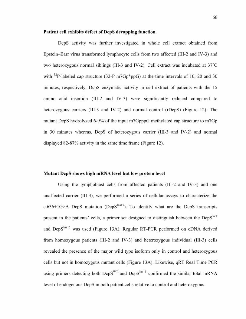
66
Patient cell exhibits defect of DcpS decapping function.
DcpS activity was further investigated in whole cell extract obtained from
Epstein–Barr virus transformed lymphocyte cells from two affected (III-2 and IV-3) and
two heterozygous normal siblings (III-3 and IV-2). Cell extract was incubated at 37˚C
with 32P-labeled cap structure (32-P m7Gp*ppG) at the time intervals of 10, 20 and 30
minutes, respectively. DcpS enzymatic activity in cell extract of patients with the 15
amino acid insertion (III-2 and IV-3) were significantly reduced compared to
heterozygous carriers (III-3 and IV-2) and normal control (rDcpS) (Figure 12). The
mutant DcpS hydrolyzed 6-9% of the input m7GpppG methylated cap structure to m7Gp
in 30 minutes whereas, DcpS of heterozygous carrier (III-3 and IV-2) and normal
displayed 82-87% activity in the same time frame (Figure 12).
Mutant DcpS shows high mRNA level but low protein level
Using the lymphoblast cells from affected patients (III-2 and IV-3) and one
unaffected carrier (III-3), we performed a series of cellular assays to characterize the
c.636+1G>A DcpS mutation (DcpSIns15). To identify what are the DcpS transcripts
present in the patients’ cells, a primer set designed to distinguish between the DcpSWT
and DcpSIns15 was used (Figure 13A). Regular RT-PCR performed on cDNA derived
from homozygous patients (III-2 and IV-3) and heterozygous individual (III-3) cells
revealed the presence of the major wild type isoform only in control and heterozygous
cells but not in homozygous mutant cells (Figure 13A). Likewise, qRT Real Time PCR
using primers detecting both DcpSWT and DcpSIns15 confirmed the similar total mRNA
level of endogenous DcpS in both patient cells relative to control and heterozygous

67
Figure 12. DcpS activity in lymphoblastoid cell lines.
Lymphoblast cell lines from homozygous affected (III-2 and IV-3) and heterozygous unaffected (III-3 and IV-2) family members were used for decapping assays. Decapping products and labeling are as in Figure 11

68
individual cells (Figure 13B). Using specific primers for DcpSWT also did not detect any
DcpSWT mRNA generated in patient cells (Figure 13B) These data demonstrate the
c.636+1G>A mutation results in an alteration of splicing that leads to exclusive
generation of variant splice pattern in homozygous individuals while heterozygous
individuals express both the WT and variant splice patterns.
We next assessed the consequences of the synthesis of the DcpS protein in patient
cells. Consistent with the mRNA data, western blot analysis with an anti-DCPS antibody
detected DcpSWT in control and heterozygous cells but not in homozygous mutant cells
(Figure 13C). A slightly larger band of DcpS likely representing the 15 amino-acid in-
frame insertion translated from mutation was detected at lower levels in both
heterozygous cells and homozygous mutant cells but not in control cells (Figure 13C).
These results suggest that the discovered splice site mutation completely abolished the
default splicing and utilize the alternative splicing site 45 nt downstream instead.
Furthermore, these data indicate that despite comparable mRNA levels for DcpSIns15 and
DcpSWT, considerably lower levels of DcpSIns15 protein are present in both homozygous
and heterozygous cells.
DcpSIns15 localized in nucleus and hetero-dimerized with wild type DcpS
DcpS is a shuttling protein that is predominantly nuclear (Shen et al, 2008). We
next determined whether DcpSIns15 is correctly localized in cells. Since the nuclear
localization sequence (NLS) in DcpS is at the N- terminus of the protein and not
disrupted by the c.636+1G>A mutation, it was anticipated to localize normally. We

69
Figure 13. Characterize DcpSIns15 mRNA and protein in patient cells.
(A) RT–PCR analysis for DCPS mRNA. RT–PCR across the DcpS splice site mutation (from exons 3 to 6) was performed using mRNA from patients (III-2 and IV-3) and an unaffected family member (III-3), and a schematic indicating the corresponding exon usage. (B) Total DcpS mRNA levels and DcpSWT mRNA levels in patients (III-2 and IV-3) and unaffected family member (III-3) were tested by real time PCR using specific primers. Wild type lymphoblast cell line C3688 was used as a control and set to 1. RNA levels are presented relative to the GAPDH mRNA and derived from three independent experiments. (C) Western Blot of DcpS. Wild-type and variant DcpS proteins from the indicated lymphoblast cell lines were detected by western blotting. A wild-type lymphoblastoid cell line is shown to indicate the position of wild-type DcpS. Actin was used as loading control.

70
confirmed this by exogenous expression of an epitope tagged DcpSIns15 protein in
GM03813 which are SMA patient fibroblast cells that express normal levels of DcpS. As
shown in Figure 14A, after infecting lentiviral particles harboring an expression system
for Flag-DcpSIns15, the majority of DcpSIns15 protein was detected in the nucleus with an
anti-Flag tag antibody as expected (Figure 14A). Infection with viral particles lacking an
exogenous expression cassette was used as the control and no Flag signal was detected
(Figure 14A).
To test the possibility that the 15 amino-acid-insertion may interfere with the
dimerization interface, we next determined whether a heterodimer between DcpSIns15 and
DcpSWT could form. HEK293T cells stably transformed with Flag-DcpSIns15 cDNA were
generated and DcpSIns15 was immunopurified on a Flag affinity column (Figure 14B).
Western blot analyses with the -DcpS antibody revealed coimmunoprecipitation of wild
type DcpS in the fractions with Flag-DcpSIns15 in a dose-dependent manner (Figure 14B)
demonstrating that DcpS and DcpSIns15 can heterodimerize in cells. Since a heterodimer
of wild type DcpS with a catalytically inactive DcpS can generate partially active protein
with one active site (Liu et al, 2008b), the DcpSIns15 containing heterodimer may contain
partial activity.
Reduced levels of DcpSIns15 protein in cells is not a consequence of protein
degradation or microRNA mediated silencing
Closer examination of figure 13 revealed a disconnection between the level of
DcpSIns15 mRNA and protein. Although mutant DcpSIns15 mRNA levels in the ID patient

71
Figure 14. DcpSIns15 localized in nucleus and was able to dimerize with wild type DcpS.
(A) GM03813 human fibroblast cells were infected with lentivirus which expressed Flag-DcpSIns15 or empty vector. The red staining by DcpS antibody denotes localization of endogenous DcpS and the green staining by Flag antibody denoted the localization of DcpSIns15. The nuclei are designated by DAPI staining. (B) Anti-Flag immunoprecipitation was performed in HEK293T cell lines constitutively express Flag-tagged DcpSIns15. Western Blot using DcpS antibody indicated the DcpSWT was co-purified with Flag-DcpSIns15. The lower band in anti-DcpS blot was not degradation product of Flag-DcpSIns15 as it was not detected in anti-Flag blot. Whole cell extract was also included to designate the position of DcpSWT and DcpSIns15.
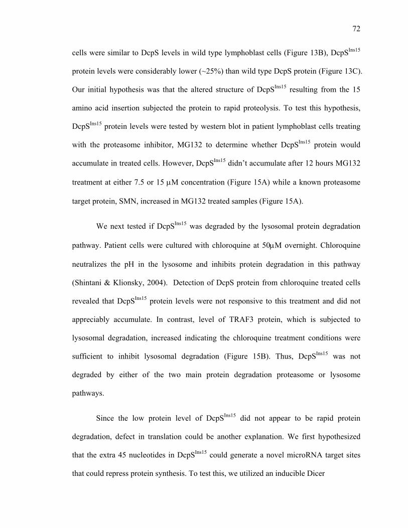
72
cells were similar to DcpS levels in wild type lymphoblast cells (Figure 13B), DcpSIns15
protein levels were considerably lower (~25%) than wild type DcpS protein (Figure 13C).
Our initial hypothesis was that the altered structure of DcpSIns15 resulting from the 15
amino acid insertion subjected the protein to rapid proteolysis. To test this hypothesis,
DcpSIns15 protein levels were tested by western blot in patient lymphoblast cells treating
with the proteasome inhibitor, MG132 to determine whether DcpSIns15 protein would
accumulate in treated cells. However, DcpSIns15 didn’t accumulate after 12 hours MG132
treatment at either 7.5 or 15 M concentration (Figure 15A) while a known proteasome
target protein, SMN, increased in MG132 treated samples (Figure 15A).
We next tested if DcpSIns15 was degraded by the lysosomal protein degradation
pathway. Patient cells were cultured with chloroquine at 50M overnight. Chloroquine
neutralizes the pH in the lysosome and inhibits protein degradation in this pathway
(Shintani & Klionsky, 2004). Detection of DcpS protein from chloroquine treated cells
revealed that DcpSIns15 protein levels were not responsive to this treatment and did not
appreciably accumulate. In contrast, level of TRAF3 protein, which is subjected to
lysosomal degradation, increased indicating the chloroquine treatment conditions were
sufficient to inhibit lysosomal degradation (Figure 15B). Thus, DcpSIns15 was not
degraded by either of the two main protein degradation proteasome or lysosome
pathways.
Since the low protein level of DcpSIns15 did not appear to be rapid protein
degradation, defect in translation could be another explanation. We first hypothesized
that the extra 45 nucleotides in DcpSIns15 could generate a novel microRNA target sites
that could repress protein synthesis. To test this, we utilized an inducible Dicer
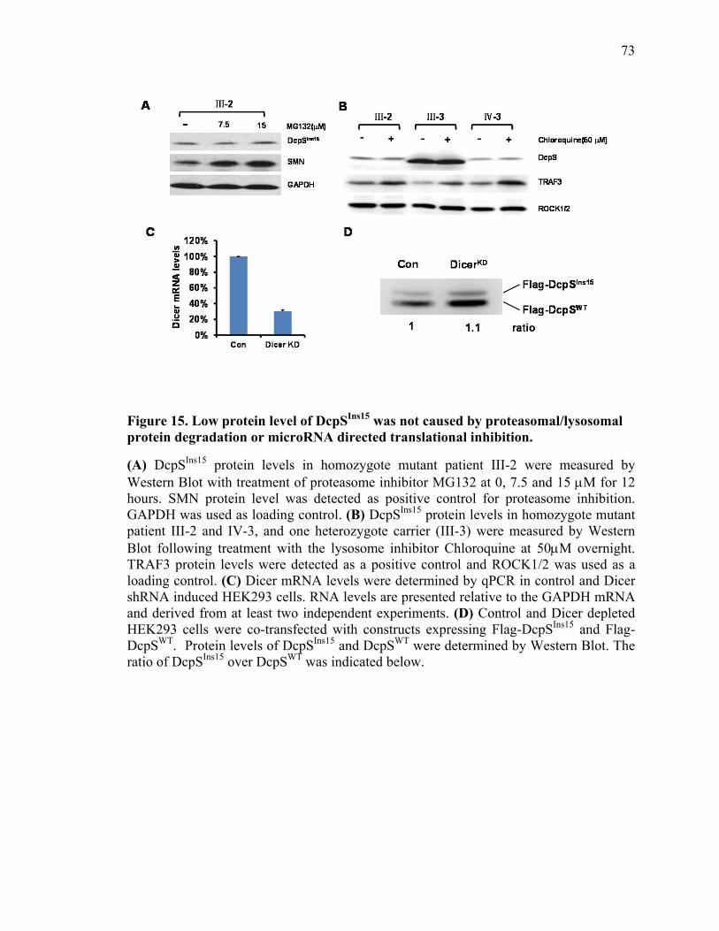
73
Figure 15. Low protein level of DcpSIns15 was not caused by proteasomal/lysosomal protein degradation or microRNA directed translational inhibition.
(A) DcpSIns15 protein levels in homozygote mutant patient III-2 were measured by Western Blot with treatment of proteasome inhibitor MG132 at 0, 7.5 and 15 M for 12 hours. SMN protein level was detected as positive control for proteasome inhibition. GAPDH was used as loading control. (B) DcpSIns15 protein levels in homozygote mutant patient III-2 and IV-3, and one heterozygote carrier (III-3) were measured by Western Blot following treatment with the lysosome inhibitor Chloroquine at 50M overnight. TRAF3 protein levels were detected as a positive control and ROCK1/2 was used as a loading control. (C) Dicer mRNA levels were determined by qPCR in control and Dicer shRNA induced HEK293 cells. RNA levels are presented relative to the GAPDH mRNA and derived from at least two independent experiments. (D) Control and Dicer depleted HEK293 cells were co-transfected with constructs expressing Flag-DcpSIns15 and Flag-DcpSWT. Protein levels of DcpSIns15 and DcpSWT were determined by Western Blot. The ratio of DcpSIns15 over DcpSWT was indicated below.

74
knockdown 293 cell line (Schmitter et al, 2006). Dicer is an essential factor for
microRNA expression and its depletion should ablate microRNA-mediated translational
silencing. Flag-DcpSIns15 plasmid was co-transfected with Flag-DcpSWT into doxycycline
induced cells and DcpS protein levels were determined by western blot 2 days after
transfection. Dicer level was about 30% in the induced cells compared to non-treated
cells as determined by real- time RT-PCR (Figure 15C). However DcpSIns15 protein
levels failed to accumulate in Dicer reduced cells after normalizing to wild type DcpS
(Figure 15D). In summary, the reduced DcpSIns15 protein level does not appear to be due
to proteasomal/lysosomal degradation or microRNA-mediated translational repression.
Possible translation repression mechanisms other than microRNA-mediated remain to be
tested.
Discussion
In this chapter, we identified mutant DcpS genes in 3 individuals with intellectual
disability from a large family with multiple branches. We identified a splice-site variant
that results in the insertion of 15 amino acids (c.636+1G>A) and a missense variant
(c.947C>T; p.Thr316Met) in the DcpS gene (Figure 9). In silico parameters and
molecular modeling suggested deleterious effects of both variants (Figure 10) and
functional analyses validated both mutations lead to abrogation of DcpS decapping
function in vitro and in cells (Figure 11C and Figure 12). Using the lymphoblast cell lines
generated from affected patients, we characterized the mutant DcpS with insertion of 15
amino acids (DcpSIns15) and analyzed its localization and capacity to form dimer with
wild type DcpS (Figure 14). It is interesting that the transcription of this mutant DcpS

75
appeared normal and mRNA level of this isoform were similar to wild type DcpS in
heterozygous cells (Figure 13B), while DcpSIns15 protein levels were significantly lower
relative to wild type DcpS (Figure 13C). This phenomenon was not due to protein
degradation by either proteasome or lysosome (Figure 15A and B), or microRNA
induced translational repression (Figure 15D). Collectively, our data demonstrate that 1)
homozygous mutations in DcpS that abolish its decapping function lead to intellectual
disability in affected individuals; and 2) the insertion of 15 amino acids in DcpS results in
reduced protein levels through a yet-to-be determined mechanism.
Although at present we only have identified disruptions in DcpS in one family,
the presence of compound heterozygous variants in DcpS in an affected person supports a
role for DcpS as the causative agent for intellectual disability in these individuals.
Moreover, the identification of a second family, of Jordanian descent, with intellectual
disability also caused by a homozygous mutation in DcpS leading to an in frame 7 amino
acid insertion at amino acid K67 and nonfunctional variant protein (Ng et al, 2015b),
further reinforces the significance of DcpS for neuronal function. The presence of
obvious physical anomalies and neuromuscular phenotype in the Jordanian family
suggests that a relatively wide clinical spectrum can be anticipated for different types and
locations of mutations in DcpS.
In cells, a disruption of DcpS decapping activity is expected to lead to the
accumulation of m7GpppN cap structure, which can sequester the nuclear cap binding
protein and decrease the efficiency of first intron splicing (Shen et al, 2008) and
potentially alter cytoplasmic mRNA translation by sequestering the cytoplasmic cap
binding protein (Bail & Kiledjian, 2008). In addition, at least in S. cerevisiae and C.

76
elegans, DcpS orthologues contribute to Xrn1-mediated exonucleolytic decay (Bosse et
al, 2013; Liu & Kiledjian, 2005; Sinturel et al, 2012). In human cells, DcpS also
demonstrates transcript specific regulation of RNA stability coupled with Xrn1 as well as
RNA decay independent regulation of RNA steady state levels (Zhou et al, 2015).
Genomic mRNA and proteomic analysis of cells homozygous c.636+1G>A mutation that
exclusively generate DcpSIns15 will enable an analysis of the underlying molecular defect
attributed to expression of this variant protein.
The mutant DcpSIns15 exhibited much lower protein levels compared to wild type
DcpS, despite similar mRNA levels of the two genes. This suggested the mutant DcpS
had lower translational efficiency or faster protein turnover. Inhibition of the two
primary protein degradation pathways mediated through the proteasome and lysosome,
did not increase mutant DcpS protein levels, indicating the low level of this protein was
not caused by impeded protein degradation. Alternatively, the insertion of 45nt could
create sites for microRNA or some RNA-binding proteins, which could in turn slow
down the progression of ribosomes and reduce translational efficiency. DcpSIns15 protein
levels were unchanged in Dicer depleted cells, suggesting DcpSIns15 was not a microRNA
target. Further work is needed to test whether the 45nt insertion recruited some protein
factors to repress translation.
In an effort to generate a DcpS null mouse, we found that DcpS is an essential
gene and its deletion is embryonic lethal (M. Kiledjian: unpublished observations),
demonstrating a role for DcpS protein in embryogenesis. CRISPR screens of human
genes also indicate the requirement of DcpS for cellular viability (Shalem et al, 2014),
underlining the importance of this protein in fundamental cellular functions. The viability

77
of individuals with a decapping-compromised DcpS protein suggests that the decapping
activity of DcpS is important for normal cognition, while an additional decapping-
independent function of DcpS must also be required for normal development and cell
survival. This additional function is yet to be determined. Further, we consider it unlikely
that fully null homozygous mutations in DcpS will be identified in a clinical context, and
that bi-allelic mutations would always be hypomorphic or with restricted effect on
specific isoforms or functions of the protein.
In conclusion, we report the investigation of a family with apparent non-specific
autosomal recessive intellectual disability leading to the identification of mutations in
DcpS. At present the molecular consequence of defects in DcpS is unknown, yet it leads
to a physiological consequence of adverse overall cognition in individuals harboring the
mutant alleles. Future identification of the downstream target mRNAs or proteins altered
in the cells of affected individuals will provide insights into the contribution of DcpS in
gene expression and cognitive function.

78
Chapter III DcpS Insertion Variant Elevates Cellular SMN2
RNA and Protein Levels
Summary
Spinal Muscular Atrophy (SMA) is a common autosomal recessive disorder that
results in progressive loss of spinal anterior horn motor neurons. It is a consequence of
low levels of survival motor neuron (SMN) protein caused by the loss or mutation of the
survival motor neuron 1 gene (SMN1). A duplicate gene, SMN2, usually remains intact
and relieves the severity of SMA, though the majority of mRNA generated from SMN2
lack exon 7 due to inappropriate splicing resulting from a single nucleotide substitution
of a C->T inside exon 7. Previously it was reported that a quinazoline derivative, RG3039,
increased survival and function in a mouse model of SMA through inhibition of the
decapping scavenger enzyme, DcpS. We now have evidence that a variant form of DcpS
that utilizes a cryptic splice site initially identified as a mutation in non-syndromic
autosomal recessive intellectual disability (ID) can significantly elevate SMN2 steady
state mRNA and SMN protein in SMA patient cells. Most significantly, utilization of the
cryptic splice site 45 nucleotides downstream of the endogenous splice site can be
promoted by oligonucleotide directed splice switching technology. We designed and
tested four oligonucleotides in an effort to alter the endogenous DcpS gene splicing
pattern to that of the DcpSIns15. Shifting of the DcpS splicing pattern to the DcpSI15
variant could be a potential therapeutic avenue to elevate full length SMN2 mRNA and
SMN protein in SMA patient cells.

79
Introduction
Spinal Muscular Atrophy (SMA) is a common autosomal recessive disorder that
results in progressive loss of spinal anterior horn motor neurons, lethal in its severe form,
with no current cure. SMA is caused by reduced levels of SMN protein, the consequence
of homozygous loss or mutation in the survival motor neuron 1 gene (SMN1) (Lefebvre
et al, 1995). A second, nearly identical gene to SMN1, termed SMN2, was found in the
human genome. It shares the same promoter and differs by only two-nucleotides in the
coding region relative to SMN1 (Monani et al, 1999a; Monani et al, 1999b). The
primarily difference is a single nucleotide substitution of a CT inside exon 7 in SMN2
that disrupts splicing of exon 7 leading predominantly to exon 7 exclusion and the
generation of nonfunctional truncated protein (Lorson et al., 1999; Monani et al., 1999).
However, due to a low level of exon 7 inclusion from the SMN2 gene (Gavrilov et al.,
1998; Lorson et al., 1999; Monani et al., 1999), an increase of SMN2 mRNA levels can
lead to dosage dependent compensation of SMN1 lose and a decrease of SMA severity in
humans (Chen et al., 1999; McAndrew et al., 1997; Wirth et al., 1999) as well as mice
model systems (Hsieh-Li et al., 2000; Monani et al., 2000).
The primary strategies of current SMA therapeutics are focused on the importance
of increasing SMN levels from three different approaches: 1) to increase SMN protein
levels; 2), to increase SMN2 transcription levels; and 3) to promote the inclusion of exon
7 in SMN2 transcripts. Based on the first approach to express exogenous SMN, the
feasibility of viral-based delivery of the SMN-coding region to restore normal SMN
expression in SMA patient-derived cells was first demonstrated using adenoviral vectors
(DiDonato et al, 2003). Later, a lentiviral vector based system was shown to retrogradely

80
increase SMN expression in motor neurons in an SMA mouse model (Azzouz et al, 2004).
Recently, an adeno-associated viral vector (AAV9) based method, which can infect
peripheral tissue as well as cross the blood–brain barrier, showed improvements in gene
delivery efficiency and led to the rescue of a severe mouse model of SMA (Dominguez et
al, 2011; Foust et al, 2010; Passini et al, 2010).
Major emphasis is also focused on an alternative approach to promote the
inclusion of exon 7 in SMN2 transcripts through antisense oligonucleotide or small
compound. The antisense oligonucleotide approach targets pre-mRNA processing,
reducing exon 7 exclusion in SMN2 mRNA and increasing full-length SMN transcripts
and protein levels. Many promising antisense oligonucleotide based methodology to
treating SMA has proven successful both in vitro and in vivo using SMA mouse models
(Cherry et al, 2013; Hua et al, 2010; Porensky et al, 2012; Rigo et al, 2014; Williams et al,
2009). Besides the antisense oligonucleotide, small molecule compounds that shift the
balance of SMN2 splicing toward the production of full-length SMN2 are also showing
therapeutic potential (Naryshkin et al, 2014). However, most strategies also have various
obvious shortcomings and challenges in patients (Lunn & Stockwell, 2005; Wee et al,
2010). For example, oligonucleotide delivery requires a direct route of administration to
the CNS, such as intrathecal or intracerebroventricular injections, and will require
additional optimization for an efficient delivery to peripheral organs (Anderton et al,
2013).
A class of C5- substituted quinazoline compounds were identified that increased
human SMN2 promoter driven expression of the bacterial -lactamase reporter in a
screen of a mouse motor neuron hybrid cell line NSC-34 (Jarecki et al, 2005). The

81
initially identified compound was optimized by a focused medicinal chemistry effort to
develop a series of modified C5-quinazolines and shown to be an inhibitor of the
scavenger decapping enzyme, DcpS (Singh et al, 2008; Thurmond et al, 2008). Although
the quinazoline compounds were capable of improving motor neuron function and
extending survival of SMA model mice (Butchbach et al, 2010; Gogliotti et al, 2013; Van
Meerbeke et al, 2013a), statistically significant increases in SMN2 mRNA or SMN
protein were not evident. The lack of detectable increase in SMN mRNA and protein in
treated animals confounds the mechanism by which the quinazoline compounds promote
survival of SMA mice. To add to this perplexing result, neither compound treatment nor
DcpS knockdown in cell lines leads to statistically significant elevated SMN2 mRNA or
protein (unpublished) suggesting that the compound likely functions through an SMN
independent and possibly DcpS independent pathway. Therefore, the efficacy of current
efforts focused on using DcpS inhibitor compounds in patients remains unclear.
In this chapter, we demonstrate that a variant form of DcpS, DcpSIns15, initially
identified as a mutation in non-syndromic autosomal recessive intellectual disability (ID)
(Chapter2) increases SMN2 mRNA expression. Lymphoblast cells from ID patients
harboring a homozygous DcpSIns15 allele contain significant elevation of SMN2 mRNA
but not in heterozygous siblings with normal intellect. More significantly, exogenous
expression of DcpSIns15 into SMA patient fibroblasts lead to elevated SMN2 mRNA and
SMN protein. The SMN2 increase is not observed when the cells are complemented with
either wild type or a catalytically inactive DcpS. Of particularly high significance for
potential SMA therapeutics, the DcpS pre-mRNA is amendable to antisense

82
oligonucleotide directed splice switching to promote generation of the DcpSIns15 variant
mRNA in SMA patient cells in a strategy to ameliorate the SMA phenotype in patients.
Results
SMN2 mRNA levels are elevated by expression of variant DcpS
The requirement of DcpS for normal neurological function (Ahmed et al, 2015b;
Ng et al, 2015b) and the potential link of DcpS to SMN2 expression (Singh et al, 2008),
prompted us to test whether SMN2 levels were altered in DcpSIns15 homozygous cells.
Quantitative reverse transcription (qRT)-PCR analysis of RNA derived from the
homozygous patients with DcpSIns15 mutation revealed a selective elevation of SMN2 in
these cells. A 2 fold increase of full length SMN2 mRNA was detected in both DcpSIns15
homozygous cell lines, but not in a heterozygous line relative to wild type lymphoblast
cells (Figure 16A).
To determine whether the increase of full length SMN2 mRNA was due to loss of
DcpS decapping activity, a 293T-DcpSKD cell line which constitutively expresses DcpS-
specific shRNA to reduce endogenous DcpS levels, was transfected with shRNA-
resistant catalytically inactive mutant of DcpS (DcpSMT), or DcpSIns15, and stable cell
populations were selected. Similar to the DcpSIns15 patient cells, expression of the
DcpSIns15 variant in DcpSKD cells resulted in a >2 fold increase in full length SMN2
mRNA compared to control transfected cells (Figure 16B). Moreover, it was evident that
the increase was not a result of a defect in DcpS decapping since expression of the
DcpSMT catalytically inactive mutant had no effect on SMN2 expression.

83
Figure 16. DcpSIns15 correlated with increasing full length SMN2 mRNA.
(A) Full length SMN2 mRNA levels were detected by Real Time PCR in patients (III-2 and IV-3) and unaffected family member (III-3 and IV-2) as well as in WT lymphoblast cell line C3688, and normalized to C3688. (B) Full length SMN2 mRNA levels were detected by Real Time PCR in HEK293T control knock down (ConKD) or DcpS knock down (DcpSKD) cells, complemented with shRNA-resistant catalytically inactive DcpSMT, or DcpSIns15. RNA levels are presented relative to the GAPDH mRNA and derived from three independent experiments. Error bars represent +/- SD. P values are denoted by asterisks; * represents p < 0.05, ** represents p < 0.01 (Student’s t test).
0
0.5
1
1.5
2
2.5
****
Ⅲ-2 Ⅲ-3 Ⅳ-3Ⅳ-2C3688
Lym
ph
ob
last
sm
RN
A
leve
ls (
fold
)SMN2 full-length
0
0.5
1
1.5
2
2.5
293T
cel
l m
RN
A
leve
ls (
fold
)
**
Vector Vector DcpSMT DcpSIns15
DcpSKDConKD
SMN2 full-length
A
B
84
These data demonstrate that the detected elevation of full length SMN2 in both
the homozygous patient cells and in 293T cells is a consequence of DcpSIns15 expression.
In addition, the enhanced level of elevation of SMN2 RNA observed with expression of
DcpSIns15 compared to catalytic inactive DcpS indicates that DcpSIns15 is likely a gain of
function variant coordinating an elevation of SMN2 in a manner independent of its
decapping activity. Furthermore, the lack of detectable SMN2 mRNA increase in
heterozygous lymphoblast cells indicated there may be a threshold level of DcpSIns15
variant that is necessary to increase SMN2 levels.
SMN RNA and protein levels were increased by DcpSIns15 in SMA patient cells
To determine whether expression of DcpSIns15 can elevate full length SMN2
mRNA in a clinically more relevant SMA type I patient-derived fibroblast cells
(GM03813), we engineered a constitutive knockdown of endogenous DcpS with DcpS-
specific shRNA. These cells were transfected with shRNA-resistant wild type DcpS
(DcpSWT), a catalytically inactive mutant of DcpS (DcpSMT), or DcpSIns15. Similar to the
293T cells, expression of the DcpSIns15 variant into GM03813 cells resulted in a >2 fold
increase of both full length and ∆7 SMN2 mRNA, compared to control transfected cells
by qRT-PCR (Figure 17A). The increase of both the full length and ∆7 transcripts
indicated the increase in full length mRNA is not a consequence of enhanced exon 7
splicing (Figure 17A).
In order to determine whether SMN protein levels could also increase as a
consequence of SMN2 full length mRNA elevation, two SMA patient fibroblast cell lines

85
GM03813 and GM00232 expressing DcpS-specific shRNA were transfected with
shRNA-resistant DcpS wild type (DcpSWT), a catalytically inactive DcpS mutant
(DcpSMT), or DcpSIns15. Western Blot showed the protein level of SMN also increased
about 2 fold in both GM03813 and GM00232 cells with DcpSIns15 expression (Figure
17B). Quantitation of the Western Blot was shown in Figure 17C. Collectively, our data
indicated that DcpSIns15 specifically elevated SMN2 mRNA and protein levels
independent of scavenger decapping activity.
Next, we determined whether DcpSIns15 could increase SMN2 without reducing
endogenous DcpS. GM00232 SMA fibroblast cells were infected with increasing amount
of lentivirus that expressed DcpSIns15. Lentiviruses expressing GFP or DcpSMT were used
as controls. Western Blot analysis showed SMN protein levels increased about 2 fold
when the expression level of DcpSIns15 reached levels comparable to endogenous wild
type DcpS (Figure 18A). With low DcpSIns15 protein level which was similar to
heterozygous individuals, there was also no SMN change (Figure 18A). Quantitation of
the Western Blot showed that SMN protein level was elevated ~2 fold by expressing
DcpSIns15 at levels that at least reached 1:1 ratio to endogenous DcpS (Figure 18B). Our
data indicates a threshold level of DcpSIns15 is necessary in order to increase SMN.
Application of antisense morpholino oligonucleotides to alter DcpS pre-mRNA
splicing in cells
The unexpected discovery of the DcpSIns15 alternative splice variant as an agent to
increase full-length SMN2 mRNA and protein levels provides a potentially novel

86
Figure 17. DcpSIns15 increased full length SMN2 mRNA and SMN protein levels in SMA patient cells.
(A) GM03813 cells were infected with control (ConKD) or DcpS shRNA expressing (DcpSKD) lentiviruses, and complemented with shRNA-resistant DcpSWT, catalytically inactive DcpSMT, or DcpSIns15. Full length and exon7 truncated SMN2 mRNA levels were detected by Real Time PCR with transcript-specific primers. RNA levels are presented relative to the GAPDH mRNA and derived from three independent experiments. (B) SMN protein levels were detected by Western Blot in GM03813 and GM00232 cells from cells subjected to the same conditions as in (A). GAPDH was detected as loading control. (C) Quantitative summary of three independent Western Blot results as (B). Protein levels are normalized to GAPDH. Error bars represent +/- SD. P values are denoted by asterisks; * represents p < 0.05, ** represents p < 0.01 (Student’s t test).

87
Figure 18. DcpSIns15 increased SMN protein levels in a dose dependent manner.
(A) SMN protein levels were detected by Western Blot in GM00232 cells infected with lentiviruses expressing GFP, catalytically inactive DcpSMT, or increasing amount of DcpSIns15. GAPDH was detected as loading control. (B) Quantitative summary of three independent Western Blot results as (A). Protein levels are normalized to GAPDH. Error bars represent +/- SD. P values are denoted by asterisks; ** represents p < 0.01 (Student’s t test).

88
therapeutic avenue for SMA. The DcpS pre-mRNA is amendable by antisense splice
switch oligonucleotide (SSO) technology to shift the endogenous DcpS splicing pattern
to a predominant DcpSIns15 variant in SMA patients. With this approach, an antisense
oligonucleotide complementary to the endogenous splice site is used to block the
endogenous splicing pattern and shift it towards the DcpSIns15 splicing pattern in cells
(Figure 18A). Our objective was to utilize SSO’s to partially mask the endogenous 5´
splice site of DcpS intron 4 and redirect splicing to the cryptic splice site 45 nucleotides
downstream to generate DcpSIns15 at levels sufficient to support increased full length
SMN2 mRNA while retaining effective levels of wild type DcpS to maintain its normal
function.
Four different vivo-morpholino antisense oligonucleotides targeting different
positions of exon4 and intron4 junction in DcpS pre-mRNA were designed to directly
switch splicing of DcpS in cells (Figure 19B). GM03813 cells were treated with a
titration of antisense morpholino oligonucleotides containing a cell permeable
guanidinium based dendramer delivery system (vivo-morpholino; GeneTools LLC) for
24 hours and splicing patterns were determined by RT- PCR. By using a pair of primers
spanning exon3 to exon 6, three possible splicing patterns were shown in Figure 20A:
wild type, insertion of the 45 nucleotides and a third product that skipped exon4
completely and spliced from exon 3 directly into exon 5. The addition of the vivo-
morpholino oligonucleotide #1, #3 and #4 almost completely inhibited utilization of the
endogenous exon 4 splice site at 1 M concentration while oligonucleotide#2 didn’t work
as efficiently (Figure 20B). However all four oligonucleotides we tested not only

89
Figure 19. Design Morpholino anti-sense oligonucleotides that shift DcpS splicing.
(A) Schematic presentation of the splicing shift of DcpS by vivo-morpholino targeting DcpS exon4/intro4 junction. (B) Schematic presentation of the four designed vivo-morpholino oligonucleotides.

90
promoted utilization of the cryptic site to generate the DcpSIns15 splice variant, but also
promoted skipping of exon 4 entirely (Figure 20B). This skipping effect was unexpected
and is detrimental because it would generate a truncated DcpS transcript bypassing exon
4 and the insert.
After screening the splicing alteration efficiency of our four Morpholino
oligonucleotides, we next checked whether DcpSIns15 protein was expressed in cells
treated with the SSO’s. GM03813 cells were treated with 0.5µM or 0.75µM Morpholino
oligonucleotide#1 and 0.75µM or 1uM oligonucleotide #3 respectively and harvested
after 6 days. Different DcpS transcripts were determined by RT-PCR using primers
spanning exon3 to exon 6 and DcpS protein. A polyclonal antibody raised against the
entire DcpS protein was use to enable detection of both DcpSIns15 and any putative
truncated DcpS from the exon4-excluded transcript. However, no DcpSIns15 protein was
detected in any of the treatment although there was DcpSIns15 mRNA expression (Figure
21). There was no truncated DcpS (lower band) detected either (Figure 21), which might
be an indication that the truncated products were unstable. Interestingly, we observed
high cell death rate after 6-day treatment with 1uM Morpholino oligonucleotide#1 and
1.25uM oligonucleotide #3, which reduced wild type DcpS transcripts more than 90%
(Figure 20B). Thus we were unable to collect and test the longer incubation samples. In
summary, although our findings demonstrate that the exon 4- intron 4 junction of DcpS is
accessible to antisense oligonucleotide directed manipulation, at least the initial set of
four antisense morpholino oligonucleotides tested do not sufficiently shift the
endogenous DcpS pre-mRNA splicing to levels that generate detectable DcpSIns15 protein.

91
Figure 20. Morpholino oligonucleotide efficiencies were screening by RT-PCR.
(A) Schematic depicting the position of primers used for the following RT-PCR screens of possible splicing patterns caused by morpholino oligonucleotides. (B) Endogenous DcpS mRNAs with different splicing patterns were detected by RT-PCR in GM03813 cells after 2-day treatment of morpholino oligonucleotides #1-#4 at the indicated concentrations. Schematic represents the exon usage.

92
Figure 21. Morpholino oligonucleotides used are capable of changing endogenous DcpS transcript splicing but not the production of DcpSIns15 protein.
GM03813 cells were harvested after 6-day treatment of morpholino oligonucleotide #1 and #3 at the indicated concentrations. Top: endogenous DcpS mRNAs with different splicing patterns were detected by RT-PCR with corresponding schematics indicating exon usage. Bottom: DcpS protein was detected by Western Blot using a DcpS polyclonal antibody. GAPDH protein was used as a loading control.

93
A panel of additional oligonucleotides that walk along the entire junction appears to be
necessary to identify oligonucleotides that can efficiently shift the splicing pattern.
Discussion
In this chapter, we observed that full length SMN2 mRNA levels increased
approximately 2 fold in DcpSIns15 homozygous patient lymphoblast cells compared to
wild type and heterozygous lymphoblast cells (Figure 16A). We next confirmed that full
length SMN2 mRNA levels also increased 2 fold in 293T DcpSKD cells expressing
exogenous DcpSIns15 but not catalytic inactive DcpSMT (Figure 16B). A similar outcome
was also detected in a the GM03813SMA patient fibroblast cell line where full length
SMN2 mRNA levels also increased 2 fold in this cell line upon expression of exogenous
DcpSIns15 (Figure 17A). The elevation of SMN2 RNA is accompanied with a
corresponding increase of SMN protein levels (Figure 17B and 18). We showed that
vivo-morpholino antisense oligonucleotides which targeted exon 4 and intron 4 junction
in DcpS pre-mRNA were able to reduce the endogenous DcpS splicing pattern and
partially shift the splicing to the DcpSIns15 isoform pattern in cells (Figure 20B). However,
we also observed an unexpected alternative splicing whereby the entire exon 4 was
skipped in morpholino treated cells (Figure 20B). Our studies establish a proof of
principle for the use of vivo-morpholino technology to shift DcpS splicing, however, the
current set of antisense oligonucleotides to not generate sufficient levels of variant
mRNA to translate into detectable DcpSIns15 protein (Figure 21).

94
DcpSIns15 is likely a gain of function variant coordinating an elevation of SMN2 in
a manner independent of its decapping activity. We performed all our initial experiments
in DcpS knocked down cells to minimize any potential interference by endogenous DcpS
since DcpS functions as a homodimer. However, we predicted that sufficient expression
of DcpSIns15 should be able to elevate SMN2 even within the normal endogenous DcpS
protein background. Indeed, in preliminary experiments we observed about 2 fold
increase of SMN protein level by overexpressing DcpSIns15 up to at least 1:1 protein ratio
to endogenous DcpS in wild type GM00232 cells (Figure 18). This is consistent with our
data that heterozygous individuals did not show increased SMN2 mRNA levels.
Therefore, to increase SMN2 mRNA required a certain threshold level of DcpSIns15
protein.
The application of vivo-morpholino antisense oligonucleotides to express
endogenous DcpSIns15 was promising but still faces a few challenges. One problem is the
unexpected exon 4 skipping caused by the morpholino oligonucleotides. While the
morpholino oligonucleotides successfully inhibited the endogenous DcpS intron 4
splicing and promoted utilization of the DcpSIns15 splicing pattern, they also generated a
splicing variant that skipped the entire exon 4. In most cases, this actually occurred more
efficient than DcpSIns15 splicing. The binding of morpholino oligonucleotides at exon 4-
intron 4 junction appear to disrupt both the endogenous and variant intron 4 splicing,
favoring a bypass splicing pattern that omits exon 4 and splices exon 3 to exon 5. To
eliminate this type of skipping, we will likely need to screen a series of oligonucleotides
that span the exon 4-intron 4 junction at single nucleotide intervals. Another problem we
faced was the low protein expression of DcpSIns15. Despite the significant expression of

95
DcpSIns15 splicing variant by morpholino oligonucleotides, we were unable to detect
production of DcpSIns15 protein consistent with the low level of DcpSIns15 protein level
observed in ID patient cells (Chapter 2). We have ruled out several possible regulatory
pathways including proteasomal/lysosomal degradation of DcpSIns15 protein or
translational repression by microRNAs (Chapter 2), but a clear understanding of why low
levels of DcpSIns15 protein are generated is still lacking. As a therapeutic agent, a fine
balance would have to be maintained between wild type and variant DcpS. Ideally we
should express as much protein as possible to reach the threshold level. On the other hand,
we would need to retain a certain level of wild type DcpS expression to maintain normal
cell function otherwise it could cause neurological defects (Ahmed et al, 2015a; Ng et al,
2015a).
In addition to the therapeutic application of DcpSIns15 in SMA, the unknown
mechanism beyond this specific regulation may also provide new perspectives in SMN2
gene regulation. Determining how DcpSIns15 upregulates SMN2 could provide novel
insights into strategies to upregulate SMN2 expression independent of DcpSIns15 and
avoid complications that could be associated with reduced wild type DcpS levels in cells.

96
Concluding remarks
Despite its initial described function in the last step of the mRNA 3´end decay
pathway, DcpS is a multifunctional protein involved in a greater network of regulated
gene expression. We identified two long non coding RNA, DRNT1 and DRNT2, as
human DcpS responsive RNAs. DRNT1 RNA stability was regulated by DcpS in a
transcript-specific manner. This modulation is dependent on its decapping activity and
mediated through Xrn1. One the other hand, the regulation of DRNT2 steady state level
by DcpS independent of transcript stability strongly suggests a role for DcpS in non-
decay regulation.
Our studies have also uncovered a link of DcpS to human cognition. We
identified mutant DcpS genes in intellectual disability patients from a Pakistan family
within consanguineous marriages. Functional analyses validated that both of the two
mutations (DcpSIns15 and DcpST316M) lead to abolishment of DcpS decapping function in
vitro and in vivo. Moreover, a different homozygous mutation in DcpS from a second
family with intellectual disability was identified independently by another group (Ng et
al, 2015a). All these findings reinforce the significance of DcpS decapping activity for
neuronal development. How DcpS decapping activity contributes to neurogenesis
remains to be determined.
The fact that DcpS deletion is lethal in cells by CRISPR (Shalem et al, 2014) and
embryonic lethal in mice (M. Kiledjian: unpublished observations), demonstrates the
requirement of DcpS for cellular viability and embryogenesis. The viability of individuals
with a decapping-defective DcpS suggests that the decapping of DcpS is only important

97
for normal cognition, while an additional decapping-independent function of DcpS must
also be required for normal development and survival. This additional importance of
DcpS in fundamental cellular functions has yet to be determined.
In a fortuitous development, we observed that lymphoblast cells from ID patients
harboring a homozygous DcpSIns15 allele contain significant elevation of SMN2 mRNA
uncovering a surprising connection between expressions of the DcpSIns15 variant with
SMA. We showed that exogenous expression of DcpSIns15 into SMA patient fibroblasts
led to elevation of SMN2 mRNA and SMN protein but not with either wild type or a
catalytically inactive DcpS. We also demonstrated the feasibility of using vivo-
morpholino antisense oligonucleotides, which targeted the exon 4 - intron 4 junction in
the DcpS pre-mRNA as a tool to switch splicing of DcpS from wild type to DcpSIns15
isoform in cells. Our findings provide a new strategy in SMA therapeutic application, but
the challenges such as low DcpSIns15 protein expression, nonproductive exon skipping as
a consequence of the antisense morpholinos thus far used and the potential risk of
intellectual disability, have yet to be overcome.
Finally, as a potential SMA drug, RG3039 is not a simple DcpS inhibitor and the
extension of survival of SMA mice by RG3039 is likely independent of DcpS and SMN.
We identified four mRNAs (PAQR8, ATOH7, RAB26 and MAOB) mediated by
RG3039 independent of DcpS. Interestingly, the regulation of PAQR8 could be attributed
to a decrease in mRNA stability, while the stability of the other three RNAs was
unchanged upon RG3039 treatment. How RG3039 modulates RNA levels by a
mechanism dependent and independent of transcript stability and independent of DcpS

98
needs to be further addressed to gain a more complete understanding of how this potential
drug candidate may function in cells.
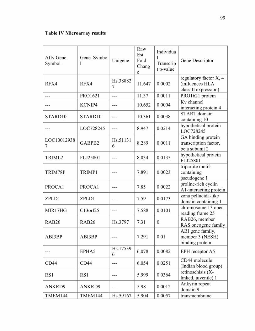
99
Table IV Microarray results
Affy Gene Symbol
Gene_Symbol
Unigene
Raw Est Fold Change
Individual Transcript p-value
Gene Descriptor
RFX4 RFX4 Hs.388827
11.647 0.0002 regulatory factor X, 4 (influences HLA class II expression)
--- PRO1621 --- 11.37 0.0011 PRO1621 protein
--- KCNIP4 --- 10.652 0.0004 Kv channel interacting protein 4
STARD10 STARD10 --- 10.361 0.0038 START domain containing 10
--- LOC728245 --- 8.947 0.0214 hypothetical protein LOC728245
LOC100129387
GABPB2 Hs.511316
8.289 0.0011 GA binding protein transcription factor, beta subunit 2
TRIML2 FLJ25801 --- 8.034 0.0135 hypothetical protein FLJ25801
TRIM78P TRIMP1 --- 7.891 0.0023 tripartite motif-containing pseudogene 1
PROCA1 PROCA1 --- 7.85 0.0022 proline-rich cyclin A1-interacting protein
ZPLD1 ZPLD1 --- 7.59 0.0173 zona pellucida-like domain containing 1
MIR17HG C13orf25 --- 7.588 0.0101 chromosome 13 open reading frame 25
RAB26 RAB26 Hs.3797 7.31 0 RAB26, member RAS oncogene family
ABI3BP ABI3BP --- 7.291 0.01 ABI gene family, member 3 (NESH) binding protein
--- EPHA5 Hs.175396
6.078 0.0082 EPH receptor A5
CD44 CD44 --- 6.054 0.0251 CD44 molecule (Indian blood group)
RS1 RS1 --- 5.999 0.0364 retinoschisis (X-linked, juvenile) 1
ANKRD9 ANKRD9 --- 5.98 0.0012 Ankyrin repeat domain 9
TMEM144 TMEM144 Hs.59167 5.904 0.0057 transmembrane
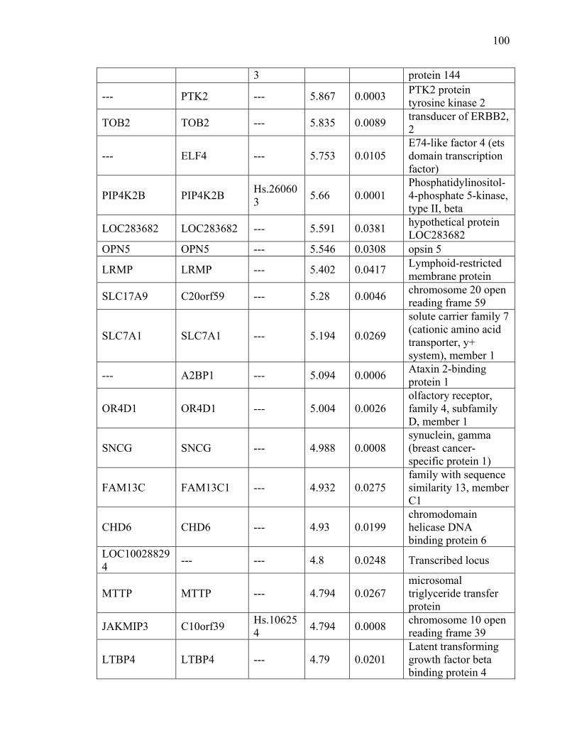
100
3 protein 144
--- PTK2 --- 5.867 0.0003 PTK2 protein tyrosine kinase 2
TOB2 TOB2 --- 5.835 0.0089 transducer of ERBB2, 2
--- ELF4 --- 5.753 0.0105 E74-like factor 4 (ets domain transcription factor)
PIP4K2B PIP4K2B Hs.260603
5.66 0.0001 Phosphatidylinositol-4-phosphate 5-kinase, type II, beta
LOC283682 LOC283682 --- 5.591 0.0381 hypothetical protein LOC283682
OPN5 OPN5 --- 5.546 0.0308 opsin 5
LRMP LRMP --- 5.402 0.0417 Lymphoid-restricted membrane protein
SLC17A9 C20orf59 --- 5.28 0.0046 chromosome 20 open reading frame 59
SLC7A1 SLC7A1 --- 5.194 0.0269
solute carrier family 7 (cationic amino acid transporter, y+ system), member 1
--- A2BP1 --- 5.094 0.0006 Ataxin 2-binding protein 1
OR4D1 OR4D1 --- 5.004 0.0026 olfactory receptor, family 4, subfamily D, member 1
SNCG SNCG --- 4.988 0.0008 synuclein, gamma (breast cancer-specific protein 1)
FAM13C FAM13C1 --- 4.932 0.0275 family with sequence similarity 13, member C1
CHD6 CHD6 --- 4.93 0.0199 chromodomain helicase DNA binding protein 6
LOC100288294
--- --- 4.8 0.0248 Transcribed locus
MTTP MTTP --- 4.794 0.0267 microsomal triglyceride transfer protein
JAKMIP3 C10orf39 Hs.106254
4.794 0.0008 chromosome 10 open reading frame 39
LTBP4 LTBP4 --- 4.79 0.0201 Latent transforming growth factor beta binding protein 4

101
SLC2A12 SLC2A12 --- 4.732 0.0264
solute carrier family 2 (facilitated glucose transporter), member 12
--- LRP4 --- 4.627 0.0012 Low density lipoprotein receptor-related protein 4
SYT12 SYT12 --- 4.61 0.002 Synaptotagmin XII MYO7A MYO7A --- 4.607 0.0439 myosin VIIA
UGT1A UGT1A6 --- 4.534 0.0455
UDP glucuronosyltransferase 1 family, polypeptide A9
C20orf96 C20orf96 --- 4.496 0.0469 chromosome 20 open reading frame 96
HCG22 HCG22 --- 4.488 0.0009 HLA complex group 22
ARHGEF12 ARHGEF12 --- 4.476 0.0451 Rho guanine nucleotide exchange factor (GEF) 12
EBF3 EBF3 --- 4.436 0.0284 early B-cell factor 3
IL1RAP IL1RAP --- 4.344 0.0288 interleukin 1 receptor accessory protein
RAB26 RAB26 --- 4.254 0.0002 RAB26, member RAS oncogene family
KCNJ3 KCNJ3 --- 4.247 0.0066
potassium inwardly-rectifying channel, subfamily J, member 3
--- --- Hs.370762
4.155 0.0004 Similar to Laminin receptor 1
RANBP3L RANBP3L --- 4.138 0.0153 RAN binding protein 3-like
CLEC11A CLEC11A Hs.512680
4.131 0.0019 C-type lectin domain family 11, member A
ZNF831 C20orf174 --- 4.113 0.0136 chromosome 20 open reading frame 174
--- EHMT2 --- 3.99 0.0117 Euchromatic histone-lysine N-methyltransferase 2
GRM1 GRM1 --- 3.872 0.0396 glutamate receptor, metabotropic 1
TCF3 TCF3 --- 3.863 0.0012 Transcription factor 3 (E2A immunoglobulin

102
enhancer binding factors E12/E47)
--- CCND2 --- 3.852 0.0202 Cyclin D2
VPS13B VPS13B --- 3.851 0.0188 vacuolar protein sorting 13 homolog B (yeast)
--- GATA6 --- 3.786 0.0356 GATA binding protein 6
ABCA1 ABCA1 --- 3.692 0.0382 ATP-binding cassette, sub-family A (ABC1), member 1
--- HMGB2 Hs.434953
3.677 0.0003 High-mobility group box 2
LOC100272217
LOC158301 --- 3.534 0.0021 Hypothetical protein LOC158301
C9orf45 C9orf45 --- 3.529 0.028
chromosome 9 open reading frame 45 /// chromosome 9 open reading frame 45
--- CHRNA10 --- 3.515 0.0444 Cholinergic receptor, nicotinic, alpha polypeptide 10
HIST2H2BE HIST2H2BE Hs.2178 3.417 0 histone cluster 2, H2be
--- RHBDD1 --- 3.384 0.0141 Rhomboid domain containing 1
PTGIR PTGIR --- 3.363 0 prostaglandin I2 (prostacyclin) receptor (IP)
LOC400960 LOC400960 Hs.516253
3.33 0.0003 hypothetical gene supported by BC040598
FAM45A FAM45A --- 3.321 0.0447 family with sequence similarity 45, member A
FLJ39739 FLJ39739 --- 3.311 0.021 FLJ39739 protein LOC100272217
LOC158301 --- 3.301 0.002 Hypothetical protein LOC158301
SIGLEC9 SIGLEC9 --- 3.272 0.0437 sialic acid binding Ig-like lectin 9
BC011766 LOC728210 --- 3.271 0.0006 Hypothetical protein LOC728210
CD79B CD79B --- 3.256 0.0035 CD79b molecule, immunoglobulin-associated beta

103
RNF165 RNF165 Hs.501114
3.249 0 ring finger protein 165
LOC100288271
LOC730496 --- 3.209 0.0188 hypothetical protein LOC730496
--- TJP2 --- 3.18 0.029 Tight junction protein 2 (zona occludens 2)
GAL3ST2 GAL3ST2 --- 3.16 0.0206 galactose-3-O-sulfotransferase 2
LOC253044 LOC253044 --- 3.093 0.0056 hypothetical protein LOC253044
VWA5A LOH11CR2A
--- 3.069 0.0374
loss of heterozygosity, 11, chromosomal region 2, gene A
PNCK PNCK --- 3.034 0.0215
pregnancy upregulated non-ubiquitously expressed CaM kinase
CRH CRH --- 3.028 0.0214 corticotropin releasing hormone
A2M A2M --- 3.01 0.0259 alpha-2-macroglobulin
LOC401588 LOC401588 --- 2.982 0 hypothetical LOC401588
HIST1H1A HIST1H1A --- 2.965 0.0257 histone cluster 1, H1a
PRDM16 PRDM16 --- 2.959 0.0415 PR domain containing 16
ERF ERF --- 2.955 0.0121 Ets2 repressor factor
FAM187B FLJ25660 --- 2.911 0.0283 hypothetical protein FLJ25660
ADAMTSL1 ADAMTSL1 --- 2.883 0.0031 ADAMTS-like 1
C11orf71 C11orf71 Hs.91816 2.872 0 chromosome 11 open reading frame 71
CLEC11A CLEC11A --- 2.871 0.0003
C-type lectin domain family 11, member A /// C-type lectin domain family 11, member A
TFDP1 TFDP1 Hs.79353 2.87 0 transcription factor Dp-1
TMPRSS2 TMPRSS2 --- 2.847 0.0425 transmembrane protease, serine 2
--- X Kell blood group
Hs.282082
2.838 0.0001 X Kell blood group precursor-related

104
precursor-r family, member 7
LEKR1 FLJ16641 Hs.478048
2.773 0.0001 FLJ16641 protein
HIST1H2BB HIST1H2BB --- 2.763 0.0419 histone cluster 1, H2bb
C20orf112 C20orf112 Hs.516977
2.748 0 chromosome 20 open reading frame 112
ZNF518B KIAA1729 Hs.455089
2.736 0.0001 KIAA1729 protein
CDH16 CDH16 --- 2.683 0.0091 cadherin 16, KSP-cadherin
HIST1H3B HIST1H3B --- 2.656 0.0032 histone cluster 1, H3b
LOC644450 LOC644450 --- 2.643 0.0108 hypothetical protein LOC644450
LOC729013 LOC729013 Hs.34068 2.627 0.0004 hypothetical protein LOC729013
--- MGC16733 --- 2.622 0.0184 Hypothetical protein FLJ25037
CDKN2AIPNL
MGC13017 Hs.156506
2.62 0 similar to RIKEN cDNA A430101B06 gene
--- GENX-3414 --- 2.613 0.0027 Genethonin 1
FAM3D FAM3D --- 2.612 0.0496 family with sequence similarity 3, member D
--- LOC729776 Hs.645315
2.61 0 hypothetical protein LOC729776
LOC439911 LOC439911 --- 2.609 0.0001 hypothetical gene supported by NM_194304
--- NSMCE2 --- 2.608 0.043 Non-SMC element 2 homolog (MMS21, S. cerevisiae)
LOC645722 LOC645722 --- 2.582 0.0014 hypothetical LOC645722
MIB2 MIB2 Hs.135805
2.564 0.0003 mindbomb homolog 2 (Drosophila)
--- C12orf32 Hs.198853
2.555 0.0003 Chromosome 12 open reading frame 32
HIST2H2AA3 /// HIST2H2AA4
HIST2H2AA3 /// HIST2H2AA4
--- 2.532 0.0008 histone cluster 2, H2aa3 /// histone cluster 2, H2aa4
ST7OT1 ST7OT1 --- 2.518 0.0021 ST7 overlapping transcript 1 (antisense

105
non-coding RNA)
--- PBX1 --- 2.515 0.0012 Pre-B-cell leukemia transcription factor 1
LOC646903 LOC646903 Hs.632559
2.513 0 hypothetical LOC646903
LPIN3 LPIN3 --- 2.51 0.0283 lipin 3 HIST3H2A HIST3H2A --- 2.507 0.0006 histone cluster 3, H2a LOC730631 HIST2H4 Hs.55468 2.506 0 Histone 2, H4
C22orf45 ADORA2A --- 2.496 0.0068 Adenosine A2a receptor
TRIM54 TRIM54 --- 2.493 0.0105 tripartite motif-containing 54
TRADD TRADD --- 2.479 0.0265 TNFRSF1A-associated via death domain
--- LOC729705 /// LOC731763
--- 2.465 0.001
hypothetical protein LOC729705 /// hypothetical protein LOC731763
FEZF2 ZNF312 --- 2.454 0.0309 zinc finger protein 312
LOC729887 --- --- 2.452 0.0024
Similar to Serine/threonine-protein kinase PLK1 (Polo-like kinase 1) (PLK-1) (Serine-threonine protein kinase 13) (STPK13)
C9orf123 C9orf123 Hs.7517 2.432 0.0002 chromosome 9 open reading frame 123
CNR1 CNR1 --- 2.422 0.0027 Cannabinoid receptor 1 (brain)
IFNAR1 --- --- 2.415 0.0009 Transcribed locus ZNF836 FLJ16287 --- 2.393 0.0072 FLJ16287 protein
MYH6 /// MYH7
MYH6 /// MYH7
--- 2.392 0.0188
myosin, heavy chain 6, cardiac muscle, alpha (cardiomyopathy, hypertrophic 1) /// myosin, heavy chain 7, cardiac muscle, beta
BICD1 BICD1 --- 2.353 0.0003 bicaudal D homolog 1 (Drosophila)
MRGPRX3 MRGPRX3 --- 2.342 0.0359 MAS-related GPR,

106
member X3
--- EIF2AK3 --- 2.325 0.0107 Eukaryotic translation initiation factor 2-alpha kinase 3
CACNA1G CACNA1G --- 2.309 0.0171 calcium channel, voltage-dependent, alpha 1G subunit
--- ARPC5L --- 2.301 0.0007 Actin related protein 2/3 complex, subunit 5-like
NPRL3 C16orf35 --- 2.295 0.0164 Chromosome 16 open reading frame 35
--- FLJ10385 --- 2.289 0.006 WD repeat domain 79
LOC342918 LOC342918 --- 2.284 0.0099 hypothetical LOC342918
BHLHE23 BHLHB4 --- 2.281 0.0033 basic helix-loop-helix domain containing, class B, 4
SRRM2 SRRM2 --- 2.276 0.0201 Serine/arginine repetitive matrix 2
--- PRDM5 --- 2.27 0.0031 PR domain containing 5
FAIM3 FAIM3 --- 2.269 0.027
Fas apoptotic inhibitory molecule 3 /// Fas apoptotic inhibitory molecule 3
--- IMMT --- 2.254 0.0271
Inner membrane protein, mitochondrial (mitofilin)
PCDHAC1 PCDHAC1 --- 2.248 0.0112 protocadherin alpha subfamily C, 1
MAPK8IP3 MAPK8IP3 --- 2.236 0.0259 Mitogen-activated protein kinase 8 interacting protein 3
LOC284861 LOC284861 --- 2.225 0.0129 hypothetical gene supported by BC039313
--- LOC440064 Hs.534870
2.225 0.0002 LOC440064
FLJ37453 FLJ37453 --- 2.212 0.0003 hypothetical protein LOC645580
SQSTM1 SQSTM1 --- 2.209 0.0089 sequestosome 1
COX18 FLJ38991 --- 2.206 0.0034 Mitochondrial COX18

107
--- LOC727804 --- 2.193 0.0023 Hypothetical protein LOC727804
GRIA3 GRIA3 --- 2.191 0.0016 glutamate receptor, ionotrophic, AMPA 3
ZNF551 ZNF551 --- 2.186 0.0176 Zinc finger protein 551
LOC645676 LOC645676 --- 2.164 0.0002 hypothetical LOC645676
C22orf39 LOC128977 --- 2.163 0.0002 hypothetical protein LOC128977
--- GCNT2 --- 2.163 0.0149
Glucosaminyl (N-acetyl) transferase 2, I-branching enzyme (I blood group)
MST1P9 MSTP9 --- 2.154 0.0039 macrophage stimulating, pseudogene 9
DPP7 DPP7 Hs.37916 2.143 0 Dipeptidyl-peptidase 7
C9orf122 C9orf122 Hs.632652
2.132 0.0001 chromosome 9 open reading frame 122
LOC400027 LOC400027 --- 2.129 0.0023 hypothetical gene supported by BC047417
FAM200B LOC285550 Hs.399980
2.128 0 hypothetical protein LOC285550
FLJ10038 FLJ10038 --- 2.12 0.0005 hypothetical protein FLJ10038
LOH12CR2 LOH12CR2 --- 2.115 0.0061
loss of heterozygosity, 12, chromosomal region 2
LILRB3 LILRB2 /// LILRB3
--- 2.114 0.0425
leukocyte immunoglobulin-like receptor, subfamily B (with TM and ITIM domains), member 2 /// leukocyte immunoglobulin-like receptor, subfamily B (with TM and ITIM domains), member 3
C6orf226 LOC441150 --- 2.089 0.0006 similar to RIKEN cDNA 2310039H08
KCNK12 KCNK12 --- 2.083 0.0014 potassium channel, subfamily K, member

108
12
--- STK35 --- 2.065 0.0238 Serine/threonine kinase 35
--- C10orf95 --- 2.057 0.0235 Chromosome 10 open reading frame 95
SH3GLB2 SH3GLB2 --- 2.046 0.0486
SH3-domain GRB2-like endophilin B2 /// SH3-domain GRB2-like endophilin B2
KLHDC8A FLJ10748 --- 2.04 0.0488 Hypothetical protein FLJ10748
--- LOC401048 Hs.190365
2.016 0 hypothetical LOC401048
SERF2 SERF2 --- 2.006 0.0004 Small EDRK-rich factor 2
PDLIM5 PDLIM5 --- -2.155 0.0276 PDZ and LIM domain 5
ABLIM2 ABLIM2 --- -2.263 0.0419 Actin binding LIM protein family, member 2
CDC25C CDC25C --- -2.281 0.0483 cell division cycle 25 homolog C (S. cerevisiae)
PHLDA1 PHLDA1 --- -2.29 0.0389 pleckstrin homology-like domain, family A, member 1
CDH15 CDH15 --- -2.3 0.0014 cadherin 15, M-cadherin (myotubule)
RHBDL3 RHBDL4 --- -2.393 0.0014 rhomboid, veinlet-like 4 (Drosophila)
ZNF821 LOC55565 --- -2.497 0.003 hypothetical protein LOC55565
--- PACSIN2 --- -2.508 0.0467 Protein kinase C and casein kinase substrate in neurons 2
SH3YL1 SH3YL1 --- -2.62 0.0008 SH3 domain containing, Ysc84-like 1 (S. cerevisiae)
PHLDB2 PHLDB2 --- -2.999 0.0303 pleckstrin homology-like domain, family B, member 2
LAT2 LAT2 --- -3.026 0.0116 linker for activation of T cells family, member 2
--- PAX8 --- -3.048 0.0129 Paired box gene 8

109
PDLIM5 PDLIM5 --- -3.061 0.0427 PDZ and LIM domain 5
CD99L2 CD99L2 --- -3.129 0.0244 CD99 antigen-like 2
C9orf156 C9orf156 --- -3.235 0.0257 chromosome 9 open reading frame 156
--- LOC730271 --- -3.29 0.0082 Hypothetical protein LOC730271
SNORA74A SNORA74A --- -3.358 0.0255 small nucleolar RNA, H/ACA box 74A
NPPC NPPC --- -3.377 0.0178 natriuretic peptide precursor C
HCG4P6 HCG4P6 --- -3.404 0.0403 HLA complex group 4 pseudogene 6
NCKIPSD NCKIPSD --- -3.424 0.0133 NCK interacting protein with SH3 domain
--- XPO1 --- -3.536 0.0462 Exportin 1 (CRM1 homolog, yeast)
IGHM IGHM --- -3.655 0.0219 immunoglobulin heavy constant mu
CNIH3 CNIH3 Hs.28659 -3.809 0 cornichon homolog 3 (Drosophila)
--- RGNEF --- -3.823 0.0334 Rho-guanine nucleotide exchange factor
LOC100132080
PDGFA --- -3.908 0.0282 Platelet-derived growth factor alpha polypeptide
C10orf85 C10orf85 --- -3.944 0.022 chromosome 10 open reading frame 85
MAOB MAOB Hs.46732 -3.945 0.0001 monoamine oxidase B
PAQR8 PAQR8 Hs.239388
-4.149 0.0001 progestin and adipoQ receptor family member VIII
COL13A1 COL13A1 --- -4.166 0.0345 collagen, type XIII, alpha 1
C10orf79 C10orf79 --- -4.205 0.0174 chromosome 10 open reading frame 79
--- IDH3A --- -4.365 0.0106 Isocitrate dehydrogenase 3 (NAD+) alpha
NSMCE4A C10orf86 --- -4.544 0.0306 Chromosome 10 open reading frame 86
OFCC1 OFCC1 --- -4.576 0.0388 orofacial cleft 1 candidate 1

110
AGR3 BCMP11 --- -4.642 0.0389 breast cancer membrane protein 11
PTH2R PTHR2 Hs.159499
-4.703 0.0001 parathyroid hormone receptor 2
--- C21orf66 --- -4.887 0.0122 Chromosome 21 open reading frame 66
IFNA2 IFNA2 --- -5.113 0.0021 interferon, alpha 2
ZNF76 ZNF76 --- -5.187 0.0004 zinc finger protein 76 (expressed in testis)
--- USP33 --- -5.378 0.0122 ubiquitin specific peptidase 33
RIN2 RIN2 --- -5.488 0.0058 Ras and Rab interactor 2
TRAK1 TRAK1 --- -5.659 0.0058 trafficking protein, kinesin binding 1
--- CAPZA1 --- -5.681 0.0067
Capping protein (actin filament) muscle Z-line, alpha 1
LOC202181 LOC202181 --- -5.765 0.0112 Hypothetical protein LOC202181
KIAA0226 KIAA0226 --- -5.878 0.0068 KIAA0226
NCOR1 NCOR1 --- -5.947 0.0208 nuclear receptor co-repressor 1
--- HEG1 --- -5.992 0.0072 HEG homolog 1 (zebrafish)
FAM125B FAM125B --- -6.296 0.0021 family with sequence similarity 125, member B
FBXL21 FBXL21 --- -6.311 0.0087 F-box and leucine-rich repeat protein 21
PMS2L1 PMS2L1 --- -6.694 0.0003 postmeiotic segregation increased 2-like 1
FGF7 FGF7 --- -6.918 0.018 fibroblast growth factor 7 (keratinocyte growth factor)
RGS11 RGS11 --- -7.272 0.0084 regulator of G-protein signalling 11

111
Reference
Ahmed I, Buchert R, Zhou M, Jiao X, Mittal K, Sheikh TI, Scheller U, Vasli N, Rafiq MA, Brohi MQ, Mikhailov A, Ayaz M, Bhatti A, Sticht H, Nasr T, Carter M, Uebe S, Reis A, Ayub M, John P, Kiledjian M, Vincent JB, Jamra RJ (2015a) Mutations in DCPS and EDC3 in Autosomal Recessive Intellectual Disability Indicate a Crucial Role for mRNA Decapping in Neurodevelopment. Hum Mol Genet
Ahmed I, Buchert R, Zhou M, Jiao X, Mittal K, Sheikh TI, Scheller U, Vasli N, Rafiq MA, Brohi MQ, Mikhailov A, Ayaz M, Bhatti A, Sticht H, Nasr T, Carter MT, Uebe S, Reis A, Ayub M, John P, Kiledjian M, Vincent JB, Jamra RA (2015b) Mutations in DCPS and EDC3 in autosomal recessive intellectual disability indicate a crucial role for mRNA decapping in neurodevelopment. Human molecular genetics
Allmang C, Petfalski E, Podtelejnikov A, Mann M, Tollervey D, Mitchell P (1999) The yeast exosome and human PM-Scl are related complexes of 3' --> 5' exonucleases. Genes Dev 13: 2148-2158
Anderson JSJ, Parker RP (1998) The 3' to 5' degradation of yeast mRNAs is a general mechanism for mRNA turnover that requires the SKI2 DEVH box protein and 3' to 5' exonucleases of the exosome complex. EMBO J 17: 1497-1506
Anderton RS, Meloni BP, Mastaglia FL, Boulos S (2013) Spinal muscular atrophy and the antiapoptotic role of survival of motor neuron (SMN) protein. Molecular neurobiology 47: 821-832
Andreassi C, Jarecki J, Zhou J, Coovert DD, Monani UR, Chen X, Whitney M, Pollok B, Zhang M, Androphy E, Burghes AH (2001) Aclarubicin treatment restores SMN levels to cells derived from type I spinal muscular atrophy patients. Hum Mol Genet 10: 2841-2849
Azzouz M, Le T, Ralph GS, Walmsley L, Monani UR, Lee DC, Wilkes F, Mitrophanous KA, Kingsman SM, Burghes AH, Mazarakis ND (2004) Lentivector-mediated SMN replacement in a mouse model of spinal muscular atrophy. J Clin Invest 114: 1726-1731
Bail S, Kiledjian M (2008) DcpS, a general modulator of cap-binding protein-dependent processes? RNA Biol 5: 216-219

112
Bail S, Swerdel M, Liu H, Jiao X, Goff LA, Hart RP, Kiledjian M (2010) Differential regulation of microRNA stability. RNA 16: 1032-1039
Behrens SE, Luhrmann R (1991) Immunoaffinity purification of a [U4/U6.U5] tri-snRNP from human cells. Genes Dev 5: 1439-1452
Besser LM, Shin M, Kucik JE, Correa A (2007) Prevalence of down syndrome among children and adolescents in metropolitan Atlanta. Birth Defects Res A 79: 765-774
Black DL (2003) Mechanisms of alternative pre-messenger RNA splicing. Annu Rev Biochem 72: 291-336
Blencowe BJ, Bowman JA, McCracken S, Rosonina E (1999) SR-related proteins and the processing of messenger RNA precursors. Biochemistry and cell biology = Biochimie et biologie cellulaire 77: 277-291
Bosse GD, Ruegger S, Ow MC, Vasquez-Rifo A, Rondeau EL, Ambros VR, Grosshans H, Simard MJ (2013) The decapping scavenger enzyme DCS-1 controls microRNA levels in Caenorhabditis elegans. Mol Cell 50: 281-287
Boulisfane N, Choleza M, Rage F, Neel H, Soret J, Bordonne R (2010) Impaired minor tri-snRNP assembly generates differential splicing defects of U12-type introns in lymphoblasts derived from a type I SMA patient. Hum Mol Genet 20: 641-648
Burnett BG, Munoz E, Tandon A, Kwon DY, Sumner CJ, Fischbeck KH (2009) Regulation of SMN protein stability. Mol Cell Biol 29: 1107-1115
Bushell M, Sarnow P (2002) Hijacking the translation apparatus by RNA viruses. J Cell Biol 158: 395-399
Butchbach ME, Singh J, Thorsteinsdottir M, Saieva L, Slominski E, Thurmond J, Andresson T, Zhang J, Edwards JD, Simard LR, Pellizzoni L, Jarecki J, Burghes AH, Gurney ME (2010) Effects of 2,4-diaminoquinazoline derivatives on SMN expression and phenotype in a mouse model for spinal muscular atrophy. Hum Mol Genet 19: 454-467
Cartegni L, Chew SL, Krainer AR (2002) Listening to silence and understanding nonsense: exonic mutations that affect splicing. Nature reviews Genetics 3: 285-298

113
Chernyakov I, Whipple JM, Kotelawala L, Grayhack EJ, Phizicky EM (2008) Degradation of several hypomodified mature tRNA species in Saccharomyces cerevisiae is mediated by Met22 and the 5'-3' exonucleases Rat1 and Xrn1. Genes & development 22: 1369-1380
Cherry JJ, Evans MC, Ni J, Cuny GD, Glicksman MA, Androphy EJ (2012) Identification of novel compounds that increase SMN protein levels using an improved SMN2 reporter cell assay. Journal of biomolecular screening 17: 481-495
Cherry JJ, Osman EY, Evans MC, Choi S, Xing X, Cuny GD, Glicksman MA, Lorson CL, Androphy EJ (2013) Enhancement of SMN protein levels in a mouse model of spinal muscular atrophy using novel drug-like compounds. EMBO molecular medicine 5: 1035-1050
Chiara MD, Gozani O, Bennett M, Champion-Arnaud P, Palandjian L, Reed R (1996) Identification of proteins that interact with exon sequences, splice sites, and the branchpoint sequence during each stage of spliceosome assembly. Mol Cell Biol 16: 3317-3326
Choi KM, McMahon LP, Lawrence JC, Jr. (2003) Two motifs in the translational repressor PHAS-I required for efficient phosphorylation by mammalian target of rapamycin and for recognition by raptor. J Biol Chem 278: 19667-19673
Cioce M, Lamond AI (2005) Cajal bodies: a long history of discovery. Annual review of cell and developmental biology 21: 105-131
Cohen LS, Mikhli C, Jiao X, Kiledjian M, Kunkel G, Davis RE (2005) Dcp2 Decaps m2,2,7GpppN-capped RNAs, and its activity is sequence and context dependent. Mol Cell Biol 25: 8779-8791
Collins VR, Muggli EE, Riley M, Dip G, Palma S, Halliday JL (2008) Is down syndrome a disappearing birth defect? J Pediatr 152: 20-24
Conti E, Izaurralde E (2005) Nonsense-mediated mRNA decay: molecular insights and mechanistic variations across species. Current opinion in cell biology 17: 316-325
Cooper TA, Wan L, Dreyfuss G (2009) RNA and disease. Cell 136: 777-793

114
Darras BT, Kang PB (2007) Clinical trials in spinal muscular atrophy. Current opinion in pediatrics 19: 675-679
De Benedetti A, Harris AL (1999) eIF4E expression in tumors: its possible role in progression of malignancies. Int J Biochem Cell Biol 31: 59-72
Deshmukh MV, Jones BN, Quang-Dang DU, Flinders J, Floor SN, Kim C, Jemielity J, Kalek M, Darzynkiewicz E, Gross JD (2008) mRNA Decapping Is Promoted by an RNA-Binding Channel in Dcp2. Mol Cell 29: 324-336
Devys D, Lutz Y, Rouyer N, Bellocq JP, Mandel JL (1993) The FMR-1 protein is cytoplasmic, most abundant in neurons and appears normal in carriers of a fragile X premutation. Nature genetics 4: 335-340
DiDonato CJ, Parks RJ, Kothary R (2003) Development of a gene therapy strategy for the restoration of survival motor neuron protein expression: implications for spinal muscular atrophy therapy. Human gene therapy 14: 179-188
Doma MK, Parker R (2006) Endonucleolytic cleavage of eukaryotic mRNAs with stalls in translation elongation. Nature 440: 561-564
Dominguez E, Marais T, Chatauret N, Benkhelifa-Ziyyat S, Duque S, Ravassard P, Carcenac R, Astord S, Pereira de Moura A, Voit T, Barkats M (2011) Intravenous scAAV9 delivery of a codon-optimized SMN1 sequence rescues SMA mice. Hum Mol Genet 20: 681-693
Donovan J, Copeland PR (2010) The efficiency of selenocysteine incorporation is regulated by translation initiation factors. J Mol Biol 400: 659-664
Eberle AB, Lykke-Andersen S, Muhlemann O, Jensen TH (2009) SMG6 promotes endonucleolytic cleavage of nonsense mRNA in human cells. Nature Structural & Molecular Biology 16: 49-55
Fortner DM, Troy RG, Brow DA (1994) A stem/loop in U6 RNA defines a conformational switch required for pre-mRNA splicing. Genes Dev 8: 221-233
Foust KD, Wang X, McGovern VL, Braun L, Bevan AK, Haidet AM, Le TT, Morales PR, Rich MM, Burghes AH, Kaspar BK (2010) Rescue of the spinal muscular atrophy

115
phenotype in a mouse model by early postnatal delivery of SMN. Nature biotechnology 28: 271-274
Fraser CS, Doudna JA (2007) Quantitative studies of ribosome conformational dynamics. Q Rev Biophys 40: 163-189
Gabanella F, Butchbach ME, Saieva L, Carissimi C, Burghes AH, Pellizzoni L. (2007) Ribonucleoprotein assembly defects correlate with spinal muscular atrophy severity and preferentially affect a subset of spliceosomal snRNPs. PLoS One, Vol. 2, p. e921.
Gallie DR (1998) A tale of two termini: a functional interaction between the termini of an mRNA is a prerequisite for efficient translation initiation. Gene 216: 1-11
Gallie DR (2002) Protein-protein interactions required during translation. Plant molecular biology 50: 949-970
Ghosh T, Peterson B, Tomasevic N, Peculis BA (2004) Xenopus U8 snoRNA Binding Protein Is a Conserved Nuclear Decapping Enzyme. Mol Cell 13: 817-828
Gilbert J, Baker SD, Bowling MK, Grochow L, Figg WD, Zabelina Y, Donehower RC, Carducci MA (2001) A phase I dose escalation and bioavailability study of oral sodium phenylbutyrate in patients with refractory solid tumor malignancies. Clinical cancer research : an official journal of the American Association for Cancer Research 7: 2292-2300
Gingras AC, Raught B, Sonenberg N (1999) eIF4 initiation factors: effectors of mRNA recruitment to ribosomes and regulators of translation. Annu Rev Biochem 68: 913-963
Gogliotti RG, Cardona H, Singh J, Bail S, Emery C, Kuntz N, Jorgensen M, Durens M, Xia B, Barlow C, Heier CR, Plasterer HL, Jacques V, Kiledjian M, Jarecki J, Rusche J, DiDonato CJ (2013) The DcpS inhibitor RG3039 improves survival, function and motor unit pathologies in two SMA mouse models. Hum Mol Genet 22: 4084-4101
Golembe TJ, Yong J, Dreyfuss G (2005) Specific sequence features, recognized by the SMN complex, identify snRNAs and determine their fate as snRNPs. Mol Cell Biol 25: 10989-11004
Graveley BR (2000) Sorting out the complexity of SR protein functions. RNA 6: 1197-1211

116
Gu M, Fabrega C, Liu SW, Liu H, Kiledjian M, Lima CD (2004) Insights into the structure, mechanism, and regulation of scavenger mRNA decapping activity. Mol Cell 14: 67-80
Haghighat A, Mader S, Pause A, Sonenberg N (1995) Repression of cap-dependent translation by 4E-binding protein 1: competition with p220 for binding to eukaryotic initiation factor-4E. EMBO J 14: 5701-5709
Haghighat A, Sonenberg N (1997) eIF4G dramatically enhances the binding of eIF4E to the mRNA 5'-cap structure [published erratum appears in J Biol Chem 1997 Nov 14;272(46):29398]. J Biol Chem 272: 21677-21680
Hansen RS, Gartler SM, Scott CR, Chen SH, Laird CD (1992) Methylation analysis of CGG sites in the CpG island of the human FMR1 gene. Human molecular genetics 1: 571-578
Harper JE, Manley JL (1991) A novel protein factor is required for use of distal alternative 5' splice sites in vitro. Mol Cell Biol 11: 5945-5953
Hastings ML, Berniac J, Liu YH, Abato P, Jodelka FM, Barthel L, Kumar S, Dudley C, Nelson M, Larson K, Edmonds J, Bowser T, Draper M, Higgins P, Krainer AR (2009) Tetracyclines that promote SMN2 exon 7 splicing as therapeutics for spinal muscular atrophy. Science translational medicine 1: 5ra12
Hastings ML, Krainer AR (2001) Pre-mRNA splicing in the new millennium. Current opinion in cell biology 13: 302-309
Hebert MD, Szymczyk PW, Shpargel KB, Matera AG (2001) Coilin forms the bridge between Cajal bodies and SMN, the spinal muscular atrophy protein. Genes Dev 15: 2720-2729
Ho CK, Martins A, Shuman S (2000) A yeast-based genetic system for functional analysis of viral mRNA capping enzymes. J Virol 74: 5486-5494
Hofmann Y, Wirth B (2002) hnRNP-G promotes exon 7 inclusion of survival motor neuron (SMN) via direct interaction with Htra2-beta1. Hum Mol Genet 11: 2037-2049

117
Houseley J, LaCava J, Tollervey D (2006) RNA-quality control by the exosome. Nat Rev Mol Cell Biol 7: 529-539
Hsu CL, Stevens A (1993) Yeast cells lacking 5'-->3' exoribonuclease 1 contain mRNA species that are poly(A) deficient and partially lack the 5' cap structure. Mol Cell Biol 13: 4826-4835
Hu W, Petzold C, Coller J, Baker KE (2010) Nonsense-mediated mRNA decapping occurs on polyribosomes in Saccharomyces cerevisiae. Nat Struct Mol Biol 17: 244-247
Hua Y, Sahashi K, Hung G, Rigo F, Passini MA, Bennett CF, Krainer AR (2010) Antisense correction of SMN2 splicing in the CNS rescues necrosis in a type III SMA mouse model. Genes Dev 24: 1634-1644
Huntzinger E, Kashima I, Fauser M, Sauliere J, Izaurralde E (2008) SMG6 is the catalytic endonuclease that cleaves mRNAs containing nonsense codons in metazoan. Rna-a Publication of the Rna Society 14: 2609-2617
Imataka H, Gradi A, Sonenberg N (1998) A newly identified N-terminal amino acid sequence of human eIF4G binds poly(A)-binding protein and functions in poly(A)-dependent translation. EMBO J 17: 7480-7489
Izaurralde E, Lewis J, McGuigan C, Jankowska M, Darzynkiewicz E, Mattaj IW (1994) A nuclear cap binding protein complex involved in pre-mRNA splicing. Cell 78: 657-668
Jackson RJ, Hellen CU, Pestova TV (2010) The mechanism of eukaryotic translation initiation and principles of its regulation. Nat Rev Mol Cell Biol 11: 113-127
Jarecki J, Chen X, Bernardino A, Coovert DD, Whitney M, Burghes A, Stack J, Pollok BA (2005) Diverse small-molecule modulators of SMN expression found by high-throughput compound screening: early leads towards a therapeutic for spinal muscular atrophy. Hum Mol Genet 14: 2003-2018
Kieft JS (2008) Viral IRES RNA structures and ribosome interactions. Trends Biochem Sci 33: 274-283
Kolupaeva VG, Pestova TV, Hellen CU, Shatsky IN (1998) Translation eukaryotic initiation factor 4G recognizes a specific structural element within the internal ribosome entry site of encephalomyocarditis virus RNA. J Biol Chem 273: 18599-18604

118
Korneeva NL, Lamphear BJ, Hennigan FL, Rhoads RE (2000) Mutually cooperative binding of eukaryotic translation initiation factor (eIF) 3 and eIF4A to human eIF4G-1. J Biol Chem 275: 41369-41376
Koromilas AE, Lazaris-Karatzas A, Sonenberg N (1992) mRNAs containing extensive secondary structure in their 5' non-coding region translate efficiently in cells overexpressing initiation factor eIF-4E. EMBO J 11: 4153-4158
Kozak M (1986) Point mutations define a sequence flanking the AUG initiator codon that modulates translation by eukaryotic ribosomes. Cell 44: 283-292
Kramer A (1996) The structure and function of proteins involved in mammalian pre-mRNA splicing. Annu Rev Biochem 65: 367-409
Kremer EJ, Pritchard M, Lynch M, Yu S, Holman K, Baker E, Warren ST, Schlessinger D, Sutherland GR, Richards RI (1991) Mapping of DNA Instability at the Fragile-X to a Trinucleotide Repeat Sequence P(Ccg)N. Science 252: 1711-1714
Lall S, Piano F, Davis RE (2005) Caenorhabditis elegans decapping proteins: localization and functional analysis of Dcp1, Dcp2, and DcpS during embryogenesis. Mol Biol Cell 16: 5880-5890
Lebreton A, Tomecki R, Dziembowski A, Seraphin B (2008) Endonucleolytic RNA cleavage by a eukaryotic exosome. Nature 456: 993-996
Lefebvre S, Burglen L, Reboullet S, Clermont O, Burlet P, Viollet L, Benichou B, Cruaud C, Millasseau P, Zeviani M, et al. (1995) Identification and characterization of a spinal muscular atrophy-determining gene. Cell 80: 155-165
Leonard H, Wen X (2002) The epidemiology of mental retardation: challenges and opportunities in the new millennium. Mental retardation and developmental disabilities research reviews 8: 117-134
Li Y, Dai J, Song M, Fitzgerald-Bocarsly P, Kiledjian M (2012) Dcp2 Decapping Protein Modulates mRNA Stability of the Critical Interferon Regulatory Factor (IRF) IRF-7. Mol Cell Biol 32: 1164-1172

119
Li Y, Ho ES, Gunderson SI, Kiledjian M (2009) Mutational analysis of a Dcp2-binding element reveals general enhancement of decapping by 5'-end stem-loop structures. Nucleic Acids Res 37: 2227-2237
Li Y, Song MG, Kiledjian M (2008) Transcript-specific decapping and regulated stability by the human Dcp2 decapping protein. Mol Cell Biol 28: 939-948
Lima CD, Klein MG, Hendrickson WA (1997) Structure-based analysis of catalysis and substrate definition in the HIT protein family. Science 278: 286-290.
Liu H, Kiledjian M (2005) Scavenger decapping activity facilitates 5' to 3' mRNA decay. Mol Cell Biol 25: 9764-9772
Liu H, Rodgers ND, Jiao X, Kiledjian M (2002) The scavenger mRNA decapping enzyme DcpS is a member of the HIT family of pyrophosphatases. EMBO J 21: 4699-4708
Liu Q, Dreyfuss G (1996) A novel nuclear structure containing the survival of motor neurons protein. EMBO J 15: 3555-3565
Liu Q, Greimann JC, Lima CD (2006) Reconstitution, activities, and structure of the eukaryotic RNA exosome. Cell 127: 1223-1237
Liu SW, Jiao X, Welch S, Kiledjian M (2008a) Analysis of mRNA decapping. Methods Enzymol 448: 3-21
Liu SW, Rajagopal V, Patel SS, Kiledjian M (2008b) Mechanistic and kinetic analysis of the DcpS scavenger decapping enzyme. J Biol Chem 283: 16427-16436
Lopez-Lastra M, Rivas A, Barria MI (2005) Protein synthesis in eukaryotes: the growing biological relevance of cap-independent translation initiation. Biological research 38: 121-146
Lorson CL, Hahnen E, Androphy EJ, Wirth B (1999) A single nucleotide in the SMN gene regulates splicing and is responsible for spinal muscular atrophy. Proc Natl Acad Sci U S A 96: 6307-6311

120
Lotti F, Imlach WL, Saieva L, Beck ES, Hao le T, Li DK, Jiao W, Mentis GZ, Beattie CE, McCabe BD, Pellizzoni L (2012) An SMN-dependent U12 splicing event essential for motor circuit function. Cell 151: 440-454
Lunn MR, Stockwell BR (2005) Chemical genetics and orphan genetic diseases. Chem Biol 12: 1063-1073
Lykke-Andersen J (2002) Identification of a human decapping complex associated with hUpf proteins in nonsense-mediated decay. Mol Cell Biol 22: 8114-8121
Massenet S, Pellizzoni L, Paushkin S, Mattaj IW, Dreyfuss G (2002) The SMN complex is associated with snRNPs throughout their cytoplasmic assembly pathway. Mol Cell Biol 22: 6533-6541
Matlin AJ, Clark F, Smith CW (2005) Understanding alternative splicing: towards a cellular code. Nat Rev Mol Cell Biol 6: 386-398
Merrick WC (2004) Cap-dependent and cap-independent translation in eukaryotic systems. Gene 332: 1-11
Mitchell P, Tollervey D (2003) An NMD pathway in yeast involving accelerated deadenylation and exosome-mediated 3'-->5' degradation. Mol Cell 11: 1405-1413
Monani UR, Lorson CL, Parsons DW, Prior TW, Androphy EJ, Burghes AH, McPherson JD (1999a) A single nucleotide difference that alters splicing patterns distinguishes the SMA gene SMN1 from the copy gene SMN2. Hum Mol Genet 8: 1177-1183
Monani UR, McPherson JD, Burghes AH (1999b) Promoter analysis of the human centromeric and telomeric survival motor neuron genes (SMNC and SMNT). Biochim Biophys Acta 1445: 330-336
Monani UR, Sendtner M, Coovert DD, Parsons DW, Andreassi C, Le TT, Jablonka S, Schrank B, Rossol W, Prior TW, Morris GE, Burghes AH (2000) The human centromeric survival motor neuron gene (SMN2) rescues embryonic lethality in Smn(-/-) mice and results in a mouse with spinal muscular atrophy. Hum Mol Genet 9: 333-339
Mouaikel J, Narayanan U, Verheggen C, Matera AG, Bertrand E, Tazi J, Bordonne R (2003) Interaction between the small-nuclear-RNA cap hypermethylase and the spinal muscular atrophy protein, survival of motor neuron. EMBO Rep 4: 616-622

121
Muhlrad D, Decker CJ, Parker R (1995) Turnover mechanisms of the stable yeast PGK1 mRNA. Mol Cell Biol 15: 2145-2156
Nagarajan VK, Jones CI, Newbury SF, Green PJ (2013) XRN 5'-->3' exoribonucleases: structure, mechanisms and functions. Biochim Biophys Acta 1829: 590-603
Najmabadi H, Motazacker MM, Garshasbi M, Kahrizi K, Tzschach A, Chen W, Behjati F, Hadavi V, Nieh SE, Abedini SS, Vazifehmand R, Firouzabadi SG, Jamali P, Falah M, Seifati SM, Gruters A, Lenzner S, Jensen LR, Ruschendorf F, Kuss AW, Ropers HH (2007) Homozygosity mapping in consanguineous families reveals extreme heterogeneity of non-syndromic autosomal recessive mental retardation and identifies 8 novel gene loci. Human genetics 121: 43-48
Narayanan U, Ospina JK, Frey MR, Hebert MD, Matera AG (2002) SMN, the spinal muscular atrophy protein, forms a pre-import snRNP complex with snurportin1 and importin beta. Hum Mol Genet 11: 1785-1795
Naryshkin NA, Weetall M, Dakka A, Narasimhan J, Zhao X, Feng Z, Ling KK, Karp GM, Qi H, Woll MG, Chen G, Zhang N, Gabbeta V, Vazirani P, Bhattacharyya A, Furia B, Risher N, Sheedy J, Kong R, Ma J, Turpoff A, Lee CS, Zhang X, Moon YC, Trifillis P, Welch EM, Colacino JM, Babiak J, Almstead NG, Peltz SW, Eng LA, Chen KS, Mull JL, Lynes MS, Rubin LL, Fontoura P, Santarelli L, Haehnke D, McCarthy KD, Schmucki R, Ebeling M, Sivaramakrishnan M, Ko CP, Paushkin SV, Ratni H, Gerlach I, Ghosh A, Metzger F (2014) Motor neuron disease. SMN2 splicing modifiers improve motor function and longevity in mice with spinal muscular atrophy. Science 345: 688-693
Ng C, Shboul M, Taverniti V, Bonnard C, Ascia E, Nelson SF, Al-Raqad M, Altawalbeh S, Séraphin B, Reversade B (2015a) Loss of the Scavenger mRNA Decapping Enzyme DCPS Causes Syndromic Intellectual Disability with Neuromuscular Defects. Hum Mol Genet
Ng CK, Shboul M, Taverniti V, Bonnard C, Lee H, Eskin A, Nelson SF, Al-Raqad M, Altawalbeh S, Seraphin B, Reversade B (2015b) Loss of the scavenger mRNA decapping enzyme DCPS causes syndromic intellectual disability with neuromuscular defects. Human molecular genetics
Oberle I, Rousseau F, Heitz D, Kretz C, Devys D, Hanauer A, Boue J, Bertheas MF, Mandel JL (1991) Instability of a 550-base pair DNA segment and abnormal methylation in fragile X syndrome. Science 252: 1097-1102

122
Orban TI, Izaurralde E (2005a) Decay of mRNAs targeted by RISC requires XRN1, the Ski complex, and the exosome. Rna-a Publication of the Rna Society 11: 459-469
Orban TI, Izaurralde E (2005b) Decay of mRNAs targeted by RISC requires XRN1, the Ski complex, and the exosome. RNA 11: 459-469
Passini MA, Bu J, Roskelley EM, Richards AM, Sardi SP, O'Riordan CR, Klinger KW, Shihabuddin LS, Cheng SH (2010) CNS-targeted gene therapy improves survival and motor function in a mouse model of spinal muscular atrophy. J Clin Invest 120: 1253-1264
Patrizi AL, Tiziano F, Zappata S, Donati MA, Neri G, Brahe C (1999) SMN protein analysis in fibroblast, amniocyte and CVS cultures from spinal muscular atrophy patients and its relevance for diagnosis. European journal of human genetics : EJHG 7: 301-309
Pause A, Belsham GJ, Gingras AC, Donze O, Lin TA, Lawrence JC, Jr., Sonenberg N (1994) Insulin-dependent stimulation of protein synthesis by phosphorylation of a regulator of 5'-cap function. Nature 371: 762-767
Pellizzoni L (2007) Chaperoning ribonucleoprotein biogenesis in health and disease. EMBO Rep 8: 340-345
Pellizzoni L, Yong J, Dreyfuss G (2002) Essential role for the SMN complex in the specificity of snRNP assembly. Science 298: 1775-1779
Pestova TV, Hellen CU (2003) Translation elongation after assembly of ribosomes on the Cricket paralysis virus internal ribosomal entry site without initiation factors or initiator tRNA. Genes Dev 17: 181-186
Pestova TV, Hellen CU, Shatsky IN (1996) Canonical eukaryotic initiation factors determine initiation of translation by internal ribosomal entry. Mol Cell Biol 16: 6859-6869
Pestova TV, Kolupaeva VG, Lomakin IB, Pilipenko EV, Shatsky IN, Agol VI, Hellen CU (2001) Molecular mechanisms of translation initiation in eukaryotes. Proc Natl Acad Sci U S A 98: 7029-7036
Pestova TV, Shatsky IN, Fletcher SP, Jackson RJ, Hellen CU (1998) A prokaryotic-like mode of cytoplasmic eukaryotic ribosome binding to the initiation codon during internal

123
translation initiation of hepatitis C and classical swine fever virus RNAs. Genes Dev 12: 67-83
Piccirillo C, Khanna R, Kiledjian M (2003) Functional characterization of the mammalian mRNA decapping enzyme hDcp2. Rna 9: 1138-1147
Pickering BM, Willis AE (2005) The implications of structured 5' untranslated regions on translation and disease. Seminars in cell & developmental biology 16: 39-47
Pieretti M, Zhang FP, Fu YH, Warren ST, Oostra BA, Caskey CT, Nelson DL (1991) Absence of expression of the FMR-1 gene in fragile X syndrome. Cell 66: 817-822
Porensky PN, Mitrpant C, McGovern VL, Bevan AK, Foust KD, Kaspar BK, Wilton SD, Burghes AH (2012) A single administration of morpholino antisense oligomer rescues spinal muscular atrophy in mouse. Hum Mol Genet 21: 1625-1638
Poulin F, Gingras AC, Olsen H, Chevalier S, Sonenberg N (1998) 4E-BP3, a new member of the eukaryotic initiation factor 4E-binding protein family. J Biol Chem 273: 14002-14007
Ranu RS, London IM (1979) Regulation of protein synthesis in rabbit reticulocyte lysates: additional initiation factor required for formation of ternary complex (eIF-2.GTP.Met-tRNAf) and demonstration of inhibitory effect of heme-regulated protein kinase. Proc Natl Acad Sci U S A 76: 1079-1083
Rigo F, Chun SJ, Norris DA, Hung G, Lee S, Matson J, Fey RA, Gaus H, Hua Y, Grundy JS, Krainer AR, Henry SP, Bennett CF (2014) Pharmacology of a central nervous system delivered 2'-O-methoxyethyl-modified survival of motor neuron splicing oligonucleotide in mice and nonhuman primates. J Pharmacol Exp Ther 350: 46-55
Roeleveld N, Zielhuis GA, Gabreels F (1997) The prevalence of mental retardation: a critical review of recent literature. Developmental medicine and child neurology 39: 125-132
Ropers HH (2006) X-linked mental retardation: many genes for a complex disorder. Current opinion in genetics & development 16: 260-269
Ropers HH (2010) Genetics of early onset cognitive impairment. Annual review of genomics and human genetics 11: 161-187

124
Ropers HH, Hamel BC (2005) X-linked mental retardation. Nature reviews Genetics 6: 46-57
Ruby SW, Abelson J (1988) An early hierarchic role of U1 small nuclear ribonucleoprotein in spliceosome assembly. Science 242: 1028-1035
Ruskin B, Zamore PD, Green MR (1988) A factor, U2AF, is required for U2 snRNP binding and splicing complex assembly. Cell 52: 207-219
Salehi Z, Geffers L, Vilela C, Birkenhager R, Ptushkina M, Berthelot K, Ferro M, Gaskell S, Hagan I, Stapley B, McCarthy JE (2002) A nuclear protein in Schizosaccharomyces pombe with homology to the human tumour suppressor Fhit has decapping activity. Mol Microbiol 46: 49-62
Sanchez R, Sali A (2000) Comparative protein structure modeling. Introduction and practical examples with modeller. Methods Mol Biol 143: 97-129
Schmid M, Jensen TH (2008) The exosome: a multipurpose RNA-decay machine. Trends in biochemical sciences 33: 501-510
Schmitter D, Filkowski J, Sewer A, Pillai RS, Oakeley EJ, Zavolan M, Svoboda P, Filipowicz W (2006) Effects of Dicer and Argonaute down-regulation on mRNA levels in human HEK293 cells. Nucleic Acids Res
Seal SN, Schmidt A, Marcus A (1983) Eukaryotic initiation factor 4A is the component that interacts with ATP in protein chain initiation. Proc Natl Acad Sci U S A 80: 6562-6565
Seraphin B (1992) The HIT protein family: a new family of proteins present in prokaryotes, yeast and mammals. DNA Seq 3: 177-179
Shalem O, Sanjana NE, Hartenian E, Shi X, Scott DA, Mikkelsen TS, Heckl D, Ebert BL, Root DE, Doench JG, Zhang F (2014) Genome-scale CRISPR-Cas9 knockout screening in human cells. Science 343: 84-87
Shen V, Kiledjian M (2006) A view to a kill: structure of the RNA exosome. Cell 127: 1093-1095

125
Shen V, Liu H, Liu SW, Jiao X, Kiledjian M (2008) DcpS scavenger decapping enzyme can modulate pre-mRNA splicing. RNA 14: 1132-1142
Sheth U, Parker R (2003) Decapping and decay of messenger RNA occur in cytoplasmic processing bodies. Science 300: 805-808
Shintani T, Klionsky DJ (2004) Autophagy in health and disease: a double-edged sword. Science 306: 990-995
Shukla S, Parker R (2014) Quality control of assembly-defective U1 snRNAs by decapping and 5'-to-3' exonucleolytic digestion. Proc Natl Acad Sci U S A 111: E3277-3286
Singh J, Salcius M, Liu SW, Staker BL, Mishra R, Thurmond J, Michaud G, Mattoon DR, Printen J, Christensen J, Bjornsson JM, Pollok BA, Kiledjian M, Stewart L, Jarecki J, Gurney ME (2008) DcpS as a therapeutic target for spinal muscular atrophy. ACS Chem Biol 3: 711-722
Sinturel F, Brechemier-Baey D, Kiledjian M, Condon C, Benard L (2012) Activation of 5'-3' exoribonuclease Xrn1 by cofactor Dcs1 is essential for mitochondrial function in yeast. Proc Natl Acad Sci U S A 109: 8264-8269
Song MG, Bail S, Kiledjian M (2013) Multiple Nudix family proteins possess mRNA decapping activity. RNA 19: 390-399
Song MG, Li Y, Kiledjian M (2010) Multiple mRNA decapping enzymes in mammalian cells. Mol Cell 40: 423-432
Spellman R, Rideau A, Matlin A, Gooding C, Robinson F, McGlincy N, Grellscheid SN, Southby J, Wollerton M, Smith CW (2005) Regulation of alternative splicing by PTB and associated factors. Biochem Soc Trans 33: 457-460
Steiger M, Carr-Schmid A, Schwartz DC, Kiledjian M, Parker R (2003) Analysis of recombinant yeast decapping enzyme. RNA 9: 231-238
Sterne-Weiler T, Howard J, Mort M, Cooper DN, Sanford JR (2011) Loss of exon identity is a common mechanism of human inherited disease. Genome Res 21: 1563-1571

126
Stevens A, Poole TL (1995) 5'-exonuclease-2 of Saccharomyces cerevisiae. Purification and features of ribonuclease activity with comparison to 5'-exonuclease-1. J Biol Chem 270: 16063-16069
Sumner CJ, Huynh TN, Markowitz JA, Perhac JS, Hill B, Coovert DD, Schussler K, Chen X, Jarecki J, Burghes AH, Taylor JP, Fischbeck KH (2003) Valproic acid increases SMN levels in spinal muscular atrophy patient cells. Annals of neurology 54: 647-654
Svitkin YV, Herdy B, Costa-Mattioli M, Gingras AC, Raught B, Sonenberg N (2005) Eukaryotic translation initiation factor 4E availability controls the switch between cap-dependent and internal ribosomal entry site-mediated translation. Mol Cell Biol 25: 10556-10565
Taggart AJ, DeSimone AM, Shih JS, Filloux ME, Fairbrother WG (2012) Large-scale mapping of branchpoints in human pre-mRNA transcripts in vivo. Nature structural & molecular biology 19: 719-721
Takahashi S, Araki Y, Sakuno T, Katada T (2003) Interaction between Ski7p and Upf1p is required for nonsense-mediated 3 '-to-5 ' mRNA decay in yeast. Embo Journal 22: 3951-3959
Tan D, Zhou M, Kiledjian M, Tong L (2014) The ROQ domain of Roquin recognizes mRNA constitutive-decay element and double-stranded RNA. Nat Struct Mol Biol 21: 679-685
Taverniti V, Seraphin B (2014) Elimination of cap structures generated by mRNA decay involves the new scavenger mRNA decapping enzyme Aph1/FHIT together with DcpS. Nucleic Acids Res
Taylor MJ, Peculis BA (2008) Evolutionary conservation supports ancient origin for Nudt16, a nuclear-localized, RNA-binding, RNA-decapping enzyme. Nucleic Acids Res 36: 6021-6034
Thach RE, Dewey KF, Brown JC, Doty P (1966) Formylmethionine codon AUG as an initiator of polypeptide synthesis. Science 153: 416-418
Thurmond J, Butchbach ME, Palomo M, Pease B, Rao M, Bedell L, Keyvan M, Pai G, Mishra R, Haraldsson M, Andresson T, Bragason G, Thosteinsdottir M, Bjornsson JM, Coovert DD, Burghes AH, Gurney ME, Singh J (2008) Synthesis and Biological

127
Evaluation of Novel 2,4-Diaminoquinazoline Derivatives as SMN2 Promoter Activators for the Potential Treatment of Spinal Muscular Atrophy. J Med Chem 51: 449-469
van Dijk E, Cougot N, Meyer S, Babajko S, Wahle E, Seraphin B (2002) Human Dcp2: a catalytically active mRNA decapping enzyme located in specific cytoplasmic structures. EMBO J 21: 6915-6924.
van Dijk EL, Chen CL, d'Aubenton-Carafa Y, Gourvennec S, Kwapisz M, Roche V, Bertrand C, Silvain M, Legoix-Ne P, Loeillet S, Nicolas A, Thermes C, Morillon A (2011) XUTs are a class of Xrn1-sensitive antisense regulatory non-coding RNA in yeast. Nature 475: 114-117
van Hoof A, Frischmeyer PA, Dietz HC, Parker R (2002) Exosome-mediated recognition and degradation of mRNAs lacking a termination codon. Science 295: 2262-2264.
Van Meerbeke JP, Gibbs RM, Plasterer HL, Miao W, Feng Z, Lin MY, Rucki AA, Wee CD, Xia B, Sharma S, Jacques V, Li DK, Pellizzoni L, Rusche JR, Ko CP, Sumner CJ (2013a) The DcpS inhibitor RG3039 improves motor function in SMA mice. Hum Mol Genet 22: 4074-4083
Van Meerbeke JP, Gibbs RM, Plasterer HL, Miao W, Feng Z, Lin MY, Rucki AA, Wee CD, Xia B, Sharma S, Jacques V, Li DK, Pellizzoni L, Rusche JR, Ko CP, Sumner CJ (2013b) The DcpS inhibitor RG3039 improves motor function in SMA mice. Hum Mol Genet
Vincent A, Heitz D, Petit C, Kretz C, Oberle I, Mandel JL (1991) Abnormal pattern detected in fragile-X patients by pulsed-field gel electrophoresis. Nature 349: 624-626
Wang Z, Jiao X, Carr-Schmid A, Kiledjian M (2002) The hDcp2 protein is a mammalian mRNA decapping enzyme. Proc Natl Acad Sci U S A 99: 12663-12668.
Wang Z, Kiledjian M (2001) Functional Link between the Mammalian Exosome and mRNA Decapping. Cell 107: 751-762.
Wee CD, Kong L, Sumner CJ (2010) The genetics of spinal muscular atrophies. Curr Opin Neurol 23: 450-458

128
Williams JH, Schray RC, Patterson CA, Ayitey SO, Tallent MK, Lutz GJ (2009) Oligonucleotide-mediated survival of motor neuron protein expression in CNS improves phenotype in a mouse model of spinal muscular atrophy. J Neurosci 29: 7633-7638
Yu S, Pritchard M, Kremer E, Lynch M, Nancarrow J, Baker E, Holman K, Mulley JC, Warren ST, Schlessinger D, et al. (1991) Fragile X genotype characterized by an unstable region of DNA. Science 252: 1179-1181
Zhou M, Bail S, Plasterer HL, Rusche J, Kiledjian M (2015) DcpS is a transcript-specific modulator of RNA in mammalian cells. RNA
Zhuang YA, Goldstein AM, Weiner AM (1989) UACUAAC is the preferred branch site for mammalian mRNA splicing. Proc Natl Acad Sci U S A 86: 2752-2756